WO2017010080A1 - 高品質なiPS細胞の製造方法 - Google Patents
高品質なiPS細胞の製造方法 Download PDFInfo
- Publication number
- WO2017010080A1 WO2017010080A1 PCT/JP2016/003282 JP2016003282W WO2017010080A1 WO 2017010080 A1 WO2017010080 A1 WO 2017010080A1 JP 2016003282 W JP2016003282 W JP 2016003282W WO 2017010080 A1 WO2017010080 A1 WO 2017010080A1
- Authority
- WO
- WIPO (PCT)
- Prior art keywords
- gene
- cells
- h1foo
- cell
- ips
- Prior art date
- Legal status (The legal status is an assumption and is not a legal conclusion. Google has not performed a legal analysis and makes no representation as to the accuracy of the status listed.)
- Ceased
Links
Images








Classifications
-
- C—CHEMISTRY; METALLURGY
- C07—ORGANIC CHEMISTRY
- C07K—PEPTIDES
- C07K14/00—Peptides having more than 20 amino acids; Gastrins; Somatostatins; Melanotropins; Derivatives thereof
- C07K14/435—Peptides having more than 20 amino acids; Gastrins; Somatostatins; Melanotropins; Derivatives thereof from animals; from humans
- C07K14/46—Peptides having more than 20 amino acids; Gastrins; Somatostatins; Melanotropins; Derivatives thereof from animals; from humans from vertebrates
- C07K14/47—Peptides having more than 20 amino acids; Gastrins; Somatostatins; Melanotropins; Derivatives thereof from animals; from humans from vertebrates from mammals
- C07K14/4701—Peptides having more than 20 amino acids; Gastrins; Somatostatins; Melanotropins; Derivatives thereof from animals; from humans from vertebrates from mammals not used
- C07K14/4702—Regulators; Modulating activity
-
- C—CHEMISTRY; METALLURGY
- C12—BIOCHEMISTRY; BEER; SPIRITS; WINE; VINEGAR; MICROBIOLOGY; ENZYMOLOGY; MUTATION OR GENETIC ENGINEERING
- C12N—MICROORGANISMS OR ENZYMES; COMPOSITIONS THEREOF; PROPAGATING, PRESERVING, OR MAINTAINING MICROORGANISMS; MUTATION OR GENETIC ENGINEERING; CULTURE MEDIA
- C12N15/00—Mutation or genetic engineering; DNA or RNA concerning genetic engineering, vectors, e.g. plasmids, or their isolation, preparation or purification; Use of hosts therefor
- C12N15/01—Preparation of mutants without inserting foreign genetic material therein; Screening processes therefor
-
- C—CHEMISTRY; METALLURGY
- C12—BIOCHEMISTRY; BEER; SPIRITS; WINE; VINEGAR; MICROBIOLOGY; ENZYMOLOGY; MUTATION OR GENETIC ENGINEERING
- C12N—MICROORGANISMS OR ENZYMES; COMPOSITIONS THEREOF; PROPAGATING, PRESERVING, OR MAINTAINING MICROORGANISMS; MUTATION OR GENETIC ENGINEERING; CULTURE MEDIA
- C12N15/00—Mutation or genetic engineering; DNA or RNA concerning genetic engineering, vectors, e.g. plasmids, or their isolation, preparation or purification; Use of hosts therefor
- C12N15/09—Recombinant DNA-technology
-
- C—CHEMISTRY; METALLURGY
- C12—BIOCHEMISTRY; BEER; SPIRITS; WINE; VINEGAR; MICROBIOLOGY; ENZYMOLOGY; MUTATION OR GENETIC ENGINEERING
- C12N—MICROORGANISMS OR ENZYMES; COMPOSITIONS THEREOF; PROPAGATING, PRESERVING, OR MAINTAINING MICROORGANISMS; MUTATION OR GENETIC ENGINEERING; CULTURE MEDIA
- C12N5/00—Undifferentiated human, animal or plant cells, e.g. cell lines; Tissues; Cultivation or maintenance thereof; Culture media therefor
- C12N5/06—Animal cells or tissues; Human cells or tissues
- C12N5/0602—Vertebrate cells
- C12N5/0696—Artificially induced pluripotent stem cells, e.g. iPS
-
- C—CHEMISTRY; METALLURGY
- C12—BIOCHEMISTRY; BEER; SPIRITS; WINE; VINEGAR; MICROBIOLOGY; ENZYMOLOGY; MUTATION OR GENETIC ENGINEERING
- C12N—MICROORGANISMS OR ENZYMES; COMPOSITIONS THEREOF; PROPAGATING, PRESERVING, OR MAINTAINING MICROORGANISMS; MUTATION OR GENETIC ENGINEERING; CULTURE MEDIA
- C12N5/00—Undifferentiated human, animal or plant cells, e.g. cell lines; Tissues; Cultivation or maintenance thereof; Culture media therefor
- C12N5/10—Cells modified by introduction of foreign genetic material
-
- C—CHEMISTRY; METALLURGY
- C07—ORGANIC CHEMISTRY
- C07K—PEPTIDES
- C07K14/00—Peptides having more than 20 amino acids; Gastrins; Somatostatins; Melanotropins; Derivatives thereof
- C07K14/435—Peptides having more than 20 amino acids; Gastrins; Somatostatins; Melanotropins; Derivatives thereof from animals; from humans
- C07K14/46—Peptides having more than 20 amino acids; Gastrins; Somatostatins; Melanotropins; Derivatives thereof from animals; from humans from vertebrates
- C07K14/47—Peptides having more than 20 amino acids; Gastrins; Somatostatins; Melanotropins; Derivatives thereof from animals; from humans from vertebrates from mammals
-
- C—CHEMISTRY; METALLURGY
- C12—BIOCHEMISTRY; BEER; SPIRITS; WINE; VINEGAR; MICROBIOLOGY; ENZYMOLOGY; MUTATION OR GENETIC ENGINEERING
- C12N—MICROORGANISMS OR ENZYMES; COMPOSITIONS THEREOF; PROPAGATING, PRESERVING, OR MAINTAINING MICROORGANISMS; MUTATION OR GENETIC ENGINEERING; CULTURE MEDIA
- C12N2501/00—Active agents used in cell culture processes, e.g. differentation
- C12N2501/60—Transcription factors
- C12N2501/602—Sox-2
-
- C—CHEMISTRY; METALLURGY
- C12—BIOCHEMISTRY; BEER; SPIRITS; WINE; VINEGAR; MICROBIOLOGY; ENZYMOLOGY; MUTATION OR GENETIC ENGINEERING
- C12N—MICROORGANISMS OR ENZYMES; COMPOSITIONS THEREOF; PROPAGATING, PRESERVING, OR MAINTAINING MICROORGANISMS; MUTATION OR GENETIC ENGINEERING; CULTURE MEDIA
- C12N2501/00—Active agents used in cell culture processes, e.g. differentation
- C12N2501/60—Transcription factors
- C12N2501/603—Oct-3/4
-
- C—CHEMISTRY; METALLURGY
- C12—BIOCHEMISTRY; BEER; SPIRITS; WINE; VINEGAR; MICROBIOLOGY; ENZYMOLOGY; MUTATION OR GENETIC ENGINEERING
- C12N—MICROORGANISMS OR ENZYMES; COMPOSITIONS THEREOF; PROPAGATING, PRESERVING, OR MAINTAINING MICROORGANISMS; MUTATION OR GENETIC ENGINEERING; CULTURE MEDIA
- C12N2501/00—Active agents used in cell culture processes, e.g. differentation
- C12N2501/60—Transcription factors
- C12N2501/604—Klf-4
-
- C—CHEMISTRY; METALLURGY
- C12—BIOCHEMISTRY; BEER; SPIRITS; WINE; VINEGAR; MICROBIOLOGY; ENZYMOLOGY; MUTATION OR GENETIC ENGINEERING
- C12N—MICROORGANISMS OR ENZYMES; COMPOSITIONS THEREOF; PROPAGATING, PRESERVING, OR MAINTAINING MICROORGANISMS; MUTATION OR GENETIC ENGINEERING; CULTURE MEDIA
- C12N2501/00—Active agents used in cell culture processes, e.g. differentation
- C12N2501/60—Transcription factors
- C12N2501/605—Nanog
-
- C—CHEMISTRY; METALLURGY
- C12—BIOCHEMISTRY; BEER; SPIRITS; WINE; VINEGAR; MICROBIOLOGY; ENZYMOLOGY; MUTATION OR GENETIC ENGINEERING
- C12N—MICROORGANISMS OR ENZYMES; COMPOSITIONS THEREOF; PROPAGATING, PRESERVING, OR MAINTAINING MICROORGANISMS; MUTATION OR GENETIC ENGINEERING; CULTURE MEDIA
- C12N2510/00—Genetically modified cells
Definitions
- the present invention relates to an iPS cell quality improving agent, a method for producing iPS cells, an iPS cell produced by such a production method, and a composition for producing iPS cells.
- iPS (induced pluripotent cells) cells are produced from somatic cells by introducing Oct3 / 4, Sox2, Klf4 and c-Myc. (Non-patent document 1, Patent document 1). This can be achieved by reprogramming the parental cell transcriptional network and epigenetic signature.
- iPS cells offer various benefits for basic research, pharmaceutical innovation, and regenerative medicine. However, it is still a serious problem that the generated iPS cell population is of heterogeneous quality compared to embryonic stem cell (ES cell) population.
- ES cells variation in properties from cell to cell is small, and almost any cell can be differentiated into a target cell, whereas iPS cells have large variation in properties from cell to cell. There were often cells that could not be differentiated into cells. It is important for basic research and clinical purposes that all iPS cells show consistent and high quality.
- Patent Document 2 when a predetermined amount of Oct3 / 4 gene, Klf4 gene, c-Myc gene and Sox2 gene is introduced into somatic cells a predetermined number of times, the production efficiency and stability of iPS cells can be improved. The effect is described.
- the Oct3 / 4 gene or its gene product, the Sox2 gene or its gene product, the Klf4 gene or its gene product, and the c-Myc gene or its gene product, the Prdm14 gene or its gene product By introducing the Esrrb gene or its gene product and the Sall4a gene or its gene product into somatic cells, it is possible to produce artificial pluripotent stem cells (iPS cells) excellent in quality in an efficient manner in a short period of time. Is described.
- Patent Document 4 in addition to Oct3 / 4 gene or its gene product, Sox2 gene or its gene product, Klf4 gene or its gene product and c-Myc gene or its gene product, Jarid2 mutant gene or its gene product It is described that an artificial pluripotent stem cell (iPS cell) excellent in quality can be produced efficiently and in a short period of time by introducing into a somatic cell. However, there was still room for improvement in the quality of iPS cells. Therefore, development of a method for producing iPS cells with higher quality and less variation in quality has been demanded.
- iPS cell artificial pluripotent stem cell
- the linker histone H1 family binds to the linker DNA and generates a higher order chromatin structure in order to control gene expression.
- the members of the linker histone H1 family include histones H1a, H1b, H1c, H1d, H1e, H1foo, H1x, H1.0, H1t, H1T2, and HILS1.
- Most members of the linker histone family consist of somatic linker histones that condense chromatin. Therefore, such a structure generally suppresses the overall gene transcription activity (Non-Patent Documents 2 and 3).
- An object of the present invention is to provide an iPS cell quality improving agent, a method for producing iPS cells, an iPS cell produced by such a production method, and a composition for producing iPS cells.
- the present inventors have introduced not only a nuclear reprogramming substance but also a nuclear reprogramming substance in a method for producing iPS cells by introducing a nuclear reprogramming substance into a somatic cell. It has been found that introduction of the “H1foo gene or gene product thereof” into somatic cells can produce iPS cells with higher quality and less quality variation, and the present invention has been completed. It was surprising to those skilled in the art that by using “H1foo gene or gene product thereof” in combination with a nuclear reprogramming substance, iPS cells with higher quality and fewer variations in quality could be produced.
- the present invention (1) an iPS cell quality improving agent characterized by comprising an H1foo gene or a gene product thereof, (2) The iPS cell quality improving agent according to (1) above, which comprises an expression vector containing an H1foo gene.
- the present invention also provides: (3) (a) a nuclear reprogramming substance, and (b) a method for producing iPS cells, comprising the step of introducing the H1foo gene or gene product thereof into a somatic cell, (4) Nuclear reprogramming substances are derived from Oct gene family genes, Sox gene family genes, Klf gene family genes, Myc gene family genes, Lin gene family genes, and Nanog genes, and their gene products.
- the method for producing iPS cells according to (3) above comprising at least one selected from the group consisting of: (5)
- the nuclear reprogramming substance comprises the Oct3 / 4 gene or gene product thereof, the Sox2 gene or gene product thereof, the Klf4 gene or gene product thereof, and the L-Myc gene or gene product thereof.
- the method for producing iPS cells according to any one of 1) is provided.
- the present invention provides (8) iPS cells produced by the iPS cell production method according to any one of (1) to (7) above, (9) Provided is a composition for producing iPS cells, comprising (a) a nuclear reprogramming substance, and (b) an H1foo gene or a gene product thereof.
- the present invention can provide an iPS cell quality improving agent, a method for producing iPS cells, an iPS cell produced by such a production method, and a composition for producing iPS cells. According to the present invention, iPS cells with higher quality and less quality variation can be produced.
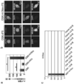
- FIG. 1 a It is the image by the microscope of the tail fibroblast (henceforth a "mouse fibroblast") of the C57BL / 6J mouse which expressed exogenous (mouse) H1foo.
- the “BF”, “DAPI”, “H1foo”, and “MERGE” images in FIG. 1 a are obtained by superimposing a phase difference image, a DAPI fluorescence stained image, an H1foo fluorescence stained image, and an image obtained by superimposing the three types of images, respectively. Show.
- the left and right images in FIG. 1b show two-dimensional and 2.5-dimensional images with H1foo fluorescence staining, respectively.
- FIG. 1c show electron microscope images of control cells (mouse fibroblasts) and mouse fibroblasts expressing exogenous H1foo, respectively.
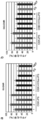
- FIG. 2a shows three types of nuclear reprogramming substances (mouse Oct3 / 4, Sox2 and Klf4 genes [OSK gene]), OSK gene and linker histone H1 gene (OSK + H1c gene), or OSK gene and H1foo gene (OSK + H1foo gene). It is a figure which shows the result of having introduce
- FIG. 2 b shows three types of pluripotency markers (Oct3 / Oc) in iPS cells (hereinafter referred to as “OSK-iPS cells” and “OSK + H1foo-iPS cells”) produced by introducing the OSK gene or OSK + H1foo gene, respectively. 4, Nanog, and SSEA1) are analysis results of the expression.
- FIG. 2c is a diagram showing the results of analyzing the expression of endogenous H1foo on days 1 to 5 after introduction of the OSK gene, or OSK gene, and c-Myc gene (OSKM gene).
- “MEF + H1foo” in FIG. 2c shows the result of analyzing mouse embryonic fibroblasts (MEF) introduced with the H1foo gene.
- FIG. 4a is a diagram showing the results of comparison of whole gene transcriptome profiles between OSK + H1foo-iPS cells and ES cells.
- FIG. 4b is a diagram showing the results of comparison of whole gene transcriptome profiles between OSK + H1foo-iPS cells and OSK-iPS cells. It is a figure which shows the result of having analyzed the DNA methylation level of IG-DMR (FIG.
- FIG. 5a Gtl2-DMR (FIG. 5b) of an OSK-iPS cell and an OSK + H1foo-iPS cell.
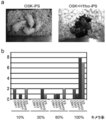
- ES and “MEF” in the figure indicate the results of analyzing ES cells and MEF cells, respectively. It is a figure which shows the result of having analyzed the form of the embryoid body (EB) formed from the OSK-iPS cell and the OSK + H1foo-iPS cell. It is a figure which shows the result of having analyzed the ratio of the apoptotic cell of EB formed from the OSK-iPS cell and the OSK + H1foo-iPS cell.
- EB embryoid body
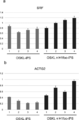
- FIG. 11 shows the results of quantitative RT-PCR analysis of the SRF gene (FIG. 11a) and the ACTG2 gene (FIG. 11b) in the iPS cells produced as described above. Numerical values 1 to 4 on the horizontal axis of the graph represent clone numbers.
- the iPS cell production method of the present invention comprises (a) a nuclear reprogramming substance, and (b) a step of introducing an H1foo gene or gene product thereof into a somatic cell (hereinafter also simply referred to as “introduction step”). And may include other steps as necessary.
- the introduction step is a step of introducing at least (a) a nuclear reprogramming substance and (b) an H1foo gene or a gene product thereof into a somatic cell.
- a gene product means mRNA (messenger RNA) transcribed from a gene and / or a protein translated from the mRNA.
- said H1foo gene means the polynucleotide which codes H1foo protein.
- the H1foo gene or its gene product can be used as an agent for improving the quality of iPS cells.
- the vector containing the H1foo gene described later can also be used as an agent for improving the quality of iPS cells.
- H1foo gene There is no restriction
- the sequence information of the H1foo gene can be obtained from a known database.
- GenBank can obtain accession numbers BC047943 (human), AY158091 (human), and BC137916 (mouse).
- the nucleotide sequence of the human H1foo gene (BC047943) is shown in SEQ ID NO: 1, and the amino acid sequence of the human H1foo protein (protein encoded by the H1foo gene of BC047943) is shown in SEQ ID NO: 2.
- the nucleotide sequence of the human H1foo gene (AY158091) is represented by SEQ ID NO: 59, and the amino acid sequence of the human H1foo protein (protein encoded by the H1foo gene of AY158091) is represented by SEQ ID NO: 60.
- the nucleotide sequence of the mouse H1foo gene (BC137916) is represented by SEQ ID NO: 3, and the amino acid sequence of the mouse H1foo protein (protein encoded by the BC137916 H1foo gene) is represented by SEQ ID NO: 4.
- the nucleotide sequence of the H1foo gene and the mRNA nucleotide sequence thereof may be the same as the wild-type H1foo gene nucleotide sequence and the mRNA nucleotide sequence, or may contain mutations.
- the nucleotide sequence containing such a mutation includes: “a nucleotide sequence of a wild-type H1foo gene (for example, a nucleotide sequence represented by SEQ ID NO: 1 or 59 or 3) or a nucleotide sequence of mRNA thereof, preferably 1 to 30, preferably 1 to 20, more preferably 1 to 15, more preferably 1 to 10, more preferably 1 to 5, more preferably 1 to 3 nucleotides have been deleted, substituted, inserted, or added.
- a nucleotide sequence of a wild-type H1foo gene for example, a nucleotide sequence represented by SEQ ID NO: 1 or 59 or 3
- a nucleotide sequence of mRNA thereof preferably 1 to 30, preferably 1 to 20, more preferably 1 to 15, more preferably 1 to 10, more preferably 1 to 5, more preferably 1 to 3 nucleotides have been deleted, substituted, inserted, or added.
- nucleotide sequence of the nucleotide sequence of the wild-type H1foo gene (for example, SEQ ID NO: 1 or 59 or 3 in the nucleotide sequence encoding the protein having H1foo activity)
- Nucleotide sequence represented by or a nucleotide sequence of mRNA thereof A nucleotide sequence having a sequence identity of 70% or more, preferably 80% or more, more preferably 85% or more, more preferably 90% or more, more preferably 95% or more, and further preferably 98% or more.
- nucleotide sequence encoding a protein having H1foo activity for example, SEQ ID NO: 1 or 59 or 3 in the nucleotide sequence encoding the protein having H1foo activity
- the amino acid sequence of the H1foo protein may be the same as the amino acid sequence of the wild-type H1foo protein (for example, the amino acid sequence represented by SEQ ID NO: 2 or 60 or 4), or may contain a mutation.
- the protein containing such a mutation include: “1-30, preferably 1-20, more preferably in the amino acid sequence of the wild-type H1foo protein (for example, the amino acid sequence represented by SEQ ID NO: 2 or 60 or 4).
- protein having H1foo activity refers to a high-quality iPS cell when introduced into a somatic cell together with a nuclear reprogramming substance as compared with a case where only the nuclear reprogramming substance is introduced into a somatic cell. Is a protein that can be produced in a larger amount.
- nuclear reprogramming substance means that a somatic cell can be induced into an iPS cell by introducing the substance alone or a combination of the substance and another substance into the somatic cell.
- a nuclear reprogramming substance if it is a substance (group) capable of inducing iPS cells from somatic cells, such as a gene (including a form incorporated in an expression vector) or a gene product thereof, a low molecular weight compound, etc. Any substance may be used.
- a gene that is a nuclear reprogramming substance means a polynucleotide that encodes a protein that is a nuclear reprogramming substance.
- the nuclear reprogramming substance is a gene or gene product thereof, Oct gene family gene, Sox gene family gene, Klf gene family gene, Myc gene family gene, Lin gene family gene, Nanog gene, and At least one selected from the group consisting of the gene products (WO2007 / 69666; Patent No. 5696282; Science, 2007, 318: 1917-1920), among which two or more selected from such a group 3 or more are more preferable, and 2 to 4 ranges selected from such a group are preferable, and 3 or 4 is more preferable. Specific examples of these family genes and combinations thereof are listed below. In the following, only the name of a gene is described, but the case of using the gene product is also included.
- A one nuclear reprogramming substance consisting of genes of the Oct gene family;
- B a combination of two nuclear reprogramming substances consisting of genes of the Oct gene family and Sox gene family;
- C a combination of two kinds of nuclear reprogramming substances consisting of genes of the Oct gene family and Klf gene family;
- D a combination of two nuclear reprogramming substances consisting of genes of the Oct gene family and Nanog genes;
- E a combination of three nuclear reprogramming substances consisting of genes of the Oct gene family, the Sox gene family, and the Klf gene family;
- F A combination of three kinds of nuclear reprogramming substances consisting of genes of the Oct gene family, Klf gene family and Myc gene family.
- (G) a combination of four nuclear reprogramming substances consisting of genes of the Oct gene family, Sox gene family, Klf gene family and Myc gene family; and (h) Oct gene family of genes, Sox gene A combination of four nuclear reprogramming substances consisting of a family gene, a Lin gene family gene, and a Nanog gene; Alternatively, it may be a combination in which another nuclear reprogramming substance (gene or gene product thereof) is further added to any one of the nuclear reprogramming substances (a) to (h) or a combination thereof.
- a ′ a combination of nuclear reprogramming substances including one nuclear reprogramming substance consisting of genes of the Oct gene family
- B ′ a combination of nuclear reprogramming substances including two nuclear reprogramming substances consisting of genes of the Oct gene family and Sox gene family
- C ′ a combination of nuclear reprogramming substances including two kinds of nuclear reprogramming substances consisting of genes of the Oct gene family and Klf gene family
- D ′ a combination of nuclear reprogramming substances including two nuclear reprogramming substances consisting of genes of the Oct gene family and Nanog genes
- E ′ a combination of nuclear reprogramming substances including three kinds of nuclear reprogramming substances consisting of genes of Oct gene family, Sox gene family, and Klf gene family
- F ′ A combination of nuclear reprogramming substances including three kinds of nuclear reprogramming substances consisting of genes of the Oct gene family, Klf gene family and Myc gene family.
- G ′ a combination of nuclear reprogramming substances including four nuclear reprogramming substances consisting of genes of the Oct gene family, Sox gene family, Klf gene family and Myc gene family
- h ′ A combination of nuclear reprogramming substances including four nuclear reprogramming substances consisting of genes of the Oct gene family, Sox gene family, Lin gene family, and Nanog gene;
- Oct3 / 4 gene, Klf4 gene, c-Myc gene (2) Oct3 / 4 gene, Sox2 gene, Klf4 gene, c-Myc gene (where Sox2 gene can be replaced by Sox1 gene, Sox3 gene, Sox15 gene, Sox17 gene or Sox18 gene.
- Sox2 gene can be replaced by Sox1 gene, Sox3 gene, Sox15 gene, Sox17 gene or Sox18 gene.
- the Klf1 gene, the Klf2 gene, or the Klf5 gene can be replaced.
- the c-Myc gene can be replaced with the T58A (active mutant) gene, the N-Myc gene, and the L-Myc gene).
- Oct gene family member genes eg, Oct1A, Oct6, etc.
- Sox2 gene or the Sox1 gene, the Sox3 gene, the Sox15 gene, the Sox17 gene, the Sox18 gene
- Sox7 gene a member gene of another Lin gene family
- Lin28b gene a member gene of another Lin gene family
- nuclear reprogramming substances selected from the group consisting of Fbx15 gene, ERas gene, ECAT15-2 gene, Tcl1 gene, and ⁇ -catenin gene may be combined.
- ECAT1 gene Esg1 gene, Dnmt3L gene, ECAT8 gene, Gdf3 gene, Mybl2 gene, ECAT15-1 gene, Fthl17 gene, Sall4 gene, Rex1 gene, UTF1 gene, Stella gene, Stat3 gene, and Grb2 gene
- One or more kinds of nuclear reprogramming substances selected from the group can also be combined. These combinations are specifically described in WO2007 / 69666.
- Preferred nuclear reprogramming substances are selected from the group consisting of Oct3 / 4 gene, Sox2 gene, Klf4 gene, c-Myc gene (or L-Myc gene), Lin28 gene, Nanog gene, and their gene products. At least one, preferably two or more, more preferably three or more are examples of preferable nuclear reprogramming materials.
- Particularly preferred nuclear reprogramming substance combinations include (1) Oct3 / 4 gene or gene product thereof, Sox2 gene or gene product thereof, and Klf4 gene or gene product thereof, (2) Oct3 / 4 gene or gene product thereof, Sox2 gene or gene product thereof, Klf4 gene or gene product thereof, and c-Myc gene or gene product thereof, (3) Oct3 / 4 gene or gene product thereof, Sox2 gene or gene product thereof, Klf4 gene or gene product thereof, And L-Myc gene or gene product thereof, among them, Oct3 / 4 gene or gene product thereof, Sox2 gene or gene product thereof, and combination of Klf4 gene or gene product thereof, Oct3 / 4 gene or Gene product, Sox2 inheritance
- the gene product, the Klf4 gene or the gene product thereof, and the combination of the L-Myc gene or the gene product thereof can be preferably exemplified.
- the combination of the Oct3 / 4 gene, the Sox2 gene, and the Klf4 gene, or the Oct3 / 4 More preferred examples include a combination of a gene, a Sox2 gene, a Klf4 gene, and an L-Myc gene.
- the c-Myc gene or its gene product may be used as one of the nuclear reprogramming substances used in the present invention, but it is preferable not to use the c-Myc gene or its gene product. This is because c-Myc has been reported to reduce the proportion of Nanog-GFP positive colonies and increase the carcinogenicity of cells (Nakagawa, M., et al. Generation of induced pluripotent stem cells without Myc from mouse and human fibroblasts. Nature biotechnology 26, 101-106 (2008)).
- the origin of the gene is not particularly limited and can be appropriately selected according to the purpose.
- human, mouse, rat, cow, sheep, horse, Any mammal such as monkeys can be mentioned.
- Mouse and human cDNA sequence information of each of the above nuclear reprogramming substances can be obtained by referring to GenBank accession numbers described in WO2007 / 069666. Note that the Nanog gene is described as “ECAT4” in the publication. Moreover, the mouse
- the cDNA sequence of the human Oct3 / 4 gene is shown in SEQ ID NO: 47
- the amino acid sequence of the human Oct3 / 4 protein is shown in SEQ ID NO: 48
- the cDNA sequence of the human Sox2 gene is shown in SEQ ID NO: 49
- the human The amino acid sequence of the Sox2 protein is shown in SEQ ID NO: 50
- the cDNA sequence of the human Klf4 gene is shown in SEQ ID NO: 51
- the amino acid sequence of the human Klf4 protein is shown in SEQ ID NO: 52
- the cDNA sequence of the human L-Myc gene is The amino acid sequence of human L-Myc protein is shown in SEQ ID NO: 53.
- nucleotide sequence in the case where the nuclear reprogramming substance is each of the above genes or mRNA thereof is not particularly limited as long as the effect of the present invention is not impaired, and the portion of each nucleotide sequence of each of the above genes that is translated into a protein It may be only, and other parts may be included.
- nucleotide sequence of each gene and the nucleotide sequence of the mRNA may be the same as the nucleotide sequence of the wild-type gene and the nucleotide sequence of the mRNA, or may contain a mutation.
- nucleotide sequences containing such mutations include: “1-30 nucleotides, preferably 1-20, more preferably 1-15, and even more preferably in the nucleotide sequence of each of the above wild-type genes or the nucleotide sequence thereof.
- the nuclear reprogramming substance is a protein encoded by each of the above genes (that is, a protein translated from mRNA transcribed from each of the above genes), the amino acid sequence thereof is encoded by each of the wild type each of the above genes. It may be the same as the protein or may contain a mutation. Examples of the protein containing such a mutation include: “1-30 amino acids, preferably 1-20, more preferably 1-15, and even more preferably 1-10 in the amino acid sequence of the protein encoded by each wild-type gene.
- sequence identity with the amino acid sequence of the protein encoded by each gene of the type is 70% or more, preferably 80% or more, more preferably 85% or more, still more preferably 90% or more, more preferably 95% or more, More preferred is a protein having an amino acid sequence of 98% or more and having a nuclear reprogramming action.
- somatic cells There is no restriction
- the mature somatic cells include mesenchymal stem cells, hematopoietic stem cells, adipose tissue-derived stromal cells, adipose tissue-derived stromal stem cells, neural stem cells, sperm stem cells, and other tissue stem cells (somatic stem cells); tissue precursors Cells; already differentiated cells such as fibroblasts, epithelial cells, lymphocytes, muscle cells, and the like.
- the biological species from which the somatic cells are derived is not particularly limited and may be appropriately selected depending on the intended purpose. Examples thereof include any mammals such as humans, mice, rats, cows, sheep, horses and monkeys. It is done. Moreover, the biological species from which the somatic cell is derived and the biological species from which the gene to be introduced into the somatic cell is not necessarily the same, but are preferably the same.
- the individual from which the somatic cells are collected is not particularly limited and can be appropriately selected according to the purpose.
- the individual itself from the viewpoint of rejection Alternatively, other individuals having the same or substantially the same type of MHC are preferred.
- the type of MHC is substantially the same as that when the cells obtained by inducing differentiation from the somatic cell-derived iPS cells are transplanted into an individual by using an immunosuppressant or the like. It means that the type of MHC matches to the extent that it can be worn.
- the somatic cells may be recombinant so as to facilitate selection of iPS cells.
- the recombinant cell include a recombinant cell in which at least one of a reporter gene and a drug resistance gene is incorporated into a gene locus that is specifically highly expressed in a pluripotent cell.
- the gene that is specifically highly expressed in the pluripotent cells include Fbx15 gene, Nanog gene, Oct3 / 4 gene, and the like.
- the reporter gene include green fluorescent protein (GFP) gene, luciferase gene, ⁇ -galactosidase gene and the like.
- the drug resistance gene include blasticidin gene, hygromycin gene, puromycin resistance gene, neomycin resistance gene and the like.
- the somatic cell culture conditions are not particularly limited and may be appropriately selected depending on the intended purpose. Examples thereof include a culture temperature of about 37 ° C. and a CO 2 concentration of about 2% to 5%.
- the medium used for culturing the somatic cells is not particularly limited and may be appropriately selected depending on the intended purpose. For example, minimal essential medium (MEM) containing 5% to 20% by mass of serum, Dulbecco's modification Examples thereof include a medium (DMEM), RPMI 1640 medium, 199 medium, and F12 medium.
- the method for introducing (a) nuclear reprogramming substance and (b) H1foo gene or gene product thereof into a somatic cell is not particularly limited and can be appropriately selected according to the purpose.
- the method to use, the method using mRNA, the method using a recombinant protein, etc. are mentioned.
- the nuclear reprogramming substance is preferably introduced into somatic cells in the form of genes or in the form of mRNA of the genes rather than introduced into somatic cells in the form of proteins. More preferably, it is introduced into a somatic cell in the form of a gene.
- Such a gene may be DNA, RNA, or DNA / RNA chimera, but is preferably DNA from the viewpoint of stability.
- the gene may be double-stranded or single-stranded, but preferably is double-stranded.
- Preferred examples of the nuclear reprogramming substance include cDNA of each of the above genes. Among them, double-stranded cDNA of each of the above genes is preferably exemplified.
- the “H1foo gene or gene product thereof” is expressed in the form of a gene or in the form of mRNA of the gene rather than being introduced into a somatic cell in the form of a protein. It is preferable to introduce it into a cell, and it is more preferable to introduce it into a somatic cell in the form of a gene.
- the H1foo gene may be DNA, RNA, or a DNA / RNA chimera, but is preferably DNA from the viewpoint of stability.
- the H1foo gene may be double-stranded or single-stranded, but preferably is double-stranded.
- Preferred embodiments of the “H1foo gene or gene product thereof” include H1foo gene cDNA. Among them, a double-stranded cDNA of H1foo gene is preferred.
- Expression vector When a nuclear reprogramming substance is introduced into a somatic cell in the form of a gene or when an H1foo gene is introduced into a somatic cell, the nuclear reprogramming is performed with an appropriate expression vector containing a promoter that can function in the host somatic cell.
- An expression vector incorporating a substance or the H1foo gene can be preferably used.
- the expression vector for incorporating the nuclear reprogramming substance and the H1foo gene is not particularly limited and may be appropriately selected depending on the intended purpose. Examples thereof include episomal vectors, artificial chromosome vectors, plasmid vectors, and viral vectors. It is done.
- promoters used in the above expression vectors include SR ⁇ promoter, SV40 early promoter, retrovirus LTR, CMV (cytomegalovirus) promoter, RSV (rous sarcoma virus) promoter, HSV-TK (herpes simplex virus thymidine kinase). ) Promoter, EF1 ⁇ promoter, metallothionein promoter, heat shock promoter and the like.
- an enhancer of human CMV IE gene may be used together with a promoter.
- CAG promoter including cytomegalovirus enhancer, chicken ⁇ -actin promoter and poly A signal site of ⁇ -globin gene
- CAG promoter including cytomegalovirus enhancer, chicken ⁇ -actin promoter and poly A signal site of ⁇ -globin gene
- the above expression vector may contain an enhancer, a poly A addition signal, a marker gene, a replication origin, a gene encoding a protein that binds to the replication origin and controls replication, if desired.
- the above marker gene refers to a gene that enables selection and selection of cells by introducing the marker gene into cells.
- Specific examples of the marker gene include a drug resistance gene, a fluorescent protein gene, a luminescent enzyme gene, and a chromogenic enzyme gene. These may be used individually by 1 type and may use 2 or more types together.
- the drug resistance gene examples include a neomycin resistance gene, a tetracycline resistance gene, a kanamycin resistance gene, a zeocin resistance gene, and a hygromycin resistance gene.
- Specific examples of the fluorescent protein gene include a green fluorescent protein (GFP) gene, a yellow fluorescent protein (YFP) gene, and a red fluorescent protein (RFP) gene.
- Specific examples of the luminescent enzyme gene include a luciferase gene.
- the chromogenic enzyme gene include ⁇ -galactosidase gene, ⁇ -glucuronidase gene, alkaline phosphatase gene, and the like.
- the above episomal vector is a vector capable of autonomous replication outside the chromosome. Specific means using an episomal vector is disclosed in Yu et al., Science, 324, 797-801 (2009).
- an episomal vector in which loxP sequences are arranged in the same direction on the 5 'and 3' sides of vector elements necessary for episomal vector replication may be used.
- Episomal vectors can autonomously replicate outside the chromosome, so they can provide stable expression in host cells even if they are not integrated into the genome. However, once iPS cells are established, the vectors are quickly removed. It is desirable.
- the autonomous replication ability of the episomal vector can be lost. Can be removed from iPS cells at an early stage.
- Examples of the episomal vector used in the present invention include a vector containing a sequence necessary for autonomous replication derived from EBV, SV40 or the like as a vector element.
- vector elements necessary for autonomous replication include a replication origin and a gene that encodes a protein that binds to the replication origin and regulates replication.
- the replication origin oriP And EBNA-1 gene and SV40 includes the replication origin ori and the SV40LT gene.
- examples of the artificial chromosome vector include a YAC (Yeast Artificial Chromosome) vector, a BAC (Bacterial Artificial Chromosome) vector, a PAC (P1-derived Artificial Chromosome) vector, and the like.
- the plasmid vector is not particularly limited as long as it is a plasmid vector that can be expressed in the introduced somatic cell.
- somatic cell is a mammal, pA1-11, pXT1, pRc / CMV, pRc / RSV And plasmid vectors for expression of animal cells such as pcDNAI / Neo.
- viral vectors examples include retrovirus (including lentivirus) vectors, adenovirus vectors, adeno-associated virus vectors, Sendai virus vectors, herpes virus vectors, vaccinia virus vectors, pox virus vectors, polio virus vectors, silbis virus vectors, Examples include a rhabdovirus vector, a paramyxovirus vector, and an orthomyxovirus vector.
- the method for introducing the above expression vector into the above somatic cell is not particularly limited and may be appropriately selected depending on the intended purpose.
- lipofection method, microinjection method, DEAE dextran method, gene gun method, electro Examples thereof include a poration method and a calcium phosphate method.
- viral particles obtained using a packaging cell may be used.
- a packaging cell is a cell into which a gene encoding a structural protein of a virus has been introduced, and when a recombinant virus vector incorporating the target gene is introduced into the cell, recombinant virus particles incorporating the target gene are produced.
- the packaging cell is not particularly limited and may be appropriately selected according to the purpose.
- packaging cells based on human kidney-derived HEK293 cells or mouse fibroblast-derived NIH3T3 cells PLAT-E cells designed to express virus-derived envelope glycoproteins
- PLAT-A cells designed to express envelope glycoproteins derived from Amphotropic virus, designed to express envelope glycoproteins derived from vesicular stomatitis virus PLAT-GP cells that have been used.
- PLAT-A cells and PLAT-GP cells are preferable from the viewpoint of host orientation when a recombinant viral vector is introduced into human somatic cells.
- the method for introducing the viral vector into the packaging cell is not particularly limited and may be appropriately selected depending on the intended purpose.
- Examples thereof include a lipofection method, an electroporation method, and a calcium phosphate method.
- a lipofection method an electroporation method
- a calcium phosphate method an electroporation method
- the polybrene method is mentioned.
- one gene When introducing a nuclear reprogramming substance or H1foo gene into a somatic cell using the above expression vector, one gene may be incorporated into one expression vector, or two or more genes may be incorporated. By incorporating two or more genes into one vector, the two or more genes can be expressed simultaneously (hereinafter sometimes referred to as “co-expression”). Further, all nuclear reprogramming substances to be introduced into somatic cells and the H1foo gene may be incorporated into one expression vector.
- the method for incorporating two or more genes into one vector is not particularly limited and may be appropriately selected according to the purpose. However, it is preferable to incorporate the two or more genes through a linking sequence. .
- sequence According to the objective, it can select suitably, For example, the gene sequence which codes 2A peptide derived from foot-and-mouth disease virus (Picornaviridae Aphthovirus), IRES (internal ribosome entry sites), etc. are mentioned. .
- a nuclear reprogramming substance and (b) a gene product of the H1foo gene may be introduced into somatic cells in the form of mRNA.
- mRNA messenger RNA
- a well-known method can be selected suitably and can be used.
- a commercially available RNA transfection reagent such as Lipofectamine (registered trademark) MessengerMAX (manufactured by Life Technologies) can be used.
- a nuclear reprogramming substance and (b) a gene product of the H1foo gene may be introduced into a somatic cell in the form of a protein.
- transducing this protein into the said somatic cell A well-known method can be selected suitably and can be used. Examples of such a method include a method using a protein introduction reagent, a method using a protein introduction domain (PTD) fusion protein, and a microinjection method.
- PTD protein introduction domain
- Protein introduction reagents include cationic lipid-based BioPOTER (registered trademark) Protein Delivery Reagent (Gene Therapy Systmes) and Pro-Ject TM Protein Transfection Reagent (PIERCE), lipid-based Profect-1 Targeting TM Systems), Penetratin Peptide (Q biogene) and Chariot Kit (Active Motif) based on membrane-permeable peptides, GenomONE (Ishihara Sangyo) using HVJ envelope (inactivated Sendai virus) Etc. are commercially available.
- the introduction can be carried out according to the protocol attached to these reagents, but the general procedure is as follows.
- (A) Nuclear reprogramming substance” or “(b) gene product of H1foo gene”, which is a protein form, is diluted in an appropriate solvent (for example, a buffer solution such as PBS or HEPES), and an introduction reagent is added to room temperature. Incubate for 5 to 15 minutes to form a complex, which can be added to cells exchanged in serum-free medium and incubated at 37 ° C. for 1 to several hours. Thereafter, the medium can be removed and replaced with a serum-containing medium.
- an appropriate solvent for example, a buffer solution such as PBS or HEPES
- Such a PTD is produced by recombinant expression of a fusion protein expression vector incorporating the cDNA of the “(a) nuclear reprogramming substance” or “(b) gene product of the H1foo gene” and the PTD sequence, and the fusion protein is recovered. And can be used for introduction.
- a fusion protein can be introduced in the same manner as described above except that no protein introduction reagent is added.
- the microinjection method described above is a method in which a protein solution is put into a glass needle having a tip diameter of about 1 ⁇ m and puncture is introduced into the cell, and the protein can be reliably introduced into the cell.
- CPP cell-penetrating peptide
- the number of introductions of “(a) nuclear reprogramming substance” and “(b) H1foo gene or gene product thereof” into a somatic cell may be 1 or 2 It may be more than once.
- the introduction time is not particularly limited and can be appropriately selected depending on the purpose. All of the “(a) nuclear reprogramming substance” and “(b) H1foo gene or its gene product” to be introduced are simultaneously generated. May be introduced at a different time.
- the “H1foo gene or gene product thereof” may be an embodiment using only the gene, an embodiment using only the gene product, or an embodiment using both the gene and the gene product. May be.
- the nuclear reprogramming substance is a gene or a gene product thereof
- an embodiment using only the gene may be used
- an embodiment using only the gene product may be used, or both the gene and the gene product may be used.
- An aspect may be sufficient.
- the nuclear reprogramming substance is a gene or a gene product thereof and two or more kinds of “genes or gene products thereof” are used in combination
- the gene product is used for a certain gene and the other genes are An embodiment using a gene may also be used.
- the amount of H1foo gene or its gene product introduced into somatic cells is (a) a nuclear reprogramming substance, and (b) when H1foo gene or its gene product is introduced into a somatic cell, it is of higher quality and quality. There is no particular limitation as long as iPS cells with less variation can be produced.
- the nuclear reprogramming substance is a gene or a gene product thereof
- the amount of the gene or gene product introduced into a somatic cell is not particularly limited as long as the somatic cell nucleus can be reinitialized. All genes or their gene products may be introduced in equal amounts or in different amounts.
- the Oct3 / 4 gene is introduced in a large amount, for example, about 3 times the amount of the Sox2 gene, the Klf4 gene, or the c-Myc gene (PNAS 106 (31): 12759-12764 (2009), J. Biol. Chem. 287 (43): 36273-36282 (2012)).
- the contact of the low molecular weight compound with a somatic cell is performed by dissolving the low molecular weight compound in an aqueous or non-aqueous solvent at an appropriate concentration.
- a medium suitable for culturing somatic cells for example, a minimum essential medium (MEM) containing about 5 to 20% fetal calf serum, Dulbecco's modified Eagle medium (DMEM), RPMI 1640 medium, 199 medium, F12 medium, etc.
- MEM minimum essential medium
- DMEM Dulbecco's modified Eagle medium
- RPMI 1640 medium 199 medium
- F12 medium F12 medium
- the low-molecular compound solution is added so that the concentration of the low-molecular compound is sufficient to cause nuclear reprogramming in somatic cells and no cytotoxicity is observed, and the cells are cultured for a certain period of time.
- the concentration of the low molecular weight compound as the nuclear reprogramming substance varies depending on the type of the low molecular weight compound used, but can be appropriately selected within the range of about 0.1 nM to about 100 nM.
- the contact period is not particularly limited as long as it is a time sufficient for the nuclear reprogramming of the
- the iPS cell production method of the present invention includes (a) a nuclear reprogramming substance and (b) a step of introducing an H1foo gene or a gene product thereof into a somatic cell (“introduction step”). Depending on the, further steps may be included. The other steps are not particularly limited as long as the effects of the present invention are not impaired, and can be appropriately selected according to the purpose.
- the nuclear reprogramming substance and (b) the H1foo gene or Examples include a step of culturing a somatic cell into which the gene product has been introduced (hereinafter also simply referred to as “introduced cell”) (hereinafter also simply referred to as “introduced cell culture step”).
- the introduced cell culturing step is a step of culturing a somatic cell into which (a) the nuclear reprogramming substance and (b) the H1foo gene or its gene product have been introduced.
- the culture conditions for the introduced cells are not particularly limited, and examples thereof include conditions suitable for ES cell culture. Examples of such conditions include a culture temperature of about 37 ° C. and a CO 2 concentration of about 2% to 5%.
- transduced cell According to the objective, it can select suitably.
- LIF Leukemia Inhibitory Factor
- bFGF basic fibroblast growth factor
- SCF stem cell factor
- the cells are cultured as feeder cells in the presence of mouse embryo-derived fibroblasts (MEF) that have been treated with radiation or antibiotics to stop cell division.
- MEF mouse embryo-derived fibroblasts
- STO cells STO cells are usually used, but SNL cells (McMahon, AP & Bradley, A. Cell 62, 1073-1085 (1990)) and the like are frequently used for induction of iPS cells.
- Co-culture with feeder cells may be started before the introduction of (a) nuclear reprogramming substance and (b) H1foo gene or gene product thereof, or at the time of or after the introduction (for example, 1 It may start from (after 10 days).
- the period of the introduced cell culturing step is not particularly limited and can be appropriately selected depending on the purpose.
- iPS Cell The iPS cell of the present invention produced by the above-described iPS cell production method has pluripotency and self-renewal ability.
- the term “pluripotency” means that all three germ layers can be differentiated.
- the self-replicating ability means the ability to proliferate while maintaining an undifferentiated state.
- the method for confirming whether or not the cells produced by the above-described iPS cell production method are iPS cells is not particularly limited, and can be appropriately selected according to the purpose.
- a somatic cell a reporter gene at a gene locus that is specifically highly expressed in differentiated pluripotent cells (for example, Fbx15, Nanog, Oct3 / 4, etc., preferably Nanog or Oct3 / 4), and
- a recombinant cell incorporating at least one of the drug resistance gene it can be confirmed using the above reporter gene or drug resistance gene.
- a green fluorescent protein (GFP) gene for example, a method of confirming GFP positive cells with a flow cytometer can be mentioned.
- a puromycin resistance gene it can be confirmed by administering puromycin to the cells.
- high-quality iPS cells refers to iPS cells with higher quality than iPS cells produced by the same method except that the H1foo gene or gene product thereof is not introduced into somatic cells.
- iPS cells with little quality variation means iPS cells with less quality variation than iPS cells produced by the same method except that the H1foo gene or its gene product is not introduced into somatic cells. Means a cell. These qualities may be one type or two or more types. Specific examples of “high-quality iPS cells” or “iPS cells with little variation in quality” include iPS cells having one or more characteristics selected from the following (a) to (q): .
- iPS cells prepared by the same method except that the H1foo gene or its gene product is not introduced into a somatic cell (preferably, “OSK-iPS cell” or “OSKL-iPS cell” in Examples described later)
- iPS cells in which the number of embryoid bodies formed when cultured for 5 days by the method of [Embryoid body (EB) formation] described later is improved by 5% or more, preferably 10% or more.
- somatic cells preferably “OSK-iPS cells” or “OSKL-iPS cells” in Examples described later
- the variation ( ⁇ 2 ) in the size ( ⁇ m 2 ) of the embryoid body when cultured for 5 days by the method of “embryoid body (EB) formation” described later is 25% or more, preferably 40%.
- the variation ( ⁇ 2 ) in the size ( ⁇ m 2 ) of the embryoid body when cultured for 5 days by the method of “embryoid body (EB) formation” described below is 10000 ( ⁇ m 2 ) or less, preferably 8000 ( iPS cells that are [mu] m ⁇ 2 >) or less.
- the ratio of viable cells (annexin V ( ⁇ ) / PI ( ⁇ ) cells) measured by the method of [Apoptosis assay] described later was improved 1.1 times or more, preferably 1.2 times or more.
- iPS cells iPS cells having a ratio of viable cells (annexin V ( ⁇ ) / PI ( ⁇ ) cells) of 68% or more, preferably 73% or more, measured by the method of [Apoptosis assay] described later.
- the ratio of apoptotic cells is 0. IPS cells reduced to 75-fold or less, preferably 0.67-fold or less.
- the ratio of apoptotic cells (total of annexin V (+) / PI ( ⁇ ) cells and annexin V (+) / PI (+) cells) measured by the method of [Apoptosis assay] described later is 30% or less, IPS cells that are preferably 25% or less.
- IPS cells that are 0.65 times or more, preferably 0.73 times or more.
- K an iPS cell produced by the same method except that the H1foo gene or its gene product is not introduced into a somatic cell (preferably, “OSK-iPS cell” or “OSKL-iPS cell” in Examples described later) IPS cells with higher chimera-forming ability.
- L iPS cells prepared by the same method except that the H1foo gene or gene product thereof is not introduced into somatic cells (preferably “OSK-iPS cells” or “OSKL-iPS cells” in Examples described later) In comparison, iPS cells with a higher ability to move to the gonads.
- the expression level of the SRF gene (known as a chromosomal abnormality marker whose expression level decreases when it has a chromosomal abnormality) measured by the method described in the section of [Quantitative RT-PCR analysis] described later IPS cells improved by 1.1 times, preferably 1.2 times.
- N an iPS cell prepared by the same method except that the H1foo gene or its gene product is not introduced into a somatic cell (preferably, “OSKL-iPS cell” or “OSK-iPS cell” in Examples described later)
- the expression level of the ACTG2 gene (known as a chromosomal abnormality marker whose expression level decreases when it has a chromosomal abnormality), measured by the method described in [Quantitative RT-PCR analysis] described later IPS cells improved by 1.2 times, preferably 1.5 times.
- the variation in expression level ( ⁇ 2 ) of Oct3 / 4 gene (undifferentiated marker of stem cells) measured by the method described in the item of “Quantitative RT-PCR analysis” described later is 10% or more, preferably IPS cells reduced by 20% or more.
- P an iPS cell produced by the same method (preferably “OSKL-iPS cell” or “OSK-iPS cell” in Examples described later) except that the H1foo gene or its gene product is not introduced into somatic cells.
- iPS cells in which the number of viable cells measured by the method described in the item “Comparison of cell viability in the early stage of differentiation induction” described later is improved 1.2 times, preferably 1.5 times.
- an iPS cell produced by the same method except that the H1foo gene or its gene product is not introduced into a somatic cell preferably, “OSKL-iPS cell” or “OSK-iPS cell” in Examples described later
- the variation in the expression level of Oct3 / 4 gene was measured by the method described in the item of Comparative differentiation inducing early undifferentiated markers residual amount of iPS cells] described later (undifferentiated marker for stem cells) (sigma 2 IPS cells reduced by 10% or more, preferably 20% or more.
- the biological species from which the above iPS cells are derived is not particularly limited and can be appropriately selected according to the purpose.
- any mammal such as human, mouse, rat, cow, sheep, horse, monkey, etc. Can be mentioned.
- the iPS cell quality improving agent of the present invention includes at least the H1foo gene or a gene product thereof, and may further include other components as necessary.
- the H1foo gene or gene product thereof is the same as that described in the above-described method for producing iPS cells. Further, the H1foo gene or its gene product may contain the same mutation as described in the above iPS cell production method.
- what was integrated in said expression vector containing the promoter which can function by the somatic cell used as a host as a H1foo gene may be used.
- composition for iPS cell production of the present invention comprises (a) a nuclear reprogramming substance, and (b) an H1foo gene or a gene product thereof, and may further comprise other components as necessary. .
- the (a) nuclear reprogramming substance and (b) the H1foo gene or gene product thereof are the same as those described in the above-described iPS cell production method.
- the H1foo gene or its gene product, the gene that is a nuclear reprogramming substance or its gene product may contain the same mutations as described in the above-mentioned iPS cell production method.
- Preferred forms of the nuclear reprogramming substance in the composition for producing iPS cells include a form in which a gene is incorporated into a vector, a form of synthetic mRNA, a form of a protein (preferably a recombinant protein), and the like.
- a vector the thing similar to what was described by the manufacturing method of said iPS cell is mentioned.
- Said synthetic mRNA and said protein (preferably recombinant protein) can be manufactured by a well-known method.
- composition for producing iPS cells may be one in which the above (a) nuclear reprogramming substance and the above (b) H1foo gene or gene product thereof are divided into individual containers, What was put together in one container may be sufficient, and what was put together in the container for every arbitrary number may be sufficient.
- the amount of the above-mentioned (a) nuclear reprogramming substance and (b) the H1foo gene or gene product thereof in the composition for producing iPS cells is not particularly limited, and all nuclear reprogramming substances, H1foo The gene or its gene product may be equal or different.
- the above composition for producing iPS cells may contain a gene other than the above (a) nuclear reprogramming substance, the above (b) H1foo gene or its gene product, or a gene product thereof.
- a packaging cell may be included. Examples of the genes or gene products other than the genes or gene products thereof and the packaging cells include those described in the method for producing iPS cells.
- a method for improving the quality of iPS cells comprising the step of introducing the H1foo gene or gene product thereof into a somatic cell”; “Use of the H1foo gene or its gene product in the production of an iPS cell quality improving agent”, “Use of (a) nuclear reprogramming substance and (b) H1foo gene or gene product thereof in the production of a composition for producing iPS cells” Is also included.
- the step of introducing the H1foo gene or its gene product into the somatic cell in the above-mentioned “method for improving the quality of iPS cells” is the same as the introducing step in the method for producing iPS cells of the present invention. By including such a step, the quality of iPS cells can be improved and high-quality iPS cells can be obtained.
- H1foo cDNA and H1cDNA (Teranishi, T., et al. Rapid replacement of somatic linker histones with the oocyte-specific linker histone H1foo in nuclear transfer. Developmental Biology 266, 76-86 (2004).) It was inserted into BamH1-Sal1 site and EcoR1-Sal1 site, and it was confirmed by DNA sequencing that H1foo cDNA and H1cDNA were inserted.
- mice iPS cells and cell culture method 1) The production of mouse iPS cells is described in literature (Takahashi, K., Okita, K., Nakagawa, M. & Yamanaka, S. Induction of pluripotent stem cells from fibroblast cultures. Nature protocols 2, 3081-3089 (2007)). Was performed according to the protocol described in. However, in this test, the H1foo gene was also used in addition to the genes described in the literature.
- a pMXs retroviral vector containing the mouse Oct3 / 4, Sox2, Klf4, and H1foo genes nucleotide sequence of SEQ ID NO: 3: BC137916
- mouse fibroblasts or Nanog-GFP-IRES-puro transgenic mice
- Nanog-GFP-IRES-puro transgenic mice Okita, K., Ichisaka, T. & Yamanaka, S. Generation of germline-competent induced pluripotent stem cells. Nature 448, 313-317 (2007)
- IPS cells were produced from “blast cells”.
- the DsRed gene was used instead of the H1foo gene.
- the experimental mice were managed in accordance with the Keio University animal and DNA experiment guidelines.
- Mouse ES cells (B6J-23 UTR ) (Tanimoto, Y., et al. Embryonic stem cells derived from C57BL / 6J and C57BL / 6N mice. Comparative medicine 58, 347-352 (2008)) Obtained from the Resource Center and used in accordance with the Ministry of Education, Culture, Sports, Science and Technology guidelines on the distribution and use of human ES cells.
- Mouse iPS and ES cell lines are 20% KnockOut Serum Replacement (Gibco), 1 mM GlutaMAX (Gibco), 1 mM non-essential amino acid (Sigma-Aldrich), 0.1 mM 2-mercaptoethanol, 50U In a DMEM (Sigma-Aldrich) culture medium (hereinafter referred to as “iPS cell culture medium”) containing penicillin, 50 mg / ml sputomycin (Gibco), and mouse leukemia inhibitory factor (LIF), Cultured and maintained on mouse embryonic fibroblasts (iMEF feeder cells) irradiated with X-rays derived from type ICR mice. The mouse iPS cell culture medium was changed every 2 to 3 days, and the cells were passaged every 2 to 3 days using 0.5 mM trypsin-EDTA (Gibco).
- EB formation of iPS cells was performed by seeding 5 ⁇ 10 4 iPS cells collected using 1 mg / ml collagenase IV on a 100 mm low adhesion plate (manufactured by AGC), 20% FBS (manufactured by Gibco), Minimum essential containing 2 mM GlutaMAX (Gibco), 0.1 mM non-essential amino acid (Sigma-Aldrich), 0.1 mM 2-mercaptoethanol, 50 U / ml penicillin, and 50 mg / ml streptomycin (Gibco) This was carried out by culturing for 5 days in the presence of Medium Alpha Medium (Gibco) (hereinafter referred to as “EB-forming culture medium”). The culture medium for EB formation was changed every 2 to 3 days. In addition, ES cells were used as a control.
- EB-forming culture medium Medium Alpha Medium
- mice manufactured by Claire Japan
- ketamine 50 mg / kg
- xylazine 10 mg / kg
- chlorpromazine 1.25 mg / kg
- mice were sacrificed by cervical dislocation, and tissue sections injected with iPS cells were fixed in 10% paraformaldehyde (PFA) overnight, embedded in paraffin, and then treated with hematoxylin. Confirmed by eosin (HE) staining.
- PFA paraformaldehyde
- HE eosin stain
- the anesthesia treatment of the mouse was appropriately performed by monitoring the mouse heart rate, muscle relaxation, and sensory reflex response (that is, no response to tail pinch).
- IPS cells and fibroblasts seeded on a glass bottom dish were washed once with PBS and fixed with 4% paraformaldehyde (manufactured by Muto Chemical) at 4 ° C. for 15 minutes.
- Fixed cells were permeabilized with 0.5% Triton X-100 in PBS for 10 minutes at room temperature.
- ImmunoBlock DS Pharma Biomedical
- four types of primary antibodies anti-Oct3 / 4 antibody [Oct3 / 4 antibody sc-8629, Santa Cruz Biotechnology)], anti-Nanog antibody [RCAB0001P Reprocell], anti-SSEA1 antibody [sc-21702; Santa Cruz Biotechnology], and anti-H1foo antibody [HPA037992; Sigma-Aldrich]
- ImmunoBlock DS Pharma Secondary antibodies (anti-rabbit IgG antibody and anti-mouse IgG or IgM antibody conjugated with Alexa Fluor 488 or Alexa Fluor 568 [Life Technologies]) corresponding to each primary antibody Incubate for 60 minutes at room temperature and with 6-diamidino-2-phenylindole (DAPI; Life Technologies) After staining the cell nucleus, equipped with a color charge coupled device camera (BZ-9000; manufactured by Keyence), an optical microscope (IX71; manufactured by
- RNA samples were isolated using TRIZOL reagent (Life Technologies) according to the instructions attached to the product. The concentration and purity of the RNA sample were measured with an ND-1000 spectrophotometer (Thermo Fisher Scientific), and cDNA was prepared using ReverTra Ace qPCR RT Master Mix (Toyobo). Quantitative PCR (QT-PCR) was performed with a 7500 real-time PCR system (Life Technologies) using SYBR Premix ExTaq (Takara Bio). The amount of mRNA was normalized by the amount of GAPDH mRNA. The nucleotide sequence of the primer set used for quantitative PCR is shown in Table 1 below.
- IPS cells on the first day after culturing in the culture medium for EB formation were trypsinized and suspended in annexin A5 binding buffer (Takara Bio Inc.), and then annexin according to the instructions attached to the product.
- -A5-FITC and propidium iodide (PI) (manufactured by BD) stained early and late apoptotic cells, respectively.
- the cells were filtered through a 70 mm pore nylon membrane, the cells stained with FACS Aria3 (BD) flow cytometry were analyzed using CellQuest software (BD), and FlowJo software (Tree Star) was used. The percentage of apoptotic cells was measured.
- genomic DNA was analyzed from cell samples using the SV genomic DNA purification kit (Promega). Was isolated and purified. Using the EZ DNA methylation gold kit (manufactured by ZYMO RESEARCH), the purified genomic DNA is converted from unmethylated cytosine (C) to uracil (U) according to the instructions attached to the product, and input DNA for bisulfite PCR Was prepared. Using this input DNA, primer set, and TaKaRa EpiTaq HS (TaKaRa), PCR reaction conditions (one cycle of denaturation treatment at 98 ° C.
- Bisulfite PCR was performed under 40 cycles of 60 seconds of amplification treatment; and 1 cycle of final extension treatment at 72 ° C. for 5 minutes.
- the nucleotide sequence of the primer set used for bisulfite PCR is shown in Table 2 below.
- the PCR product was purified, TA-cloned into a pGEM-T vector (Promega), and sequenced.
- IG-DMR intergenic methylation variable region
- Gtl2-DMR Gtl2 methylation variable region
- pyrosequence was performed using PyroMark Q24 (manufactured by QIAGEN) according to the instructions attached to the product. The nucleotide sequence of the primer set used for the pyrosequence is shown in Table 3 below.
- ES cells and iPS cells Two-cell embryos were harvested from ICR (CD-1®) female mice that were superovulated and spontaneously mated, and then cultured to the blastocyst stage. ES cells and iPS cells were dissociated with 0.25% trypsin immediately before co-culture aggregation, and 15-20 cells were aggregated with 8-cell stage blastomeres from which the transparent membrane was removed. A blastocyst stage chimeric embryo was introduced into the uterine horn of a 2.5 dpc (days of post-coitus) ICR pseudopregnant mouse.
- Total RNA was isolated from iPS cells and ES cells, cyanine-labeled antisense RNA was amplified using Quick Amp Labeling Kit (Agilent Technologies), and the whole mouse genome oligo microarray (Agilent Technologies) was amplified using a gene expression hybridization kit. And analyzed using an Agilent microarray scanner. Data was analyzed with GeneSpring GX12.0 software (Agilent Technologies).
- H1foo gene (OSK + H1foo gene) is introduced into a somatic cell using a retrovirus vector, and ALP (Alkaline phosphatase) staining using TRACP & 20ALP double- stain Kit (Takara Bio) on the 20th day The number of colonies that were ALP positive and composed of ES-like cells was measured.
- ALP is known as a stem cell marker.
- the OSK + H1foo gene was introduced into Nanog-GFP-expressing fibroblasts, and the quality of iPS cells (FIG. 3a) produced using the GFP signal as an indicator was examined.
- Nanog-GFP positive cells are known as high quality stem cell markers.
- the H1foo gene is introduced into the Nanog-GFP-expressing fibroblasts in addition to the OSK gene, the number of colonies of Nanog-GFP-positive ES-like cells (iPS cells) is about compared to the case where the OSK gene is introduced. An 8-fold increase was shown (FIG. 3b).
- the ratio of Nanog-GFP positive ES-like cells (iPS cells) in the produced iPS cell population was as high as 90% or more (FIG. 3c).
- OSK gene and c-Myc gene OSKM gene
- H1foo gene OSKM + H1foo gene
- the OSK + H1foo gene is introduced into the Nanog-GFP-expressing fibroblasts in addition to the OSKM gene
- the OSK + H1foo gene is introduced.
- the number of Nanog-GFP positive colonies per se was large (FIG. 3b), but the proportion of Nanog-GFP positive cells in the produced iPS cell population was low (FIG. 3c).
- OSK + H1foo-iPS cells The properties of OSK + H1foo-iPS cells were analyzed in detail. According to the method described in the above [Quantitative RT-PCR analysis], the expression of four pluripotency markers (Oct3 / 4, Sox2, Rex1, and Sall4) was analyzed. As a result, OSK + H1foo-iPS cells -Similar to iPS cells, it was shown to express the pluripotency markers (Table 4). In addition, the expression of foreign genes including the H1foo gene was suppressed (Table 5). Further, when the cell proliferation rate was analyzed, the proliferation level of OSK + H1foo-iPS cells was the same as that of OSK-iPS cells.
- Table 4 shows five types of pluripotency in ES cells (ES), OSK-iPS cell 3 clones (OSK-iPS1-3), OSK + H1foo-iPS cell 3 clones (OSKH-iPS1-3), and MEF cells (MEF). It is a table
- surface which shows the result of having analyzed the expression of the sex marker (Nanog, Oct3 / 4, Sox2, Rex1, and Sall4).
- the expression level of each pluripotency marker is shown as a relative value when the result of ES cells is 1.
- Table 5 shows ES cells (ES), OSKH gene-introduced MEF cells (MEF + OSKH), OSK-iPS cell 3 clones (OSK-iPS1-3), OSK + H1foo-iPS cell 3 clones (OSKH-iPS1-3), and MEF cells. It is a table
- surface which shows the result of having analyzed the expression of three types of foreign proteins (Oct3 / 4, Sox2, and H1foo) in (MEF). The expression level of each pluripotency marker is shown as a relative value when the result of the OSKH gene-introduced MEF cell is 1.
- OSK + H1foo-iPS cells like ES cells and OSK-iPS cells, have two types of pluripotency marker (Oct3 / 4 and Nanog) gene promoter regions, IG-DMR, and Gtl2-DMR DNA. It was demethylated ( Figures 5a and b).
- OSK-iPS cells variations in demethylation levels were observed among clones (cell groups), whereas in OSK + H1foo-iPS cells, the degree of such variations was low (FIGS. 5a and b).
- EBs derived from OSK + H1foo-iPS cells In the case of inducing differentiation from iPS cells to various cells, usually, EB is first formed, and then differentiation is further induced by a differentiation induction method specific to various cells. Since the number of EBs formed and the uniformity of the EB size are thought to affect the subsequent differentiation induction efficiency, iPS cells that are uniform in size and capable of forming a large number of EBs are considered desirable. It was. Therefore, the EB formation ability of OSK + H1foo-iPS cells was analyzed.
- the ratio of viable cells (annexin V ( ⁇ ) / PI ( ⁇ ) cells) in EBs derived from OSK + H1foo-iPS cells was improved by about 1.2 times compared to EBs derived from OSK-iPS cells.
- EBs derived from OSK + H1foo-iPS cells are apoptotic cells (total of annexin V (+) / PI ( ⁇ ) cells and annexin V (+) / PI (+) cells) compared to EBs derived from OSK-iPS cells. The ratio decreased to about 0.65 times.
- OSK + H1foo-iPS cell differentiation pluripotency The differentiation pluripotency of OSK + H1foo-iPS cells was analyzed. According to the method described in the above item [Co-aggregation method of ES cells and iPS cells], a chimeric mouse is prepared using two clones (OSKH1 and 3) having normal chromosome number among OSK + H1foo-iPS cells. did. As a control, chimera mice were prepared according to the same method using two clones (OSK1 and 2) having normal chromosome numbers among OSK-iPS cells.
- Germline transmission potential also known as gonadal transfer potential
- pluripotent stem cells such as ES cells and iPS cells can differentiate into germ cells, and the next generation of genes derived from pluripotent stem cells via chimeras Ability that can be communicated to.
- This germline transmission potential is one of the most stringent characteristics of being a pluripotent stem cell, and is one of the characteristics indicating that the pluripotent stem cell is of high quality.
- IVF in vitro fertilization
- OSKH1, OSKH3-1, OSKH3-2, and OSKH3-3) were 2 types of OSK-iPS cell-derived chimeric mice (OSK2-1 and OSK2-2). It showed higher fertility (Fig. 10a) and gave birth to many colored hair pups (Fig. 10b and c). No phenotypic abnormality was observed in such pups. The above results indicate that OSK + H1foo-iPS cells have high pluripotency.
- Human iPS cells are produced in the literature (Seki, T., Yuasa, S., Fukuda, K., Generation of induced pluripotent stem cells from a small amount of human peripheral blood using a combination of activated T cells and Sendai virus, Nature Protocols 7, 718-728, 2012).
- Sendai virus vectors containing human Oct3 / 4, Sox2, Klf4, L-Myc and H1foo genes nucleotide sequence of SEQ ID NO: 1 BC047943
- iPS cells from human peripheral blood lymphocytes or human dermal fibroblasts ( OSKL + H1foo-iPS cells).
- OSKL + H1foo-iPS cells As a control, iPS cells (OSKL-iPS cells) were produced using the Azami-Green gene instead of the H1foo gene. This experiment was conducted in accordance with Keio University guidelines for genetic modification experiments.
- human iPS cell culture solution The culture medium of human iPS cells was changed every 2 days, and the cells were passaged every 5-7 days using StemPro®Accutase (Gibco).
- Such quantitative RT-PCR analysis was performed using 4 clones of OSKL + H1foo-iPS cells and 4 clones of OSKL-iPS cells (control) according to the method described in the above [Quantitative RT-PCR analysis].
- the results of quantitative RT-PCR analysis for the SRF gene are shown in FIG. 11a, and the results of quantitative RT-PCR analysis for the ACTG2 gene are shown in FIG. 11b.
- the OSKL + H1foo-iPS cell group showed significantly higher expression levels than the OSKL-iPS cell group (control) for both the SRF gene and the ACTG2 gene. From this result, it was shown that when the H1foo gene was used in addition to the nuclear reprogramming substance, chromosomal abnormalities that could occur during the iPS cell establishment process were suppressed, and a higher-quality iPS cell population could be produced.
- FIG. 13 shows the results of calculating the number of sex cells for 4 clones of OSKL + H1foo-iPS cells and 4 clones of OSKL-iPS cells (control).
- the OSKL + H1foo-iPS cell group has a significantly larger number of viable cells after differentiation induction and has higher adaptability to the differentiation induction environment than the OSKL-iPS cell group (control).
- a trend was observed. From this result, in addition to the nuclear reprogramming substance, when using the H1foo gene, a higher-quality iPS cell population with a higher number of viable cells after differentiation induction and higher adaptability to the differentiation induction environment can be obtained. It was shown that it can be produced.
- differentiation induction culture solution [DMEM F12 HAM (Sigma Aldrich) + FBS (Gibco) ) + GlutaMAX (manufactured by Gibco) + PenStrep (manufactured by Gibco) +2 mercaptoethanol (manufactured by Sigma Aldrich)]
- DMEM F12 HAM Sigma Aldrich
- FBS Gibco
- GlutaMAX manufactured by Gibco
- PenStrep manufactured by Gibco
- 2 mercaptoethanol manufactured by Sigma Aldrich
- iPS cells prepared using the H1foo gene when cultured in a differentiation-inducing medium, rapidly lose the expression of undifferentiated markers and have little variation in their disappearance. It has been shown. Thus, it was shown that the use of the H1foo gene in addition to the nuclear reprogramming substance can produce an iPS cell population with less quality variation.
- the present invention can provide an iPS cell quality improving agent, a method for producing iPS cells, an iPS cell produced by such a production method, and a composition for producing iPS cells. According to the present invention, iPS cells with higher quality and less quality variation can be produced.
Landscapes
- Health & Medical Sciences (AREA)
- Life Sciences & Earth Sciences (AREA)
- Genetics & Genomics (AREA)
- Engineering & Computer Science (AREA)
- Chemical & Material Sciences (AREA)
- Organic Chemistry (AREA)
- Zoology (AREA)
- Biomedical Technology (AREA)
- Biotechnology (AREA)
- Wood Science & Technology (AREA)
- Bioinformatics & Cheminformatics (AREA)
- General Engineering & Computer Science (AREA)
- General Health & Medical Sciences (AREA)
- Biochemistry (AREA)
- Microbiology (AREA)
- Molecular Biology (AREA)
- Biophysics (AREA)
- Cell Biology (AREA)
- Plant Pathology (AREA)
- Physics & Mathematics (AREA)
- Toxicology (AREA)
- Gastroenterology & Hepatology (AREA)
- Medicinal Chemistry (AREA)
- Proteomics, Peptides & Aminoacids (AREA)
- Developmental Biology & Embryology (AREA)
- Transplantation (AREA)
- Micro-Organisms Or Cultivation Processes Thereof (AREA)
Abstract
Description
(1)H1foo遺伝子又はその遺伝子産物を含むことを特徴とするiPS細胞の品質改善剤や、
(2)H1foo遺伝子を含む発現ベクターを含むことを特徴とする上記(1)に記載のiPS細胞の品質改善剤を提供する。
(3)(a)核初期化物質、及び(b)H1foo遺伝子又はその遺伝子産物を体細胞に導入する工程を含むことを特徴とするiPS細胞の製造方法や、
(4)核初期化物質が、Oct遺伝子ファミリーの遺伝子、Sox遺伝子ファミリーの遺伝子、Klf遺伝子ファミリーの遺伝子、Myc遺伝子ファミリーの遺伝子、Lin遺伝子ファミリーの遺伝子、及びNanog遺伝子、並びに、それらの遺伝子産物からなる群から選択される少なくとも1つを含む上記(3)に記載のiPS細胞の製造方法や、
(5)核初期化物質が、Oct遺伝子ファミリーの遺伝子又はその遺伝子産物、Sox遺伝子ファミリーの遺伝子又はその遺伝子産物、及びKlf遺伝子ファミリー遺伝子又はその遺伝子産物からなる上記(3)又は(4)に記載のiPS細胞の製造方法や、
(6)核初期化物質が、Oct3/4遺伝子又はその遺伝子産物、Sox2遺伝子又はその遺伝子産物、及びKlf4遺伝子又はその遺伝子産物からなる上記(3)~(5)のいずれかに記載のiPS細胞の製造方法や、
(7)核初期化物質が、Oct3/4遺伝子又はその遺伝子産物、Sox2遺伝子又はその遺伝子産物、Klf4遺伝子又はその遺伝子産物、及びL-Myc遺伝子又はその遺伝子産物からなる上記(3)~(5)のいずれかに記載のiPS細胞の製造方法を提供する。
(8)上記(1)~(7)のいずれかに記載のiPS細胞の製造方法により製造されるiPS細胞や、
(9)(a)核初期化物質、及び(b)H1foo遺伝子又はその遺伝子産物を含むことを特徴とするiPS細胞製造用組成物を提供する。
本発明のiPS細胞の製造方法は、(a)核初期化物質、及び(b)H1foo遺伝子又はその遺伝子産物を体細胞に導入する工程(以下、単に「導入工程」とも表示する。)を少なくとも含み、必要に応じて更にその他の工程を含み得る。
上記導入工程は、少なくとも、(a)核初期化物質、及び(b)H1foo遺伝子又はその遺伝子産物を体細胞に導入する工程である。核初期化物質だけでなく、H1foo遺伝子又はその遺伝子産物を体細胞に導入することにより、品質の高いiPS細胞をより多く製造することができる。本明細書において、遺伝子産物とは、遺伝子から転写されるmRNA(メッセンジャーRNA)、及び/又は、該mRNAから翻訳されるタンパク質を意味する。また、上記のH1foo遺伝子とは、H1fooタンパク質をコードするポリヌクレオチドを意味する。なお、H1foo遺伝子又はその遺伝子産物は、iPS細胞の品質改善剤として使用することができる。また、後述するH1foo遺伝子を含むベクターもまた、iPS細胞の品質改善剤として使用することができる。
上記H1foo遺伝子の由来としては、特に制限はなく、目的に応じて適宜選択することができ、例えば、ヒト、マウス、ラット、ウシ、ヒツジ、ウマ、サルなどの任意の哺乳動物が挙げられる。上記H1foo遺伝子の配列情報は、公知のデータベースから得ることができ、例えば、GenBankでは、アクセッション番号BC047943(ヒト)、AY158091(ヒト)、BC137916(マウス)で入手することができる。ヒトのH1foo遺伝子(BC047943)のヌクレオチド配列を配列番号1に表し、ヒトのH1fooタンパク質(BC047943のH1foo遺伝子がコードするタンパク質)のアミノ酸配列を配列番号2に表す。ヒトのH1foo遺伝子(AY158091)のヌクレオチド配列を配列番号59に表し、ヒトのH1fooタンパク質(AY158091のH1foo遺伝子がコードするタンパク質)のアミノ酸配列を配列番号60に表す。また、マウスのH1foo遺伝子(BC137916)のヌクレオチド配列を配列番号3に表し、マウスのH1fooタンパク質(BC137916のH1foo遺伝子がコードするタンパク質)のアミノ酸配列を配列番号4に表す。
本明細書において「核初期化物質」とは、その物質単独、又はその物質と他の物質との組み合わせを体細胞の細胞内に導入することにより、体細胞をiPS細胞に誘導することができる物質(群)を意味する。かかる核初期化物質としては、体細胞からiPS細胞を誘導することができる物質(群)であれば、遺伝子(発現ベクターに組み込まれた形態を含む)又はその遺伝子産物、あるいは低分子化合物等のいかなる物質であってもよい。核初期化物質である遺伝子とは、核初期化物質であるタンパク質をコードするポリヌクレオチドを意味する。核初期化物質が遺伝子又はその遺伝子産物の場合、Oct遺伝子ファミリーの遺伝子、Sox遺伝子ファミリーの遺伝子、Klf遺伝子ファミリーの遺伝子、Myc遺伝子ファミリーの遺伝子、Lin遺伝子ファミリーの遺伝子、及びNanog遺伝子、並びに、それらの遺伝子産物からなる群から選択される少なくとも1つを挙げることができ(WO2007/69666;特許第5696282号; Science, 2007, 318:1917-1920)、中でも、かかる群から選択される2つ以上が好ましく、3つ以上がより好ましく挙げられ、また、かかる群から選択される2~4つの範囲内が好ましく、3つ又は4つがより好ましく挙げられる。これらのファミリーの遺伝子及びその組み合わせの具体例を以下に列挙する。なお、以下においては遺伝子の名称のみを記載するが、その遺伝子産物を用いる場合も含まれる。
(b)Oct遺伝子ファミリーの遺伝子及びSox遺伝子ファミリーの遺伝子からなる2種の核初期化物質の組み合わせ;
(c)Oct遺伝子ファミリーの遺伝子及びKlf遺伝子ファミリーの遺伝子からなる2種の核初期化物質の組み合わせ;
(d)Oct遺伝子ファミリーの遺伝子及びNanog遺伝子からなる2種の核初期化物質の組み合わせ;
(e)Oct遺伝子ファミリーの遺伝子、Sox遺伝子ファミリーの遺伝子、及びKlf遺伝子ファミリーの遺伝子からなる3種の核初期化物質の組み合わせ;
(f)Oct遺伝子ファミリーの遺伝子、Klf遺伝子ファミリーの遺伝子及びMyc遺伝子ファミリーの遺伝子からなる3種の核初期化物質の組み合わせ。
(g)Oct遺伝子ファミリーの遺伝子、Sox遺伝子ファミリーの遺伝子、Klf遺伝子ファミリーの遺伝子及びMyc遺伝子ファミリーの遺伝子からなる4種の核初期化物質の組み合わせ;並びに
(h)Oct遺伝子ファミリーの遺伝子、Sox遺伝子ファミリーの遺伝子、Lin遺伝子ファミリーの遺伝子、及びNanog遺伝子からなる4種の核初期化物質の組み合わせ;
あるいは、上記(a)~(h)のいずれかの核初期化物質又はその組み合わせに、さらに他の核初期化物質(遺伝子又はその遺伝子産物)を追加した組み合わせであってもよい。具体的に記載すると、
(a’)Oct遺伝子ファミリーの遺伝子からなる1種の核初期化物質を含む核初期化物質の組み合わせ;
(b’)Oct遺伝子ファミリーの遺伝子及びSox遺伝子ファミリーの遺伝子からなる2種の核初期化物質を含む核初期化物質の組み合わせ;
(c’)Oct遺伝子ファミリーの遺伝子及びKlf遺伝子ファミリーの遺伝子からなる2種の核初期化物質を含む核初期化物質の組み合わせ;
(d’)Oct遺伝子ファミリーの遺伝子及びNanog遺伝子からなる2種の核初期化物質を含む核初期化物質の組み合わせ;
(e’)Oct遺伝子ファミリーの遺伝子、Sox遺伝子ファミリーの遺伝子、及びKlf遺伝子ファミリーの遺伝子からなる3種の核初期化物質を含む核初期化物質の組み合わせ;
(f’)Oct遺伝子ファミリーの遺伝子、Klf遺伝子ファミリーの遺伝子及びMyc遺伝子ファミリーの遺伝子からなる3種の核初期化物質を含む核初期化物質の組み合わせ。
(g’)Oct遺伝子ファミリーの遺伝子、Sox遺伝子ファミリーの遺伝子、Klf遺伝子ファミリーの遺伝子及びMyc遺伝子ファミリーの遺伝子からなる4種の核初期化物質を含む核初期化物質の組み合わせ;並びに
(h’)Oct遺伝子ファミリーの遺伝子、Sox遺伝子ファミリーの遺伝子、Lin遺伝子ファミリーの遺伝子、及びNanog遺伝子からなる4種の核初期化物質を含む核初期化物質の組み合わせ;
(1)Oct3/4遺伝子、Klf4遺伝子、c-Myc遺伝子;
(2)Oct3/4遺伝子、Sox2遺伝子、Klf4遺伝子、c-Myc遺伝子(ここで、Sox2遺伝子はSox1遺伝子、Sox3遺伝子、Sox15遺伝子、Sox17遺伝子又はSox18遺伝子で置換可能である。また、Klf4遺伝子はKlf1遺伝子、Klf2遺伝子又はKlf5遺伝子で置換可能である。さらに、c-Myc遺伝子は、T58A(活性型変異体)遺伝子、N-Myc遺伝子、L-Myc遺伝子で置換可能である。);
(3)Oct3/4遺伝子、Sox2遺伝子、Klf4遺伝子、c-Myc遺伝子、Fbx15遺伝子、Nanog遺伝子、Eras遺伝子、ECAT15-2遺伝子、TclI遺伝子、β-catenin(活性型変異体S33Y);
(4)Oct3/4遺伝子、Sox2遺伝子、Klf4遺伝子、c-Myc遺伝子、hTERT遺伝子、SV40 Large T antigen(以下、SV40LT)遺伝子;
(5)Oct3/4遺伝子、Sox2遺伝子、Klf4遺伝子、c-Myc遺伝子、hTERT遺伝子、HPV16 E6遺伝子;
(6)Oct3/4遺伝子、Sox2遺伝子、Klf4遺伝子、c-Myc遺伝子、hTERT遺伝子、HPV16 E7遺伝子;
(7)Oct3/4遺伝子、Sox2遺伝子、Klf4遺伝子、c-Myc遺伝子、hTERT遺伝子、HPV6 E6遺伝子、HPV16 E7遺伝子;
(8)Oct3/4遺伝子、Sox2遺伝子、Klf4遺伝子、c-Myc遺伝子、hTERT遺伝子、Bmil遺伝子;
(上記(1)~(8)の組み合わせについては、WO2007/069666を参照(但し、上記(2)の組み合わせにおいて、Sox2遺伝子からSox18遺伝子への置換、Klf4遺伝子からKlf1遺伝子若しくはKlf5遺伝子への置換については、Nature Biotechnology, 26, 101-106 (2008)を参照)。「Oct3/4遺伝子、Sox2遺伝子、Klf4遺伝子、c-Myc遺伝子」の組み合わせについては、Cell, 126, 663-676 (2006)、Cell, 131, 861-872 (2007) 等も参照。「Oct3/4遺伝子、Sox2遺伝子、Klf2(又はKlf5)遺伝子、c-Myc遺伝子」の組み合わせについては、Nat. Cell Biol., 11, 197-203 (2009) も参照。「Oct3/4遺伝子、Sox2遺伝子、Klf4遺伝子、c-Myc遺伝子、hTERT遺伝子、SV40LT遺伝子」の組み合わせについては、Nature, 451, 141-146 (2008)も参照。)
(9)Oct3/4遺伝子、Sox2遺伝子、Klf4遺伝子(Nature Biotechnology, 26, 101-106 (2008)を参照);
(10)Oct3/4遺伝子、Sox2遺伝子、Nanog遺伝子、Lin28遺伝子(Science, 318, 1917-1920 (2007)を参照);
(11)Oct3/4遺伝子、Sox2遺伝子、Nanog遺伝子、Lin28遺伝子、hTERT遺伝子、SV40LT遺伝子(Stem Cells, 26, 1998-2005 (2008)を参照);
(12)Oct3/4遺伝子、Sox2遺伝子、Klf4遺伝子、c-Myc遺伝子、Nanog遺伝子、Lin28遺伝子(Cell Research (2008) 600-603を参照);
(13)Oct3/4遺伝子、Sox2遺伝子、Klf4遺伝子、c-Myc遺伝子、SV40LT遺伝子(Stem Cells, 26, 1998-2005 (2008)も参照);
(14)Oct3/4遺伝子、Klf4遺伝子(Nature 454:646-650 (2008)、Cell Stem Cell, 2:525-528(2008))を参照);
(15)Oct3/4遺伝子、c-Myc遺伝子(Nature 454:646-650 (2008)を参照);
(16)Oct3/4遺伝子、Sox2遺伝子(Nature, 451, 141-146 (2008), WO2008/118820を参照);
(17)Oct3/4遺伝子、Sox2遺伝子、Nanog遺伝子(WO2008/118820を参照);
(18)Oct3/4遺伝子、Sox2遺伝子、Lin28遺伝子(WO2008/118820を参照);
(19)Oct3/4遺伝子、Sox2遺伝子、c-Myc遺伝子、Esrrb遺伝子(ここで、Essrrb遺伝子はEsrrg遺伝子で置換可能である。Nat. Cell Biol., 11, 197-203 (2009) を参照);
(20)Oct3/4遺伝子、Sox2遺伝子、Esrrb遺伝子(Nat. Cell Biol., 11, 197-203 (2009) を参照);
(21)Oct3/4遺伝子、Klf4遺伝子、L-Myc遺伝子;
(22)Oct3/4遺伝子、Nanog遺伝子;
(23)Oct3/4遺伝子;
(24)Oct3/4遺伝子、Klf4遺伝子、c-Myc遺伝子、Sox2遺伝子、Nanog遺伝子、Lin28遺伝子、SV40LT遺伝子(Science, 324: 797-801 (2009)を参照);
また、上記(1)~(24)のいずれかの組み合わせそのものではないが、上記(1)~(24)のいずれかにおける構成要素をすべて含み、且つ任意の他の物質(好ましくは他の核初期化物質)をさらに含む組み合わせも、本発明における「核初期化物質」の範疇に含まれ得る。また、核初期化の対象となる体細胞が上記(1)~(24)の組み合わせのいずれかにおける構成要素の一部を、核初期化のために十分なレベルで内在的に発現している条件下にあっては、当該構成要素を除いた残りの構成要素のみの組み合わせもまた、本発明における「核初期化物質」の範疇に含まれ得る。
遺伝子名 マウス ヒト
Oct3/4 NM_013633 NM_002701
Sox2 NM_011443 NM_003106
Klf4 NM_010637 NM_004235
L-Myc NM_008506 NM_001033081
なお、ヒトのOct3/4遺伝子のcDNA配列を配列番号47に示し、ヒトのOct3/4タンパク質のアミノ酸配列を配列番号48に示し、ヒトのSox2遺伝子のcDNA配列を配列番号49に示し、ヒトのSox2タンパク質のアミノ酸配列を配列番号50に示し、ヒトのKlf4遺伝子のcDNA配列を配列番号51に示し、ヒトのKlf4タンパク質のアミノ酸配列を配列番号52に示し、ヒトのL-Myc遺伝子のcDNA配列を配列番号53に示し、ヒトのL-Mycタンパク質のアミノ酸配列を配列番号54に示す。
遺伝子名 マウス ヒト
Lin28 NM_145833 NM_024674
Lin28b NM_001031772 NM_001004317
Esrrb NM_011934 NM_004452
Esrrg NM_011935 NM_001438
上記体細胞としては、特に制限はなく、目的に応じて適宜選択することができ、例えば、胎児期の体細胞、成熟した体細胞などが挙げられる。上記成熟した体細胞の具体例としては、間葉系幹細胞、造血幹細胞、脂肪組織由来間質細胞、脂肪組織由来間質幹細胞、神経幹細胞、精子幹細胞等の組織幹細胞(体性幹細胞);組織前駆細胞;線維芽細胞、上皮細胞、リンパ球、筋肉細胞等の既に分化した細胞;などが挙げられる。
(a)核初期化物質や、(b)H1foo遺伝子又はその遺伝子産物を、体細胞へ導入する方法としては、特に制限はなく、目的に応じて適宜選択することができ、例えば、発現ベクターを用いる方法、mRNAを用いる方法、組換えタンパク質を用いる方法、などが挙げられる。体細胞への導入の容易さを考慮すると、核初期化物質は、タンパク質の形態で体細胞へ導入するよりも、遺伝子の形態や、該遺伝子のmRNAの形態で体細胞へ導入する方が好ましく、遺伝子の形態で体細胞へ導入することがより好ましい。かかる遺伝子はDNAであってもRNAであってもよく、あるいはDNA/RNAキメラであってもよいが、安定性の観点から、DNAであることが好ましい。また、該遺伝子は二本鎖であっても一本鎖であってもよいが、二本鎖であることが好ましい。好ましい核初期化物質としては、上記各遺伝子のcDNAが挙げられ、中でも、上記各遺伝子の二本鎖のcDNAが好ましく挙げられる。
核初期化物質を遺伝子の形態で体細胞へ導入する場合や、H1foo遺伝子を体細胞へ導入する場合には、宿主となる体細胞で機能し得るプロモーターを含む適当な発現ベクターに、核初期化物質やH1foo遺伝子を組み込んだ発現ベクターを好適に用いることができる。核初期化物質やH1foo遺伝子を組み込むための発現ベクターとしては、特に制限はなく、目的に応じて適宜選択することができ、例えば、エピソーマルベクター、人工染色体ベクター、プラスミドベクター、ウイルスベクターなどが挙げられる。
前述したように、本発明のiPS細胞の製造方法は、(a)核初期化物質及び(b)H1foo遺伝子又はその遺伝子産物を体細胞に導入する工程(「導入工程」)を少なくとも含み、必要に応じて更にその他の工程を含み得る。上記のその他の工程としては、本発明の効果を損なわない限り特に制限はなく、目的に応じて適宜選択することができ、例えば、上記の(a)核初期化物質及び(b)H1foo遺伝子又はその遺伝子産物が導入された体細胞(以下、単に「導入細胞」とも表示する。)を培養する工程(以下、単に「導入細胞培養工程」とも表示する。)などが挙げられる。
上記の導入細胞培養工程は、上記の(a)核初期化物質及び(b)H1foo遺伝子又はその遺伝子産物が導入された体細胞を培養する工程である。上記の導入細胞の培養条件としては、特に制限はなく、例えばES細胞の培養に適した条件を挙げることができる。かかる条件として例えば、培養温度は約37℃、CO2濃度は約2%~5%などが挙げられる。また、上記の導入細胞の培養に用いる培地としては、特に制限はなく、目的に応じて適宜選択することができる。マウス細胞の場合、通常の培地に分化抑制因子としてLeukemia Inhibitory Factor(LIF)を添加して培養を行う。一方、ヒト細胞の場合には、LIFの代わりに塩基性線維芽細胞増殖因子(bFGF)および/または幹細胞因子(SCF)を添加することが望ましい。また通常、細胞は、フィーダー細胞として、放射線や抗生物質で処理して細胞分裂を停止させたマウス胎仔由来の線維芽細胞(MEF)の共存下で培養される。MEFとしては、通常STO細胞等がよく使われるが、iPS細胞の誘導には、SNL細胞(McMahon, A. P. & Bradley, A. Cell 62, 1073-1085 (1990))等がよく使われている。フィーダー細胞との共培養は、(a)核初期化物質及び(b)H1foo遺伝子又はその遺伝子産物の導入より前から開始してもよいし、該導入時から、あるいは該導入より後(例えば1~10日後)から開始してもよい。
上記のiPS細胞の製造方法により製造される本発明のiPS細胞は、分化多能性及び自己複製能を有する。前記分化多能性とは、三胚葉系列すべてに分化できることを意味する。また、前記自己複製能とは、未分化状態を保持したまま増殖できる能力を意味する。
(a)H1foo遺伝子又はその遺伝子産物を体細胞に導入しないこと以外は、同じ方法で作製したiPS細胞(好ましくは、後述の実施例における「OSK-iPS細胞」又は「OSKL-iPS細胞」)と比較して、後述の[胚様体(EB)形成]の方法で5日間培養したときの胚様体形成数が割合として5%以上、好ましくは10%以上向上したiPS細胞。
(b)後述の[胚様体(EB)形成]の方法で5日間培養したときの胚様体形成数が78個以上、好ましくは80個以上であるiPS細胞。
(c)H1foo遺伝子又はその遺伝子産物を体細胞に導入しないこと以外は、同じ方法で作製したiPS細胞(好ましくは、後述の実施例における「OSK-iPS細胞」又は「OSKL-iPS細胞」)と比較して、後述の[胚様体(EB)形成]の方法で5日間培養したときの胚様体のサイズ(μm2)のばらつき(σ2)が割合として25%以上、好ましくは40%以上低下したiPS細胞。
(d)後述の[胚様体(EB)形成]の方法で5日間培養したときの胚様体のサイズ(μm2)のばらつき(σ2)が10000(μm2)以下、好ましくは8000(μm2)以下であるiPS細胞。
(e)H1foo遺伝子又はその遺伝子産物を体細胞に導入しないこと以外は、同じ方法で作製したiPS細胞(好ましくは、後述の実施例における「OSK-iPS細胞」又は「OSKL-iPS細胞」)と比較して、後述の[アポトーシスアッセイ]の方法で測定した生細胞(アネキシンV(-)/PI(-)細胞)の割合が、1.1倍以上、好ましくは1.2倍以上に向上したiPS細胞。
(f)後述の[アポトーシスアッセイ]の方法で測定した生細胞(アネキシンV(-)/PI(-)細胞)の割合が68%以上、好ましくは73%以上であるiPS細胞。
(g)H1foo遺伝子又はその遺伝子産物を体細胞に導入しないこと以外は、同じ方法で作製したiPS細胞(好ましくは、後述の実施例における「OSK-iPS細胞」又は「OSKL-iPS細胞」)と比較して、後述の[アポトーシスアッセイ]の方法で測定したアポトーシス細胞(アネキシンV(+)/PI(-)細胞とアネキシンV(+)/PI(+)細胞の合計)の割合が、0.75倍以下、好ましくは0.67倍以下に低下したiPS細胞。
(h)後述の[アポトーシスアッセイ]の方法で測定したアポトーシス細胞(アネキシンV(+)/PI(-)細胞とアネキシンV(+)/PI(+)細胞の合計)の割合が30%以下、好ましくは25%以下であるiPS細胞。
(i)H1foo遺伝子又はその遺伝子産物を体細胞に導入しないこと以外は、同じ方法で作製したiPS細胞(好ましくは、後述の実施例における「OSK-iPS細胞」又は「OSKL-iPS細胞」)と比較して、後述の[定量RT-PCR解析]の項目に記載の方法で測定したKi67遺伝子又はPCNA遺伝子(両遺伝子は細胞増殖マーカーとして知られている)の発現量が1.7倍、好ましくは2.2倍に向上したiPS細胞。
(j)後述の[定量RT-PCR解析]の項目に記載の方法で測定したKi67遺伝子又はPCNA遺伝子(両遺伝子は細胞増殖マーカーとして知られている)の発現量が、ES細胞と比較して、0.65倍以上、好ましくは0.73倍以上であるiPS細胞。
(k)H1foo遺伝子又はその遺伝子産物を体細胞に導入しないこと以外は、同じ方法で作製したiPS細胞(好ましくは、後述の実施例における「OSK-iPS細胞」又は「OSKL-iPS細胞」)と比較して、キメラ形成能が高いiPS細胞。
(l)H1foo遺伝子又はその遺伝子産物を体細胞に導入しないこと以外は、同じ方法で作製したiPS細胞(好ましくは、後述の実施例における「OSK-iPS細胞」又は「OSKL-iPS細胞」)と比較して、性腺移行能が高いiPS細胞。
(m)H1foo遺伝子又はその遺伝子産物を体細胞に導入しないこと以外は、同じ方法で作製したiPS細胞(好ましくは、後述の実施例における「OSKL-iPS細胞」又は「OSK-iPS細胞」)と比較して、後述の[定量RT-PCR解析]の項目に記載の方法で測定したSRF遺伝子(SRF遺伝子は染色体異常を有すると発現量が低下する染色体異常マーカーとして知られている)の発現量が1.1倍、好ましくは1.2倍に向上したiPS細胞。
(n)H1foo遺伝子又はその遺伝子産物を体細胞に導入しないこと以外は、同じ方法で作製したiPS細胞(好ましくは、後述の実施例における「OSKL-iPS細胞」又は「OSK-iPS細胞」)と比較して、後述の[定量RT-PCR解析]の項目に記載の方法で測定したACTG2遺伝子(ACTG2遺伝子は染色体異常を有すると発現量が低下する染色体異常マーカーとして知られている)の発現量が1.2倍、好ましくは1.5倍に向上したiPS細胞。
(o)H1foo遺伝子又はその遺伝子産物を体細胞に導入しないこと以外は、同じ方法で作製したiPS細胞(好ましくは、後述の実施例における「OSKL-iPS細胞」又は「OSK-iPS細胞」)と比較して、後述の[定量RT-PCR解析]の項目に記載の方法で測定したOct3/4遺伝子(幹細胞の未分化マーカー)の発現量のばらつき(σ2)が割合として10%以上、好ましくは20%以上低下したiPS細胞。
(p)H1foo遺伝子又はその遺伝子産物を体細胞に導入しないこと以外は、同じ方法で作製したiPS細胞(好ましくは、後述の実施例における「OSKL-iPS細胞」又は「OSK-iPS細胞」)と比較して、後述の[分化誘導初期における細胞生存率の比較]の項目に記載の方法で測定した生存細胞数が、1.2倍、好ましくは1.5倍に向上したiPS細胞。
(q)H1foo遺伝子又はその遺伝子産物を体細胞に導入しないこと以外は、同じ方法で作製したiPS細胞(好ましくは、後述の実施例における「OSKL-iPS細胞」又は「OSK-iPS細胞」)と比較して、後述の[iPS細胞の分化誘導初期の未分化マーカー残存量の比較]の項目に記載の方法で測定したOct3/4遺伝子(幹細胞の未分化マーカー)の発現量のばらつき(σ2)が割合として10%以上、好ましくは20%以上低下したiPS細胞。
本発明のiPS細胞の品質改善剤は、H1foo遺伝子又はその遺伝子産物を少なくとも含み、必要に応じて更にその他の構成を含み得る。H1foo遺伝子又はその遺伝子産物は、上記のiPS細胞の製造方法で記載したものと同様である。また、H1foo遺伝子又はその遺伝子産物は、上記のiPS細胞の製造方法で記載したものと同様の変異が含まれていてもよい。また、H1foo遺伝子として、宿主となる体細胞で機能し得るプロモーターを含む上記の発現ベクターに組み込まれたものを用いてもよい。
本発明のiPS細胞製造用組成物は、(a)核初期化物質、及び(b)H1foo遺伝子又はその遺伝子産物を少なくとも含み、必要に応じて更にその他の構成を含み得る。
上記の(a)核初期化物質、及び(b)H1foo遺伝子又はその遺伝子産物は、上記のiPS細胞の製造方法で記載したものと同様である。また、H1foo遺伝子又はその遺伝子産物や、核初期化物質である遺伝子又はその遺伝子産物は、上記のiPS細胞の製造方法で記載したものと同様の変異が含まれていてもよい。
「H1foo遺伝子又はその遺伝子産物を体細胞に導入する工程を含むことを特徴とする、iPS細胞の品質の改善方法」や、
「iPS細胞の品質改善剤の製造における、H1foo遺伝子又はその遺伝子産物の使用」や、
「iPS細胞製造用組成物の製造における、(a)核初期化物質、及び(b)H1foo遺伝子又はその遺伝子産物の使用」
も含まれる。
[プラスミドの構築]
H1foocDNA及びH1ccDNA(Teranishi, T., et al. Rapid replacement of somatic linker histones with the oocyte-specific linker histone H1foo in nuclear transfer. Developmental Biology 266, 76-86 (2004).)を、それぞれpMXsプラスミドの制限酵素BamH1-Sal1部位、及びにEcoR1-Sal1部位に挿入し、DNAシークエンシングによりH1foocDNA及びH1ccDNAが挿入されたことを確認した。
1)マウスiPS細胞の産生は、文献(Takahashi, K., Okita, K., Nakagawa, M. & Yamanaka, S. Induction of pluripotent stem cells from fibroblast cultures. Nature protocols 2, 3081-3089 (2007))に記載されたプロトコールにしたがって行った。ただし、本試験では、前記文献に記載の遺伝子に加えて、H1foo遺伝子も用いた。すなわち、マウスのOct3/4、Sox2、Klf4及びH1foo遺伝子(配列番号3のヌクレオチド配列:BC137916)を含むpMXsレトロウイルスベクターを用いて、マウス線維芽細胞、又はNanog-GFP-IRES-puroトランスジェニックマウス(Okita, K., Ichisaka, T. & Yamanaka, S. Generation of germline-competent induced pluripotent stem cells. Nature 448, 313-317 (2007))の尾部線維芽細胞(以下、単に「Nanog-GFP発現線維芽細胞」という)からiPS細胞を産生した。なお、コントロールとして、H1foo遺伝子に代えてDsRed遺伝子を用いた。また、実験マウスの管理は、慶應義塾大学の動物及びDNA実験指針にしたがって行った。
iPS細胞のEB形成は、1mg/mlコラゲナーゼIVを用いて採取した5×104個のiPS細胞を、100mmの低接着プレート(AGC社製)に播種し、20% FBS(Gibco社製)、2mM GlutaMAX(Gibco社製)、0.1mM 非必須アミノ酸(Sigma-Aldrich社製)、0.1mM 2-メルカプトエタノール、50U/ml ペニシリン、及び50mg/ml ストレプトマイシン(Gibco社製)を含有するMinimum Essential Medium Alpha Medium(Gibco社製)(以下、「EB形成用培養液」という)存在下で5日間培養することにより行った。EB形成用培養液は、2~3日毎に交換した。なお、コントロールとして、ES細胞を用いた。
iPS細胞のテラトーマ形成能は、ケタミン(50mg/kg)、キシラジン(10mg/kg)及びクロルプロマジン(1.25mg/kg)の混合物を用いて麻酔処理したSCIDマウス(日本クレア社製)の精巣に、iPS細胞を注入し、約8週間後、マウスを頚椎脱臼により犠牲にし、iPS細胞を注入した組織切片を10%のパラホルムアルデヒド(PFA)に一晩固定し、パラフィンに包埋した後、ヘマトキシリン・エオシン(HE)染色法により確認した。なお、マウスの麻酔処理は、マウスの心拍数、筋肉弛緩及び感覚反射反応(すなわち、テールピンチに対して無反応)をモニターすることによって適切に行った。
ガラスボトムディッシュ(AGC社製)上に播種したiPS細胞及び線維芽細胞を、PBSで一回洗浄し、4%パラホルムアルデヒド(武藤化学社製)で4℃、15分固定処理を行った。固定処理した細胞をPBS中0.5%トリトンX-100で10分間、室温で透過処理した。その後、ImmunoBlock(DSファーマバイオメディカル社製)で20分間ブロッキング処理し、4種類の一次抗体(抗Oct3/4抗体[Oct3/4抗体sc-8629、Santa Cruz Biotechnology社製]、抗Nanog抗体[RCAB0001P;リプロセル社製]、抗SSEA1抗体[sc-21702;Santa Cruz Biotechnology社製]、及び抗H1foo抗体[HPA037992; Sigma-Aldrich社製])存在下で、室温で60分インキュベートし、ImmunoBlock(DSファーマバイオメディカル社製)で洗浄し、各々の一次抗体に対応する二次抗体(Alexa Fluor 488又はAlexa Fluor 568[Life Technologies社製]とコンジュゲートした抗ウサギIgG抗体と抗マウスIgG又はIgM抗体)と室温で60分インキュベートし、6-ジアミジノ-2-フェニルインドール(DAPI; Life Technologies社製)で細胞核を染色した後、カラー電荷結合素子カメラ(BZ-9000;キーエンス社製)、光学顕微鏡(IX71;Olympus社製)及びレーザー共焦点顕微鏡(LSM 510 META;Carl Zeiss, Jena社製)を備えた蛍光レーザー顕微鏡を用いて蛍光観察を行った。
全RNA試料は、TRIZOL試薬(Life Technologies社製)を用い、製品添付の説明書にしたがって単離した。RNA試料の濃度及び純度は、ND-1000分光測光器(Thermo Fisher Scientific社製)で測定し、cDNAの調製は、ReverTra Ace qPCR RT Master Mix(東洋紡社製)を用いて行った。定量PCR(QT-PCR)は、SYBR Premix ExTaq(タカラバイオ社製)を用い7500リアルタイムPCRシステム(Life Technologies社製)により行った。mRNAの量は、GAPDHのmRNA量により標準化した。なお、定量PCRに用いたプライマーセットのヌクレオチド配列は、以下の表1に示す。

EB形成用培養液での培養を開始してから1日目のiPS細胞をトリプシン処理し、アネキシンA5結合緩衝液(タカラバイオ社製)に懸濁した後、製品添付の説明書にしたがって、アネキシン-A5-FITC及びヨウ化プロピジウム(PI)(BD社製)により、それぞれアポトーシス初期細胞及びアポトーシス後期細胞を染色した。細胞を70mm孔ナイロン膜でろ過し、CellQuestソフトウェア(BD社製)を用いてFACS Aria3(BD社製)フローサイトメトリーにより蛍光染色した細胞を解析し、FlowJoソフトウェア(Tree Star社製)を用いてアポトーシス細胞の割合を測定した。
Oct3/4及びNanog遺伝子のプロモーター領域におけるDNAメチル化レベルを、バイサルファイトシーケンス(Bisulfite Sequence)法を用いて解析するために、細胞試料からSVゲノムDNA精製キット(Promega社製)を用いてゲノムDNAを単離・精製した。精製したゲノムDNAを、EZ DNAメチル化ゴールドキット(ZYMO RESEARCH社製)を用い、製品添付の説明書にしたがって非メチル化シトシン(C)をウラシル(U)に変換し、バイサルファイトPCR用インプットDNAを調製した。かかるインプットDNA、プライマーセット、及びTaKaRa EpiTaq HS(TaKaRa社製)を用い、PCR反応条件(98℃で10分間の変性処理を1サイクル;95℃で20秒間、55℃で30秒間、及び72℃で60秒間の増幅処理を40サイクル;並びに72℃で5分間の最終伸長処理を1サイクル)下でバイサルファイトPCRを行った。なお、バイサルファイトPCRに用いたプライマーセットのヌクレオチド配列は、以下の表2に示す。


2細胞期の胚を、過剰排卵し自然交尾したICR(CD-1(登録商標))メスマウスから回収し、その後、胚盤胞期まで培養した。ES細胞及びiPS細胞は、共培養凝集の直前に0.25%トリプシンで解離し、15~20個の細胞を、透明膜を除去した8細胞期の割球と凝集させた。胚盤胞期のキメラ胚を2.5dpc(days of post-coitus)のICR偽妊娠マウスの子宮角に導入した。
iPS細胞及びES細胞から全RNAを単離し、シアニン標識アンチセンスRNAをQuick Amp Labeling Kit(Agilent Technologies社製)を用いて増幅し、遺伝子発現ハイブリダイゼーションキットで、全マウスゲノムオリゴマイクロアレイ(Agilent Technologies社製)上にハイブリダイズし、アジレントマイクロアレイスキャナーを使用して解析した。データは、GeneSpring GX12.0 ソフトウェア(Agilent Technologies社製)で解析した。
図のエラーバー付きの棒グラフの値は、平均±標準偏差(SME)として示す。データは、StatView J-4.5 ソフトウェアを用いて解析した。2つのグループ間の比較は、Student’s t-testで行った。グループ間の比較は、ボンフェローニ事後検定(Bonferroni's post hoc test)でone-way ANOVAで行った。図中の「*」及び「**」は、有意性(それぞれP<0.05及びP<0.01)があることを示す。
[外来性H1foo遺伝子を用いたiPS細胞の産生]
H1fooの体細胞リプログラミングに対する効果について調べた。
まず、上記[免疫組織化学染色法]の項目に記載の方法を行い、外来性H1fooは細胞核にのみ発現すること(図1a)や、その多くが核周辺に局在すること(図1b)を確認した。なお、外来性H1fooの発現により、細胞核の膨張や、核膜内側の電子密度の高い領域(ヘテロクロマチン)の減少は観察されなかった(図1c)。
OSK+H1foo-iPS細胞の性質について詳細に解析を行った。上記[定量RT-PCR解析]の項目に記載の方法にしたがって、4種類の多能性マーカー(Oct3/4、Sox2、Rex1、及びSall4)の発現を解析したところ、OSK+H1foo-iPS細胞は、OSK-iPS細胞と同様に、上記多能性マーカーを発現することが示された(表4)。なお、H1foo遺伝子を含む外来性遺伝子の発現は抑制されていた(表5)。また、細胞増殖率についても解析したところ、OSK+H1foo-iPS細胞の増殖レベルはOSK-iPS細胞と同レベルであった。


iPS細胞から各種細胞へ分化誘導する場合、通常は、まずEBを形成させ、その後各種細胞に特異的な分化誘導法によりさらに分化誘導することが行われている。形成されるEBの数やEBサイズの均一性は、その後の分化誘導効率に影響を及ぼすと考えられているため、サイズが均一でかつ数の多いEBを形成できるiPS細胞が望ましいと考えられていた。そこで、OSK+H1foo-iPS細胞のEB形成能について解析した。
以上の結果は、OSK+H1foo-iPS細胞を用いてEB形成させた場合は、OSKを用いてEB形成させた場合と比べ、サイズが比較的均一で数が多く、かつ死細胞の割合の少ないEB(高品質なEB)を産生できることを示している。
OSK+H1foo-iPS細胞の分化多能性について解析を行った。上記[ES細胞及びiPS細胞の共培養凝集法]の項目に記載の方法にしたがって、OSK+H1foo-iPS細胞のうち、染色体数が正常な2種のクローン(OSKH1及び3)を用いてキメラマウスを作製した。また、コントロールとして、OSK-iPS細胞のうち、染色体数が正常な2種のクローン(OSK1及び2)を用いて、同様の方法にしたがってキメラマウスを作製した。なお、本実験で用いたiPS細胞やES細胞は、黒色マウスの体細胞から作製したため、iPS細胞やES細胞のキメラ形成能が高いほど、より黒色のマウスがより高い割合で作製されることとなる。キメラマウスの作製試験の結果、OSK+H1foo-iPS細胞の2種のクローン(OSKH1及び3)は、OSK-iPS細胞の2種のクローン(OSK1及び2)と同等かより高いキメラ形成能を有することが示された(図9a及びb)。特に、OSKH3は、ES細胞と同レベルのキメラ形成能を有することが示された(図9a及びb)。
以上の結果は、OSK+H1foo-iPS細胞は、高い分化多能性を有することを示している。
[ヒトiPS細胞の産生と細胞培養法]
ヒトiPS細胞の産生は、文献(Seki, T., Yuasa, S., Fukuda, K., Generation of induced pluripotent stem cells from a small amount of human peripheral blood using a combination of activated T cells and Sendai virus, Nature Protocols 7, 718-728, 2012)に記載されたプロトコールにしたがって行った。ヒトのOct3/4、Sox2、Klf4、L-Myc及びH1foo遺伝子(配列番号1のヌクレオチド配列:BC047943)を含むセンダイウイルスベクターを用いて、ヒト末梢血リンパ球又はヒト皮膚線維芽細胞からiPS細胞(OSKL+H1foo-iPS細胞)を産生した。なお、コントロールとして、H1foo遺伝子に代えてAzami-Green遺伝子を用いてiPS細胞(OSKL-iPS細胞)を産生した。また、本実験は慶應義塾大学の遺伝子組み換え実験指針にしたがって行った。
[iPS細胞の染色体異常を反映する遺伝子の発現量の比較]
上記[ヒトiPS細胞の産生と細胞培養法]の項目に記載の方法にしたがって樹立したヒトiPS細胞のゲノム不安定性の比較検討を行うため、染色体異常を有すると発現量が低下するSRF遺伝子、ACTG2遺伝子(Lamm, N., Kerem, B., Genomic Instability in Human Pluripotent Stem Cells Arises from Replicative Stress and Chromosome Condensation Defects Cell Stem Cell 18(2): 253-261. 2016)について、その発現量を定量RT-PCR解析により比較検討した。かかる定量RT-PCR解析は、4クローンのOSKL+H1foo-iPS細胞、及び、4クローンのOSKL-iPS細胞(コントロール)を用い、上記[定量RT-PCR解析]の項目に記載の方法にしたがって行った。SRF遺伝子に関する定量RT-PCR解析の結果を図11aに示し、ACTG2遺伝子に関する定量RT-PCR解析の結果を図11bに示す。
上記[ヒトiPS細胞の産生と細胞培養法]の項目に記載の方法にしたがって樹立したiPS細胞が、安定した未分化状態を維持しているかを調べるために、Oct3/4遺伝子(幹細胞の未分化マーカー)の発現量を定量RT-PCR解析により比較検討した。かかる定量RT-PCR解析は、4クローンのOSKL+H1foo-iPS細胞、及び、4クローンのOSKL-iPS細胞(コントロール)を用い、上記[定量RT-PCR解析]の項目に記載の方法にしたがって行った。かかる定量RT-PCR解析の結果を図12に示す。
上記[ヒトiPS細胞の産生と細胞培養法]の項目に記載の方法にしたがって樹立したヒトiPS細胞の分化誘導初期における細胞生存数を比較検討するため,一定の細胞数(1×10⁶cells/well)を播種した後に、iPS細胞培養液からRPMI-1640 (Sigma Aldrich社製)+B27 Supplement(Gibco社製)培養液へと交換し、翌日細胞を回収しトリパンブルー溶液を用いて生存細胞数を算出し比較した。かかる性細胞数の算出を、4クローンのOSKL+H1foo-iPS細胞、及び、4クローンのOSKL-iPS細胞(コントロール)について行った結果を図13に示す。
上記[ヒトiPS細胞の産生と細胞培養法]の項目に記載の方法にしたがって樹立したヒトiPS細胞の分化誘導初期におけるOct3/4遺伝子(幹細胞の未分化マーカー)の残存量の比較を行った。具体的には、一定の細胞数(1×106cells/well)を各ウェルに播種した後に、iPS細胞培養液から分化誘導培養液[DMEM F12 HAM(Sigma Aldrich社製)+FBS(Gibco社製)+GlutaMAX(Gibco社製)+PenStrep(Gibco社製)+2メルカプトエタノール(Sigma Aldrich社製)]へと交換し、培養液交換後5日目に細胞を回収し、未分化マーカーであるOct3/4遺伝子の発現量を定量RT-PCR解析を用いて比較した。かかる定量RT-PCR解析は、4クローンのOSKL+H1foo-iPS細胞、及び、4クローンのOSKL-iPS細胞(コントロール)を用い、上記[定量RT-PCR解析]の項目に記載の方法にしたがって行った。かかる定量RT-PCR解析の結果を図14に示す。
Claims (9)
- H1foo遺伝子又はその遺伝子産物を含むことを特徴とするiPS細胞の品質改善剤。
- H1foo遺伝子を含む発現ベクターを含むことを特徴とする請求項1に記載のiPS細胞の品質改善剤。
- (a)核初期化物質、及び(b)H1foo遺伝子又はその遺伝子産物を体細胞に導入する工程を含むことを特徴とするiPS細胞の製造方法。
- 核初期化物質が、Oct遺伝子ファミリーの遺伝子、Sox遺伝子ファミリーの遺伝子、Klf遺伝子ファミリーの遺伝子、Myc遺伝子ファミリーの遺伝子、Lin遺伝子ファミリーの遺伝子、及びNanog遺伝子、並びに、それらの遺伝子産物からなる群から選択される少なくとも1つを含む請求項3に記載のiPS細胞の製造方法。
- 核初期化物質が、Oct遺伝子ファミリーの遺伝子又はその遺伝子産物、Sox遺伝子ファミリーの遺伝子又はその遺伝子産物、及びKlf遺伝子ファミリー遺伝子又はその遺伝子産物からなる請求項3又は4に記載のiPS細胞の製造方法。
- 核初期化物質が、Oct3/4遺伝子又はその遺伝子産物、Sox2遺伝子又はその遺伝子産物、及びKlf4遺伝子又はその遺伝子産物からなる請求項3~5のいずれかに記載のiPS細胞の製造方法。
- 核初期化物質が、Oct3/4遺伝子又はその遺伝子産物、Sox2遺伝子又はその遺伝子産物、Klf4遺伝子又はその遺伝子産物、及びL-Myc遺伝子又はその遺伝子産物からなる請求項3~5のいずれかに記載のiPS細胞の製造方法。
- 請求項1~7のいずれかに記載のiPS細胞の製造方法により製造されるiPS細胞。
- (a)核初期化物質、及び(b)H1foo遺伝子又はその遺伝子産物を含むことを特徴とするiPS細胞製造用組成物。
Priority Applications (12)
| Application Number | Priority Date | Filing Date | Title |
|---|---|---|---|
| AU2016293647A AU2016293647B2 (en) | 2015-07-10 | 2016-07-11 | Method for producing high-quality iPS cells |
| SG11201710619RA SG11201710619RA (en) | 2015-07-10 | 2016-07-11 | METHOD FOR PRODUCING HIGH-QUALITY iPS CELLS |
| JP2017528287A JP6927578B2 (ja) | 2015-07-10 | 2016-07-11 | 高品質なiPS細胞の製造方法 |
| US15/737,988 US10544396B2 (en) | 2015-07-10 | 2016-07-11 | Method for producing high-quality iPS cells |
| CN201680037461.3A CN107709553A (zh) | 2015-07-10 | 2016-07-11 | 高品质iPS细胞的制造方法 |
| EP16824065.3A EP3333256B1 (en) | 2015-07-10 | 2016-07-11 | Method for producing high-quality ips cells |
| IL256384A IL256384B2 (en) | 2015-07-10 | 2016-07-11 | Production method for high quality iPS cells |
| HK18107080.6A HK1247640A1 (zh) | 2015-07-10 | 2016-07-11 | 高品质ips细胞的制造方法 |
| KR1020177037053A KR20180029976A (ko) | 2015-07-10 | 2016-07-11 | 고품질 iPS 세포의 제조 방법 |
| CA2990476A CA2990476C (en) | 2015-07-10 | 2016-07-11 | Method for producing high-quality ips cells |
| BR112018000168-6A BR112018000168A2 (ja) | 2015-07-10 | 2016-07-11 | A manufacturing method of a quality iPS cell |
| RU2018100118A RU2721266C1 (ru) | 2015-07-10 | 2016-07-11 | Способ получения высококачественных индуцированных плюрипотентных стволовых клеток (ипск) |
Applications Claiming Priority (2)
| Application Number | Priority Date | Filing Date | Title |
|---|---|---|---|
| JP2015-138645 | 2015-07-10 | ||
| JP2015138645 | 2015-07-10 |
Publications (1)
| Publication Number | Publication Date |
|---|---|
| WO2017010080A1 true WO2017010080A1 (ja) | 2017-01-19 |
Family
ID=57757358
Family Applications (1)
| Application Number | Title | Priority Date | Filing Date |
|---|---|---|---|
| PCT/JP2016/003282 Ceased WO2017010080A1 (ja) | 2015-07-10 | 2016-07-11 | 高品質なiPS細胞の製造方法 |
Country Status (13)
| Country | Link |
|---|---|
| US (1) | US10544396B2 (ja) |
| EP (1) | EP3333256B1 (ja) |
| JP (1) | JP6927578B2 (ja) |
| KR (1) | KR20180029976A (ja) |
| CN (1) | CN107709553A (ja) |
| AU (1) | AU2016293647B2 (ja) |
| BR (1) | BR112018000168A2 (ja) |
| CA (1) | CA2990476C (ja) |
| HK (1) | HK1247640A1 (ja) |
| IL (1) | IL256384B2 (ja) |
| RU (1) | RU2721266C1 (ja) |
| SG (1) | SG11201710619RA (ja) |
| WO (1) | WO2017010080A1 (ja) |
Cited By (3)
| Publication number | Priority date | Publication date | Assignee | Title |
|---|---|---|---|---|
| WO2019211802A1 (en) | 2018-05-03 | 2019-11-07 | Gals Bio Ltd. | Intravaginal device |
| JPWO2018143243A1 (ja) * | 2017-02-03 | 2019-11-21 | 国立大学法人神戸大学 | 人工多能性幹細胞の作製方法 |
| JPWO2022050419A1 (ja) * | 2020-09-04 | 2022-03-10 |
Families Citing this family (3)
| Publication number | Priority date | Publication date | Assignee | Title |
|---|---|---|---|---|
| JP7321247B2 (ja) * | 2019-03-07 | 2023-08-04 | オリンパス株式会社 | iPS細胞の選定方法、iPS細胞の作製方法、及び制御装置 |
| CA3164215A1 (en) * | 2019-12-11 | 2021-06-17 | The University Of Tokyo | Induction of proliferous pancreatic islet precursor cell-like cells by transient expression of mycl and induction of differentiation into insulin-positive cells |
| CN113563428B (zh) * | 2021-06-22 | 2022-08-02 | 呈诺再生医学科技(珠海横琴新区)有限公司 | 特定空间结构多肽及其在制备iPSC中的应用 |
Citations (1)
| Publication number | Priority date | Publication date | Assignee | Title |
|---|---|---|---|---|
| WO2009114949A1 (en) * | 2008-03-20 | 2009-09-24 | UNIVERSITé LAVAL | Methods for deprogramming somatic cells and uses thereof |
Family Cites Families (9)
| Publication number | Priority date | Publication date | Assignee | Title |
|---|---|---|---|---|
| KR101178786B1 (ko) | 2004-03-23 | 2012-09-07 | 다이이찌 산쿄 가부시키가이샤 | 다능성 줄기세포의 증식 방법 |
| US8048999B2 (en) | 2005-12-13 | 2011-11-01 | Kyoto University | Nuclear reprogramming factor |
| KR101564044B1 (ko) * | 2007-10-31 | 2015-10-28 | 고쿠리츠 다이가쿠 호진 교토 다이가쿠 | 핵초기화 방법 |
| CA2695590C (en) * | 2008-06-27 | 2018-01-09 | Kyoto University | Method of efficiently establishing induced pluripotent stem cells |
| JP2011004674A (ja) | 2009-06-26 | 2011-01-13 | Fujitsu Ltd | 誘導多能性幹細胞(iPS細胞)の製造方法 |
| CA2788635A1 (en) * | 2010-02-01 | 2011-08-04 | The Board Of Trustees Of The Leland Stanford Junior University | Enhanced efficiency of induced pluripotent stem cell generation |
| RU2458983C1 (ru) * | 2011-07-18 | 2012-08-20 | Российская Федерация, От Имени Которой Выступает Министерство Образования И Науки Российской Федерации | Способ получения индуцированных плюрипотентных стволовых клеток из фибробластов пациентов с болезнью хангингтона |
| JP2014217345A (ja) | 2013-05-10 | 2014-11-20 | 学校法人 埼玉医科大学 | 人工多能性幹細胞製造用組成物、及び人工多能性幹細胞の製造方法 |
| JP2014217344A (ja) | 2013-05-10 | 2014-11-20 | 学校法人 埼玉医科大学 | 人工多能性幹細胞製造用組成物、及び人工多能性幹細胞の製造方法 |
-
2016
- 2016-07-11 KR KR1020177037053A patent/KR20180029976A/ko not_active Ceased
- 2016-07-11 CN CN201680037461.3A patent/CN107709553A/zh active Pending
- 2016-07-11 IL IL256384A patent/IL256384B2/en unknown
- 2016-07-11 CA CA2990476A patent/CA2990476C/en active Active
- 2016-07-11 EP EP16824065.3A patent/EP3333256B1/en active Active
- 2016-07-11 AU AU2016293647A patent/AU2016293647B2/en active Active
- 2016-07-11 HK HK18107080.6A patent/HK1247640A1/zh unknown
- 2016-07-11 WO PCT/JP2016/003282 patent/WO2017010080A1/ja not_active Ceased
- 2016-07-11 BR BR112018000168-6A patent/BR112018000168A2/ja not_active Application Discontinuation
- 2016-07-11 SG SG11201710619RA patent/SG11201710619RA/en unknown
- 2016-07-11 RU RU2018100118A patent/RU2721266C1/ru active
- 2016-07-11 JP JP2017528287A patent/JP6927578B2/ja active Active
- 2016-07-11 US US15/737,988 patent/US10544396B2/en active Active
Patent Citations (1)
| Publication number | Priority date | Publication date | Assignee | Title |
|---|---|---|---|---|
| WO2009114949A1 (en) * | 2008-03-20 | 2009-09-24 | UNIVERSITé LAVAL | Methods for deprogramming somatic cells and uses thereof |
Non-Patent Citations (7)
| Title |
|---|
| AWE J.P . ET AL.: "Identifying candidate oocyte reprogramming factors using cross- species global transcriptional analysis.", CELLULAR REPROGRAMMING, vol. 15, no. 2, 2013, pages 126 - 133, XP002747890, DOI: doi:10.1089/cell.2012.0060 * |
| BRINK T.C. ET AL.: "The origins of human embryonic stem cells: A biological conundrum.", CELLS TISSUES ORGANS, vol. 188, 2008, pages 9 - 22, XP009508280 * |
| HAYAKAWA K. ET AL.: "Oocyte-specific linker histone Hlhoo is an epigenomic modulator that decondenses chromatin and impairs pluripotency.", EPIGENETICS, vol. 7, no. 9, 2012, pages 1029 - 1036, XP055509257 * |
| KUNITOMI A. ET AL.: "Hlfoo has a pivotal role in qualifying induced pluripotent stem cells.", STEM CELL REPORTS, vol. 6, 26 May 2016 (2016-05-26), pages 825 - 833, XP055509242 * |
| MAEKAWA M. ET AL.: "Direct reprogramming of somatic cells is promoted by maternal transcription factor Glisl.", NATURE, vol. 474, 2011, pages 225 - 229, XP055034083, DOI: doi:10.1038/nature10106 * |
| NASHUN B. ET AL.: "Reprogramming of cell fate: epigenetic memory and the erasure of memories past.", THE EMBO JOURNAL, vol. 34, 27 March 2015 (2015-03-27), pages 1296 - 1308, XP055509261 * |
| SHINAGAWA T. ET AL.: "Histone variants enriched in oocytes enhance reprogramming to induced pluripotent stem cells.", CELL STEM CELL, vol. 14, 2014, pages 217 - 227, XP002747891, DOI: doi:10.1016/j.stem.2013.12.015 * |
Cited By (8)
| Publication number | Priority date | Publication date | Assignee | Title |
|---|---|---|---|---|
| JPWO2018143243A1 (ja) * | 2017-02-03 | 2019-11-21 | 国立大学法人神戸大学 | 人工多能性幹細胞の作製方法 |
| JP7224021B2 (ja) | 2017-02-03 | 2023-02-17 | 国立大学法人神戸大学 | 人工多能性幹細胞の作製方法 |
| WO2019211802A1 (en) | 2018-05-03 | 2019-11-07 | Gals Bio Ltd. | Intravaginal device |
| EP4032512A1 (en) | 2018-05-03 | 2022-07-27 | Gals Bio Ltd. | Intravaginal device |
| JPWO2022050419A1 (ja) * | 2020-09-04 | 2022-03-10 | ||
| WO2022050419A1 (ja) | 2020-09-04 | 2022-03-10 | Heartseed株式会社 | iPS細胞の品質改善剤、iPS細胞の製造方法、iPS細胞、及びiPS細胞製造用組成物 |
| EP4209593A4 (en) * | 2020-09-04 | 2024-10-09 | Heartseed Inc. | IPS CELL QUALITY IMPROVING AGENT, METHOD FOR PRODUCING IPS CELLS, IPS CELLS, AND COMPOSITION FOR PRODUCING IPS CELLS |
| JP7808298B2 (ja) | 2020-09-04 | 2026-01-29 | Heartseed株式会社 | iPS細胞の品質改善剤、iPS細胞の製造方法、iPS細胞、及びiPS細胞製造用組成物 |
Also Published As
| Publication number | Publication date |
|---|---|
| AU2016293647A1 (en) | 2018-01-18 |
| HK1247640A1 (zh) | 2018-09-28 |
| EP3333256A4 (en) | 2019-01-16 |
| CN107709553A (zh) | 2018-02-16 |
| EP3333256B1 (en) | 2023-10-25 |
| CA2990476A1 (en) | 2017-01-19 |
| SG11201710619RA (en) | 2018-01-30 |
| IL256384B2 (en) | 2023-12-01 |
| JPWO2017010080A1 (ja) | 2018-05-24 |
| RU2721266C1 (ru) | 2020-05-18 |
| US20180298346A1 (en) | 2018-10-18 |
| KR20180029976A (ko) | 2018-03-21 |
| JP6927578B2 (ja) | 2021-09-01 |
| US10544396B2 (en) | 2020-01-28 |
| IL256384A (en) | 2018-02-28 |
| AU2016293647B2 (en) | 2019-07-11 |
| IL256384B1 (en) | 2023-08-01 |
| BR112018000168A2 (ja) | 2018-09-04 |
| CA2990476C (en) | 2021-09-07 |
| EP3333256A1 (en) | 2018-06-13 |
Similar Documents
| Publication | Publication Date | Title |
|---|---|---|
| KR101685209B1 (ko) | 유도된 다능성 줄기 세포의 효율적인 확립 방법 | |
| JP5827220B2 (ja) | 人工多能性幹細胞の樹立効率改善方法 | |
| JP5553178B2 (ja) | 効率的な人工多能性幹細胞の樹立方法 | |
| EP2536828B1 (en) | Method of efficiently establishing induced pluripotent stem cells | |
| JP6927578B2 (ja) | 高品質なiPS細胞の製造方法 | |
| KR20120003867A (ko) | 신규한 핵 재프로그래밍 물질 | |
| US20110003365A1 (en) | Method of preparing induced pluripotent stem cells deprived of reprogramming gene | |
| JP5773393B2 (ja) | 効率的な人工多能性幹細胞の樹立方法 | |
| JP7808298B2 (ja) | iPS細胞の品質改善剤、iPS細胞の製造方法、iPS細胞、及びiPS細胞製造用組成物 | |
| HK40096086A (en) | Quality improving agent for ips cells, method of producing ips cells, ips cells, and composition for producing ips cells |
Legal Events
| Date | Code | Title | Description |
|---|---|---|---|
| 121 | Ep: the epo has been informed by wipo that ep was designated in this application |
Ref document number: 16824065 Country of ref document: EP Kind code of ref document: A1 |
|
| WWE | Wipo information: entry into national phase |
Ref document number: 15737988 Country of ref document: US |
|
| ENP | Entry into the national phase |
Ref document number: 2990476 Country of ref document: CA |
|
| ENP | Entry into the national phase |
Ref document number: 20177037053 Country of ref document: KR Kind code of ref document: A |
|
| WWE | Wipo information: entry into national phase |
Ref document number: 11201710619R Country of ref document: SG |
|
| ENP | Entry into the national phase |
Ref document number: 2017528287 Country of ref document: JP Kind code of ref document: A |
|
| NENP | Non-entry into the national phase |
Ref country code: DE |
|
| ENP | Entry into the national phase |
Ref document number: 2016293647 Country of ref document: AU Date of ref document: 20160711 Kind code of ref document: A |
|
| WWE | Wipo information: entry into national phase |
Ref document number: 2018100118 Country of ref document: RU |
|
| REG | Reference to national code |
Ref country code: BR Ref legal event code: B01A Ref document number: 112018000168 Country of ref document: BR |
|
| ENP | Entry into the national phase |
Ref document number: 112018000168 Country of ref document: BR Kind code of ref document: A2 Effective date: 20180104 |