WO2017016470A1 - Dispositif d'occlusion de l'oreillette gauche amélioré - Google Patents
Dispositif d'occlusion de l'oreillette gauche amélioré Download PDFInfo
- Publication number
- WO2017016470A1 WO2017016470A1 PCT/CN2016/091700 CN2016091700W WO2017016470A1 WO 2017016470 A1 WO2017016470 A1 WO 2017016470A1 CN 2016091700 W CN2016091700 W CN 2016091700W WO 2017016470 A1 WO2017016470 A1 WO 2017016470A1
- Authority
- WO
- WIPO (PCT)
- Prior art keywords
- left atrial
- atrial appendage
- anchoring
- appendage occluder
- net
- Prior art date
- Legal status (The legal status is an assumption and is not a legal conclusion. Google has not performed a legal analysis and makes no representation as to the accuracy of the status listed.)
- Ceased
Links
Images




Classifications
-
- A—HUMAN NECESSITIES
- A61—MEDICAL OR VETERINARY SCIENCE; HYGIENE
- A61B—DIAGNOSIS; SURGERY; IDENTIFICATION
- A61B17/00—Surgical instruments, devices or methods
- A61B17/12—Surgical instruments, devices or methods for ligaturing or otherwise compressing tubular parts of the body, e.g. blood vessels or umbilical cord
- A61B17/12022—Occluding by internal devices, e.g. balloons or releasable wires
- A61B17/12099—Occluding by internal devices, e.g. balloons or releasable wires characterised by the location of the occluder
- A61B17/12122—Occluding by internal devices, e.g. balloons or releasable wires characterised by the location of the occluder within the heart
-
- A—HUMAN NECESSITIES
- A61—MEDICAL OR VETERINARY SCIENCE; HYGIENE
- A61B—DIAGNOSIS; SURGERY; IDENTIFICATION
- A61B17/00—Surgical instruments, devices or methods
- A61B17/12—Surgical instruments, devices or methods for ligaturing or otherwise compressing tubular parts of the body, e.g. blood vessels or umbilical cord
-
- A—HUMAN NECESSITIES
- A61—MEDICAL OR VETERINARY SCIENCE; HYGIENE
- A61B—DIAGNOSIS; SURGERY; IDENTIFICATION
- A61B17/00—Surgical instruments, devices or methods
- A61B17/12—Surgical instruments, devices or methods for ligaturing or otherwise compressing tubular parts of the body, e.g. blood vessels or umbilical cord
- A61B17/12022—Occluding by internal devices, e.g. balloons or releasable wires
- A61B17/12027—Type of occlusion
- A61B17/12031—Type of occlusion complete occlusion
-
- A—HUMAN NECESSITIES
- A61—MEDICAL OR VETERINARY SCIENCE; HYGIENE
- A61B—DIAGNOSIS; SURGERY; IDENTIFICATION
- A61B17/00—Surgical instruments, devices or methods
- A61B17/12—Surgical instruments, devices or methods for ligaturing or otherwise compressing tubular parts of the body, e.g. blood vessels or umbilical cord
- A61B17/12022—Occluding by internal devices, e.g. balloons or releasable wires
- A61B17/12027—Type of occlusion
- A61B17/1204—Type of occlusion temporary occlusion
- A61B17/12045—Type of occlusion temporary occlusion double occlusion, e.g. during anastomosis
-
- A—HUMAN NECESSITIES
- A61—MEDICAL OR VETERINARY SCIENCE; HYGIENE
- A61B—DIAGNOSIS; SURGERY; IDENTIFICATION
- A61B17/00—Surgical instruments, devices or methods
- A61B17/12—Surgical instruments, devices or methods for ligaturing or otherwise compressing tubular parts of the body, e.g. blood vessels or umbilical cord
- A61B17/12022—Occluding by internal devices, e.g. balloons or releasable wires
- A61B17/12131—Occluding by internal devices, e.g. balloons or releasable wires characterised by the type of occluding device
- A61B17/12168—Occluding by internal devices, e.g. balloons or releasable wires characterised by the type of occluding device having a mesh structure
-
- A—HUMAN NECESSITIES
- A61—MEDICAL OR VETERINARY SCIENCE; HYGIENE
- A61B—DIAGNOSIS; SURGERY; IDENTIFICATION
- A61B17/00—Surgical instruments, devices or methods
- A61B17/12—Surgical instruments, devices or methods for ligaturing or otherwise compressing tubular parts of the body, e.g. blood vessels or umbilical cord
- A61B17/12022—Occluding by internal devices, e.g. balloons or releasable wires
- A61B17/12131—Occluding by internal devices, e.g. balloons or releasable wires characterised by the type of occluding device
- A61B17/12168—Occluding by internal devices, e.g. balloons or releasable wires characterised by the type of occluding device having a mesh structure
- A61B17/12172—Occluding by internal devices, e.g. balloons or releasable wires characterised by the type of occluding device having a mesh structure having a pre-set deployed three-dimensional shape
-
- A—HUMAN NECESSITIES
- A61—MEDICAL OR VETERINARY SCIENCE; HYGIENE
- A61B—DIAGNOSIS; SURGERY; IDENTIFICATION
- A61B17/00—Surgical instruments, devices or methods
- A61B17/12—Surgical instruments, devices or methods for ligaturing or otherwise compressing tubular parts of the body, e.g. blood vessels or umbilical cord
- A61B17/12022—Occluding by internal devices, e.g. balloons or releasable wires
- A61B17/12131—Occluding by internal devices, e.g. balloons or releasable wires characterised by the type of occluding device
- A61B17/12168—Occluding by internal devices, e.g. balloons or releasable wires characterised by the type of occluding device having a mesh structure
- A61B17/12177—Occluding by internal devices, e.g. balloons or releasable wires characterised by the type of occluding device having a mesh structure comprising additional materials, e.g. thrombogenic, having filaments, having fibers or being coated
-
- A—HUMAN NECESSITIES
- A61—MEDICAL OR VETERINARY SCIENCE; HYGIENE
- A61B—DIAGNOSIS; SURGERY; IDENTIFICATION
- A61B17/00—Surgical instruments, devices or methods
- A61B17/00234—Surgical instruments, devices or methods for minimally invasive surgery
- A61B2017/00238—Type of minimally invasive operation
- A61B2017/00243—Type of minimally invasive operation cardiac
-
- A—HUMAN NECESSITIES
- A61—MEDICAL OR VETERINARY SCIENCE; HYGIENE
- A61B—DIAGNOSIS; SURGERY; IDENTIFICATION
- A61B17/00—Surgical instruments, devices or methods
- A61B2017/00526—Methods of manufacturing
-
- A—HUMAN NECESSITIES
- A61—MEDICAL OR VETERINARY SCIENCE; HYGIENE
- A61B—DIAGNOSIS; SURGERY; IDENTIFICATION
- A61B17/00—Surgical instruments, devices or methods
- A61B17/0057—Implements for plugging an opening in the wall of a hollow or tubular organ, e.g. for sealing a vessel puncture or closing a cardiac septal defect
- A61B2017/00575—Implements for plugging an opening in the wall of a hollow or tubular organ, e.g. for sealing a vessel puncture or closing a cardiac septal defect for closure at remote site, e.g. closing atrial septum defects
- A61B2017/00579—Barbed implements
-
- A—HUMAN NECESSITIES
- A61—MEDICAL OR VETERINARY SCIENCE; HYGIENE
- A61B—DIAGNOSIS; SURGERY; IDENTIFICATION
- A61B17/00—Surgical instruments, devices or methods
- A61B2017/00743—Type of operation; Specification of treatment sites
- A61B2017/00796—Breast surgery
-
- A—HUMAN NECESSITIES
- A61—MEDICAL OR VETERINARY SCIENCE; HYGIENE
- A61B—DIAGNOSIS; SURGERY; IDENTIFICATION
- A61B17/00—Surgical instruments, devices or methods
- A61B2017/00831—Material properties
-
- A—HUMAN NECESSITIES
- A61—MEDICAL OR VETERINARY SCIENCE; HYGIENE
- A61B—DIAGNOSIS; SURGERY; IDENTIFICATION
- A61B17/00—Surgical instruments, devices or methods
- A61B17/12—Surgical instruments, devices or methods for ligaturing or otherwise compressing tubular parts of the body, e.g. blood vessels or umbilical cord
- A61B17/12022—Occluding by internal devices, e.g. balloons or releasable wires
- A61B2017/1205—Introduction devices
Definitions
- the invention relates to the technical field of medical instruments, and in particular to an improved left atrial appendage occluder.
- Atrial fibrillation is the most common persistent arrhythmia. As you age, the incidence of atrial fibrillation increases, reaching 10% in people over 75 years of age.
- the frequency of atrial excitement during atrial fibrillation is 300-600 beats/min.
- the heart rate is often fast and irregular, and the atrium loses its effective contractile function.
- the left atrial appendage contraction force decreases during atrial fibrillation, and the morphological characteristics of the left atrial appendage and the trabecular convexity and concavity in the left atrial appendage cause vortex and flow velocity in the left atrial appendage to slow down and promote thrombosis. More than 90% of left atrial thrombus in patients with non-valvular atrial fibrillation is present in the left atrial appendage. After the thrombus falls off, it will enter the brain arterial blood vessels through the aorta to form a cerebral embolism, that is, stroke.
- Anticoagulant therapy refers to the inhibition of blood coagulation by taking anticoagulant drugs (such as warfarin, aspirin) to reduce the risk of thrombosis in the left atrial appendage, thereby reducing the incidence of stroke.
- anticoagulant drugs such as warfarin, aspirin
- Clinical trials have shown that anticoagulant therapy can significantly reduce the incidence of stroke, but anticoagulant therapy is a long process, and there are also obvious complications, mainly as bleeding complications, and may be more serious.
- Surgical treatment includes surgical resection or suturing the left atrial appendage, but the surgical trauma is large, usually completed during other operations such as valve replacement or coronary artery bypass grafting. It is generally difficult for patients to receive simple left atrial appendage surgery, especially in elderly patients.
- Percutaneous left atrial appendage occlusion treatment refers to the delivery of a left atrial appendage occluder to the left atrial appendage located in the left atrium of the heart by percutaneous puncture using a small diameter delivery sheath.
- the left atrial appendage occluder can block the opening position of the left atrial appendage, and the blood flow in the atrium cannot enter the left atrial appendage, so It can not form a thrombus, so as to prevent thromboembolism induced by atrial fibrillation.
- percutaneous left atrial appendage treatment has been put into use, and animal experiments and clinical trials have been carried out. Clinical trials have shown that left atrial appendage occlusion can effectively reduce the incidence of stroke in patients with atrial fibrillation.
- the left atrial appendage occluder is basically made of nickel-titanium alloy and polymer material coating.
- the nickel-titanium alloy mainly plays a fixed role, and the polymer material film mainly serves to block blood flow.
- left atrial appendage occlusion devices are mainly divided into two types.
- the first type is a plug structure that is placed directly into the left atrial appendage with barbs for fixation.
- This structure of the left atrial appendage occluder usually requires the sheath to be deeper into the left atrial appendage, so It is easy to pierce the left atrial appendage and cannot be repeatedly released.
- the second type consists of a occlusion disk and an anchor plate.
- the occlusion disk seals the left atrial appendage opening in the left atrium.
- the anchor plate is placed inside the left atrial appendage and has a barb.
- the second structure of the left atrial appendage occluder mainly relies on the sealing disc to block the left atrial appendage. When releasing, it is necessary to ensure that the anchoring disc is reliably fitted and sealed inside the left atrial appendage.
- the function of the left atrial appendage occluder is to completely block the left atrial appendage opening position. Therefore, the reliability of the sealing is very important. If the left atrial appendage opening position is not completely blocked, the operation will fail and the thrombus may be further increased. The risk of formation.
- the present invention provides an improved left atrial appendage occluder that can be evenly attached to the inner wall of the left atrial appendage and achieve a good sealing effect.
- An improved left atrial appendage occluder comprising an interconnecting sealing disk and an anchoring device, the sealing disk being provided with at least two flow blocking membranes.
- the improved left atrial appendage occluder provided by the present invention is delivered to the position of the left atrial appendage of the heart through a delivery catheter by means of percutaneous puncture, the anchoring device is used to anchor the entire left atrial appendage occluder, and the sealing disc will be the left atrial appendage The opening position is blocked, blocking the flow of blood into the left atrial appendage in the left atrium to prevent thrombosis in the left atrial appendage, thereby leading to stroke.
- the sealing disk has a good sealing effect, and at least two flow blocking films are disposed in the sealing disk, and the blocking film is made of a medical polymer material such as PET or PTFE which is flexible.
- the sealing disc is provided with two choke membranes disposed axially upward and downward along the left atrial appendage occluder.
- the sealing disc is provided with three choke membranes disposed axially upward, middle and lower along the left atrial appendage occluder.
- the sealing disk has a certain space, and a plurality of blocking films are sequentially stacked in the space.
- the sealing disk is woven by at least one wire, which is a superelastic wire or a memory alloy wire.
- the sealing disk is a mesh structure.
- the sealing disk includes a disk surface and a waist that are connected to each other:
- the disk surface is on a side away from the anchoring device, and the disk surface is provided with a first fixed end for connecting the conveying device;
- the waist is on a side adjacent to the anchoring device and the waist is provided with a second fixed end for attaching the anchoring device.
- the first fixed end may be disposed at the center of the disk surface or the eccentric position.
- the second fixed end may be disposed at the center of the waist or at an eccentric position.
- the flow blocking film is placed in the disk surface.
- the multilayer blocking film in order to provide a multilayer gas blocking film, the multilayer blocking film is prevented from interfering with each other, and the flow blocking effect is obtained.
- the ability to cooperate with each other significantly improves the flow blocking effect, improves the structure of the sealing disk, and increases the waist portion capable of accommodating the multilayer blocking film.
- the waist portion comprises:
- a tubular body portion is formed at the periphery of the diffusion section, the top edge of the body section is connected to the disk surface, and the bottom edge of the body section is connected to the outer edge of the diffusion section.
- the diffusion section radiates outwardly from the second fixed end to form a plane, and the second fixed end is slightly closer to the disc surface, so that a recessed area is formed in the plane.
- the body segment is a rotating curved surface, and the outer circumference of the body segment is slightly larger than the outer circumference of the anchoring device.
- the top edge of the body segment and the disk surface are smoothly connected, and a circular arc transitions between a bottom edge of the body segment and an outer edge of the diffusion segment.
- the flow blocking film is two, one on the surface of the disk and the other in the body section of the waist or on the diffusion section.
- the length of the disk surface in the axial direction is small, the choke film is located in the disk surface, the length of the waist body segment is longer in the axial direction, and the choke film may be located inside the waist body segment or on the diffusion section.
- the flow blocking film is three sheets, one on the surface of the disk, one in the body section of the waist, and one on the diffusion section.
- the anchoring device may be an anchoring device of the prior art or may take other forms.
- the anchoring device cooperates with the left atrial appendage as an anchoring mesh, and the anchoring mesh is a boneless structure.
- the anchoring net is mesh-shaped and adopts a boneless structure, and can be evenly attached to the inner wall of the left atrial appendage, thereby realizing the position of the occluder at the opening portion of the left atrial appendage, and at the same time, the anchoring net is at the position of the sealing disc Improper, can not completely fit the opening of the left atrial appendage to achieve the closure, can return to the income delivery device, achieve repeated release, significantly improve the success rate of surgery.
- the anchoring net is in surface contact with the left atrial appendage, and the anchoring force is evenly distributed on the contact surface between the anchoring net and the left atrial appendage, thereby eliminating local stress concentration. , reducing the risk of puncture of the left atrial wall.
- the anchoring device has a rolled structure from the inside to the outside, the inner edge portion of the rolled structure is connected to the sealing disk, and the peripheral portion of the rolled structure is an anchoring net that cooperates with the left atrial appendage.
- the anchoring device is an inner and outer double layer structure, and the anchoring net is located at the outer periphery of the inner edge portion.
- the anchoring mesh encloses at least one-half of the axial length of the inner edge portion.
- the anchor web is rolled over to a proximal end adjacent the inner edge portion.
- the anchoring net should have a sufficiently long extension path when rolling, and the turning angle.
- the turning angle of the rolled structure is greater than or equal to 180 degrees based on the axis of the anchoring device, as a further preferred turning angle is greater than or equal to 270 degrees.
- the proximal end of the inner edge portion is converged with the sealing disk.
- Proximal converging means that the inner edge portion is gradually gathered toward the side of the sealing disk, and then joined to the sealing disk, preferably to the central region in the radial direction of the sealing disk.
- connection method may be an inseparable fixed connection, such as a unitary structure, or welding, etc.
- Removable connections can also be used, such as with snaps, threads or transitional connections.
- the inner edge portion is a compressible hollow structure. At least it can be compressed in the radial direction in order to be inserted through the blood vessel.
- the inner edge portion is a mesh structure or a plurality of link structures.
- the anchoring device is entirely boneless.
- the anchoring device of the present invention does not have a skeleton structure.
- the force distribution is uniform when interacting with the inner wall of the left atrial appendage, especially the anchoring net has no skeleton structure, so that the anchoring net and the left atrial appendage
- the contact surface of the inner wall has a uniform distribution of forces, and the boneless structure also ensures that the anchoring device has good compliance and is easy to adapt to different internal anatomical structures of the left atrial appendage.
- the boneless structure in the present invention means that no anchoring structure is provided in the anchoring device or the anchoring net, so that the mechanical properties of each part of the anchoring device or the anchoring net are kept uniform, and stress concentration does not occur. On the skeleton structure.
- a skeletal structure is arranged in the anchoring device.
- the interaction force between the left atrial appendage and the anchoring device is concentrated on the skeletal structure, and the inner wall of the left atrial appendage is easily affected. Excessive force creates stress concentration, which in turn has the risk of piercing the left atrial appendage.
- the anchoring net is woven from a metal wire or cut into a metal tube.
- the anchoring net is a part of the anchoring device that is in contact with the left atrial appendage, and other parts of the anchoring device, such as the inner edge portion of the rolled structure, may be processed in the same or different manner as the anchoring net portion, and the same
- the anchoring device is made of a wire braid or a metal tube.
- the anchoring device is woven from at least one wire, the anchoring mesh being nested (i.e., bowl shaped).
- the wire is made of nickel-titanium wire.
- the anchoring net has a regularly distributed cell structure.
- Each cell adopts a diamond-shaped or approximately diamond-shaped structure, and the anchoring net is a rotating curved surface, and an intersection point of an arbitrary latitude circle on the rotating curved surface and a cell of the anchoring net is evenly distributed along the rotating axis.
- the distal end of the inner rim portion is open and rolled outward toward the proximal end to form the anchoring mesh, and the anchoring mesh encloses the inner rim portion.
- the distal and proximal ends of the present invention are closer to the operator than the operator in the occluder
- the proximal end is the proximal end and the distal end away from the operator is the distal end.
- the anchoring mesh engages the distal end of the inner edge portion through the arcuate transition zone.
- the inner edge portion has a conical shape, a pyramidal shape, a truncated cone shape, a prismatic shape, a cylindrical shape, a waist drum shape or a bellows shape.
- the tendency of the inner edge portion to close (close) on the side close to the sealing disk can be various, and when the inner edge portion is uniformly folded as a whole, the inner edge portion is tapered as a whole.
- the inner edge portion is only converged in a small area near the side of the sealing disk, and the portion far from the sealing disk accounts for the majority and is determined.
- the overall shape of the inner edge portion In the case of a truncated cone shape, a prismatic shape, a cylindrical shape, a waist drum shape or a bellows shape, it can be considered that the inner edge portion is only converged in a small area near the side of the sealing disk, and the portion far from the sealing disk accounts for the majority and is determined.
- the overall shape of the inner edge portion is only converged in a small area near the side of the sealing disk, and the portion far from the sealing disk accounts for the majority and is determined.
- the inner edge portion is a mesh structure
- the inner edge portion is a conical mesh
- the anchor net and the conical mesh form an inner and outer two-layer structure
- the anchor net is located at the outer periphery of the conical net, and one end of the conical net is connected with the sealing disc.
- the anchoring mesh engages the bottom edge of the tapered mesh (the distal end of the inner edge portion) through the arc transition zone.
- the anchoring net is a rotating surface, and the busbar of the rotating surface is a straight line or an arc.
- the opening of the arc faces the interior of the anchoring net
- the anchoring net has a curved surface
- the curved surface of the curved surface is smoothly connected with the arc transition zone.
- the busbar is a straight line and is parallel or inclined to the axis.
- the anchoring net When the busbar is straight and parallel to the axis, the anchoring net is nest-shaped, and the nest shape and the arc transition zone are smoothly connected.
- the anchoring net When the busbar is straight and inclined with the axis, the anchoring net has a truncated cone shape, and the lower bottom of the truncated cone (the larger bottom) is arranged close to the sealing disc, and the upper base of the truncated cone is smoothly connected with the arc transition zone.
- the anchoring device is woven from a wire.
- the nest end of the nested anchoring mesh is bent inwardly to form a cuffing region.
- the cuffing region is radially bent toward the center end, and the end point of the superelastic or memory alloy wire is hidden inside the anchoring net through the cuffing region, thereby reducing the possibility of direct contact with the inner wall of the left atrial appendage, thereby reducing the left atrial appendage Injury, all parts of the anchoring net are smooth transitions, and there is no sharp part.
- the venting area is first released by the conveying device, and is woven from the superelastic metal wire or the memory alloy wire, and the closing area is separated from the conveying device. After that, the flipping curl occurs on its own, so that the part of the anchoring device that is in contact with the left atrial appendage is always a transitional part of the arc, so as to avoid damage to the left atrial appendage.
- the anchoring net is provided with a plurality of barbs, and when the anchoring net is in an axially stretched compression state, the barbs are disposed on the anchoring net. Inner side.
- the barbs are inclined along the circumferential direction of the wire of the anchoring net. That is, the barbs are radially outwardly turned along the anchoring net, and the barbs are inclined along the axial direction of the anchoring net.
- the barbed structure provided by the invention can ensure the repeated release of the occluder.
- the barb When released in the human body, the barb can effectively penetrate the inner wall of the left atrial appendage and anchor the position of the occluder. If the position of the sealing disk is inappropriate, it cannot be Good sealing of the left atrial appendage opening position, the occluder needs to be retracted and re-released.
- the conveying device has the opposite direction of movement of the outer layer and the inner layer. When the double layer structure disappears, the outer layer is also collected in the conveying device.
- the direction of the barb is first opposite to the direction of the piercing, so that the barb is reversed. It can be smoothly extracted from the inner wall of the left atrial appendage, and then returned to the delivery device along the traction direction.
- the barb is disposed at at least one of a circular arc transition zone, an anchoring mesh, or a cuffing zone.
- the barbs are fixed to the anchoring wire by a ferrule or by welding.
- the barb is made of a wire, which is distinguished from the wire of the anchoring net, and is called barbed wire.
- the barbed wire can be made of nickel-titanium wire.
- the ferrule can be made of metal.
- the barbed root extends along the attached superelastic or memory alloy wire to the proximal end of the anchoring device (e.g., the inner end of the inner tapered mesh). That is, one end of the barbed wire is fixed to the center end of the anchoring net, and is in parallel with a wire on the anchoring net, and the ferrule (for example, a steel sleeve) is used at the other end of the barbed wire. Or welding to fix the barbed wire and the parallel wire, the end of the barbed wire extending from the fixed point of 1 to 5 mm, and opening a certain angle to the outside of the anchoring net to form the barb.
- the ferrule for example, a steel sleeve
- one end of the barbed wire is inside the ferrule, and the other end extends out of the ferrule by 1 to 5 mm to form the barb.
- one end of the barbed wire is welded and fixed to at least one wire of the braided anchoring net, and the other end extends out of the ferrule by 1 to 5 mm to form the barb.
- the length of the barbed wire is longer than Short, generally no more than 10mm.
- the improved left atrial appendage occluder provided by the invention can be anchored uniformly inside the left atrial appendage, eliminate local stress concentration, and can achieve reproducible release, and effectively and reliably block the left atrial appendage opening.
- FIG. 1 is a schematic overall view of a left atrial appendage occluder of Embodiment 1;
- FIG. 2 is a schematic view of a sealing disk in the left atrial appendage occluder of Embodiment 1;
- Figure 3a is a schematic view of the anchoring device in the left atrial appendage occluder of Embodiment 1;
- Figure 3b is a schematic view (another angle) of the anchoring device in the left atrial appendage occluder of Embodiment 1;
- Figure 4 is a schematic view showing the steel sleeve connecting the sealing disk and the anchoring device in the first embodiment
- Figure 5 is a schematic view showing the structure of a nickel-titanium wire of the frame of the braiding anchoring device in the first embodiment
- Figure 6a is a schematic view showing the connection of the nickel-titanium wire used as the barb in Example 1 to the nickel-titanium wire of the frame of the braiding anchoring device;
- Figure 6b is an enlarged view of a portion A in Figure 6a;
- FIG. 7a is a schematic view of a mesh tube woven during the process of making an anchoring device in Embodiment 1;
- Figure 7b is an enlarged view of a portion B in Figure 7a;
- Figure 8 is a schematic view showing the end of the mesh tube woven in the first embodiment after being gathered and welded;
- Figure 9 is a schematic view showing the welding of the center end of the anchoring device in Example 1 without adding a steel sleeve;
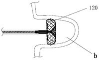
- Figure 10 is a schematic view of the left atrial appendage occluder of Example 1 after being released in the left atrial appendage;
- Figure 11a is a schematic view showing the connection of the nickel-titanium wire used as the barbed wire and the nickel-titanium wire of the frame of the braiding anchoring device in the second embodiment;
- Figure 11b is an enlarged view of a portion C of Figure 11a;
- Figure 12a is a schematic view of the left atrial appendage occluder of Embodiment 2;
- Figure 12b is an enlarged view of a portion D in Figure 12a;
- Figure 13a is a schematic view showing the connection of the nickel-titanium wire used as the barb in Example 3 to the nickel-titanium wire of the frame of the braiding anchoring device;
- Figure 13b is an enlarged view of a portion E of Figure 13a;
- Figure 14a is a schematic view of the left atrial appendage occluder of Embodiment 3;
- Figure 14b is an enlarged view of a portion F in Figure 14a;
- Figure 15a is a schematic view of the anchoring device of Embodiment 4.
- Figure 15b is a schematic view (another angle) of the anchoring device of Embodiment 4.
- Figure 16a is a schematic view of the anchoring device of Embodiment 5.
- Figure 16b is a schematic view (another angle) of the anchoring device of Embodiment 5;
- Figure 17a is a schematic view of the anchoring device of Embodiment 6;
- Figure 17b is a schematic view (another angle) of the anchoring device of Embodiment 6;
- Figure 18 is a schematic view of a sealing disk in Embodiment 7.
- Figure 19 is a schematic overall view of the left atrial appendage occluder of Embodiment 7;
- FIG. 20 is a schematic view of the left atrial appendage occluder of Example 7 after being released in the left atrial appendage;
- Figure 21a is a schematic view of the preparation of the release anchoring net in Embodiment 1;
- Figure 21b is a schematic view showing the release region of the anchoring net in Embodiment 1;
- Figure 21c is a schematic view showing the release of the anchoring device in Embodiment 1;
- Figure 22a is a schematic view showing the completion of the recovery of the sealing disk in the first embodiment, preparing the recovery anchoring device;
- Figure 22b is a schematic view showing the partial recovery of the anchoring device in the first embodiment
- Figure 22c is a schematic illustration of the recovery anchoring device of Example 1 with the barbs completely withdrawn from the left atrial appendage wall.
- the left atrial appendage occluder 100 of the present invention is comprised of a sealing disk 110 and an anchoring device 120.
- the anchoring device 120 has a rolled structure from the inside to the outside. The inner edge portion of the rolled structure is connected to the sealing disc 110.
- the peripheral portion of the rolled structure is an anchoring net 122 having a boneless structure that cooperates with the left atrial appendage.
- the sealing disk 110 is a mesh structure woven by nickel-titanium wire. As shown in FIG. 2, the sealing disk 110 is composed of a disk surface 111 and a waist portion 112. A first fixed end 113 is provided at the center of the disk surface, and the center of the waist portion 112 is provided. There is a second fixed end 114, a layer of PET blocking film 115 is slit in the disk surface, and a layer of the blocking film 116 is sewn in the waist 112.
- the first fixed end 113 is for connection to a delivery device and the second fixed end 114 is coupled to the anchoring device 120.
- the waist portion 112 includes a diffusion section 117 radiated outwardly from the second fixed end 114, and a tubular body portion 118 having a cylindrical shape at the periphery of the diffusion section 117.
- the top edge of the body section 118 is connected to the disk surface 111.
- the bottom edge of the 118 and the outer edge of the diffuser section 117 are transitionally connected by a circular arc 119, and the baffle film 116 in the waist portion 112 is located in the middle of the axial direction of the body section 118.
- the diffusion section 117 is radiated and diffused outward by the second fixed end 114 to form a plane, and the second fixed end 114 is slightly closer to the disk surface 111, so that a recessed area is formed in the plane.
- the anchoring device 120 is woven by nickel-titanium wire, and the anchoring net 122 matching the inner wall of the left atrial appendage is nest-shaped (ie, bowl-shaped), and the anchoring device 120 is a boneless structure. That is, there is no skeleton provided inside the anchoring device 120 for supporting.
- the anchoring device 120 has a two-layer structure, and the central portion of the anchoring device 120 is folded toward the sealing disk 110 to form an inner layer tapered mesh 121.
- the proximal end of the tapered mesh 121 is converged and the central end of the sealing disk 110 is closed.
- the 126 is connected, and the anchoring net 122 is at the periphery of the tapered mesh 121.
- the anchoring net 122 is rolled up to the proximal end of the adjacent tapered mesh 121, and is connected to the tapered mesh through the circular arc transition zone 124. The bottom edge.
- the outer surface of the nested anchoring net 122 is evenly distributed with six barbs 127.
- the nest-shaped nest end is bent inwardly to form a cuff region 123 (i.e., one end of the nested anchor net 122 near the sealing disk 110 extends radially toward the center end 126 to form a cuff region 123), and the center end 126 is located in the conical mesh 121.
- the axial end portion, the cuff region 123 and the nested anchor net 122 are smoothly connected by a circular transition zone 125.
- the second fixed end 114 of the sealing disk 110 and the central end 126 of the anchoring device 120 are welded together by a steel sleeve 130, combined into a left atrial appendage occluder 100 as shown in Figure 1.
- the anchoring device 120 is woven by 24 nickel-titanium wires, wherein 6 nickel-titanium wires are used for forming barbs, and the remaining 18 nickel-titanium wires are used for weaving the frame of the anchoring device, and barbed nickel-titanium wires. Same diameter as the frame nickel-titanium wire.
- the nickel-titanium wire used to braid the frame of the anchoring device was previously molded into an inverted V shape, and a total of 18 inverted V-shaped nickel-titanium wires. As shown in FIG. 6a and FIG.
- each of the nickel-titanium wires 1221 for forming the barbs is arranged in parallel with one side 1222 of one of the inverted V-shaped nickel-titanium wires of the anchoring device frame (the parallel arrangement may be performed as needed) Fixed or not fixed) and passed through a steel sleeve The stainless steel material 1223 is pressed and fixed, and the steel sleeve 1223 is close to the apex 1224 where the two sides of the inverted V-shaped nickel-titanium wire intersect.
- the distance from the apex 1224 of the nickel titanium wire is 8 mm.
- the nickel-titanium wire After the nickel-titanium wire is prepared, all the nickel-titanium wires are first woven into a continuous cylindrical mesh tube 101. As shown in FIGS. 7a and 7b, the six nickel-titanium wires used for forming the barbs are evenly distributed along the circumference of the network tube 101. .
- the tail end 1011 of the network tube 101 can be fixedly welded together by a stainless steel sleeve, and then the mesh tube is shaped into an anchoring device shape as shown in FIG. 3 by a mold and high temperature heating. At the same time, the end portion 1225 is partially opened outward at a certain angle, such as 30 degrees, to form a barb 127.
- the barbs 127 are evenly distributed along the circumferential direction of the anchoring net, and the barbs 127 are radially outwardly turned along the anchoring net 122 and are axially inclined along the anchoring net 122. More intuitively, as shown in FIG. 7a and FIG. 7b, in the process of making the anchoring device 120, the nickel-titanium wire forming the barb 127 is arranged in parallel with the nickel-titanium wire of the anchoring device frame to form the nickel-titanium of the barb 127. The wire is lifted at the end to form a non-zero angle with the nickel titanium wire from which the anchoring device frame is made, as shown in Figure 3a.
- the diameter of the nickel-titanium wire used to form the barb in this embodiment may also be larger than the diameter of the nickel-titanium wire of the frame of the anchoring device.
- the number of barbs anchored on the net may also be three, nine, or other numbers of barbs evenly distributed.
- the anchoring device frame can also be woven by using 36 or other numbers of inverted V-shaped nickel-titanium wires.
- the steel sleeve 1223 for fixing the barbs 127 in this embodiment, and the steel sleeve for anchoring the tail end 1011 of the mesh tube 101 may also be made of a nickel-titanium sleeve or other metal sleeves conforming to biocompatibility requirements.
- the nickel-titanium wire braided mesh tube 101 in this embodiment can also be shaped into a shape as shown in FIG. 9, and then the center end is welded and fixed to the shape shown in FIG.
- the left atrial appendage occluder 100 can be inserted into a small diameter (such as an internal diameter of 9F) delivery device, and then into the superior vena cava through the femoral vein, then enter the right atrium, and then enter the left atrium through the interatrial septum.
- a small diameter such as an internal diameter of 9F
- the anchoring device 120 is released inside the left atrial appendage
- the barbs 127 on the anchoring net 122 are hooked into the inner wall of the left atrial appendage; then the waist portion 112 of the sealing disk 110 is released in the left atrial appendage opening.
- a is the left atrium
- b is the left atrial appendage
- c is the left ventricle
- d is the right.
- the ventricle, e is the right atrium.
- the anchoring device 120 of the left atrial appendage occluder 100 is woven into a uniform mesh shape by using a nickel-titanium wire, and the nest-shaped anchoring net 122 of the outer layer of the anchoring device 120 can be evenly contacted with the inner wall of the left atrial appendage to ensure uniformity.
- the pressure is evenly applied to the inner wall of the left atrial appendage without local stress concentration.
- the barbs 127 on the anchor net 122 of the left atrial appendage occluder 100 are made of nickel-titanium wire, and the diameter of the barbs 127 is relatively thin, so that the phenomenon of perforation of the inner wall of the left atrial appendage is not easily generated.
- the sealing disc 110 of the left atrial appendage occluder 100 has both the waist portion 112 and the disc surface 111.
- the waist portion 112 and the disc surface 111 can both block the opening of the left atrial appendage, and the double sealing effect improves the left atrial appendage.
- the blocking function of the occluder 100 as a whole.
- the left atrial appendage occluder 100 in this embodiment is capable of achieving repeated release.
- the release process is as shown in FIG. 21a, FIG. 21b and FIG. 21c.
- the anchoring device is in a compressed state in the delivery device, and the delivery device is moved to the opening of the left atrial appendage.
- the anchoring device is gradually released from the conveying device.
- the anchoring device adopts superelastic metal wire or memory alloy wire
- the outer anchoring mesh closing area is separated from the conveying device, and then the self-rewinding is performed, so that the contact with the left atrial appendage wall is always For a smooth surface, after the anchoring device is released, the barb penetrates into the wall of the left atrial appendage, and the direction of the barb is directed toward the opening of the left atrial appendage.
- the left atrial appendage occluder needs to be recovered.
- the recovery process is shown in Figure 22a, Figure 22b and Figure 22c.
- the thorn is first completely withdrawn from the inner wall of the left atrial appendage from the direction of penetration, and then, together with the outer anchoring net, is transported into the delivery device under the traction of the delivery device.
- b is a left atrial appendage.
- Embodiment 1 differs from Embodiment 1 in that each of the nickel-titanium wires 2221 for forming the barbs and one of the inverted V-shaped nickel-titanium wires 2222 of the anchor mesh frame are joined by welding, the welded region
- the length of 2223 is 2 mm
- the weld zone 2223 is adjacent to the apex 2224 where the two sides of the inverted V-shaped nickel-titanium wire intersect.
- the nickel-titanium wire 2221 extends beyond the end 2225 of the weld zone 2223 to form a barb 227, the end
- each of the nickel-titanium wires 3221 for forming the barbs is 5 mm
- the nickel-titanium wire 3221 is divided into a fixed section and a free section, and the fixed section passes through the steel sleeve 3223 and
- Embodiment 1 differs from Embodiment 1 in that the anchoring device 420 does not include a cuffing region and includes only a central end 426 that is coupled to the sealing disk, and a conical mesh 421 that is diffused away from the sealing disk by the central end 426.
- the nested anchoring net 422 around the conical mesh 421, the conical mesh 421 and the nested anchoring net 422 are smoothly connected by a circular arc transition zone 424, and six barbs 427 are evenly distributed on the surface of the nested shape 422, such as Figure 15a, Figure 15b.
- the anchoring device 520 includes a center end 526 connected to the sealing disk, a conical mesh 521 diffused away from the sealing disk by the center end 526, and a truncated-shaped anchoring net 522 located at the periphery of the conical mesh 521.
- the net 521 and the truncated anchor net 522 are smoothly connected by a circular arc transition region 524, and six barbs 527 are uniformly distributed on the surface of the truncated cone shape 522.
- the center end 526 is located at the axial end of the conical mesh 521, and the end of the truncated anchoring net 522 adjacent to the sealing disc is connected with a cuffing region 523 extending radially toward the central end 526, and the cuff region 523 and the truncated cone shape 522 pass through the circle.
- the arc transition zone 525 is smoothly transitioned.
- the outer anchoring net structure of the anchoring device 520 is a truncated cone shape 522, as shown in Figs. 16a and 16b.
- the anchoring device 620 includes a central end 626 that is coupled to the sealing disk, a conical mesh 621 that is diffused away from the sealing disk by the central end 626, and a curved toroidal anchoring mesh 622 located at the periphery of the conical mesh 621.
- the conical mesh 621 and the curved toroidal anchoring net 622 are smoothly connected by a circular arc transition region 624, and six barbs 627 are uniformly distributed on the surface of the curved circular ring 622.
- the central end 626 is located at the axial end of the tapered mesh 621, and the end of the curved toroidal anchoring net 622 adjacent to the sealing disk is connected with a cuffing region 623 extending radially toward the central end 626, the cuffing region 623 and the curved circular anchor
- the fixed nets 622 are smoothly connected by a circular transition zone 625.
- the anchoring device 620 has an outer surface anchoring net structure of a curved circular ring 622, as shown in Figs. 17a and 17b.
- sealing disk 710 is composed only of the disk surface 711, and the first fixed end 713 and the second fixed end 714 are respectively disposed on two sides of the disk surface 711; Layer PET barrier film 715, as shown in FIG.
- the second fixed end 714 of the disk surface 711 and the center end of the anchor net 720 are joined together by a steel sleeve, as shown in FIG.
- FIG. 20 The schematic diagram of the left atrial appendage occluder 700 provided in the present embodiment is shown in FIG. 20, in which, a is the left atrium, b is the left atrial appendage, c is the left ventricle, and d is the right ventricle, e For the right atrium.
- the difference between this embodiment and the embodiment 1 is that three PET choke films are arranged in the sealing disc, one is sewn in the disc surface, one is sewn in the axial middle portion of the waist body segment, and one is sewn in the On the diffusion section.
Landscapes
- Health & Medical Sciences (AREA)
- Surgery (AREA)
- Life Sciences & Earth Sciences (AREA)
- Biomedical Technology (AREA)
- Medical Informatics (AREA)
- Vascular Medicine (AREA)
- Reproductive Health (AREA)
- Engineering & Computer Science (AREA)
- Veterinary Medicine (AREA)
- Heart & Thoracic Surgery (AREA)
- Nuclear Medicine, Radiotherapy & Molecular Imaging (AREA)
- Molecular Biology (AREA)
- Animal Behavior & Ethology (AREA)
- General Health & Medical Sciences (AREA)
- Public Health (AREA)
- Cardiology (AREA)
- Prostheses (AREA)
- Surgical Instruments (AREA)
Abstract
L'invention a pour objet des dispositifs d'occlusion de l'oreillette gauche améliorés (100, 700), lesquels dispositifs d'occlusion comprennent des poches hermétiques (110, 710) mutuellement reliées avec des dispositifs d'ancrage (120, 420, 520, 620), dans lesquelles poches hermétiques (110, 710) sont logées au moins deux membranes d'étranglement (115, 116, 715). Les dispositifs d'ancrage (120, 420, 520, 620) forment des réseaux d'ancrage (122, 422, 522, 622, 720) avec la position d'ajustement mutuelle de l'oreillette gauche (b), les réseaux d'ancrage (122, 422, 522, 622, 720) forment, à leur tour, une structure sans cadre. L'entier des dispositifs d'ancrage (120, 420, 520, 620) forme également une structure sans cadre. Les dispositifs d'ancrage (120, 420, 520, 620) sont formés de fibres métalliques, et leur extrémité distale adopte une configuration ouverte alors que l'extrémité proximale des dispositifs d'ancrage (120, 420, 520, 620) rétrécie relie les réseaux adoptant une configuration conique (121, 421, 520, 621) avec les poches hermétiques (110, 710). L'ouverture de l'extrémité distale des dispositifs d'ancrage (120, 420, 520, 620) forme, par revirement en direction de leur extrémité proximale, des réseaux d'ancrage (122, 422, 522, 622, 720), lesquels réseaux d'ancrage (122, 422, 522, 622, 720) enveloppent les réseaux adoptant une configuration conique (121, 421, 520, 621). Les dispositifs d'occlusion améliorés de l'oreillette gauche (100, 700) de la présente invention permettent l'occlusion multicouche de l'ouverture de l'oreillette gauche ainsi que l'ancrage uniforme par la force dans la partie interne de l'oreillette gauche (b), ce qui permet d'éviter l'accumulation de contrainte locale tout en assurant la libération susceptible d'être répétée, et ce, en garantissant l'occlusion fiable de la partie ouverte de l'oreillette gauche.
Priority Applications (2)
| Application Number | Priority Date | Filing Date | Title |
|---|---|---|---|
| EP16829835.4A EP3329865B1 (fr) | 2015-07-28 | 2016-07-26 | Dispositif d'occlusion d'appendice auriculaire gauche amélioré |
| US15/746,071 US10792045B2 (en) | 2015-07-28 | 2016-07-26 | Left atrial appendage occluder |
Applications Claiming Priority (2)
| Application Number | Priority Date | Filing Date | Title |
|---|---|---|---|
| CN201510452143.0A CN105054985B (zh) | 2015-07-28 | 2015-07-28 | 一种改进的左心耳封堵器 |
| CN201510452143.0 | 2015-07-28 |
Publications (1)
| Publication Number | Publication Date |
|---|---|
| WO2017016470A1 true WO2017016470A1 (fr) | 2017-02-02 |
Family
ID=54484684
Family Applications (1)
| Application Number | Title | Priority Date | Filing Date |
|---|---|---|---|
| PCT/CN2016/091700 Ceased WO2017016470A1 (fr) | 2015-07-28 | 2016-07-26 | Dispositif d'occlusion de l'oreillette gauche amélioré |
Country Status (4)
| Country | Link |
|---|---|
| US (1) | US10792045B2 (fr) |
| EP (1) | EP3329865B1 (fr) |
| CN (2) | CN108992120B (fr) |
| WO (1) | WO2017016470A1 (fr) |
Cited By (2)
| Publication number | Priority date | Publication date | Assignee | Title |
|---|---|---|---|---|
| CN114767179A (zh) * | 2022-04-09 | 2022-07-22 | 四川大学华西医院 | 一种房间隔缺损封堵器 |
| CN118750054A (zh) * | 2024-08-12 | 2024-10-11 | 山东第一医科大学第一附属医院(山东省千佛山医院) | 一种用于治疗室间隔穿孔和室壁瘤的介入封盖装置 |
Families Citing this family (38)
| Publication number | Priority date | Publication date | Assignee | Title |
|---|---|---|---|---|
| ES2403009T3 (es) * | 2010-05-23 | 2013-05-13 | Occlutech Holding Ag | Dispositivo médico trenzado y método para su fabricación |
| CA2946078C (fr) | 2014-04-30 | 2023-03-14 | Cerus Endovascular Limited | Dispositif d'occlusion |
| JP7001476B2 (ja) | 2015-02-25 | 2022-02-03 | ギャラクシー セラピューティクス インコーポレイテッド | 動脈瘤を治療するための装置 |
| CN104958087B (zh) * | 2015-07-28 | 2018-09-25 | 杭州诺茂医疗科技有限公司 | 一种左心耳封堵器 |
| CN108992120B (zh) * | 2015-07-28 | 2022-01-07 | 杭州德诺电生理医疗科技有限公司 | 一种改进的左心耳封堵器 |
| US10285711B2 (en) | 2015-12-07 | 2019-05-14 | Cerus Endovascular Limited | Occlusion device |
| EP3782576B1 (fr) | 2016-03-11 | 2025-07-16 | Stryker Ireland Technology Ltd. | Dispositif d'occlusion |
| CN107049373B (zh) * | 2017-02-08 | 2023-09-26 | 宁波迪创医疗科技有限公司 | 封堵器 |
| CN108938035B (zh) * | 2017-05-23 | 2025-09-05 | 杭州德诺电生理医疗科技有限公司 | 一种改进连接方式的左心耳封堵器 |
| CN209074751U (zh) * | 2017-05-23 | 2019-07-09 | 杭州诺茂医疗科技有限公司 | 一种提高密封效果的左心耳封堵器 |
| CN108938034B (zh) * | 2017-05-23 | 2025-09-05 | 杭州德诺电生理医疗科技有限公司 | 一种改进密封效果的左心耳封堵器及其制备方法 |
| CN109124755A (zh) * | 2017-06-16 | 2019-01-04 | 诺芮医疗器械(上海)有限公司 | 左心耳封堵消融装置 |
| CA3073232C (fr) * | 2017-08-21 | 2023-08-29 | Cerus Endovascular Limited | Dispositif d'occlusion |
| US12402885B2 (en) | 2017-09-23 | 2025-09-02 | Universität Zürich | Medical occlusion device |
| US20210251618A1 (en) * | 2017-10-13 | 2021-08-19 | Sree Chitra Tirunal Institute For Medical Sciences And Technology | Implantable atrial septal defect occlusion device with woven central section on left atrial flange |
| US11173023B2 (en) * | 2017-10-16 | 2021-11-16 | W. L. Gore & Associates, Inc. | Medical devices and anchors therefor |
| US11185335B2 (en) | 2018-01-19 | 2021-11-30 | Galaxy Therapeutics Inc. | System for and method of treating aneurysms |
| WO2019166017A1 (fr) * | 2018-03-02 | 2019-09-06 | 诺芮医疗器械(上海)有限公司 | Dispositif d'occlusion et d'ablation d'appendice auriculaire gauche |
| CN110215253A (zh) * | 2018-03-02 | 2019-09-10 | 诺芮医疗器械(上海)有限公司 | 左心耳封堵消融装置 |
| CN110215254A (zh) * | 2018-03-02 | 2019-09-10 | 诺芮医疗器械(上海)有限公司 | 一种左心耳封堵消融装置 |
| CN110811810A (zh) * | 2018-08-10 | 2020-02-21 | 诺芮医疗器械(上海)有限公司 | 左心耳消融封堵装置 |
| CN111150434B (zh) * | 2018-11-08 | 2025-02-25 | 先健科技(深圳)有限公司 | 封堵器 |
| CN110613492B (zh) * | 2019-01-28 | 2024-10-18 | 慧扬医疗科技(苏州)有限公司 | 左心耳封堵装置 |
| US12102327B2 (en) | 2019-05-25 | 2024-10-01 | Galaxy Therapeutics, Inc. | Systems and methods for treating aneurysms |
| US11166731B2 (en) | 2019-05-25 | 2021-11-09 | Galaxy Therapeutics Inc. | Systems and methods for treating aneurysms |
| CN114641242B (zh) | 2019-09-26 | 2025-11-18 | 苏黎世大学 | 左心耳闭塞装置 |
| US11406404B2 (en) | 2020-02-20 | 2022-08-09 | Cerus Endovascular Limited | Clot removal distal protection methods |
| WO2021263145A1 (fr) * | 2020-06-25 | 2021-12-30 | Squadra Lifesciences, Inc. | Méthodes et dispositifs d'occlusion de l'appendice auriculaire gauche |
| WO2022010908A1 (fr) * | 2020-07-07 | 2022-01-13 | St. Jude Medical, Cardiology Division, Inc. | Dispositifs et méthodes d'occlusion d'anormalités du système vasculaire |
| CN112155627B (zh) * | 2020-07-28 | 2025-07-25 | 宁波迪创医疗科技有限公司 | 一种具有仿生微刺依附结构的左心耳封堵器 |
| CN114680956B (zh) * | 2020-12-30 | 2025-02-25 | 先健科技(深圳)有限公司 | 封堵器械 |
| CN112315523B (zh) * | 2021-01-05 | 2021-06-04 | 先健科技(深圳)有限公司 | 封堵装置 |
| EP4284263A4 (fr) | 2021-01-27 | 2024-06-26 | Galaxy Therapeutics, Inc. | Systèmes et méthodes pour traiter des anévrismes |
| IT202100008411A1 (it) | 2021-04-02 | 2022-10-02 | Riccardo Laurenti | Occlusore di auricola atriale sinistra |
| CN115212370A (zh) * | 2021-04-14 | 2022-10-21 | 杭州诺生医疗科技有限公司 | 分流装置及分流系统 |
| CN113384308B (zh) * | 2021-06-02 | 2022-09-13 | 上海捍宇医疗科技股份有限公司 | 一种挂钩连接式可穿刺的经导管输送房间壁封堵器系统 |
| WO2024046392A1 (fr) * | 2022-08-30 | 2024-03-07 | 先健科技(深圳)有限公司 | Appareil d'occlusion |
| WO2025137233A1 (fr) * | 2023-12-20 | 2025-06-26 | Boston Scientific Scimed, Inc. | Systèmes d'implant d'occlusion |
Citations (9)
| Publication number | Priority date | Publication date | Assignee | Title |
|---|---|---|---|---|
| US20070066993A1 (en) * | 2005-09-16 | 2007-03-22 | Kreidler Marc S | Intracardiac cage and method of delivering same |
| CN104352260A (zh) * | 2014-10-13 | 2015-02-18 | 陈奕龙 | 左心耳封堵系统 |
| CN204181679U (zh) * | 2014-10-13 | 2015-03-04 | 陈奕龙 | 左心耳封堵器 |
| CN204181678U (zh) * | 2014-10-13 | 2015-03-04 | 陈奕龙 | 左心耳封堵系统 |
| CN204246182U (zh) * | 2014-11-22 | 2015-04-08 | 常州乐奥医疗科技有限公司 | 一种新型伞状左心耳封堵器 |
| CN104958087A (zh) * | 2015-07-28 | 2015-10-07 | 杭州德诺科技有限公司 | 一种左心耳封堵器 |
| CN105054985A (zh) * | 2015-07-28 | 2015-11-18 | 杭州德诺科技有限公司 | 一种改进的左心耳封堵器 |
| CN204814031U (zh) * | 2015-07-28 | 2015-12-02 | 杭州德诺科技有限公司 | 一种改进的左心耳封堵器 |
| CN204971418U (zh) * | 2015-07-28 | 2016-01-20 | 杭州德诺科技有限公司 | 一种左心耳封堵器 |
Family Cites Families (22)
| Publication number | Priority date | Publication date | Assignee | Title |
|---|---|---|---|---|
| US7128073B1 (en) | 1998-11-06 | 2006-10-31 | Ev3 Endovascular, Inc. | Method and device for left atrial appendage occlusion |
| WO2006102213A1 (fr) * | 2005-03-18 | 2006-09-28 | Nmt Medical, Inc. | Element de retention pour occluseur de pfo |
| WO2009052432A2 (fr) | 2007-10-19 | 2009-04-23 | Coherex Medical, Inc. | Dispositif médical destiné à une modification de l'appendice auriculaire gauche, systèmes et procédés apparentés |
| CN101449986B (zh) * | 2007-11-28 | 2011-08-31 | 王涛 | 主动脉导管未闭的阻塞器 |
| US20090171386A1 (en) * | 2007-12-28 | 2009-07-02 | Aga Medical Corporation | Percutaneous catheter directed intravascular occlusion devices |
| US9649115B2 (en) * | 2009-06-17 | 2017-05-16 | Coherex Medical, Inc. | Medical device for modification of left atrial appendage and related systems and methods |
| US9351716B2 (en) * | 2009-06-17 | 2016-05-31 | Coherex Medical, Inc. | Medical device and delivery system for modification of left atrial appendage and methods thereof |
| US8828051B2 (en) * | 2010-07-02 | 2014-09-09 | Pfm Medical Ag | Left atrial appendage occlusion device |
| CN101933850B (zh) * | 2010-09-16 | 2012-07-18 | 先健科技(深圳)有限公司 | 封堵器及其制造方法 |
| US8764787B2 (en) * | 2011-06-17 | 2014-07-01 | Aga Medical Corporation | Occlusion device and associated deployment method |
| EP2775932A1 (fr) * | 2011-11-09 | 2014-09-17 | Boston Scientific Scimed, Inc. | Dispositif d'occlusion |
| US9592058B2 (en) * | 2012-02-21 | 2017-03-14 | Cardia, Inc. | Left atrial appendage occlusion device |
| EP2838444A4 (fr) * | 2012-04-20 | 2016-02-24 | Inceptus Medical LLC | Dispositifs d'occlusion expansibles et procédés d'utilisation |
| CN104768476B (zh) * | 2012-07-13 | 2019-05-31 | 波士顿科学国际有限公司 | 用于心耳的封堵器 |
| US20140135817A1 (en) * | 2012-11-14 | 2014-05-15 | Boston Scientific Scimed, Inc. | Left atrial appendage closure implant |
| US10973523B2 (en) * | 2013-03-08 | 2021-04-13 | Aga Medical Corporation | Medical device for treating a target site |
| US9681861B2 (en) * | 2013-03-11 | 2017-06-20 | St. Jude Medical, Cardiology Division, Inc. | Percutaneous catheter directed collapsible medical closure device |
| CN103845096B (zh) * | 2014-03-10 | 2016-05-04 | 上海形状记忆合金材料有限公司 | 左心耳封堵器及其制作方法 |
| CN204016555U (zh) * | 2014-07-30 | 2014-12-17 | 上海形状记忆合金材料有限公司 | 一种封堵器 |
| CN104274224B (zh) * | 2014-09-29 | 2017-01-25 | 上海形状记忆合金材料有限公司 | 左心耳封堵器 |
| CN104352261B (zh) * | 2014-10-13 | 2017-11-03 | 深圳市科奕顿生物医疗科技有限公司 | 左心耳封堵器 |
| CN104398284A (zh) * | 2014-11-24 | 2015-03-11 | 上海普实医疗器械科技有限公司 | 一种新型左心耳封堵器及制造方法 |
-
2015
- 2015-07-28 CN CN201810391987.2A patent/CN108992120B/zh active Active
- 2015-07-28 CN CN201510452143.0A patent/CN105054985B/zh active Active
-
2016
- 2016-07-26 WO PCT/CN2016/091700 patent/WO2017016470A1/fr not_active Ceased
- 2016-07-26 US US15/746,071 patent/US10792045B2/en active Active
- 2016-07-26 EP EP16829835.4A patent/EP3329865B1/fr active Active
Patent Citations (9)
| Publication number | Priority date | Publication date | Assignee | Title |
|---|---|---|---|---|
| US20070066993A1 (en) * | 2005-09-16 | 2007-03-22 | Kreidler Marc S | Intracardiac cage and method of delivering same |
| CN104352260A (zh) * | 2014-10-13 | 2015-02-18 | 陈奕龙 | 左心耳封堵系统 |
| CN204181679U (zh) * | 2014-10-13 | 2015-03-04 | 陈奕龙 | 左心耳封堵器 |
| CN204181678U (zh) * | 2014-10-13 | 2015-03-04 | 陈奕龙 | 左心耳封堵系统 |
| CN204246182U (zh) * | 2014-11-22 | 2015-04-08 | 常州乐奥医疗科技有限公司 | 一种新型伞状左心耳封堵器 |
| CN104958087A (zh) * | 2015-07-28 | 2015-10-07 | 杭州德诺科技有限公司 | 一种左心耳封堵器 |
| CN105054985A (zh) * | 2015-07-28 | 2015-11-18 | 杭州德诺科技有限公司 | 一种改进的左心耳封堵器 |
| CN204814031U (zh) * | 2015-07-28 | 2015-12-02 | 杭州德诺科技有限公司 | 一种改进的左心耳封堵器 |
| CN204971418U (zh) * | 2015-07-28 | 2016-01-20 | 杭州德诺科技有限公司 | 一种左心耳封堵器 |
Cited By (2)
| Publication number | Priority date | Publication date | Assignee | Title |
|---|---|---|---|---|
| CN114767179A (zh) * | 2022-04-09 | 2022-07-22 | 四川大学华西医院 | 一种房间隔缺损封堵器 |
| CN118750054A (zh) * | 2024-08-12 | 2024-10-11 | 山东第一医科大学第一附属医院(山东省千佛山医院) | 一种用于治疗室间隔穿孔和室壁瘤的介入封盖装置 |
Also Published As
| Publication number | Publication date |
|---|---|
| US20180193027A1 (en) | 2018-07-12 |
| EP3329865A1 (fr) | 2018-06-06 |
| CN105054985A (zh) | 2015-11-18 |
| CN105054985B (zh) | 2018-05-01 |
| US10792045B2 (en) | 2020-10-06 |
| EP3329865B1 (fr) | 2023-06-21 |
| CN108992120A (zh) | 2018-12-14 |
| CN108992120B (zh) | 2022-01-07 |
| EP3329865A4 (fr) | 2019-03-27 |
Similar Documents
| Publication | Publication Date | Title |
|---|---|---|
| WO2017016470A1 (fr) | Dispositif d'occlusion de l'oreillette gauche amélioré | |
| CN104958087B (zh) | 一种左心耳封堵器 | |
| US20220125567A1 (en) | Space filling devices | |
| CN108926370B (zh) | 一种提高密封效果的左心耳封堵器及其装配方法 | |
| JP6240604B2 (ja) | 拡張可能な閉塞デバイス及び方法 | |
| CN204971418U (zh) | 一种左心耳封堵器 | |
| US20150133989A1 (en) | Expandable occlusion devices and methods of use | |
| CN204814031U (zh) | 一种改进的左心耳封堵器 | |
| EP3202333A1 (fr) | Structures tressées multicouche pour l'occlusion de défauts vasculaires | |
| CN108938035A (zh) | 一种改进连接方式的左心耳封堵器 | |
| CN108938034A (zh) | 一种改进密封效果的左心耳封堵器及其制备方法 | |
| CN207928350U (zh) | 一种改进密封效果的左心耳封堵器 | |
| CN207928351U (zh) | 一种改进连接方式的左心耳封堵器 |
Legal Events
| Date | Code | Title | Description |
|---|---|---|---|
| 121 | Ep: the epo has been informed by wipo that ep was designated in this application |
Ref document number: 16829835 Country of ref document: EP Kind code of ref document: A1 |
|
| NENP | Non-entry into the national phase |
Ref country code: DE |
|
| WWE | Wipo information: entry into national phase |
Ref document number: 2016829835 Country of ref document: EP |