WO2017086356A1 - ウシ血清組成物及びそのウシ血清組成物を添加剤として使用する細胞の培養方法 - Google Patents
ウシ血清組成物及びそのウシ血清組成物を添加剤として使用する細胞の培養方法 Download PDFInfo
- Publication number
- WO2017086356A1 WO2017086356A1 PCT/JP2016/083990 JP2016083990W WO2017086356A1 WO 2017086356 A1 WO2017086356 A1 WO 2017086356A1 JP 2016083990 W JP2016083990 W JP 2016083990W WO 2017086356 A1 WO2017086356 A1 WO 2017086356A1
- Authority
- WO
- WIPO (PCT)
- Prior art keywords
- bovine serum
- serum composition
- blood
- platelets
- glass
- Prior art date
- Legal status (The legal status is an assumption and is not a legal conclusion. Google has not performed a legal analysis and makes no representation as to the accuracy of the status listed.)
- Ceased
Links
Images



Classifications
-
- C—CHEMISTRY; METALLURGY
- C12—BIOCHEMISTRY; BEER; SPIRITS; WINE; VINEGAR; MICROBIOLOGY; ENZYMOLOGY; MUTATION OR GENETIC ENGINEERING
- C12N—MICROORGANISMS OR ENZYMES; COMPOSITIONS THEREOF; PROPAGATING, PRESERVING, OR MAINTAINING MICROORGANISMS; MUTATION OR GENETIC ENGINEERING; CULTURE MEDIA
- C12N5/00—Undifferentiated human, animal or plant cells, e.g. cell lines; Tissues; Cultivation or maintenance thereof; Culture media therefor
- C12N5/06—Animal cells or tissues; Human cells or tissues
- C12N5/0602—Vertebrate cells
- C12N5/0652—Cells of skeletal and connective tissues; Mesenchyme
- C12N5/0662—Stem cells
- C12N5/0664—Dental pulp stem cells, Dental follicle stem cells
-
- C—CHEMISTRY; METALLURGY
- C12—BIOCHEMISTRY; BEER; SPIRITS; WINE; VINEGAR; MICROBIOLOGY; ENZYMOLOGY; MUTATION OR GENETIC ENGINEERING
- C12N—MICROORGANISMS OR ENZYMES; COMPOSITIONS THEREOF; PROPAGATING, PRESERVING, OR MAINTAINING MICROORGANISMS; MUTATION OR GENETIC ENGINEERING; CULTURE MEDIA
- C12N5/00—Undifferentiated human, animal or plant cells, e.g. cell lines; Tissues; Cultivation or maintenance thereof; Culture media therefor
- C12N5/06—Animal cells or tissues; Human cells or tissues
- C12N5/0602—Vertebrate cells
- C12N5/0652—Cells of skeletal and connective tissues; Mesenchyme
- C12N5/0662—Stem cells
-
- A—HUMAN NECESSITIES
- A61—MEDICAL OR VETERINARY SCIENCE; HYGIENE
- A61K—PREPARATIONS FOR MEDICAL, DENTAL OR TOILETRY PURPOSES
- A61K35/00—Medicinal preparations containing materials or reaction products thereof with undetermined constitution
- A61K35/12—Materials from mammals; Compositions comprising non-specified tissues or cells; Compositions comprising non-embryonic stem cells; Genetically modified cells
- A61K35/14—Blood; Artificial blood
- A61K35/16—Blood plasma; Blood serum
-
- A—HUMAN NECESSITIES
- A61—MEDICAL OR VETERINARY SCIENCE; HYGIENE
- A61K—PREPARATIONS FOR MEDICAL, DENTAL OR TOILETRY PURPOSES
- A61K35/00—Medicinal preparations containing materials or reaction products thereof with undetermined constitution
- A61K35/12—Materials from mammals; Compositions comprising non-specified tissues or cells; Compositions comprising non-embryonic stem cells; Genetically modified cells
- A61K35/14—Blood; Artificial blood
- A61K35/17—Lymphocytes; B-cells; T-cells; Natural killer cells; Interferon-activated or cytokine-activated lymphocytes
-
- A—HUMAN NECESSITIES
- A61—MEDICAL OR VETERINARY SCIENCE; HYGIENE
- A61K—PREPARATIONS FOR MEDICAL, DENTAL OR TOILETRY PURPOSES
- A61K35/00—Medicinal preparations containing materials or reaction products thereof with undetermined constitution
- A61K35/12—Materials from mammals; Compositions comprising non-specified tissues or cells; Compositions comprising non-embryonic stem cells; Genetically modified cells
- A61K35/14—Blood; Artificial blood
- A61K35/19—Platelets; Megacaryocytes
-
- C—CHEMISTRY; METALLURGY
- C12—BIOCHEMISTRY; BEER; SPIRITS; WINE; VINEGAR; MICROBIOLOGY; ENZYMOLOGY; MUTATION OR GENETIC ENGINEERING
- C12N—MICROORGANISMS OR ENZYMES; COMPOSITIONS THEREOF; PROPAGATING, PRESERVING, OR MAINTAINING MICROORGANISMS; MUTATION OR GENETIC ENGINEERING; CULTURE MEDIA
- C12N5/00—Undifferentiated human, animal or plant cells, e.g. cell lines; Tissues; Cultivation or maintenance thereof; Culture media therefor
- C12N5/06—Animal cells or tissues; Human cells or tissues
- C12N5/0602—Vertebrate cells
- C12N5/0652—Cells of skeletal and connective tissues; Mesenchyme
- C12N5/0662—Stem cells
- C12N5/0668—Mesenchymal stem cells from other natural sources
-
- C—CHEMISTRY; METALLURGY
- C12—BIOCHEMISTRY; BEER; SPIRITS; WINE; VINEGAR; MICROBIOLOGY; ENZYMOLOGY; MUTATION OR GENETIC ENGINEERING
- C12N—MICROORGANISMS OR ENZYMES; COMPOSITIONS THEREOF; PROPAGATING, PRESERVING, OR MAINTAINING MICROORGANISMS; MUTATION OR GENETIC ENGINEERING; CULTURE MEDIA
- C12N2500/00—Specific components of cell culture medium
-
- C—CHEMISTRY; METALLURGY
- C12—BIOCHEMISTRY; BEER; SPIRITS; WINE; VINEGAR; MICROBIOLOGY; ENZYMOLOGY; MUTATION OR GENETIC ENGINEERING
- C12N—MICROORGANISMS OR ENZYMES; COMPOSITIONS THEREOF; PROPAGATING, PRESERVING, OR MAINTAINING MICROORGANISMS; MUTATION OR GENETIC ENGINEERING; CULTURE MEDIA
- C12N2501/00—Active agents used in cell culture processes, e.g. differentation
- C12N2501/10—Growth factors
- C12N2501/115—Basic fibroblast growth factor (bFGF, FGF-2)
-
- C—CHEMISTRY; METALLURGY
- C12—BIOCHEMISTRY; BEER; SPIRITS; WINE; VINEGAR; MICROBIOLOGY; ENZYMOLOGY; MUTATION OR GENETIC ENGINEERING
- C12N—MICROORGANISMS OR ENZYMES; COMPOSITIONS THEREOF; PROPAGATING, PRESERVING, OR MAINTAINING MICROORGANISMS; MUTATION OR GENETIC ENGINEERING; CULTURE MEDIA
- C12N2501/00—Active agents used in cell culture processes, e.g. differentation
- C12N2501/10—Growth factors
- C12N2501/135—Platelet-derived growth factor [PDGF]
-
- C—CHEMISTRY; METALLURGY
- C12—BIOCHEMISTRY; BEER; SPIRITS; WINE; VINEGAR; MICROBIOLOGY; ENZYMOLOGY; MUTATION OR GENETIC ENGINEERING
- C12N—MICROORGANISMS OR ENZYMES; COMPOSITIONS THEREOF; PROPAGATING, PRESERVING, OR MAINTAINING MICROORGANISMS; MUTATION OR GENETIC ENGINEERING; CULTURE MEDIA
- C12N2501/00—Active agents used in cell culture processes, e.g. differentation
- C12N2501/10—Growth factors
- C12N2501/15—Transforming growth factor beta (TGF-β)
-
- C—CHEMISTRY; METALLURGY
- C12—BIOCHEMISTRY; BEER; SPIRITS; WINE; VINEGAR; MICROBIOLOGY; ENZYMOLOGY; MUTATION OR GENETIC ENGINEERING
- C12N—MICROORGANISMS OR ENZYMES; COMPOSITIONS THEREOF; PROPAGATING, PRESERVING, OR MAINTAINING MICROORGANISMS; MUTATION OR GENETIC ENGINEERING; CULTURE MEDIA
- C12N2501/00—Active agents used in cell culture processes, e.g. differentation
- C12N2501/10—Growth factors
- C12N2501/165—Vascular endothelial growth factor [VEGF]
-
- C—CHEMISTRY; METALLURGY
- C12—BIOCHEMISTRY; BEER; SPIRITS; WINE; VINEGAR; MICROBIOLOGY; ENZYMOLOGY; MUTATION OR GENETIC ENGINEERING
- C12N—MICROORGANISMS OR ENZYMES; COMPOSITIONS THEREOF; PROPAGATING, PRESERVING, OR MAINTAINING MICROORGANISMS; MUTATION OR GENETIC ENGINEERING; CULTURE MEDIA
- C12N2501/00—Active agents used in cell culture processes, e.g. differentation
- C12N2501/20—Cytokines; Chemokines
-
- C—CHEMISTRY; METALLURGY
- C12—BIOCHEMISTRY; BEER; SPIRITS; WINE; VINEGAR; MICROBIOLOGY; ENZYMOLOGY; MUTATION OR GENETIC ENGINEERING
- C12N—MICROORGANISMS OR ENZYMES; COMPOSITIONS THEREOF; PROPAGATING, PRESERVING, OR MAINTAINING MICROORGANISMS; MUTATION OR GENETIC ENGINEERING; CULTURE MEDIA
- C12N2501/00—Active agents used in cell culture processes, e.g. differentation
- C12N2501/20—Cytokines; Chemokines
- C12N2501/21—Chemokines, e.g. MIP-1, MIP-2, RANTES, MCP, PF-4
-
- C—CHEMISTRY; METALLURGY
- C12—BIOCHEMISTRY; BEER; SPIRITS; WINE; VINEGAR; MICROBIOLOGY; ENZYMOLOGY; MUTATION OR GENETIC ENGINEERING
- C12N—MICROORGANISMS OR ENZYMES; COMPOSITIONS THEREOF; PROPAGATING, PRESERVING, OR MAINTAINING MICROORGANISMS; MUTATION OR GENETIC ENGINEERING; CULTURE MEDIA
- C12N2501/00—Active agents used in cell culture processes, e.g. differentation
- C12N2501/20—Cytokines; Chemokines
- C12N2501/22—Colony stimulating factors (G-CSF, GM-CSF)
-
- C—CHEMISTRY; METALLURGY
- C12—BIOCHEMISTRY; BEER; SPIRITS; WINE; VINEGAR; MICROBIOLOGY; ENZYMOLOGY; MUTATION OR GENETIC ENGINEERING
- C12N—MICROORGANISMS OR ENZYMES; COMPOSITIONS THEREOF; PROPAGATING, PRESERVING, OR MAINTAINING MICROORGANISMS; MUTATION OR GENETIC ENGINEERING; CULTURE MEDIA
- C12N2501/00—Active agents used in cell culture processes, e.g. differentation
- C12N2501/20—Cytokines; Chemokines
- C12N2501/23—Interleukins [IL]
- C12N2501/2301—Interleukin-1 (IL-1)
-
- C—CHEMISTRY; METALLURGY
- C12—BIOCHEMISTRY; BEER; SPIRITS; WINE; VINEGAR; MICROBIOLOGY; ENZYMOLOGY; MUTATION OR GENETIC ENGINEERING
- C12N—MICROORGANISMS OR ENZYMES; COMPOSITIONS THEREOF; PROPAGATING, PRESERVING, OR MAINTAINING MICROORGANISMS; MUTATION OR GENETIC ENGINEERING; CULTURE MEDIA
- C12N2501/00—Active agents used in cell culture processes, e.g. differentation
- C12N2501/20—Cytokines; Chemokines
- C12N2501/23—Interleukins [IL]
- C12N2501/2304—Interleukin-4 (IL-4)
-
- C—CHEMISTRY; METALLURGY
- C12—BIOCHEMISTRY; BEER; SPIRITS; WINE; VINEGAR; MICROBIOLOGY; ENZYMOLOGY; MUTATION OR GENETIC ENGINEERING
- C12N—MICROORGANISMS OR ENZYMES; COMPOSITIONS THEREOF; PROPAGATING, PRESERVING, OR MAINTAINING MICROORGANISMS; MUTATION OR GENETIC ENGINEERING; CULTURE MEDIA
- C12N2501/00—Active agents used in cell culture processes, e.g. differentation
- C12N2501/20—Cytokines; Chemokines
- C12N2501/23—Interleukins [IL]
- C12N2501/2305—Interleukin-5 (IL-5)
-
- C—CHEMISTRY; METALLURGY
- C12—BIOCHEMISTRY; BEER; SPIRITS; WINE; VINEGAR; MICROBIOLOGY; ENZYMOLOGY; MUTATION OR GENETIC ENGINEERING
- C12N—MICROORGANISMS OR ENZYMES; COMPOSITIONS THEREOF; PROPAGATING, PRESERVING, OR MAINTAINING MICROORGANISMS; MUTATION OR GENETIC ENGINEERING; CULTURE MEDIA
- C12N2501/00—Active agents used in cell culture processes, e.g. differentation
- C12N2501/20—Cytokines; Chemokines
- C12N2501/23—Interleukins [IL]
- C12N2501/2306—Interleukin-6 (IL-6)
-
- C—CHEMISTRY; METALLURGY
- C12—BIOCHEMISTRY; BEER; SPIRITS; WINE; VINEGAR; MICROBIOLOGY; ENZYMOLOGY; MUTATION OR GENETIC ENGINEERING
- C12N—MICROORGANISMS OR ENZYMES; COMPOSITIONS THEREOF; PROPAGATING, PRESERVING, OR MAINTAINING MICROORGANISMS; MUTATION OR GENETIC ENGINEERING; CULTURE MEDIA
- C12N2501/00—Active agents used in cell culture processes, e.g. differentation
- C12N2501/20—Cytokines; Chemokines
- C12N2501/23—Interleukins [IL]
- C12N2501/2307—Interleukin-7 (IL-7)
-
- C—CHEMISTRY; METALLURGY
- C12—BIOCHEMISTRY; BEER; SPIRITS; WINE; VINEGAR; MICROBIOLOGY; ENZYMOLOGY; MUTATION OR GENETIC ENGINEERING
- C12N—MICROORGANISMS OR ENZYMES; COMPOSITIONS THEREOF; PROPAGATING, PRESERVING, OR MAINTAINING MICROORGANISMS; MUTATION OR GENETIC ENGINEERING; CULTURE MEDIA
- C12N2501/00—Active agents used in cell culture processes, e.g. differentation
- C12N2501/20—Cytokines; Chemokines
- C12N2501/23—Interleukins [IL]
- C12N2501/2308—Interleukin-8 (IL-8)
-
- C—CHEMISTRY; METALLURGY
- C12—BIOCHEMISTRY; BEER; SPIRITS; WINE; VINEGAR; MICROBIOLOGY; ENZYMOLOGY; MUTATION OR GENETIC ENGINEERING
- C12N—MICROORGANISMS OR ENZYMES; COMPOSITIONS THEREOF; PROPAGATING, PRESERVING, OR MAINTAINING MICROORGANISMS; MUTATION OR GENETIC ENGINEERING; CULTURE MEDIA
- C12N2501/00—Active agents used in cell culture processes, e.g. differentation
- C12N2501/20—Cytokines; Chemokines
- C12N2501/23—Interleukins [IL]
- C12N2501/2309—Interleukin-9 (IL-9)
-
- C—CHEMISTRY; METALLURGY
- C12—BIOCHEMISTRY; BEER; SPIRITS; WINE; VINEGAR; MICROBIOLOGY; ENZYMOLOGY; MUTATION OR GENETIC ENGINEERING
- C12N—MICROORGANISMS OR ENZYMES; COMPOSITIONS THEREOF; PROPAGATING, PRESERVING, OR MAINTAINING MICROORGANISMS; MUTATION OR GENETIC ENGINEERING; CULTURE MEDIA
- C12N2501/00—Active agents used in cell culture processes, e.g. differentation
- C12N2501/20—Cytokines; Chemokines
- C12N2501/23—Interleukins [IL]
- C12N2501/231—Interleukin-10 (IL-10)
-
- C—CHEMISTRY; METALLURGY
- C12—BIOCHEMISTRY; BEER; SPIRITS; WINE; VINEGAR; MICROBIOLOGY; ENZYMOLOGY; MUTATION OR GENETIC ENGINEERING
- C12N—MICROORGANISMS OR ENZYMES; COMPOSITIONS THEREOF; PROPAGATING, PRESERVING, OR MAINTAINING MICROORGANISMS; MUTATION OR GENETIC ENGINEERING; CULTURE MEDIA
- C12N2501/00—Active agents used in cell culture processes, e.g. differentation
- C12N2501/20—Cytokines; Chemokines
- C12N2501/23—Interleukins [IL]
- C12N2501/2312—Interleukin-12 (IL-12)
-
- C—CHEMISTRY; METALLURGY
- C12—BIOCHEMISTRY; BEER; SPIRITS; WINE; VINEGAR; MICROBIOLOGY; ENZYMOLOGY; MUTATION OR GENETIC ENGINEERING
- C12N—MICROORGANISMS OR ENZYMES; COMPOSITIONS THEREOF; PROPAGATING, PRESERVING, OR MAINTAINING MICROORGANISMS; MUTATION OR GENETIC ENGINEERING; CULTURE MEDIA
- C12N2501/00—Active agents used in cell culture processes, e.g. differentation
- C12N2501/20—Cytokines; Chemokines
- C12N2501/23—Interleukins [IL]
- C12N2501/2313—Interleukin-13 (IL-13)
-
- C—CHEMISTRY; METALLURGY
- C12—BIOCHEMISTRY; BEER; SPIRITS; WINE; VINEGAR; MICROBIOLOGY; ENZYMOLOGY; MUTATION OR GENETIC ENGINEERING
- C12N—MICROORGANISMS OR ENZYMES; COMPOSITIONS THEREOF; PROPAGATING, PRESERVING, OR MAINTAINING MICROORGANISMS; MUTATION OR GENETIC ENGINEERING; CULTURE MEDIA
- C12N2501/00—Active agents used in cell culture processes, e.g. differentation
- C12N2501/20—Cytokines; Chemokines
- C12N2501/24—Interferons [IFN]
-
- C—CHEMISTRY; METALLURGY
- C12—BIOCHEMISTRY; BEER; SPIRITS; WINE; VINEGAR; MICROBIOLOGY; ENZYMOLOGY; MUTATION OR GENETIC ENGINEERING
- C12N—MICROORGANISMS OR ENZYMES; COMPOSITIONS THEREOF; PROPAGATING, PRESERVING, OR MAINTAINING MICROORGANISMS; MUTATION OR GENETIC ENGINEERING; CULTURE MEDIA
- C12N2501/00—Active agents used in cell culture processes, e.g. differentation
- C12N2501/20—Cytokines; Chemokines
- C12N2501/25—Tumour necrosing factors [TNF]
-
- C—CHEMISTRY; METALLURGY
- C12—BIOCHEMISTRY; BEER; SPIRITS; WINE; VINEGAR; MICROBIOLOGY; ENZYMOLOGY; MUTATION OR GENETIC ENGINEERING
- C12N—MICROORGANISMS OR ENZYMES; COMPOSITIONS THEREOF; PROPAGATING, PRESERVING, OR MAINTAINING MICROORGANISMS; MUTATION OR GENETIC ENGINEERING; CULTURE MEDIA
- C12N2502/00—Coculture with; Conditioned medium produced by
- C12N2502/11—Coculture with; Conditioned medium produced by blood or immune system cells
Definitions
- the present invention relates to a bovine serum composition and a cell culture method using the bovine serum composition as an additive.
- bovine serum contains many humoral factors including growth factors and has become a standard for cell culture.
- bovine spongiform encephalopathy BSE
- fetal bovine serum is widely used in Oceania from countries other than BSE-producing countries, leading to higher prices. It is known that adult bovine serum not derived from fetus can be used for cell culture, but does not contain humoral factors including growth factors as fetal bovine serum.
- platelet-rich plasma prepared by concentrating platelets to a high concentration is prepared from whole human blood that has been treated with anticoagulation by centrifugation, etc. It is reported to be useful for (Non-patent Document 1).
- Platelet-rich plasma contains platelet-derived transforming growth factor- ⁇ (TGF- ⁇ ), platelet-derived growth factor-BB (PDGF-BB), vascular endothelial growth factor (VEGF), epidermal growth factor (EGF), etc. Many are included, and these induce cell proliferation (Non-patent Document 2).
- TGF- ⁇ platelet-derived transforming growth factor- ⁇
- PDGF-BB platelet-derived growth factor-BB
- VEGF vascular endothelial growth factor
- EGF epidermal growth factor
- blood cell components contained in whole blood include leukocytes in addition to platelets. It is known that leukocytes secrete platelet activating factor (PAF) in addition to interleukin, interferon and colony-stimulating factor, which activate platelets (Non-patent Document 4).
- PAF platelet activating factor
- Buffy coat is obtained from human whole blood that has undergone anticoagulation treatment by standing or simple centrifugation, and in the case of humans, the specific gravity is clearly different from plasma and red blood cells. Presents a white layer containing. Therefore, serum that is rich in various humoral factors including the above-mentioned growth factors, chemotaxis factors, and activation factors secreted by platelets and leukocytes, which mutually activate platelets and leukocytes contained in the buffy coat layer Is considered suitable for cell culture.
- Non-patent Document 5 there is a report that administration of non-existing cells, for example intravenously, induces blood coagulation. Therefore, establishment of a cell culture method that hardly induces blood coagulation is important for realizing regenerative medicine.
- Patent Document 1 describes a proliferation promoting agent that promotes the proliferation of mesenchymal stem cells containing a liquid component of coagulated cord blood.
- an erythrocyte sedimentation agent is added to non-coagulated umbilical cord blood and separated into a fraction containing erythrocytes and a supernatant fraction, and then, with respect to the supernatant fraction, a precipitated fraction containing hematopoietic stem cells. And a liquid fraction containing platelets. Then, a glass bead is brought into contact with the liquid fraction containing platelets to produce a growth promoter. That is, the sediment fraction in which leukocytes including hematopoietic stem cells are present is not included in the growth promoter.
- the present invention activates the interaction between white blood cells and platelets in the buffy coat over a certain period of time at a certain temperature, and recoagulates the blood component containing the obtained liquid component.
- This invention is completely different from the invention described in Patent Document 1.
- Patent Document 2 discloses a serum in which an adsorption member made of a glass material is introduced into plasma after separating platelet-rich plasma and formed components from blood containing an anticoagulant, and fibrinogen in the plasma is adsorbed and removed. The separation method is described.
- a glass material is used as an adsorbing material for removing the fibrinogen in plasma by utilizing the fact that the fibrinogen adheres to the surface of the glass material.
- the addition of a glass material further activates the interaction between white blood cells and platelets in the buffy coat, and the present invention is completely different from the invention described in Patent Document 2.
- Patent Document 3 the collected human blood is centrifuged without an anticoagulant before clotting, 80% of the supernatant plasma is removed, and the remaining blood components including platelets are well suspended.
- glass beads as a blood coagulation promoting material, platelets are activated and blood coagulated, and growth factors are released from the platelets to obtain concentrated serum.
- Patent Document 4 an anticoagulant is added to the collected human blood, centrifuged, 80% of the supernatant plasma is removed, and the remaining blood components including platelets are well suspended to serve as a blood coagulation promoter.
- an aqueous calcium chloride solution By adding an aqueous calcium chloride solution, platelets are activated and blood coagulated, and growth factors are released from the platelets to obtain concentrated serum.
- bovine blood to which an anticoagulant is added includes a buffy coat and a fraction having a higher specific gravity than buffy coat (containing a leukocyte in the case of bovine). Is a serum component obtained by activating the interaction between white blood cells and platelets over a certain period of time at a certain temperature, and recoagulating the blood component containing the obtained liquid component. This is completely different from the inventions described in Patent Documents 3 and 4.
- the present invention provides an unprecedented method for producing a bovine serum composition, specifically, an inexpensive bovine serum composition containing a number of factors that are beneficial to cell proliferation and factors that inhibit blood coagulation by cells. It aims at providing the manufacturing method of a thing.
- the method for producing a bovine serum composition according to the present invention includes a step of anticoagulating bovine whole blood with an anticoagulant, and obtaining a buffy coat and a specific gravity fraction heavier than the buffy coat from the anticoagulated bovine whole blood. Including the humoral factor while secreting or releasing the humoral factor from the leukocyte and / or platelet by activating the process and the interaction between the acquired leukocyte and platelet over a certain time at a certain temperature. And a step of recoagulating the blood component with a recoagulant.
- the bovine serum composition obtained by the method for producing a bovine serum composition according to the present invention is inexpensive and includes many cell growth factors. That is, according to the present invention, a serum composition having a growth promoting effect equivalent to or higher than that of fetal bovine serum can be produced from, for example, bovine whole blood. Since fetal bovine serum is obtained by slaughtering the fetus, a blood test that takes into account the window period (blank period during which no infection can be confirmed by the test) cannot be performed later for infectious diseases. If the infectious disease is 3 months, a blood test is performed 3 months after the production of the serum composition, and a highly safe serum composition can be obtained by completely denying the infection.
- the bovine serum composition obtained by the present invention is cheaper and safer, has a cell growth ability equal to or higher than that of fetal bovine serum, and is less likely to induce blood coagulation by cells.
- the bovine serum composition obtained by the present invention is extremely excellent as an additive for cell culture.
- the bovine serum composition according to this embodiment is (i) Anticoagulation treatment step: anticoagulation treatment of bovine whole blood with an anticoagulant, (ii) white blood cell and platelet obtaining step: obtaining a buffy coat and a specific gravity fraction heavier than the buffy coat from the anticoagulated whole blood; (iii) Leukocyte and platelet activation / recoagulation treatment process: Activate the interaction between the acquired leukocyte and platelet by taking a certain amount of time at a certain temperature and adding a glass material and allowing it to stand as necessary. It contains a humoral factor secreted / released by leukocytes and / or platelets, and is obtained by recoagulating a blood component containing this humoral factor with a recoagulant.
- Anticoagulation treatment step anticoagulation treatment of bovine whole blood with an anticoagulant
- white blood cell and platelet obtaining step obtaining a buffy coat and a specific gravity fraction heavier than the buffy coat from the anticoagulated whole blood
- Whole blood is derived from bovine, and any of adult, young, newborn or fetal bovine can be applied.
- Whole blood can be collected in a blood collection bag or the like through a needle through a blood vessel and a tube.
- Whole blood consists of blood cell components (red blood cells, white blood cells, platelets) and plasma, which is a liquid component. Plasma contains clotting components, whereas serum contains little or no clotting components.
- An anticoagulation treatment using an anticoagulant is performed during or after collecting bovine whole blood. Since blood contains blood coagulation factors such as fibrinogen (blood coagulation factor I), prothrombin (blood coagulation factor II), blood coagulation factor V, and blood coagulation factor VIII, buffing from whole blood as described later. This is to prevent blood coagulation when obtaining the coat.
- the anticoagulant is not particularly limited, and examples thereof include sodium citrate, citric acid, heparin and the like.
- (ii) Leukocyte and platelet acquisition step Acquisition of leukocytes and platelets from anticoagulated bovine whole blood is performed, for example, by a centrifuge or a continuous centrifuge.
- the buffy coat is a layer of white blood cells and platelets generated between the red blood cell layer and plasma when uncoagulated blood is centrifuged.
- the specific gravity of red blood cells is lighter than that of humans, as will be described later, there are portions where the specific gravity of white blood cells and red blood cells overlap, and as a result, the buffy coat is an unclear layer because it contains less white blood cells. .
- the specific gravity of bovine platelets is, for example, 1.032 to 1.058
- the specific gravity of bovine leukocytes is, for example, 1.032 to 1.084
- the specific gravity of bovine erythrocytes is, for example, 1.071 to 1.110.
- the buffy coat located between the red blood cell layer and the plasma is a layer with a specific gravity of 1.032 to 1.071, but this layer has a higher specific gravity than the buffy coat, for example 1.071 to 1.084 because it contains less white blood cells.
- the fraction is also acquired.
- plasma rich in both white blood cells and platelets is prepared from anticoagulated bovine whole blood, for example, by using a centrifuge or a continuous centrifuge, excluding plasma and heavy red blood cells.
- the conditions for the centrifugation are not particularly limited, and for example, the centrifugal acceleration can be 2900 to 11760 m / s 2 (300 to 1200 ⁇ g), the temperature is 4 to 37 ° C., and the time is 3 minutes or more.
- the obtained plasma containing a large amount of leukocytes and platelets is allowed to stand at room temperature to 40 ° C. for a certain period of time to promote the interaction between leukocytes and platelets.
- normal temperature is 10 ° C to 30 ° C.
- the glass material or the like comes into contact, the interaction between leukocytes and platelets is further activated, and a large amount of humoral factor is secreted and released from the leukocytes and / or platelets.
- the glass material is a glass having components composed of soda-lime glass, lead glass, borosilicate glass, or a mixture thereof.
- the activation temperature and time conditions are preferably 37 ° C. for 1 hour or longer, but can be, for example, normal temperature to 40 ° C. for 5 minutes or longer, or 37 ° C. to 40 ° C. for 1 to 3 hours. In addition, it is possible to set the temperature between 38 ° C. and 40 ° C. for 1 to 3 hours. Also, during the activation time, a recoagulant is added and the next recoagulation treatment step is performed simultaneously. It is also possible.
- the shape of the glass material to be input is not particularly limited, and for example, it can be a granular material, a sheet, a block, or the like.
- the granular material is, for example, a glass bead shape, a marbled shape, an eyeball shape, a dice shape, a cylindrical shape, a prismatic shape, a hollow cylindrical shape, a donut shape, a teardrop shape, a plate shape, or an irregular shape such as a cullet. Is possible. Glass beads are preferred.
- the particle size of the granular material is not particularly limited, and can be, for example, 1 to 20 mm, preferably 5 to 9 mm.
- the liquid factor secreted / released by increasing the concentration by adding a glass material includes those derived from platelets, leukocytes, or both.
- Platelet-derived humoral factors include, for example, TGF-beta1, Basic FGF, G-CSF, IFN-gamma, IL-10, IL-1RA, IL-1b, IL-4, IL-6, IL-8, TNF -alpha etc. are mentioned.
- humoral factors derived from leukocytes include Eotaxin, IL-12 (p70), and the like.
- humoral factors derived from both platelets and leukocytes include IL-5, IL-9, IP-10, MCP-1, and PDGF-BB.
- the blood component containing the above-mentioned liquid factor is recoagulated with a recoagulant to obtain a serum component as a supernatant.
- the recoagulant is not particularly limited and can be appropriately set according to the type of anticoagulant, and examples thereof include calcium chloride and protamine.
- the recoagulant is preferably a calcium solution such as a calcium chloride solution, and when the anticoagulant is heparin, the recoagulant is preferably, for example, protamine.
- the bovine serum composition obtained through the above steps contains a large number of cell growth factors, and a serum composition having excellent cell growth ability can be obtained. Furthermore, the obtained cells have higher safety because induction of blood coagulation is suppressed.
- the cell culture method of the present invention promotes cell proliferation by culturing cells in a medium containing as an additive the bovine serum composition obtained by the method for producing a bovine serum composition according to the present invention, A step of suppressing the induction of blood coagulation.
- the cells are not particularly limited, and are, for example, mesenchymal stem cells or mesenchymal cells derived from fetal appendages including bone marrow, adipose tissue, or amniotic membrane / umbilical cord.
- the origin of the cell is not particularly limited, and examples thereof include humans and non-human mammals such as rodents, livestock, and primates excluding humans.
- the medium in which the bovine serum composition is included as an additive is not particularly limited, and examples thereof include a medium that can be used for culturing mesenchymal stem cells.
- a-Minimal essential medium examples include a medium (aMEM medium), Dulbecco's modified eagle medium medium (DMEM medium), and the like.
- the concentration of the bovine serum composition in the medium is not particularly limited, and the concentration of the humoral factor included in the serum composition can be, for example, 0.01 to 20 v / v%.
- the number of cells per 1 mL of the medium can be, for example, 1000 to 100,000.
- the cell culture conditions are not particularly limited.
- the temperature is 37 ° C.
- the time is 2 to 10 days.
- the medium is preferably replaced with a new medium every 1 to 5 days, for example.
- the medium can be replaced by, for example, a method of replacing the medium every predetermined time, a method of supplying a new medium continuously or intermittently, etc. Can be given. In the latter case, it is preferable to discard a part of the old medium continuously or intermittently, for example, in accordance with the supply of a new medium.
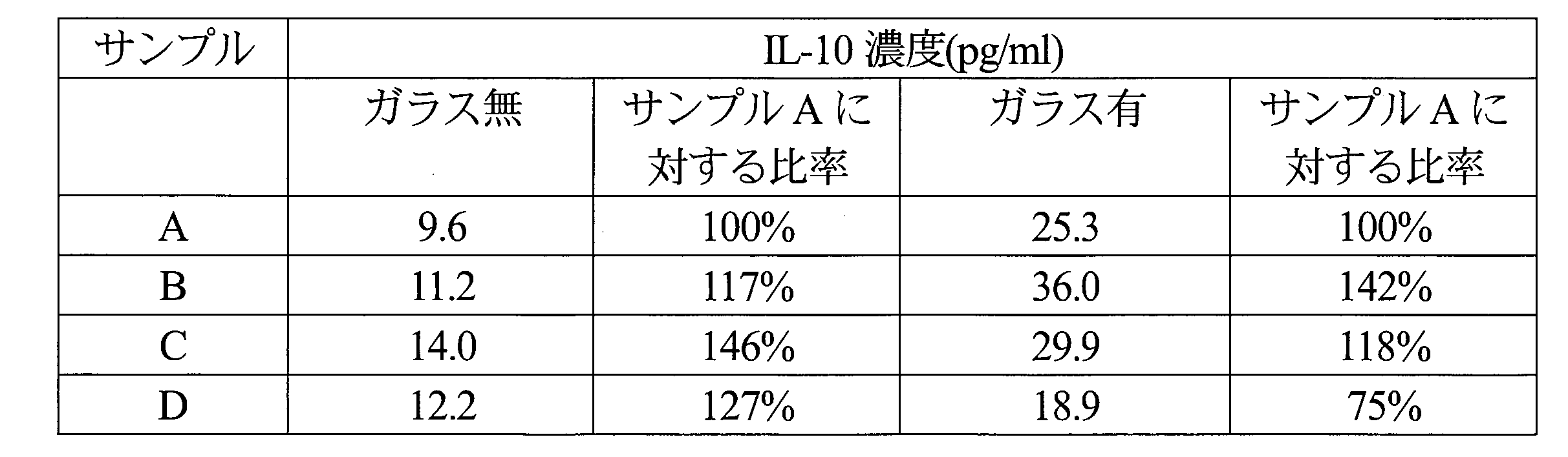
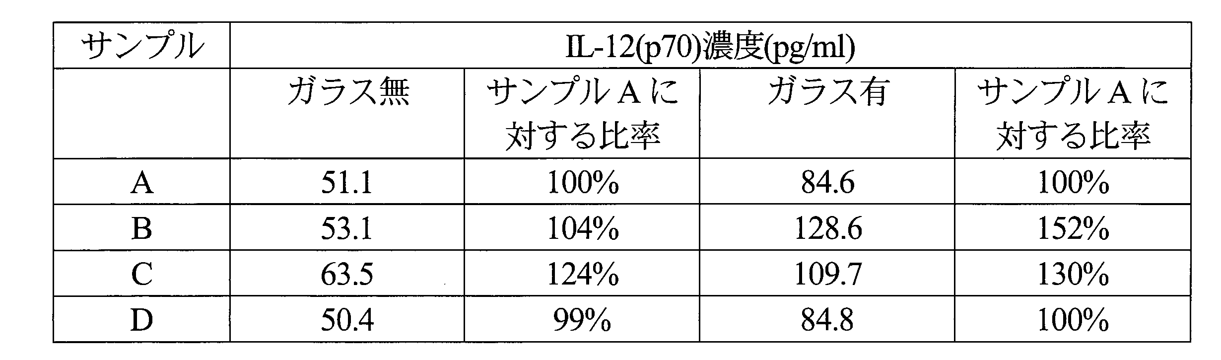
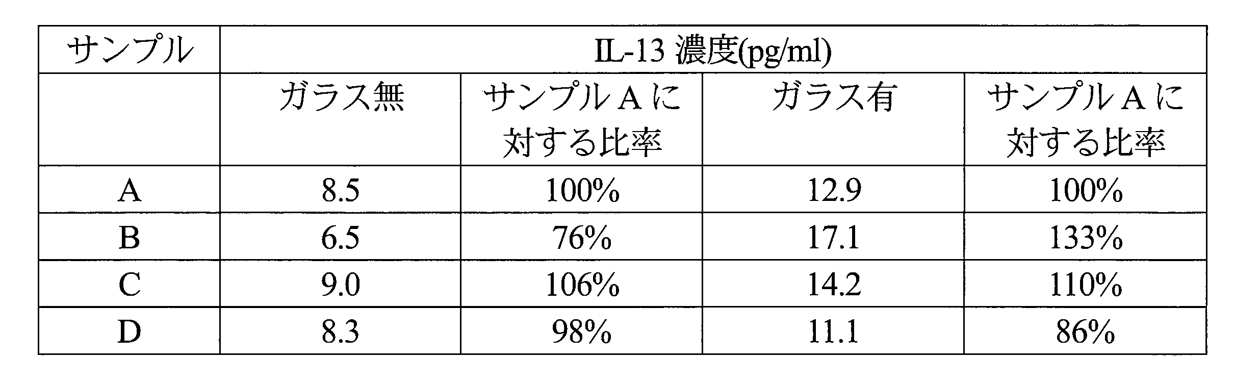
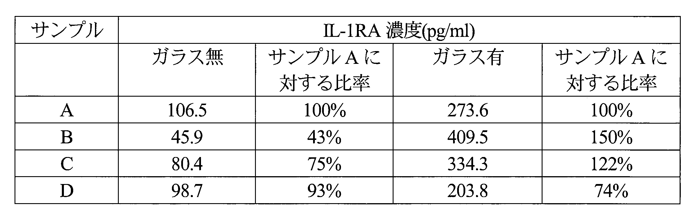
- Example 1 Using a centrifugal blood component separator (component collection system: CCS, manufactured by Hemonetics), leukocyte and platelet fractions of various concentrations are collected from anticoagulated blood, and after reactivation of leukocytes and platelets, recoagulation is performed. The serum composition was adjusted by, and the humoral factor was analyzed.
- CCS centrifugal blood component separator
- a blood circuit dedicated to the device (971J, manufactured by Hemonetics) is installed, and the anticoagulant ACD-A solution (sodium citrate hydrate 2.20%, citric acid hydrate 0.80%, glucose 2.20%,
- the anticoagulated blood obtained by mixing Terumo Co., Ltd. and donor blood at a ratio of 1:10 is used to obtain various concentrations of white blood
- the platelet fraction was collected in portions over time.
- the obtained leukocyte / platelet fraction (buffy coat) was measured for leukocyte / platelet count with an automatic blood cell counter (LC-660, manufactured by Fukuda Denshi). The results are shown in Table 2.
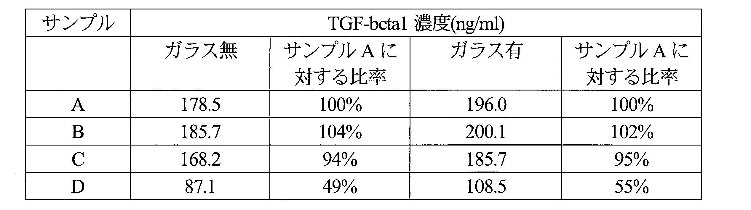
- TGF-beta1 concentration was sufficient even without glass, but an increase in concentration was observed when glass was used. Looking at the ratio to sample A, it was highly correlated with the platelet count data in Table 2, and it was speculated that TGF-beta1 in the serum composition was mostly derived from platelets. Comparing samples A and C, the platelet count and TGF-beta1 concentration were almost the same, strongly suggesting that TGF-beta1 is derived from platelets.
- IL-12 (p70) concentration was sufficient even without glass, but an increase in concentration was observed when glass was used. Looking at the ratio to sample A, a high correlation was found with the data on the number of leukocytes in Table 2, and it was assumed that IL-12 (p70) in the serum composition was derived from a large amount of leukocytes. Comparing samples A and D, the white blood cell count and IL-12 (p70) concentration were almost the same, strongly suggesting that IL-12 (p70) is derived from white blood cells.
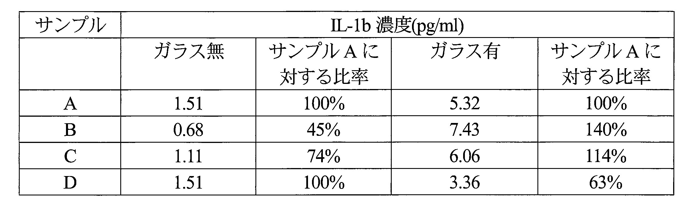
- IL-1b concentration was found to increase significantly when glass was used. Looking at the ratio to sample A (with glass), a high correlation with the platelet count data in Table 2 was observed, and it was speculated that IL-1b in the serum composition was mostly derived from platelets. Comparing samples A and C, the platelet count and IL-1b concentration were almost the same, strongly suggesting that IL-1b is derived from platelets.
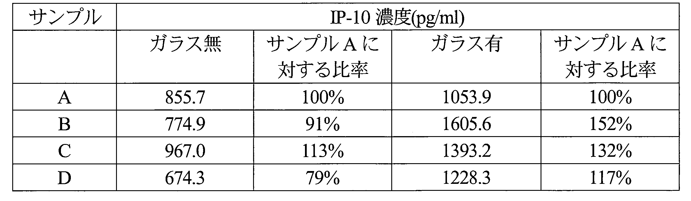
- the IP-10 concentration was sufficient even without glass, but an increase in concentration was observed when glass was used. Looking at the ratio to sample A, it is difficult to recognize the correlation with the data on the white blood cell count and platelet count in Table 2. (1) In the absence of glass, the trend is closely related to the platelet count, but sample D has half the platelet count compared to A, but the IP-10 concentration is about 80%, (2) with glass In this case, the tendency is closely related to the white blood cell count, but when comparing samples B and C, the platelet count was the same, but a difference was observed in the IP-10 concentration. From these, it is highly possible that IP-10 is derived from both leukocytes and platelets. As the basis for the interaction due to the use of glass, sample B has a relatively high white blood cell count and the highest platelet count, but the IP-10 concentration is (1) below the average in the absence of glass, ( 2) When glass is present, the most common points can be pointed out.
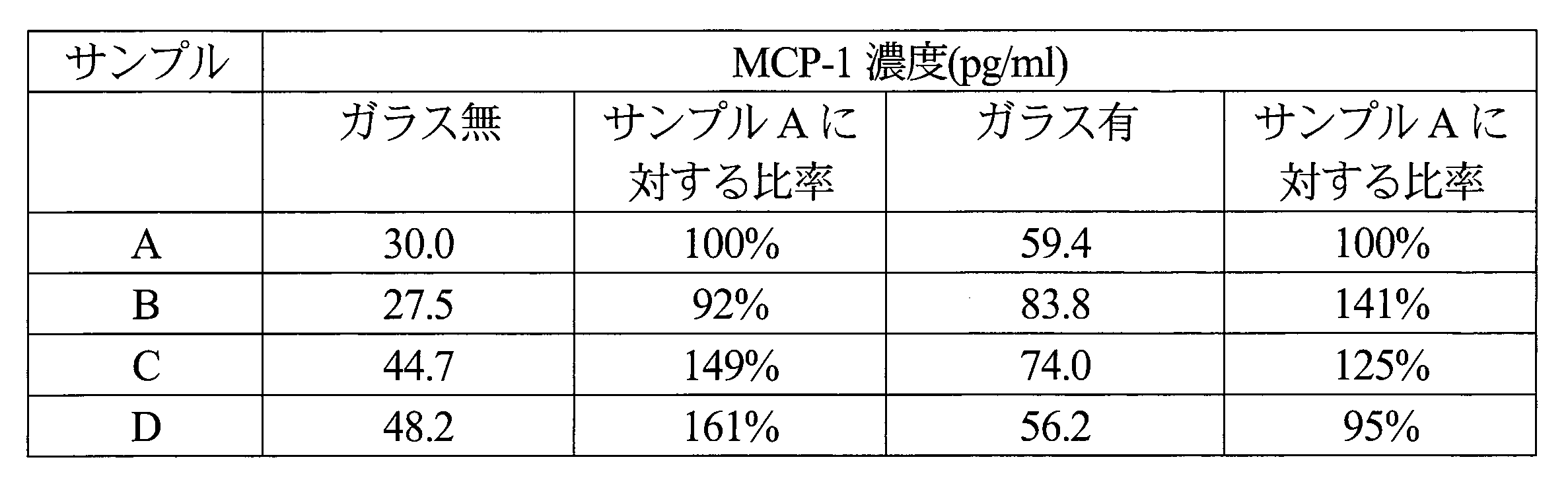
- the MCP-1 concentration was a sufficient concentration even without glass, but an increase in concentration was observed when glass was used. Looking at the ratio to sample A, it is difficult to recognize the correlation with the data on the white blood cell count and platelet count in Table 2.
- sample B has a relatively high white blood cell count and the highest platelet count, but the MCP-1 concentration is (1) the lowest without glass ( 2) When glass is present, the most common points can be pointed out.
- the MIP-1b concentration did not increase when glass was used. However, when looking at the ratio to sample A, there was a correlation with the leukocyte count data in Table 2, and it was speculated that MIP-1b in the serum composition was mostly derived from leukocytes. Comparing samples A and D, the white blood cell count and MIP-1b concentration were almost the same, suggesting that MIP-1b is derived from white blood cells.
- the PDGF-BB concentration was sufficient even without glass, but an increase in concentration was observed when glass was used. Looking at the ratio to sample A, it is difficult to recognize the correlation with the data on the white blood cell count and platelet count in Table 2.
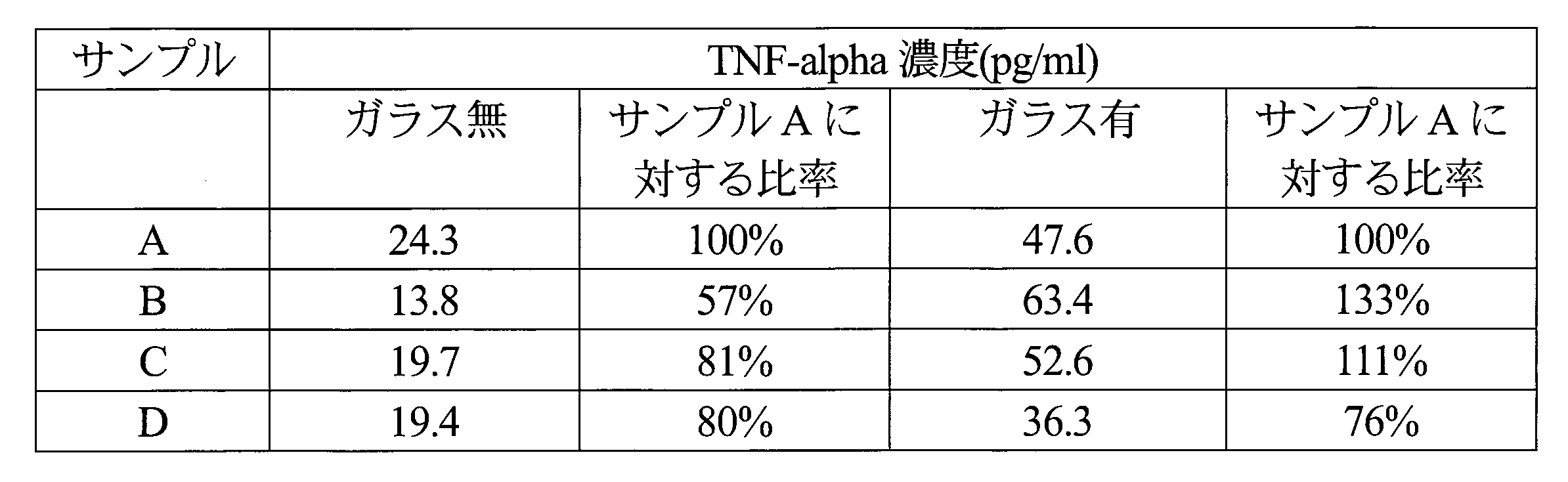
- TNF-alpha concentration was found to increase significantly when glass was used. Looking at the ratio to sample A (with glass), a high correlation with the platelet count data in Table 2 was observed, suggesting that TNF-alpha in the serum composition is mostly derived from platelets. Comparing samples A and C, the platelet count and IL-8 concentration were almost the same, strongly suggesting that TNF-alpha is derived from platelets.
- the VEGF concentration did not increase when glass was used. Looking at the ratio to sample A, it is difficult to recognize the correlation with the data on the white blood cell count and platelet count in Table 2. In the presence of glass, the trend is closely related to the white blood cell count, but when comparing samples A and D, for example, there was no correlation between platelet count and VEGF concentration, indicating that VEGF is derived from both white blood cells and platelets. There is no denying the possibility.
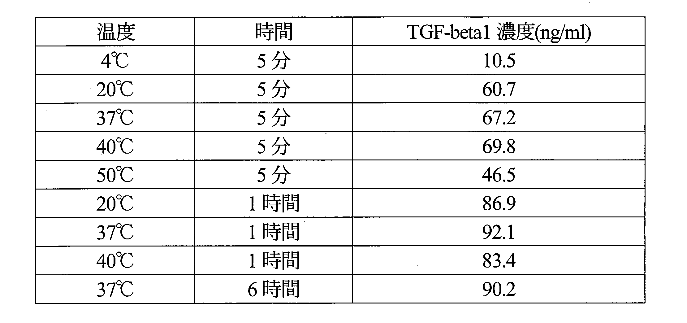
- Example 2 Furthermore, studies were carried out for the purpose of optimizing temperature and time conditions in the mutual activation of the obtained leukocyte / platelet fraction.
- Example 1 a blood circuit dedicated to the same device (971J, manufactured by Hemonetics) was attached, and the anticoagulant ACD-A solution (sodium citrate hydrate 2.20%, citrate hydrate 0.80%) , Glucose 2.20%, manufactured by Terumo Co., Ltd.) and blood of donor blood at a ratio of 1:10, anticoagulated blood was obtained by continuously centrifuging leukocytes and platelets using a centrifuge bowl equipped with the blood circuit. Fractions were collected. To the obtained leukocyte / platelet fraction, an aqueous calcium chloride solution was added so that the final addition concentration was 5 mM.
- ACD-A solution sodium citrate hydrate 2.20%, citrate hydrate 0.80%
- Glucose 2.20% manufactured by Terumo Co., Ltd.
- the TGF-beta1 concentration was high at 20 ° C. to 40 ° C. with respect to the temperature, and the time was high at 1 hour or more. Based on the above, it was considered that the temperature and time conditions for the mutual activation of leukocyte and platelet fractions were from room temperature to 40 ° C. for 1 hour or more.
- Example 3 Regarding the collection of leukocyte / platelet fractions from whole blood treated with anticoagulation, the difference by animal species was examined.
- the anticoagulant ACD-A solution sodium citrate hydrate 2.20%, citric acid hydrate 0.80%, glucose 2.20%, manufactured by Terumo Corporation
- human or bovine blood 1:10
- a cooling centrifuge 5922 manufactured by Kubota Corporation
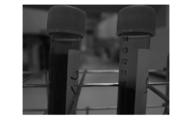
- FIG. 2 the left is a cow and the right is a human.
- bovines have a relatively low specific gravity of erythrocytes and erythrocytes do not settle easily, whereas humans have a high erythrocyte specific gravity and erythrocytes easily settle.
- red blood cells are difficult to settle by standing or simple centrifugation, and as a result, a relatively lighter white blood cell / platelet fraction does not appear easily.
- Example 4 Therefore, the specific gravity of each blood component in cattle was examined.
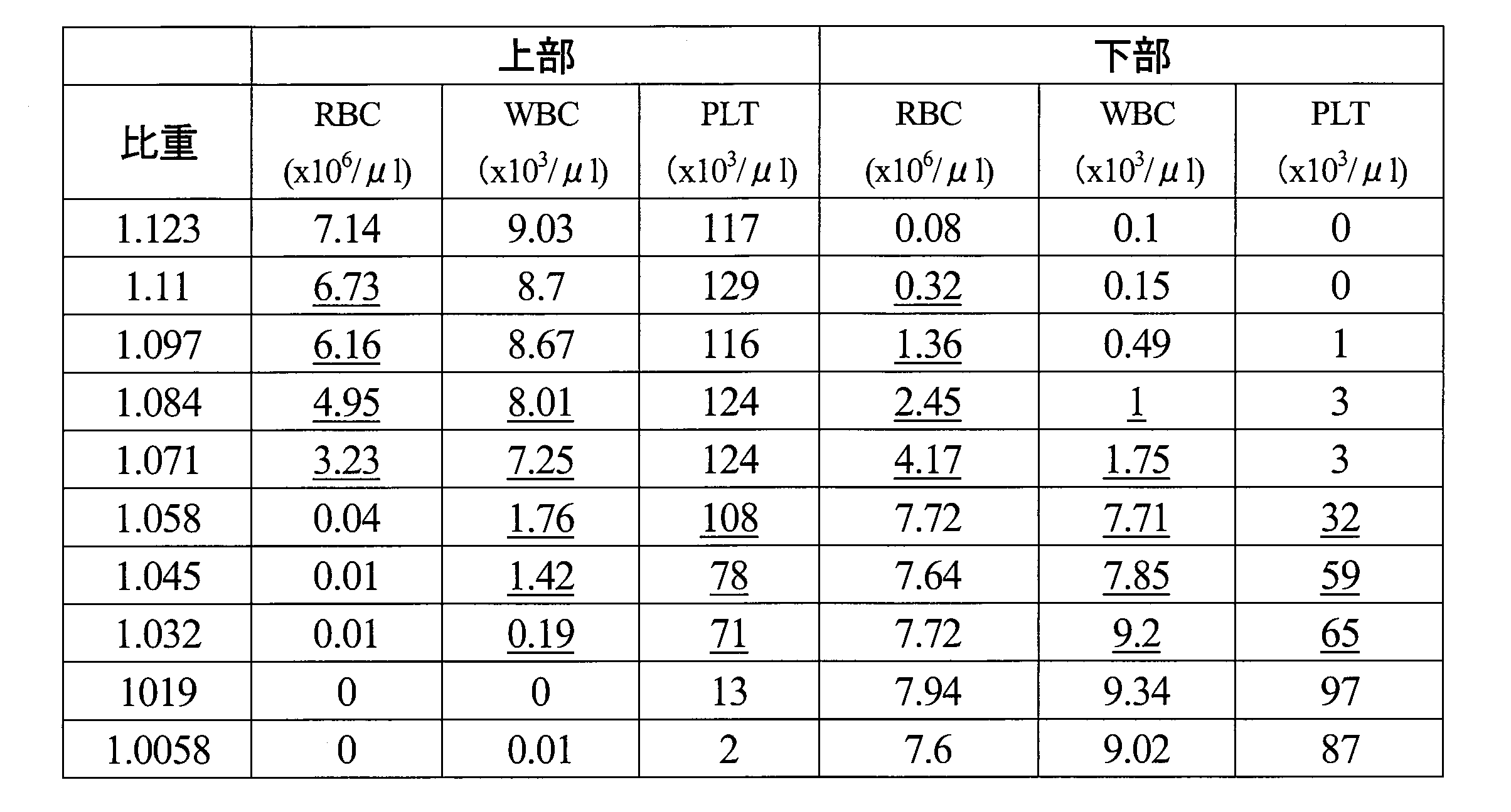
- Percoll undiluted solution (1.130 g / ml, manufactured by GE Healthcare Biosciences Co., Ltd., 17-0891-01), 1.5M NaCl, and sterilized water are mixed in appropriate amounts, specific gravity 1.123, 1.110, 1.097, 1.084, 1.071, 1.058, 1.045 , 1.032, 1019, 1.006 isotonic Percoll solutions were prepared. 3 ml of these isotonic Percoll solutions were added to a 15 ml polypropylene tube (2325-015, manufactured by AGC Techno Glass Co., Ltd.).
- anticoagulant ACD-A solution sodium citrate hydrate 2.20%, citric acid hydrate 0.80%, glucose 2.20%, manufactured by Terumo Corporation
- bovine blood 3 ml of the obtained anticoagulated blood was layered on a 15 ml polypropylene tube (2325-015, manufactured by AGC Techno Glass Co., Ltd.) to which isotonic Percoll solution was added. Centrifugation was performed at 400 ⁇ g and room temperature for 10 minutes (universal cooling centrifuge 5922, manufactured by Kubota Corporation), and the state was photographed. The results are shown in FIG.
- bovine blood contains many red blood cells that are suspended in an isotonic Percoll solution having a specific gravity of 1.071 and lighter than a specific gravity of 1.071.
- each tube (6 ml) was divided into an upper 3 ml and a lower 3 ml, and the blood cell components in each were measured with an automatic blood cell counter (IDEXX Laboratories Proteosite Dx). The results are shown in Table 28.
- leukocyte / platelet fraction (so-called buffy coat) appears by a simple operation, but in the case of cattle, there are many parts where the specific gravity of leukocytes and erythrocytes overlaps. In operation, it became clear that it was difficult to separate all leukocytes.
- Example 5 Therefore, we aimed to collect a stable leukocyte / platelet fraction from bovine whole blood and examined it using a continuous centrifuge.
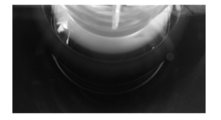
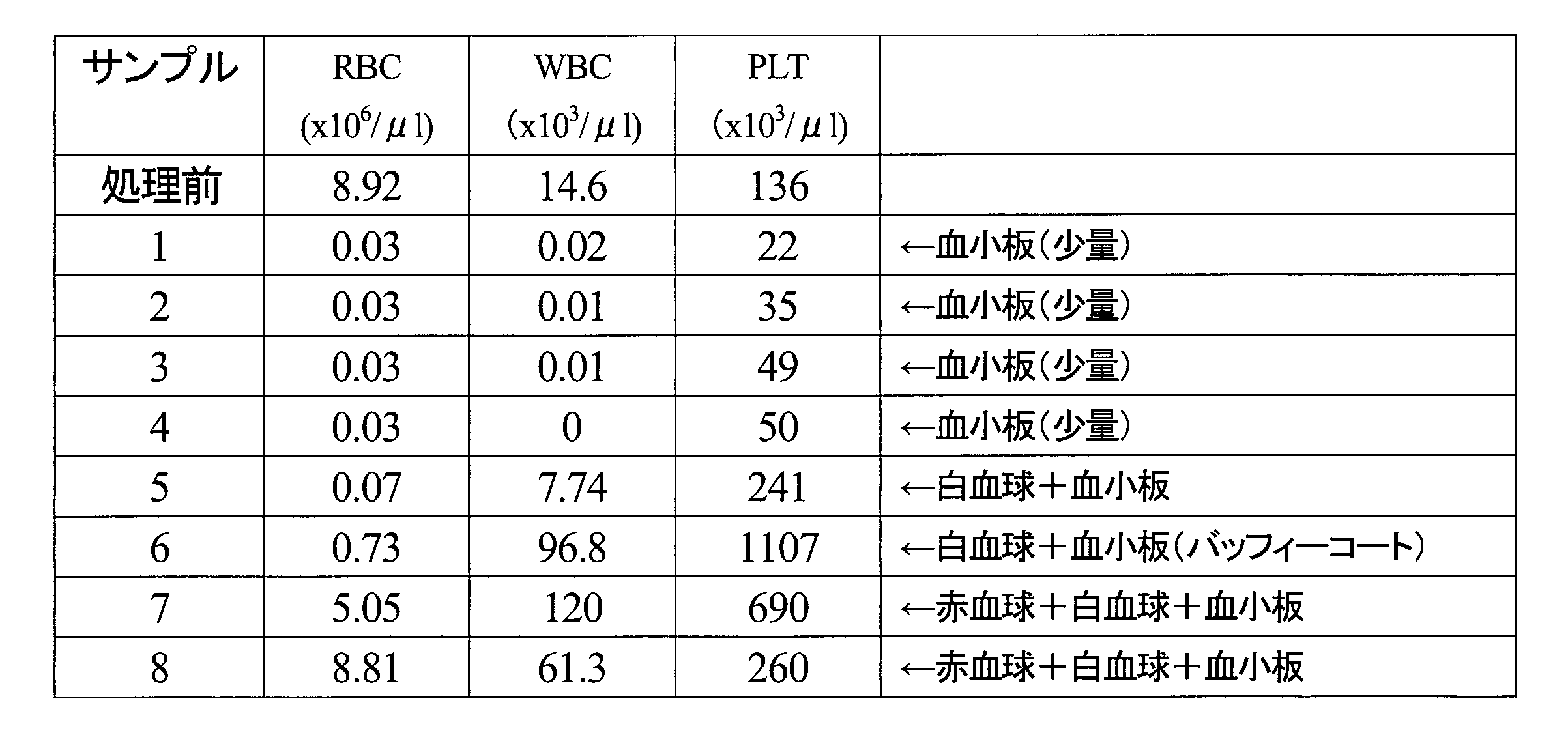
- a centrifugal blood component separator (CCS, manufactured by Hamonetics) is equipped with a dedicated blood circuit (971J, manufactured by Hamonetics), and the anticoagulant ACD-A solution (sodium citrate hydrate 2.20%, (Citrate hydrate 0.80%, glucose 2.20%, manufactured by Terumo Corporation) and bovine whole blood in a ratio of 1:10, using a centrifuge bowl equipped with the blood circuit Then, continuous centrifugation was performed, and a photograph of the white blood cell / platelet fraction that appeared was taken. The results are shown in FIG.
- a buffy coat (white blood cell / platelet fraction) in which a white belt-like circle is drawn clearly exists between the red blood cells located outside the centrifuge bowl and the plasma located inside.
- the amount of blood processed once is large (> 250 ml), so it seems that buffy coat appeared relatively easily even if there was a slight difference in specific gravity.
- Sample 6 located in the buffy coat was proved to be a leukocyte + platelet fraction.
- Sample 6 having a heavier specific gravity than the buffy coat had the most white blood cells.
- it was confirmed that it is necessary to obtain a fraction including a fraction with a higher specific gravity than buffy coat ( leukocytes in bovines). It was.
- Example 6 Therefore, with respect to bovine whole blood that had undergone anticoagulation treatment, a centrifugal blood component separation device (CCS, manufactured by Hamonetics) was used to collect leukocyte and platelet fractions, which were activated by adding glass beads, and then recoagulated.
- a serum composition (hereinafter referred to as the present serum composition) was prepared, and the possibility as an additive for the purpose of cell culture was examined.
- a blood circuit dedicated to the device (970E, manufactured by Hemonetics) is attached, and the anticoagulant titramine solution “Fuso” 4% (sodium citrate hydrate 4%, manufactured by Fuso Pharmaceutical Co., Ltd.) and bovine
- the anticoagulated blood obtained by mixing the blood at a ratio of 1:16 was collected from the white blood cell / platelet fraction by continuous centrifugation using a centrifuge bowl provided in the blood circuit.
- Glass beads (1 per 50 ml of blood component: BZ-6, manufactured by ASONE) are added to the obtained leukocyte / platelet fraction, and an aqueous calcium chloride solution is added to a final addition concentration of 5 mM to 37 ° C. Left for more than 6 hours.
- the mixture was centrifuged at 3000 rpm for 10 minutes, and the supernatant (serum composition) was collected and stored at ⁇ 20 degrees.
- the serum composition was thawed and added to aMEM medium (Life Technologies, Inc., 41061) at a rate of 10% to obtain a cell culture medium.
- the control was an aMEM medium supplemented with 10% fetal bovine serum (Moregate Biotech).
- proliferation curves were prepared using commercially available human bone marrow-derived mesenchymal stem cells (Lonza, PT-2501) and human amnion-derived mesenchymal stem cells established by us.
- the human amnion-derived mesenchymal stem cells were cultured in the following steps. That is, the amniotic membrane obtained by cesarean section was treated with collagenase and thermolysin for 30 minutes at 35 ° C., and the cells were collected by passing the enzyme-treated amniotic membrane through a mesh with a pore size of 100 ⁇ m. Cultured. The concentration of collagenase was 500 CDU / ml and the concentration of thermolysin was 400 PU / ml.
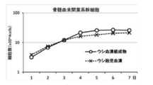
- the serum composition has superior cell proliferation ability compared to fetal bovine serum usually used for cell culture in both bone marrow and amnion-derived mesenchymal stem cells. It turned out to be a thing.
- Example 7 In addition, the effect of the serum composition on the cell diameter was examined. As in Example 6 above, using the aMEM medium supplemented with this serum composition at a rate of 10% (the control is the aMEM medium supplemented with 10% fetal bovine serum), we established human amnion-derived mesenchymal stem cells. Culture was performed and subcultured appropriately. The average cell diameter ( ⁇ m) was measured at each passage, and the difference between the two was examined. The results are shown in Table 31.
- the human amnion-derived mesenchymal stem cells cultured with this serum composition always had a smaller cell diameter at each passage than the fetal bovine serum.
- this serum composition was shown to have a higher cell proliferation ability than fetal bovine serum, and it was considered that the time until cell division was earlier.
- Example 8 In intravenous administration of cell preparations using cultured mesenchymal stem cells, the promotion of blood coagulation by the administered cells often becomes a problem. Therefore, it was examined whether cells cultured with this serum composition induce blood coagulation.
- human amnion-derived mesenchymal stem cells cultured in 10% serum composition-added aMEM medium were adjusted with physiological saline to 1 ⁇ 10 5 cells / ml.
- the control was the same cells cultured in aMEM medium supplemented with 10% fetal bovine serum.
- 40 ⁇ L of human plasma was added to 160 ⁇ L of these cell suspensions, and after incubation at 37 ° C. for 5 minutes, 100 ⁇ L of 20 ⁇ M CaCl 2 was added, and the clotting time was measured with an automatic blood coagulation measuring device (KC1 Delta, manufactured by Tcoag). .
- the results are shown in Table 32.
- the human amnion-derived mesenchymal stem cells cultured with this serum composition were found to be prone to induce blood coagulation with an increase in clotting time compared to fetal bovine serum.
- Example 9 Therefore, an investigation was conducted for the purpose of searching for a factor in which cells cultured with this serum composition hardly induce blood coagulation.
- TFPI tissue factor pathway inhibitor
- the concentration of the stem cell (passage number 4) in the culture supernatant was measured using an ELISA kit (Human TFPI Quantikine ELISA Kit) (DTFP10, R & D systems).
- the controls were the culture supernatant of the same cells cultured in 10% fetal bovine serum-added aMEM medium, and these media before cell culture. The results are shown in Table 33.
- Human amnion-derived mesenchymal stem cells cultured with this serum composition secrete more TFPI than fetal bovine serum, indicating that they are difficult to induce blood clotting. .
Landscapes
- Health & Medical Sciences (AREA)
- Life Sciences & Earth Sciences (AREA)
- Engineering & Computer Science (AREA)
- Biomedical Technology (AREA)
- Zoology (AREA)
- Biotechnology (AREA)
- Cell Biology (AREA)
- Chemical & Material Sciences (AREA)
- Developmental Biology & Embryology (AREA)
- General Health & Medical Sciences (AREA)
- Hematology (AREA)
- Bioinformatics & Cheminformatics (AREA)
- Genetics & Genomics (AREA)
- Immunology (AREA)
- Wood Science & Technology (AREA)
- Organic Chemistry (AREA)
- Medicinal Chemistry (AREA)
- Virology (AREA)
- Pharmacology & Pharmacy (AREA)
- Epidemiology (AREA)
- Animal Behavior & Ethology (AREA)
- Public Health (AREA)
- Veterinary Medicine (AREA)
- General Engineering & Computer Science (AREA)
- Biochemistry (AREA)
- Microbiology (AREA)
- Rheumatology (AREA)
- Medicines Containing Material From Animals Or Micro-Organisms (AREA)
- Micro-Organisms Or Cultivation Processes Thereof (AREA)
Abstract
Description
以下、添付の図面を参照して本発明の実施形態について具体的に説明するが、当該実施形態は本発明の原理の理解を容易にするためのものであり、本発明の範囲は、下記の実施形態に限られるものではなく、当業者が以下の実施形態の構成を適宜置換した他の実施形態も、本発明の範囲に含まれる。
(i)抗凝固処理工程:抗凝固剤でウシ全血を抗凝固処理し、
(ii)白血球および血小板取得工程:抗凝固処理された全血からバフィーコート及びバフィーコートより重い比重分画を取得し、
(iii)白血球および血小板活性化・再凝固処理工程:取得した白血球と血小板との相互作用を一定の温度で一定以上時間をかけ、必要に応じガラス材料の添加及び静置により、活性化させることにより白血球及び/又は血小板により分泌・放出される液性因子を含有し、この液性因子を含む血液成分を再凝固剤により再凝固処理する
ことにより得られる。以下、各工程について説明する。
全血は、ウシ由来であり、成獣・幼若・新生児又は胎児ウシの何れも適用可能である。全血は血管に針を刺し、針からチューブを介して採血バッグ等に採取することが可能である。全血は、血球成分(赤血球、白血球、血小板)と液体成分である血漿とからなる。血漿は凝固成分を含むのに対し、血清は凝固成分をほとんど含まないか、含んだとしても少量の液体である。
抗凝固処理されたウシ全血からの白血球および血小板の取得は、例えば遠心機又は連続遠心機によりなされる。バフィーコートとは、凝固していない血液を遠心分離した際に、赤血球層と血漿との間に生じる白血球と血小板との層である。しかしながら、ウシの場合、後述するようにヒトと比べ赤血球の比重が軽いことから、白血球と赤血球の比重が重なる部分があり、結果、バフィーコートは白血球の含有が少ないことから不明瞭な層となる。従って、白血球および血小板両者を採取するには、バフィーコートより比重が重い分画(=ウシの場合、白血球を含有)も含め取得する必要性が、本願発明において明らかとなっている。後述するように、ウシ血小板の比重は例えば1.032~1.058であり、ウシ白血球の比重は例えば1.032~1.084であり、ウシ赤血球の比重は例えば1.071~1.110である。そのためウシ全血の場合、赤血球層と血漿との間に位置するバフィーコートは比重1.032~1.071の層となるが、この層は白血球の含有が少ないためバフィーコートより比重が重い例えば1.071~1.084の分画も取得する。このようにして、抗凝固処理されたウシ全血から、例えば遠心機又は連続遠心機により、血漿および比重の重い赤血球を除いた、白血球と血小板双方を多く含む血漿が調整される。

次に、取得した白血球及び血小板を多く含有する血漿を常温~40℃にて一定時間静置し、白血球と血小板との相互作用を促す。ここで常温とは10℃~30℃とする。この際、ガラス材料等が接触することにより、白血球と血小板との相互作用が更に活性化され、白血球及び/又は血小板から大量の液性因子が分泌・放出される。ガラス材料は、ソーダ石灰ガラス、鉛ガラス、ホウケイ酸ガラス、又はこれらの混合からなる成分を有するガラスである。
本発明の細胞の培養方法は、本発明にかかるウシ血清組成物の製造方法で得られたウシ血清組成物を添加剤として含む培地にて細胞を培養することにより、細胞の増殖を促進し、血液凝固の誘導を抑制する工程を含む。
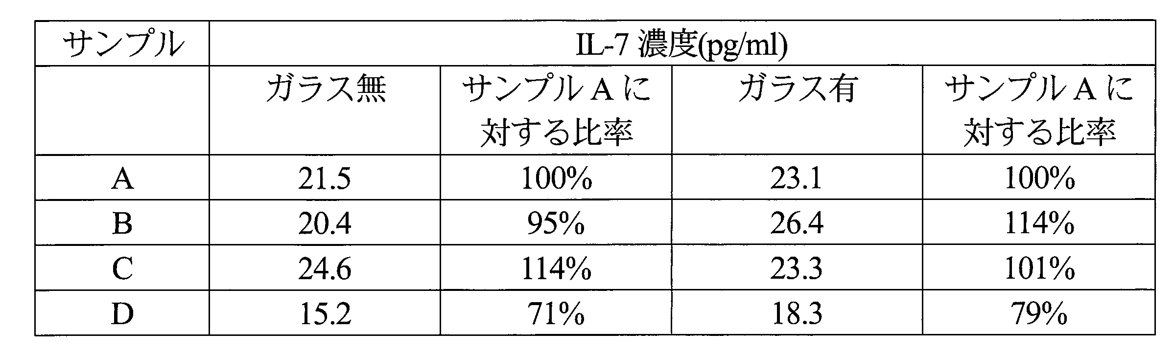
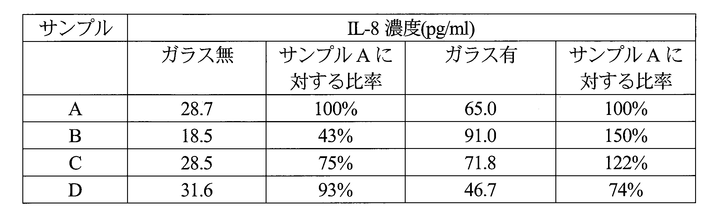
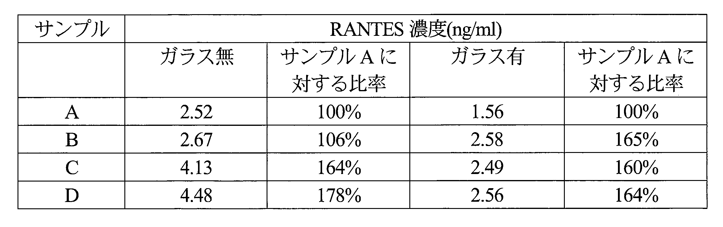
遠心型血液成分分離装置(コンポーネントコレクションシステム:CCS、ヘモネティクス製)を用い、抗凝固処理血液から様々な濃度の白血球・血小板分画を採取し、白血球・血小板を相互活性化後、再凝固処理により血清組成物を調整し、その液性因子を解析した。

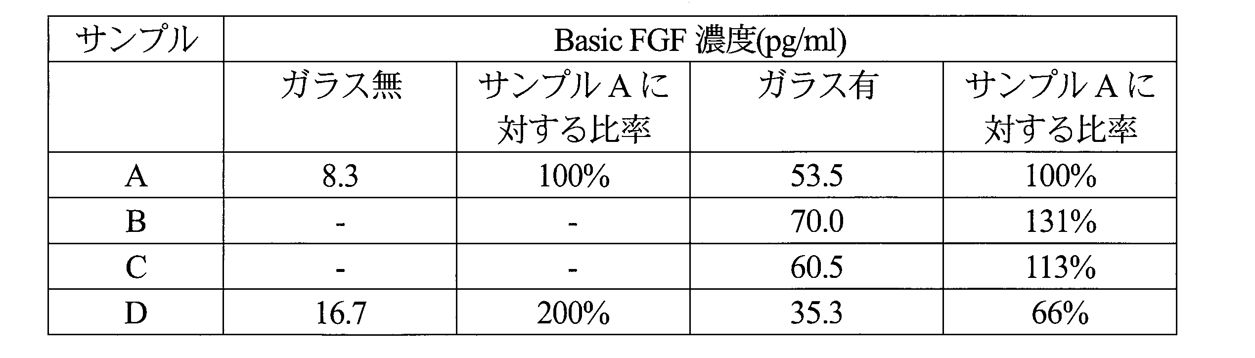
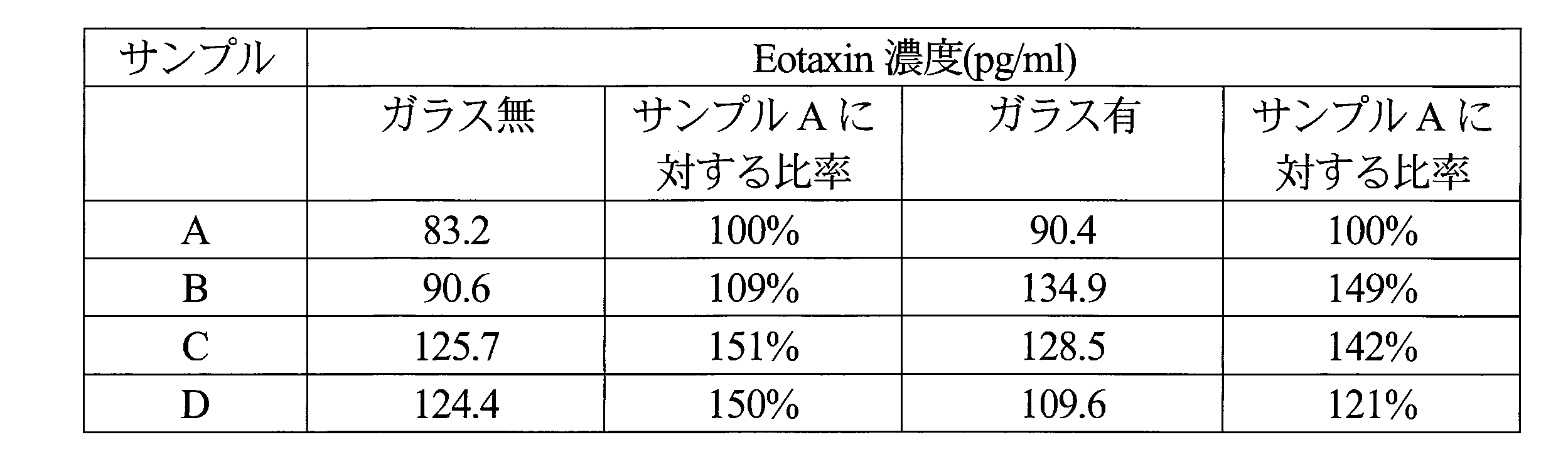
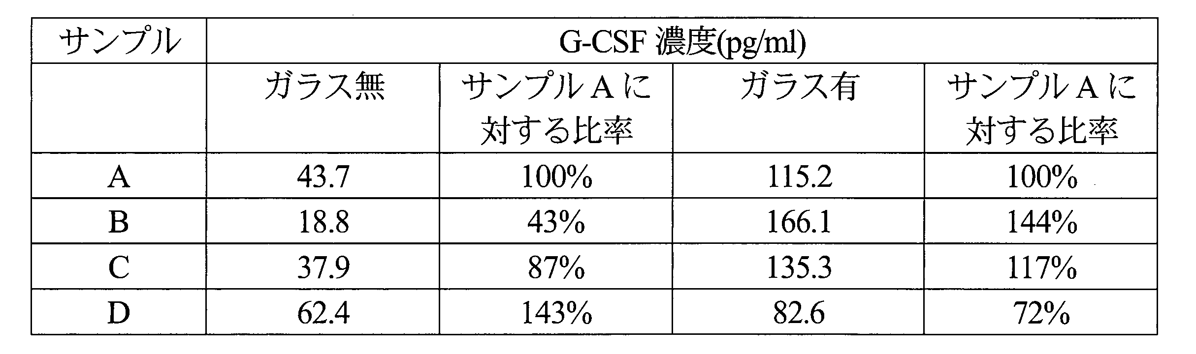
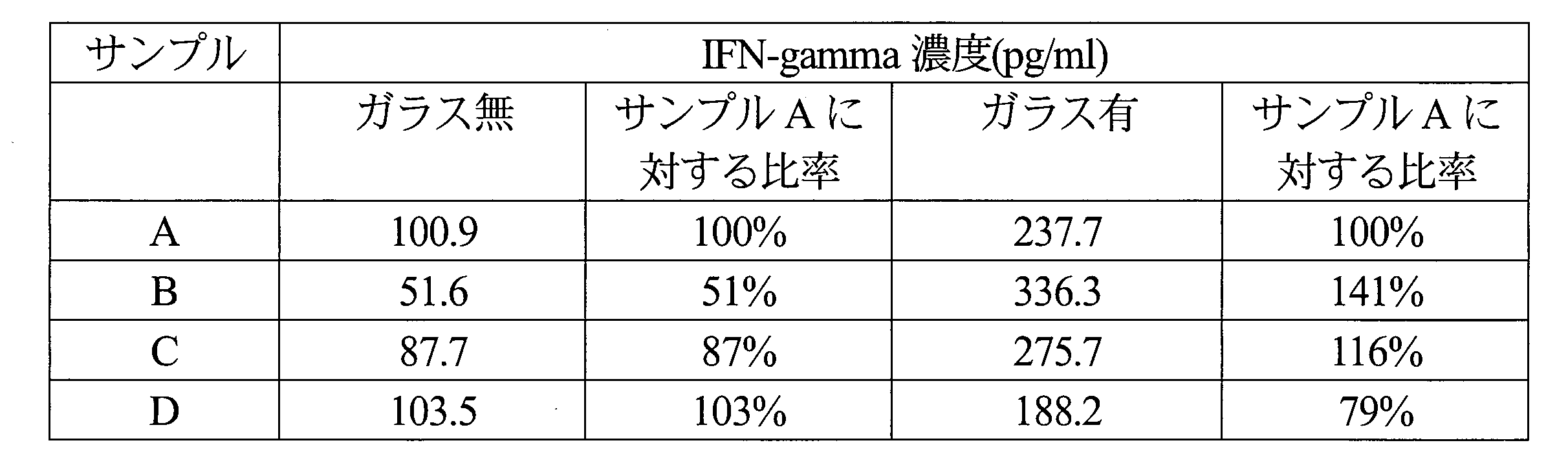







更に、得られた白血球・血小板分画の相互活性化における温度・時間条件の至適化を目的とした検討を行った。
抗凝固処理された全血からの白血球・血小板分画採取に関し、動物種による違いを検討した。
そこで、ウシにおける各血液成分の比重検討を行った。Percoll原液(1.130g/ml、GEヘルスケアバイオサイエンス株式会社製、17-0891-01)、1.5M NaCl、及び滅菌水を適量混合し、比重1.123, 1.110, 1.097, 1.084, 1.071, 1.058, 1.045, 1.032, 1019, 1.006の等張Percoll液を調整した。15mlポリプロピレン製チューブ(2325-015, AGCテクノグラス株式会社製)に、これら等張Percoll液を3ml添加した。抗凝固剤ACD-A液(クエン酸ナトリウム水和物2.20%、クエン酸水和物0.80%、ブドウ糖2.20%、テルモ株式会社製)と、ウシ血液を1:10の割合で混合することに得られた抗凝固処理血液3mlを、等張Percoll液を加えた15mlポリプロピレン製チューブ(2325-015, AGCテクノグラス株式会社製)に3ml重層した。400×g、常温にて、10分遠心し(ユニバーサル冷却遠心機5922、久保田商事株式会社製)、状態を写真撮影した。結果を図3に示す。

そこで、ウシ全血からの安定した白血球・血小板分画の採取を目指し、連続遠心機を用いた検討を行った。
そこで、抗凝固処理を行ったウシ全血に関し、遠心型血液成分分離装置(CCS、ヘモネティクス製)を用い、白血球・血小板分画を採取し、ガラスビース添加により活性化後、再凝固処理により血清組成物(以下本血清組成物)を調整し、細胞培養を目的とした添加剤としての可能性を検討した。
また、本血清組成物が細胞径に与える影響を検討した。上記実施例6と同様、本血清組成物を10%の割合で添加したaMEM培地(対照はウシ胎児血清を10%添加したaMEM培地)を用い、我々が樹立したヒト羊膜由来間葉系幹細胞の培養を行い、適宜継代培養した。継代ごとに平均細胞径(μm)を測定し、両者の差違を検討した。結果を表31に示す。

培養間葉系幹細胞を用いた細胞製剤の経静脈的投与において、しばしば投与細胞による血液凝固促進が問題になる。そこで、本血清組成物にて培養した細胞が血液凝固を誘導するかについて、検討を行った。実施例6・7同様、10%本血清組成物添加aMEM培地にて培養したヒト羊膜由来間葉系幹細胞を、1×10⁵細胞/mlとなるように生理食塩水にて調整した。対照は10%ウシ胎児血清添加aMEM培地で培養した同細胞とした。これら細胞懸濁液160μLにヒト血漿40μLを添加し、37℃ 5分間インキュベーションの後、20μM CaCl2を100μL添加し、血液凝固自動測定装置(KC1 Delta、Tcoag社製)にて凝固時間を測定した。結果を表32に示す。

そこで、本血清組成物にて培養した細胞が血液凝固を誘導しにくい要因の探索を目的とした検討を行った。細胞が分泌し、細胞膜に結合している強力な凝固阻止因子である組織因子経路インヒビター(tissue factor pathway inhibitor:TFPI)に関し、10%本血清組成物添加aMEM培地にて培養したヒト羊膜由来間葉系幹細胞(継代数4)の培養上清における濃度測定を、ELISAキット(Human TFPI Quantikine ELISA Kit)(DTFP10、R&D systems社)を用いて行った。対照は10%ウシ胎児血清添加aMEM培地にて培養した同細胞の培養上清、及び細胞培養前のこれら培地とした。結果を表33に示す。

Claims (12)
- ウシ全血を抗凝固剤で抗凝固処理する工程と、
抗凝固処理されたウシ全血からバフィーコート及びバフィーコートより重い比重分画を取得する工程と、
取得した白血球と血小板を一定の温度で一定以上時間をかけることで相互作用を促し活性化させることにより、白血球及び/又は血小板から液性因子を分泌又は放出させつつ、この液性因子を含む血液成分を再凝固剤で再凝固処理する工程と、
を有することを特徴とするウシ血清組成物の製造方法。 - 前記バフィーコートより重い比重分画は、1.071~1.084の比重分画であることを特徴とする、請求項1に記載のウシ血清組成物の製造方法。
- 前記一定の温度は、常温~40℃、時間は5分以上の条件であることを特徴とする、請求項1又は2に記載のウシ血清組成物の製造方法。
- 前記バフィーコートの更なる活性化に、ガラス材料を添加することを特徴とする、請求項1乃至3の何れか1項に記載のウシ血清組成物の製造方法。
- 前記抗凝固剤は、クエン酸ナトリウム又はクエン酸であることを特徴とする請求項1乃至4の何れか1項に記載のウシ血清組成物の製造方法。
- 前記再凝固剤は、塩化カルシウム溶液であることを特徴とする請求項1乃至5の何れか1項に記載のウシ血清組成物の製造方法。
- 前記バフィーコートの取得は、遠心機又は連続遠心機によることを特徴とする請求項1乃至6の何れか1項に記載のウシ血清組成物の製造方法。
- 前記液性因子は、TGF-beta1、Basic FGF、Eotaxin、G-CSF、IFN-gamma、IL-10、IL-12(p70)、IL-13、IL-1RA、IL-1b、IL-4、IL-5、IL-6、IL-8、IL-9、IP-10、MCP-1、PDGF-BB、TNF-alphaであることを特徴とする請求項1乃至7の何れか1項に記載のウシ血清組成物の製造方法。
- ウシ全血を抗凝固剤で抗凝固処理し、
抗凝固処理された全血からバフィーコート及びバフィーコートより重い比重分画を取得し、取得した白血球と血小板を常温~40℃の温度で5分以上時間をかけることで相互作用を促し活性化させることにより白血球及び/又は血小板により分泌・放出される液性因子を含み、再凝固剤により再凝固処理することにより得られるウシ血清組成物。 - 請求項1乃至8の何れか1項に記載のウシ血清組成物の製造方法で得られたウシ血清組成物を添加剤として使用することにより、増殖性に優れた細胞を培養する方法。
- 請求項1乃至8の何れか1項に記載のウシ血清組成物の製造方法で得られたウシ血清組成物を添加剤として使用することにより、組織因子経路インヒビターの発現を誘導し、血液凝固を誘導しにくい細胞を培養する方法。
- 前記細胞は、骨髄、脂肪組織、又は羊膜・臍帯を含む胎児付属物を由来とする間葉系幹細胞又は間葉系細胞であることを特徴とする請求項10又は11に記載の細胞を培養する方法。
Priority Applications (5)
| Application Number | Priority Date | Filing Date | Title |
|---|---|---|---|
| EP16866357.3A EP3378935B1 (en) | 2015-11-16 | 2016-11-16 | Bovine serum composition and method for culturing cells using said bovine serum composition as additive |
| CN201680066959.2A CN108291202A (zh) | 2015-11-16 | 2016-11-16 | 牛血清组合物和使用所述牛血清组合物作为添加剂培养细胞的方法 |
| AU2016356125A AU2016356125B2 (en) | 2015-11-16 | 2016-11-16 | Bovine serum composition and method for culturing cells using said bovine serum composition as additive |
| US15/776,662 US11085023B2 (en) | 2015-11-16 | 2016-11-16 | Bovine serum composition and method for culturing cells using said bovine serum composition as additive |
| JP2017507906A JP6212723B1 (ja) | 2015-11-16 | 2016-11-16 | ウシ血清組成物及びそのウシ血清組成物を添加剤として使用する細胞の培養方法 |
Applications Claiming Priority (2)
| Application Number | Priority Date | Filing Date | Title |
|---|---|---|---|
| JP2015-224075 | 2015-11-16 | ||
| JP2015224075 | 2015-11-16 |
Publications (1)
| Publication Number | Publication Date |
|---|---|
| WO2017086356A1 true WO2017086356A1 (ja) | 2017-05-26 |
Family
ID=58718954
Family Applications (1)
| Application Number | Title | Priority Date | Filing Date |
|---|---|---|---|
| PCT/JP2016/083990 Ceased WO2017086356A1 (ja) | 2015-11-16 | 2016-11-16 | ウシ血清組成物及びそのウシ血清組成物を添加剤として使用する細胞の培養方法 |
Country Status (6)
| Country | Link |
|---|---|
| US (1) | US11085023B2 (ja) |
| EP (1) | EP3378935B1 (ja) |
| JP (1) | JP6212723B1 (ja) |
| CN (1) | CN108291202A (ja) |
| AU (1) | AU2016356125B2 (ja) |
| WO (1) | WO2017086356A1 (ja) |
Cited By (3)
| Publication number | Priority date | Publication date | Assignee | Title |
|---|---|---|---|---|
| WO2018159431A1 (ja) * | 2017-03-03 | 2018-09-07 | ロート製薬株式会社 | 間葉系幹細胞及び肝疾患治療剤 |
| WO2019038803A1 (ja) * | 2017-08-21 | 2019-02-28 | Aof株式会社 | 判定システム及び判定方法 |
| WO2023047832A1 (ja) * | 2021-09-27 | 2023-03-30 | 国立大学法人 東京大学 | 細胞培養用成分、細胞培養用培地、血清の製造方法、及び、細胞の製造方法 |
Families Citing this family (3)
| Publication number | Priority date | Publication date | Assignee | Title |
|---|---|---|---|---|
| CN105769909A (zh) * | 2016-05-13 | 2016-07-20 | 云南舜喜再生医学工程有限公司 | 一种直接获得富含细胞因子血清的采血器及方法 |
| CN116926001A (zh) * | 2022-04-06 | 2023-10-24 | 上海我武干细胞科技有限公司 | 血清替代物及其制备方法与应用 |
| CN118979010B (zh) * | 2024-08-28 | 2025-05-23 | 广州蕊特生物科技有限公司 | 一种牛全血提取物冻干粉的制备方法 |
Citations (11)
| Publication number | Priority date | Publication date | Assignee | Title |
|---|---|---|---|---|
| JPH11502435A (ja) * | 1995-03-03 | 1999-03-02 | クウォンティック バイオメディカル パートナーズ | 血小板グルー創傷密封材 |
| JP2001505906A (ja) * | 1996-12-10 | 2001-05-08 | ハダジット メディカル リサーチ サービシズ アンド ディベラップメント カンパニー リミテッド | 細胞の分化を誘導する血清由来因子およびその医薬的使用 |
| JP2005530768A (ja) * | 2002-05-09 | 2005-10-13 | メディジーンズ | 血漿または血清を含有した創傷治療用医薬組成物 |
| JP2008246200A (ja) * | 2007-03-07 | 2008-10-16 | Jms Co Ltd | 血清調製方法及び血清調製装置 |
| JP2009506992A (ja) * | 2005-09-01 | 2009-02-19 | ケンブリッジ・サイエンティフィク・ピーティーワイ・リミテッド | 組織破壊治療及びその治療に使用するための組成物 |
| JP2010531142A (ja) * | 2007-06-22 | 2010-09-24 | サークル バイオロジクス、 エルエルシー. | 液体濃縮装置、オートロガスな濃縮体液、およびそれらの使用方法 |
| JP2011160799A (ja) * | 2010-01-15 | 2011-08-25 | Jms Co Ltd | 間葉系幹細胞の増殖促進剤、それを用いた間葉系幹細胞の増殖促進方法および製造方法 |
| JP2013132240A (ja) * | 2011-12-26 | 2013-07-08 | Otsuka Pharmaceut Factory Inc | 細胞分離装置及び遠心分離方法 |
| JP2013132241A (ja) * | 2011-12-26 | 2013-07-08 | Otsuka Pharmaceut Factory Inc | 細胞分離装置 |
| JP2014117347A (ja) * | 2012-12-13 | 2014-06-30 | Jms Co Ltd | 血清の調製方法及び血清 |
| WO2014126931A1 (en) * | 2013-02-15 | 2014-08-21 | Victor Steven | Stable platelet- rich-plasma compositions and methods of use |
-
2016
- 2016-11-16 US US15/776,662 patent/US11085023B2/en active Active
- 2016-11-16 EP EP16866357.3A patent/EP3378935B1/en active Active
- 2016-11-16 JP JP2017507906A patent/JP6212723B1/ja active Active
- 2016-11-16 AU AU2016356125A patent/AU2016356125B2/en active Active
- 2016-11-16 WO PCT/JP2016/083990 patent/WO2017086356A1/ja not_active Ceased
- 2016-11-16 CN CN201680066959.2A patent/CN108291202A/zh active Pending
Patent Citations (11)
| Publication number | Priority date | Publication date | Assignee | Title |
|---|---|---|---|---|
| JPH11502435A (ja) * | 1995-03-03 | 1999-03-02 | クウォンティック バイオメディカル パートナーズ | 血小板グルー創傷密封材 |
| JP2001505906A (ja) * | 1996-12-10 | 2001-05-08 | ハダジット メディカル リサーチ サービシズ アンド ディベラップメント カンパニー リミテッド | 細胞の分化を誘導する血清由来因子およびその医薬的使用 |
| JP2005530768A (ja) * | 2002-05-09 | 2005-10-13 | メディジーンズ | 血漿または血清を含有した創傷治療用医薬組成物 |
| JP2009506992A (ja) * | 2005-09-01 | 2009-02-19 | ケンブリッジ・サイエンティフィク・ピーティーワイ・リミテッド | 組織破壊治療及びその治療に使用するための組成物 |
| JP2008246200A (ja) * | 2007-03-07 | 2008-10-16 | Jms Co Ltd | 血清調製方法及び血清調製装置 |
| JP2010531142A (ja) * | 2007-06-22 | 2010-09-24 | サークル バイオロジクス、 エルエルシー. | 液体濃縮装置、オートロガスな濃縮体液、およびそれらの使用方法 |
| JP2011160799A (ja) * | 2010-01-15 | 2011-08-25 | Jms Co Ltd | 間葉系幹細胞の増殖促進剤、それを用いた間葉系幹細胞の増殖促進方法および製造方法 |
| JP2013132240A (ja) * | 2011-12-26 | 2013-07-08 | Otsuka Pharmaceut Factory Inc | 細胞分離装置及び遠心分離方法 |
| JP2013132241A (ja) * | 2011-12-26 | 2013-07-08 | Otsuka Pharmaceut Factory Inc | 細胞分離装置 |
| JP2014117347A (ja) * | 2012-12-13 | 2014-06-30 | Jms Co Ltd | 血清の調製方法及び血清 |
| WO2014126931A1 (en) * | 2013-02-15 | 2014-08-21 | Victor Steven | Stable platelet- rich-plasma compositions and methods of use |
Non-Patent Citations (3)
| Title |
|---|
| LOH Y.S.: "LYSATES PRODUCED FROM IRRADIATED AND EXPIRED BUFFY COAT-DERIVED PLATELETS STORED IN ADDITIVE SOLUTION SUPPORT CELL PROLIFERATION", VOX SANGUINIS, vol. 109, no. 1, June 2015 (2015-06-01), pages 198, XP055383589 * |
| See also references of EP3378935A4 * |
| SOUSA A.P.: "VALORIZATION OF OUTDATED BLOOD PRODUCTS FOR REGENERATIVE MEDICENE", VOX SANGUINIS, vol. 101, no. 1, pages 49, XP055383581 * |
Cited By (5)
| Publication number | Priority date | Publication date | Assignee | Title |
|---|---|---|---|---|
| WO2018159431A1 (ja) * | 2017-03-03 | 2018-09-07 | ロート製薬株式会社 | 間葉系幹細胞及び肝疾患治療剤 |
| US11253550B2 (en) | 2017-03-03 | 2022-02-22 | Rohto Pharmaceutical Co., Ltd. | Method for treating fibrotic liver disease |
| WO2019038803A1 (ja) * | 2017-08-21 | 2019-02-28 | Aof株式会社 | 判定システム及び判定方法 |
| WO2023047832A1 (ja) * | 2021-09-27 | 2023-03-30 | 国立大学法人 東京大学 | 細胞培養用成分、細胞培養用培地、血清の製造方法、及び、細胞の製造方法 |
| JP2023047560A (ja) * | 2021-09-27 | 2023-04-06 | 国立大学法人 東京大学 | 細胞培養用成分、細胞培養用培地、血清の製造方法、及び、細胞の製造方法 |
Also Published As
| Publication number | Publication date |
|---|---|
| AU2016356125A1 (en) | 2018-06-21 |
| CN108291202A (zh) | 2018-07-17 |
| JPWO2017086356A1 (ja) | 2017-11-16 |
| EP3378935A1 (en) | 2018-09-26 |
| JP6212723B1 (ja) | 2017-10-18 |
| EP3378935A4 (en) | 2018-12-05 |
| EP3378935B1 (en) | 2020-09-02 |
| US11085023B2 (en) | 2021-08-10 |
| US20180371417A1 (en) | 2018-12-27 |
| AU2016356125B2 (en) | 2019-07-11 |
Similar Documents
| Publication | Publication Date | Title |
|---|---|---|
| JP6212723B1 (ja) | ウシ血清組成物及びそのウシ血清組成物を添加剤として使用する細胞の培養方法 | |
| RU2735139C2 (ru) | Полученные из лизата тромбоцитов человека внеклеточные везикулы для применения в медицине | |
| CN104307208B (zh) | 一种富集纯化血小板的方法 | |
| JP5763894B2 (ja) | 細胞の分離方法 | |
| CN105339007A (zh) | 使用蛋白质溶液治疗外周血管疾病的方法 | |
| EP4292585A2 (en) | Methods for isolating umbilical cord blood plasma products, tissue and cellular exosomes, and compositions and methods of use thereof | |
| JP2022524764A (ja) | 免疫調節性間葉系幹細胞 | |
| ES2705704T3 (es) | Método de obtención de una composición rica en citocinas y composición obtenida mediante este método | |
| JP2011160799A (ja) | 間葉系幹細胞の増殖促進剤、それを用いた間葉系幹細胞の増殖促進方法および製造方法 | |
| Even et al. | Comparing the immunomodulatory properties of equine BM-MSCs culture expanded in autologous platelet lysate, pooled platelet lysate, equine serum and fetal bovine serum supplemented culture media | |
| CN113667636A (zh) | 猪血小板裂解液及其制备方法 | |
| US10406181B2 (en) | Method for reducing the inflammatory activity of a stem cell transplant and use thereof | |
| Munley et al. | Effects of trauma plasma-derived exosomes on hematopoietic progenitor cells | |
| CN111849897A (zh) | 一种细胞因子诱导杀伤细胞的体外活化方法 | |
| Arko et al. | Role of Erythrocyte Sedi-mentation Rate (ESR) in preparation of platelet and extracellular vesicles rich plasma | |
| CN109055310A (zh) | 一种基于tws119的nk细胞培养方法 | |
| CN109288868A (zh) | M-prp保存方法、制备方法及其应用 | |
| JP5769136B2 (ja) | 間葉系幹細胞の分化促進剤およびそれを用いた分化促進方法 | |
| HK40104400A (en) | Methods for isolating umbilical cord blood plasma products, tissue and cellular exosomes, and compositions and methods of use thereof | |
| WO2025121386A1 (ja) | 血漿溶液及び血小板溶解物並びにそれらの製造方法 | |
| Nelson et al. | Haemorrhagic thrombocythaemia | |
| JPH03109330A (ja) | 造血機能回復促進剤 | |
| JPS6252620B2 (ja) |
Legal Events
| Date | Code | Title | Description |
|---|---|---|---|
| ENP | Entry into the national phase |
Ref document number: 2017507906 Country of ref document: JP Kind code of ref document: A |
|
| 121 | Ep: the epo has been informed by wipo that ep was designated in this application |
Ref document number: 16866357 Country of ref document: EP Kind code of ref document: A1 |
|
| NENP | Non-entry into the national phase |
Ref country code: DE |
|
| WWE | Wipo information: entry into national phase |
Ref document number: 2016866357 Country of ref document: EP |
|
| ENP | Entry into the national phase |
Ref document number: 2016356125 Country of ref document: AU Date of ref document: 20161116 Kind code of ref document: A |
|
| ENP | Entry into the national phase |
Ref document number: 2016866357 Country of ref document: EP Effective date: 20180618 |