WO2017110094A1 - カスパーゼ阻害剤を含む、TGF-βに起因する障害を治療または予防するための医薬およびその応用 - Google Patents
カスパーゼ阻害剤を含む、TGF-βに起因する障害を治療または予防するための医薬およびその応用 Download PDFInfo
- Publication number
- WO2017110094A1 WO2017110094A1 PCT/JP2016/005216 JP2016005216W WO2017110094A1 WO 2017110094 A1 WO2017110094 A1 WO 2017110094A1 JP 2016005216 W JP2016005216 W JP 2016005216W WO 2017110094 A1 WO2017110094 A1 WO 2017110094A1
- Authority
- WO
- WIPO (PCT)
- Prior art keywords
- corneal endothelial
- corneal
- caspase
- cells
- tgf
- Prior art date
- Legal status (The legal status is an assumption and is not a legal conclusion. Google has not performed a legal analysis and makes no representation as to the accuracy of the status listed.)
- Ceased
Links
Images







Classifications
-
- A—HUMAN NECESSITIES
- A61—MEDICAL OR VETERINARY SCIENCE; HYGIENE
- A61K—PREPARATIONS FOR MEDICAL, DENTAL OR TOILETRY PURPOSES
- A61K31/00—Medicinal preparations containing organic active ingredients
- A61K31/16—Amides, e.g. hydroxamic acids
- A61K31/165—Amides, e.g. hydroxamic acids having aromatic rings, e.g. colchicine, atenolol, progabide
-
- A—HUMAN NECESSITIES
- A61—MEDICAL OR VETERINARY SCIENCE; HYGIENE
- A61P—SPECIFIC THERAPEUTIC ACTIVITY OF CHEMICAL COMPOUNDS OR MEDICINAL PREPARATIONS
- A61P27/00—Drugs for disorders of the senses
- A61P27/02—Ophthalmic agents
-
- A—HUMAN NECESSITIES
- A61—MEDICAL OR VETERINARY SCIENCE; HYGIENE
- A61K—PREPARATIONS FOR MEDICAL, DENTAL OR TOILETRY PURPOSES
- A61K31/00—Medicinal preparations containing organic active ingredients
- A61K31/12—Ketones
- A61K31/122—Ketones having the oxygen directly attached to a ring, e.g. quinones, vitamin K1, anthralin
-
- A—HUMAN NECESSITIES
- A61—MEDICAL OR VETERINARY SCIENCE; HYGIENE
- A61K—PREPARATIONS FOR MEDICAL, DENTAL OR TOILETRY PURPOSES
- A61K31/00—Medicinal preparations containing organic active ingredients
- A61K31/185—Acids; Anhydrides, halides or salts thereof, e.g. sulfur acids, imidic, hydrazonic or hydroximic acids
- A61K31/19—Carboxylic acids, e.g. valproic acid
- A61K31/195—Carboxylic acids, e.g. valproic acid having an amino group
- A61K31/197—Carboxylic acids, e.g. valproic acid having an amino group the amino and the carboxyl groups being attached to the same acyclic carbon chain, e.g. gamma-aminobutyric acid [GABA], beta-alanine, epsilon-aminocaproic acid or pantothenic acid
-
- A—HUMAN NECESSITIES
- A61—MEDICAL OR VETERINARY SCIENCE; HYGIENE
- A61K—PREPARATIONS FOR MEDICAL, DENTAL OR TOILETRY PURPOSES
- A61K31/00—Medicinal preparations containing organic active ingredients
- A61K31/33—Heterocyclic compounds
- A61K31/395—Heterocyclic compounds having nitrogen as a ring hetero atom, e.g. guanethidine or rifamycins
- A61K31/435—Heterocyclic compounds having nitrogen as a ring hetero atom, e.g. guanethidine or rifamycins having six-membered rings with one nitrogen as the only ring hetero atom
- A61K31/47—Quinolines; Isoquinolines
- A61K31/472—Non-condensed isoquinolines, e.g. papaverine
- A61K31/4725—Non-condensed isoquinolines, e.g. papaverine containing further heterocyclic rings
-
- A—HUMAN NECESSITIES
- A61—MEDICAL OR VETERINARY SCIENCE; HYGIENE
- A61K—PREPARATIONS FOR MEDICAL, DENTAL OR TOILETRY PURPOSES
- A61K38/00—Medicinal preparations containing peptides
- A61K38/04—Peptides having up to 20 amino acids in a fully defined sequence; Derivatives thereof
- A61K38/05—Dipeptides
-
- A—HUMAN NECESSITIES
- A61—MEDICAL OR VETERINARY SCIENCE; HYGIENE
- A61K—PREPARATIONS FOR MEDICAL, DENTAL OR TOILETRY PURPOSES
- A61K38/00—Medicinal preparations containing peptides
- A61K38/04—Peptides having up to 20 amino acids in a fully defined sequence; Derivatives thereof
- A61K38/06—Tripeptides
-
- A—HUMAN NECESSITIES
- A61—MEDICAL OR VETERINARY SCIENCE; HYGIENE
- A61K—PREPARATIONS FOR MEDICAL, DENTAL OR TOILETRY PURPOSES
- A61K45/00—Medicinal preparations containing active ingredients not provided for in groups A61K31/00 - A61K41/00
- A61K45/06—Mixtures of active ingredients without chemical characterisation, e.g. antiphlogistics and cardiaca
-
- A—HUMAN NECESSITIES
- A61—MEDICAL OR VETERINARY SCIENCE; HYGIENE
- A61K—PREPARATIONS FOR MEDICAL, DENTAL OR TOILETRY PURPOSES
- A61K9/00—Medicinal preparations characterised by special physical form
- A61K9/0012—Galenical forms characterised by the site of application
- A61K9/0048—Eye, e.g. artificial tears
Definitions
- the present invention relates to techniques, methods, and methods for treating or preventing a corneal endothelial condition, disorder or disease resulting from at least one of transforming growth factor- ⁇ (TGF- ⁇ ) signal and mitochondrial abnormality in corneal endothelial cells, and
- TGF- ⁇ transforming growth factor- ⁇
- the present invention relates to a drug for this purpose, and a corneal endothelial cell preservation technique using this technique.
- Visual information is transmitted from the cornea, the transparent tissue on the foreground of the eyeball, to reach the retina and excite the neurons in the retina. To be recognized.
- the cornea needs to be transparent.
- the transparency of the cornea is maintained by keeping the water content constant by the pump function and the barrier function of corneal endothelial cells.
- Fuchs corneal endothelial dystrophy is a disease in which the endothelial cells inside the cornea become abnormal and the density of the corneal endothelial cells is remarkably reduced, resulting in corneal edema, the cause of which is unknown.
- extracellular matrix such as collagen and fibronectin is deposited on a part of the rear surface of the Descemet's membrane at the back of the cornea, resulting in thickening of the Corneal guttae and Descemet's membrane.
- Corneal guttae and Descemet's thickening are the cause of photophobia and fog in patients with Fuchs's corneal endothelial dystrophy and significantly impair the patient's quality of life.
- extracellular matrices such as fibronectin are also associated with symptoms that cause vision loss such as warts on the corneal endothelium and opacity of the Descemet's membrane, such as cloudiness, corneal opacity and vitiligo It can be a major cause of corneal endothelial damage related to corneal turbidity.
- Fuchs corneal endothelial dystrophy has no effective treatment other than corneal transplantation, but the cornea donation in Japan is insufficient, and it is conducted in Japan annually for about 2600 patients waiting for corneal transplantation.
- the number of corneal transplants is about 1700.
- Non-patent Documents 1 and 3 there are cultures of corneal endothelial cells derived from patients with Fuchs cornea (Non-patent Documents 1 and 3) and reports of immortalization (Non-patent Document 2). Since there are no reports of cells suitable for screening for therapeutic drugs and prophylactic drugs that maintain the characteristics of the disease, there is a limit to the development of such therapeutic drugs, and there are currently no therapeutic drugs in clinical use. I have to rely on corneal transplantation.
- Patent Document 1 discloses a TGF- ⁇ 1 inhibitor peptide for treating corneal fibrosis and / or turbidity.
- Patent Document 2 discloses an antibody that binds to TGF- ⁇ 1,2,3.
- U.S. Patent No. 6,057,031 discloses that Nrf2 agonists or activators can be used to treat corneal endothelial disorders, and that Nrf2 agonists or activators can be used to treat corneal endothelial disorders.
- Patent Document 4 discloses a peptide that can bind to the transforming growth factor TGF- ⁇ 1 (TGF- ⁇ 1) and is a potent inhibitor of the biological activity of TGF- ⁇ 1 by direct binding to cytokines.
- Patent document 5 discloses the scar formation inhibitor containing BMP-7 polypeptide.
- Patent Document 6 generally describes corneal disorders as diseases in which the TGF- ⁇ inhibitory action is therapeutically or prophylactically effective.
- TGF- ⁇ signal is impaired by using an agent represented by transforming growth factor- ⁇ 2 (TGF- ⁇ 2) in cells of a corneal endothelial dysfunction model of Fuchs corneal endothelial dystrophy.
- TGF- ⁇ 2 transforming growth factor- ⁇ 2
- the present inventors have found that such disorders can be surprisingly treated with caspase inhibitors.
- the inventors have also found that mitochondrial abnormalities can be cured by caspase inhibitors.
- the present invention provides caspase inhibitors for the treatment or prevention of corneal endothelial disorders caused by transforming growth factor- ⁇ (TGF- ⁇ ) and / or mitochondrial abnormalities (particularly corneal endothelial disorders of Fuchs corneal endothelial dystrophy). As a result, the present invention has been completed.
- extracellular matrix for example, fibronectin, etc.
- ECM extracellular matrix
- the present inventors have improved caspase inhibitors to improve corneal endothelial dysfunction caused by overexpression of extracellular matrix (eg, gutta, thickening of desme layer, corneal turbidity, turbidity symptoms such as vitiligo), It was found that it can also be applied to treatment or prevention. Furthermore, the present inventors have also found that caspase inhibitors suppress cell damage due to cryopreservation of corneal endothelial cells.
- extracellular matrix eg, gutta, thickening of desme layer, corneal turbidity, turbidity symptoms such as vitiligo
- the present invention provides, for example, the following items.
- (Item 1) To treat or prevent a corneal endothelial symptom, disorder or disease caused by at least one of transforming growth factor- ⁇ (TGF- ⁇ ) signal and mitochondrial abnormality in corneal endothelial cells, including a caspase inhibitor. Medicines.
- (Item 2) The medicament according to item 1, wherein the symptom, disorder or disease is associated with TGF- ⁇ signal and mitochondrial abnormality.
- the mitochondrial abnormality is selected from any one or more of mitochondrial membrane potential decrease, mitochondrial morphological abnormality, and mitochondrial biosynthesis decrease.
- the symptoms, disorders or diseases include Fuchs corneal endothelial dystrophy, corneal transplantation disorder, corneal endotheliitis, trauma, ophthalmic surgery, disorder after ophthalmic laser surgery, aging, posterior polymorphic corneal dystrophy (PPD)
- PPD posterior polymorphic corneal dystrophy
- the medicament according to item 1 selected from the group consisting of: congenital hereditary corneal endothelial dystrophy (CHED) and idiopathic corneal endothelial dysfunction.
- CHED congenital hereditary corneal endothelial dystrophy
- idiopathic corneal endothelial dysfunction idiopathic corneal endothelial dysfunction.
- (Item 6) The medicament according to item 5, wherein the medicament prevents progression of Fuchs corneal endothelial dystrophy by suppressing a decrease in mitochondrial membrane potential of corneal endothelial cells in Fuchs corneal endothelial dystrophy.
- (Item 7) A composition for storage of corneal endothelial cells or culture after storage, comprising a caspase inhibitor.
- (Item 8) The composition according to item 7, wherein the preservation is cryopreservation.
- (Item 9) A composition for promoting proliferation of corneal endothelial cells, comprising a caspase inhibitor.
- (Item 10) The composition according to item 9, further comprising p38MAP kinase.
- (Item 11) The medicament or composition according to any one of Items 1 to 10, wherein the caspase inhibitor is a caspase-3 inhibitor.
- (Item 11A) The medicament or composition according to any one of Items 1 to 11, wherein the caspase inhibitor is a pan caspase inhibitor.
- (Item 12) The medicament or composition according to any one of items 1 to 11 or 11A, wherein the caspase inhibitor is selected from the group consisting of Z-VD-FMK, Z-VAD-FMK, emlicasan and nibocasan.
- (Item 13) The medicament or composition according to item 12, wherein the concentration of Z-VD-FMK is about 3 ⁇ M to about 100 ⁇ M.
- (Item 14) The medicament or composition according to item 12, wherein the concentration of Z-VAD-FMK is about 3 ⁇ M to about 30 ⁇ M.
- (Item 15) The medicament or composition according to item 12, wherein the concentration of the emlicasan is about 1 ⁇ M to about 100 ⁇ M.
- (Item 16) The medicament or composition according to item 12, wherein the concentration of nibocasan is about 30 ⁇ M to about 300 ⁇ M.
- (Item 17) The medicament or composition according to any of items 1 to 11, 11A, or 12 to 16, wherein the caspase inhibitor is water-soluble.
- (Item 18) The medicament according to items 1 to 17, wherein the caspase inhibitor is provided as an eye drop.
- the present invention provides the following items, for example.
- (Item X1) To treat or prevent a corneal endothelial symptom, disorder or disease caused by at least one of transforming growth factor- ⁇ (TGF- ⁇ ) signal and mitochondrial abnormality in a corneal endothelial cell, comprising a caspase inhibitor Medicines.
- (Item X2) The medicament according to item X1, wherein the symptom, disorder or disease is associated with TGF- ⁇ signal and mitochondrial abnormality.
- the mitochondrial abnormality is selected from any one or more of mitochondrial membrane potential decrease, mitochondrial morphological abnormality, and mitochondrial biosynthesis decrease.
- the symptom, disorder or disease is Fuchs corneal endothelial dystrophy, corneal transplantation disorder, corneal endotheliitis, trauma, ophthalmic surgery, disorder after ophthalmic laser surgery, aging, posterior polymorphic corneal dystrophy (PPD)
- CHED congenital hereditary corneal endothelial dystrophy
- idiopathic corneal endothelial dysfunction selected from the group consisting of: congenital hereditary corneal endothelial dystrophy (CHED), and idiopathic corneal endothelial dysfunction.
- the symptom consists of warts on the corneal endothelium (guttata), opacity of the Descemet's membrane, thickening of the Descemet's membrane, foggy vision, halo, glare, visual acuity, corneal opacity, vitiligo and abnormal visual sensation
- the symptom, disorder or disease is from Fuchs corneal endothelial dystrophy, gut formation, Descemet's thickening, corneal thickening, turbidity, scar, corneal fluke, corneal plaque, corneal vitiligo, photophobia, and fog vision
- (Item X12) The medicament according to any one of items X9 to X11, wherein the symptom, disorder or disease includes Fuchs corneal endothelial dystrophy.
- the symptom, disorder or disease is an item selected from the group consisting of Fuchs corneal endothelial dystrophy, other corneal endothelial dystrophy, and corneal endothelial disorders caused by drugs, surgery, trauma, infection, or uveitis
- (Item X19) A composition for promoting the proliferation of corneal endothelial cells, comprising a caspase inhibitor.
- (Item X20) The composition according to Item X19, further comprising p38MAP kinase.
- (Item X21) The medicament or composition according to any of items X1 to X20, wherein the caspase inhibitor is a caspase-3 inhibitor.
- (Item X21A) The medicament or composition according to any of items 1 to 21, wherein the caspase inhibitor is a pan caspase inhibitor.
- (Item X22) The medicament or composition according to any one of items X1 to X21 or X21A, wherein the caspase inhibitor is selected from the group consisting of Z-VD-FMK, Z-VAD-FMK, emlicasan and nibocasan.
- the caspase inhibitor is selected from the group consisting of Z-VD-FMK, Z-VAD-FMK, emlicasan and nibocasan.
- (Item X24) The medicament or composition according to Item X22, wherein the concentration of Z-VAD-FMK is about 3 ⁇ M to about 30 ⁇ M.
- the present invention provides a medicament capable of treating or preventing a disorder or disease caused by transforming growth factor- ⁇ (TGF- ⁇ ) and / or a disease caused by mitochondrial abnormality in Fuchs corneal endothelial dystrophy.
- a drug capable of treating or preventing a disease caused by corneal endothelial dysfunction caused by excessive production of an extracellular matrix for example, fibronectin
- an extracellular matrix for example, fibronectin
- the present invention provides a composition for preservation of corneal endothelial cells or a composition for promoting proliferation of corneal endothelial cells.
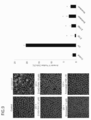
- FIG. 1 shows a microscopic image of corneal endothelial cells after UV irradiation and a graph of caspase 3/7 activity.
- the left panel shows phase contrast microscopic images of a control group, a UV irradiation group, a UV irradiation + emricasan addition group, a UV irradiation + Z-VD-FMK addition group, and a UV irradiation + Z-VAD-FMK addition group.
- the vertical axis indicates caspase 3/7 activity (%) relative to the UV irradiation group
- the horizontal axis indicates, from the left, control, UV irradiation group, UV irradiation + emlicasan added group (10 ⁇ M), UV irradiation + Z ⁇ .
- the VD-FMK addition group (10 ⁇ M) and the UV irradiation + Z-VAD-FMK addition group (10 ⁇ M) are shown. * Indicates statistical significance (p ⁇ 0.01).
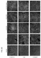
- FIG. 2 shows microscopic images of corneal endothelial cells after UV irradiation and the results of Western blotting in the Z-VD-FMK addition group.
- the left panel shows phase contrast microscopic images of a control group, a UV irradiation group, a UV irradiation + Z-VAD-FMK addition group (10 ⁇ M), and a UV irradiation + Z-VD-FMK addition group (3, 10, 20, 30, 100 ⁇ M). (Magnification ⁇ 200) is shown. The intensity of the UV was used 100J / m 2.
- the right panel shows the results of Western blot of caspase 3, PARP and GAPDH for each of the above groups.
- FIG. 3 shows a microscopic image of a corneal endothelial cell after UV irradiation and a Western blot result in the emlicasan addition group.
- the left panel shows a phase contrast microscope (magnification ⁇ 200) of a control group, a UV irradiation group, a UV irradiation + Z-VD-FMK addition group (10 ⁇ M), and a UV irradiation + emricasan addition group (1, 3, 10, 30, 100 ⁇ M). ).
- the intensity of the UV was used 100J / m 2.
- the right panel shows the results of Western blot of caspase 3, PARP and GAPDH for each of the above groups.
- FIG. 4 shows microscopic images of corneal endothelial cells after UV irradiation and the results of Western blotting in the nibokasan addition group.
- the left panel shows a phase contrast microscope (magnification ⁇ 200) of a control group, a UV irradiation group, a UV irradiation + Z-VD-FMK addition group (10 ⁇ M), and a UV irradiation + nibokasan addition group (1, 3, 10, 30, 100 ⁇ M). ).
- the intensity of the UV was used 100J / m 2.
- the right panel shows the results of Western blot of caspase 3, PARP and GAPDH for each of the above groups.
- FIG. 5 shows the effect of a caspase inhibitor on cell damage caused by hydrogen peroxide in cultured monkey corneal endothelial cells.
- FIG. 6 shows the effect of a caspase inhibitor on cell damage caused by MG132 in cultured monkey corneal endothelial cells.
- the left panel shows a control group of corneal endothelial cells, a group added with MG132 (10 ⁇ M), a group added with MG132 + Z-VAD-FMK (10 ⁇ M), a group added with MG132 + Z-VD-FMK (10 ⁇ M), a group added with MG132 + emricasan (10 ⁇ M), and MG132 +
- the phase-contrast microscope image (magnification x200) of a nibokasan addition group (100 micromol) is shown.
- the right panel shows the results of Western blot of caspase 3, PARP, GAPDH and CHOP of each of the above groups.
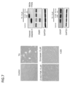
- FIG. 7 shows the effect of a caspase inhibitor on cell damage caused by thapsigargin (TG) in cultured monkey corneal endothelial cells.
- the left panel shows phase contrast microscopic images (magnification ⁇ 200) of a control group of corneal endothelial cells, a TG addition group (10 ⁇ M), a TG + Z-VAD-FMK addition group (10 ⁇ M), and a TG + emricasan addition group (10 ⁇ M).
- the right panel shows the results of Western blot of caspase 3, PARP, GAPDH and CHOP of each of the above groups.
- FIG. 8 shows the effect of caspase inhibitors on CCCP-induced cell damage in cultured monkey corneal endothelial cells.
- FIG. 9 shows fluorescence observation of Annexin V in rabbit corneal endothelial cells irradiated with UV.
- the left panel shows a control group, a UV irradiation group (250 J / m 2 ), a UV + Z-VD-FMK addition group (10 ⁇ M), a UV + Z-VAD-FMK addition group (10 ⁇ M), a UV + emricasan addition group (10 ⁇ M), and a UV + nibokasan addition
- a control group a UV irradiation group (250 J / m 2 )
- a UV + Z-VD-FMK addition group (10 ⁇ M
- a UV + Z-VAD-FMK addition group 10 ⁇ M
- a UV + emricasan addition group (10 ⁇ M) a UV + nibokasan addition
- a confocal microscope image of a group (100 ⁇ M) is shown.
- FIG. 10 shows observation of dysfunction and morphological abnormality of UV-irradiated rabbit corneal endothelial cells.
- the upper row shows the fluorescence of N-cadherin
- the middle row shows ZO-1
- the lower row shows phalloidin fluorescence.
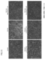
- FIG. 11 shows the effect of caspase inhibitors on TGF- ⁇ 2 cytotoxicity of immortalized Fuchs corneal endothelial dystrophy patient-derived cells (iFECD).
- FIG. 12 shows the results of flow cytometry for measuring programmed cell death induced by TGF- ⁇ 2.
- FIG. 13 shows a graph of Annexin V positive cell rate measured by flow cytometry.
- the vertical axis shows the value (ratio) of Q1-LR in FIG.
- FIG. 14 shows the effect of caspase inhibitors on TGF- ⁇ 2 cell damage in Fuchs corneal endothelial dystrophy disease cell model. The results of Western blotting of caspase 3, PARP and GAPDH are shown.
- FIG. 15 shows the effect of caspase inhibitor using rabbit cornea on corneal preservation.
- the upper part of the left panel shows a fluorescent image of N-cadherin, the middle part shows ZO-1, the lower part shows Phalloidin, the left column shows the control group, and the right column shows the emlicasan added group (10 ⁇ M).
- FIG. 16 shows cell viability measurements using various cryopreservation solutions.
- the left graph shows trypan blue negative cells (%).
- the right graph shows the cell viability (%) measured by CellTiter-Glo Luminescent Cell Viability Assay.
- FIG. 17 shows phase contrast microscopic images obtained by observing cell engraftment after freezing of corneal endothelial cells in various cryopreservation solutions.
- the upper row is a phase contrast microscopic image of corneal endothelial cells cultured in the absence of Y27632, and the lower row is a phase contrast microscopic image of corneal endothelial cells cultured in the presence of Y27632.
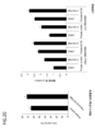
- FIG. 18 shows a graph of the number of cells after freezing of corneal endothelial cells in various cryopreservation solutions. The vertical axis indicates the number of cells (cells). From the left, CELL BANKER PLUS, CELL BANKER 2, STEM-CELLBANKER, KM BANKER, CnT-CRYO, OptiMEM + 10% FBS + 10% DMSO, and subcultured cells that have not undergone cryopreservation Please confirm. Indicates.
- FIG. 18 shows a graph of the number of cells after freezing of corneal endothelial cells in various cryopreservation solutions. The vertical axis indicates the number of cells (cells). From the left, CELL BANKER PLUS, CELL BANKER 2, STEM-CELLBANKER, KM BANKER, CnT-CRYO, OptiMEM + 10% FBS + 10% DMSO, and subcultured cells that have not undergone cryopreservation Please confirm. Indicates.
- FIG. 19 shows the effect of caspase inhibition of Z-VD-FMK on corneal endothelial cells after corneal endothelial cryopreservation.
- the image on the left shows the corneal endothelial cells of the control (using KM BANKER as the medium) with DMSO added without adding the caspase inhibitor, and the image on the right shows the corneal endothelial cells with Z-VD-FMK added Is shown.
- FIG. 20 shows the reduction of cell damage in corneal endothelial cryopreservation by Z-VD-FMK.
- the left graph shows the cell viability (%) immediately after recovery from freezing to room temperature, the left is Freeze control without adding Z-VD-FMK to the cryopreservation solution, and the right is Z-VD in the cryopreservation solution. -Freeze + Z-VD-FMK with FMK added.
- the right graph shows the number of cells 24 hours after thawing of cells and addition of DMSO or Z-VD-FMK.
- the four on the left are groups to which SB203580 is not added, and the four on the right are groups to which SB203580 is added.
- FIG. 21 shows that Z-VD-FMK promotes cell culture after freezing in addition to multiple storage solvents.
- DMSO added to culture medium, Cell Banker, and KM Bank used groups are shown as cryopreservation solutions. In each group, no addition after cryopreservation (DMSO group), Y27632 added (5 ⁇ M) , And Z-VD-FMK added (5 ⁇ M).
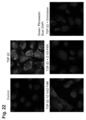
- FIG. 22 shows the inhibitory effect of a caspase inhibitor on fibronectin overexpression by TGF- ⁇ 2 in immortalized Fuchs corneal endothelial dystrophy patient-derived cells (iFECD).
- FIG. 23 shows Western blot results for fibronectin, p-Smad2, Smad2 and GAPDH in immortalized Fuchs corneal endothelial dystrophy patient-derived cells (iFECD).
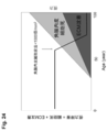
- FIG. 24 shows a schematic diagram of the relationship between visual acuity, corneal endothelial cells and ECM deposition. It is shown that visual acuity decreases as corneal endothelial cells and ECM deposition increase.
- Thickening of the Gutte or Desme layer due to ECM deposition generally begins in the 30s and 40s in Fuchs corneal endothelial dystrophy patients and progresses throughout life. Progression causes visual impairment such as foggy vision, halo, glare, and vision loss. At the same time, corneal endothelial cell death proceeds, but until the corneal endothelial cell density falls below about 1000 cells / mm 2 , the transparency of the cornea is maintained by compensating for the corneal endothelium that has left the pump function. On the other hand, when the rate is less than about 1000 / mm 2 , the anterior aqueous humor enters the cornea to cause corneal edema, resulting in severe visual impairment. This technique can maintain visual function by suppressing both ECM deposition and corneal endothelial cell death.
- caspase is a general term for endopeptidases having cysteine as an active center and hydrolyzing peptide bonds on the C-terminal side of aspartic acid. Caspases function in the processing of cytokines (such as interleukin 1 ⁇ ) and are known to be involved in the execution of programmed cell death and inflammatory responses. All caspases are translated as enzyme precursors and activation is activated by degradation by itself or other caspases and functions in a cascaded manner. Caspases are numbered in the order in which they are discovered, and caspase 1 to caspase 14 are known in mammals to date. About 10 types have been found in human cells. For example, caspase 1 functions to induce inflammation by cytokine processing, caspase 3 is directly involved in the execution of programmed cell death, and caspase 8 is located upstream of the cascade and is responsible for signaling of programmed cell death.
- cytokines such as interleukin 1 ⁇
- caspase inhibitor refers to any drug that inhibits signal transduction of any caspase.
- a caspase inhibitor is a compound that can inhibit any one or more of the caspase family.
- the caspase inhibitor is preferably water-soluble. This is because if it is not water-soluble, it may be necessary to use a solvent that is not compatible with the body. Whether it is water-soluble or not can be classified based on the pharmacopoeia solubility definition.
- the amount of solvent required to dissolve 1 g or 1 mL of solute is extremely soluble: less than 1 mL; easily soluble: 1 mL or more but less than 10 mL; slightly soluble: 10 mL or more but less than 30 mL; : 100 mL or more and less than 1000 mL; extremely difficult to dissolve: 1000 mL or more and less than 10000 mL; almost insoluble: defined as 10000 mL or more, and similarly evaluated in this specification.
- water-soluble means that any water-soluble one can be used as long as it dissolves an effective amount when water is used as a solvent. Such water-soluble components are also advantageously used as eye drops.
- the caspase inhibitor that can be used in the present invention is not particularly limited as long as it is a compound having caspase inhibitory activity.
- JP2012-036150, JP2007-308501, JP2005-089324 and compounds described in JP-A No. 2002-338474, JP-A No. 2001-302516, JP-T 2009-542689, WO 2006/054757, and the like.
- FMK pan ([pan] caspase inhibitor), Z-VD-FMK (caspase 1,3,6,7,8 and 9 inhibitor and substantially pan caspase inhibitor), Emricasan (Emricasan) ) (Total caspase inhibitor), Nivocasan (Nibocasan) (caspase 1, 3, 7 Z-YVAD-FMK (caspase 1 inhibitor), Z-VDVAD-FMK (caspase 2 inhibitor), Z-DEVD-FMK (caspase 3 inhibitor) Agent), Z-LEVD-FMK (caspase 4 inhibitor), Z-WEHD-FMK (caspase 5 inhibitor), Z-VEID-FMK (caspase 6 inhibitor), Z-IETD-FMK (caspase 8 inhibitor) Z-LEHD-FMK (caspase 9 inhibitor), Z-AEVD-FMK (caspase 10 inhibitor), and Z-LEED-FMK (caspase 13 inhibitor), but are not limited thereto.
- Preferred caspase inhibitors include, but are not limited to, emlicasan.
- Other preferred caspase inhibitors include, but are not limited to, Z-VAD-FMK, Z-VD-FMK, and nibokasan.
- the caspase inhibitor is preferably capable of inhibiting all caspases, but may be a selective caspase inhibitor.
- a selective caspase inhibitor refers to a caspase inhibitor that inhibits one caspase of the caspase family or a plurality of caspases.
- caspase 3 is directly involved in programmed cell death, so that inhibiting caspase 3 is direct and effective, but upstream in the caspase cascade Inhibiting caspase 3 (eg, caspase 2, 8, 9, 10, etc.) may be used to indirectly inhibit caspase 3.
- caspase 6 or caspase 7, which are the execution factors of programmed cell death may be inhibited in the same manner as caspase 3, or caspase 6 or caspase 7 may be indirectly inhibited by inhibiting upstream caspase in the caspase cascade. It may be an inhibitor.
- the caspase inhibitor used is advantageously a “pan caspase inhibitor”.
- caspase inhibitor that directly or indirectly inhibits a caspase inhibitor that inhibits caspase 8, which is regarded as an important molecule for signaling signal of programmed cell death. May be.
- caspase inhibitors that can be used in the present invention include, for example, neutralizing antibodies against caspases, compounds that inhibit caspase activity, compounds that inhibit transcription of genes encoding caspases (eg, antisense nucleic acids). , RNAi, ribozyme), peptides, and plant components.
- the concentration used is exemplified by about 50 nmol / l to 100 ⁇ mol / l, usually about 0.001 to 100 ⁇ mol / l, preferably about 0.01 to 75 ⁇ mol / l, about 0.05 to 50 ⁇ mol / l, About 1-10 ⁇ mol / l, about 0.01-10 ⁇ mol / l, about 0.05-10 ⁇ mol / l, about 0.075-10 ⁇ mol / l, about 0.1-10 ⁇ mol / l, about 0.5-10 ⁇ mol / l 1, about 0.75-10 ⁇ mol / l, about 1.0-10 ⁇ mol / l, about 1.25-10 ⁇ mol / l, about 1.5-10 ⁇ mol / l, about 1.75-10 ⁇ mol / l, about 2.
- 0-10 ⁇ mol / l about 2.5-10 ⁇ mol / l, about 3.0-10 ⁇ mol / l, about 4.0-10 ⁇ mol / l, about 5.0-10 ⁇ mol / l, about 6.0-10 ⁇ mol / l , About 7.0 ⁇ 0 ⁇ mol / l, about 8.0 to 10 ⁇ mol / l, about 9.0 to 10 ⁇ mol / l, about 0.01 to 50 ⁇ mol / l, about 0.05 to 5.0 ⁇ mol / l, about 0.075 to 5.0 ⁇ mol / L, about 0.1 to 5.0 ⁇ mol / l, about 0.5 to 5.0 ⁇ mol / l, about 0.75 to 5.0 ⁇ mol / l, about 1.0 to 5.0 ⁇ mol / l, about 1.
- the antisense nucleic acid used in the present invention may inhibit the expression and / or function of a gene (nucleic acid) encoding a member of a caspase signal transduction pathway or the like by any of the actions described above.
- a gene nucleic acid
- an antisense sequence complementary to the untranslated region near the 5 'end of mRNA of a gene encoding the above-mentioned caspase or the like is designed, it is considered effective for inhibiting translation of the gene.
- a sequence complementary to the coding region or the 3 'untranslated region can also be used.
- the nucleic acid containing the antisense sequence of the sequence of the untranslated region is included in the antisense nucleic acid used in the present invention.
- the antisense nucleic acid used is linked downstream of a suitable promoter, and preferably a sequence containing a transcription termination signal is linked on the 3 'side.
- the nucleic acid thus prepared can be transformed into a desired animal (cell) using a known method.
- the sequence of the antisense nucleic acid is preferably a sequence complementary to a gene encoding caspase or the like possessed by the animal (cell) to be transformed or a part thereof, but as long as the expression of the gene can be effectively suppressed, It may not be completely complementary.
- the transcribed RNA preferably has a complementarity of 90% or more, most preferably 95% or more, to the transcript of the target gene.
- the length of the antisense nucleic acid is preferably at least 12 bases and less than 25 bases, but the antisense nucleic acid of the present invention is not necessarily of this length.
- the antisense nucleic acid may be composed only of DNA, but may contain nucleic acid other than DNA, for example, locked nucleic acid (LNA).
- the antisense nucleic acid used in the present invention may be an LNA-containing antisense nucleic acid containing LNA at the 5 'end and LNA at the 3' end.
- an antisense nucleic acid for example, Hirashima and Inoue, Shinsei Kagaku Kogaku Kenkyu 2 (Replication and Expression of Nucleic Acid IV Gene, edited by the Japanese Biochemical Society, Tokyo Chemical Dojin, 1993, 319-347. Can be used to design antisense sequences based on nucleic acid sequences such as caspases.
- ribozymes refers to an RNA molecule having catalytic activity. Although ribozymes have various activities, research focusing on ribozymes as enzymes that cleave RNA has made it possible to design ribozymes that cleave RNA in a site-specific manner. Some ribozymes have a size of 400 nucleotides or more, such as group I intron type and M1 RNA contained in RNase P, but some have an active domain of about 40 nucleotides called hammerhead type or hairpin type. (Makoto Koizumi and Eiko Otsuka, protein nucleic acid enzyme, 1990, 35, 2191.).
- the self-cleaving domain of hammerhead ribozyme cleaves 3 ′ of C15 in the sequence G13U14C15, but base pairing between U14 and A9 is important for its activity, and instead of C15, A15 or U15 However, it has been shown that it can be cut (Koizumi, M. et al., FEBS Lett, 1988, 228, 228.).
- a restriction enzyme-like RNA-cleaving ribozyme that recognizes the sequence UC, UU or UA in the target RNA can be produced (Koizumi, M.
- Hairpin ribozymes are also useful for the purposes of the present invention.
- This ribozyme is found, for example, in the minus strand of satellite RNA of tobacco ring spot virus (Buzayan, JM., Nature, 1986, 323, 349.). It has been shown that target-specific RNA-cleaving ribozymes can also be produced from hairpin ribozymes (Kikuchi, Y. & Sasaki, N., Nucl. Acids Res, 1991, 19, 6751., Hiroshi Kikuchi, Chemistry and Biology , 1992, 30, 112.). Thus, the expression of the gene can be inhibited by specifically cleaving the transcript of the gene encoding caspase or the like using a ribozyme.
- RNA interference RNA interference
- RNAi RNA interference
- siRNA short-chain dsRNA
- siRNA is an RNA molecule having a double-stranded RNA portion consisting of 15 to 40 bases, cleaves the mRNA of the target gene having a sequence complementary to the antisense strand of the siRNA, It has a function of suppressing the expression of the target gene.
- the siRNA in the present invention comprises a sense RNA strand comprising a sequence homologous to a continuous RNA sequence in mRNA such as caspase, and an antisense RNA strand comprising a sequence complementary to the sense RNA sequence.
- RNA containing a single-stranded RNA portion The design and production of such siRNAs and mutant siRNAs described below are within the skill of the artisan.
- RNA region of mRNA that is a transcription product of a sequence of caspase or the like and prepare a double-stranded RNA corresponding to this region within the scope of normal trials. Is to get.
- selection of siRNA sequences having a stronger RNAi effect from mRNA sequences that are transcripts of the sequences can also be appropriately performed by those skilled in the art by known methods. If one strand is known, those skilled in the art can easily know the base sequence of the other strand (complementary strand). siRNA can be appropriately prepared by those skilled in the art using a commercially available nucleic acid synthesizer. In addition, for synthesis of a desired RNA, a general synthesis contract service can be used.
- the length of the double-stranded RNA portion is 15 to 40 bases, preferably 15 to 30 bases, more preferably 15 to 25 bases, still more preferably 18 to 23 bases, and most preferably 19 to 21 bases as a base. . It is understood that these upper and lower limits are not limited to these specific ones and may be any combination of those listed.
- the terminal structure of the sense strand or antisense strand of siRNA is not particularly limited and may be appropriately selected depending on the intended purpose. For example, it may have a blunt end or a protruding end (overhang) It is preferable that the 3 ′ end protrudes.
- siRNA having an overhang consisting of several bases, preferably 1 to 3 bases, more preferably 2 bases, at the 3 ′ end of the sense RNA strand and the antisense RNA strand suppresses the expression of the target gene. In many cases, the effect is large, which is preferable.
- the type of the overhanging base is not particularly limited, and may be either a base constituting RNA or a base constituting DNA.
- Preferred overhang sequences include dTdT (deoxy T 2 bp) at the 3 'end.
- preferred siRNAs include, but are not limited to, those in which dTdT (deoxy T is 2 bp) is attached to the 3 'end of the sense / antisense strands of all siRNAs.
- siRNA in which 1 to several nucleotides are deleted, substituted, inserted and / or added in one or both of the sense strand or antisense strand of the siRNA can also be used.
- the 1 to several bases are not particularly limited, but are preferably 1 to 4 bases, more preferably 1 to 3 bases, and most preferably 1 to 2 bases.
- Such mutations include those in which the number of bases in the 3 ′ overhang portion is 0 to 3, or the base sequence in the 3 ′ overhang portion is changed to another base sequence, or base insertion or addition Or, the length of the sense RNA strand and the antisense RNA strand differ by 1 to 3 bases due to deletion, and the sense strand and / or antisense strand in which the base is replaced with another base.
- the sense strand and the antisense strand can hybridize in these mutant siRNAs, and that these mutant siRNAs have the ability to suppress gene expression equivalent to siRNA having no mutation.
- the siRNA may be a molecule having a structure in which one end is closed, for example, an siRNA having a hairpin structure (Short Hairpin RNA; shRNA).
- shRNA is a RNA comprising a sense strand RNA of a specific sequence of a target gene, an antisense strand RNA consisting of a sequence complementary to the sense strand sequence, and a linker sequence connecting both strands, and a sense strand portion and an antisense strand The portions hybridize to form a double stranded RNA portion.
- siRNA does not show a so-called off-target effect in clinical use.
- the off-target effect refers to the action of suppressing the expression of another gene that is partially homologous to the used siRNA in addition to the target gene.
- NCBI National Center for Biotechnology Information
- RNA of the present invention In order to produce the siRNA of the present invention, a known method such as a method using chemical synthesis or a method using a gene recombination technique can be appropriately used.
- double-stranded RNA can be synthesized by a conventional method based on sequence information.
- an expression vector encoding a sense strand sequence or an antisense strand sequence is constructed, and the sense strand RNA or antisense strand RNA generated by transcription after introducing the vector into a host cell. It can also be produced by acquiring each of the above.
- a desired strand can be expressed by expressing a shRNA that forms a hairpin structure, including a sense strand of a specific sequence of a target gene, an antisense strand consisting of a sequence complementary to the sense strand sequence, and a linker sequence that connects both strands.
- the double-stranded RNA can also be prepared.
- all or part of the nucleic acid constituting the siRNA may be a natural nucleic acid or a modified nucleic acid.
- the siRNA in the present invention does not necessarily need to be a set of double-stranded RNAs for the target sequence, and a plurality of sets for the region containing the target sequence (this “plurality” is not particularly limited, but preferably 2 to 5) It may be a mixture of double-stranded RNA.
- siRNA as a nucleic acid mixture corresponding to the target sequence can be appropriately prepared by a person skilled in the art using a commercially available nucleic acid synthesizer and a DICER enzyme. Synthetic contract service can be used.
- the siRNA of the present invention includes so-called “cocktail siRNA”. In the siRNA of the present invention, not all nucleotides are necessarily ribonucleotides (RNA).
- the one or more ribonucleotides constituting the siRNA may be corresponding deoxyribonucleotides.
- This “corresponding” refers to the same base species (adenine, guanine, cytosine, thymine (uracil)) although the structures of the sugar moieties are different.
- deoxyribonucleotide corresponding to ribonucleotide having adenine refers to deoxyribonucleotide having adenine.
- a DNA (vector) capable of expressing the RNA of the present invention is also included in a preferred embodiment of a nucleic acid capable of suppressing the expression of caspase and the like.
- the DNA (vector) capable of expressing the double-stranded RNA of the present invention is a DNA encoding one strand of the double-stranded RNA and a DNA encoding the other strand of the double-stranded RNA, Each DNA has a structure linked to a promoter so that it can be expressed.
- the expression vector of the present invention can be prepared by appropriately inserting DNA encoding the target RNA into various known expression vectors.
- a modified nucleic acid may be used as the nucleic acid that suppresses the expression of the target gene.
- the modified nucleic acid means one having a structure different from that of a natural nucleic acid, in which a nucleoside (base site, sugar site) and / or internucleoside binding site is modified.
- Examples of the “modified nucleoside” constituting the modified nucleic acid include an abasic nucleoside; an arabino nucleoside, 2′-deoxyuridine, ⁇ -deoxyribonucleoside, ⁇ -L-deoxyribonucleoside, and other sugars.
- nucleosides having modifications include peptide nucleic acids (PNA), peptide nucleic acids to which phosphate groups are bound (PHONA), locked nucleic acids (LNA), morpholino nucleic acids and the like.
- PNA peptide nucleic acids
- PONA peptide nucleic acids to which phosphate groups are bound
- LNA locked nucleic acids
- nucleoside having a sugar modification include substituted pentose monosaccharides such as 2′-O-methylribose, 2′-deoxy-2′-fluororibose, and 3′-O-methylribose; 1 ′, 2′-deoxyribose Arabinose; substituted arabinose sugars; nucleosides with hexose and alpha-anomeric sugar modifications.
- These nucleosides may be modified bases with modified base sites. Examples of such modified bases include pyrimidines such as 5-hydroxycytosine, 5-fluorouracil and
- modified internucleoside linkage constituting the modified nucleic acid
- examples of the “modified internucleoside linkage” constituting the modified nucleic acid include, for example, alkyl linker, glyceryl linker, amino linker, poly (ethylene glycol) linkage, methylphosphonate internucleoside linkage; methylphosphonothioate, phosphotriester , Phosphothiotriester, phosphorothioate, phosphorodithioate, triester prodrug, sulfone, sulfonamide, sulfamate, formacetal, N-methylhydroxylamine, carbonate, carbamate, morpholino, boranophosphonate, phosphoramidate, etc.
- Non-natural internucleoside linkages include, for example, alkyl linker, glyceryl linker, amino linker, poly (ethylene glycol) linkage, methylphosphonate internucleoside linkage; methylphosphonothioate
- nucleic acid sequence contained in the double-stranded siRNA of the present invention examples include siRNA for caspase or other caspase signaling members.
- siRNA shows very excellent specific post-transcriptional repression effect in vitro, but in vivo it is rapidly degraded by the nuclease activity in serum, so its duration is limited and more optimal and effective delivery System development has been demanded.
- One example is Ochiya, T et al., Nature Med.
- Atelocollagen a biocompatible material
- nucleic acid is mixed with nucleic acid to form a complex, which protects the nucleic acid from degrading enzymes in the body and is very suitable as a carrier of siRNA.
- the method of introducing the nucleic acid, therapeutic or prophylactic agent of the present invention is not limited thereto. In this way, in the living body, it is rapidly degraded by the action of the nucleolytic enzyme in the serum, so that a long-term effect can be achieved. For example, in Takeshita F. PNAS.
- atelocollagen derived from bovine skin forms a complex with nucleic acid, It has been reported that it has an action of protecting nucleic acids from degrading enzymes and is very suitable as a carrier of siRNA, and such a technique can be used.
- iFECD immobilized Fuchs' endothelial corneal dystrophy
- iFECD immobilized Fuchs' endothelial corneal dystrophy
- HCEC human Corneal Endothelial cells
- IHCEC is an abbreviation for immortalized human corneal endothelial cells.
- programmed cell death refers to a phenomenon in which cells spontaneously die at a predetermined time or environment as if they were programmed in advance. Programmed cell death is used to mean, for example, “apoptosis”.
- transforming growth factor- ⁇ transformed growth factor- ⁇ ; also abbreviated as TGF- ⁇
- TGF- ⁇ transformed growth factor- ⁇
- various sclerosis properties responsible for the pathogenesis of diseases, rheumatoid arthritis and proliferative vitreoretinopathy It is a homodimeric multifunctional cytokine with a molecular weight of 25 kD that exhibits a variety of biological activities, such as preventing falling and suppressing the growth of cancer cells.
- a “TGF- ⁇ signal” refers to a signal mediated by TGF- ⁇ and triggered by TGF- ⁇ .
- TGF- ⁇ signals include, for example, signals mediated by TGF- ⁇ 2, and other examples include signals mediated by TGF- ⁇ 1, TGF- ⁇ 3, and the like.
- TGF- ⁇ there are three isoforms with about 70% homology from TGF- ⁇ 1 to ⁇ 3 in humans, and their actions are similar.
- TGF- ⁇ is produced as an inactive latent form having a molecular weight of about 300 kD that cannot bind to the receptor, and is activated on the surface of the target cell or its surroundings to become an active form that can bind to the receptor and exerts its action.
- TGF- ⁇ protein phosphorylation pathways responsible for information transmission
- Smad protein phosphorylation pathways responsible for information transmission
- a receptor complex consisting of two type II receptor molecules and two type I TGF- ⁇ receptor molecules is formed. Phosphorylates type I receptors.
- phosphorylated type I receptor phosphorylates Smad2 or Smad3
- phosphorylated Smad2 and Smad3 form a complex with Smad4 and move to the nucleus, and are called CAGA boxes that exist in the target gene promoter region. It is said to bind to the target sequence and induce transcriptional expression of the target gene together with the coactivator.
- TGF- ⁇ Transforming (transforming) growth factor- ⁇ (TGF- ⁇ ) signaling pathway is regulated by regulation of its target genes, cell proliferation and differentiation, growth arrest, programmed cell death, and epithelial-mesenchymal transdifferentiation (EMT; epithelium) Many cell activities such as mesenchymal transition can be regulated.
- TGF- ⁇ family including TGF- ⁇ itself (eg, TGF- ⁇ 1, TGF- ⁇ 2 and TGF- ⁇ 3), activin and bone morphogenetic protein (BMP) are cell proliferation, differentiation, migration and programmed cell death. Is a powerful regulator.
- TGF- ⁇ is an approximately 24 Kd protein produced by many cells and many other cell types, including B lymphocytes, T lymphocytes and activated macrophages.
- B lymphocytes include B lymphocytes, T lymphocytes and activated macrophages.
- TGF- ⁇ is thought to be involved in a variety of pathological conditions (Border et al. (1992) J. Clin. Invest. 90: 1) and functions as either a tumor suppressor or tumor promoter. Is well supported.
- TGF- ⁇ mediates its signaling by two serine / threonine kinase cell surface receptors, TGF- ⁇ RII and ALK5.
- TGF- ⁇ signaling is initiated by ligand-induced receptor dimerization that allows TGF- ⁇ RII to phosphorylate the ALK5 receptor.
- the phosphorylation activates ALK5 kinase activity, which in turn activates downstream effector Smad protein (vertebrate homologue of MAD, or “Mothers against DPP” protein), Smad2 or 3 Is phosphorylated.
- Smad protein verebrate homologue of MAD, or “Mothers against DPP” protein
- Smad2 or 3 Is phosphorylated The p-Smad2 / 3 complex with Smad4 enters the nucleus and activates transcription of the target gene.
- Smad3 is a member of Smad's R-Smad (receptor-activated Smad) subgroup and is a direct mediator of transcriptional activation by the TGF- ⁇ receptor.
- TGF- ⁇ stimulation results in phosphorylation and activation of Smad2 and Smad3, which form a complex with Smad4 (“common Smad” or “co-Smad” in vertebrates), which together with the nucleus Accumulate and regulate transcription of target genes.
- R-Smad localizes in the cytoplasm and, upon ligand-induced phosphorylation by the TGF- ⁇ receptor, forms a complex with co-Smad and translocates to the nucleus where they are chromatin and synergistic Regulates gene expression associated with transcription factors.
- Smad6 and Smad7 are inhibitory Smads (“I-Smad”), ie, transcriptionally induced by TGF- ⁇ and function as inhibitors of TGF- ⁇ signaling (Feng et al. (2005) Annu. Rev. Cell. Dev.Biol.21: 659).
- Smad6 / 7 exerts their inhibitory effects by preventing receptor-mediated activation of R-Smad; they are associated with type I receptors that competitively prevent R-Smad mobilization and phosphorylation.
- Smad6 and Smad7 are known to recruit E3 ubiquitin ligase, which leads to ubiquitination and degradation of Smad6 / 7 interacting proteins.
- TGF- ⁇ signaling pathway J. Massaggu'e, Annu. Rev. Biochem. 1998. 67: 753-91; Vilar JMG, Jansen R, Sander C (2006) PLoS Comput Biol 2 (1): e3; Leask, A., Abraham, D.J.FASEB J.18, 816-827 (2004); Coert Margadant & Arnoud Sonnenberg EMBO reports (2010) 11, 97-105; Joel Rosenbloom et al., Ann Intern Med. 2010 ; See 152: 159-166, etc.
- a corneal endothelial symptom, disorder or disease caused by transforming growth factor- ⁇ refers to any corneal endothelial symptom or disorder induced by TGF- ⁇ in corneal endothelial cells. Or refers to a disease.
- TGF- ⁇ transforming growth factor- ⁇
- exposure of corneal endothelial cells, eg, Fuchs corneal endothelial dystrophy model cells (eg, iFECD) to TGF- ⁇ 2
- iFECD corneal endothelial dystrophy model cells
- Such a phenomenon has not been well elucidated.
- the inventors further analyzed the symptoms, disorders or diseases of the corneal endothelium caused by the TGF- ⁇ signal, and unexpectedly found that the disorders can be suppressed by caspase inhibitors. It was. Symptoms, disorders or diseases of the corneal endothelium caused by TGF- ⁇ signal are different from the caspase signaling pathway, and the caspase inhibitor used does not suppress the TGF- ⁇ signaling pathway Therefore, it can be said that the route of the development of unexplained diseases and disorders and the mode of treatment or prevention thereof have been found so far. Therefore, it is considered that the present invention can be positioned as providing a new treatment / prevention technique for the corneal endothelium.
- Symptoms, disorders or diseases of corneal endothelium caused by TGF- ⁇ signal include, for example, Fuchs corneal endothelial dystrophy, corneal transplantation disorders, corneal endotheliitis, trauma, disorders after ophthalmic surgery, disorders after ophthalmic laser surgery, TGF- ⁇ expression is observed in age, posterior polymorphic corneal dystrophy (PPD), congenital hereditary endothelial dystrophy (CHED), idiopathic corneal endothelial dystrophy, etc. But are not limited to these.
- corneal endothelial cells or corneal endothelial tissues in which the expression of TGF- ⁇ 2 is increased more than usual it is considered that the disorder found in the present invention or a disorder related thereto is expressed or increased.
- Any corneal endothelial symptom, disorder or disease in which corneal endothelial cells or corneal endothelial tissue are found is specifically contemplated as a subject of the present invention.
- symptom, disorder or disease of corneal endothelium due to mitochondrial abnormality refers to a symptom, disorder or disease of corneal endothelium due to mitochondrial abnormality.
- Symptoms, disorders or diseases of corneal endothelium due to mitochondrial abnormalities include, for example, Fuchs corneal endothelial dystrophy, corneal transplantation disorders, corneal endotheliitis, trauma, disorders after ophthalmic surgery, disorders after ophthalmic laser surgery, aging, Examples include posterior polymorphic corneal dystrophy (PPD), congenital hereditary endothelial dystrophy (CHED), and idiopathic corneal endothelial dystrophy with any mitochondrial abnormality. Yes, but not limited to them.
- extracellular matrix in corneal endothelial cells refers to expression of extracellular matrix at an abnormal level compared to the expression level of extracellular matrix in normal corneal endothelial cells. “Expressing an extracellular matrix at an abnormal level” means that an amount of an extracellular matrix protein such as fibronectin is produced more than the amount produced in the extracellular matrix in a normal form. The production status includes not only stimulation but also that the expression level is increased by a response to transforming growth factor (TGF) ⁇ as necessary.
- TGF transforming growth factor
- the amount of extracellular matrix in normal is about 1.1 times or more, about 1.2 times or more, about 1.3 times or more, about 1.4 times or more, about 1.5 times. Times or more, about 1.6 times or more, about 1.7 times or more, about 1.8 times or more, about 1.9 times or more, or about 2.0 times or more. Differences from normal are preferably statistically significant, but not necessarily significant, as long as they are medically significant.
- cornereal endothelial disorder caused by excessive production of extracellular matrix (ECM) or “symptom” thereof refers to a disorder mainly related to turbidity, deposition, thickening or the like caused by extracellular matrix or its It is a symptom, and is related to symptoms that cause a decrease in visual acuity, such as a thickening of the Descemet's membrane, such as warts (guttata) on the corneal endothelium and opacity of the Descemet's membrane.
- a caspase inhibitor can suppress the cause of corneal endothelial injury caused by “turbidity” or “deposition”. This indicates that the present invention is based on “extracellular matrix (ECM)”. It can be said that it supports the improvement, treatment, or prevention of corneal endothelial disorder caused by overproduction of and the related symptoms.
- corneal endothelial disorders caused by overproduction of extracellular matrix (ECM) or “symptoms” thereof include, but are not limited to, turbidity, scar, corneal cloud, corneal plaque, corneal vitiligo, etc. . *
- the symptom, disorder or disease targeted by the present invention is a disorder related to Fuchs corneal endothelial dystrophy.
- Fuchs corneal endothelial dystrophy it has been shown that TGF- ⁇ induction in corneal endothelial cells is involved, and it can also be involved in cell loss in FECD. Therefore, it is naturally expected that inhibition of the TGF- ⁇ signaling pathway may be an effective treatment for FECD.
- the present inventors have unexpectedly found that caspase inhibitors can suppress damage caused by TGF- ⁇ signal.
- the medicament of the present invention can treat cell damage induced by TGF- ⁇ 2, which causes one of the important abnormalities or disorders among Fuchs corneal endothelial dystrophy, and is useful for the treatment or prevention of Fuchs corneal endothelial dystrophy. It is understood that In particular, the present invention was able to suppress cell damage or programmed cell death induced by TGF- ⁇ 2 in the Fuchs corneal endothelial dystrophy model in the Examples. It can be used to treat severely related patients. Moreover, the medicament of the present invention can unexpectedly suppress the overexpression of extracellular matrix (ECM), and can treat or prevent disorders in the corneal endothelium such as deposition of ECM in the desme layer. .
- ECM extracellular matrix
- the present invention provides corneal endothelial cell damage in Fuchs corneal endothelial dystrophy, corneal endothelial density reduction, guttae formation, Descemet's thickening, corneal thickening, corneal epithelial disorder, turbidity, scar, corneal stroma Cloudiness, photophobia, foggy vision, vision impairment, eye pain, lacrimation, hyperemia, pain, bullous keratopathy, ocular discomfort, reduced contrast, halo, glare and corneal edema can be treated or prevented.
- the present invention can also suppress mitochondrial abnormalities such as mitochondrial membrane potential decrease, mitochondrial morphological abnormality, mitochondrial biosynthesis decrease, and the like.
- the present invention relates to a medicament for treating or preventing a corneal endothelial symptom, disorder or disease caused by a transforming growth factor- ⁇ (TGF- ⁇ ) signal in a corneal endothelial cell, comprising a caspase inhibitor.
- TGF- ⁇ transforming growth factor- ⁇
- the present invention provides a medicament for treating or preventing a corneal endothelial symptom, disorder or disease caused by a mitochondrial abnormality in a corneal endothelial cell, comprising a caspase inhibitor.
- the present invention is for the treatment or prevention of a corneal endothelium symptom, disorder or disease resulting from transforming growth factor- ⁇ (TGF- ⁇ ) signal and mitochondrial abnormalities in corneal endothelial cells, comprising a caspase inhibitor.
- TGF- ⁇ transforming growth factor- ⁇
- the medicine is provided.
- Caspases are thought to be involved in various signal transductions and are also thought to be involved in inflammation, but in the corneal endothelium, not all of the mechanisms have been elucidated and TGF- ⁇ signaling, mitochondrial damage or both It could not be expected to be effective in the cure or prevention of corneal endothelial dysfunction caused by.
- the corneal endothelium symptom, disorder or disease caused by transforming growth factor- ⁇ (TGF- ⁇ ) in corneal endothelial cells is Fuchs corneal endothelial dystrophy, corneal transplantation disorder, corneal endotheliitis, trauma, Disorders after ophthalmic surgery, disorders after ophthalmic laser surgery, aging, posterior polymorphic corneal dystrophy (PPD), congenital hereditary endothelial dystrophy (CHED), and idiopathic cornea Selected from the group consisting of endothelial disorders.
- the corneal endothelium symptom, disorder or disease resulting from mitochondrial abnormalities in corneal endothelial cells is Fuchs corneal endothelial dystrophy, corneal transplantation disorder, corneal endotheliitis, trauma, post-ophthalmic surgery disorder, ophthalmic laser surgery Selected from the group consisting of posterior disorders, aging, posterior polymorphic corneal dystrophy (PPD), congenital hereditary endothelial dystrophy (CHED), and idiopathic corneal endothelial dystrophy .
- the corneal endothelium symptom, disorder or disease resulting from a TGF- ⁇ signal is a corneal endothelium symptom, disorder or disease resulting from TGF- ⁇ 2.
- mitochondrial abnormalities include, but are not limited to, mitochondrial membrane potential reduction, mitochondrial morphological abnormalities, mitochondrial biogenesis, and the like.
- the present invention has the effect of treating or preventing symptoms caused by excessive production of extracellular matrix in Fuchs corneal endothelial dystrophy, and a medicament for treating or preventing such symptoms Or provide treatment or prevention.
- symptoms include warts on the corneal endothelium, opacity of the Descemet's membrane, thickening of the Descemet's membrane, foggy vision, halo, glare, vision loss, corneal opacity, vitiligo, and abnormal visual sensation. be able to. Symptoms resulting from overproduction of the extracellular matrix are further described below.
- the present invention provides a medicament for treating or preventing a corneal endothelial symptom, disorder or disease caused by overexpression of an extracellular matrix in corneal endothelial cells, comprising a caspase inhibitor.
- the caspase inhibitor can treat or prevent corneal endothelial damage and the like caused by TGF- ⁇ signal and mitochondrial abnormality. It was surprising that it was possible to suppress. This suggests that caspase inhibitors can simultaneously treat corneal endothelial damage due to TGF- ⁇ signaling, mitochondrial abnormalities, and extracellular matrix overexpression in corneal endothelial cells.
- Fuchs corneal endothelial dystrophy has a marked decrease in the density of corneal endothelial cells due to TGF- ⁇ signal and mitochondrial abnormalities, and the extracellular matrix is deposited on the Descemet's membrane, resulting in thickening of Corneal guttae and Descemet's membrane. Suppressing extracellular matrix overexpression, as it is a disease that occurs, can mean significant improvements in the treatment and prevention of Fuchs corneal endothelial dystrophy, and in some cases, complete healing. ing.
- Corneal guttae and Descemet's thickening that may occur due to excessive production of extracellular matrix in corneal endothelial disorders such as Fuchs' corneal endothelial dystrophy, and other symptoms related to turbidity and deposition (irreversible due to prolonged corneal edema) Corneal opacity etc.) can be improved, treated or prevented.
- a corneal endothelial symptom, disorder or disease resulting from overexpression of extracellular matrix in corneal endothelial cells may be due to overexpression of fibronectin in corneal endothelial cells.
- a corneal endothelial symptom, disorder or disease resulting from extracellular matrix overexpression in corneal endothelial cells is Fuchs corneal endothelial dystrophy, gut formation, Descemet's thickening, corneal thickening, turbidity, Selected from the group consisting of scar, corneal opacity, corneal epithelial edema, corneal epithelial disorder, photophobia, and fog vision.
- the present invention is for treating or preventing a corneal endothelial symptom, disorder or disease resulting from TGF- ⁇ signaling, mitochondrial abnormalities and extracellular matrix overexpression in corneal endothelial cells, comprising a caspase inhibitor.
- the medicine is provided.
- a caspase inhibitor can simultaneously treat or prevent corneal endothelial damage resulting from TGF- ⁇ signaling, mitochondrial abnormalities, and extracellular matrix overexpression in corneal endothelial cells.
- a corneal endothelial symptom, disorder or disease resulting from TGF- ⁇ signaling, mitochondrial abnormalities and extracellular matrix overexpression in corneal endothelial cells is Fuchs corneal endothelial dystrophy, other corneal endothelial dystrophy, and drugs Selected from the group consisting of corneal endothelial disorders due to surgery, trauma, infection, uveitis and the like.
- the corneal endothelial condition, disorder or disease resulting from TGF- ⁇ signaling, mitochondrial abnormalities and extracellular matrix overexpression in corneal endothelial cells comprises Fuchs corneal endothelial dystrophy.
- Fuchs corneal endothelial dystrophy has a marked decrease in the density of corneal endothelial cells due to TGF- ⁇ signal and mitochondrial abnormalities, and the extracellular matrix is deposited on the Descemet's membrane, resulting in disorders such as Corneal guttae and Descemet's thickening Suppressing extracellular matrix overexpression means that Fuchs corneal endothelial dystrophy can be significantly improved, and in some cases, can be completely cured .
- the use of the present invention includes, for example, eye drops, but is not limited thereto, injection into the anterior chamber, impregnation into a sustained-release agent, subconjunctival injection, systemic administration (internal use) And administration methods such as intravenous injection).
- the caspase inhibitor used in the present invention is of any type as long as it is effective in treating or preventing any corneal endothelial symptom, disorder or disease caused by TGF- ⁇ signal or mitochondria
- a caspase-3 inhibitor more preferably a pan caspase inhibitor.
- Specific caspase inhibitors include Z-VAD-FMK, Z-VD-FMK, Emricasan, Nivocasan, Z-YVAD-FMK, Z-VDVAD-FMK, Z-DEVD-FMK, Z -At least one selected from the group consisting of LEVD-FMK, Z-WEHD-FMK, Z-VEID-FMK, Z-IETD-FMK, Z-LEHD-FMK, Z-AEVD-FMK, Z-LEED-FMK including.
- the caspase inhibitor may be used alone or in combination.
- the concentration of the caspase inhibitor used in the present invention is usually about 0.1 to 300 ⁇ M ( ⁇ mol / l), preferably about 1 to 150 ⁇ M, more preferably about 3 to 30 ⁇ M.
- Two or more caspase inhibitors Can be appropriately changed when used in combination. Examples of other concentration ranges are usually about 0.001 to 300 ⁇ M, about 0.01 to 150 ⁇ M, about 0.001 to 100 ⁇ M, and about 0.01.
- ⁇ 75 ⁇ M about 0.05-50 ⁇ M, about 1-10 ⁇ M, about 0.01-10 ⁇ M, about 0.05-10 ⁇ M, about 0.075-10 ⁇ M, about 0.1-10 ⁇ M, about 0.5-10 ⁇ M, About 0.75-10 ⁇ M, about 1.0-10 ⁇ M, about 1.25-10 ⁇ M, about 1.5-10 ⁇ M, about 1.75-10 ⁇ M, about 2.0-10 ⁇ M, about 2.5-10 ⁇ M, about 3.0 to 10 ⁇ M, about 4.0 ⁇ 10 ⁇ M, about 5.0-10 ⁇ M, about 6.0-10 ⁇ M, about 7.0-10 ⁇ M, about 8.0-10 ⁇ M, about 9.0-10 ⁇ M, about 0.01-50 ⁇ M, about 0.05- 5.0 ⁇ M, about 0.075 to 5.0 ⁇ M, about 0.1 to 5.0 ⁇ M, about 0.5 to 5.0 ⁇ M, about 0.75 to 5.0 ⁇ M, about 1.0 to 5.0 ⁇ M, about 1.25 to 5.0 ⁇ M, about
- ⁇ M about 2.0 to 3.0 ⁇ M
- the caspase inhibitor is, for example, N- (benzyloxycarbonyl) -L-valyl-DL-aspartyl-fluoromethylketone (Z-VD-FMK), ⁇ benzyloxycarbonyl-L-valyl-L- Alanyl-[(2s) -2-amino-3- (methoxycarbonyl) propionyl] ⁇ fluoromethane (Z-VAD-FMK), 3- [2-[(2-tert-butyl-phenylaminooxalyl) -amino] -Propionylamino] -4-oxo-5- (2,3,5,6-tetrafluoro-phenoxy) -pentanoic acid (emricasan) and (R) -N-((2S, 3S) -2- (fluoromethyl ) -2-Hydroxy-5-oxotetrahydrofuran-3-yl) -5-iso
- the caspase inhibitor is Z-VD-FMK.
- concentration of Z-VD-FMK used is usually about 1 ⁇ M to about 150 ⁇ M, preferably about 3 ⁇ M to about 100 ⁇ M, more preferably about 10 ⁇ M to about 50 ⁇ M.
- the caspase inhibitor is Z-VAD-FMK.
- concentration of Z-VAD-FMK used is usually about 1 ⁇ M to about 100 ⁇ M, preferably about 3 ⁇ M to about 30 ⁇ M, more preferably about 10 ⁇ M.
- the caspase inhibitor is emlicasan.
- concentration of emlicasan used is usually about 0.3 ⁇ M to about 150 ⁇ M, preferably about 1 ⁇ M to about 100 ⁇ M, more preferably about 10 ⁇ M.
- the caspase inhibitor is nivocasan.
- concentration of nibocasan used is usually about 1 ⁇ M to about 300 ⁇ M, preferably about 30 ⁇ M to about 300 ⁇ M, more preferably about 100 ⁇ M.
- emlicasan is used. While not wishing to be bound by theory, treatment with emricasan has shown significant therapeutic results compared to other caspase inhibitors, particularly transforming growth factor- ⁇ 2 (TGF-, such as Fuchs endothelial dystrophy) This is because it has been found that the healing performance of corneal endothelium disease or disorder related to ⁇ 2) or corneal endothelium disease or disorder related to mitochondrial abnormality is remarkably improved.
- TGF- transforming growth factor- ⁇ 2
- the medicament for treating or preventing the present invention can be directed to any animal having a corneal endothelium, such as a mammal, preferably for the purpose of treating or preventing a primate corneal endothelium.
- the subject of treatment or prevention is human corneal endothelium.
- the present invention provides at least for transforming growth factor- ⁇ (TGF- ⁇ ) signaling and mitochondrial abnormalities in corneal endothelial cells comprising administering an effective amount of a caspase inhibitor to a subject in need thereof.
- TGF- ⁇ transforming growth factor- ⁇
- Methods are provided for the treatment or prevention of corneal endothelium symptoms, disorders or diseases caused by one.
- the term ⁇ subject '' refers to the subject of administration (transplantation) of the medicament or method for the treatment and prevention of the present invention, and the subject is a mammal (e.g., human, mouse, rat, hamster, Rabbits, cats, dogs, cows, horses, sheep, monkeys, etc.), primates are preferred, and humans are particularly preferred.
- a mammal e.g., human, mouse, rat, hamster, Rabbits, cats, dogs, cows, horses, sheep, monkeys, etc.
- primates are preferred, and humans are particularly preferred.
- the effective amount of the medicament of the present invention that is effective in the treatment of a particular disease, disorder or condition can vary depending on the nature of the disorder or condition, but can be determined by those skilled in the art according to standard clinical techniques based on the description herein. is there. Furthermore, if necessary, in vitro assays can be used to help identify optimal dosage ranges.
- the exact dose to be used in the formulation can also vary depending on the route of administration and the severity of the disease or disorder and should be determined according to the judgment of the attending physician and the circumstances of each patient. However, the dose is not particularly limited, and may be, for example, 0.001, 1, 5, 10, 15, 100, or 1000 mg / kg body weight per dose, and within the range of any two of these values There may be.
- the dosing interval is not particularly limited.
- the present invention may be administered once or twice per 1, 7, 14, 21, or 28 days, or once or twice per any two of these ranges. Also good.
- the dose, number of administrations, administration interval, and administration method may be appropriately selected depending on the age and weight of the patient, symptoms, administration form, target organ, and the like.
- the present invention can be used as eye drops.
- the medicament of the present invention can also be injected into the anterior chamber.
- the therapeutic agent preferably contains a therapeutically effective amount or an effective amount of an active ingredient that exhibits a desired action. If the therapeutic marker is significantly decreased after administration, it may be determined that there is a therapeutic effect. Effective doses can be extrapolated from dose-response curves derived from in vitro or animal model test systems.
- the present invention provides a composition for the preservation of corneal endothelial cells, comprising a caspase inhibitor.
- the storage is cryopreservation.
- the caspase inhibitor used in the present invention can be used in any form described herein, for example, an embodiment described as a medicine, which is suitable as a preservation composition.
- the term “preservation composition” refers to a composition for preserving corneal endothelial cells extracted from a donor for a period until transplantation to a recipient, or pre-proliferated or proliferated corneal endothelial cells. It is.
- the preservation composition of the present invention can be prepared by adding the caspase inhibitor of the present invention to a conventionally used preservative or preservation solution.
- a corneal preservation solution include a preservation solution usually used at the time of corneal transplantation (a corneal piece preservation solution (Optisol GS: registered trademark), an eye preservation solution for corneal transplantation (EPII: registered trademark)), physiological saline, phosphorus Examples include acid buffered saline (PBS).
- the preservation composition of the present invention is used for preservation of the cornea used for organ transplantation and the like. Further, the preservation composition of the present invention is also used as a preservation solution or a component thereof for cryopreserving corneal endothelial cells.
- the preservation composition containing the caspase inhibitor of the present invention can be added to an existing cryopreservation solution for use.
- cryopreservation solution examples include CELLBANKER (registered trademark) series (CELL BANKER PLUS (catalog number: CB021), CELL BANKER 2 (catalog number: CB031), STEM-CELLBANKER (catalog number: CB043) provided by Takara Bio.
- cryopreservation solution used may be KM BANKER.
- a suitable modified cryopreservation solution can be used by appropriately changing the components of the cryopreservation solution or adding additional components.
- cryopreservation for example, glycerol, dimethyl sulfoxide, propylene glycol, acetamide and the like may be further added to the preservation solution of the present invention.
- the present invention provides a composition for promoting proliferation of corneal endothelial cells. It is understood that the caspase inhibitor used in the present invention can be used in any form described herein, for example, an embodiment described as a medicine, which is suitable as a preservation composition.
- the composition for promoting growth of the present invention can be prepared by adding the caspase inhibitor of the present invention to a conventionally used medium.
- cryopreservation solution examples include CELLBANKER (registered trademark) series (CELL BANKER PLUS (catalog number: CB021), CELL BANKER 2 (catalog number: CB031), STEM-CELLBANKER (catalog number: CB043) provided by Takara Bio.
- cryopreservation solution used is preferably KM BANKER, but is not limited thereto.
- the composition for promoting proliferation of corneal endothelial cells of the present invention may further comprise a p38 MAP kinase inhibitor.
- P38 MAP kinase inhibitor also referred to as“ p38 MAPK inhibitor ” refers to any agent that inhibits MAP kinase signaling associated with p38, eg, 4- (4-fluorophenyl) -2- (4-Methylsulfinylphenyl) -5- (4-pyridyl) -1H-imidazole (SB-203580), 1- (carbamoyl-6- (2,4-difluorophenyl) pyridin-2-yl) -1- ( 2,6-difluorophenyl) urea (VX-702), and 3- [3-bromo-4-[(2,4-difluorophenyl) methoxy] -6-methyl-2-oxopyridin-1-yl]- Non-limiting
- the concentration of the p38 MAP kinase inhibitor contained can be appropriately selected by those skilled in the art.
- the concentration is about 0.1 to 100 ⁇ M ( ⁇ mol / l), preferably about 0.1 to 30 ⁇ M, more preferably Other concentration ranges are, for example, usually about 0.001 to 100 ⁇ M, preferably about 0.01 to 75 ⁇ M, about 0.05 to 50 ⁇ M, about 1 to 10 ⁇ M, about 0.1 .mu.M.
- composition for promoting proliferation of the present invention can be used in the culture of corneal endothelial cells.
- the culture of corneal endothelial cells is typically performed as follows.
- Corneal endothelial cells are collected in a conventional manner from the recipient himself or from the cornea of an appropriate donor.
- allogeneic corneal endothelial cells may be prepared. For example, after detaching the Descemet's membrane and endothelial cell layer of corneal tissue from the corneal stroma, it is transferred to a culture dish and treated with dispase or the like. As a result, corneal endothelial cells are detached from the Descemet's membrane. Corneal endothelial cells remaining on the Desme membrane can be removed by pipetting or the like.
- corneal endothelial cells are cultured in the culture medium of the present invention.
- a culture container (culture dish) whose surface is coated with type I collagen, type IV collagen, fibronectin, laminin, or an extracellular matrix of bovine corneal endothelial cells.
- an ordinary culture vessel treated with a commercially available coating agent such as FNC coating mix (registered trademark) (50 ml (AES-0407), ATHENA, catalog number: 0407) may be used.
- the temperature conditions for culturing the corneal endothelial cells are not particularly limited as long as the corneal endothelial cells grow. For example, about 25 ° C. to about 45 ° C., and considering the growth efficiency, preferably about 30 ° C. to about 40 ° C. More preferably, it is about 37 ° C.
- the culture method is performed in a normal cell culture incubator under humidification and in an environment having a CO 2 concentration of about 5 to 10%.
- Subculture Subculture can be performed after the corneal endothelial cells subjected to the culture have proliferated. Preferably, subculture is performed when sub-confluent or confluent. Subculture can be performed as follows. First, the cells are detached from the surface of the culture container by treating with trypsin-EDTA or the like, and then the cells are recovered. A medium or culture solution containing the composition for promoting growth of the present invention is added to the collected cells to obtain a cell suspension. Centrifugation is preferably performed when cells are collected or after collection. A cell suspension having a high cell density can be prepared by such centrifugation treatment. A preferred cell density is about 1-2 ⁇ 10 6 cells / mL.
- examples of the conditions for the centrifugal treatment include 500 rpm (30 g) to 1000 rpm (70 g) and 1 to 10 minutes.
- the cell suspension is seeded in a culture vessel in the same manner as in the above primary culture and is subjected to culture.
- the dilution factor at the time of passage varies depending on the state of the cells, but is about 1: 2 to 1: 4, preferably about 1: 3.
- Subculture can be performed under the same culture conditions as the above-mentioned primary culture.
- the culture time varies depending on the state of the cells used, but is, for example, 7 to 30 days.
- the above subculture can be performed multiple times as necessary.
- iFECD immortalized corneal endothelial cell line
- an immortalized corneal endothelial cell line was prepared from corneal endothelial cells derived from a patient with Fuchs corneal endothelial dystrophy.
- SB431542 (1 ⁇ mol / l) and SB203580 (4- (4-fluorophenyl) -2- (4-methylsulfonylphenyl) -5 (4-pyridyl) imidazole ⁇ 4- [4- (4-fluoro (Phenyl) -2- (4-methylsulfinylphenyl) -1H-imidazol-5-yl] pyridine) (1 ⁇ mol / l) was added (also referred to herein as “SB203580 + SB431542 + 3T3 conditioned medium”).
- corneal endothelial cells were enzymatically collected and cultured in SB203580 + SB431542 + 3T3 conditioned medium.
- Corneal endothelial cells derived from a patient with Fuchs corneal endothelial dystrophy were amplified by SV40 large T antigen and hTERT gene by PCR and introduced into a lentiviral vector (pLenti6.3_V5-TOPO; Life Technologies Inc).
- the lentiviral vector was transformed into 293T cells (RCB2202; Riken) using three types of helper plasmids (pLP1, pLP2, pLP / VSVG; Life Technologies Inc.) and transfection reagent (Fugene HD; Promega Corp., Madison, WI). Bioresource Center, Ibaraki, Japan).
- the culture supernatant containing the virus was collected and added to a culture of corneal endothelial cells derived from a patient with Fuchs corneal endothelial dystrophy using 5 ⁇ g / ml of polybrene to obtain SV40 large T antigen and The hTERT gene was introduced.
- iHCEC immortalized corneal endothelial cell line
- iFECD immortalized corneal endothelial cell line
- Example 1 Inhibition of caspase 3 activation by caspase inhibitor after UV irradiation
- Example 2 Inhibition of caspase 3 activation by caspase inhibitor after UV irradiation
- DMEM Dulbecco's Modified Eagle Medium
- each inhibitor was added at the above concentration and incubated at 37 ° C. under 5% CO 2 for 18 hours.
- DMSO Dimethylsulfoxide, Sterile-filtered
- nacalai test, 13408-64 as a solvent of each reagent was added to the control group and the UV irradiation group.
- the medium used was Gibco DMEM + 1% P / S (penicillin-streptomycin).
- the graph of FIG. 1 shows the ratio of caspase 3/7 activity when each caspase inhibitor is added to caspase 3/7 activity when UV is irradiated without adding caspase. As shown, when each inhibitor was added, caspase 3/7 activity equivalent to that of the control group not added with UV was shown, and the caspase inhibitor suppressed activation of caspase 3 at the time of cell injury. It was shown that.
- Example 2 Examination of effect of caspase inhibitor on apoptosis of cultured monkey corneal endothelial cells
- the effect of the caspase inhibitor Z-VD-FMK on apoptosis of cultured monkey corneal endothelial cells was examined.
- the cell supernatant was removed, and the cells were irradiated with UV (100 J / m 2 ). After irradiation, the medium containing each inhibitor was added to the cells again and cultured for 9 hours.
- the cell morphology was observed under a phase contrast microscope. After observation, the protein was subjected to Western blotting according to the following procedure.
- the collected liquid was pulverized in cold water for 30 seconds and 3 times in an ultrasonic apparatus (BIORUPTOR, manufactured by TOSHO DENKI), and then centrifuged at 4 ° C. and 15000 rpm for 10 minutes to recover a protein supernatant. 2) Western blotting 8 ⁇ g of the extracted protein was separated by SDS-PAGE and transferred to a nitrocellulose membrane.
- As the primary antibody rabbit anti-caspase 3 antibody (Cell Signaling, 9662), rabbit anti-PARP antibody (Cell Signaling, 9542), and mouse anti-GAPDH antibody (MBL, M171-3) were used.
- the secondary antibody used was an anti-rabbit antibody or anti-mouse antibody (GE Healthcare Biosciences, NA934V, NA931V) labeled with peroxidase.
- the primary antibodies were rabbit anti-caspase 3 antibody and rabbit anti-PARP antibody: diluted 1000 times, mouse anti-GAPDH antibody: diluted 3000 times, and the secondary antibody was diluted 5000 times.
- Chemi Lumi ONE Ultra (nacalai tesque, 11644-40) was used for detection. The intensity of the detected band was analyzed with a lumino image analyzer LAS-4000mini (Fuji Film) and ImageQuant TM software (GE Healthcare).
- Example 3 Effect of caspase inhibitor on cell damage caused by hydrogen peroxide in cultured monkey corneal endothelial cells
- the effect of a caspase inhibitor on cell damage caused by hydrogen peroxide in cultured monkey corneal endothelial cells was examined.
- the cell morphology and apoptosis were observed under a phase contrast microscope. After observation, the protein was subjected to Western blotting according to the following procedure.
- the collected liquid was pulverized in cold water for 30 seconds and 3 times in an ultrasonic apparatus (BIORUPTOR, manufactured by TOSHO DENKI), and then centrifuged at 4 ° C. and 15000 rpm for 10 minutes to recover a protein supernatant.
- 2) Western blotting 9.6 ⁇ g of the extracted protein was separated by SDS-PAGE and transferred to a nitrocellulose membrane.
- Primary antibodies include rabbit anti-caspase 3 antibody (Cell Signaling, 9662), rabbit anti-PARP antibody (Cell Signaling, 9542), mouse anti-GAPDH antibody (MBL, M171-3), mouse anti-CHOP antibody (Cell Signaling, 2895). ) was used.
- the secondary antibody used was an anti-rabbit antibody or anti-mouse antibody (GE Healthcare Biosciences, NA934V, NA931V) labeled with peroxidase.
- the primary antibodies were rabbit anti-caspase 3 antibody: 1000-fold diluted, rabbit anti-PARP antibody: 2000-fold diluted, mouse anti-GAPDH antibody: 3000-fold diluted, and secondary antibody was diluted 5000-fold.
- Chemi Lumi ONE Ultra (nacalai tesque, 11644-40) was used for detection. The intensity of the detected band was analyzed with a lumino image analyzer LAS-4000mini (Fuji Film) and ImageQuant TM software (GE Healthcare).
- Example 4 Effect of caspase inhibitor on cell damage caused by MG132 in cultured monkey corneal endothelial cells
- the effect of a caspase inhibitor on cell damage caused by MG132 in cultured monkey corneal endothelial cells was examined.
- each inhibitor was added at the above concentration and incubated at 37 ° C. under 5% CO 2 for 6 hours.
- DMSO Dimethyl Sulfoxide, Sterile-filtered
- nacalai tests, 13408-64 as a solvent of each reagent was added to the control group and the MG132 group.
- the medium used was Gibco DMEM + 1% P / S.
- the cell morphology and apoptosis were observed under a phase contrast microscope. After observation, the protein was subjected to Western blotting according to the following procedure.
- the collected liquid was pulverized in cold water for 30 seconds and 3 times in an ultrasonic apparatus (BIORUPTOR, manufactured by TOSHO DENKI), and then centrifuged at 4 ° C. and 15000 rpm for 10 minutes to recover a protein supernatant.
- BIOUPTOR manufactured by TOSHO DENKI
- the primary antibodies were rabbit anti-caspase 3 antibody: 1000-fold diluted, rabbit anti-PARP antibody: 2000-fold diluted, mouse anti-GAPDH antibody: 3000-fold diluted, and secondary antibody was diluted 5000-fold.
- Chemi Lumi ONE Ultra (nacalai tesque, 11644-40) was used for detection. The intensity of the detected band was analyzed with a lumino image analyzer LAS-4000mini (Fuji Film) and ImageQuant TM software (GE Healthcare).
- MG132 is a proteasome inhibitor, resulting in an unfolded protein, resulting in endoplasmic reticulum stress. Accumulation of endoplasmic reticulum activates caspase 3 and damages cells.
- the inhibitory effect of cell damage induced by MG132 in the caspase inhibitor added group was confirmed. The results are shown in FIG. The caspase 3 activation observed in the MG132 group was not observed in the caspase inhibitor added group. Therefore, the caspase inhibitor suppresses the activation of caspase 3 by ER stress induced by MG132.
- Example 5 Examination of effect of caspase inhibitor on cell damage caused by thapsigargin (TG) in cultured monkey corneal endothelial cells
- TG thapsigargin
- each inhibitor was added at the above concentration and incubated at 37 ° C. under 5% CO 2 for 18 hours.
- DMSO Dimethyl Sulphoxide, Sterile-filtered
- nacalai tests, 13408-64 as a solvent of each reagent was added to the control group and the TG group.
- the medium used was Gibco DMEM, 1% P / S.
- TG and DMSO Dimethylsulfoxide, Sterile-filtered
- the cell morphology was observed under a phase contrast microscope. After observation, the protein was subjected to Western blotting according to the following procedure.
- the collected liquid was pulverized in cold water for 30 seconds and 3 times in an ultrasonic apparatus (BIORUPTOR, manufactured by TOSHO DENKI), and then centrifuged at 4 ° C. and 15000 rpm for 10 minutes to recover a protein supernatant.
- BIOUPTOR manufactured by TOSHO DENKI
- the primary antibodies were rabbit anti-caspase 3 antibody: 1000-fold diluted, rabbit anti-PARP antibody: 2000-fold diluted, mouse anti-GAPDH antibody: 3000-fold diluted, and secondary antibody was diluted 5000-fold.
- Chemi Lumi ONE Ultra (nacalai tesque, 11644-40) was used for detection. The intensity of the detected band was analyzed with a lumino image analyzer LAS-4000mini (Fuji Film) and ImageQuant TM software (GE Healthcare).
- Example 6 Effect of caspase inhibitor on cell damage caused by CCCP in cultured monkey corneal endothelial cells
- the effect of a caspase inhibitor on cell damage caused by CCCP in cultured monkey corneal endothelial cells was examined.
- DMSO Dimethyl Sulfoxide, Sterile-filtered
- nacalai tests, 13408-64 a solvent for each reagent was added to the control group and the CCCP group.
- the medium used was Gibco DMEM + 1% P / S.
- CCCP and DMSO Dimethylsulfoxide, Sterile-filtered
- the cell morphology was observed under a phase contrast microscope. After observation, the protein was subjected to Western blotting according to the following procedure.
- the collected liquid was pulverized in cold water for 30 seconds and 3 times in an ultrasonic apparatus (BIORUPTOR, manufactured by TOSHO DENKI), and then centrifuged at 4 ° C. and 15000 rpm for 10 minutes to recover a protein supernatant. 2) Western blotting 5 ⁇ g of the extracted protein was separated by SDS-PAGE and transferred to a nitrocellulose membrane.
- As the primary antibody rabbit anti-caspase 3 antibody (Cell Signaling, 9662), rabbit anti-PARP antibody (Cell Signaling, 9542), and mouse anti-GAPDH antibody (MBL, M171-3) were used.
- the secondary antibody used was an anti-rabbit antibody or anti-mouse antibody (GE Healthcare Biosciences, NA934V, NA931V) labeled with peroxidase.
- the primary antibodies were rabbit anti-caspase 3 antibody: 1000-fold diluted, rabbit anti-PARP antibody: 2000-fold diluted, mouse anti-GAPDH antibody: 3000-fold diluted, and secondary antibody was diluted 5000-fold.
- Chemi Lumi ONE Ultra (nacalai tesque, 11644-40) was used for detection. The intensity of the detected band was analyzed with a lumino image analyzer LAS-4000mini (Fuji Film) and ImageQuant TM software (GE Healthcare).
- CCCP mitochondrial membrane potential decreases due to uncoupling, and mitochondrial damage is induced. Mitochondrial damage induces programmed cell death, ie caspases are activated. Therefore, when CCCP was added, cleaved caspase 3 of about 17 kDa was observed, and activation of caspase 3 was observed (FIG. 8). However, when a caspase inhibitor was added, activation of caspase 3 was suppressed. From these results, it was revealed that caspase inhibitors can suppress cell damage due to mitochondrial damage.
- Example 7 Fluorescence observation of Annexin V in UV-irradiated rabbit corneal endothelial cells
- fluorescence observation of Annexin V was performed in UV-irradiated rabbit corneal endothelial cells.
- Rabbit eyeballs 0-24 hours after euthanasia were used in this example.
- the sclera was excised along the corneal limbus using a spring cutting blade, and the lens and iris were removed to create a cornea piece.
- the cornea piece was divided into 4 groups, which were used as a control group and a caspase inhibitor added group.
- the control group was Optisol-GS (registered trademark) (Bausch & Lomb) with DMSO added
- the caspase inhibitor added group was Optisol-GS (registered trademark) added with each inhibitor for 16 hours. Was carried out in the dark.
- corneal endothelial cells were irradiated with UV 250 J / m 2 and stored again at 4 ° C. for 24 hours under light shielding.
- DMSO was used for the control group
- Optisol-GS registered trademark
- the cornea fragments were washed with PBS ( ⁇ ) and stained with MEBCYTO-Apoptosis Kit (Annexin V-FITC Kit) (manufacturer: MBL, Code: 4700) at 37 ° C. for 15 minutes. Thereafter, it was fixed with 4% formaldehyde for 10 minutes. After fixing, it was stained with DAPI Solution (manufacturer: DOJINDO, Code: GA098) for 30 minutes, and added with an anti-fading agent and enclosed. Annexin V and nuclei were observed by fluorescence observation with a confocal microscope. Moreover, the ratio of Annexin V positive cells was measured by flow cytometry.
- Annexin V positive apoptotic cells were observed in the UV irradiation group, and the ratio was also significantly high.
- almost no Annexin V fluorescence was observed in the caspase inhibitor-added group, and the ratio of Annexin V-positive cells was lower than that of the control group without UV irradiation, or the Annexin V-positive cells equivalent to the control group. The percentage of was shown.
- Example 8 Observation of dysfunction and morphological abnormality of UV-irradiated rabbit corneal endothelial cells
- dysfunction and morphological abnormality of UV-irradiated rabbit corneal endothelial cells were observed with a confocal microscope.
- Rabbit eyeballs 0-24 hours after euthanasia were used in this example.
- the sclera was excised along the corneal limbus using a spring cutting blade, and the lens and iris were removed to create a cornea piece.
- the cornea piece was divided into 4 groups, which were used as a control group and a caspase inhibitor added group.
- the control group shields 16 hours of pretreat with Optisol-GS (registered trademark) (Bausch & Lomb) with DMSO, and the caspase inhibitor added group with Optisol-GS (registered trademark) with each reagent added. I went below.
- Optisol-GS registered trademark
- DMSO an inhibitor solvent
- the cornea piece was washed with PBS ( ⁇ ), fixed with 4% formaldehyde for 10 minutes at room temperature (RT), and incubated with 1% bovine serum albumin (BSA) for 60 minutes.
- BSA bovine serum albumin
- Antibodies against N-cadherin (BD Bioscience) and ZO-1 (Zymed Laboratories) were performed using a 1: 300 dilution.
- the secondary antibodies used were Alexa Fluor® 488-labeled goat anti-mouse IgG (Life Technologies) and Alexa Fluor® (Invitrogen, A11009) 1: 1000 dilution.
- DAPI Cell stain DAPI Solution
- Dojindo Kumamoto, Japan
- fluorescence observation was performed using a confocal microscope (TCS SP2, AOBS; Leica Microsystems, Wetzlar Germany).
- N-cadherin and ZO-1 are proteins involved in adherence junctions and tight junctions, and the barrier function that is one of the functions of corneal endothelial cells can be evaluated by observing these fluorescences.
- the UV irradiation group almost no fluorescence of N-cadherin is observed, indicating that the adherence junction is impaired.
- fluorescence of ZO-1 it is understood that the tight junction is obstructed. Since phalloidin binds to actin having various roles including maintenance of cell morphology, cell morphology can be evaluated.
- Example 9 Effect of caspase inhibitor on TGF- ⁇ 2 cytotoxicity of immortalized Fuchs corneal endothelial dystrophy patient-derived cells (iFECD)
- iFECD immortalized Fuchs corneal endothelial dystrophy patient-derived cells
- each inhibitor was added at a concentration of 10 ⁇ M and incubated at 37 ° C. under 5% CO 2 for 24 hours.
- the medium was changed.
- DMEM + 2% FBS + 1% P / S was used as the medium.
- the following reagents were used:
- TGF- ⁇ 2 (manufacturer: R & D Systems, Inc., distributor: Wako Pure Chemical Industries, Ltd./manufacturer code 302-B2-002, 302-B2-010, distributor code: 553-62881, 559-62883 ) (10 ng / mL) alone or both TGF- ⁇ 2 (10 ng / mL) and each inhibitor (10 ⁇ M) were added and cell morphology and programmed cell death were observed using a phase contrast microscope 24 hours later.
- FIG. 12 and FIG. 13 show the results of flow cytometry.
- the caspase inhibitor added group had a lower Annexin V positive cell rate than the TGF- ⁇ 2 added group. This means that the caspase inhibitor strongly suppressed the damage of corneal endothelial cells induced by TGF- ⁇ 2 to the same extent regardless of the route different from TGF- ⁇ signal inhibition.
- Example 10 Activation of caspase 3 by TGF- ⁇ 2 in immortalized Fuchs corneal endothelial dystrophy patient-derived cells (iFECD)
- iFECD immortalized Fuchs corneal endothelial dystrophy patient-derived cells
- IFECD was cultured in the same manner as in Example 9.
- the collected liquid was subjected to ultrasonic waves for 3 minutes in cold water using an ultrasonic device (BIORUPTOR, manufactured by TOSHO DENKI), and then centrifuged at 4 ° C., 1500 rpm for 10 minutes to recover the protein supernatant.
- BoORUPTOR manufactured by TOSHO DENKI
- the above extracted protein was run at 9 ⁇ l, and the amount of protein was about 5.4 ⁇ g for Cleaved-Caspase3 and GAPDH, and about 6.2 ⁇ g for PARP) was separated by SDS-PAGE. And transferred to a nitrocellulose membrane.
- Anti-GAPDH mAb (Medical and Biological Laboratories / M171-3), PARP Antibody (CST Japan Co., Ltd./9542S) and Caspase-3 Antibody (CST Japan Co., Ltd./9662S) were used as primary antibodies. Secondary antibodies were ECL Mouse IgG, HRP-Linked Whole Ab (derived from sheep) (GE Healthcare Life Sciences / NA931V), ECL Rabbit IgG, HRP-linked whole Ab (derived from donkey) GE Hec th . For the primary antibody, Anti-GAPDH mAb was diluted 3000 times, PARP Antibody was diluted 2000 times, Caspase-3 Antibody was diluted 1000 times, and the secondary antibody was diluted 5000 times.
- Chemi-Lumi One Ultra (Nacalai Tesque / 11644-40) was used for detection.
- the intensity of the detected band was analyzed with a lumino image analyzer LAS-4000mini (Fuji Film) and ImageQuant TM software (GE Healthcare).
- Example 11 Examination of effect of caspase inhibitor on cornea preservation using rabbit cornea
- the effect of caspase inhibitor using rabbit cornea on corneal preservation was examined.
- the cornea piece was washed with PBS ( ⁇ ), fixed with 4% formaldehyde for 10 minutes at room temperature (RT), and incubated with 1% bovine serum albumin (BSA) for 60 minutes.
- BSA bovine serum albumin
- Antibodies against N-cadherin (BD Bioscience) and ZO-1 (Zymed Laboratories) were used at a 1: 300 dilution.
- As the secondary antibody a 1: 1000 diluted Alexa Fluor (registered trademark) 488-labeled goat anti-mouse IgG (Life Technologies, A11009) was used.
- a 1: 400 dilution of phalloidin (Life Technologies) 546 was used for actin staining.
- DAPI Cell stain DAPI Solution
- TCS SP2, AOBS; Leica Microsystems, Wetzlar Germany Based on the fluorescence microscope image, the cells in which the actin contraction ring was observed were counted, and the ratio was measured.
- Example 12 Examination of cryopreservation solution
- a cryopreservation solution was examined.
- cryopreservation solutions CELL BANKER PLUS (Takara Bio catalog number: CB021), CELL BANKER 2 (Takara Bio catalog number: CB031), STEM-CELLBANKER (Takara Bio catalog number: CB043), KM BANKER (Kojin Bio catalog number) : KOJ-16092005), Freezing Medium, Animal Component Free, CRYO Defined (Cnt-CRYO) (CELLNTEC catalog number: CnT-CRYO-50) and OptiMEM (INVITROGEN) 10% (v) Fetal serum and DMSO (Nacalai Tesque) were added and used as a cryopreservation solution.
- the medium was Opti-MEM I Reduced-Serum Medium, Liquid (INVITROGEN catalog number: 31985-070) + 8% fetal bovine serum (FBS) (BIOEST, catalog number: S1820-500) + 200 mg / ml CaCl 2 ⁇ 2H 2.
- the cells were suspended in each cryopreservation solution to a cell concentration of 100,000 cells / ml, and 1 mL each was placed in a cryotube (CORNING catalog number: 430488).
- the frozen tube was placed in a bicell (Japan freezer catalog number: BICELL), stored at ⁇ 80 ° C. for 10 days, and then immersed in a 37 ° C. water bath for thawing. After thawing, the cells were washed with a medium and the number of live cells and dead cells were measured by trypan blue dye exclusion method.
- Example 13 Cell engraftment after freezing of corneal endothelial cells in various cryopreservation solutions
- cell engraftment after freezing of corneal endothelial cells in various cryopreservation solutions was observed.
- corneal endothelial cells were cryopreserved and thawed.
- the cells were seeded at 5000 cells per well in a 96-well plate coated with laminin E8 (Veritas) and cultured in the presence and absence of Y27632. After 3 days of culture, a phase contrast microscope image was obtained (magnification: 100 times).
- KM bank promotes cell engraftment after freezing of corneal endothelial cells
- FIG. 17 shows that cell engraftment was promoted when KM banker was used.
- FIG. 18 when KM banker was used, the number of cells was slightly smaller than that of subcultured cells that had not undergone cryopreservation, but compared with the case where other cryopreservation solutions were used. Markedly higher cell number. This indicates that KM bank promotes cell engraftment after cryopreservation of corneal endothelial cells.
- Example 14 Promotion of corneal endothelial cell culture after cryopreservation of corneal endothelium by caspase inhibition of Z-VD-FMK
- the effect of caspase inhibition of Z-VD-FMK in corneal endothelial cell culture after corneal endothelial cryopreservation was examined.
- Example 15 Reduction of cell damage in corneal endothelium cryopreservation by Z-VD-FMK
- the reduction of cell damage in corneal endothelial cryopreservation by Z-VD-FMK was examined.
- the tube After storing at ⁇ 80 ° C. for 3 days, the tube was immersed in a 37 ° C. water bath and thawed. After thawing, the cells were washed with a medium and the number of live cells and dead cells were measured by trypan blue dye exclusion method. In addition, cells were seeded at 10,000 cells in a 96-well plate coated with laminin E8, and SB203580 (Cayman at a final concentration of 10 ⁇ mol / l or Z-VD-FMK at a final concentration of 5 ⁇ mol / l at the time of seeding. , Catalog number: 13067). DMSO was added to the group to which Z-VAD-FMK and SB203580 were not added. 24 hours after sowing, CellTiter-Glo Luminescent Cell Viability Assay (Promega catalog number: G7570) was performed to measure the amount of luminescence.
- Example 16 Z-VD-FMK promotes cell culture after freezing in addition to multiple cryopreservation solutions
- MSC-CM human corneal endothelial cells cultured in MSC-CM were used. The medium was removed from the culture dish in which human corneal endothelial cells were being cultured, and PBS ( ⁇ ) preliminarily warmed to 37 ° C. was added and washed. This operation was repeated twice. After removing PBS ( ⁇ ), TrypLE Select ( ⁇ 10) (GIBCO, A12177-01) was added and incubated at 37 ° C. (5% CO 2 ) for 10 minutes.
- the cells were cryopreserved using Cell Banker and KM Banker as cryopreservation solutions.
- DMSO was used for control. After storing at ⁇ 80 ° C. for 3 days, the tube was immersed in a 37 ° C. water bath and thawed.
- Example 17 Inhibitory effect of caspase inhibitor on fibronectin production by TGF ⁇
- a caspase inhibitor suppresses extracellular matrix production of corneal endothelial cells by TGF- ⁇ 2.
- SB431542 (Wako Pure Chemical Industries, Ltd./192-16541), Z-VD-FMK (isomer mixture) (Wako Pure Chemical Industries, Ltd./262-02061), Emlicasan (CHEMSCENE, LLC / CS-0599) 10 ⁇ M And incubated for 24 hours.
- the medium was exchanged between the control group and the TGF- ⁇ 2 addition group.
- As the medium DMEM + 2% FBS + 1% P / S was used. Thereafter, only TGF- ⁇ 2 (10 ng / mL) or both TGF- ⁇ 2 (10 ng / mL) and each inhibitor (10 ⁇ M) were added, and immunostaining was performed by the following method 24 hours later.
- iFECD was seeded at a rate of 1.0 ⁇ 10 5 in a 12-well plate and cultured at 37 ° C. (5% CO 2 ) until 60% to 70% confluent.
- the medium used was Dulbecco's modified Eagle medium (DMEM, nacalai tesque, 08456-36) + 10% FBS (Thermo Fisher Scientific, S1820-500) + 1% penicillin-streptomycin (nacalai tesque, 26252-94).
- a mouse anti-Fibronectin antibody was diluted 20000 times
- a rabbit anti-Smad2 antibody and a rabbit anti-p-Smad2 antibody were diluted 1000 times
- GAPDH was diluted 3000 times
- a secondary antibody was diluted 5000 times.
- Chemi Lumi ONE Ultra (nacalai tesque, 11644-40) was used for detection. The intensity of the detected band was analyzed with Amersham TM Imager 600 (GE Healthcare).
- the effect of a caspase inhibitor on extracellular matrix production by TGF- ⁇ 2 stimulation was examined.
- the expression of fibronectin was increased compared to the control.
- expression of phosphorylated Smad2 (p-Smad2) was observed in the TGF- ⁇ 2 addition group and the TGF- ⁇ 2 + caspase inhibitor.
- Fuchs corneal endothelial dystrophy there are problems such as the formation of Descemet's membrane thickening and the formation of guttae due to overproduction of extracellular matrix such as fibronectin and deposition on the Descemet's membrane. These disorders generally begin in the 30s and 40s in patients with Fuchs corneal endothelial dystrophy and progress throughout life. Progression causes visual impairment such as foggy vision, halo, glare, and vision loss. Furthermore, Fuchs's corneal endothelial dystrophy continuously damages corneal endothelial cells, but until the cell density falls below about 1000 cells / mm 2 , the corneal endothelium that has been left with a pump function compensates for the transparency of the cornea.
- the anterior aqueous humor enters the cornea, causing corneal edema and visual impairment (FIG. 23).
- corneal endothelial dystrophy patients visual dysfunction occurs mainly due to two causes of extracellular matrix overproduction and corneal endothelial cell death.
- the caspase inhibitor in the present invention is particularly useful for the treatment of Fuchs corneal endothelial dystrophy by acting on both suppression of extracellular matrix production and suppression of corneal endothelial cell death.
- Example 18 Formulation example: corneal preservation solution containing caspase inhibitor
- a corneal preservation solution containing a caspase inhibitor is produced as follows.
- Emricasan can be manufactured by Chemscene.
- Example 19 Preparation example of eye drops
- Emlicasan 1-10mM 569.5-5695mg
- the appropriate concentration of other caspase inhibitors sodium chloride 0.85 g Sodium dihydrogen phosphate dihydrate 0.1g Benzalkonium chloride 0.005g Sodium hydroxide
- Purified water Appropriate amount Total amount 100 mL (pH 7.0)
- the concentration may be diluted using a base consisting of: Sodium chloride 0.85g Sodium dihydrogen phosphate dihydrate 0.1g Benzalkonium chloride 0.005g Sodium hydroxide Appropriate amount Purified water Appropriate amount Total amount 100 mL (pH 7.0)
- Example 20 Treatment example
- Fuchs corneal endothelial dystrophy and related corneal endothelial diseases are diagnosed (specific examples are 1) gut formation by slit lamp microscopy, Descemet's membrane thickening, corneal epithelial edema, observation of corneal edema, 2) specular Use a microscope to observe a Gutte image, corneal endothelium lesion image, 3) observe corneal edema using a pentacam, OCT, ultrasonic corneal thickness measurement device, etc. 4) use a genetic diagnosis that is judged to be high risk).
- Possible examples include eye drops, intra-anterior injection, administration with sustained release, intravitreal injection, subconjunctival injection and the like.
- each component other than the active ingredient for example, commercially available products that are compatible with the Japanese Pharmacopeia or its equivalent can be used.
- medicaments for the treatment or prevention of corneal endothelial disorders resulting from overexpression of transforming growth factor- ⁇ (TGF- ⁇ ) signal, mitochondrial abnormalities, and / or extracellular matrix in corneal endothelial cells in particular Fuchs
- TGF- ⁇ transforming growth factor- ⁇
- mitochondrial abnormalities mitochondrial abnormalities
- extracellular matrix in corneal endothelial cells
- Fuchs A medicament for treating or preventing corneal endothelial dysfunction of corneal endothelial dystrophy has been provided.
- technologies that can be used in industries (pharmaceuticals, etc.) related to technologies related to formulations based on such technologies are provided.
Landscapes
- Health & Medical Sciences (AREA)
- Life Sciences & Earth Sciences (AREA)
- Veterinary Medicine (AREA)
- Pharmacology & Pharmacy (AREA)
- Chemical & Material Sciences (AREA)
- Public Health (AREA)
- Medicinal Chemistry (AREA)
- General Health & Medical Sciences (AREA)
- Animal Behavior & Ethology (AREA)
- Epidemiology (AREA)
- Engineering & Computer Science (AREA)
- Bioinformatics & Cheminformatics (AREA)
- Ophthalmology & Optometry (AREA)
- Organic Chemistry (AREA)
- Nuclear Medicine, Radiotherapy & Molecular Imaging (AREA)
- General Chemical & Material Sciences (AREA)
- Chemical Kinetics & Catalysis (AREA)
- Gastroenterology & Hepatology (AREA)
- Immunology (AREA)
- Proteomics, Peptides & Aminoacids (AREA)
- Pharmaceuticals Containing Other Organic And Inorganic Compounds (AREA)
- Medicines That Contain Protein Lipid Enzymes And Other Medicines (AREA)
Abstract
Description
(項目1)カスパーゼ阻害剤を含む、角膜内皮細胞におけるトランスフォーミング増殖因子-β(TGF-β)シグナルおよびミトコンドリア異常の少なくとも1つに起因する角膜内皮の症状、障害または疾患を治療または予防するための医薬。
(項目2)前記症状、障害または疾患はTGF-βシグナルおよびミトコンドリア異常に関連するものである、項目1に記載の医薬。
(項目3)前記ミトコンドリア異常が、ミトコンドリア膜電位低下、ミトコンドリアの形態異常、およびミトコンドリア生合成の低下のうちのいずれか1つまたは複数から選択される、項目1に記載の医薬。
(項目4)前記症状、障害または疾患は、フックス角膜内皮ジストロフィ、角膜移植後障害、角膜内皮炎、外傷、眼科手術、眼科レーザー手術後の障害、加齢、後部多形性角膜ジストロフィ(PPD)、先天性遺伝性角膜内皮ジストロフィ(CHED)、および特発性角膜内皮障害からなる群より選択される、項目1に記載の医薬。
(項目5)前記症状、障害または疾患は、フックス角膜内皮ジストロフィを含む、項目1~4のいずれかに記載の医薬。
(項目6)前記医薬は、フックス角膜内皮ジストロフィにおける角膜内皮細胞のミトコンドリア膜電位低下を抑制することにより、フックス角膜内皮ジストロフィの進行を防止するものである、項目5に記載の医薬。
(項目7)カスパーゼ阻害剤を含む、角膜内皮細胞の保存または保存後の培養のための組成物。
(項目8)前記保存は凍結保存である、項目7に記載の組成物。
(項目9)カスパーゼ阻害剤を含む、角膜内皮細胞の増殖を促進するための組成物。
(項目10)p38MAPキナーゼをさらに含む、項目9に記載の組成物。
(項目11)前記カスパーゼ阻害剤は、カスパーゼ-3阻害剤である、項目1~10のいずれかに記載の医薬または組成物。
(項目11A)前記カスパーゼ阻害剤は、panカスパーゼ阻害剤である、項目1~11のいずれかに記載の医薬または組成物。
(項目12)前記カスパーゼ阻害剤は、Z-VD-FMK、Z-VAD-FMK、エムリカサンおよびニボカサンからなる群より選択される、項目1~11または11Aのいずれかに記載の医薬または組成物。
(項目13)前記Z-VD-FMKの濃度は、約3μM~約100μMである、項目12に記載の医薬または組成物。
(項目14)前記Z-VAD-FMKの濃度は、約3μM~約30μMである、項目12に記載の医薬または組成物。
(項目15)前記エムリカサンの濃度は、約1μM~約100μMである、項目12に記載の医薬または組成物。
(項目16)前記ニボカサンの濃度は、約30μM~約300μMである、項目12に記載の医薬または組成物。
(項目17)前記カスパーゼ阻害剤が水溶性である、項目1~11、11A、または12~16のいずれかに記載の医薬または組成物。
(項目18)前記カスパーゼ阻害剤が点眼剤として提供される、項目1~17に記載の医薬。
(項目X1)カスパーゼ阻害剤を含む、角膜内皮細胞におけるトランスフォーミング増殖因子-β(TGF-β)シグナルおよびミトコンドリア異常の少なくとも1つに起因する角膜内皮の症状、障害または疾患を治療または予防するための医薬。
(項目X2)前記症状、障害または疾患はTGF-βシグナルおよびミトコンドリア異常に関連するものである、項目X1に記載の医薬。
(項目X3)前記ミトコンドリア異常が、ミトコンドリア膜電位低下、ミトコンドリアの形態異常、およびミトコンドリア生合成の低下のうちのいずれか1つまたは複数から選択される、項目X1またはX2に記載の医薬。
(項目X4)前記症状、障害または疾患は、フックス角膜内皮ジストロフィ、角膜移植後障害、角膜内皮炎、外傷、眼科手術、眼科レーザー手術後の障害、加齢、後部多形性角膜ジストロフィ(PPD)、先天性遺伝性角膜内皮ジストロフィ(CHED)、および特発性角膜内皮障害からなる群より選択される、項目X1~X3のいずれか1項に記載の医薬。
(項目X5)前記症状、障害または疾患は、フックス角膜内皮ジストロフィを含む、項目X1~X4のいずれかに記載の医薬。
(項目X6)前記医薬は、フックス角膜内皮ジストロフィにおける角膜内皮細胞のミトコンドリア膜電位低下を抑制することにより、フックス角膜内皮ジストロフィの進行を防止するものである、項目X1~X5のいずれか1項に記載の医薬。
(項目X7)前記医薬は、フックス角膜内皮ジストロフィにおける細胞外マトリクスの過剰産生に起因する症状を治療または予防するものである、項目X5に記載の医薬。
(項目X8)前記症状は、角膜内皮面の疣贅(グッタータ)、デスメ膜の混濁グッテー、デスメ膜の肥厚、霧視、ハロー、グレア、視力低下、角膜混濁、白斑および視感覚の異常からなる群より選択される少なくとも1つを含む、項目X7に記載の医薬。
(項目X9)カスパーゼ阻害剤を含む、角膜内皮細胞における細胞外マトリックスの過剰発現に起因する角膜内皮の症状、障害または疾患を治療または予防するための医薬。
(項目X10)前記症状、障害または疾患は、角膜内皮細胞におけるフィブロネクチンの過剰発現に起因する、項目X9に記載の医薬。
(項目X11)前記症状、障害または疾患は、フックス角膜内皮ジストロフィ、グッテーの形成、デスメ膜の肥厚、角膜厚の肥厚、混濁、瘢痕、角膜片雲、角膜斑、角膜白斑、羞明、および霧視からなる群より選択される項目X7またはX8に記載の医薬。
(項目X12)前記症状、障害または疾患は、フックス角膜内皮ジストロフィを含む、項目X9~X11のいずれかに記載の医薬。
(項目X13)前記症状、障害または疾患は、フックス角膜内皮ジストロフィにおける、グッテーの形成およびデスメ膜の肥厚から選択される少なくとも1つを含む、項目X9~X12のいずれか1項に記載の医薬。
(項目X14)カスパーゼ阻害剤を含む、角膜内皮細胞におけるTGF-βシグナル、ミトコンドリア異常および細胞外マトリックスの過剰発現に起因する角膜内皮の症状、障害または疾患を治療または予防するための医薬。
(項目X15)前記症状、障害または疾患は、フックス角膜内皮ジストロフィ、その他の角膜内皮ジストロフィ、ならびに、薬物、手術、外傷、感染症、またはぶどう膜炎による角膜内皮障害からなる群より選択される項目X14に記載の医薬。
(項目X16)前記症状、障害または疾患は、フックス角膜内皮ジストロフィを含む、項目X14またはX15に記載の医薬。
(項目X17)カスパーゼ阻害剤を含む、角膜内皮細胞の保存または保存後の培養のための組成物。
(項目X18)前記保存は凍結保存である、項目X17に記載の組成物。
(項目X19)カスパーゼ阻害剤を含む、角膜内皮細胞の増殖を促進するための組成物。
(項目X20)p38MAPキナーゼをさらに含む、項目X19に記載の組成物。
(項目X21)前記カスパーゼ阻害剤は、カスパーゼ-3阻害剤である、項目X1~X20のいずれかに記載の医薬または組成物。
(項目X21A)前記カスパーゼ阻害剤は、panカスパーゼ阻害剤である、項目1~21のいずれかに記載の医薬または組成物。
(項目X22)前記カスパーゼ阻害剤は、Z-VD-FMK、Z-VAD-FMK、エムリカサンおよびニボカサンからなる群より選択される、項目X1~X21またはX21Aのいずれかに記載の医薬または組成物。
(項目X23)前記Z-VD-FMKの濃度は、約3μM~約100μMである、項目X22に記載の医薬または組成物。
(項目X24)前記Z-VAD-FMKの濃度は、約3μM~約30μMである、項目X22に記載の医薬または組成物。
(項目X25)前記エムリカサンの濃度は、約1μM~約100μMである、項目X22に記載の医薬または組成物。
(項目X26)前記ニボカサンの濃度は、約30μM~約300μMである、項目X22に記載の医薬または組成物。
本明細書において「カスパーゼ」とは、システインを活性中心に持ち、アスパラギン酸のC末端側でペプチド結合を加水分解するエンドペプチダーゼの総称である。カスパーゼは、サイトカイン(インターロイキン1β等)のプロセッシングに機能しており、プログラム細胞死の実行や炎症反応に関与していることが知られている。すべてのカスパーゼは酵素前駆体として翻訳され、活性化は自分自身、または他のカスパーゼによる分解によって活性化され、カスケードの形式で機能する。カスパーゼは、発見された順に番号が付けられており、現在に至るまで哺乳類においてカスパーゼ1からカスパーゼ14までが知られている。ヒトの細胞においては10種類程度発見されている。例えば、カスパーゼ1はサイトカインのプロセシングによる炎症誘導に機能し、カスパーゼ3はプログラム細胞死の実行に直接関与し、カスパーゼ8はカスケードの上流に位置しプログラム細胞死のシグナル伝達を担っている。
混合し複合体を形成させると、生体中の分解酵素から核酸を保護する作用がありsiRNAのキャリアーとして非常に適しているキャリアーであると報告されており、このような形態を利用することができるが、本発明の核酸、治療または予防薬の導入の方法はこれには限られない。このようにして、生体内においては血清中の核酸分解酵素の働きにより、速やかに分解されてしまうため長時間の効果の継続を達成することができる。例えば、Takeshita F. PNAS.(2003) 102(34) 12177-82、Minakuchi Y Nucleic Acids Reserch(2004) 32(13) e109では、牛皮膚由来のアテロコラーゲンが核酸と複合体を形成し、生体内の分解酵素から核酸を保護する作用があり、siRNAのキャリアーとして非常に適していると報告されており、このような技術を用いることができる。
本明細書において用いられる分子生物学的手法、生化学的手法、微生物学的手法は、当該分野において周知であり慣用されるものであり、例えば、Sambrook J.et al.(1989).Molecular Cloning:A Laboratory Manual,Cold Spring Harborおよびその3rd Ed.(2001); Ausubel,F.M.(1987).Current Protocols in Molecular Biology,Greene Pub.Associates and Wiley-Interscience; Ausubel,F.M.(1989).Short Protocols in Molecular Biology:A Compendium of Methods from Current Protocols in Molecular Biology,Greene Pub.Associates and Wiley-Interscience; Innis,M.A.(1990).PCR Protocols:A Guide to Methods and Applications,Academic Press; Ausubel,F.M.(1992).Short Protocols in Molecular Biology:A Compendium of Methods from Current Protocols in Molecular Biology,Greene Pub.Associates; Ausubel,F.M.(1995).Short Protocols in Molecular Biology:A Compendium of Methods from Current Protocols in Molecular Biology,Greene Pub.Associates; Innis,M.A.et al.(1995).PCR Strategies,Academic Press; Ausubel,F.M.(1999).Short Protocols in Molecular Biology:A Compendium of Methods from Current Protocols in Molecular Biology,Wiley,and annual updates; Sninsky,J.J.et al.(1999).PCR Applications:Protocols for Functional Genomics,Academic Press、Gait,M.J.(1985).Oligonucleotide Synthesis:A Practical Approach,IRLPress; Gait,M.J.(1990).Oligonucleotide Synthesis:A Practical Approach,IRL Press; Eckstein,F.(1991).Oligonucleotides and Analogues:A Practical Approach,IRL Press; Adams,R.L.etal.(1992).The Biochemistry of the Nucleic Acids,Chapman&Hall; Shabarova,Z.et al.(1994).Advanced Organic Chemistry of Nucleic Acids,Weinheim; Blackburn,G.M.et al.(1996).Nucleic Acids in Chemistry and Biology,Oxford University Press; Hermanson,G.T.(I996).Bioconjugate Techniques, Academic Press、別冊実験医学「遺伝子導入&発現解析実験法」羊土社、1997などに記載されている。角膜内皮細胞については、Nancy Joyceらの報告{Joyce, 2004 #161} {Joyce, 2003 #7}がよく知られているが、前述のごとく長期培養、継代培養により線維芽細胞様の形質転換を生じ、効率的な培養法の研究が現在も行われている。これらは本明細書において関連する部分(全部であり得る)が参考として援用される。
以下に好ましい実施形態の説明を記載するが、この実施形態は本発明の例示であり、本発明の範囲はそのような好ましい実施形態に限定されないことが理解されるべきである。当業者はまた、以下のような好ましい実施例を参考にして、本発明の範囲内にある改変、変更などを容易に行うことができることが理解されるべきである。これらの実施形態について、当業者は適宜、任意の実施形態を組み合わせ得る。
1つの局面において、本発明は、カスパーゼ阻害剤を含む、角膜内皮細胞におけるトランスフォーミング増殖因子-β(TGF-β)シグナルに起因する角膜内皮の症状、障害または疾患の治療または予防のための医薬を提供する。
別の局面において、本発明は、カスパーゼ阻害剤を含む、角膜内皮細胞の保存のための組成物を提供する。好ましい実施形態では、保存は凍結保存である。本発明において用いられるカスパーゼ阻害剤は、本明細書において説明される任意の形態、例えば、医薬として説明されている実施形態のうち、保存用組成物として適切なものを用いることができると理解される。
別の局面において、本発明は、角膜内皮細胞の増殖を促進するための組成物を提供する。本発明において用いられるカスパーゼ阻害剤は、本明細書において説明される任意の形態、例えば、医薬として説明されている実施形態のうち、保存用組成物として適切なものを用いることができると理解される。
角膜内皮細胞はレシピエント自身または適切なドナーの角膜から常法で採取される。本発明における移植条件を考慮すれば、同種由来の角膜内皮細胞を準備すればよい。例えば、角膜組織のデスメ膜と内皮細胞層を角膜実質から剥離した後、培養皿に移し、ディスパーゼなどで処理する。これによって角膜内皮細胞はデスメ膜より脱落する。デスメ膜に残存している角膜内皮細胞はピペッティングなどによって脱落させることができる。デスメ膜を除去した後、本発明の培養液中で角膜内皮細胞を培養する。培地または培養液としては例えば市販のDMEM(Dulbecco’s Modified Eagle’s Medium)(例えば、THERMO-FISCHER=INVITROGEN、カタログ番号:12320等を)にFBS(ウシ胎仔血清)(例えば、BIOWEST、カタログ番号:S1820-500)、b-FGF(塩基性線維芽細胞増殖因子)(例えば、THERMO-FISCHER=INVITROGEN、カタログ番号:13256-029)、およびペニシリン、ストレプトマイシンなどの抗生物質を適宜添加し、さらに本発明の増殖促進用組成物の成分を添加したものを使用することができる。培養容器(培養皿)にはその表面にI型コラーゲン、IV型コラーゲン、フィブロネクチン、ラミニンまたはウシ角膜内皮細胞の細胞外マトリックスなどをコーティングしてあるものを使用することが好ましい。あるいは、通常の培養容器をFNC coating mix(登録商標)(50ml(AES-0407)、ATHENA、カタログ番号:0407)等の市販のコーティング剤で処理したものを用いてもよい。
培養に供された角膜内皮細胞が増殖した後に継代培養を行うことができる。好ましくはサブコンフルエントないしコンフルエントになった時点で継代培養を行う。継代培養は次のように行うことができる。まずトリプシン-EDTA等で処理することによって細胞を培養容器表面から剥がし、次いで細胞を回収する。回収した細胞に本発明の増殖促進用組成物を含む培地または培養液を加えて細胞浮遊液とする。細胞を回収する際、あるいは回収後に遠心処理を行うことが好ましい。かかる遠心分離処理によって細胞密度の高い細胞浮遊液を調製することができる。好ましい細胞密度は、約1~2×10⁶個/mLである。尚、ここでの遠心処理の条件としては、例えば、500rpm(30g)~1000rpm(70g)、1~10分を挙げることができる。
本実施例では、フックス角膜内皮ジストロフィ患者由来の角膜内皮細胞から不死化角膜内皮細胞株(iFECD)を作製した。
シアトルアイバンクから購入した研究用角膜より角膜内皮細胞を基底膜とともに機械的に剥離し、コラゲナーゼを用いて基底膜よりはがして回収後、初代培養を行った。培地はOpti-MEM I Reduced-Serum Medium, Liquid(INVITROGEN カタログ番号:31985-070)に、8%FBS(BIOWEST、カタログ番号:S1820-500)、200mg/ml CaCl2・2H2O(SIGMA カタログ番号:C7902-500G)、0.08% コンドロイチン硫酸(SIGMA カタログ番号:C9819-5G)、20μg/mlアスコルビン酸(SIGMA カタログ番号:A4544-25G)、50μg/mlゲンタマイシン(INVITROGEN カタログ番号:15710-064)および5ng/ml EGF(INVITROGEN カタログ番号:PHG0311)を加えた3T3フィーダー細胞用の馴化させたものを基本培地として用いた。また、基本培地にSB431542(1μmol/l)およびSB203580(4-(4-フルオロフェニル)-2-(4-メチルスルホニルフェニル)-5(4-ピリジル)イミダゾール<4-[4-(4-フルオロフェニル)-2-(4-メチルスルフィニルフェニル)-1H-イミダゾール-5-イル]ピリジン)(1μmol/l)を添加したもの(本明細書では「SB203580+SB431542+3T3馴化培地」ともいう)で培養した。
フックス角膜内皮ジストロフィの臨床診断により水疱性角膜症に至り、角膜内皮移植(デスメ膜内皮角膜移植=DMEK)を実施されたヒト患者3名より文書による同意および倫理員会の承認のもと角膜内皮細胞を得た。DMEKの際に機械的に病的な角膜内細胞と基底膜であるデスメ膜とともに剥離し、角膜保存液であるOptisol-GS(ボシュロム社)に浸漬した。その後、コラゲナーゼ処理を行い酵素的に角膜内皮細胞を回収して、SB203580+SB431542+3T3馴化培地により培養した。培養したフックス角膜内皮ジストロフィ患者由来の角膜内皮細胞はSV40ラージT抗原およびhTERT遺伝子をPCRにより増幅して、レンチウイルスベクター(pLenti6.3_V5-TOPO; Life Technologies Inc)に導入した。その後、レンチウイルスベクターを3種類のヘルパープラスミド(pLP1、pLP2、pLP/VSVG; Life Technologies Inc.)とともにトランスフェクション試薬(Fugene HD; Promega Corp., Madison, WI)を用いて293T 細胞 (RCB2202; Riken Bioresource Center, Ibaraki, Japan)に感染させた。48時間の感染後にウイルスを含む培養上清を回収して、5μg/mlのポリブレンを用いて、培養したフックス角膜内皮ジストロフィ患者由来の角膜内皮細胞の培養液に添加して、SV40ラージT抗原およびhTERT遺伝子を導入した。フックス角膜内皮ジストロフィ患者由来の不死化角膜内皮細胞株(iFECD)の位相差顕微鏡像を確認した。コントロールとしてシアトルアイバンクから輸入した研究用角膜より培養した角膜内皮細胞を同様の方法で不死化し、正常角膜内皮細胞の不死化細胞株を作製した(iHCEC)。健常ドナー由来の不死化角膜内皮細胞株(iHCEC)および不死化角膜内皮細胞株(iFECD)の位相差顕微鏡像をみると、iHCECおよびiFECDはいずれも正常の角膜内皮細胞同様に一層の多角形の形態を有する。iHCECおよびiFECDはDMEM+10%FBSにより維持培養を行った。
本実施例では、UV照射後におけるカスパーゼ阻害剤によるカスパーゼ3の活性化の抑制を調べた。
培養サル角膜内皮細胞をFNC Coating Mixをコートした96ウェルプレートに5×10³個播種し、37℃で5%CO2の条件下にてコンフルエントに到達するまで培養した。培地はダルベッコ改変イーグル培地(DMEM)(Gibco,12320-032)+10% FBS+1%ペニシリン-ストレプトマイシン(nacalai tesque、26252-94)を使用した。
Z-VD-FMK (10μM)
Z-VAD-FMK (10μM)
エムリカサン (10μM)
(カスパーゼ阻害剤は細胞障害時のカスパーゼ3の活性化を抑制している)
図1のグラフは、カスパーゼ添加なしでUVを照射した場合のカスパーゼ3/7活性に対する、各カスパーゼ阻害剤を添加した場合のカスパーゼ3/7活性の割合を示している。示される通り、各阻害剤を添加した場合は、UVを添加していないコントロール群と同等のカスパーゼ3/7活性を示しており、カスパーゼ阻害剤が細胞障害時のカスパーゼ3の活性化を抑制していることが示された。
本実施例では、培養サル角膜内皮細胞のアポトーシスに対するカスパーゼ阻害剤Z-VD-FMKの効果の検討を行った。
培養サル角膜内皮細胞をFNC Coating Mixをコートした12ウェルプレートに1×10⁵個播種し、37℃で5%CO2の条件下にてコンフルエントに到達するまで培養した。培地はダルベッコ改変イーグル培地(DMEM)(Gibco,12320-032)+10% FBS+1% Penicillin-Streptomycin(ペニシリン-ストレプトマイシン)(nacalai tesque,26252-94)を使用した。
Z-VAD-FMK (10μM)
Z-VD-FMK (3、10、20、30、50μM)
エムリカサン (1、3、10、30、100μM)
ニボカサン (1、3、10、30、100μM)
浮遊および死細胞も回収するため、氷上で培地を回収し、細胞を1×PBS(-)で2回洗浄した溶液も回収し、4℃、800g、5分遠心し上清を捨て、沈殿物を得た。洗浄した細胞は、氷上でタンパク質抽出用緩衝液(RIPA;50mM Tris-HCl(pH7.4)、150mM NaCl、1mM EDTA、0.1% SDS、0.5% DOC、1%NP-40)を加えてタンパク質を抽出した。その後、上記浮遊および死細胞の遠心後の沈殿物も一緒に懸濁して抽出した。回収した液を超音波装置(BIORUPTOR、TOSHO DENKI製)にて冷水中で30sec、3回粉砕後に、4℃、15000rpm、10分遠心し、タンパク質の上清を回収した。
2)ウェスタンブロット法
上記抽出したタンパク質8μgをSDS-PAGEにて分離し、ニトロセルロース膜に転写した。1次抗体は、ウサギ抗カスパーゼ3抗体(Cell Signaling、9662)、ウサギ抗PARP抗体(Cell Signaling、9542)、マウス抗GAPDH抗体(MBL社、M171-3)を用いた。2次抗体はペルオキシダーゼで標識した抗ウサギ抗体、抗マウス抗体(GE Healthcare Biosciences、NA934V,NA931V)を用いた。1次抗体はウサギ抗カスパーゼ3抗体およびウサギ抗PARP抗体:1000倍希釈、マウス抗GAPDH抗体:3000倍希釈し、2次抗体は5000倍希釈した。検出にはChemi Lumi ONE Ultra(nacalai tesque、11644-40)を使用した。検出したバンドの強度は、ルミノ・イメージアナライザーLAS-4000mini(富士フィルム社)およびImageQuantTM software(GE Healthcare社)により解析した。
(カスパーゼ阻害剤は細胞障害時のカスパーゼ3の活性化を抑制し細胞死を抑える)
結果を図2~4に示す。位相差顕微鏡像からUV照射群は顕著に細胞障害を受けている様子が観察された。さらに、ウェスタンブロットの結果から、UV照射群において活性型である切断された約17kDaのカスパーゼ3が認められた。UV照射+Z-VAD-FMK添加群では、試験したすべての濃度域において約17kDaの活性型切断カスパーゼ3は観察されず、非活性型である切断された約19kDaのカスパーゼ3が観察された(図2)。エムリカサン添加群においては、試験したすべての濃度域において活性型切断カスパーゼ3は観察されず、その上、約19kDaの非活性型切断カスパーゼ3も、1および3μM添加時においてわずかに観察されるだけで、その他の濃度では観察されなかった(図3)。これは、カスパーゼ3の活性化を強く抑制していることを意味している。ニボカサン添加群においては、100μMの濃度でカスパーゼ3の抑制効果が観察された(図4)。これらの結果から、カスパーゼ阻害剤は細胞障害時のカスパーゼ3の活性化を抑制していることが明らかになった。
本実施例では、培養サル角膜内皮細胞の過酸化水素による細胞障害に対するカスパーゼ阻害剤の効果を検討した。
培養サル角膜内皮細胞をFNC Coating Mixをコートした12ウェルプレートに1×10⁵個播種し、37℃で5%CO2の条件下にてコンフルエントに到達するまで培養した。培地は、ダルベッコ改変イーグル培地(Gibco,12320-032)+10% FBS+1% Penicillin-Streptomycin(nacalai tesque, 26252-94)を使用した。
Z-VAD-FMK (10μM)
Z-VD-FMK (10μM)
エムリカサン (10μM)
ニボカサン (100μM)
浮遊および死細胞も回収するため、氷上で培地を回収し、細胞を1×PBS(-)で2回洗浄した溶液も回収し、4℃、800g、5分遠心し上清を捨て、沈殿物を得た。洗浄した細胞は、氷上でタンパク質抽出用緩衝液(RIPA;50mM Tris-HCl(pH7.4)、150mM NaCl、1mM EDTA、0.1% SDS、0.5%
DOC、1%NP-40)を加えてタンパク質を抽出した。その後、上記浮遊および死細胞の遠心後の沈殿物も一緒に懸濁して抽出した。回収した液を超音波装置(BIORUPTOR、TOSHO DENKI製)にて冷水中で30秒、3回粉砕後に、4℃、15000rpm、10分遠心し、タンパク質の上清を回収した。
2)ウェスタンブロット法
上記抽出したタンパク質9.6μgをSDS-PAGEにて分離し、ニトロセルロース膜に転写した。1次抗体は、ウサギ抗カスパーゼ3抗体(Cell Signaling、9662)、ウサギ抗PARP抗体(Cell Signaling、9542)、マウス抗GAPDH抗体(MBL社、M171-3)、マウス抗CHOP抗体(Cell Signaling、2895)を用いた。2次抗体はペルオキシダーゼで標識した抗ウサギ抗体、抗マウス抗体(GE Healthcare Biosciences、NA934V,NA931V)を用いた。1次抗体はウサギ抗カスパーゼ3抗体:1000倍希釈、ウサギ抗PARP抗体:2000倍希釈、マウス抗GAPDH抗体:3000倍希釈し、2次抗体は5000倍希釈した。検出にはChemi Lumi ONE Ultra(nacalai tesque、11644-40)を使用した。検出したバンドの強度は、ルミノ・イメージアナライザーLAS-4000mini(富士フィルム社)およびImageQuantTM software(GE Healthcare社)により解析した。
(カスパーゼ阻害剤は過酸化水素による細胞障害時のカスパーゼ3の活性化を抑制する)
過酸化水素は、その強力な酸化作用に起因して細胞に障害を与えることが知られている。本実施例において、カスパーゼ阻害剤が過酸化水素による細胞障害を抑制することを確認した。結果を図5に示す。示される通り、過酸化水素のみ添加した群では、活性型切断カスパーゼ3が認められるのに対し、過酸化水素+カスパーゼ阻害剤添加群においては、活性型切断カスパーゼ3は認められず、細胞障害を抑制していた。したがって、カスパーゼ阻害剤は過酸化水素による細胞障害時のカスパーゼ3の活性化を抑制する。
本実施例では、培養サル角膜内皮細胞のMG132による細胞障害に対するカスパーゼ阻害剤の効果を検討した。
培養サル角膜内皮細胞を、FNC Coating Mixをコートした12ウェルプレートに1×10⁵個播種し、37℃で5%CO2の条件下にてコンフルエントに到達するまで培養した。培地はダルベッコ改変イーグル培地(Gibco, 12320-032)+10% FBS+1% Penicillin-Streptomycin(nacalai tesque, 26252-94)を使用した。
Z-VAD-FMK (10μM)
Z-VD-FMK (10μM)
エムリカサン (10μM)
ニボカサン (100μM)
浮遊および死細胞も回収するため、氷上で培地を回収し、細胞を1×PBS(-)で2回洗浄した溶液も回収し、4℃、800g、5分遠心し上清を捨て、沈殿物を得た。洗浄した細胞は、氷上でタンパク質抽出用緩衝液(RIPA;50mM Tris-HCl(pH7.4)、150mM NaCl、1mM EDTA、0.1% SDS、0.5% DOC、1%NP-40)を加えてタンパク質を抽出した。その後、上記浮遊および死細胞の遠心後の沈殿物も一緒に懸濁して抽出した。回収した液を超音波装置(BIORUPTOR、TOSHO DENKI製)にて冷水中で30秒、3回粉砕後に、4℃、15000rpm、10分遠心し、タンパク質の上清を回収した。
上記抽出したタンパク質10μgをSDS-PAGEにて分離し、ニトロセルロース膜に転写した。1次抗体は、ウサギ抗カスパーゼ3抗体(Cell Signaling、9662)、ウサギ抗PARP抗体(Cell Signaling、9542)、マウス抗GAPDH抗体(MBL社、M171-3)、マウス抗CHOP抗体(Cell Signaling、2895)を用いた。2次抗体はペルオキシダーゼで標識した抗ウサギ抗体、抗マウス抗体(GE Healthcare Biosciences、NA934V,NA931V)を用いた。1次抗体はウサギ抗カスパーゼ3抗体:1000倍希釈、ウサギ抗PARP抗体:2000倍希釈、マウス抗GAPDH抗体:3000倍希釈し、2次抗体は5000倍希釈した。検出にはChemi Lumi ONE Ultra(nacalai tesque、11644-40)を使用した。検出したバンドの強度は、ルミノ・イメージアナライザーLAS-4000mini(富士フィルム社)およびImageQuantTM software(GE Healthcare社)により解析した。
(カスパーゼ阻害剤はMG132により誘導されるunfolded proteinによる細胞障害を抑制し、小胞体ストレスによるカスパーゼ3の活性化を抑制する)
MG132はプロテアソーム阻害剤であり、これにより折り畳まれなかったタンパク質(unfolded protein)がもたらされ、小胞体ストレスが生じる。小胞体ストレスの蓄積によりカスパーゼ3が活性化され、細胞が障害を受ける。本実施例では、カスパーゼ阻害剤添加群におけるMG132により誘導される細胞障害の抑制効果を確認した。結果を図6に示す。MG132群において観察されるカスパーゼ3の活性化は、カスパーゼ阻害剤添加群においては観察されなかった。したがって、カスパーゼ阻害剤はMG132により誘導される小胞体ストレスによるカスパーゼ3の活性化を抑制する。
本実施例では、培養サル角膜内皮細胞のTGによる細胞障害対するカスパーゼ阻害剤の効果の検討を行った。
培養サル角膜内皮細胞をFNC Coating Mixをコートした12ウェルプレートに1×10⁵個播種し、37℃で5%CO2の条件下にてコンフルエントに到達するまで培養した。培地はダルベッコ改変イーグル培地(Gibco, 12320-032)+10% FBS+1% Penicillin-Streptomycin(nacalai tesque, 26252-94)を使用した。
Z-VAD-FMK (10μM)
エムリカサン (10μM)
浮遊および死細胞も回収するため、氷上で培地を回収し、細胞を1×PBS(-)で2回洗浄した溶液も回収し、4℃、800g、5分遠心し上清を捨て、沈殿物を得た。洗浄した細胞は、氷上でタンパク質抽出用緩衝液(RIPA;50mM Tris-HCl(pH7.4)、150mM NaCl、1mM EDTA、0.1% SDS、0.5% DOC、1%NP-40)を加えてタンパク質を抽出した。その後、上記浮遊および死細胞の遠心後の沈殿物も一緒に懸濁して抽出した。回収した液を超音波装置(BIORUPTOR、TOSHO DENKI製)にて冷水中で30秒、3回粉砕後に、4℃、15000rpm、10分遠心し、タンパク質の上清を回収した。
上記抽出したタンパク質5.7μgをSDS-PAGEにて分離し、ニトロセルロース膜に転写した。1次抗体は、ウサギ抗カスパーゼ 3抗体(Cell Signaling、9662)、ウサギ抗PARP抗体(Cell Signaling、9542)、マウス抗GAPDH抗体(MBL社、M171-3)、マウス抗CHOP抗体(Cell Signaling、2895)を用いた。2次抗体はペルオキシダーゼで標識した抗ウサギ抗体、抗マウス抗体(GE Healthcare Biosciences、NA934V,NA931V)を用いた。1次抗体はウサギ抗カスパーゼ3抗体:1000倍希釈、ウサギ抗PARP抗体:2000倍希釈、マウス抗GAPDH抗体:3000倍希釈し、2次抗体は5000倍希釈した。検出にはChemi Lumi ONE Ultra(nacalai tesque、11644-40)を使用した。検出したバンドの強度は、ルミノ・イメージアナライザーLAS-4000mini(富士フィルム社)およびImageQuantTM software(GE Healthcare社)により解析した。
(カスパーゼ阻害剤はタプシガルギン(TG)により誘導されるunfolded proteinによる細胞障害を抑制し小胞体ストレスによるカスパーゼ3の活性化を抑制する)
タプシガルギン(TG)は、MG132と同様、小胞体ストレスを誘導する。本実施例では、カスパーゼ阻害剤添加群におけるTGにより誘導される細胞障害の抑制効果を確認した。結果を図7に示す。TGのみ添加した場合、活性型である切断された約17kDaのカスパーゼが観察されるのに対し、カスパーゼ阻害剤を添加した場合は、活性型切断カスパーゼは観察されなかった。したがって、カスパーゼ阻害剤はTGにより誘導される小胞体ストレスによるカスパーゼ3の活性化を抑制する。
本実施例では、培養サル角膜内皮細胞のCCCPによる細胞障害に対するカスパーゼ阻害剤の効果を検討した。
培養サル角膜内皮細胞をFNC Coating Mixをコートした12ウェルプレートに1×10⁵個播種し、37℃で5%CO2の条件下にてコンフルエントに到達するまで培養した。培地は、ダルベッコ改変イーグル培地(Gibco, 12320-032)+10% FBS+1% Penicillin-Streptomycin(nacalai tesque, 26252-94)を使用した。
Z-VAD-FMK (10μM)
Z-VD-FMK (10μM)
エムリカサン (10μM)
浮遊および死細胞も回収するため、氷上で培地を回収し、細胞を1×PBS(-)で2回洗浄した溶液も回収し、4℃、800g、5分遠心し上清を捨て、沈殿物を得た。洗浄した細胞は、氷上でタンパク質抽出用緩衝液(RIPA;50mM Tris-HCl(pH7.4)、150mM NaCl、1mM EDTA、0.1% SDS、0.5% DOC、1%NP-40)を加えてタンパク質を抽出した。その後、上記浮遊および死細胞の遠心後の沈殿物も一緒に懸濁して抽出した。回収した液を超音波装置(BIORUPTOR、TOSHO DENKI製)にて冷水中で30秒、3回粉砕後に、4℃、15000rpm、10分遠心し、タンパク質の上清を回収した。
2)ウェスタンブロット法
上記抽出したタンパク質5μgをSDS-PAGEにて分離し、ニトロセルロース膜に転写した。1次抗体は、ウサギ抗カスパーゼ3抗体(Cell Signaling、9662)、ウサギ抗PARP抗体(Cell Signaling、9542)、マウス抗GAPDH抗体(MBL社、M171-3)を用いた。2次抗体はペルオキシダーゼで標識した抗ウサギ抗体、抗マウス抗体(GE Healthcare Biosciences、NA934V,NA931V)を用いた。1次抗体はウサギ抗カスパーゼ3抗体:1000倍希釈、ウサギ抗PARP抗体:2000倍希釈、マウス抗GAPDH抗体:3000倍希釈し、2次抗体は5000倍希釈した。検出にはChemi Lumi ONE Ultra(nacalai tesque、11644-40)を使用した。検出したバンドの強度は、ルミノ・イメージアナライザーLAS-4000mini(富士フィルム社)およびImageQuantTM software(GE Healthcare社)により解析した。
(カスパーゼ阻害剤はCCCPにより誘導されるミトコンドリア膜電位低下によるミトコンドリア依存性のプログラム細胞死を抑制する)
脱共役剤であるCCCPを添加した場合、脱共役に起因してミトコンドリア膜電位が低下し、ミトコンドリア障害が誘導される。ミトコンドリア障害はプログラム細胞死を誘導し、すなわちカスパーゼが活性化される。そのため、CCCPを添加した場合は、約17kDaの切断されたカスパーゼ3が観察され、カスパーゼ3の活性化が認められた(図8)。しかしながら、カスパーゼ阻害剤を添加した場合は、カスパーゼ3の活性化が抑制された。これらの結果から、カスパーゼ阻害剤は、ミトコンドリア障害による細胞障害を抑制することができることが明らかになった。
本実施例では、UV照射されたウサギ角膜内皮細胞におけるAnnexin Vの蛍光観察を行った。
本実施例では、以下のカスパーゼ阻害剤を使用した。
Z-VAD-FMK (10μM)
Z-VD-FMK (10μM)
エムリカサン (10μM)
ニボカサン (100μM)
(カスパーゼ阻害剤は細胞障害による角膜内皮細胞のプログラム細胞死を抑制する)
図9に示される通り、UV照射群では、Annexin V陽性のアポトーシス細胞が観察され、その割合も顕著に高かった。他方で、カスパーゼ阻害剤添加群においては、Annexin Vの蛍光はほとんど観察されず、Annexin V陽性細胞の割合に関しても、UV照射なしのコントロール群よりも低いか、コントロール群と同等のAnnexin V陽性細胞の割合を示した。
本実施例では、UV照射されたウサギ角膜内皮細胞の機能障害および形態異常を共焦点顕微鏡で観察した。
本実施例では、以下のカスパーゼ阻害剤を使用した。
Z-VD-FMK (10μM)
Z-VAD-FMK (10μM)
エムリカサン (10μM)
(カスパーゼ阻害剤は細胞障害刺激による角膜内皮細胞の機能障害および形態異常を抑制する)
結果を図10に示す。N-カドヘリン、ZO-1は、アドへレンスジャンクションやタイトジャンクションに関与するタンパク質であり、これらの蛍光観察により角膜内皮細胞の機能の一つであるバリア機能を評価することができる。UV照射群では、N-カドヘリンの蛍光がほとんど観察されず、アドへレンスジャンクションが障害されていることがわかる。また、ZO-1の蛍光観察からは、タイトジャンクションが障害されていることがわかる。phalloidinは、細胞形態の維持等を含む様々な役割を持つアクチンに結合するため、細胞形態を評価することができる。phalloidinの蛍光画像からわかるように、UV照射群では、コントロールでは細胞の皮質部分に局在するアクチンが細切れになり異常な局在を示している。これらのUV対照群における観察とは対照的に、UV照射+カスパーゼ阻害剤添加群においては、コントロール群(UV照射なし)と同様に細胞の機能・形態が維持されていることが認められる。
本実施例では、不死化フックス角膜内皮ジストロフィ患者由来細胞(iFECD)のTGF-β2による細胞障害に対するカスパーゼ阻害剤の効果を検討した。
iFECDをコーティングされていない12ウェルプレートに1.2×10⁵ずつ播種し、37℃で5%CO2の条件下にて24時間培養した。培地はダルベッコ改変イーグル培地(nacalai tesque,26252-94)+10% FBS(Biological Industries/04-001-1A)+1% Penicillin-Streptomycin(nacalai tesque, 26252-94)を使用した。
試薬は以下のものを使用した:
(カスパーゼ阻害剤はフックス角膜内皮ジストロフィ疾患細胞モデルの角膜内皮細胞のアポトーシスを抑制する)
図11に示される通り、TGF-β2群において顕著な細胞死が観察されたが、各カスパーゼ阻害剤が添加された群においては、TGF-β2阻害剤であるSB431542を添加した群と同様に、プログラム細胞死が抑制されていたことが観察された。この結果は、カスパーゼ阻害剤がTGF-β2刺激による細胞障害を抑制することができることを示している。
本実施例では、不死化フックス角膜内皮ジストロフィ患者由来細胞(iFECD)におけるTGF-β2によるカスパーゼ3の活性化に対するカスパーゼ阻害剤の抑制効果を調べた。
IFECDの培養は実施例9と同様の手順で行った。
浮遊および死細胞も回収するため、氷上で培地を回収し、細胞をダルベッコPBS(-)(ニッスイ/05913)で2回洗浄した溶液も回収し、4℃、800g、5分遠心し上清を捨て、沈殿物を得た。洗浄した細胞は、氷上でタンパク質抽出用緩衝液(RIPA;50mM Tris-HCl(pH7.4)、150mM NaCl、1mM EDTA、0.1% SDS、0.5% DOC、1% NP-40)を加えてタンパク質を抽出した。その後、上記浮遊および死細胞の遠心後の沈殿物も一緒に懸濁して抽出した。回収した液を超音波装置(BIORUPTOR、TOSHO DENKI製)にて冷水中で3分間超音波を当てたのち、4℃、1500rpm、10分遠心し、タンパク質の上清を回収した。
2)ウェスタンブロット法
上記抽出したタンパク質(各ウェルにそれぞれ9μl流し、タンパク質量は、Cleaved-Caspase3およびGAPDHは約5.4μg、PARPは約6.2μgであった。)をSDS-PAGEにて分離し、ニトロセルロース膜に転写した。1次抗体は、Anti-GAPDH mAb(株式会社医学生物学研究所/M171-3)、PARP Antibody(CSTジャパン株式会社/9542S)、Caspase-3 Antibody(CSTジャパン株式会社/9662S)を用いた。2次抗体はECL Mouse IgG, HRP-Linked Whole Ab (ヒツジ由来)(GE Healthcare Life Sciences/ NA931V)、ECL Rabbit IgG, HRP-linked whole Ab(ロバ由来)(GE Healthcare Life Sciences/ NA934V)を用いた。1次抗体はAnti-GAPDH mAbを3000倍希釈、PARP Antibodyを2000倍希釈、Caspase-3 Antibodyを1000倍希釈し、2次抗体は5000倍希釈した。検出にはChemi-Lumi One Ultra(ナカライテスク株式会社/11644-40)を使用した。検出したバンドの強度は、ルミノ・イメージアナライザーLAS-4000mini(富士フィルム社)およびImageQuantTM software(GE Healthcare社)により解析した。
(カスパーゼ阻害剤はフックス角膜内皮ジストロフィ疾患細胞モデルのカスパーゼ3の活性化を抑制する)
ウェスタンブロットの結果を図14に示す。示される通り、カスパーゼ阻害剤添加群においては、活性化型の切断カスパーゼ3のバンド(約17kDa)が観察されず、カスパーゼ阻害剤が、フックス角膜内皮ジストロフィ疾患細胞モデルのカスパーゼ3の活性化を抑制することが示された。
本実施例では、ウサギ角膜を用いたカスパーゼ阻害剤の角膜保存に対する効果を検討した。
安楽死後0~24時間のウサギ眼球を実験に用いた。実体顕微鏡下で角膜輪部に沿ってスプリング剪刃を用いて強膜を切除し、水晶体および虹彩を取り除き強角膜片を作成した。強角膜片を2分割し、コントロール群およびエムリカサン添加群とした。角膜の分割後、コントロール群はDMSOを添加したOptisol-GS(登録商標)(Bausch&Lomb)、カスパーゼ阻害剤添加群ではエムリカサンを添加したOptisol-GS(登録商標)にて2週間4℃で静置した。
(Emricasanは角膜保存液への添加により角膜保存中の角膜内皮障害を抑制する)
結果を図15に示す。4℃で2週間保存後の蛍光画像からわかるように、コントロール群は細胞障害を受けているのに対し、エムリカサン添加群は、4℃で2週間保存してもほとんど細胞障害を受けず、細胞の形態異常が観察されなかった。また、細胞障害に際して認められるアクチンの収縮環の出現をエムリカサンが有意に抑制した。
本実施例では、凍結保存液の検討を行った。
市販される凍結保存液、CELL BANKER PLUS(タカラバイオ カタログ番号:CB021)、CELL BANKER 2(タカラバイオ カタログ番号:CB031)、STEM-CELLBANKER(タカラバイオ カタログ番号:CB043)、KM BANKER(コージンバイオ カタログ番号:KOJ-16092005)、Freezing Medium, Animal Component Free, CRYO Defined(Cnt-CRYO)(CELLNTEC カタログ番号:CnT-CRYO-50)、およびOptiMEM(INVITROGEN)に10%(v/v)となるようにウシ胎児血清とDMSO(ナカライテスク)を添加し、凍結保存液として用いた。
生存率=生細胞数/(生細胞数+死細胞数)×100
(市販される各種凍結保存液においてKM bankerは角膜内皮細胞の細胞障害抑制に優れる)
結果を図16に示す。トリパンブルー陰性細胞の割合は凍結保存液間で差は認められなかった。しかしながら、より代謝活性のある細胞に由来するATPを定量することにより算出された細胞生存率は、Km bankerが最も高く、コントロールとほとんど変わらなかった。
本実施例では、各種凍結保存液における角膜内皮細胞の凍結後の細胞生着を観察した。
(材料および方法)
(KM bankerは角膜内皮細胞の凍結後の細胞生着を促進する)
図17に示される顕微鏡像からわかるように、KM bankerを使用した場合、細胞生着が促進されていることが観察された。さらに、図18に示されるように、KM bankerを使用した場合、凍結保存を経ていない継代培養細胞と比較してわずかに細胞数が少なかったが、他の凍結保存液を使用した場合と比較して顕著に高い細胞数を示した。これは、KM bankerは、角膜内皮細胞の凍結保存後の細胞生着を促進していることを示している。
本実施例では、角膜内皮凍結保存後の角膜内皮細胞培養におけるZ-VD-FMKのカスパーゼ阻害の効果を検証した。
試験にはMSC-CM(MSC順化培地)で培養したヒト角膜内皮細胞を用いた。ヒト角膜内皮細胞を培養中の培養皿から培地を除去し、事前に37℃に温めておいたPBS(-)を添加し、洗浄を行った。この作業を2回繰り返した。PBS(-)除去後、TrypLE Select(×10)(GIBCO、A12177-01)を添加し、37℃(5% CO2)で10分インキュベートした。その後、KM BANKERで凍結保存した。-80℃で3日間保存した後、37℃水浴にてチューブを浸漬して解凍した。また細胞をラミニンE8をコーティングした12ウェルプレートに100,000細胞播種し、播種時に最終濃度5μmol/lとなるようにZ-VD-FMK添加した。Z-VAD-FMKを添加しない群に関してはDMSOを添加した。
(Z-VD-FMKは角膜内皮凍結保存後の角膜内皮細胞培養を促進する)
図19に示される通り、カスパーゼ阻害剤であるZ-VD-FMKを添加した場合は、凍結保存後の角膜内皮細胞培養において細胞増殖を促進した。したがって、カスパーゼ阻害剤は、角膜内皮細胞の増殖に有用であることが明らかになった。
本実施例では、Z-VD-FMKによる角膜内皮凍結保存における細胞障害の減少を調べた。
試験にはMSC-CMで培養したヒト角膜内皮細胞を用いた。ヒト角膜内皮細胞を培養中の培養皿から培地を除去し、事前に37℃に温めておいたPBS(-)を添加し、洗浄を行った。この作業を2回繰り返した。PBS(-)除去後、TrypLE Select(×10)(GIBCO、A12177-01)を添加し、37℃(5% CO2)で10分インキュベートした。その後、KM BANKERに最終濃度10μmol/lとなるようにZ-VD-FMK(和光純薬工業 カタログ番号:262-02061)を加えて凍結保存した。なお、コントロール群には、試薬の溶媒であるジメチルスルホキシド(DMSO)(Dimethyl Sulfoxide,Sterile-filtered;nacalai tesque,13408-64)を添加して凍結した。
(Z-VD-FMKによるカスパーゼ阻害は角膜内皮凍結保存による細胞障害を減らす)
本実施例では、凍結保存におけるカスパーゼ阻害剤の細胞生存率に対する影響および凍結保存後に添加したカスパーゼ阻害剤の細胞数に対する影響を調べた。凍結から常温に回復した直後の細胞生存率は、Z-VD-FMKを凍結保存液に添加した場合(Freeze+Z-VD-FMK)、添加していない場合(Freeze control)と比べ細胞生存率が高かった(図20左)。Freeze control群において、凍結保存後にZ-VD-FMKを添加した場合、細胞障害が抑制され、細胞数は、凍結保存後にDMSOを添加した場合と比較しておよそ1.7倍であった。Freeze+Z-VD-FMK群においては、凍結保存後にDMSOを添加した場合、細胞数はおよそ2.2倍であり、凍結保存後にさらにZ-VD-FMKを添加した場合は、およそ2.5倍であった。また、p38MAPK阻害剤であるSB203580を添加した場合は相乗効果が確認された。
(材料および方法)
試験にはMSC-CMで培養したヒト角膜内皮細胞を用いた。ヒト角膜内皮細胞を培養中の培養皿から培地を除去し、事前に37℃に温めておいたPBS(-)を添加し、洗浄を行った。この作業を2回繰り返した。PBS(-)除去後、TrypLE Select(×10)(GIBCO、A12177-01)を添加し、37℃(5% CO2)で10分インキュベートした。その後、Cell BANKER、KM BANKERを凍結保存液として使用して凍結保存した。コントロールにはDMSOを使用した。-80℃で3日間保存した後、37℃水浴にてチューブを浸漬して解凍した。
(Z-VD-FMKは複数の凍結保存液への添加における凍結後の細胞培養を促進する)
結果を図21に示す。示される通り、凍結保存後にZ-VAD-MKを添加した場合、添加していない場合と比べて顕著に細胞数が多いことが認められる。また、Cell Bankerを凍結保存液として使用した場合でも、KM Bankerを凍結保存液として使用した場合でも、Z-VAD-MKの効果は同等であることがわかる。したがって、Z-VAD-MKは、いずれの凍結保存液において添加しても同様に細胞培養を促進することが示された。
本実施例では、カスパーゼ阻害剤がTGF-β2による角膜内皮細胞の細胞外マトリックス産生を抑制することを確認した。
(材料および方法)
24ウェルプレートに丸ガラスを置き、エタノールで5分間消毒しラミニン-511 E8 Fragmentでコーティングした。その後、iFECDを4.0×10⁵ずつ播種し、6割~7割コンフルエントになるまで37℃(5% CO2)で培養した。培地はダルベッコ改変イーグル培地(DMEM、nacalai tesque、08456-36)+10%FBS(Thermo Fisher Scientific、S1820-500)+1%ペニシリン-ストレプトマイシン(nacalai tesque、26252-94)を使用した。その後、SB431542(和光純薬工業株式会社/192-16541)、Z-VD-FMK(異性体混合物)(和光純薬株式会社/262-02061)、エムリカサン(CHEMSCENE、LLC/CS-0599)を10μMの濃度で添加し24時間インキュベートした。コントロール群、TGF-β2添加群は培地交換を行った。培地はDMEM+2%FBS+1%P/Sを用いた。その後、TGF-β2(10ng/mL)のみまたはTGF-β2(10ng/mL)と各阻害剤(10μM)の両方を添加し24時間後に下記の方法で免疫染色を行った。
(材料および方法)
iFECDを12ウェルプレートに1.0×10⁵ずつ播種し、6割~7割コンフルエントになるまで37℃(5%CO2)で培養した。培地はダルベッコ改変イーグル培地(DMEM、nacalai tesque、08456-36)+10%FBS(Thermo Fisher Scientific、S1820-500)+1%ペニシリン-ストレプトマイシン(nacalai tesque、26252-94)を使用した。その後、SB431542(和光純薬工業株式会社/192-16541)、Z-VD-FMK(異性体混合物)(和光純薬株式会社/262-02061)エムリカサン(CHEMSCENE、LLC/CS-0599)を10μMの濃度で添加し24時間インキュベートした。コントロール群、TGF-β2添加群は培地交換を行った。培地はDMEM+2%FBS+1%P/Sを用いた。その後、TGF-β2(10ng/mL)のみまたはTGF-β2(10ng/mL)と各阻害剤(10μM)の両方を添加し24時間後に下記の方法でタンパクを回収した。
浮遊及び死細胞も回収するため、氷上で培地を回収し、細胞を1×PBS(-)で2回洗浄した溶液も回収し、4℃、800g、15分遠心し上清を捨て、沈殿物を得た。洗浄した細胞は、氷上でタンパク質抽出用緩衝液(RIPA;50mM Tris-HCl(pH7.4)、150mM NaCl、1mM EDTA、0.1% SDS、0.5% DOC、1%NP-40)を加えてタンパク質を抽出した。その後上記浮遊および死細胞の遠心後の沈殿物も一緒に懸濁して抽出した。回収した液を超音波装置(BIORUPTOR、TOSHO DENKI製)にて3分粉砕後、10分遠心し(4℃、15000rpm)、タンパク質の上清を回収した。
上記抽出したタンパク質8μgをSDS-PAGEにて分離し、ニトロセルロース膜に転写した。1次抗体にはマウス抗フィブロネクチン抗体(BD Bioscinece,610077)、ウサギ抗Smad2抗体(cell signaling、5339P)、ウサギ抗p-Smad2抗体(cell signaling、3108S)、マウス抗GAPDH抗体(MBL社、M171ー3)を用いた。2次抗体はペルオキシターゼで標識した抗ウサギ抗体、抗マウス抗体(GE Healthcare Biosciences、NA934V、NA931V)を用いた。1次抗体はマウス抗Fibronectin抗体を20000倍希釈、ウサギ抗Smad2抗体およびウサギ抗p-Smad2抗体を1000倍希釈、GAPDHを3000倍希釈し、二次抗体は5000倍希釈した。
結果を図22および23に示す。本実施例では、TGF-β2刺激による細胞外マトリックス産生に対するカスパーゼ阻害剤の効果について検討を行った。TGF-β2添加群において、フィブロネクチンの発現がコントロールに比べ増加していた。それに対し、TGF-β2+カスパーゼ阻害剤添加群において発現が減少していた。また、TGF-β2添加群、TGF-β2+カスパーゼ阻害剤においてリン酸化Smad2(p-Smad2)の発現が認められた。
図22および図23から明らかなように、TGF-β2添加群においてはフィブロネクチンの過剰発現が認められ、TGF-β2阻害剤であるSB431542添加群においては、TGF-β2によるフィブロネクチンの過剰発現が抑制されており、またTGF-β2刺激によりリン酸化されるSmad2の存在も認められなかった。他方で、カスパーゼ阻害剤であるZ-VD-FMKまたはエムリカサンを添加した群では、フィブロネクチンの過剰発現が抑制されていたにもかかわらず、p-Smad2の存在が認められた。これは、カスパーゼ阻害剤が、TGF-β2シグナルとは異なる経路でフィブロネクチンの過剰発現を抑制していることが示唆される。実際、カスパーゼは細胞死に関与していることが知られているが、細胞外マトリックスの発現に関与していることは知られていなかった。したがって、カスパーゼ阻害剤が、TGF-β2シグナルとは異なる経路でフィブロネクチンの過剰発現を抑制することは予想外であった。
本実施例では、製剤例として、カスパーゼ阻害剤を含有する角膜保存液を以下のように製造する。
エムリカサン 0.5695mg
Optisol-GS(Bausch-Lomb)適量
全量100mL
各濃度の被験物質の組成を以下に示す。
エムリカサン 1~10mM(569.5-5695mg)
または、他のカスパーゼ阻害剤の適切な濃度
塩化ナトリウム 0.85g
リン酸二水素ナトリウム二水和物 0.1g
ベンザルコニウム塩化物 0.005g
水酸化ナトリウム 適量
精製水 適量
全量100mL(pH7.0)
塩化ナトリウム 0.85g
リン酸二水素ナトリウム二水和物 0.1g
ベンザルコニウム塩化物 0.005g
水酸化ナトリウム 適量
精製水 適量
全量100mL(pH7.0)
フックス角膜内皮ジストロフィおよび類縁の角膜内皮疾患と診断された際に(具体例としては、1)細隙灯顕微鏡検査によるグッテー形成、デスメ膜肥厚、角膜上皮浮腫、角膜実質浮腫の観察、2)スペキュラマイクロスコープによるグッテー像、角膜内皮障害像の観察、3)ペンタカム、OCT、超音波角膜厚測定装置などによる角膜浮腫の観察、4)遺伝子診断により高リスクと判断された場合)使用する。想定例としては、点眼薬、前房内注射、徐放剤を用いた投与、硝子体内注射、結膜下注射などがある。
Claims (26)
- カスパーゼ阻害剤を含む、角膜内皮細胞におけるトランスフォーミング増殖因子-β(TGF-β)シグナルおよびミトコンドリア異常の少なくとも1つに起因する角膜内皮の症状、障害または疾患を治療または予防するための医薬。
- 前記症状、障害または疾患はTGF-βシグナルおよびミトコンドリア異常に関連するものである、請求項1に記載の医薬。
- 前記ミトコンドリア異常が、ミトコンドリア膜電位低下、ミトコンドリアの形態異常、およびミトコンドリア生合成の低下のうちのいずれか1つまたは複数から選択される、請求項1に記載の医薬。
- 前記症状、障害または疾患は、フックス角膜内皮ジストロフィ、角膜移植後障害、角膜内皮炎、外傷、眼科手術、眼科レーザー手術後の障害、加齢、後部多形性角膜ジストロフィ(PPD)、先天性遺伝性角膜内皮ジストロフィ(CHED)、および特発性角膜内皮障害からなる群より選択される、請求項1に記載の医薬。
- 前記症状、障害または疾患は、フックス角膜内皮ジストロフィを含む、請求項1~4のいずれかに記載の医薬。
- 前記医薬は、フックス角膜内皮ジストロフィにおける角膜内皮細胞のミトコンドリア膜電位低下を抑制することにより、フックス角膜内皮ジストロフィの進行を治療または予防するものである、請求項5に記載の医薬。
- 前記医薬は、フックス角膜内皮ジストロフィにおける細胞外マトリクスの過剰産生に起因する症状を治療または予防するものである、請求項5に記載の医薬。
- 前記症状は、角膜内皮面の疣贅(グッタータ)、デスメ膜の混濁グッテー、デスメ膜の肥厚、霧視、ハロー、グレア、視力低下、角膜混濁、白斑および視感覚の異常からなる群より選択される少なくとも1つを含む、請求項7に記載の医薬。
- カスパーゼ阻害剤を含む、角膜内皮細胞における細胞外マトリックスの過剰発現に起因する角膜内皮の症状、障害または疾患を治療または予防するための医薬。
- 前記症状、障害または疾患は、角膜内皮細胞におけるフィブロネクチンの過剰発現に起因する、請求項9に記載の医薬。
- 前記症状、障害または疾患は、フックス角膜内皮ジストロフィ、グッテーの形成、デスメ膜の肥厚、角膜厚の肥厚、混濁、瘢痕、角膜片雲、角膜斑、角膜白斑、羞明、および霧視からなる群より選択される請求項9に記載の医薬。
- 前記症状、障害または疾患は、フックス角膜内皮ジストロフィを含む、請求項9~11のいずれかに記載の医薬。
- 前記症状、障害または疾患は、フックス角膜内皮ジストロフィにおける、グッテーの形成およびデスメ膜の肥厚から選択される少なくとも1つを含む、請求項12に記載の医薬。
- カスパーゼ阻害剤を含む、角膜内皮細胞におけるTGF-βシグナル、ミトコンドリア異常および細胞外マトリックスの過剰発現に起因する角膜内皮の症状、障害または疾患を治療または予防するための医薬。
- 前記症状、障害または疾患は、フックス角膜内皮ジストロフィ、その他の角膜内皮ジストロフィ、ならびに、薬物、手術、外傷、感染症、またはぶどう膜炎による角膜内皮障害からなる群より選択される請求項12に記載の医薬。
- 前記症状、障害または疾患は、フックス角膜内皮ジストロフィを含む、請求項14または15に記載の医薬。
- カスパーゼ阻害剤を含む、角膜内皮細胞の保存または保存後の培養のための組成物。
- 前記保存は凍結保存である、請求項17に記載の組成物。
- カスパーゼ阻害剤を含む、角膜内皮細胞の増殖を促進するための組成物。
- p38MAPキナーゼをさらに含む、請求項19に記載の組成物。
- 前記カスパーゼ阻害剤は、カスパーゼ-3阻害剤である、請求項1~20のいずれかに記載の医薬または組成物。
- 前記カスパーゼ阻害剤は、Z-VD-FMK、Z-VAD-FMK、エムリカサンおよびニボカサンからなる群より選択される、請求項1~19のいずれかに記載の医薬または組成物。
- 前記Z-VD-FMKの濃度は、約3μM~約100μMである、請求項22に記載の医薬または組成物。
- 前記Z-VAD-FMKの濃度は、約3μM~約30μMである、請求項221に記載の医薬または組成物。
- 前記エムリカサンの濃度は、約1μM~約100μMである、請求項22に記載の医薬または組成物。
- 前記ニボカサンの濃度は、約30μM~約300μMである、請求項22に記載の医薬または組成物。
Priority Applications (4)
| Application Number | Priority Date | Filing Date | Title |
|---|---|---|---|
| US16/065,609 US11446263B2 (en) | 2015-12-24 | 2016-12-22 | Caspase inhibitor-containing drug for treating or preventing disorders caused by TGF-β, and applications thereof |
| ES16877998T ES2896504T3 (es) | 2015-12-24 | 2016-12-22 | Fármaco que contiene inhibidor de caspasa para el tratamiento o la prevención de trastornos causados por TGF-beta y aplicaciones del mismo |
| JP2017547583A JP6273636B2 (ja) | 2015-12-24 | 2016-12-22 | カスパーゼ阻害剤を含む、TGF−βに起因する障害を治療または予防するための医薬およびその応用 |
| EP16877998.1A EP3395364B1 (en) | 2015-12-24 | 2016-12-22 | Caspase inhibitor-containing drug for treating or preventing disorders caused by tgf-beta , and applications thereof |
Applications Claiming Priority (2)
| Application Number | Priority Date | Filing Date | Title |
|---|---|---|---|
| JP2015251787 | 2015-12-24 | ||
| JP2015-251787 | 2015-12-24 |
Publications (1)
| Publication Number | Publication Date |
|---|---|
| WO2017110094A1 true WO2017110094A1 (ja) | 2017-06-29 |
Family
ID=59089988
Family Applications (1)
| Application Number | Title | Priority Date | Filing Date |
|---|---|---|---|
| PCT/JP2016/005216 Ceased WO2017110094A1 (ja) | 2015-12-24 | 2016-12-22 | カスパーゼ阻害剤を含む、TGF-βに起因する障害を治療または予防するための医薬およびその応用 |
Country Status (5)
| Country | Link |
|---|---|
| US (1) | US11446263B2 (ja) |
| EP (1) | EP3395364B1 (ja) |
| JP (2) | JP6273636B2 (ja) |
| ES (1) | ES2896504T3 (ja) |
| WO (1) | WO2017110094A1 (ja) |
Cited By (8)
| Publication number | Priority date | Publication date | Assignee | Title |
|---|---|---|---|---|
| WO2018230712A1 (ja) * | 2017-06-16 | 2018-12-20 | 学校法人同志社 | TGF-β阻害剤の新規スクリーニング法 |
| WO2020004652A1 (ja) | 2018-06-29 | 2020-01-02 | 学校法人同志社 | エムリカサンを含む製剤 |
| JP2020054330A (ja) * | 2018-10-02 | 2020-04-09 | 学校法人同志社 | 角膜内皮細胞を保存するための方法および容器 |
| EP3639855A4 (en) * | 2017-06-16 | 2021-03-17 | The Doshisha | COMPOUNDS HAVING A CASPASE-INHIBITING EFFECT, PHARMACEUTICAL AGENTS CONTAINING THESE COMPOUNDS FOR THE TREATMENT OR PREVENTION OF CORNEAL DOTHELIC SYMPTOMS, DISORDERS OR DISEASES AND USE OF THIS PHARMACEUTICAL AGENT |
| WO2022085680A1 (ja) | 2020-10-22 | 2022-04-28 | 京都府公立大学法人 | ヒト角膜内皮細胞及び/又はヒト角膜内皮前駆細胞の保存方法 |
| JP2022529871A (ja) * | 2019-02-28 | 2022-06-27 | イー. クルーゼ,フリードリッヒ | 細胞外マトリックス調節剤 |
| US11433090B2 (en) | 2017-06-16 | 2022-09-06 | The Doshisha | mTOR-inhibitor-containing medicine for treating or preventing ophthalmic symptoms, disorders, or diseases, and application thereof |
| JP2025504136A (ja) * | 2022-02-23 | 2025-02-06 | エルジー・ケム・リミテッド | イソキサゾリン誘導体を含む注射用製剤及びこの製造方法 |
Families Citing this family (4)
| Publication number | Priority date | Publication date | Assignee | Title |
|---|---|---|---|---|
| KR102366226B1 (ko) | 2019-05-31 | 2022-02-22 | 주식회사 엘지화학 | 캐스파제 저해제 프로드럭을 함유하는 주사용 조성물 및 이의 제조 방법 |
| BR112022026725A2 (pt) * | 2020-08-05 | 2023-02-14 | Lg Chemical Ltd | Composição farmacêutica para aliviar ou tratar osteoartrite |
| CN115197913A (zh) * | 2021-04-13 | 2022-10-18 | 江苏齐氏生物科技有限公司 | 一种原代角膜内皮细胞培养液及其应用 |
| US20250107999A1 (en) * | 2022-01-24 | 2025-04-03 | Lg Chem, Ltd. | Injectable formulation containing isoxazoline derivative |
Citations (4)
| Publication number | Priority date | Publication date | Assignee | Title |
|---|---|---|---|---|
| JP2003511463A (ja) * | 1999-10-12 | 2003-03-25 | サイトビア インコーポレイテッド | 化学療法および放射線治療に誘導される細胞死の処置および予防のためのカスパーゼインヒビター |
| JP2014510155A (ja) * | 2011-04-06 | 2014-04-24 | ザ トラスティーズ オブ プリンストン ユニバーシティ | 抗ウイルス組み合わせ治療薬 |
| WO2015064768A1 (ja) * | 2013-10-31 | 2015-05-07 | 京都府公立大学法人 | 角膜内皮の小胞体細胞死関連疾患治療薬 |
| WO2015072580A1 (ja) * | 2013-11-14 | 2015-05-21 | 学校法人同志社 | 細胞増殖促進または細胞障害抑制による角膜内皮治療薬 |
Family Cites Families (2)
| Publication number | Priority date | Publication date | Assignee | Title |
|---|---|---|---|---|
| WO2011133964A2 (en) * | 2010-04-23 | 2011-10-27 | Massachusetts Eye And Ear Infirmary | Methods and compositions for preserving photoreceptor and retinal pigment epithelial cells |
| KR20140094227A (ko) | 2013-01-21 | 2014-07-30 | 경북대학교병원 | 골수유래 줄기세포의 천연 냉동보존제 및 이를 이용한 골수유래 줄기세포의 냉동보존방법 |
-
2016
- 2016-12-22 WO PCT/JP2016/005216 patent/WO2017110094A1/ja not_active Ceased
- 2016-12-22 JP JP2017547583A patent/JP6273636B2/ja active Active
- 2016-12-22 US US16/065,609 patent/US11446263B2/en active Active
- 2016-12-22 EP EP16877998.1A patent/EP3395364B1/en active Active
- 2016-12-22 ES ES16877998T patent/ES2896504T3/es active Active
-
2017
- 2017-12-21 JP JP2017245257A patent/JP2018065860A/ja not_active Withdrawn
Patent Citations (4)
| Publication number | Priority date | Publication date | Assignee | Title |
|---|---|---|---|---|
| JP2003511463A (ja) * | 1999-10-12 | 2003-03-25 | サイトビア インコーポレイテッド | 化学療法および放射線治療に誘導される細胞死の処置および予防のためのカスパーゼインヒビター |
| JP2014510155A (ja) * | 2011-04-06 | 2014-04-24 | ザ トラスティーズ オブ プリンストン ユニバーシティ | 抗ウイルス組み合わせ治療薬 |
| WO2015064768A1 (ja) * | 2013-10-31 | 2015-05-07 | 京都府公立大学法人 | 角膜内皮の小胞体細胞死関連疾患治療薬 |
| WO2015072580A1 (ja) * | 2013-11-14 | 2015-05-21 | 学校法人同志社 | 細胞増殖促進または細胞障害抑制による角膜内皮治療薬 |
Non-Patent Citations (2)
| Title |
|---|
| CORWIN, W.L. ET AL.: "In vitro assessment of apoptosis and necrosis following cold storage in Human Corneal Endothelial Cells", CRYOBIOLOGY, vol. 66, no. 3, 2013, pages 355, XP028534096 * |
| See also references of EP3395364A4 * |
Cited By (14)
| Publication number | Priority date | Publication date | Assignee | Title |
|---|---|---|---|---|
| EP3639855A4 (en) * | 2017-06-16 | 2021-03-17 | The Doshisha | COMPOUNDS HAVING A CASPASE-INHIBITING EFFECT, PHARMACEUTICAL AGENTS CONTAINING THESE COMPOUNDS FOR THE TREATMENT OR PREVENTION OF CORNEAL DOTHELIC SYMPTOMS, DISORDERS OR DISEASES AND USE OF THIS PHARMACEUTICAL AGENT |
| US12343356B2 (en) | 2017-06-16 | 2025-07-01 | The Doshisha | mTOR-inhibitor-containing medicine for treating or preventing ophthalmic symptoms, disorders, or diseases, and application thereof |
| WO2018230712A1 (ja) * | 2017-06-16 | 2018-12-20 | 学校法人同志社 | TGF-β阻害剤の新規スクリーニング法 |
| US11433090B2 (en) | 2017-06-16 | 2022-09-06 | The Doshisha | mTOR-inhibitor-containing medicine for treating or preventing ophthalmic symptoms, disorders, or diseases, and application thereof |
| JP7340863B2 (ja) | 2018-06-29 | 2023-09-08 | 学校法人同志社 | エムリカサンを含む製剤 |
| JPWO2020004652A1 (ja) * | 2018-06-29 | 2021-07-15 | 学校法人同志社 | エムリカサンを含む製剤 |
| CN112334151A (zh) * | 2018-06-29 | 2021-02-05 | 学校法人同志社 | 包含恩利卡生的制剂 |
| WO2020004652A1 (ja) | 2018-06-29 | 2020-01-02 | 学校法人同志社 | エムリカサンを含む製剤 |
| JP2020054330A (ja) * | 2018-10-02 | 2020-04-09 | 学校法人同志社 | 角膜内皮細胞を保存するための方法および容器 |
| JP2022529871A (ja) * | 2019-02-28 | 2022-06-27 | イー. クルーゼ,フリードリッヒ | 細胞外マトリックス調節剤 |
| JP2025061389A (ja) * | 2019-02-28 | 2025-04-10 | イー. クルーゼ,フリードリッヒ | 細胞外マトリックス調節剤 |
| WO2022085680A1 (ja) | 2020-10-22 | 2022-04-28 | 京都府公立大学法人 | ヒト角膜内皮細胞及び/又はヒト角膜内皮前駆細胞の保存方法 |
| US12550891B2 (en) | 2020-10-22 | 2026-02-17 | Kyoto Prefectural Public University Corporation | Storage method of human corneal endothelial cells and/or human corneal endothelial precursor cells |
| JP2025504136A (ja) * | 2022-02-23 | 2025-02-06 | エルジー・ケム・リミテッド | イソキサゾリン誘導体を含む注射用製剤及びこの製造方法 |
Also Published As
| Publication number | Publication date |
|---|---|
| JP6273636B2 (ja) | 2018-02-07 |
| JPWO2017110094A1 (ja) | 2017-12-28 |
| EP3395364B1 (en) | 2021-10-20 |
| EP3395364A4 (en) | 2019-09-04 |
| US11446263B2 (en) | 2022-09-20 |
| US20210196656A1 (en) | 2021-07-01 |
| EP3395364A1 (en) | 2018-10-31 |
| JP2018065860A (ja) | 2018-04-26 |
| ES2896504T3 (es) | 2022-02-24 |
Similar Documents
| Publication | Publication Date | Title |
|---|---|---|
| JP6273636B2 (ja) | カスパーゼ阻害剤を含む、TGF−βに起因する障害を治療または予防するための医薬およびその応用 | |
| US20230057984A1 (en) | Normalization of culture of corneal endothelial cells | |
| JPWO2015064768A1 (ja) | 角膜内皮の小胞体細胞死関連疾患治療薬 | |
| US10980787B2 (en) | Drug for treating or preventing disorder caused by TGF-B signals, and application thereof | |
| US12343356B2 (en) | mTOR-inhibitor-containing medicine for treating or preventing ophthalmic symptoms, disorders, or diseases, and application thereof | |
| JPWO2015015654A1 (ja) | 角膜内皮ecm治療薬 | |
| US10959997B2 (en) | Combined agent for cell therapy of corneal endothelial cell | |
| JP7398729B2 (ja) | (t)ew-7197を含む角膜内皮疾患を治療または予防するための組成物または方法 | |
| RU2782613C2 (ru) | СОДЕРЖАЩЕЕ ИНГИБИТОР mTOR ЛЕКАРСТВЕННОЕ СРЕДСТВО ДЛЯ ЛЕЧЕНИЯ ИЛИ ПРОФИЛАКТИКИ ГЛАЗНЫХ СИМПТОМОВ, НАРУШЕНИЙ ИЛИ ЗАБОЛЕВАНИЙ И ЕГО ПРИМЕНЕНИЕ | |
| JP2018138621A (ja) | 角膜内皮ecm治療薬 | |
| RU2798396C2 (ru) | Композиция или способ, включающие (t)ew-7197, для лечения или предотвращения заболеваний эндотелия роговицы | |
| JP2023139279A (ja) | 角膜内皮ecm治療薬 |
Legal Events
| Date | Code | Title | Description |
|---|---|---|---|
| 121 | Ep: the epo has been informed by wipo that ep was designated in this application |
Ref document number: 16877998 Country of ref document: EP Kind code of ref document: A1 |
|
| ENP | Entry into the national phase |
Ref document number: 2017547583 Country of ref document: JP Kind code of ref document: A |
|
| NENP | Non-entry into the national phase |
Ref country code: DE |
|
| WWE | Wipo information: entry into national phase |
Ref document number: 2016877998 Country of ref document: EP |
|
| ENP | Entry into the national phase |
Ref document number: 2016877998 Country of ref document: EP Effective date: 20180724 |