WO2017155166A1 - Procédé de reprogrammation cellulaire utilisant l'imposition d'une transition environnementale médiée par une stimulation physique - Google Patents
Procédé de reprogrammation cellulaire utilisant l'imposition d'une transition environnementale médiée par une stimulation physique Download PDFInfo
- Publication number
- WO2017155166A1 WO2017155166A1 PCT/KR2016/008754 KR2016008754W WO2017155166A1 WO 2017155166 A1 WO2017155166 A1 WO 2017155166A1 KR 2016008754 W KR2016008754 W KR 2016008754W WO 2017155166 A1 WO2017155166 A1 WO 2017155166A1
- Authority
- WO
- WIPO (PCT)
- Prior art keywords
- cells
- differentiated
- cell
- differentiation
- culture medium
- Prior art date
- Legal status (The legal status is an assumption and is not a legal conclusion. Google has not performed a legal analysis and makes no representation as to the accuracy of the status listed.)
- Ceased
Links
Images













Classifications
-
- C—CHEMISTRY; METALLURGY
- C12—BIOCHEMISTRY; BEER; SPIRITS; WINE; VINEGAR; MICROBIOLOGY; ENZYMOLOGY; MUTATION OR GENETIC ENGINEERING
- C12N—MICROORGANISMS OR ENZYMES; COMPOSITIONS THEREOF; PROPAGATING, PRESERVING, OR MAINTAINING MICROORGANISMS; MUTATION OR GENETIC ENGINEERING; CULTURE MEDIA
- C12N13/00—Treatment of microorganisms or enzymes with electrical or wave energy, e.g. magnetism, sonic waves
Definitions
- the present invention relates to a cell reprogramming method using environmental influx by physical stimulation such as ultrasound, laser or heat treatment.
- the national R & D project supporting the present invention is a high-tech medical technology development project managed by the Ministry of Health and Welfare and the Korea Institute of Health Industry Promotion, and assigned a unique project number "HI14C3297", titled “In vivo in vivo using micro RNA tracking system in ischemic brain injury model.” Development of Stem Cell Distribution and Neurodifferentiation Monitoring Method, ”and was supported by the clergyman Industry-Academic Cooperation Group.
- the national R & D project supporting the present invention is a mid-sized researcher support project organized by the Ministry of Science, ICT and Future Planning, and the project's unique number "2013R1A2A2A01068140", titled “Micro RNA-based Stem Cell Differentiation Tracking Radiation Biomolecular Imaging Development "and was supported by the clergy Kanto University Industry-Academic Cooperation Group.
- the medium component contains various substances and ions, and the intracellular inflow of such an environment may be a breakthrough method for promoting cellular change.
- the cells are not easily delivered by the cell membrane composed of phospholipids.
- ultrasonic waves cause ATP, and it has been reported that such ATP reacts with receptors on cell membranes to cause mass transport.
- the present inventors devised a method of delivering a variety of substances into the cell by using a physical stimulation such as ultrasound to temporarily damage the somatic cell membrane and at the same time using the cavitation effect of the medium caused by the ultrasonic wave.
- the present invention has been completed by developing a cell reprogramming method using environmental influx, aka "Physical stimulation-mediated permeation of En vironmental t ransition guided e llular r eprogramming, ENTER cell".
- the present invention provides a physical stimulus capable of promoting environmental influx in a mixture of differentiated or undifferentiated cells and culture medium,
- a method for reprogramming cells comprising culturing the mixture provided with the physical stimulus for a period of time to obtain reprogrammed cells.
- the invention also provides a physical stimulus capable of promoting environmental influx in a mixture of differentiated or undifferentiated cells and culture medium,
- Exosome-containing extracellular vesicles (extracellular vesicles) separated from the above cultures and differentiated or undifferentiated cells are mixed and cultured for a period of time to provide a reprogrammed cell, comprising a method for reprogramming the cells.
- the present invention provides differentiated cells with physical stimuli that can promote environmental influx, such as ultrasound, laser, or heat treatment, without the introduction of derivatization inducers and chemicals into differentiated cells, thereby pluripotent cells by inflow of external environment. It can be reprogrammed into any differentiated cell that has a different phenotype from the cells or differentiated cells, and this induction has an effect of increasing the possibility of autologous cell treatment because the production process is simple and efficient.
- FIG. 1 is a conceptual view of a wide variety ENTER (En vironmental ransition guided t c e r llular eprogramming) reprogramming method of a cell according to the present invention.
- ENTER En vironmental ransition guided t c e r llular eprogramming
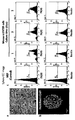
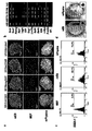
- Figure 2a-b is a result showing the cell membrane damage and recovery by ultrasonic stimulation
- Figure 2a is a cell surface image taken by SEM
- Figure 2b is a live / dead kit staining image.
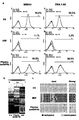
- 3A-C show the results of RT-PCR analysis (c) for intracellular Ca influx assay (a), ATP response assay (b) and ATP receptor gene expression.
- Figure 4a-c is a result showing the influx of foreign substances in the cell by ultrasonic stimulation using QD605
- Figure 4a is a result of QD605 influx analysis in the ultrasound-exposed medium and cells (usMC-S) spheroid and single cells
- 4b and 4c show the QD605 influx analysis and transcription factor expression results in usMC-S spheroid (b) and single cell (c) for each environmental condition.
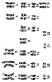
- Figure 5 shows the results of RT-PCR analysis of RNA of exosomes in the sonicated culture.
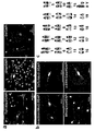
- Figure 6a-c is a result showing the mass transfer by exosomes
- Figure 6a is an image of the process of the QD605 is moved to another cell by the exosomes
- Figure 6b shows the expression of protein markers in the exosomes
- 6C shows the results of poly (A) 27 -Cy5.5 delivery experiment by exosomes.
- Figure 7a-b shows the change of cells according to the co-culture of exosomes and non-sonicated cells in the culture of sonicated cells
- Figure 7a is a change in cell morphology with incubation time
- Figure 7b is an exosome
- the results show the expression of Oct4 in cells cultured for 6 days.
- FIG. 8 illustrates a direct differentiation method of human dermal fibroblasts according to the present invention.
- Figure 9a-b shows the results of differentiation of the human dermal fibroblasts sonicated into adipocytes in the adipocyte differentiation medium
- Fig. 9a is a result of the change of cells and oil red O staining of adipocytes 20 days after induction of differentiation
- 9B shows the results of RT-PCR analysis of adipocyte marker gene expression of differentiation-induced cells.
- FIG. 10a-c shows the results of differentiation of neural stem cell differentiation-inducing medium and ultrasound-treated human dermal fibroblasts into neural progenitor cells
- FIG. 10a shows changes in cells at 3 days after induction of differentiation
- FIG. Cell Marker Expression shows the results of analysis of neural progenitor cell marker expression in attached cells.
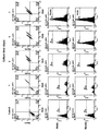
- FIG. 11 is a flow cytometry analysis result of analyzing the expression of Pax6 and Nestin in cells differentiated into neural progenitor cells (n / ENTER cells).
- FIG. 13A shows the process of cell proliferation in a single sphere of n / ENTER cells
- FIG. 13B shows the result of maintaining the properties of neuroprogenitor cells in the proliferated cells.
- FIG. 14A-B show differentiation after transplanting n / ENTER cells into the rat brain
- FIG. 14A shows expression of astrocytic markers (Gfap) in transplanted cells (HNA stained cells)
- FIG. 14B shows transplantation Post secretion of synapsin (Syn1) in differentiated cells.
- FIG. 15A-C show the differentiation of MEFs into neural progenitor cells by neural stem cell differentiation induction medium and ultrasound stimulation.
- FIG. 15A is a change in cell morphology
- FIG. 15B is an expression of neurosphere progenitor markers of spear
- FIG. 15C is Expression analysis results of neural progenitor cell markers using flow cytometry are shown.
- FIG. 16 shows the results of differentiation marker expression analysis of differentiation-induced neuron progenitor cells (mouse n / ENTER cells) in MEF.
- 17 is a schematic showing direct differentiation into hepatocytes.
- Figures 18a-b is the result of differentiation of hepatocyte differentiation medium and sonicated HDF into hepatocytes
- Figure 18a shows the change in cell morphology
- Figure 18b shows the expression of hepatocyte markers.
- 19A-C shows the differentiation of hepatocyte differentiation-inducing medium and heLa cells treated with ultrasound into HeLa cells (HeLa h / ENTER), FIG. 19A shows the change in cell morphology, FIG. 19B shows the expression of hepatocyte markers using qPCR, and FIG. 19C shows Expression results of hepatocyte markers are shown using immunocytochemistry.
- Figures 20a-b shows the differentiation of hepatocyte differentiation induction medium and sonicated Hep3B cells into hepatocytes (Hep3B h / ENTER cells), Figure 20a shows the change in cell morphology, Figure 20b shows the expression of hepatocyte markers.
- Figure 21a-b is a result of differentiation of human ES culture medium and ultrasonic treated HDF into es / ENTER cells, Figure 21a shows the change in cell morphology, Figure 21b shows the change of Oct4 expression with incubation time.
- 22A-B show expression of pluripotent marker genes (a) and protein (b) in es / ENTER cells.
- Figure 23a-c shows the results of pluripotency analysis in es / ENTER cells
- Figure 23a shows a pluripotency marker analysis using flow cytometry
- Figure 23b shows a pluripotency gene expression pattern analysis using a microarray
- Figure 23c Is the result of methylation of Oct4 and Nanog promoter using bisulfite sequencing.
- Figures 24A-B show expression of trioderm marker genes (a) and proteins (b) in es / ENTER cells.
- Figures 26a-b show the results of the expression of triploid marker protein in adherent cultured es / ENTER cells (a) and the methylation analysis (b) of triploid marker DNA of es / ENTER cells using bisulfite sequencing.
- Figure 27a-c shows the results of in vitro differentiation of es / ENTER cells into neurons (a), cardiomyocytes (b) and hepatocytes (c).
- Figure 28a-c shows the results of analyzing the expression of neurons (a), cardiomyocytes (b) and hepatocytes (c) markers in HDF.
- 29a-c show the results of RT-PCR analysis on the expression of neuronal (a), cardiomyocyte (b) and hepatocyte (c) differentiation marker genes of differentiation-induced es / ENTER cells.
- FIG. 31A-B show the results of muscle transplantation and in vivo differentiation of es / ENTER cells
- FIG. 31A shows the result of differentiation of the transplanted cells into skeletal muscle
- FIG. 31B shows the presence or absence of Oct4 expression and proliferation (Ki67) of the transplanted cells. Shows.
- FIG. 32A-C show the results of transplantation and in vivo differentiation of es / ENTER cells into the rat brain
- FIG. 32A shows the coordinates of transplantation of cells
- FIG. 32B shows the astrocytic differentiation markers (Gfap) and neuronal functional markers of transplanted cells.
- Gfap astrocytic differentiation markers
- FIG. 32C shows Oct4 expression and proliferation (Ki67) in transplanted cells.
- FIG. 33a-c shows the differentiation of MEF into mouse es / ENTER cells by human ES culture medium and ultrasonic stimulation
- FIG. 33a shows the change in cell morphology and Oct4-GFP expression according to the culture time
- FIG. 33b shows the culture.
- FIG. 33C shows the change in Oct4 expression.
- Figure 34a-d is a result of pluripotent property analysis in mouse es / ENTER cells
- Figure 34a is the expression of pluripotent marker protein by immunocytochemistry
- Figure 34b is the expression of pluripotent marker gene by RT-PCR
- Figure 34c shows the expression ratio analysis of the pluripotent marker gene by flow cytometry
- Figure 34d shows the alkaline phosphatase (AP) staining results of mouse es / ENTER cells.
- AP alkaline phosphatase
- Figure 35a-c is a result of analyzing the three germ cells characteristics in mouse es / ENTER cells
- Figure 35a is the expression of the three germ cells markers by immunocytochemistry
- Figures 35b and 35c is the three germ marker genes by culture time (b) And analysis results of expression patterns of protein (c).
- 36A-C show in vitro differentiation of mouse es / ENTER cells into neurons (a) and cardiomyocytes (b), and karyotyping results (c) by chromosomal G-band analysis of mouse es / ENTER cells. will be.
- FIG. 37a-c shows the differentiation of L132 cells into L132 es / ENTER cells by human ES culture medium and ultrasonic stimulation
- FIG. 37a shows cell morphology change according to culture time
- FIGS. 37b and 37c show L132 es / ENTER.
- the pluripotent (b) and trioderm (c) properties of the cells are shown.
- FIG. 38A-C shows the differentiation of MSCs into MSC es / ENTER cells by human ES culture medium and ultrasonic stimulation.
- FIG. 38A shows cell morphology changes with culture time
- FIGS. 38B and 38C show MSC es / ENTER cells. Shows pluripotency (b) and trioderm (c) properties.
- Figure 39a-c shows the differentiation of human skin fibroblasts into SF es / ENTER cells by the human ES culture medium and ultrasonic stimulation
- Figure 39a is a change in cell morphology with culture time
- Figures 39b and 39c is SF es / ENTER cells show pluripotent (b) and trioderm (c) properties.
- FIGS. 40B and 40C show pluripotency of es / ENTER cells.
- FIGS. 40B and 40C show pluripotency of es / ENTER cells.
- c triplet
- FIG. 41A-C show the differentiation of HDF into es / ENTER cells using laser stimulation and hES medium.
- FIG. 41A shows differentiation induced HDF spheroids.
- FIGS. 41B and 41C show pluripotency of es / ENTER cells.
- FIG. 44 shows changes in cell morphology of cultured human fibroblasts treated with EVs during es / ENTER induction.
- 46a-b shows the results of confirming the expression of pluripotent marker protein (a: ICC image) and gene (b: qPCR analysis) in 3 days cultured HDF treated with EVs recovered upon es / ENTER induction.
- 47a-b is a result confirming the expression of neural stem cell marker protein (a: ICC image) and gene (b: qPCR analysis) in 3 days cultured HDF treated EVs recovered when n / ENTER induction.
- the present invention provides a physical stimulus capable of promoting environmental influx in a mixture of differentiated or undifferentiated cells and a culture medium, and culturing the mixture provided with the physical stimulus for a period of time to obtain reprogrammed cells. It relates to a method of reprogramming cells.
- the present invention provides pluripotency by culturing differentiated or undifferentiated cells in any medium capable of inducing the reprogrammed cells of interest while providing physical stimulation to promote the influx of environment such as ultrasound, laser or heat treatment to differentiated or undifferentiated cells.
- (pluripotency) cells or any differentiated cells that differ in phenotype from the differentiated or undifferentiated cells, such as hepatocytes, osteoblasts, adipocytes, muscle cells, neurons, astrocytes, keratinocytes, hair follicle cells, pancreatic beta cells or cardiomyocytes. It is characterized in that the reprogramming of.
- the differentiated cells when reprogrammed cells are intended for pluripotent cells, the differentiated cells may be reprogrammed into pluripotent cells by mixing differentiated cells with stem cell culture medium and providing a physical stimulus to culture for a period of time. .
- the differentiated cells and the differentiated induction medium of the desired differentiated cells are mixed and cultured for a period of time by providing a physical stimulus.
- differentiated cells can be reprogrammed to any differentiated cell with a different phenotype.
- the differentiation of undifferentiated cells such as induced pluripotent stem cells or embryonic stem cells with the differentiation-inducing medium of the desired differentiation cells, and provides a physical stimulus to incubate for a certain period of time to differentiate the desired differentiation compared to the existing technology Can be reprogrammed into cells.
- the reprogramming method of cells of the present invention appears to induce reprogramming of differentiated or undifferentiated cells in response to extracellular environmental influx through physical stimulation to differentiated or undifferentiated cells.
- influx may include extracellular vesicles containing genetic material, chemicals, small molecules, exosomes, or exosomes, released from differentiated cells that have been provided with physical stimuli; Or influx into neighboring differentiated or undifferentiated cells such as culture medium components.
- environmental influx into differentiated or undifferentiated cells is characterized by recombination into pluripotent cells stably expressing pluripotent markers and trioderm markers and to differentiated cells having different phenotypes from the differentiated or undifferentiated cells. It seems that programming direction can be determined.
- the reprogramming directionality seems to be determined by the type of culture medium.
- reprogramming from differentiated or undifferentiated cells to pluripotent cells can be induced when physical stimulation is provided to the mixture of differentiated cells and stem cell culture medium, and any Reprogramming to differentiated cells can be induced when a physical stimulus is provided to a mixture of differentiated cells and any differentiated cells, and a physical stimulus to a mixture of undifferentiated cells and differentiation induced media of any differentiated cells. If provided, it can be reprogrammed to any differentiated cell.
- the inventors considered cell membrane damage and cell secreting substances (exosomes or exosome-containing extracellular vesicles) in particular by physical stimulation. That is, ultrasonic waves, lasers, or heat treatments induce temperature rise due to energy, vibration of microbubbles generated by ultrasonic waves, and induction of flow of liquids, that is, microstream generation along the cell membranes.
- the Ca 2 + concentration rapidly increases immediately after sonication and gradually decreases so as not to process the ultrasonic wave. By decreasing to the level of the control group it can be seen that after the damage of the cell membrane is induced.
- the generation and increase of ATP due to ultrasound is known to induce endocytosis in response to various cellular stresses and in response to intracellular membrane ATP receptors.
- exosomes or exosome-containing extracellular vesicles are known to contain genetic information materials (DNA, mRNA, microRNA, protein) inside, exosomes or exosome-containing cells outside the cell membrane through cell membrane damage
- Genetic information material present inside the exosomes or exosome-containing extracellular vesicles can be delivered by the endoplasmic reticulum reenters other surrounding cells.
- stimulation by sonication induces or promotes the expression of pluripotent markers, trioderm markers, or differentiated cell markers that have been under-expressed or remained suppressed in the cell, and damage the cell membrane.
- exosomes or exosome-containing extracellular vesicles present inside the cell including pluripotent markers, trioderm markers, or differentiated cell markers, whose expression is induced or promoted, are discharged to the outside and delivered to surrounding cells.
- Peripheral cells also have a partially damaged cell membrane, which increases cell membrane fluidity, resulting in higher efficiency of exosomes or exosome-containing extracellular vesicles inside the cell than in normal conditions.
- the expression induced or promoted pluripotency, developmental and differentiation tracts present inside the outer endoplasmic reticulum The genetic information is transferred pluripotent cells, or any of differentiated cells was thought to be made.
- the culture medium is recovered during the pluripotent cell induction process, and the exosome or exosome-containing extracellular vesicles are extracted from the medium to determine whether there are pluripotent cell-related pluripotency markers or differentiation markers therein.
- pluripotency markers known pluripotency markers, differentiation markers were identified with a high degree of expression, supporting the hypothesis of the present inventors.
- ultrasonic, laser, or heat treatments were found to be normal without karyotyping.
- somatic cells including dermal fibroblasts, dermal fibroblasts and the like derived from mammals; Cancer cells including uterine cancer cells (HeLa), liver cancer cells (Hep3B), and the like; Or organ tissue cells, including lung epithelial cells (L132 cell) can be used.
- the term "somatic cell” refers to a cell that constitutes an adult and is limited in the capacity for differentiation and self-production.
- the somatic cells may be somatic cells constituting the skin, hair, fat of the mammal, preferably fibroblasts derived from mammals, but is not limited thereto.
- undifferentiated cell means a cell having differentiation capacity and self-producing capacity.
- induced pluripotent stem cells embryonic stem cells, progenitor cells, etc. may be mentioned.
- pluripotency refers to pluripotency after physical stimulation, in a strict sense ultrasound, laser, magnetic field, plasma, light-emitting diode, electrical stimulation, chemical exposure, heat treatment, or acid treatment. Refers to the obtained cell.
- the pluripotency means a state of stably expressing a pluripotency marker expressed in stem cells. In addition, it means a state expressing three kinds of endoderm, ectoderm and mesoderm three germ layers markers.
- the pluripotent cells are "E mbryonic s tem cell-based media En vironmental ransition t-guided c e llular r eprogramming (es / ENTER) cells can be used.
- the pluripotent cells according to the present invention are well differentiated from the induced pluripotent stem cells in that differentiation is well induced according to the external environment, and that the properties of the progenitor cells, which have strong differentiation properties compared to those of the stem cells, are stronger. Has characteristics. In other words, when embryonic stem cells such as induced pluripotent stem cells are used as cell therapeutics, a preparatory step of undergoing a differentiation process is required, which includes a risk factor that can turn into cancer, and a virus for introducing a differentiation inducer.
- pluripotent cells of the present invention are induced without introducing a differentiation-inducing substance such as a back-differentiation inducer or a chemical for genetic variation, so co-culture with other types of cells
- a differentiation-inducing substance such as a back-differentiation inducer or a chemical for genetic variation
- the pluripotent cells of the present invention have the advantage that the induction process is simple and short, so that time can be dramatically reduced until transplantation by treating autologous cells.
- the pluripotent cells are pluripotent markers or mesoderm of any one of OCT3 / 4, SOX2, NANOG, c-MYC, KLF4, TDGF1, SSEA4, TRA-1-60, PAX6, Nestin, Brachyury, SMA, GATA4, or AFP. And it is characterized by stably expressing the three germ seed marker gene consisting of endoderm.
- differentiated cells may be reprogrammed into pluripotent cells or any differentiated cells of interest that differ in phenotype from differentiated cells upon receiving a physical stimulus capable of promoting environmental influx.
- undifferentiated cells can be reprogrammed to any differentiated cell with a markedly superior differentiation rate upon receiving a physical stimulus that can promote environmental influx.
- the differentiated cells are, for example, neurons expressing any one of PAX6, SOX1, SOX2, Nestin, MAP2, TuJ1, GFAP or O4 (called “ n euronal stem cell media-based ENTER, n / ENTER”); Myocytes expressing any of Desmin, Pax3, Actinin, SMA, GATA4 or NKX2-5 (called “ m uscle differentiation media-based ENTER, m / ENTER”); Hepatocytes expressing any of AFP, HNF1a, HNF4a, CK18 or ALB (called “ h epatocyte differentiation media-based ENTER, h / ENTER”); Or adipocytes expressing any one of Pparc2, C / ebpa, aP2 or Fabp4 (referred to as “ a dipocyte differentiation media-based ENTER, a / ENTER”), but are not limited thereto.
- culture medium is a medium used for in vitro cell culture in a comprehensive sense, and means a stem cell culture medium or differentiation induction medium in the present invention, the stem cell culture medium more specifically embryonic stem cell culture Mean badge.
- differentiation induction medium is a medium used for induction of normal stem cells into differentiated cells, for example, multipotent cell differentiation induction medium, hepatocyte differentiation induction medium, bone formation differentiation induction medium, fat Cell differentiation induction media, muscle cell differentiation induction media, astrocyte differentiation induction media, neuronal cell differentiation induction media, vascular endothelial cell differentiation media, keratinocyte differentiation media, pancreatic beta cell differentiation media or cardiomyocyte differentiation media Can be used, but is not limited thereto.
- the culture medium and the differentiated or undifferentiated cells are mixed and the mixture is provided with a physical stimulus.
- physical stimulation Prior to providing a physical stimulus to a mixture comprising differentiated or undifferentiated cells, physical stimulation may be provided to the culture medium to enhance the reprogramming efficiency of the cells.
- the physical stimulus may be any one of ultrasonic wave, laser, plasma, light-emitting diode, electrical stimulation, chemical exposure, heat treatment or acid treatment.
- Ultrasonic treatment of the culture medium has an output intensity of 1W / cm 2 1 to 20 minutes to the ultrasound of 20W / cm 2, specifically, the output strength of 2W / cm 2 To 10 W / cm 2 of ultrasound for 5 to 15 minutes, more specifically, output intensity 3 W / cm 2 To 7 W / cm 2 may be performed for 7 to 13 minutes.
- Laser treatment of the culture medium is performed for 1 minute to 20 minutes for the pulsed laser beam in the 300 to 900 nm wavelength band, more specifically for 3 minutes to 10 minutes for the pulsed laser beam in the wavelength band, more specifically for the wavelength.
- the pulsed laser beam of the band may be irradiated for 4 to 6 minutes.
- the wavelength band may use a wavelength of 400 nm, 808 nm, and 880 nm.
- Heat treatment of the culture medium may be carried out for 5 to 20 minutes at a temperature condition of 40 to 50 °C.
- the sonication of the culture medium and the mixture of differentiated or undifferentiated cells is performed for 1 to 5 seconds at a power intensity of 0.5 W / cm 2 to 3 W / cm 2 , more specifically, at a power intensity of 0.7 W / cm 2 to 2 W / cm. 2 to 1 to 5 seconds, more specifically, the output intensity may be performed for 1 to 5 seconds at 0.8W / cm 2 to 1.5W / cm 2 .
- Laser treatment of the culture medium and the mixture of differentiated or undifferentiated cells can be performed from 1 to 20 seconds for a pulsed laser beam in the 300 to 900 nm wavelength band, and more specifically 3 to 10 seconds for a pulsed laser beam in the wavelength band.
- the pulsed laser beam of the wavelength band may be irradiated for 4 seconds to 6 seconds.
- the wavelength band may use a wavelength of 400 nm, 808 nm, and 880 nm.
- the heat treatment of the mixture of the culture medium and the differentiated or undifferentiated cells may be performed by exposing for 1 to 10 minutes at a temperature condition of 40 to 50 ° C. and then exposing for 5 to 10 seconds at a temperature condition of 0 ° C. to 4 ° C. have.
- the mixture provided with the physical stimulus is incubated for a period of time to obtain reprogrammed cells.
- Incubation of the mixture provided with the physical stimulus is carried out during the period of 2 to 10 days in which spheroids stably express pluripotency markers or differentiation markers are formed through a suspended culture or monolayer culture. It may be performed, but is not particularly limited thereto.
- the floating culture exhibits higher spheroid formation efficiency than the adherent culture.
- the floating culture has a larger number and size of spheroids than the attached culture, and shows a constant size distribution.
- the reprogramming starts from this time by increasing or stabilizing the expression of the pluripotency marker or the differentiation marker from about 3 days in the suspension culture of the human skin fibroblasts treated with ultrasound or laser.
- the expression of pluripotency markers increased or stabilized from about 8 days, and reprogramming started from this time.
- pluripotency markers for example, OCT3 / 4, SOX2, NANOG, c-MYC, KLF4, TDGF1, SSEA4, TRA-1-60, it can be confirmed that spheroids have pluripotency.
- Identification of pluripotency markers may be analyzed by RT-PCR or immunocytochemistry, but is not particularly limited thereto.
- the pluripotent cells of the present invention are characterized by expressing high levels of trioderm markers, ie ectoderm (PAX6, Nestin), mesoderm (Brachyury, SMA), endoderm (GATA4, AFP) markers.
- trioderm markers ie ectoderm (PAX6, Nestin), mesoderm (Brachyury, SMA), endoderm (GATA4, AFP) markers.
- spheroids when providing physical stimulation to skin fibroblasts in differentiation-inducing media, may form between about 1 and 20 days after culture.
- Differentiation markers may be one or more of PAX6, SOX1, SOX2, Nestin, MAP2, TuJ1, GFAP or O4 when reprogrammed into neurons.
- reprogrammed into muscle cells it may be one or more of Desmin, Actinin, Pax3, SMA, GATA4 or NKX2-5.
- telomeres When reprogrammed into hepatocytes, it may be one or more of AFP, HNF1a, HNF4a, CK18 or ALB.
- oil red O staining may be any of Pparc2, C / ebpa, aP2 or Fabp4.
- the pluripotent cells of the present invention are characterized by having proliferative capacity by expressing the proliferation marker protein Ki-67.
- the co-culture of the reprogrammed pluripotent cells with feeder cells can increase the proliferation of pluripotent cells.
- the reprogramming method of cells of the present invention may further comprise the step of culturing the pluripotent cells in differentiation induction medium.
- Pluripotent cells may be differentiated into desired differentiated cells according to the type of differentiation-inducing medium.
- differentiation induction medium multipotent cell differentiation induction medium, hepatocyte differentiation induction medium, osteoblast differentiation induction medium, adipocyte differentiation induction medium, muscle cell differentiation induction medium, astrocytic differentiation induction medium, neuronal cell differentiation induction medium , Vascular endothelial cell differentiation induction medium, keratinocyte differentiation induction medium, pancreatic beta cell differentiation induction medium or cardiomyocyte differentiation induction medium and the like can be used, but is not particularly limited thereto.
- the invention also provides a physical stimulus capable of promoting environmental influx in a mixture of differentiated or undifferentiated cells and culture medium,
- Exosome-containing extracellular vesicles isolated from the above cultures and to differentiate or undifferentiated cells and to culture for a period of time to obtain a reprogrammed cell, comprising a method for reprogramming the cells.
- Reprogramming method of the cells of the present invention is characterized in that the exosome-containing extracellular vesicles isolated from differentiated or undifferentiated cells subjected to physical stimulation can be reprogrammed to any differentiated cells by incubating with differentiated or undifferentiated cells for a predetermined time. do.
- the exosome-containing extracellular vesicles provide a physical stimulus capable of promoting environmental influx into a mixture of differentiated or undifferentiated cells and culture medium, and the mixture subjected to the physical stimulus is cultured for 1 to 6 days to centrifugation. Can be recovered.
- the exosome-containing extracellular vesicles are pluripotent of any one of OCT3 / 4, SOX2, NANOG, c-MYC, KLF4, TDGF1, SSEA4, TRA-1-60, PAX6, Nestin, Brachyury, SMA, GATA4, or AFP.
- Marker or triploid marker Neuronal markers of any one of PAX6, Nestin, Sox1, Sox2, MAP2, TuJ1, GFAP or O4; Myocyte markers of any of Desmin, Pax3, Actinin, SMA, GATA4 or NKX2-5; Hepatocyte markers of any of AFP, HNF1a, HNF4a, CK18 or ALB; Or may be oil red O staining, expressing any one of the adipocyte marker of Pparc2, C / ebpa, aP2 or Fabp4.
- the differentiated cells when the exosome-containing extracellular vesicles contain pluripotent markers, the differentiated cells can be reprogrammed into pluripotent cells when cultured with differentiated cells.
- cultured with the differentiated cells can be reprogrammed to any differentiated cells with different phenotypes.
- the exosome-containing extracellular vesicles when they contain differentiation markers, they can be reprogrammed to any differentiated cells when cultured with undifferentiated cells.
- pluripotency markers in extracellular vesicles (EVs) stained with CD63, an exosome marker recovered during es / ENTER induction, was confirmed, and in normal human somatic cells treated with the EVs After 3 days of culture, pluripotent markers Oct4, Sox2 and Nanog were expressed to confirm reprogramming of the cells.
- neuronal stem cells such as Pax6 was detected in extracellular vesicles (EVs) stained with CD63, an exosome marker recovered during n / ENTER induction, and cultured in normal human somatic cells treated with the EVs after 3 days.
- EVs extracellular vesicles
- Sox1, Sox2, Pax6, Nestin expression was confirmed.
- EVs extracellular vesicles
- CD63 an exosome marker recovered from m / ENTER induction
- CD63 an exosome marker recovered from h / ENTER induction
- the physically stimulated differentiated or undifferentiated cells secrete extracellular vesicles containing the reprogramming factor, and treated them to differentiated or undifferentiated cells for 1 to 20 days through suspension culture or adherent culture.
- Culture can be reprogrammed to any pluripotent, or differentiated, cell.
- Cells that can be reprogrammed through the reprogramming method of the cells of the present invention may be pluripotent cells or differentiated cells of the kind described above, and the substrate is omitted to avoid overlapping substrates.
- This example demonstrates the environmental influx into cells by physical stimulation.
- the cells were prepared by adding 10% FBS (Gibco) and 1% penicillin / streptomycin (Gibco) to primary HDF cells purchased from invitrogen. Cultured in DMEM, sonication of the culture solution was performed at 5W / cm 2 for 10 minutes, and cell treatment was carried out for 1 ⁇ 10 6 cells at 1W / cm 2 for 5 seconds, followed by 2 ⁇ in a 35 mm culture dish with the treated solution. 10 5 cells were cultured.
- Untreated HDF cells for SEM image analysis and cells cultured for 2 hours immediately after treatment as described above and in a 37 ° C., 5% CO 2 incubator for 4 hours at 4 ° C. were then fixed with 0.1% tannic acid. 1 hour in solution, 2 hours in 1% osmium tetroxide solution, dehydrated with acetone at different concentrations, dried cells with liquid CO 2, and immobilized on a gold-palladium-coated surface for electron microscopy (1555 VP-FESEM). Carl Zeiss) was observed for the cells.
- live / dead image analysis cells were treated with live / dead viability / cytotoxicity assay kit (Molecular Probes, Eugene, OR) immediately after sonication with untreated HDF and incubated for 2 hours in a 37 ° C, 5% CO 2 incubator. , USA).
- the staining process was performed by adding 2 ⁇ M live cell staining dye and 4 ⁇ M ethidium homodimer-1 (EthD-1, dead cell staining dye) to the cell culture and then in a 30 ° C. in a 5% CO 2 incubator.
- the cell membrane is damaged by ultrasound to form a hole for entering the external environment, such damage was recovered after 2 hours.
- the cells were stained using a Live / dead kit used to analyze cell death in order to confirm the recovery of damaged cells and cells after ultrasonic stimulation.
- usMC u ltra s ound- exposed m edium and c ells
- usMC-S refers to a suspension culture usMC
- intracellular receptors may be used for Expression of the ATP receptor, which was known to open, was analyzed by RT-PCR.
- ATP was measured using Adenosine 5'-triphosphate (ATP) Bioluminescent Assay Kit.
- Cells were treated directly with untreated cells and with ultrasound (1 W / cm 2 , 5 sec) and then exposed to sonicated medium (5 W / cm 2 , 10 min) (usMC-S) in 96-well plates. After dispensing 3 ⁇ 10 4 cells per well, 100 ⁇ l of ATP assay mix and ATP standard were dispensed per well, followed by 3 minute incubation at room temperature, and the luminescence intensity was measured by a Varioskan Flash Fluorescent Microplate Fluorometer (Thermo Fisher Scientific).
- ATP Adenosine 5'-triphosphate
- RT-PCR for ATP receptor expression analysis extracted RNA using RNeasy plus mini kit (Qiagen, Hilden, Germany) and synthesized cDNA with Super Scrip II kit (Invitrogen, Carlsbad CA, USA). PCR was performed by mixing cDNA and primers in a PCR premix (Bioneer, Daejeon, Korea), using a thermal cycler dice PCR machine (TP600, TAKARA, Otsu, Japan). -65 ° C.) was performed at 30 cycles and 72 ° C. for 1 minute under 35 cycles, and 72 ° C. 15 minute conditions.
- intracellular calcium influx increased up to 60 seconds after ultrasound stimulation, and intracellular ATP concentration increased maximally at 60 minutes, and expression of ATP receptor in the cell membrane was also increased at 1 and 4 hours. Appeared. This was confirmed by the increase in the concentration of calcium that the foreign material is introduced into the early cells by the ultrasonic stimulation, ATP is generated by the ultrasound, it can be seen that the ATP receptor reacts to open the cell membrane passages to allow foreign material inflow.
- the external material was defined as QD605 and it was confirmed whether QD605 was introduced into the cell by ultrasound.
- QD605 is a fluorescent nanomaterial that is known to be less penetrable in living cells. Thus, QD605 confirmed the influx of foreign substances into cells by ultrasonic waves using QD605.
- HDF was treated with 100 pmol of QD605 after ultrasonic stimulation as in Example 1, and after 24 hours, the presence of QD605 in single cells and spheroids was confirmed.
- the exosomes in the culture medium discharged from the sonicated cells may contain genetic material, which is one of the substances secreted from the reprogrammed cells. It has the potential to contain genetic material that plays an important role in programming, so that the exosomes in cultured culture medium after sonication are recovered at the time of medium exchange, and the RNA of the exosomes in the culture medium is changed to Amicon Ultra-0.5 kit (Millipore).
- cDNA synthesis was performed using a Super Scrip II kit (Invitrogen, Carlsbad CA, USA). PCR was performed by mixing cDNA and primers in a PCR premix (Bioneer, Daejeon, Korea) and then denatured at 95 ° C for 5 minutes using a thermal cycler dice PCR machine (TP600, TAKARA, Otsu, Japan), 30 seconds at 95 ° C, gradient 30 RT-PCR analysis was performed at 35 cycles of 1 second at 72 ° C. and 15 minutes at 72 ° C. (Table 2).
- usMC refers to the case of ultrasonic treatment for each cell and culture medium
- usMC-A refers to the attachment culture usMC
- PCR primer list of pluripotency marker gene Genetic code Primer sequence (5'-3 ') Annealing Temperature (°C) Forward direction Reverse Oct4 (POU5F1) GACAGGGGGAGGGGAGGAGCTAGG CTTCCCTCCAACCAGTTGCCCCAAAC 60 Sox2 GGGAAATGGGAGGGGTGCAAAAGAGG TTGCGTGAGTGTGGATGGGATTGGTG 63 Nanog CAGCCCCGATTCTTCCACCAGTCCC CGGAAGATTCCCAGTCGGGTTCACC 64 Utf1 CCGTCGCTGAACACCGCCCTGCTG CGCGCTGCCCAGAATGAAGCCCAC 65 Lin28a AGCGCAGATCAAAAGGAGACA CCTCTCGAAAGTAGGTTGGCT 50 Rex1 CAGATCCTAAACAGCTCGCAGAAT GCGTACGCAAATTAAAGTCCAGA 52 Fgf4 CTACAACGCCTACGAGTCCTACA GTTGCACCAGAAAAGTCAGAGTTG 55 Foxd3 AAGCTGGTCGAGCAAACT
- QD605 was added to capture the image of living cells. As shown in FIG. 6A, the QD605 of the cells into which QD605 was introduced was separated with a part of the cytoplasm and moved to another cell as shown in FIG. 6A. I could confirm it.
- those suspected to be part of the cytoplasm that appeared around the cells were stained on CD63 and pluripotent, such as Oct4 and Nanog, in extracellular vesicles (EVs) stained with the exosome marker CD63 upon es / ENTER induction.
- EVs extracellular vesicles
- expression of sex cell markers was confirmed, and expression of neural stem cells such as Pax6 was detected in extracellular vesicles (EVs) stained with CD63, an exosome marker upon n / ENTER induction, and exosome marker upon m / ENTER induction.
- the exosome is released from the cytoplasm, it contains a genetic material and proteins, it is hypothesized that the transfer to the peripheral cells can induce changes in the peripheral cells.
- the exosomes were extracted and stained with exosomes with CD63 (staining to distinguish them from newly-injected exosomes because the exosomes were present in the cells in culture) and then poly (A) in the exosomes.
- poly (A) 27 -Cy5.5 was thought to be delivered by the exosome.
- exosomes stained with CD63 was found in the cells, it was confirmed that cy5.5 is expressed like CD63. This means that the gene (poly-A) injected by the exosomes was delivered.
- exosomes secreted from cells cultured in human ES medium can also alter the properties of surrounding cells or untreated cells.
- Exosome was extracted from the culture medium of 2 days of sonicated cells cultured in ES medium environment and cultured for 6 days by mixing exosome extract in the process of culturing cells not treated with human ES medium and fibroblast culture medium DMEM. It was.
- human fibroblasts were collected into 1 ⁇ 10 6 cells with 1 mL of differentiation-inducing medium as shown in FIG. 8, followed by ultrasound for 5 seconds at 1 W / cm 2 intensity and then 2 ⁇ 10 in a 35 mm culture dish or 6-well plate. After dispensing at 5 / Well, ultrasonic waves were incubated in 2mL differentiation induction medium treated with 10W / cm 2 intensity for 10 minutes.
- spheroids were formed between about 2 days and 6 days after incubation.
- RNA of the cell by extracting the RNA of the cell, the expression of the adipocyte marker genes, Pparc2, C / ebpa, aP2, and Fabp4 using RT-PCR was found to increase the expression after differentiation induction (Fig. 9b).
- neural stem cells neural progenitor cells
- neural precursor cell markers Oct4
- Expression of Sox2, Pax6, Nestin was confirmed by staining by immunocytochemical method.
- Figure 10a is the appearance of differentiation-induced cells
- Figure 10b shows the neural precursor cell markers in the spheroid, when the differentiation-induced expression of Oct4 is reduced, it was confirmed that the expression of Sox2, Pax6 and Nestin is high.
- Figure 10c shows the expression pattern in the attached cells, showing the same expression pattern as above.
- the markers of neural progenitor cells or neural stem cells are Sox2, Pax6, and Nestin.
- Oct4 is a pluripotent marker, and in the case of adult stem cells or progenitor cells, expression of Oct4 is decreased.
- composition of differentiation induction medium ingredient content Neuroprogenitor Cell Differentiation Induction Medium DMEM F12 bFGF 20ng / mL EGF 20ng / mL B27 supplement ( ⁇ 50) 1/50 N2 supplement ( ⁇ 100) 1/100
- FIG. 11 shows Pax6 / Nestin expression patterns in differentiation-induced cells for 7 days after differentiation induction by flow cytometry. As a result, Pax6 and Nestin expressions were expressed more than 50% on day 1 after treatment. At 3 days, the expression of Pax6 and Nestin was the highest.
- FIG. 12 is a result of confirming the expression of ki67 to confirm the proliferation in the cells expressing neuronal progenitor markers (Pax6 / nestin) among the cells 3 days after induction of differentiation, the cells indicated by the white arrow, the cells expressing Nestin Ki67 expression at was confirmed. These results indicate that differentiation-induced cells have proliferative capacity.
- the arrow indicates that the cells stained with Nestin are proliferating.
- FIG. 13 is an experimental result confirming self-renewal of differentiation-induced cells (n / ENTER cells), and FIG. 13A confirms that cells proliferated from one spheroid express Pax6 and nestin in video. By doing so, the neuroprogenitor cell properties were propagated to the proliferated cells.
- neuroprogenitor markers Oct4, Sox2, Pax6, Nestin
- the spheroid and attached cells were fixed in 4% paraformaldehyde for 10 minutes and soaked in PBS containing 0.1% Triton X-100 for 40 minutes.
- Block for 1 hour with PBS containing 5% (v / v) goat serum and Oct4 (1: 200), Sox2 (1: 200), Pax6 (1; 200), Nestin (1: 200, Cell Signaling Technology) and others were stained overnight at 4 °C.
- FIG. 15C shows the expression patterns of Pax6 / Nestin in cells differentiated for three days after induction of differentiation through flow cytometry. At least one day after treatment, expression of Pax6 and Nestin was expressed at 70% or higher. Pax6, Nestin expression was the highest.
- FIG. 16 shows the results of staining the expression of neuroprogenitor markers (Sox2, Pax6, Nestin) by immunocytochemical method in 20 days after induction of differentiation.
- the top and middle photographs show high expression of Sox2, Pax6 and Nestin.
- the bottom photo shows the expression of oligodendrocyte markers, which also showed the same expression pattern.
- HeLa cells and Hep3B cells were collected into 1 ⁇ 10 6 cells with 1 mL of differentiation-inducing medium, and then treated with ultrasonic waves at 1 W / cm 2 intensity for 5 seconds, followed by 2 ⁇ in a 35 mm Laminin coated culture dish. After dispensing at 10 5 , ultrasound was incubated in 2 mL of hepatocyte differentiation medium treated with 10 W / cm 2 intensity for 10 minutes. Differentiation-induced cells were named h / ENTER using hepatocyte differentiation medium.
- Figure 18 induced the differentiation of hepatocyte differentiation medium and ultrasound treated HDF into hepatocytes (h / ENTER). After 20 days of induction of differentiation of HDF, the cell appearance was changed. Hepatocellular markers (AFP, HNF4a, CK18, ALB) were confirmed by immunocytochemistry 20 days after induction of HDF differentiation. Expression was confirmed.
- HeLa h / ENTER differentiation of hepatocyte differentiation induction medium and sonicated HeLa cells into hepatocytes.
- HeLa cells showed changes in appearance after 19 days of induction of differentiation of HeLa cells, and hepatocyte markers (ALB, HNF4a, CYP3A4F, CYP3A7F, and AIAT) through qPCR 20 days after HeLa cell differentiation (HeLa h / ENTER). , SOX7, GATA6) was confirmed.
- hepatocyte markers HNF4a, CK18, and ALB
- Hep3B h / ENTER cells differentiation of hepatocyte differentiation-inducing medium and sonicated Hep3B cells into hepatocytes.
- Hep3B h / ENTER cells The expression of hepatocyte markers (HNF4a, CK18, ALB) was confirmed by immunocytochemistry after 19 days of differentiation of Hep3B cells and 3 weeks after Hep3B cell differentiation.
- Hep3B h / ENTER hepatocyte markers in Hep3B cells
- Figure 21a shows the change in cell morphology according to the culture time
- Figure 21b shows the difference in Oct4 expression according to the culture time
- spheroids were formed on day 1 after induction into differentiation into human ES medium
- pluripotency marker Oct4 Expression increased with incubation time.
- the spheroids formed by incubating for 6 days were recovered and pluripotent marker expression was confirmed through RT-PCR and ICC.
- expression of the pluripotent marker gene (a) and protein (b) was confirmed in es / ENTER cells.
- FIG. 24 shows the expression (a) and protein expression (b) of the three germline marker genes in es / ENTER cells
- Figure 25 shows the change in Oct4 and three germline marker gene expression in es / ENTER cells over time incubation
- FIG. 26 shows the results of triploid marker DNA methylation analysis (b) of es / ENTER cells using bisulfite sequencing and expression of triploid marker proteins in adherent cultured es / ENTER cells.
- PCR primer list of differentiation marker genes Genetic code Primer sequence (5-'3 ') Annealing Temperature (°C) Forward direction Reverse Endoderm Afp AGCAGCTTGGTGGTGGATGA CCTGAGCTTGGCACAGATCC 63 Foxa2 TTCAGGCCCGGCTAACTCTG CCTTGCGTCTCTGCAACACC 58 Gata6 TGTGCGTTCATGGAGAAGATCA TTTGATAAGAGACCTCATGAACCGACT 60 Ectoderm Nestin GAAACAGCCATAGAGGGCAAA TGGTTTTCCAGAGTCTTCAGTGA 50 Pax6 ACCCATTATCCAGATGTGTTTGCCCGAG ATGGTGAAGCTGGGCATAGGCGGCAG 58 Mesoderm, Cardiomyocyte Acta2 (a-SMA) CTATGAGGGCTATGCCTTGCC GCTCAGCAGTAGTAACGAAGGA 50 Mesoderm Brachyury (T) GCCCTCTCCCCTCCACGCACAG CGGCCGTTGCTCACAGACCACA
- FIG. 27 shows the results of in vitro differentiation of es / ENTER cells into neurons (a), cardiomyocytes (b) and hepatocytes (c). Induction of es / ENTER cells into cells differentiated from each of the three germ layers. Induction of differentiation for 4 weeks using, induced differentiation into neurons (ectoderm), cardiomyocytes (mesoderm), hepatocytes (endoderm), and expressed differentiation markers.
- Figure 28 shows the expression of differentiation markers of neurons (a), cardiomyocytes (b) and hepatocytes (c) in HDF, the differentiation marker was not expressed in HDF.
- 29 shows the results of RT-PCR analysis showing the expression of differentiation marker genes of neurons (a), cardiomyocytes (b) and hepatocytes (c) of differentiation-induced es / ENTER cells. Expression of differentiation marker genes was increased. These results demonstrate the multiplicity of es / ENTER.
- es / ENTER cells were transplanted into rat brains to confirm in vivo differentiation.
- 4 weeks after es / ENTER cells were transplanted into the brains of 5 week old SCID mice, the cells transplanted using HNA were identified.
- MEFs mouse fetal fibroblasts
- hES medium which is the same as HDF.
- the MEF used in this experiment was performed with fetal fibroblasts from mice transfected with the Oct4 promoter with OG2-MEF. These cells were used to observe the expression of Oct4 as the cells expressing GFP fluorescence when Oct4 is expressed.
- the cells induced through sonication increased the number and size of spheroids and GFP expression over time.
- pluripotency was analyzed in the mouse es / ENTER cells.
- FIG. 36 shows in vitro differentiation of mouse es / ENTER into neurons (a) and cardiomyocytes (b), and FIG. 36c shows karyotype analysis by chromosome G band analysis of mouse es / ENTER. Karyotyping was performed using GTG banding chromosome analysis (GenDix, Inc. Seoul, Korea).
- neuronal and cardiomyocyte differentiation markers were identified in mouse es / ENTER cells to confirm that they were differentiated.
- L132 lung epithelial cell
- MSC mesenchymal stem cell
- Skin fibroblast patient-derived dermal fibroblast
- FIG. 37 shows the differentiation of L132 cells into L132 es / ENTER cells by human ES culture medium and ultrasonic stimulation.
- FIG. 37A shows the change in cell morphology according to culture time
- FIGS. 37B and 37C show the Pluripotent (b) and trioderm (c) properties are shown.
- FIG. 38 shows the differentiation of MSCs into MSC es / ENTER cells by human ES culture medium and ultrasonic stimulation.
- FIG. 38A shows the change in cell morphology according to the culture time. The viability (b) and triploid (c) properties are shown.
- FIG. 39 shows the differentiation of human skin fibroblasts into SF es / ENTER cells by ultrasonic stimulation with human ES culture medium.
- FIG. 39A shows the change in cell morphology according to culture time
- FIGS. 39B and 39C show SF es / ENTER.
- the pluripotent (b) and trioderm (c) properties of the cells are shown.
- Heat shock and laser were used as physical stimuli to induce differentiation in the same medium environment called human ES culture.
- FIGS. 40A and 40C show the pluripotency (b) and triploid (c) properties of es / ENTER cells.
- FIG. 41A shows differentiation induced HDF spheroids
- Figures 41B and 41C show the pluripotency (b) and triploid (c) properties of es / ENTER cells.
- the culture solution was recovered and washed twice with D-PBS, and then es / ENTER in embryonic stem cell medium or neural stem cell differentiation medium (Gibco), respectively. And 10 ⁇ l / mL (v / v) of concentrated EVs recovered from the culture medium on day 1 of n / ENTER were added and then mixed with the washed HDF and incubated for 3 days.
- the EVs obtained after daily culturing of the cells subjected to physical stimulation as in Example 12 were concentrated and labeled with EVs using Did dye, and the EVs were delivered to normal somatic cells. In the delivered cells, expression of each pluripotent marker Oct4 and neural stem cell marker Pax6 was confirmed.
- Example 12 50 ⁇ l of EVs obtained in Example 12 was diluted with 450 ⁇ l of D-PBS, and 2.5 ⁇ l of Vybrant DiD cell-labelling solution (molecular probe, excitation / emission, 644/667 nm) was added thereto. And stained exosomes for 30 minutes at 37 °C. After staining, centrifuge again at 14000 rpm for 20 minutes with Amicon Ultra centrifugal filter (Millipore) to concentrate Did stained EVs and repeat diluting with D-PBS twice. 3mL HDF broth (DMEM with 5% FBS) (Gibco culture) and then incubated for 24 hours at 37 °C, 5% CO 2 .
- Vybrant DiD cell-labelling solution moleukin-labelling solution
- HDF cultured for 24 hours was fixed for 10 minutes with 4% paraformaldehyde and permeated for 10 minutes with 0.2% triton X100 in PBS buffer. After blocking for 1 hour with 3% BSA in PBS buffer and then stained overnight at 4 °C with rabbit-anti Oct4 (1: 250, abcam) and Pax6 (1: 200, abcam), the primary antibody and then secondary antibody Anti-rabbit conjugated Alexa-488 (1: 1000, Thermo, excitation / emission, 495/519 nm) was stained for 1 hour.
- EVs secreted from cells after physical stimulation included genes and proteins of various pluripotency markers according to the cell medium environment, and these factors could be delivered to adjacent cells by EVs. It was confirmed that there is.
- the results suggest that EVs secreted from physically stimulated cells in various media environments are likely to induce reprogramming of normal somatic cells.
- Example 13 the EVs secreted from the cells stimulated in various media environments may induce reprogramming of normal somatic cells, and thus, to verify this, EVs cultured in human fibroblast culture medium DMEM medium and human embryonic stem cells or iPS cells.
- the experiment was carried out using hESC medium which is a culture medium of.
- the control group was incubated for 3 days in each medium without added EVs, and the treated group was incubated for 3 days by adding 10 ⁇ l / mL (v / v) of EVs to each medium.
- the cultured cells were rabbit-anti-Oct4 (1: 250, abcam) as the primary antibody, anti-rabbit conjugated Alexa-488 (1: 1000, Thermo excitation / emission, 495/519 nm), mounted with a mounting solution containing DAPI, and analyzed by confocal laser microscopy.
- hESC represents human ESC medium
- DMEM represents fibroblast culture medium
- EVs represent EVs recovered upon es / ENTER induction.
- Oct4 expression was not observed in the control group, but Oct4 expression and cells formed spheroids were observed in the treated group.
- the results indicate that cell reprogramming was induced by EVs and not by the culture medium.
- the morphology of the cells was changed according to the incubation time, and spheroid formation was observed on the third day.
- the amount of EVs was added at different amounts and treated in HDF and incubated for 6 days.
- the EVs recovered during es / ENTER induction were used, cells expressing the pluripotent marker Oct4 were analyzed by flow cytometry.
- the EVs recovered during es / ENTER induction were added at 37 ° C. and 5% CO 2 conditions at concentrations of 0, 5, 12.5, 25, 50 and 100 ⁇ l / mL (v / v), respectively. Incubated daily.
- the cultured cells were rabbit-anti-Oct4 (1: 250, abcam) as the primary antibody, anti-rabbit conjugated Alexa-488 (1: 1000, Thermo excitation / emission, 495/519 nm) and analyzed using BD AccuriTM C6 Flow cytometry (BD biosciences).
- Oct4 expression was observed in the largest 84.6% of cells when treated with 12.5 ⁇ l / mL (v / v) of EVs.
- qPCR analysis was performed by using Trizol (Takara) to recover the total RNA in cells cultured for 3 days and synthesized cDNA with the Superscrip 2 kit (Invitrogen). PCR analysis was performed using a real time PCR instrument (ab step one plus, AB) for the pluripotency markers Oct4, Sox2 and Nanog.
- ICC analysis showed the expression of pluripotent markers Oct4, Sox2 and Nanog in human fibroblast nuclei, and qPCR analysis of gene expression by real time PCR showed that Oct4, Sox2 and Nanog genes were treated with EVs. It was overexpressed about 50 times compared to normal fibroblasts.
- Example 18 Neural stem cell marker expression confirmation experiment in 3 days cultured HDF treated with EVs recovered when n / ENTER induction
- Anti-Nestin (1: 250, Thermo Scientific), anti-rabbit conjugated Alexa-488 (1: 1000, Thermo excitation / emission, 495/519 nm) and anti-mouse conjugated Alexa-594 with secondary antibody (1: 1000, Thermo, alexa 488 excitation / emission, 495/519 nm; alexa 594 excitation / emission, 590/617 nm), mounted with a mounting solution containing DAPI, and then imaged using a confocal laser microscope Was analyzed.
- qPCR analysis was performed by using Trizol (Takara) to recover the total RNA in cells cultured for 3 days and synthesized cDNA with the Superscrip 2 kit (Invitrogen). PCR analysis was carried out using a real time PCR device (ab step one plus, AB) for the neural stem cell markers Sox1, Sox2, Pax6 and Nestin.
- ICC analysis showed expression of the neural stem cell markers Sox1, Sox2 and Pax6 in the human fibroblast nucleus and Nestin expression in the cytoplasm.
- Sox1, Sox2, Pax6 and Nestin genes were overexpressed about 200 times compared to normal fibroblasts without EVs treatment.
- the present invention can be used in the field of cell therapy.
Landscapes
- Zoology (AREA)
- Life Sciences & Earth Sciences (AREA)
- Engineering & Computer Science (AREA)
- Bioinformatics & Cheminformatics (AREA)
- Health & Medical Sciences (AREA)
- Organic Chemistry (AREA)
- Genetics & Genomics (AREA)
- Chemical & Material Sciences (AREA)
- Wood Science & Technology (AREA)
- Biomedical Technology (AREA)
- Microbiology (AREA)
- Biochemistry (AREA)
- General Engineering & Computer Science (AREA)
- General Health & Medical Sciences (AREA)
- Biotechnology (AREA)
- Micro-Organisms Or Cultivation Processes Thereof (AREA)
Abstract
Priority Applications (5)
| Application Number | Priority Date | Filing Date | Title |
|---|---|---|---|
| US16/082,926 US11859175B2 (en) | 2016-03-11 | 2016-08-09 | Cell reprogramming method using physical stimulation-mediated environmental influx |
| JP2018567550A JP7219618B2 (ja) | 2016-03-11 | 2016-08-09 | 物理的刺激による環境流入を用いた細胞のリプログラミング方法 |
| CN201680084450.0A CN109089423A (zh) | 2016-03-11 | 2016-08-09 | 利用由物理刺激引起的环境流入的细胞重编程方法 |
| EP16893680.5A EP3428275B1 (fr) | 2016-03-11 | 2016-08-09 | Procédé de reprogrammation cellulaire utilisant l'imposition d'une transition environnementale médiée par une stimulation physique |
| US18/512,026 US20240101998A1 (en) | 2016-03-11 | 2023-11-17 | Cell reprogramming method using imposition of physical stimulation-mediated environmental transition |
Applications Claiming Priority (4)
| Application Number | Priority Date | Filing Date | Title |
|---|---|---|---|
| KR10-2016-0029611 | 2016-03-11 | ||
| KR20160029611 | 2016-03-11 | ||
| KR1020160071852A KR101855967B1 (ko) | 2016-03-11 | 2016-06-09 | 물리적 자극에 의한 환경유입을 이용한 세포 리프로그래밍 방법 |
| KR10-2016-0071852 | 2016-06-09 |
Related Child Applications (2)
| Application Number | Title | Priority Date | Filing Date |
|---|---|---|---|
| US16/082,926 A-371-Of-International US11859175B2 (en) | 2016-03-11 | 2016-08-09 | Cell reprogramming method using physical stimulation-mediated environmental influx |
| US18/512,026 Division US20240101998A1 (en) | 2016-03-11 | 2023-11-17 | Cell reprogramming method using imposition of physical stimulation-mediated environmental transition |
Publications (1)
| Publication Number | Publication Date |
|---|---|
| WO2017155166A1 true WO2017155166A1 (fr) | 2017-09-14 |
Family
ID=59789914
Family Applications (1)
| Application Number | Title | Priority Date | Filing Date |
|---|---|---|---|
| PCT/KR2016/008754 Ceased WO2017155166A1 (fr) | 2016-03-11 | 2016-08-09 | Procédé de reprogrammation cellulaire utilisant l'imposition d'une transition environnementale médiée par une stimulation physique |
Country Status (1)
| Country | Link |
|---|---|
| WO (1) | WO2017155166A1 (fr) |
Cited By (8)
| Publication number | Priority date | Publication date | Assignee | Title |
|---|---|---|---|---|
| US20200102547A1 (en) * | 2018-10-02 | 2020-04-02 | Stemon Inc. | Reprosomes, as exosomes capable of inducing reprogramming of cells and preparation method thereof |
| WO2020243091A1 (fr) * | 2019-05-24 | 2020-12-03 | University Of Central Florida Research Foundation, Inc. | Reprogrammation sans xéno et sans transgène de cellules souches mésenchymateuses en des cellules progénitrices neurales |
| CN113061579A (zh) * | 2019-12-12 | 2021-07-02 | 中国科学院深圳先进技术研究院 | 一种外泌体及其制备方法及应用 |
| JP2022502032A (ja) * | 2018-10-02 | 2022-01-11 | ステムオン インコーポレーテッドStemon Inc. | 誘導されたエキソソームを含む皮膚再生及び創傷治癒用組成物 |
| CN113930392A (zh) * | 2020-08-20 | 2022-01-14 | 北京沃森百欧生物科技研究院有限公司 | 一种间充质干细胞外泌体及其制备方法和应用 |
| US20220073901A1 (en) * | 2018-10-02 | 2022-03-10 | Stemon Inc. | Composition for extending telomere of cell and preparation method therefor |
| CN116716288A (zh) * | 2023-04-28 | 2023-09-08 | 四川大学 | 一种声波振动提高外泌体产量的方法 |
| JP7668012B2 (ja) | 2018-10-02 | 2025-04-24 | ステムオン インコーポレーテッド | エキソソームを含む毛髪再生用組成物の製造方法 |
Citations (3)
| Publication number | Priority date | Publication date | Assignee | Title |
|---|---|---|---|---|
| US7052720B1 (en) * | 1999-06-17 | 2006-05-30 | University Of Wales College Of Medicine | Spheroid preparation |
| KR20140031943A (ko) * | 2011-05-13 | 2014-03-13 | 더 리전트 오브 더 유니버시티 오브 캘리포니아 | 세포의 선택적 트랜스펙션을 위한 광열 기판 |
| KR20150045935A (ko) * | 2012-04-24 | 2015-04-29 | 더 브리검 앤드 우먼즈 하스피털, 인크. | 다능성 세포를 다시 생성하는 방법 |
-
2016
- 2016-08-09 WO PCT/KR2016/008754 patent/WO2017155166A1/fr not_active Ceased
Patent Citations (3)
| Publication number | Priority date | Publication date | Assignee | Title |
|---|---|---|---|---|
| US7052720B1 (en) * | 1999-06-17 | 2006-05-30 | University Of Wales College Of Medicine | Spheroid preparation |
| KR20140031943A (ko) * | 2011-05-13 | 2014-03-13 | 더 리전트 오브 더 유니버시티 오브 캘리포니아 | 세포의 선택적 트랜스펙션을 위한 광열 기판 |
| KR20150045935A (ko) * | 2012-04-24 | 2015-04-29 | 더 브리검 앤드 우먼즈 하스피털, 인크. | 다능성 세포를 다시 생성하는 방법 |
Non-Patent Citations (3)
| Title |
|---|
| LV , YONGGANG ET AL.: "Effects of Low-intensity Pulsed Ultrasound on Cell Viability, Proliferation and Neural Differentiation of Induced Pluripotent Stem cells-derived Neural Crest Stem Cells", BIOTECHNOLOGY LETTERS, vol. 35, no. 12, 28 September 2013 (2013-09-28), pages 2201 - 2212, XP055551630 * |
| See also references of EP3428275A4 * |
| UCHUGONOVA, AISADA ET AL.: "Optical Reprogramming of Human Cells in an Ultrashort Femtosecond Laser Microfluidic Transfection Platform", JOURNAL OF BIOPHOTONICS, vol. 9, no. 9, 5 November 2015 (2015-11-05), pages 942 - 947, XP055261723 * |
Cited By (14)
| Publication number | Priority date | Publication date | Assignee | Title |
|---|---|---|---|---|
| JP7249693B2 (ja) | 2018-10-02 | 2023-03-31 | ステムオン インコーポレーテッド | 誘導されたエキソソームを含む皮膚再生及び創傷治癒用組成物 |
| JP7668012B2 (ja) | 2018-10-02 | 2025-04-24 | ステムオン インコーポレーテッド | エキソソームを含む毛髪再生用組成物の製造方法 |
| US20220073901A1 (en) * | 2018-10-02 | 2022-03-10 | Stemon Inc. | Composition for extending telomere of cell and preparation method therefor |
| US20200102547A1 (en) * | 2018-10-02 | 2020-04-02 | Stemon Inc. | Reprosomes, as exosomes capable of inducing reprogramming of cells and preparation method thereof |
| JP2022502032A (ja) * | 2018-10-02 | 2022-01-11 | ステムオン インコーポレーテッドStemon Inc. | 誘導されたエキソソームを含む皮膚再生及び創傷治癒用組成物 |
| JP2020054329A (ja) * | 2018-10-02 | 2020-04-09 | ステムオン インコーポレーテッドStemon Inc. | 細胞のリプログラミングを誘導するリプロソーム及びその製造方法 |
| US20220031759A1 (en) * | 2018-10-02 | 2022-02-03 | Stemon Inc. | Composition for skin regeneration and wound healing, comprising induced exosomes |
| US20220348867A1 (en) * | 2019-05-24 | 2022-11-03 | University Of Central Florida Research Foundation, Inc. | Xeno-free and transgene-free reprograming of mesenchymal stem cells toward neural progenitor cells |
| WO2020243091A1 (fr) * | 2019-05-24 | 2020-12-03 | University Of Central Florida Research Foundation, Inc. | Reprogrammation sans xéno et sans transgène de cellules souches mésenchymateuses en des cellules progénitrices neurales |
| US12577532B2 (en) | 2019-05-24 | 2026-03-17 | University Of Central Florida Research Foundation, Inc. | Xeno-free and transgene-free reprograming of mesenchymal stem cells toward neural progenitor cells |
| CN113061579A (zh) * | 2019-12-12 | 2021-07-02 | 中国科学院深圳先进技术研究院 | 一种外泌体及其制备方法及应用 |
| CN113930392A (zh) * | 2020-08-20 | 2022-01-14 | 北京沃森百欧生物科技研究院有限公司 | 一种间充质干细胞外泌体及其制备方法和应用 |
| CN116716288A (zh) * | 2023-04-28 | 2023-09-08 | 四川大学 | 一种声波振动提高外泌体产量的方法 |
| CN116716288B (zh) * | 2023-04-28 | 2024-04-26 | 四川大学 | 一种声波振动提高外泌体产量的方法 |
Similar Documents
| Publication | Publication Date | Title |
|---|---|---|
| US20240101998A1 (en) | Cell reprogramming method using imposition of physical stimulation-mediated environmental transition | |
| JP7508118B2 (ja) | in vitroでの内側神経節隆起前駆細胞の作製 | |
| US11560546B2 (en) | Methods for neural conversion of human embryonic stem cells | |
| WO2010147395A2 (fr) | Composition de milieu comprenant un neuropeptide y pour la génération, le maintien, la croissance non différenciée prolongée de cellules souches pluripotentes et procédé de culture de cellules souches pluripotentes l'utilisant | |
| WO2011043592A2 (fr) | Compositions pour induire une différenciation en cellules rétiniennes à partir de cellules progénitrices rétiniennes ou pour induire une prolifération de cellules rétiniennes comprenant des activateurs de la voie de signalisation wnt | |
| WO2016032263A1 (fr) | Procédé pour la différenciation de cellules souches en cellules ganglionnaires rétiniennes | |
| WO2017179767A1 (fr) | Procédé destiné à l'induction de la différenciation de cellules souches adipeuses en cellules souches neurales, en neurones et en neurones à acide gamma-aminobutyrique et procédé pour l'induction de la différenciation de cellules souches humaines qui sécrètent de grandes quantités de facteurs de croissance à partir de cellules souches mésenchymateuses issues de moelle osseuse humaine | |
| EP2486126A2 (fr) | Procédé pour la différenciation en cellules rétiniennes à partir de cellules souches | |
| WO2013180395A1 (fr) | Métabolite pour favoriser la génération, la conservation et la prolifération de cellules souches pluripotentes, et composition et procédé de culture le comprenant | |
| WO2021045374A1 (fr) | Composition de milieu pour la différenciation d'organoïde hépatique proliférant et procédé de préparation d'organoïde hépatique l'utilisant | |
| JP7252076B2 (ja) | エネルギーを利用した多能性細胞誘導装置及び方法 | |
| WO2014163425A1 (fr) | Procédé pour produire une cellule souche neuronale dérivée reprogrammée à partir d'une cellule non neuronale au moyen de hmga2 | |
| WO2015016420A1 (fr) | Méthode de préparation de progéniteur neuronal dopaminergique induit par reprogrammation directe | |
| WO2019083281A2 (fr) | Nouvelle cellule souche musculo-squelettique | |
| WO2017051978A1 (fr) | Procédé de différenciation de cellules souches pluripotentes induites, qui sont préparées à partir de cellules souches adultes dérivées de l'endocarde, en cellules cardiovasculaires, et utilisation associée | |
| WO2018135907A1 (fr) | Précurseur de cellules de schwann et procédé de préparation de cellules de schwann différenciées à partir dudit précurseur | |
| CA3224981A1 (fr) | Organoides structurellement complets | |
| Ladewig et al. | Lineage selection of functional and cryopreservable human embryonic stem cell-derived neurons | |
| Lee et al. | Self-reprogramming of spermatogonial stem cells into pluripotent stem cells without microenvironment of feeder cells | |
| WO2020139041A1 (fr) | Modèle de maladie de la sclérodermie généralisée et son utilisation | |
| WO2016089178A1 (fr) | Dispositif et procédé d'induction de cellules pluripotentes utilisant de l'énergie | |
| WO2013162330A1 (fr) | Population de cellules souches mésenchymateuses chimériques et son procédé de préparation, et procédé de production de parathormones à l'aide de cellules souches dérivées d'amygdales | |
| WO2021145467A1 (fr) | Procédé de différenciation pour fournir une grande quantité de cellules par découpe d'organoïdes 3d préparés à partir de cellules souches pluripotentes humaines | |
| WO2017014513A1 (fr) | Procédé de conservation et système de banque de cellules nt | |
| Solari et al. | Induced pluripotent stem cells’ self-renewal and pluripotency is maintained by a bovine granulosa cell line-conditioned medium |
Legal Events
| Date | Code | Title | Description |
|---|---|---|---|
| ENP | Entry into the national phase |
Ref document number: 2018567550 Country of ref document: JP Kind code of ref document: A |
|
| NENP | Non-entry into the national phase |
Ref country code: DE |
|
| WWE | Wipo information: entry into national phase |
Ref document number: 2016893680 Country of ref document: EP |
|
| ENP | Entry into the national phase |
Ref document number: 2016893680 Country of ref document: EP Effective date: 20181011 |
|
| 121 | Ep: the epo has been informed by wipo that ep was designated in this application |
Ref document number: 16893680 Country of ref document: EP Kind code of ref document: A1 |