WO2017160079A2 - 디케토피페라진을 유효성분으로 포함하는 전신염증질환의 치료용 약학적 조성물 - Google Patents
디케토피페라진을 유효성분으로 포함하는 전신염증질환의 치료용 약학적 조성물 Download PDFInfo
- Publication number
- WO2017160079A2 WO2017160079A2 PCT/KR2017/002796 KR2017002796W WO2017160079A2 WO 2017160079 A2 WO2017160079 A2 WO 2017160079A2 KR 2017002796 W KR2017002796 W KR 2017002796W WO 2017160079 A2 WO2017160079 A2 WO 2017160079A2
- Authority
- WO
- WIPO (PCT)
- Prior art keywords
- diketopiperazine
- systemic inflammatory
- hmgb1
- inflammatory disease
- formula
- Prior art date
- Legal status (The legal status is an assumption and is not a legal conclusion. Google has not performed a legal analysis and makes no representation as to the accuracy of the status listed.)
- Ceased
Links
- 0 CC(C)[C@](C(N1[C@]2CC(C)C*1)=O)NC2O Chemical compound CC(C)[C@](C(N1[C@]2CC(C)C*1)=O)NC2O 0.000 description 1
Classifications
-
- A—HUMAN NECESSITIES
- A61—MEDICAL OR VETERINARY SCIENCE; HYGIENE
- A61K—PREPARATIONS FOR MEDICAL, DENTAL OR TOILETRY PURPOSES
- A61K9/00—Medicinal preparations characterised by special physical form
-
- A—HUMAN NECESSITIES
- A61—MEDICAL OR VETERINARY SCIENCE; HYGIENE
- A61K—PREPARATIONS FOR MEDICAL, DENTAL OR TOILETRY PURPOSES
- A61K31/00—Medicinal preparations containing organic active ingredients
- A61K31/33—Heterocyclic compounds
- A61K31/395—Heterocyclic compounds having nitrogen as a ring hetero atom, e.g. guanethidine or rifamycins
- A61K31/495—Heterocyclic compounds having nitrogen as a ring hetero atom, e.g. guanethidine or rifamycins having six-membered rings with two or more nitrogen atoms as the only ring heteroatoms, e.g. piperazine or tetrazines
- A61K31/4985—Pyrazines or piperazines ortho- or peri-condensed with heterocyclic ring systems
-
- A—HUMAN NECESSITIES
- A61—MEDICAL OR VETERINARY SCIENCE; HYGIENE
- A61K—PREPARATIONS FOR MEDICAL, DENTAL OR TOILETRY PURPOSES
- A61K9/00—Medicinal preparations characterised by special physical form
- A61K9/06—Ointments; Bases therefor; Other semi-solid forms, e.g. creams, sticks, gels
-
- A—HUMAN NECESSITIES
- A61—MEDICAL OR VETERINARY SCIENCE; HYGIENE
- A61K—PREPARATIONS FOR MEDICAL, DENTAL OR TOILETRY PURPOSES
- A61K9/00—Medicinal preparations characterised by special physical form
- A61K9/20—Pills, tablets, discs, rods
Definitions
- composition for the treatment of systemic inflammatory diseases containing diketopiperazine as an active ingredient
- the present invention relates to a pharmaceutical composition for treating systemic inflammatory diseases comprising diketopiperazine as an active ingredient, and more particularly, diketopiperazine (diketopiperazine) represented by any one of Formulas 1 to 3 is effective. It relates to a pharmaceutical composition for the treatment of systemic inflammatory disease comprising a component.
- HMGB1 known as a nuclear transcription factor
- HMGB1 is involved as a late medicator of sepsis (Wang et al., 2009; Yang et al., 2004). Therefore, high levels of HMGB1 in the serum of patients with severe sepsis may increase mortality (Karlsson et al., 2008).
- HMGB1 is a molecular target that provides opportunities for clinical intervention in sepsis as well as other inflammatory diseases.
- HMGBK high mobility group box 1 is a non-histone nuclear protein that is actively produced from activated macrophages, dendritic cells, and natural killer cells, and is additionally secreted from necrotic cells, and has an important structural role. It regulates gene transcription and has intracellular nuclear function, and has extracellular activity.
- HMGB1 is present in humans at high concentrations in the thymus, lymph nodes, testicles, fetal liver, etc. Except for hepatocytes and brain cells In most cells, they are mainly present in the nucleus, but are secreted out of the cell when the cell or tissue is activated by an agent that causes inflammation. In cells it binds to DNA, which determines transcriptional regulation and chromosome structure, and in damaged tissues acts as a risk signal for controlling homeostatic reactions (Gallucci and Matzinger, 2001).
- HMGB1 is a mediator of inflammation and binds with high affinity to RAGE receptor for advanced glycation end products), TLRCtoll like receptor) -2, and TLR-4, and in tumor cells, tumor necrosis factor (TNF), inter leukin (IL) _6 Increases the secretion of NF and increases the phosphorylation of nuclear factor (NF)- ⁇ 'and extracellular regulated kinase (ERK) 1 and 2, inducing inflammation reactions by HMGB1 (Bae, 2012; Hori et al., 1995; Park et al., 2004).
- TNF tumor necrosis factor
- IL inter leukin
- ERK extracellular regulated kinase
- HNGB1 also enhances DNA-binding activity of NF- ⁇ subunits, promoting NF- ⁇ dependent expression of cell adhesion molecules (CAMs) (Agresti et al., 2003), endothelial Induces ⁇ expression of cell adhesion molecules such as VCAM-1, ICA-1 and E-slection in cells (Andersson et al., 2000; Bae, 2012; Lotze and Tracey, 2005; Yang et al., 2006). This increases inflammation through leukocyte recruitment.
- the compounds containing plin can be obtained from fermentation strains, gourd strains and other microorganisms (Cuong et al., 2015; Park and Strobel, 1994; Adamczeski et al., 1995).
- diketopiperazines Although several of the diketopiperazines have been known to have a variety of biological activities, including antibiotic and anti-inflammatory activity, the use of plinol-containing diketopiperazine for HMGB1- or CLP-mediated septic responses in human endothelial cells and mice. Antiseptic effects are not known.
- an object of the present invention is to provide a pharmaceutical composition for treating systemic inflammatory disease, which comprises diketopiperazine represented by any one of the following Chemical Formulas 1 to 3 as an active ingredient.
- Another object of the present invention is to provide a use of diketopiperazine (diketopiperazine) represented by any one of the following formula (1) to the preparation for the treatment of systemic inflammatory disease (systemic inflammatory disease).
- diketopiperazine represented by any one of the following formula (1)
- Another object of the present invention is to administer an effective amount of diketopiperazine (Diketopiperazine) represented by any one of the following formulas (1) to (3) of systemic inflammatory disease (characteristic inflammatory disease) characterized by To provide a method of treatment.
- Diketopiperazine represented by any one of the following formulas (1) to (3) of systemic inflammatory disease (characteristic inflammatory disease) characterized by To provide a method of treatment.
- the present invention is a pharmaceutical composition for the treatment of systemic inflammatory disease (systemic inflammatory disease) comprising a diketopiperazine (diketopiperazine) represented by any one of the following formulas (1) to (3) as an active ingredient To provide.
- systemic inflammatory disease systemic inflammatory disease
- diketopiperazine diketopiperazine
- diketopiperazine represented by any one of the following formula 1 to the formula for the preparation of a therapeutic agent for systemic inflammatory diseases (systemi c inf lammatory di sease) .
- systemic inflammatory diseases systemi c inf la atory di sease
- an effective amount of diketopiperazine, represented by one is administered to a subject in need thereof.
- the present invention provides a pharmaceutical composition for preventing or treating systemic inf lammatory di sease comprising diketopiperazine represented by any one of the following Chemical Formulas 1 to 3 as an active ingredient.
- 'diketopiperazine' is a common name of 2.5-diketopiperazine as a kind of organic compound.
- glycine and plinster esters are liable to diketopiperazine, and in this case it is not easy to obtain free esters.
- Various diketopiperazines are produced when the protein is hydrolyzed in dilute acid, especially at high temperatures, or when heated to around 180 ° C in glycerol.
- Bacillus genus HC001 and Pi ci coccus genus 12L081 bacteria were extracted from precipitates and sponges collected in Jeju Island. After culturing the extracted bacteria, the cultured medium was extracted with an organic solvent and concentrated. The concentrate was fractionated by VLC to obtain diketopiperazine of Chemical Formulas 1 to 3.
- VLC fractionated by VLC to obtain diketopiperazine of Chemical Formulas 1 to 3.
- HUVEC cells which are vascular endothelial cells.
- LPS vascular endothelial cells
- three diketopiperazines inhibited the secretion of GBl mediated by LPS.
- TLR2, TLR4, and RAGE which are HMGB1 receptors, and inhibited cytoskeletal redistribution by HMGB1.
- diketopiperazine inhibited the migration of white blood cells in HMGB1 mediated endothelial cells, inhibited the expression of VCAM-1, ICAM-1, and E-selection, and inhibited the signal transduction pathway of HMGB1.
- diketopiperazine can activate the inflammatory signaling molecules nuclear factor-KB (NF- ⁇ ), extracellular regulated kinases l / 2 (ERKl / 2), and tumor necrosis factor-a (TNF-a) and interleukin-6 ( Production of IL-6) was inhibited.
- CLP cecal ligation and puncture
- 'Systemic inflammatory disease' of the present invention is a systemic inflammatory response accompanied by fever or tachycardia, tachypnea, leukocytosis and the like caused by inflammation. In addition to infection, it can be caused by acute pancreatitis, burns, trauma or surgery.
- AOSD Still's disease
- Behcet's disease disease Crohn's disease
- juvenile le idiopathic arthritis systemic rheumatic disease
- skin rash uveitis or systemic vasculitis.
- HMGBU High mobility group box 1 HMGBU High mobility group box 1
- HMGBU High mobility group box 1 HMGBU High mobility group box 1
- the type is not particularly limited, for example, Systemic inflammatory response syndrome (SIRS), sepsis (sepsis), septic It is characterized in that it is selected from the group consisting of shock, acute lung injury, acute kidney injury, and multiple organ dysfunction syndrome.
- SIRS Systemic Inflammatory Response Syndrome
- SIRS is a systemic inflammatory response to various severe clinical injuries, and at least two of the following conditions are within 24 hours according to the definition of ACCP / SCCM. Can be diagnosed by:
- RR -Respiratory rate
- P C02 arterial carbon dioxide partial pressure
- WBC Leukocyte count
- 'sepsis' is a systemic inflammatory reaction syndrome caused by infection, hypotension, hypoxia, and urinary tract syndrome. Metabolic acidosis, thrombocytopenia, and consciousness disorders may appear. Sepsis is the result of hyper-inflammatory response due to overproduction of proinflammatory cytokines such as tumor necrosis f actor- ⁇ (TNF- ⁇ ) and abnormal immune system activation. These abnormal over-inflammatory reactions lead to tissue damage and organ failure.
- TNF- ⁇ tumor necrosis f actor- ⁇
- inflammation is an adjustable reaction to defend the human body.
- cytokine secretion and leukocyte activation occur adequately to remove the substances that cause inflammation.
- sepsis may occur due to over-inflammatory reactions caused by innate innate immune response to infection, resulting in multiple organ failure due to excessive secretion of inflammatory mediators.
- Acute lung injury, also called acute respiratory di stress syndrome (ARDS) is acute pulmonary edema that begins acutely due to extra-cardiac causes. Cause trouble. It usually occurs at several hours after exposure to the causative agent and is caused by a variety of causes.
- Acute kidney injury Acute kidney injury of the present invention is also called acute renal failure (ARF), it refers to a sharp decrease in kidney function over several hours to days. Nitrogen wastes accumulate in the body as a result of kidney function deterioration, resulting in high nitricemia in the blood and abnormalities in fluid and electrolyte balance.
- Causes include insufficient blood to the kidneys due to a decrease in overall body function (bleeding, vomiting, diarrhea, hypotension, heart failure, etc.), abnormalities of the kidney itself (kidney vascular abnormalities, high blood pressure, acute glomerulonephritis, vasculitis, etc.), Urethral and bladder abnormalities (prostate enlargement, tumor, etc.).
- Symptoms include consciousness, convulsions, high blood pressure, arrhythmia, pulmonary edema, pulmonary bleeding, anorexia, vomiting, intestinal paralysis, gastrointestinal bleeding, and ischemic heart disease.
- MODS Multiple organ dysfunct ion syndrome
- MOF mul t iple organ fai lure
- the major organs of the body namely, the heart, lungs, liver, kidneys, such as dysfunction occurs more than two or simultaneously.
- the mortality rate is very high because life support activities become extremely difficult.
- Mortality rates between two organ failures are 60-70% and three organ failures are 80-90%.
- Infectious disease and en dotoxemia play an important role in increasing the number of failure organs.
- Multiple organ failure is preceded by multiple organ failure due to external stratification such as a traffic accident, which causes the host to fall into immune failure.
- the pharmaceutical composition according to the present invention may contain diketopiperazine alone or may be formulated in a suitable form with a pharmaceutically acceptable carrier and may further contain excipients or diluents.
- 'pharmaceutically acceptable' refers to a nontoxic composition that, when administered physiologically and is human, typically does not cause allergic reactions or similar reactions, such as gastrointestinal disorders, dizziness, and the like.
- Pharmaceutically acceptable carriers may further include, for example, carriers for oral administration or carriers for parenteral administration.
- Carriers for oral administration may include lactose, starch, cellulose derivative magnesium stearate, stearic acid and the like. In addition, it may include various drug delivery materials used for oral administration to the peptide formulation.
- carriers for parenteral administration may include water, suitable oils, saline, aqueous glucose, glycols, and the like, and may further include stabilizers and preservatives. Suitable stabilizers include antioxidants such as sodium hydrogen sulfite, sodium sulfite or ascorbic acid. Suitable preservatives include benzalkonium chloride, methyl- or propyl-paraben and chlorobutane.
- the pharmaceutical composition of the present invention may further include a lubricant, a humectant, a sweetener, a flavoring agent, an emulsifier, a suspension agent, and the like.
- composition of the present invention may be administered to any mammal, including humans. For example, it can be administered orally or parenterally.
- Parenteral administration methods include, but are not limited to, intravenous, intramuscular, intraarterial, intramedullary, intradural, intracardiac, transdermal, subcutaneous, intraperitoneal, intranasal, intestinal, topical, sublingual or rectal administration.
- the pharmaceutical composition of the present invention may be formulated into a preparation for oral or parenteral administration according to the route of administration as described above.
- compositions of the present invention are formulated using powders, granules, tablets, pills, sugar tablets, capsules, liquids, gels, syrups, slurries, suspensions and the like known in the art.
- oral formulations can be obtained in tablets or dragees by combining the active ingredient with a solid brother and then grinding it, adding suitable auxiliaries and processing it into a granular mixture.
- excipients include lactose, Starch, celrose, including sugar and corn starch, wheat starch, rice starch and potato starch, including dextrose, sucrose, solbi, manny, xili, erysri and malt Layer presuppositions such as cellulose, gelatin, polyvinylpyridone, and the like, including methyl salose, sodium carboxymethylcellose, hydroxypropyl methyl-celose and the like.
- crosslinked polyvinylpyridone, agar, alginic acid or sodium alginate may be added as a disintegrant.
- the pharmaceutical composition of the present invention may further include an anticoagulant, a lubricant, a humectant, a perfume, an emulsifier and a preservative.
- Formulations for parenteral administration may be formulated by methods known in the art in the form of injections, creams, lotions, external ointments, oils, humectants, gels, aerosols and nasal inhalants. These formulations are described in Remington's Pharmaceut i Cal Science, 19th ed., Mack Publ i Shing Company, Easton, PA, 1995, a prescription generally known in all pharmaceutical chemistries.
- the total effective amount of the composition of the present invention may be administered to a patient in a single dose, and may be administered by a fract ionated treatment protocol which is administered for a long time in a mul t iple dose. Can be.
- the pharmaceutical composition of the present invention may vary the content of the active ingredient depending on the extent of the disease.
- the preferred total dose of the pharmaceutical composition of the present invention is about O. per kg of patient weight per day. Ol ⁇ g to 10,000 mg, most preferably 0.1 / g to 500 mg.
- the dosage of the pharmaceutical composition is effective for the patient in consideration of various factors such as the age, weight, health condition, sex, severity of the disease, diet and excretion rate, as well as the formulation method, route of administration, and number of treatments. Since the dosage is to be determined, one of ordinary skill in the art, in light of this, will be able to determine the appropriate effective dosage of the composition of the present invention.
- the pharmaceutical composition according to the present invention is not particularly limited to its formulation, route of administration and method of administration as long as the effect of the present invention is shown.
- the present invention provides the use of diketopiperazine represented by any one of the following Chemical Formula 1 to Chemical Formula 3 for the preparation of a therapeutic agent for systemic inflammatory diseases (systemi c inf lammatory di sease).
- the present invention provides a systemic disease comprising administering to a subject in need thereof an effective amount of a composition comprising diketopiperazine represented by any one of the following Chemical Formulas 1 to 3 as an active ingredient: It provides a method of treating atory disease).
- the term 'effective amount' of the present invention when administered to an individual, refers to an amount that improves, treats, prevents, detects, or diagnoses a systemic inflammatory disease.
- the term 'individual' means an animal, preferably a mammal, particularly It may be an animal including a human, and may be cells, tissues, organs, etc. derived from the animal. The subject is a patient in need of the effect
- treatment' of the present invention broadly refers to ameliorating symptoms of systemic or systemic inflammation-related diseases or pre-neuro-inflammatory diseases, which can cure, substantially prevent, or ameliorate such diseases. It may include, but is not limited to, alleviating, healing or preventing one symptom or most of the symptoms resulting from systemic inflammatory diseases or systemic inflammation related diseases.
- the present invention provides a pharmaceutical composition for the prevention or treatment of the vascular disease comprising diketopiperazine as an active ingredient.
- the method of the present invention is effective in the prevention and treatment of systemic inflammatory diseases by inhibiting the secretion of HMGB High Mobity group box 1), regulating proinflammatory signaling, and inhibiting inflammatory responses.
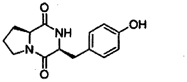
- [Brief Description of Drawings] 1 shows the formulas of three diketopiperazines (A: formula 1 combined with valine, B: formula 2 combined with tyrosine, C: leucine (Leu)
- Topiperazine was injected intravenously, 24 hours after CLP, plasma HMGB1 level was measured by ELISA (B), Confluent HUVEC cells were treated with HMGBKl g / mL) for 16 hours, and 1 diketopiperazine was treated. When treated for 6 hours, the expression level of TLR-2 (white bar), TLR-4 (gray bar), RAGE (black bar) receptor was measured by cell-based ELISA (C), by MTT assay method.
- FIG. 3 shows HUVEC cells with LPS (A, 100 ng / mL, 4 h) or Evans blue-binding across HUVEC monolayers after stimulation with HMGB1 (B, 1 // g / mL, 16 h), followed by treatment of three diketopiperazines at each concentration for 6 hours each.
- HUVEC cells were activated with HMGBKl ⁇ g / mL, 16 h) and treated with different concentrations and diketopiperazine for 6 hours, followed by the three diketopiperazine for phospho-p38 expression mediated by the dried GB1.
- D is shown as a result of measuring the effect by ELISA.
- E stimulated the HUVEC cell monolayers cultured on glass cover slip with HMGBl (l / g / mL) for 1 hour and treated with diketopiperazine for 10 hours for 6 hours each for 6 hours. Fluorescence stained with actin.
- HUVEC cells were stimulated with HMGBIU / g / mL for 16 hours and treated with three diketopiperazines at various concentrations for 6 hours, followed by HMGB1-mediated VCAM-l, ICAM-1 and E-select in (A) shows the degree of expression of human neutrophils attached to the HUVEC monolayer (B) and image (C), the migration of human neutrophils through the HUVEC monolayer (migrat ion) a will (d) shown (p: PBS vehicle control, Comp : diketopiperazine, results of five independent chambers, indicated as mean + SEM of the test, * p ⁇ 0.05 versus HMGBl) .
- HMGB1 labeled H, 3 ⁇ 4 «g per mouse, i.v.
- CLP-operation labeleled C, 24h after CLP surgery
- lysozyme (0.57-5.72 ng per mouse
- i.vj was injected to analyze peritoneal motility of the leukocytes mediated by HMGBl or CLP (E).
- H / C means CLP or HMGB1 treatment
- 'CLP / HMGB1 P PBS vehicle control
- Comp diketopiperazine
- FIG. 5 shows HUVEC cells HMGBUl / g / raL).
- A degree of HMGB1-mediated TNF- ⁇ production was measured
- B analysis of the activation level of HMGB1-mediated NF-KB p65
- D analysis of the activation level of HMGB1-mediated ERK1 / 2
- P PBS vehicle control, Comp:? Diketopiperazine
- Results are indicated as mean SEM of five independent experiments ⁇ , * / ⁇ 0.05 versus HMGBl).
- Bacteria lipopoIysaccharide LPS; serotype: 0111: B4, L5293), Evans blue dye, HPLC sorting solvent, crystal violet dye and antibiotics (penicillin G and streptomycin) were purchased from Sigma (St. Louis, M0).
- Human recombination HMGB1 was purchased from Abnova Taipei City, Taiwan, and fetal bovine serum (FBS) and Vybrant DiD were purchased from Invitrogen Cellsbad, CA.
- Methane (MeOH), ethyl acetate (EtOA), and n-nucleic acid (n-hezne), which are organic solvents for extraction and fractionation, were purchased from Duksan Chemical, Korea.
- HC001 is a genus of Bacillus
- 12L081 is Pi ci coccus genus.
- Bacillus genus HC001 and Pi ci coccus genus 12L081 bacteria were incubated in 35 L of SYP SW medium at 150 rpm and 25 ° C for 7 days, respectively, and then each culture medium was extracted twice with 35 L of EtOA.
- Bacillus genus HC001 and Pi ci coccus genus 12L081 was prepared by concentrating the extract. Extract of HC001 (1.9g) was fractionated by silica gel VLC by step-gradient elution method.
- Diketopiperazine 1 is cycVoO-Pro-d-Val
- diketopiperazine 2 is cc / o (l-Pro-l-Tyr)
- diketopi by comparison of spectroscopic data described in Ferrazine 3 was identified by l-Pro-d-Leu).
- mice Six to seven week old C57BL / 6 male mice weighing 27 g were purchased from Onient Bio Co. CSungnam, KyungKiDo, Republic of Korea) and used for this study after 12 days of acclimatization period. Five animals per polycarbonate cage were raised at 20-25 ° C, 403 ⁇ 4-45% humidity, and 12-hour day / night cycling conditions. During this period, the experimental animals were fed free rodent pellet diet and water (J / MW). All animal experiments were conducted under the approval of the Institutional Animal Care and Use Committee of Kyungpook National University in Korea (IRB approval number, KNU2012-13).
- mice were injected 2% through a small rodent gas anesthesia machine (RC2, Vet equip, Pleasant on, CA). Anesthesia was performed with oxygen containing isof lurane (Forane, JW Pharmaceutical, South Korea), and anesthesia was first anesthetized in a breathing chamber and then a facemask was used to allow for natural hops during the experiment.
- CLP-induced systemic disease models are described in Bae JS et al. , As described in the 2014 study. Briefly, a 2 cm midline incision was performed to expose the cecum and adjacent small intestine.
- HUVECs Primary human umbilical vein endothelial eel Is (HUVECs) were obtained from Cambrex Bioscience (Charles City, IA) and maintained as described in previous studies (Bae et al., 2014; u and Bae, Ku et al., 2015a; Ku et al., 2015b; Ku et al., 2015c; Lee et al., 2015a; Lee et al., 2015b; Yoon et al., 2015). Briefly, cells were incubated in endothelia basal medium (EBM-2) containing growth aid (Cambrex Bio Science), 371: and 5% CO 2 . HUVEC cells are described in Lee W et al. Passages were cultured and used as described in the 2013 study. Human neutrophi 1 was isolated from 15 mL of whole blood obtained by venipuncture from five healthy volunteers and maintained as described in the Hofbauer R et al., 1998 study.
- EBM-2 endothelia bas
- HUVEC monolayer (HUVEC monolayer) in was treated for 16 hours with LPS 100 ng / mL), after that the 0 to 10 ⁇ '3 gaji diketopiperazine was treated for 6 hours. Then, cell culture fluid was collected to check the concentration of HMGB1.
- 96-well flat plastic microtiter plates (Corning Inc., Corning, NY) were coated overnight with HMGB1 protein at 4 ° C, at 20 mM with 0.02% sodium azide.
- the plates are then washed three times with PBS-T solution and at room temperature with peroxidase-conjugated ant i -rabbit IgG ant i body (diluted 1: 2000 in PBS-T, GE Healthcare Life Science, Pittsburgh). Incubated for 90 minutes, washed three times with PBS-T solution, and the substrate solution of (100 ug / mL ⁇ phenylenedi amine and
- HMGB1 (1 «g / mL) was added to a confluent monolayer of HUVEC cells for 16 hours (experiment for VCAM-1 and ICAM—1) or 22 hours (experiment for E-selectin).
- mice ant i -human monoclonal ant i body meaning antibodies to VCAM-1, ICAM-1 and E-selectin, respectively, diluted 1:50 in PBS-T, Temecula, CA
- PBS-T Temecula
- CA Temecula
- HMGB1 receptors such as TLR2, TLR4 and RAGE, wherein Santa Cruz Biotechnology Inc.
- the receptor specific antibodies (A_9, H-80 and A-9, respectively) obtained from (Santa Cruz, CA) were used.
- MTT assay method was used as an indicator of cell survival.
- Cells were cultured at 96 x well plates at a density of 5 x 10 3 cells / well. After 24 hours, cells were washed with fresh medium and treated with three diketopiperazines 1-3. After 48 hours of incubation, the cells were washed and MTTCl mg / mL of
- DMSO dimethyl sul foxide
- Endothelial cell permeabi 1ity is described in Qureshi et al.
- the outflow of Evans blue-bound albumin across a functional cell monolayer was quantified by spectrophotometry (measurement) using a two-compartment chamber model described in, 2008. Briefly, HUVEC cells were dispensed at 5 XloVwell in 3 ⁇ pore size, 12 mm diameter transwells and incubated for 3 days. The resulting HUVEC fusion monolayers (Confluent monolayers of HUVEC) were treated with HMGBlO «g / mL, 16h) and then treated with 3 Taketopiperazines for 6 hours.
- F-actin labeled with fluorescein phalloidin F 432; Molecular Probes, Invitrogen.
- Nuclei were counterstained with 4,6-diamidino— 2-phenylindole dihydrochloride (DAPI) and visualized by confocal microscopy (TCS to Sp5, Leica microsystem, Germany) at 630x magnificat m. Quantification of p65 nuclear transfer as described in Fuseler et al., 2006 was calculated.
- HUVEC cells (6 ⁇ 1 () 4 ) were incubated for 3 days to obtain a fused endothelial monolayer.
- the cell monolayer was treated with ⁇ GBlCU / g / mL) for 16 hours, treated with three diketopiperazines for 6 hours, and human neutrophils were added to the upper compartment of the transwell plate.
- the transwell plate was then incubated for 2 hours at 37 ° C, 5% C0 2 conditions.
- the cells in the upper chamber were then aspirated and the non-mobile cells at the top of the filter were removed with a cotton swab.
- the human neutrophils at the bottom of the filter were fixed with 8% g hit araldehyde. And 20% methanol (w / v) containing 0.25% crystal violet was added. Each experiment was repeated twice in duplicate wells and the number of mobile cells was counted in nine randomly selected high power microscopic fields (HPF; 200 ⁇ ). This result is represented as a migration index.
- mice Male mice were anesthetized with isoflulan (isoflulan) in oxygen delivered via a small rodent gas anesthesia machine and allowed to breathe with a facial mask. Injecting HMGBl ( ⁇ g / mouse) into mice intravenously (iv) Treatment for 16 hours followed by three diketopiperazine treatments for 6 hours.
- isoflulan isoflulan
- 1% Evans blue dye solution in normal saline was intravenously injected into each mouse. After 30 minutes, each mouse was sacrificed and peritoneal exudate was collected by washing the peritoneal cavity with 5 mL of normal saline and centrifuged at 200 ⁇ g for 10 minutes. The absorbance of the supernatant was measured at 650 nm.
- Vascular permeability is determined by Bae JS et al. , As described in the 2012 study, as the amount of dye leaked into the peritoneal cavity according to the standard curve of Evans blue dye (! Per mouse).
- mice and mice were treated with HMGBl (g / mouse, in normal saline) intravenously (i. V.) For 16 hours and then treated with three diketopiperazines, respectively. After 6 hours, the mice were sacrificed and the peritoneal cavity was washed with 5 mL of normal saline. The peritoneal fluid was mixed with 0.38 mL Turk's solution (0.01% crystal violet in 3% acetic acid), and the number of white blood cells was counted using an optical microscope.
- HMGBl g / mouse, in normal saline
- three diketopiperazines respectively.
- mice were sacrificed and the peritoneal cavity was washed with 5 mL of normal saline.
- the peritoneal fluid was mixed with 0.38 mL Turk's solution (0.01% crystal violet in 3% acetic acid), and the number of white blood cells was counted using an optical microscope.
- neutrophils 1.5 loVmL, 200 ⁇ / wel 1
- the neutrophil cells were added to HUVEC cells labeled with Vybrant DiD dy and washed and stimulated.
- the stimulated HUVEC cells were treated with HUVEC cell monolayers for 16 hours with HMGBK1; wg / raL and again with 3 diketopiperazines (0-10 ⁇ ) for 6 hours, respectively.
- mice Five male C57BL / 6 mice were subjected to CLP and injected 12 diketopiperazines intravenously after 12 and 50 hours, respectively. Mice were lacrimated 96 hours after CLP. Lung samples were extracted from each mouse to analyze the phenotypic changes of lung tissue in the mice, and washed 3 times with PBS (pH 7.4) to remove residual blood, followed by 4% formaldehyde solution (in PBSCpH 7.4), Junsei, Tokyo, Japan) was fixed at 4 ° C for 20 hours. After the fixation, each sample was dehydrated with a series of increasing concentrations of aqueous ethanol (ethanol series), and after paraffin embedding, 4 ⁇ two Sectioned and placed on a slide.
- PBS pH 7.4
- formaldehyde solution in PBSCpH 7.4
- the slides were deparaffinized in a 60 ° C. oven, rehydrated and stained with hematoxylin (Sigma). To remove excess staining, each slide was quick-dipped 3 times in 0.3% acid alcoh and counterstained with eosin (Sigma). Thereafter, prior to mounting the slides with coverslips, excess staining was removed using a series of increasing concentrations of aqueous ethanol series and xylene. Optical microscopy of lung specimens to assess pulmonary architecture, tissue edema and infiltration of inflammatory cells was performed by blind observers as described above. (Ozdulger et al., 2003).
- Grade 1 represents normal histopathology
- Grade 2 represents a small amount of neutrophil leukocyte infiltration
- Grade 3 is moderate neutrophil leukocyte infiltration
- grade 4 means including dense neutrophil leukocyte infiltration, abscess formation and complete lung tissue structure destruction.
- HMGB1 has been shown to be secreted in human endothelial and marine macrophages by LPS (Bae and Rezaie, 2011; El Gazzar, 2007; Mull ins et al., 2004). Consistent with the results of Chen et al., 2004, LPS 100 ng / mL) stimulated HMGB1 secretion from HUVEC cells.
- LPS 100 ng / mL
- endothelial cells were stimulated by treatment with lOOng / mL LPS for 16 hours and the three diketopiperazines 6 of FIG. 1 at 0-10 ⁇ concentrations 6 Treated for hours.
- MTT assay was performed on HUVEC cells treated with diketopiperazine for 24 hours. As a result, it could be confirmed that diketopiperazine did not affect cell survival (see FIG. 2D).
- HMGB1 The high plasma concentrations of HMGB1 in patients with inflammatory diseases are known to be associated with poor prognosis and high mortality. Moreover, pharmaceutical inhibition of HMGB1 is known to increase survival in acutely inflammatory animal models as a response to endotoxin (Sama et al., 2004). Therefore, inhibition of CLP-induced HMGB1 secretion by diketopiperazine indicates that it can be used to treat vascular inflammatory diseases.
- Permeability assays were performed to investigate the effect of diketopy perrazine on barrier integrity of HUVEC cells (barrier integrity). Treatment with three diketopiperazines ( ⁇ ) alone did not alter the barrier integri ty (see Figure 3A). In contrast, LPS is known to induce the division and disruption of endothelial membrane barriers (Lee et al., 2014; Wo I f son et al., 2011; Yang et al., 2005). HUVEC cells After treatment with LPS (100 ng / mL) or HMGB1 (1 ⁇ g / mL), three diketopi perazines of varying concentrations were treated for 6 hours. As a result, as shown in FIG.
- HMGB1 is also known to induce division and disruption of barrier integrity.
- Three diketopiperazines resulted in a concentration-dependent decrease in GB1-mediated cell membrane disruption (see FIG. 3B).
- GB1 is known to induce proinflame atory reactions by promoting phosphorylation of p38 MAPK (Qin et al., 2009; Sun et al., 2009).
- diketopiperazine inhibits the activation of p38 MAPK induced by HMGB1 in HUVEC cells.
- the cells were treated with diketopiperazine for 6 hours after activation with HMGB1, followed by phosphorylated p38 MAPK. The level of was calculated.
- HMGB1 increased the expression of phosphorylated p38, which was shown to be significantly inhibited when treated with diketopiperazine (see FIG. 3D).
- Cytoskeletal proteins are important for maintaining the integrity and appearance of cells (Schnit tier et al., 2001).
- redistribution of actin cytoskeleton, cell detachment and loss of cell-cell contact due to cytokine stimulation may lead to endothelial monolayer permeability. Is related to the increase. Therefore, the effects of three diketopiperazines on act in cytoskeletal arrangement in HUVEC cells by immunofluorescence staining with HUVEC monolayers with F-act in—fluorescein phalloidin It was.
- the HUVEC cells in the control group showed a random distribution of F-actin throughout the cell, with several actin filament bundles located at the cell boundary.
- HMGB1-mediated cel l-adhesion molecules CAMs
- HMGB1 mediates the inflammatory response by increasing the cell surface expression of CAMs such as ICAM-1, VCAM-1 and E-select in on the surface of endothelial cells, and thus mediates leukocytes in the site of inflammation across the endothelial endothelium. Promote migration and attachment (Andersson et al., 2000; Fiuza et al., 2003; Park et al., 2003; Treut iger et al., 2003).
- HMGB1 increases the amount of surface expression of VCAM-l, ICAM-1, E-select ion, diketopiperazine expression of VCAM-l, ICAM-1, E-select ion Has been shown to inhibit (see Figure 4A).
- diketopiperazine was shown to effectively inhibit the attachment of human neutrophils to HUVB cells stimulated with HMGB1 (see FIGS. 4B and C).
- FIGS. 4B and C the adhesion of neutrophils to HUVEC cells and subsequent (subsequent)
- a relationship between neutrophil TEM and diketopiperazine has been shown to effectively inhibit this step (see FIG. 4D).
- mice were examined for leukocyte migration induced by HMGB1- or CLP- under treatment of diketopiperazine.
- HMGBl and CLP significantly promoted leukocyte migration into the peritoneal cavity of mice, but diketopiperazine was found to significantly inhibit leukocyte migration with concentration (see FIG. 4E).
- diketopiperazine not only inhibits endotoxin-mediated HMGBl secretion from endothelial cells, but also inhibits the proinflammatory signaling effect of secreted HMGB1, and thus, It inhibits amplification processes in inflammatory pathways such as increased CAMs induced, leukocyte adhesion and migration.
- Inflammatory cytokines are required for the initiation of an effective inflammatory process for infection, but overproduction is associated with organ dysfunction and mortality in many systems (Blackwell and Christman, 1996; Pinsky et al., 1993).
- Both HMGBl and LPS induce a significant increase in nuclear translocation of NF- ⁇ , phosphorylation of p38 MAPK in human endothelial cells.
- Activation of NF- ⁇ and ERK1 / 2 is required for proinflammatory reaction.
- Previous studies have reported the activation of NF- ⁇ and ERK 1/2 by HMGB1 in vascular inflammatory reactions.
- TNF-a (see FIG. 5A) and IL-6 (see FIG. 5B) were increased by HMGB1, but decreased according to the treatment concentration of diketopiperazine.
- NF-kB (see FIG. 5C)
- ERK1 / 2 (see FIG. 5D) showed that activation was increased by HMGB1, but activation was decreased depending on the treatment concentration of diketopiperazine.
- Diketopiperazine also inhibits the production of inflammatory markers such as IL-la and IL- ⁇ (data not shown).
- mice were injected with three diketopi perazines after CLP surgery. Twenty four hours after CLP surgery, the animals developed symptoms of sepsis, such as shivering, bristled hair, and weakness. Twelve diketopiperazines (1 (3.93 mg / mouse), 2 (5.21 mg / mouse), and 3 (4.20 mg / mouse)) 12 hours after CLP surgery do not prevent death induced by CLP. (Data not shown). Therefore, diketopiperazine was injected twice after 12 and 50 hours after CLP, and the survival rate was increased by Kaplan-Meier survival analysis (p ⁇ 0.0001, see FIG. 6A).
- the present invention provides a pharmaceutical composition for preventing or treating severe vascular diseases including diketopiperazine as an active ingredient.
- the method of the present invention HMGBK High mobility group box 1) can be useful for the prevention and treatment of systemic inflammatory diseases by inhibiting secretion, controlling proinflammatory signaling and inhibiting inflammatory reactions.
Landscapes
- Health & Medical Sciences (AREA)
- Life Sciences & Earth Sciences (AREA)
- Animal Behavior & Ethology (AREA)
- Pharmacology & Pharmacy (AREA)
- Epidemiology (AREA)
- Medicinal Chemistry (AREA)
- Chemical & Material Sciences (AREA)
- General Health & Medical Sciences (AREA)
- Public Health (AREA)
- Veterinary Medicine (AREA)
- Pharmaceuticals Containing Other Organic And Inorganic Compounds (AREA)
- Biophysics (AREA)
- Molecular Biology (AREA)
- Dermatology (AREA)
Abstract
본 발명은 디케토피페라진을 유효성분으로 포함하는 전신염증질환의 예방 또는 치료용 약학적 조성물에 관한 것으로, 보다 상세하게는 화학식 1 내지 화학식 3 중 어느 하나로 표시되는 디케토피페라진(diketopiperazine)을 유효성분으로 포함하는 전신염증질환(systemic inflammatory disease)의 예방 또는 치료용 약학적 조성물에 관한 것이다. 본 발명의 방법은 HMGB1(High mobility group box 1)의 분비를 억제하며 전염증성 시크널링을 조절하고 혈관 염증성 반응을 억제하여 전신염증질환의 예방 및 치료하는 데에 유용하게 이용될 수 있다.
Description
【명세서】
【발명의 명칭】
디케토피페라진을 유효성분으로 포함하는 전신염증질환의 치료용 약학적 조 성물 【기술분야】
본 출원은 2016년 3월 15일에 출원된 대한민국 특허출원 제 10-2016-0030680 호를 우선권으로 주장하고, 상기 명세서 전체는 본 출원의 참고문헌이다. 본 발명은 디케토피페라진을 유효성분으로 포함하는 전신염증질환의 치료용 약학적 조성물에 관한 것으로, 보다 상세하게는 화학식 1 내지 화학식 3중 어느 하 나로 표시되는 디케토피페라진 (diketopiperazine)을 유효성분으로 포함하는 전신염 증질환 (systemic inflammatory disease)의 치료용 약학적 조성물에 관한 것이다.
【배경기술】
패혈증은 심각한 감염에 의한 전신성 염증 반웅으로 즉시 치료하지 않으면 쇼크, 여러 장기의 손상 및 사망을 유발할 수 있고, 최신 치료법에도 불구하고, 중 환자실에서 사망의 주요 원인이다. 핵 전사 인자로 알려진 HMGB1은 패혈증의 후기 단계 매개물질 (late medicator)로서 관여한다 (Wang et al ., 2009; Yang et al., 2004). 따라서 심각한 패혈증 환자의 혈청에 HMGB1의 농도가 높으면 사망률이 증가 할 가능성이 있다 (Karlsson et al . , 2008) .
이에 치명적인 전신성 염증의 후기 단계 매개물질을 표적으로 하는 것은 치 료 범위를 확대하고, 연속적인 염증 반응의 유해한 효과를 억제하기 위한 새로운 접근 방식을 의미한다. HMGB1은 패혈증뿐만 아니라 다른 염증성 질환에서 임상적 개입에 대한 기회를 제공하는 분자 표적이다.
HMGBKhigh mobility group box 1)은 비히스톤 핵 단백질로, 활성화된 대식 세포, 수지상 세포 및 NK세포 (natural killer cell)로부터 활발히 생성되며 부수적 으로 괴저성 세포 (necrotic cell)로부터 수동적 분비되고, 중요한 구조적 역할 및 유전자 전사 조절과 함깨 내 핵 기능 (intracellular nuclear function)하며, 그리 고 세포 외 활성 (extracellular activity)을 가진다. HMGB1은 인체내에서 흉선, 림 프절, 고환, 태아의 간 등에서 높은 농도로 존재하며, 간세포와 뇌세포를 제외하면
대부분의 세포에서는 주로 핵 내에 존재하지만, 염증을 유발하는 물질에 의해 세포 나 조직이 활성화되면 세포 밖으로 분비된다. 세포 내에서는 전사 조절 및 염색체 구조를 결정하는 DNA에 결합하며, 손상된 조직에서는 항상성 반웅을 조절하는 위험 신호로서 작용한다 (Gallucci and Matzinger, 2001).
HMGB1은 염증의 매개 물질로서, RAGE receptor for advanced glycation end products), TLRCtoll like receptor)-2, TLR-4에 높은 친화력으로 결합하며, 내피 세포에서 tumor necrosis factor (TNF), inter leukin (IL)_6의 분비를 증가하고, nuclear factor (NF)- Β ', extracellular regulated kinase (ERK) 1과 2의 인산화 를 증가시켜 , HMGB1에 의한 염증반웅을 유발한다 (Bae, 2012; Hori et al . , 1995; Park et al ., 2004) . 또한 HNGB1은 NF-κΒ 소단위의 DNA-결합 활성 (DNA-binding activity)을 향상시킴으로서 세포 접착 분자 (cell adhesion molecules, CAMs)의 NF- Β 의존적 발현을 촉진하고 (Agresti et al . , 2003) , 내피세포에서 VCAM-l, ICA -1 및 E-slection과 같은 세포 접착 분자의 · 발현을 유도한다 (Andersson et al. , 2000; Bae, 2012; Lotze and Tracey, 2005; Yang et al ., 2006). 이는 백혈구 의 소집 (leukocyte recruitment)올 통하여 염증을 증가시킨다. 천연물에서 방부제 (anti-septic)를 스크리닝 하는 과정에서, 해양 자원으로 부터 추출하여 독립적인 두 가지 발효 균주의 에틸 아세테이트 추출물은 HUVEC세포 에서 HMGB1의 발현을 억제하는 효과를 나타냈다. 크로마토그래피를 반복하여, 3가 지 디케토피페라진 (eye lo (L-Pro-D-Val), cyclo (L-Pro-L-Tyr) , cyclo (L-Pro-D- Leu))을 획득하였고, 이와 같이 프를린을 포함하는 화합물은 발효 균주, 곰광이 균 주 및 다른 미생물로부터 획득할 수 있다 (Cuong et al. , 2015; Park and Strobel, 1994; Adamczeski et al. , 1995) .
여러 디케토피페라진 중 몇몇은 항생제와 소염 활동을 포함한 다양한 생물학 적 활성이 있다고 알려졌지만, 인간 내피 세포 및 마우스에서 HMGB1- 또는 CLP- 매 개된 패혈성 반응에 대한 프를린올 함유하는 디케토피페라진의 항패혈성 효과는 알 려져 있지 않다.
【발명의 상세한 설명】
【기술적 과제】
이에 본 발명자들은 전신염증질환에 대한 치료물질을 연구하던 중, 디케토피 페라진이 HMGB1의 분비를 억제하고 이와 관련된 분자적 매커니즘을 조절하여 HMGB1
에 의해 매개된 염증성 반웅을 억제하고, 전신염증질환 동물 모델에서 치료효과가 있는 것을 확인하여 본 발명을 완성하였다. 따라서 본 발명의 목적은 하기 화학식 1 내지 화학식 3중 어느 하나로 표시 되는 디케토피페라진 (diketopiperazine)을 유효성분으로 포함하는 전신염증질환 (systemic inflammatory disease)의 치료용 약학적 조성물을 제공하는 것이다.
<화학식 1> '

본 발명의 다른 목적은 전신염증질환 (systemic inflammatory disease)의 치 료용 제제를 제조하기 위한 하기 화학식 1 내지 화학식 3중 어느 하나로 표시되는 디케토피페라진 (diketopiperazine)의 용도를 제공하는 것이다.
<화학식 1>


본 발명의 다른 목적은 하기 화학식 1 내지 화학식 3중 어느 하나로 표시되 는 디케토피페라진 (diketopiperazine)의 유효량을 이를 필요로 하는 개체에 투여하 는 것올 특징으로 하는 전신염증질환 (systemic inflammatory disease)의 치료 방법 을 제공하는 것이다.
<화학식 1>

<화학식 2>

【기술적 해결방법】
상기와 같은 목적을 달성하기 위하여, 본 발명은 하기 화학식 1 내지 화학식 3중 어느 하나로 표시되는 디케토피페라진 (diketopiperazine)을 유효성분으로 포함 하는 전신염증질환 (systemic inflammatory disease)의 치료용 약학적 조성물을 제 공한다.
<화학식 1>

<화학식 2>


본 발명의 다른 목적을 달성하기 위하여 전신염증질환 (systemi c inf lammatory di sease)의 치료용 제제를 제조하기 위한 하기 화학식 1 내지 화학식 중 어느 하나로 표시되는 디케토피페라진 (diketopiperazine)의 용도를 제공한다.
<화학식 1>

<화학식 2>

본 발명의 다른 목적을 달성하기 위하여 하기 화학식 1 내지 화학식 3중 어
느 하나로 표시되는 디케토피페라진 (diketopiperazine)의 유효량을 이를 필요로 하 는 개체에 투여하는 것을 특징으로 하는 전신염증질환 (systemi c inf la瞧 atory di sease)의 치료 방법을 제공한다.
<화학식 1>

<화학식 2>
<화학식 3>

이하 본 발명을 상세히 설명한다. 본 발명은 하기 화학식 1 내지 화학식 3중 어느 하나로 표시되는 디케토피페 라진 (diketopiperazine)을 유효성분으로 포함하는 전신염증질환 (systemic inf lammatory di sease)의 예방 또는 치료용 약학적 조성물을 제공한다.
<화학식 1>


본 발명에서 '디케토피페라진 (diketopiperazine)' 은 유기화합물의 일종으 로 2.5-디케토피페라진의 관용명이다. 이는 글리신무수물에 해당하며, 넓은 뜻으로 는 치환기가 있는 경우에도 디케토피페라진이라고 한다. 이들은 결정성인 고체로서 α -아미노산이나 디펩티드를 에틸렌글리콜과 같은 끓는점이 높은 용매 내에서 끓이 거나 이들 에스테르 (유리형)를 가열하면 쉽게 생성된다. 특히 글리신 및 프를린에 스테르는 디케토피페라진화되기가 쉬워 이 경우는 유리에스테를 얻는 것이 쉽지 않 다. 단백질을 묽은 산 중에 특히 고온에서 가수분해할 때, 혹은 글리세롤에서 180 °C도 부근으로 가열하면 여러 가지 디케토피페라진이 생성된다.
본 발명에서는 제주도에서 수집한 침전물과 해면동물에서 Bacillus속 HC001 및 Pi ci coccus 속 12L081 박테리아를 추출하였다. 추출한 박테리아를 각각 배양한 뒤, 배양한 배지를 유기용매로 추출한 뒤 농축하였다. 농축물을 VLC로 분획하여 화 학식 1 내지 화학식 3의 디케토피페라진을 획득하였다.
본 발명자들은 혈관내피세포에서 3가지 디케토피페라진이 HMGBKHigh mobility group box 1)의 분비 억제뿐만 아니라 전염증성 시그날링 (proinflammatory signaling)을 억제한다는 사실을 처음으로 규명하였으며, 실제로 전신염증질환 동물 모델에서 디케토피페라진이 예방 또는 치료효과가 있다는 사실 을 확인하였다. 이는 본 발명의 명세서 실시예에 잘 나타나 있다. 본 발명의 실시예에서 혈관내피세포인 HUVEC 세포를 이용하여 in vitro 상에 서 디케토피페라진의 기능을 평가하였다. 혈관내피세포에 LPS를 처리하여 HMGB1의 분비를 유도한 뒤 3가지 디케토피페라진을 각각 처리한 결과, 3가지 디케토피페라 진이 LPS에 의해 매개된 GBl의 분비를 억제하였다. 또한 HMGB1의 수용체인 TLR2, TLR4, RAGE의 발현을 억제하였으며, HMGB1에 의한 세포골격 재분배를 억제하였다. 또한 디케토피페라진이 HMGB1에 매개된 내피세포에서 백혈구의 이동을 억제하고, VCAM-1, ICAM-1, E-selection의 발현을 억제하여, HMGB1의 신호 전달 경로를 억제 하였다. 게다가 디케토피페라진은 염증성 시그널 분자인 nuclear factor- KB(NF- κ Β) , extracellular regulated kinases l/2(ERKl/2)의 활성화 및 tumor necrosis factor- a(TNF-a), interleukin-6(IL-6)의 생성을 억제하였다.
상기와 같은 효과를 in vivo 상에서 확인하기 위하여, CLP(cecal ligation and puncture)를 실시하여 전신염증질환 마우스 모델을 제작한 뒤 디케토피페라진 의 영향을 확인하였다. 그 결과 디케토피페라진이 CLP 수술에 의한 HMGB1의 분비 백혈구의 이동을 억제하고, 생존율을 높이며, 폐 손상을 억제하여 현저한 치료효과 를 나타내었다.
상기와 같이 in vitro 및 in vivo 실험을 통하여, 3가지 디케토피페라진은 HMGB1의 분비를 억제하고, HMGB1에 의해 매개되는 염증성 반응을 억제하여, 전신염 증질환을 치료하는 효과가 있다. 본 발명의 '전신염증질환 (systemic inflammatory disease)' 은 염증에 의해 유발되는 발열이나 빈맥, 빈호흡, 백혈구 증가증 등을 동반하는 전신적인 염증반응 이다. 이는 감염 이외에도 급성 췌장염, 화상, 외상, 수술 등으로 발생할 수 있는 것으로, 성인형 스틸씨병 (adult onset Still' s disease, AOSD) , 베체트병 (Behcet
disease), 크론씨병 (Crohn' s disease), 연소형 특발성관절염 (Juveni le idiopathic arthritis), 전신성 류마티스 질환, 피부발진, 포도막염 또는 전신 혈관염 등을 포 함하며, 이에 제한되는 것은 아니다.
바람직하게는 HMGBUHigh mobility group box 1)에 의해 매개되는 염증성 질 환일 수 있고, 그 종류가 특별히 제한되지 않으나 예를 들어, 전신염증반옹증후군 (Systemic inflammatory response syndrome , SIRS) , 패혈증 (sepsis), 패혈성 쇼크 (septic shock) , 급성 폐손상 (Acute lung injury) , 급성 신장 손상 (Acute kidney injury) , 다발성 장기 기능부전 증후군 (Multiple organ dysfunction syndrome)으로 이루어진 군에서 선택되는 것을 특징으로 한다. 상기 '전신염증반웅증早군 (Systemic inflammatory response syndrome , SIRS) ' 은 다양한 중증 임상적 손상에 대한 전신성인 염증반웅으로, ACCP/SCCM의 정의에 따라 24시간 이내에 다음의 조건 중 2개 이상의 증상 ᅵ 의해 진단할 수 있다:
- 체온 38°C(100.4°F) 초과 또는 36°C(96.8°F) 미만;
- 심박수 (HR) 90회 박동 /분 초과;
- 호흡수 (RR) 20회 호흡 /분 초과, 또는 동맥혈이산화탄소분압 (PC02) 32 mmHg 미만, 또는 기계적 환기 필요; 및
- 백혈구 계수 (WBC) 12.0 X 10⁹/L 초과 또는 4.0 X 1()9/L미만, 또는 10%초 과의 미성숙 대상형 포함. 본 발명에서 '패혈증 (sepsis)'은 감염에 의한 전신염증반웅증후군으로, 저 혈압, 저산소증, 핍뇨증. 대사성산증, 혈소판감소증, 의식장애 등이 나타날 수 있 다. 패혈증은 tumor necrosis f actor- α (TNF—α)와 같은 전염증성 cytokine 의 과 다한 생성과 비정상적인 면역체계의 활성화에 의한 과염증반웅 (hyper-inflammatory response)의 결과이다. 이러한 비정상적인 과염증반응은 조직손상과 장기부전을 초 래하게 된다. 단핵구와 대식세포는 다양한 감염성이나 비감염성 자극들에 반웅하여 전염증반응 (proinflammatory response)에 관여하는 다양한 염증 매개물질들을 분 비한다. 정상적인 상태에서 염증은 인체를 방어하기 위한 조절 가능한 반웅으로, 정상적인 상태에서 cytokine의 분비와 백혈구의 활성화는 염증을 유발하는 물질들 을 제거할 정도로 적절하게 일어난다. 그러나 감염에 대한 부적절한 선천성 면역반 응에 의해 유발된 과염증반웅으로 인하여 패혈증이 발생하게 되고, 이에 따른 염증 매개물질들의 과도한 분비로 다발성 장기부전을 유발할 수 있다.
본 발명의 '급성 폐손상 (Acute lung injury) ' 은 급성 호흡곤란 증후군 ( acute respi ratory di stress syndrome , ARDS)라고도 불리며, 심장 외의 원인으로 인해 급성으로 시작된 폐 부종으로 고농도의 산소를 공급하여도 호흡곤란을 일으킨 다. 이는 보통 원인 인자에 노출된 후 보통 수시간에서 이를 정도에 나타나며, 다 양한 원인으로 인해 발생한다. 가장 흔한 원인은 패혈증, 심한 외상이며, 세균이나 신체 손상에 의해 혈액으로 화학적 물질이 방출되어, 이것이 폐에 도달해 폐에서 심한 염증이 발생한다. 본 발명의 '급성 신장 손상 (Acute kidney injury) ' 은 급성 신부전 (acute renal fai lure , ARF)이라고도 불리며, 신장 기능이 수시간에서 수일에 걸쳐 급격하 게 저하되는 것을 말한다. 신장 기능 저하의 결과로 신체 내에 질소 노폐물이 축적 되어 혈액 내에 고질소혈증이 일어나고, 체액 및 전해질 균형에 이상이 생긴다. 원 인으로는 전반적인 신체기능 저하로 인하여 신장으로 가는 혈액이 부족한 경우 (출 혈, 구토, 설사, 저혈압, 심부전 등) , 신장 자체의 이상 (신장 혈관 이상, 고혈압, 급성 사구체 신염, 혈관염 등), 요도 및 방광 이상 (전립선 비대증, 종양 등)이 있 다. 증상은 의식 흔탁, 경련, 고혈압, 부정맥, 폐부종, 폐출혈, 식욕부진, 구토, 장 마비, 위장관 출혈, 허혈성 심장질환 등이 있다. 본 발명의 '다발성 장기 기능부전 증후군 (Mul t iple organ dysfunct ion syndrome , MODS) ' 은 다발성 장기부전 (mul t iple organ fai lure , MOF)이라고도 블리 며, 외상, 수술 후 쇼크 또는 중증 감염증 등 치료경과중에 신체의 주요장기, 즉 심장, 폐, 간, 신장 등의 기능부전이 2개 이상 동시 또는 연속적으로 발생하는 상 태를 말한다. 생명유지 활동이 극단적으로 어려워지기 때문에 치사율이 매우 높다. 2개 장기부전에서는 사망률 60-70%, 3개 장기 부전에서는 80-90%라고 한다. 장기부 전 수가 늘어남에 따라 사망률은 상승하는데, 부전장기수를 증가시키는 기반에는 감염증이나 그에 따른 en dotoxemia가 중요한 역할을 하고 있다. 다발성 장기 부전 은 교통사고와 같은 외부의 층격으로 인하여 다발성 장기부전이 선행하고 이를 계 기로 숙주가 면역부전에 빠져. 감염증이 합병해서 더 악화하는 타입인 pr imary M0F 와, 중증감염증이나 패혈증이 선행해서 terminal infect ion에서 다발성 장기부전이 발생하는 타입인 Secondary M0F로 구분한다.
본 발명에 따른 약학적 조성물은 디케토피페라진을 단독으로 함유하거나 약 학적으로 허용되는 담체와 함께 적합한 형태로 제형화 될 수 있으며, 부형제 또는 희석제를 추가로 함유할 수 있다. 상기에서 '약학적으로 허용되는'이란 생리학적으 로 허용되고 인간에게 투여될 때, 통상적으로 위장 장애, 현기증 등과 같은 알레르 기 반응 또는 이와 유사한 반웅을 일으키지 않는 비독성의 조성물을 말한다 . 약학적으로 허용되는 담체로는 예컨대, 경구 투여용 담체 또는 비경구 투여 용 담체를 추가로 포함할 수 있다. 경구 투여용 담체는 락토스, 전분, 셀를로스 유 도체 마그네슘 스테아레이트, 스테아르산 등을 포함할 수 있다. 아울러, 펩티드 제제에 대한 경구투여용으로 사용되는 다양한 약물전달물질을 포함할 수 있다. 또 한, 비경구 투여용 담체는 물, 적합한 오일, 식염수, 수성 글루코오스 및 글리콜 등을 포함할 수 있으며, 안정화제 및 보존제를 추가로 포함할 수 있다. 적합한 안 정화제로는 아황산수소나트륨, 아황산나트륨 또는 아스코르브산과 같은 항산화제가 있다. 적합한 보존제로는 벤즈알코늄 클로라이드, 메틸 -또는 프로필-파라벤 및 클 로로부탄을이 있다. 본 발명의 약학적 조성물은 상기 성분들 이외에 윤활제, 습윤 제, 감미제, 향미제, 유화제, 현택제 등을 추가로 포함할 수 있다. 그 밖의 약학적 으로 허용되는 담체 및 제제는 다음의 문헌에 기재되어 있는 것을 참고로 할 수 있
^(Remington' s Pharmaceut i cal Sci ences , 19th ed. , Mack Publ i shing Company, Easton, PA, 1995) . 본 발명의 조성물은 인간을 비롯한 포유동물에 어떠한 방법으로도 투여할 수 있다. 예를 들면, 경구 또는 비경구적으로 투여할 수 있다. 비경구적인 투여방법으 로는 이에 한정되지는 않으나, 정맥내, 근육내, 동맥내, 골수내, 경막내, 심장내, 경피, 피하, 복강내, 비강내, 장관, 국소, 설하 또는 직장내 투여일 수 있다. 본 발명의 약학적 조성물은 상술한 바와 같은 투여 경로에 따라 경구 투여용 또는 비경구 투여용 제제로 제형화 할 수 있다.
경구 투여용 제제의 경우에 본 발명의 조성물은 분말, 과립, 정제, 환제, 당 의정제, 캡슐제, 액제, 겔제, 시럽제, 슬러리제, 현탁액 등으로 당업계에 공지된 방법올 이용하여 제형화될 수 있다. 예를 들어, 경구용 제제는 활성성분을 고체 부 형제와 배합한 다음 이를 분쇄하고 적합한 보조제를 첨가한 후 과립 흔합물로 가공 함으로써 정제 또는 당의정제를 수득할 수 있다. 적합한 부형제의 예로는 락토즈,
덱스트로즈, 수크로즈, 솔비를, 만니를, 자일리를, 에리스리를 및 말티틀 등을 포 함하는 당류와 옥수수 전분, 밀 전분, 쌀 전분 및 감자 전분 등을 포함하는 전분 류, 셀를로즈,메틸 샐를로즈, 나트륨 카르복시메틸셀를로오즈 및 하이드록시프로필 메틸-셀를로즈 등을 포함하는 셀를로즈류, 젤라틴 , 폴리비닐피를리돈 등과 같은 층 전제가 포함될 수 있다. 또한, 경우에 따라 가교결합 폴리비닐피를리돈, 한천, 알 긴산 또는 나트륨 알기네이트 등을 붕해제로 첨가할 수 있다. 나아가, 본 발명의 약학적 조성물은 항응집제, 윤활제, 습윤제, 향료, 유화제 및 방부제 등을 추가로 포함할 수 있다.
비경구 투여용 제제의 경우에는 주사제, 크림제, 로션제, 외용연고제, 오일 제, 보습제, 겔제, 에어로졸 및 비강 흡입제의 형태로 당업계에 공지된 방법으로 제형화할 수 있다. 이들 제형은 모든 제약 화학에 일반적으로 공지된 처방서인 문 헌 (Remington ' s Pharmaceut i cal Sci ence , 19th ed. , Mack Publ i shing Company , Easton , PA, 1995)에 기재되어 있다. 본 발명의 조성물의 총 유효량은 단일 투여량 (single dose)으로 환자에게 투여될 수 있으며, 다중 투여량 (mul t iple dose)으로 장기간 투여되는 분할 치료 방 법 ( fract ionated treatment protocol )에 의해 투여될 수 있다. 본 발명 약학적 조성물은 질환의 정도에 따라 유효성분의 함량을 달리할 수 있다. 바람직하게 본 발명의 약학적 조성물의 바람직한 전체 용량은 1일당 환자 체증 1kg 당 약 O . Ol^g 내지 10 , 000mg, 가장 바람직하게는 0. 1//g 내지 500mg일 수 있다. 그러나 상기 약학 적 조성물의 용량은 제제화 방법, 투여 경로 및 치료 흿수뿐만 아니라 환자의 연 령, 체중, 건강 상태, 성별, 질환의 증증도, 식이 및 배설율등 다양한 요인들을 고 려하여 환자에 대한 유효 투여량이 결정되는 것이므로, 이러한 점을 고려할 때 당 분야의 통상적인 지식을 가진 자라면 본 발명의 조성물의 적절한 유효 투여량을 결 정할 수 있을 것이다. 본 발명에 따른 약학적 조성물은 본 발명의 효과를 보이는 한 그 제형, 투여 경로 및 투여 방법에 특별히 제한되지 아니한다. 본 발명은 전신염증질환 (systemi c inf lammatory di sease)의 치료용 제제를 제조하기 위한 하기 화학식 1 내자 화학식 3중 어느 하나로 표시되는 디케토피페라 진 (diketopiperazine)의 용도를 제공한다.
<화학식 1>

<화학식 2>


본 발명은 하기 화학식 1 내지 화학식 3중 어느 하나로 표시되는 디케토피페 라진 (diketopiperazine)을 유효성분으로 포함하는 조성물의 유효량올 이를 필요로 하는 개체에 투여하는 것을 특징으로 하는 전신염 질환 (systemic infla議 atory disease)의 치료 방법을 제공한다.
<화학식 1>

<화학식 2>


본 발명의 상기 '유효량' 이란 개체에게 투여하였을 때, 전신염증질환의 개 선, 치료, 예방, 검출 또는 진단 효과를 나타내는 양을 말하며, 상기 '개체' 란 동 물, 바람직하게는 포유동물, 특히 인간을 포함하는 동물일 수 있으며, 동물에서 유 래한 세포, 조직, 기관 등일 수도 있다. 상기 개체는 상기 효과가 필요한 환자
(pat ient ) 일 수 있다. '
본 발명의 상기 '치료 는 전신염증질환 또는 전신염증 관련 질환 또는 전 신염증 관련 질환의 증상을 개선시키는 것을 포괄적으로 지칭하고, 이는 이러한 질 환을 치유하거나, 실질적으로 예방하거나, 또는 상태를 개선시키는 것을 포함할 수 있으며, 전신염증질환 또는 전신염증 관련 질환으로부터 비롯된 한 가지 증상 또는 대부분의 증상을 완화시키거나, 치유하거나 예방하는 것을 포함하나, 이에 제한되 는 것은 아니다.
【유리한 효과】
따라서, 본 발명은 디케토피페라진을 유효성분으로 포함하는 증증혈관질환의 예방 또는 치료용 약학적 조성물을 제공한다. 본 발명의 방법은 HMGB High mobi l i ty group box 1)의 분비를 억제하며 전염증성 시그널링을 조절하고 혈관 염 증성 반응올 억제하여 전신염증질환의 예방 및 치료에 효과가 있다.
【도면의 간단한 설명】
도 1은 3가지 디케토피페라진 (diketopiperazine)의 화학식을 나타낸 것이다 (A: 발린 (Val)이 결합된 1번 화학식, B: 티로신 (Tyr)이 결합된 2번 화학식, C: 류 신 (Leu)이 결합된 3번 화학식). 도 2는 3가지 디케토피페라진 (diketopiperazine)이 匪 GB1의 분비와 HMGB1 수 용체의 발현에 미치는 영향을 나타낸 것으로, HUVEC 세포에 LPS 100 ng/mL)로 16시 간 동안 자극한 후, 3가지 디케토피페라진 각각을 표시된 농도로 처리하였을 때 HMGB1의 분비량을 ELISA 방법으로 측정한 것 (A), CLP를 수행한 수컷 C57BL/6 마우 스 (n=5)에 CLP로부터 12시간 후에 3가자 디케토피페라진을 각각 정맥주사하고, CLP 로부터 24시간 후에 혈장 속 HMGB1 level을 ELISA로 측정한 것 (B), Confluent HUVEC 세포에 HMGBKl g/mL)을 16시간 처리하고, 1번 디케토피페라진을 6시간 동 안 처리하였을 때, TLR-2(white bar), TLR-4(gray bar), RAGE (black bar) 수용체의 발현량을 cell-based ELISA로 측정한 것 (C), MTT assay 방법으로 세포 생존성 (cell viability)에 대한 3가지 디케토피페라진의 영향을 측정하여 5회의 독립적 실험의 mean 土 SEM으로 나타낸 것이다 (LPS: 1 ipopolysacchar ide, *ρ<0.05 versus LPS alone(A), CLP alone(B), or HMGB1 alone(O). 도 3은 HUVEC 세포를 LPS(A, 100 ng/mL, 4 h) 또는 HMGB1(B,1 //g/mL, 16 h) 으로 자극시킨 후, 각각의 농도로 3가지 디케토피페라진을 각각 6시간 처리하였을 때, HUVEC 단일층을 가로지른 Evans blue-결합된 알부민의 유출정도 측정에 의해 모니터링하여 세포의 투과성을 평가한 결과, HMGB1 H로 표시, 2 /ig/mouse, i.v.) 또는 CLP(C로 표시, CLP 후 24h)로 마우스에 혈관투과 (vascular permeabi 1 ity)를 유도한 후, 디케토피페라진을 주입하여, 복강 세척액 (peritoneal washes)에 포함된 Evans blue 염료의 양을 측정하여 투과성을 평가하는 것으로 이의 효과를 확인한 결과 (C, expressed as /g/mouse, n = 5)를 나타낸 것이다. 또한, HUVEC 세포들을 HMGBKl ^g/mL, 16 h)으로 활성화시키고 서로 다른 농도와 디케토피페라진을 6시간 처리한 후, 服 GB1에 의해 매개된 phospho-p38 발현에 대한 3가지 디케토피페라진의 영향을 ELISA로 측정한 결과 (D)를 나타낸 것이다. E는 glass cover slip 위에서 배 양한 HUVEC 세포 단일층을 HMGBl(l/g/mL)으로 1시간동안 자극하고 6시간 동안 각각 10씩 디케토피페라진을 6시간동안 처리한 후, 형광표지된 F-actin으로 형광염색한 모습을 나타낸 것이다. 화살표는 세포사이의 틈 (intercellular gap)을 나타낸다 (P: PBS vehicle control , Comp: diketopiperazine, LPS: 1 ipopolysacchar ide, 결과는
5회의 독립적 실험의 mean 士 SEM으로서 나타냄, H/C는 CLP 또는 HMGB1 처리를 의 미하는 것으로 ' CLP/HMGB1 '과 같은 의미) . *p < 0.05 versus LPS alone(A) or HMGBl alone(B, C, D) . 도 4는 HUVEC 세포를 HMGBIU / g/mL)으로 16시간동안 자극하고 다양한 농도 로 3가지 디케토피페라진을 6시간 동안 처리한 후, HMGB1-매개 VCAM-l , ICAM-1 및 E-select in의 발현 정도를 나타낸 것 (A) , HUVEC monolayer에 대한 인간 호중구의 부착정도를 그래프 (B)와 이미지 (C)로 나타낸 것, HUVEC monolayer를 통한 인간 호 중구의 이동 (migrat ion)을 분석한 결과를 나타낸 것 (d)이다 (P: PBS vehicle control , Comp: diketopiperazine, 결과는 5회의 독립적 실'험의 mean + SEM으로서 나타냄, *p < 0.05 versus HMGBl) . 또한, C57BL/6 수컷 마우스를 HMGB1(H로 표시, ¾«g per mouse, i .v. ) 또는 CLP-수술 (C로 표시, CLP 수술 후 24h)로 자극하고 lysozyme( 0.57-5.72 ng per mouse, i .vj을 주입하여, 상기 HMGBl 또는 CLP에 의해 매개된 백혈구의 복막강으로꾀 이동성을 분석한 결과 (E)를 나타낸 것이다 (H/C는 CLP 또는 HMGB1 처리를 의미하는 것으로 'CLP/HMGB1 '과 같은 의미. P: PBS vehicle control , Comp: diketopiperazine, 결과는 5회의 독립적 실험의 mean + SEM으로서 나타냄, *p < 0.05 versus CLP/HMGB1) . 도 5는 HUVEC 세포를 HMGBUl / g/raL)으로 16시간동안 자극하고 다양한 농도 로 3가지 디케토피페라진을 6시간 동안 처리한 후, HMGB1-매개 TNF- α생성 정도를 측정한 결과 (A) , HMGB1-매개 IL-6 생성 정도를 측정한 결과 (B) , HMGB1-매개 NF- K B p65의 활성화 정도를 분석한 결과 (C) 및 HMGB1-매개 ERK1/2의 활성화 정도를 분석 한 결과 (D)를 나타낸 것이다 (P: PBS vehicle control , Comp : diketopiperazine , 결 과는 5회의 독립적 실험의 mean 士 SEM으로서 나타냄, */? < 0.05 versus HMGBl) . 도 6은 수컷 C57BL/6 마우스들 (n=20)에 CLP 후 12시간째 및 50시간째에 3가 지 디케토피페라진을 각각 정맥투여하고, CLP 후 132시간까지 6시간마다 실험동물 의 생존을 모니터링한 결과를 나타낸 것으로, 처리하지 않은 CLP !!! ^書로 표시) 및 sham-operated mice(0로 표시)들에는 살균한 식염수를 투여하였다 .Kaplan- Meier survival analysis 방법으로 CLP군에 대비한 (versus) 전체 생존률을 그래프 (A)로 나타내었다 (Comp: diketopiperazine, □: CLP 후 12시간 및 50시간째에 3,93 씩 1번 디케토피페라진 투여군, 誦: CLP 후 12시간 및 50시간째에 5.21씩 1번 디케
토피페라진 투여군. Δ: CLP 후 12시간 및 50시간째에 4.20씩 3번 디케토피페라진 투여군). 또한 CLP 후 96시간째에 안락사시켜 관찰한 폐조직의 조직병리학적 스코 어 (Histopathological score)를 나타낸 그래프 (B)와 CLP후 96시간째에 안락사시켜 수득한 폐조직을 H&E staining하여 현미경 (200x)으로 관찰한 사진 (C)을 나타낸 것 이다. 이미지는 세 번의 독립적 실험을 대표한다.
【발명의 실시를 위한 형태】
이하 본 발명을 상세히 설명한다.
단, 하기 실시예는 본 발명을 예시하는 것일 뿐, 본 발명의 내용이 하기 실 시예에 한정되는 것은 아니다.
<실험방법>
1. 시약
박테리아 lipopoIysaccharide LPS; serotype: 0111:B4, L5293), Evans blue 염료, HPLC 분류 용제, 크리스탈 바이올렛 (crystal violet) 염료 및 항생제 (페니실 리린 G 및 스트렙토마이신)는 Sigma(St. Louis, M0)로부터 구입하였다. 인간 재조 합 HMGB1은 Abnova Taipei City, Taiwan)로부터 구입하였으며, FBS(Fetal bovine serum) 및 Vybrant DiD는 InvitrogenCCarlsbad, CA)으로부터 구입하였다. 추출 및 분류를 위한 유기용제인 메탄을 (MeOH), 에틸 아세테이트 (EtOA), n-핵산 (n-hezne)은 덕산 화학 (Duksan Chemical , Korea)에서 구입하였다. 박층 크로마토그래피 (Thin layer chromatography)는 실리카겔로 코팅된 플레이트 (Kieselgel 60F254, Germany) 를 사용하였고, 실리카겔 컬럼 크로마토그래피는 Kiegelgel 60(Art. No. 9385)을 사용하였다. UV 스펙트럼 및 HR-FABMS는 시마주 UV-1800(Shimadzu, Japan) 및 JMS 700(JE0L, Japan)을 각각 사용하였으며, NMR(nuclear magnetic resonance) 스펙트 럼은 내부 표준에서 테트라메틸실란 (tetramethylsilane, TMS)로 Bruker Avance Digital 500 NMR spectrometer (Karlsruhe, Germany)를 이용하여 측정하였다. TMS에 대한 화학 변화는 ppm(parts per mil lion)으로 표시하였다. MPLC(Medi m pressure liquid chromatography)는 Water 510 펌프 (Waters, USA)와 연결된 Prep UV- 10V(Yamazen, Japan)로 실시하였다. 모든 용매는 감압하에 40°C이하에서 증발시켰 다.
2. 디케토피페라진의 획득
제주도에서 수집한 침전물과 해면 동물 (marine sponge)에서 두 종의 박테리 아, HC001 및 12L081을 추출하여 동정하였다. 두 종을 각각 동정한 결과 HC001은 Bacillus속이며, 12L081은 Pi ci coccus속인 것을 확인하였다.
Bacillus 속 HC001 및 Pi ci coccus 속 12L081 박테리아를 7일 동안 150rpm, 25°C의 조건으로 SYP SW 배지 35L에서 각각 배양한 뒤, 각 배양 배지를 EtOA 35L로 두 번 추출하였다.' Bacillus속 HC001 및 Pi ci coccus 속 12L081의 추출물을 농축하 여 제조하였다. HC001의 추출물 (1.9g)을 step-gradient elution 방법으로 실리카겔 VLC로 분획하였다. 디케토피페라진 l(2.5mg)과 디케토피페라진 2(l,6mg)를 획득하 기 위하여, 분획물 E(C¾Cl2:Me0H=10:l로 추출, 195.5mg)로 예비 RP-
HPLCCPhenomenex Luna 10 μ C18(2) 100, 250 x 21.20 mm, 10 μιτι, 6 mL/min, 20% CH3CNin¾0)를 실시하였다. 12L081의 추출물 (2.7g)은 VLC로 분획하였고, 디케토피페 라진 3(6.6mg)을 획득하기 위하여 분획물 D(CH2Cl2:MeOH=20:l로 추출, 539.1mg)를 예비 RP-HPLC(Phenomenex Luna 10 y C18(2) 100 A , 250 x 21.20 mm, 10 μιιι, 6 mL/min, 17% C¾CNin¾0)를 사용하여 추가로 정제하였다. 선행문헌 (Fdhila et al.,
2003; Jayatilake et al . , 1996; Stierle et al . , 1988)에 기재되어 있는 분광 데 이터의 비교에 의해 디케토피페라진 1은 cycVoO-Pro-d-Val), 디케토피페라진 2는 c c/o(l-Pro-l-Tyr), 디케토피페라진 3은 l—Pro-d—Leu)로 확인하였다.
3. 실험동물 및 사육
몸무게가 27g인 6 내지 7주령 C57BL/6 수컷 마우스들을 Onient Bio Co.CSungnam, KyungKiDo, Republic of Korea)로부터 구입하였고, 12일의 적웅기간 (acclimatization period) 후에 본 연구에 이용하였다. 실험동물¾은 polycarbonate cage 당 5마리씩, 20-25 °C의 온도, 40¾-45%의 습도 및 12시간 주기 의 낮 /밤 순환 조건에서 사육되었다. 상기 적웅기간 동안, 실험동물들은 일반 설치 류 펠렛 사료 (rodent pellet diet) 및 물을 자유식이 //J/ MW)하였다. 모든 동 물 실험은 한국 Institutional Animal Care and Use Committee of Kyungpook National University 의 승인 하에 수행되었다 (IRB approval number , KNU2012-13).
4. CLP(Cecal ligation and puncture)
심각한 혈관염증을 유발하기 위하여, 수컷 마우스들은 small rodent gas anesthesia machine(RC2, Vet equip, Pleasant on, CA)을 통하여 주입된 2%
isof lurane(Forane, JW Pharmaceutical, South Korea)이 포함된 산소로 마취되었' 고, 최초에는 breathing chamber에서 마취하고 그 후에는 facemask를 사용하여 실 험과정동안 자연스럽게 호홉할 수 있도록 하였다. CLP-유도 전신염증질환 모델은 Bae JS et al . , 2014의 연구에서 묘사된 바와 같이 수행되었다. 간략하게 , 2cm의 정중절개 (midline incision)를 수행하여 맹장 및 인접한 소장을 노출시켰다. 그 후 맹장 끝부분 (cecal tip)으로부터 5.0mm 떨어진 부분을 3.0-비단봉합사 (si lk suture)로 단단히 묶고, 높은 수준의 혈관염증을 유발하기 위하여 22-게이지의 바 늘로 맹장에 구멍을 냈다 (Rittirsch et al . , 2009). 그 후, 상기 구멍 부위로부터 배설물을 압출시키기 위하여 맹장을 부드럽게 짜내었고 (squeezed), 상기 맹장을 다 시 복막강 (peritoneal cavity)으로 복귀시켰다. 마지막으로 개복 부위 (laparotomy site)를 4.0-silk로 봉합하였다. sham control 군의 실험동물은 맹장을 노출시킨 후 봉합 또는 구멍을 내는 과정 없이 복강으로 복귀시켰다. 상기 실험 프로토콜은 실험 전 the Animal Care Committee at Kyungpook National UniversitydRB No. KNU 2012-13)에 의해 승인 받았다.
5. 세포 배양
인간 탯줄 정맥 내피세포 (Primary human umbilical vein endothelial eel Is, HUVECs)는 Cambrex Bioscience (Charles City, IA)로부터 수득하였고, 이전 연구에 묘사된 바와 같이 유지시켰다 (Bae et al . , 2014; u and Bae, 2014; Ku et al . , 2015a; Ku et al. , 2015b; Ku et al . , 2015c; Lee et al . , 2015a; Lee et al . , 2015b; Yoon et al. , 2015). 간략하게, 세포는 성장보조제 (Cambrex Bio Science)가 포함된 EBM-2(endotheliaI basal medium), 371: 및 5% C02에서 포화상태로 배양하였 다. HUVEC 세포는 Lee W et al. , 2013 연구에서 묘사된 바와 같이 3-5계대 (passage) 배양하고 사용하였다. 인간 호중구 (neutrophi 1 )는 다섯명의 건강한 지원 자로부터 정맥천자 (venipuncture)에 의해 수득된 15 mL의 전혈로부터 분리되었고, Hofbauer R et al ., 1998의 연구에서 묘사된 바와 같이 유지되었다.
6. EL ISA (enzyme- linked immunosorbent assay)
세포배양 배지 또는 마우스 혈장에 포함되어 있는 HMGB1의 농도를 조사하기 위하여, Lee W et al ., 2012의 연구에서 묘사된 바와 같이 경쟁적 ELISA를 수행하 였다. 구체적으로 in vitro 실험에서 HUVEC 단일층 (HUVEC monolayer)은 LPS 100 ng/mL)로 16시간 동안 처리되었으며, 그 후 0 내지 10 μΜ의' 3가지 디케토피페라진
으로 6시간동안 처리되었다. 그 다음에, HMGB1의 농도를 조사하기 위하여 세포배양 액을 수집하였다. ELISA를 수행하기 위하여, 96-well flat plastic microtiter plates(Corning Inc. , Corning, NY)를 HMGB1 단백질과 함께 4°C에서 오버나잇 (overnight)하여 코팅하였으며, 이때 0.02% sodium azide가 포함된 20 mM carbonate-bicarbonate buffer(pH 9.6)를 사용하였다. 상기 pi ate를 0.05% Tween 20이 포함된 PBS 용액 (PBS-T)으로 3회 세척한 뒤, 4°C에서 보관하였다. 상기 수집 된 세포배양액은 동결건조 (Lyophilization)시킨 후, 이를 anti_HMGBl antibody (PBS-T에 1:1000로 회석됨, Abnova, Taipei City, Taiwan)와 함께 96-well round plastic microtiter plate에서 37 °C , 90분 동안 사전 -배양 (pre—incubate)하 였고, 이를 상기 HMGB1으로 코팅된 플레이트에 옮긴 후, 30분 동안 실온에서 배양 하였다. 그 후, 상기 플레이트를 PBS-T 용액으로 3회 세척하고, peroxidase- conjugated ant i -rabbit IgG ant i body (PBS-T에 1:2000로 희석됨, GE Healthcare Life Science, Pittsburgh)와 함께 실온에서 90분 동안 배양하였으며, 다시 PBS-T 용액으로 3회 세척하고, 의 기질용액 (100 ug/mL ^phenylenedi amine 및
0.003% ¾ )을 첨가하여 암실에서 상온으로 60분 동안 배양하였다. 반웅은 8N H2S04 를 처리하여 종료하였고, 490nm에서 흡광도를 측정하였다.
HUVEC 세포에서 VCAM-1, ICAM-1 및 E-selectin과 같은 CAM들의 농도를 조사 하기 위하여, Che et al. , 2002 및 Kim et al ., 2012의 연구에서 기술된 바와 같이 전세포 ELISA(whole cell ELISA) 방법이 적용되었다. 간략하게, HUVEC 세포의 융합 된 단층 (confluent monolayer)에 HMGB1(1 «g/mL)을 16시간 (VCAM-1 및 ICAM—1에 대 한 실험) 또는 22시간 (E-selectin에 대한 실험) 동안 처리하고, 그 후 0 내지 10 μ Μ의 3가지 디케토피페라진을 각각 6시간 동안 처리한 후, 배지를 제거하고 PBS로 세척한 뒤 1¾ paraformaldehyde(50/^)로 상온에서 15분동안 고정시켰다. 3회 세척 후, mouse ant i -human monoclonal ant i body (VCAM-1 , ICAM-1 및 E-selectin 각각에 대한 항체를 의미, PBS-T에 1:50으로 희석됨, Temecula, CA)를 씩 첨가하고, 37°C, 5% C02 조건에서 1시간동안 배양하였다. 상기 배양된 세포들을 세척한 후, per oxidase— conjugated ant i -mouse IgG antibody (Sigma)를 1:2000으로 희석시켜 를 1시간동안 처리하였으며, 3회 세척한 후 o-phenylenedi amine substrate(Sigma)를 이용하여 상기한 바와 같이 실험을 전개하였다. 비색 분석은 490nm에서 흡광도를 측정하였으며, 3중으로 실시하였다.
상기와 같은 실험과정이 TLR2, TLR4 및 RAGE와 같은 HMGB1 수용체의 세포표 면 발현을 모니터링하기 위해 이용되었으며, 이때 Santa Cruz Biotechnology
Inc. (Santa Cruz, CA)로부터 수득한 상기 리셉터별 특수항체 (각각 A_9, H-80 및 A- 9)들을 사용하였다.
인산화된 p38 MAPK, nuclear factor(NF)- Β, TNF- α, ERK 1/2, IL-Ιβ 및 IL-6의 농도를 조사하기 위하여, 상업용 ELISA kit를 제조사의 프로토콜에 따라 사 용하였다; IL-6, 전체 (total) 및 인산화된 ERK 1/2, TNF-α를 조사하는 데는 R&D SystemsCMinneapolis, 丽)로부터 구입한 ELISA kit를 사용하였고, 전체 (total) 및 인산화된 p38, 전체 (total) 및 인산화된 p65 NF-κΒ를 조사하는데는 Cell Signaling Technology(Danvers , MA, Catalog NO. #7170, #7252)로부터 구입한 ELISA kit를 사용하였다. ELISA plate reader (Tecan, Austria GmbH, Austria)를 이 용하여 값을 측정하였다.
7. 세포 생존 에세이 (Cell viability assay)
세포 생존의 지표로서 MTT assay 방법을 이용하였다. 세포들은 96-well plates에서 5 x 10³ cells/well 의 밀도로 배양되었다. 24시간 후에, 세포들을 새 배지 (fresh medium)로 세척하였으며, 3가지 디케토피페라진 1 내지 3을 각각 처리 하였다. 48시간의 배양 후에, 세포들을 세척하고 의 MTTCl mg/mL)를 첨가하여
4시간 배양하였다. 마지막으로 dimethyl sul foxide(DMSO) 150^를 첨가하고, 생성 된 formazan salt의 양을 microplate reader (Tecan Austria GmbH, Austria)를 사용 하여 540 nm에서 0D(optical density)를 측정하였다.
8. in vitro투과성 에세이 (Permeabi 1 ity assay)
내피세포의 투과성 (Endothelial cell permeabi 1 ity)은 Qureshi et al . , 2008 에 기술된 two-compartment chamber model을 이용하여 기능적 세포 단일층 (functional cell monolayer)을 가로지른 Evans blue-결합된 알부민의 유출을 분광 광도 측정 (spectrophotometry: measurement )하는 방법에 의해 정량하였다. 간략하 게, HUVEC 세포들은 3μηι pore size, 12mm 직경의 transwell 에 5 XloVwell 로 분 주되었고, 3일 동안 배양되었다. 이렇게 생성된 HUVEC 융합 단층 (Confluent monolayers of HUVEC)에 HMGBlO«g/mL, 16h)을 처리하고, 그 후 3가지 다케토피페 라진을 6시간동안 처리하였다.
9. 면역형광염색
10% FBS를 포함하는 완전배지 (complete media)에서 0.05% Poly-L— Lysine으로
코팅된 glass cover slip 위에 HUVEC 세포들을 융합 (confluence) 상태까지 배양하 여, 48시간동안 유지하였다. 세포들을 HMGBl( ig/raL)으로 1시간동안 자극시킨 후, 6시간동안 3가지 디케토피페라진을 처리 또는 비처리하였다. 세포골격 염색을 위하 예 세포들을 4% formaldehyde용액 (in PBS, v/v)으로 실온에서 15분동안 고정하고, 0.05% Triton X-100 용액 (in PBS)을 15분 처리하여 투과화 (permeabi 1 ize) 시킨 후, 상기 투과화된 세포에 blocking buffer (5% BSA in PBS)를 4°C에서 오버나잇 처리하 여 블로킹 (blocking)하였다. 그 후, 세포들을 fluorescein phalloidin(F 432; Molecular Probes, Invitrogen)으로 표지된 F-actin과 함께 4°C에서 오버나잇 배양 하였다. 핵은 4,6-diamidino— 2-phenylindole dihydrochlor ide(DAPI )로 대비염색하 였고, 630x magnificat m에서 confocal microscopy(TCS~Sp5, Leica microsystem, Germany)로 시각화하였다 . (Fuseler et al . , 2006에 기재된 바와 같치 p65 핵 전이 의 정량을 계산하였다.
10. In vitro이동성 에세이 (migration assay)
세포 이동성 에세이는 8μπι pore size filter를 포함하는 6.5睡직경의 transwell plate에서 수행되었다. HUVEC 세포 (6 X 1()4)들을 3일동안 배양하여, 융합 된 내피세포 단일층을 수득하였다. 상기 세포 단층을 匿 GBlCU/g/mL)으로 16시간 처 리하고 3가지 디케토피페라진을 6시간동안 처리한 후, transwell plate의 상부 (upper compartment)에 인간 호증구를 첨가하였다. 그 후 상기 Transwell plate 는 37 °C, 5% C02 조건에서 2시간동안 배양되었다. 그 후 upper chamber의 세포들은 흡 인되었으며 (aspirated), 필터의 윗부분에 위치한 비이동성 세포들은 면봉을 이용하 여 제거되었다ᅳ 필터의 아랫부분에 위치한 인간 호중구 세포들은 8% g hit ar aldehyde로 고정된 후, 0.25% crystal violet을 포함하는 20% methanol (w/v) 을 첨가하여 염색되었다. 각각의 실험은 duplicate well에서 두 번 반복되었으며, 무작위로 선택된 9개의 high power microscopic field(HPF; 200 x)들에서 이동성 세포의 개수가 카운팅 (counting)되었다. 이러한 결과는 이동 지표 (migration index)로서 나타내어진다.
11. In wVo투과성 및 백혈구 이동성 에세어
in vivo실험을 위하여, 수컷 마우스들을 작은 설치류 가스 마취 기계를 통 해 전달되는 산소 내에 이소플루란 (isoflulan, 제약)으로 마취한 뒤, 안면 마 스크로 호흡할 수 있도록 하였다. 마우스에 HMGBl(^g/mouse)을 정맥주사 (i. v.)하
여 16시간동안 처리한 뒤 6시간 동안 3가지 디케토피페라진을 처리하였다.
in vivo 투과성 분석을 위해, 1% Evans blue dye solution(in normal saline)을 각 마우스에 정맥주사하였다. 30분 후에, 각 마우스들을 희생시켰으며, 5mL의 일반 식염수로 복막강을 세척하여 복막삼출물 (peritoneal exudate)들을 수집 하고 10분 동안 200Xg으로 원심분리하였다. 상등액의 흡광도는 650nm에서 측정되었 다. 혈관 투과성은 Bae JS et al. , 2012의 연구에서 기술된 바와 같이, Evans blue dye의 표준 곡선에 따른 복막강 (peritoneal cavity)으로 누설 (leak)된 염료의 양( ! per mouse)으로서 나타내었다.
백혈구 이동을 평가하기 위하여, CLP-받은 마우스 및 마우스에 HMGBl( g /mouse, in normal saline)을 정맥주사 (i. v.)하여 16시간동안 처리한 뒤 3가지 디 케토피페라진을 각각 처리하였다. 6시간 후에, 상기 마우스들을 희생시키고 복막강 을 5 mL의 일반 식염수로 세척하였다. 의 상기 복막액 (peritoneal fluid)을 0.38 mL Turk' s solution(0.01% crystal violet in 3% acetic acid)과 흔합하고, 광학현미경을 이용하여 백혈구의 수를 카운팅 (counting)하였다.
12. 세포 -세포 부착 어세이
내피 세포의 단핵 세포 부착정도는 이전 연구에서 기술된 바와 같이 단핵 세 포의 형광표지방법에 의해 평가되었다 (Akeson and Woods , 1993; Bae and Bae,
2011; Kim et al ., 2001). 간략하게, 호중구 (1.5 loVmL, 200^/wel 1 )세포들을
Vybrant DiD dy로 표지하고, 세척 및 자극된 HUVEC 세포들에 상기 호중구 세포들 을 첨가하였다. 상기 자극된 HUVEC 세포는, HUVEC 세포 단층을 HMGBK1 ; wg/raL)으로 16시간동안 처리하고 다시 6시간동안 3가지 디케토피페라진을 (0 내지 10 μΜ) 각각 처리하였다.
13. 헤마특실린 & 에오신 염색 및 병리조직학적 시험
5 마리의 수컷 C57BL/6 마우스들에 CLP 를 수행하고 각각 12시간 및 50시간 후에, 3가지 디케토피페라진을 정맥주사하였다. 마우스들은 CLP 후 96시간째에 안 락사 시켰다. 상기 마우스들에서 폐조직의 표현형 변화를 분석하기 위하여 각 마우 스로부터 폐 샘플들을 적출하였고, 잔류 혈액을 제거하기 위하여 PBS(pH 7.4)로 3 회 세척한 후 4% formaldehyde solution(in PBSCpH 7.4), Junsei , Tokyo, Japan)으 로 4°C에서 20시간동안 고정하였다. 상기 고정 후, 각 샘플들은 일련의 증가되는 농도의 에탄올 수용액 (ethanol series)으로 탈수되었고, 파라핀 포매된 후, 4μπι두
께로 절편화되어 슬라이드 (slide)에 올려 놓았다. 상기 슬라이드들은 60°C 오븐에 서 탈파라핀화 (deparaffinize)되었고, 재수화 (rehydrate)된후, hematoxylin( Sigma) 으로 염색되었다. 과도한 염색을 제거하기 위하여 각 슬라이드들을 0.3% acid alcoh 에 3회 급속침지 (quick-dip)시켰으며, eosin(Sigma)으로 대비염색하였다. 그 후, 커버슬립 (coverslip)으로 상기 슬라이드를 마운팅 (mount ing)하기 전에, 일 련의 증가되는 농도의 에탄올 수용액 (ethanol series) 및 자일펜 (xylene)을 이용하 여 과도한 염색을 제거하였다. 폐 조직구조 (pulmon ry architecture), 조직 부종 (tissue edema) 및 염증성 세포의 침윤 (infiltration of inflammatory cell)을 평 가하기 위한 폐 표본에 대한 광학현미경분석은, 전술한 바와 같이 블라인드 관찰자 (blinded observation)에 의해 수행되었다 (Ozdulger et al . , 2003) . 상기 관찰 결 과는 하기의 4 등급으로 분류되었다: 등급 1은 정상 조직병리 (normal histopathology)를 나타내고, 등급 2는 소량의 호중구 백혈구 침윤을 나타내며, 등 급 3은 중간정도의 호중구 백혈구 침윤, 혈관주위 부종 (perivascular edema) 형성 및 폐 조직구조의 부분적 파괴를 나타내고, 등급 4는 밀도 높은 호중구 백혈구 침 윤, 농양 (abscess) 형성 및 완전한 폐 조직구조 파괴를 포함하는 것을 의미한다.
14. 통계적 분석
각 실험은 적어도 3회씩 독립적으로 반복되었다. 모든 데이터들은 평균士 평 균의 표준오차 (mean土 standard error of the mean(SEM))로 나타내었다. 생존 분석 은 KaplanpMeier method를 이용하여 수행되었다. 통계적 분석은 SPSS for Windows, version 16.0(SPSS, Chicago, IL)을 이용하여 수행되었다. 0.05 미만의 P—값은 통 계학적으로 유의미한 것으로 생각되었다.
<실시예 1>
LPS- 및 CLP-매개 (mediated) HMGB1분비에 있어서 디케토피페라진의 영향
HMGB1은 LPS에 의해 인간 내피세포 및 해양 대식세포에서 분비되는 것을 밝 혔다 (Bae and Rezaie, 2011; El Gazzar , 2007; Mull ins et al ., 2004) . Chen et al., 2004의 연구결과와 일치하게, LPS 100 ng/mL)는 HUVEC 세포로부터 HMGB1의 분 비를 자극하였다. LPS-매개 HMGB1 분비에 대한 디케토피페라진의 영향을 조사하기 위하여, 내피세포들을 lOOng/mL LPS로 16시간동안 처리하여 자극하고, 0 내지 10 μ Μ농도의 도 1의 3가지 디케토피페라진올 6시간동안 처리하였다. 또한 in vivo상에 서의 효과를 확인하기 위하여, CLP를 실시한 마우스모델을 사용하였다.
그 결과 도 2에서 보이는 바와 같이, LPS를 처리한 HUVEC세포에 3가지 디케 토피페라진을 처리하였을 때 LPS에 의해 유도된 HMGB1의 분비량이 농도에 따라 감 소하는 것으로 나타났으며, 디케토피페라진을 단독으로 처리한 경우에는 丽 GB1 분 비에 영향을 미치지 않는 것을 확인할 수 있었다 (도 2A 참조) . 또한, CLP에 의해 전신염증질환휼 유발시킨 마우스에 디케토피페라진을 처리하였을 때, HMGB1의 분비 량이 농도에 따라 감소하는 것으로 나타났으며, 정상 마우스에 디케토피페라진을 처리한 경우는 HMGB1의 분비에 영향을 미치지 않는 것을 확인할 수 있었다 (도 2B 참조) .
한편, HUVEC 세포에 디케토피페라진을 처리하였을 때 H GB1 수용체인 TLR2 , TLR4, RAGE의 발현량을 확인하였다. HMGB1을 처리한 결과 TLR2, TLR4, RAGE의 발현 량이 증가하였으나, 디케토피페라진 (화합식 1)을 처리하였을 때, 농도에 따라 TLR2, TLR4, RAGE의 발현량이 감소하는 것을 확인하였다 (도 2C 참조) .
디케토피페라진이 세포 생존성에 미치는 영향을 확인하기 위하여, 24시간 동 안 디케토피페라진을 처리한 HUVEC세포에 대한 MTT asssay를 실시하였다. 그 결과, 디케토피페라진은 세포 생존에 영향을 미치지 않는 것을 확인할 수 있었다 (도 2D 참조) .
이에 염증성 질환의 환자에서 HMGB1의 높은 혈장 농도는 나쁜 예후와 높은 사망률과 관계된 것으로 알려져 있다. 게다가 HMGB1의 약학적 억제는 엔도톡신 (endotoxin)에 대한 반웅으로 급성 염증 동물 모델에서 생존율을 증가시키는 것으 로 알려져 있다 (Sama et al ., 2004) . 그러므로 CLP-유도된 HMGB1의 분비가 디케토 피페라진에 의해 억제되는 것은 혈관 염증질환의 치료에 사용될 수 있다는 것을 나 타낸다.
<실시예 2>
LPS-. HMGB1-또는 CLP-매개 세포장벽 (barrier) 붕괴에 있어 디케토피페라진 의 효과
HUVEC세포의 장벽 온전성 (barrier integr i ty, 장벽 통합성)에 대한 디케토피 페라진의 효과를 조사하기 위하여 투과 에세이 (permeabi l ity assay)가 수행되었다. 3가지 디케토피페라진 (ΙΟ μ Μ)의 단독처리는 장벽 온전성 (barrier integri ty)을 변 화시키지 못했다 (도 3A 참조) . 대조적으로, LPS는 내피세포의 세포막장벽 (endothel ial membrane barr iers)의 분열 및 붕괴를 유도하는 것으로 알려져 있다 (Lee et al . , 2014; Wo I f son et al . , 2011; Yang et al . , 2005) . HUVEC 세포들은
LPS (100 ng/mL) 또는 HMGB1(1 μ g/mL)을 처리한 후, 다양한 농도의 3가지 디케토피 페라진을 6시간동안 처리하였다. 그 결과 도 3에서 보이는 바와 같이, 3가지 디케토 피페라진은 LPS-매개 세포막 붕괴를 농도 의존적으로 억제하는 것으로 나타냈다 (도 3A 참조). 또한 HMGB1은 장벽 온전성 (barrier integrity)의 분열 및 붕괴를 유도하 는 것으로 알려져 있다. 3가지 디케토피페라진은 丽 GB1-매개 세포막 붕괴를 농도- 의존적으로 감소시키는 결과를 나타냈다 (도 3B 참조).
in 상에서 상기 효과를 확인하기 위하여 , 마우스에서 HMGB1- 또는 CLP- 에 의해 유도된 혈관 투과성 (vascular permeabi 1 ity)을 평가한 결과, 디케토피페라 진은 HMGB1- 또는 CLP-에 의해 유도되는 evans blue 염료의 복막 누설 (per itoneal leakage)을 현저하게 억제하는 것을 확인할 수 있었다 (도 3C 참조).
丽 GB1은 p38 MAPK의 인산화를 촉진하는 것에 의해 전염증성 (proinfla隱 atory) 반웅을 유도하는 것으로 알려져 있다 (Qin et al . , 2009; Sun et al . , 2009). HUVEC세포에서 HMGB1에 의해 유도된 p38 MAPK의 활성화를 3가지 디케 토피페라진이 억제하는지를 알아보기 위하여, 상기 세포들은 HMGB1으로 활성화시킨 후, 6시간 동안 디케토피페라진을 처리한 뒤, 인산화된 p38 MAPK의 수준 (level)을 산정하였다. 그 결과, HMGB1은 인산화된 p38의 발현을 증가시켰으며, 이는 디케토 피페라진을 처리하였을 때 현저히 억제되는 것으로 나타났다 (도 3D 참조). 이러한 결과는 디케토피페라진의 처리가 마우스에서 丽 GB1-매개 내피세포 붕괴를 억제하 고, 인간 내피세포의 장벽 온전성 (barrier integrity)을 유지한다는 것을 증명한 다ᅳ
세포골격 단백질 (Cytoskeletal protein)은 세포의 온전성 ( integrity) 및 모 양을 유지하기 위해 중요하다 (Schnit tier et al . , 2001). 게다가, 사이토카인 자극 (cytokine stimulation)에 기인하는 액틴 (act in) 세포골격의 재분배, 세포의 분리 및 세포 -세포 접촉의 상실 (loss of cell-cell contact)은 내피세포 단층 투과성 (endothelial monolayer permeability) 증가와 관계되어있다. 그러므로, HUVEC세포 단층 (HUVEC monolayer)을 F-act in— labeled fluorescein phalloidin으로 면역형광 염색하는 방법에 의해 HUVEC 세포에서 액틴 세포골격 배치 (act in cytoskeletal arrangement)에 대한 3가지 디케토피페라진의 영향을 조사하였다. Control군의 HUVEC세포들은 몇몇 액틴 필라멘트 다발들 (actin filament bundles)이 세포 경계에 위치함과 함깨, 세포 전체적으로 F-actin의 무작위 분포를 나타냈다. HUVEC 세포에 서 HMGBl( ig/mL) 처리에 의하여 유도된 장벽의 붕괴 (Barrier disruption)는 세포 사이의 름 (paracellular gap) 생성을 동반하였다 (도 3E에서 화살표로 표시). 디케
토피페라진 (ΙΟ μ Μ)의 처리는 밀집한 (dense) F-act in ring들의 생성과 함께 HMGB1에 의해 유도된 세포사이의 틈 생성을 억제하였다 (도 3E 참조) .
이는 디케토피페라진이 내피세포에서 F-act in 재분배와 관련되어 HMGB1-매개 형태학적 변화 및 틈 (gap) 형성을 억제함을 제시하며, 그렇게 함으로서 혈관 장벽 의 온전성 (vascular barrier integr ity)을 증가시키는 것을 확인할 수 있었다.
<실시예 3>
HMGB1-매개된 CAM(cel l-adhesion molecule)들의 발현. 호중구 접착 및 이주 에 대한 디케토피페라진의 효과
HMGB1이 내피세포의 표면에서 ICAM-l , VCAM-1 및 E-select in과 같은 CAM들의 세포표면 발현을 증가시킴으로서 염증성 반응을 매개하고, 그 때문에 내피 (endothel ium)를 가로질러 염증 부위에 백혈구의 이동 및 부착을 촉진한다 (Andersson et al . , 2000; Fiuza et al . , 2003; Park et al . , 2003; Treut iger et al . , 2003) .
HMGB1으로 자극된 내피세포에서 C崖들의 발현에 대한 디케토피페라진의 영향 을 조사하기위하여, HMGB1으로 자극된 후 디케토피페라진으로 처리된 HUVEC 세포에 서 VCAM-l , ICAM-1 및 E—select in의 발현을 ELISA 방법으로 모니터링하였다. 또한, 내피세포에 백혈구의 부착 및 백혈구의 내피세포 투과 이동 (transendothel ial migrat ion, TEM)은 전염증성 반웅에 있어서 중요한 단계이다. 그러므로, CAM들의 발현이 향상된 백혈구 부착과 부합하는지와 디케토피페라진이 HMGB1으로 자극된 HUVEC세포에 대하여 단핵구 (monocyte)의 부착올 억제할 수 있는지를 조사하였다. 그 결과 도 4에 나타난 바와 같이, HMGB1은 VCAM-l , ICAM-1 , E— select ion의 표면 발현량을 증가시키고, 디케토피페라진은 VCAM-l , ICAM-1 , E-select ion의 발현 을 억제하는 것으로 나타났다 (도 4A 참조) . 이는 디케토피페라진의 CAM들의 발현 억제효과는 HMGB1의 신호 전달 경로를 억제하는 것을 통해 매개되는 것을 의미한 다. 또한, 디케토피페라진은 HMGB1으로 자극된 HUVEC세포에 대한 인간 호중구의 부 착을 효과적으로 억제하는 것으로 나타났으며 (도 4B, C 참조), 추가 연구에서 HUVEC 세포에 호중구의 부착과 차후의 (subsequent ) 호중구 TEM 사이에 관계가 있음 을 밝혔고, 디케토피페라진은 이러한 단계를 효과적으로 억제하는 것으로 나타났다 (도 4D 참조) .
in Vo상에서 상기 효과를 확인하기 위하여, 마우스에서 디케토피페라진의 처리 하에 HMGB1- 또는 CLP-에 의해 유도된 백혈구 이동을 조사하였다. 그 결과,
HMGBl 및 CLP는 마우스의 복막강 (peritoneal cavity)으로 백혈구의 이동을 상당히 촉진하였으나, 디케토피페라진은 농도에 따라 백혈구의 이동을 현저히 억제하는 것 으로 나타났다 (도 4E 참조).
이러한 결과는 디케토피페라진이 내피세포로부터 내독소 -매개 (endotoxin- mediated) HMGBl의 분비를 억제하는 것뿐만 아니라 분비된 HMGB1의 전염증성 시그 날링 효과 (proinflammatory signaling effect)를 억제하며, 그 때문에 HMGB1에 의 해 유도된 CAM들의 증가, 백혈구 부착 및 이동과 같은 염증성 경로 (inflammatory pathway)에서 증폭과정을 억제한다는 것을 나타낸다.
<실시예 4>
HMGB1에 의해 자극된 NF-κΒ 및 ERK의 활성화 및 HMGB1에 의해 자극된 TNF- α 및 IL-6의 생성에 대한 디케토피페라진의 효과
염증성 사이토카인은 감염에 대한 효과적인 염증 과정의 개시에 필요하나, 과잉 생산은 여러 시스템의 장기 기능 장애 및 사망률과 관련되어 있다 (Blackwell and Christman, 1996; Pinsky et al. , 1993) . HMGBl 및 LPS는 모두 인간 내피세포 에서 NF-κΒ의 핵 전좌 (nuclear translocation), p38 MAPK의 인산화가 상당히 증가 되도록 유도한다. NF-κΒ 및 ERK1/2의 활성화는 전염증성 반웅을 위해 필요하다. 이전의 연구들은 혈관 염증성 반웅에 있어서 HMGB1에 의한 NF-κΒ 및 ERK 1/2의 활 성화를 보고하였다. 따라서 HMGB1에 의해 활성화된 HUVEC 세포에서 염증성 시그널 분자들의 활성화 및 TNF-α및 IL-6의 생산에 대한 디케토피페라진의 잠재적 영향을 조사하기 위하여, 세포들에 HMGB1을 16시간 처리하여 활성화시키고, 6시간동안 디 케토피페라진을 처리하여 인큐베이션하였다.
그 결과 도 5에서 보이는 바와 같이, TNF— a (도 5A 참조), IL-6(도 5B 참조) 가 HMGB1에 의해 생성이 증가되었으나, 디케토피페라진의 처리 농도에 따라 감소하 는 것으로 나타났다. 또한, NF-kB (도 5C 참조), ERK1/2 도 5D 참조)는 HMGB1에 의 해 활성화가 증가하는 것으로 나타났으나 디케토피페라진의 처리 농도에 따라 활성 화가 감소하는 것으로 나타났다. 또한 디케토피페라진은 IL-la 및 IL-Ιβ와 같은 염증성 마커들의 생성도 억제한다 (데이터 미도시). 이러한 결과는 디케토피페라진 이 인간 내피세포에서 전염증성 반응의 유도와 관련된 중요한 신호에 영향을 미친 다는 것을 나타낸다.
<실시예 5>
CLP-유도 패혈증의 사망률에 있어 디케토피페라진의 보호 효과
디케토피페라진이 CLP-유도 패혈증의 치사 (lethality)로부터 마우스를 보호 할 수 있는지 조사하기 위하여, 마우스들을 대상으로 CLP 수술 후 3가지 디케토피 페라진을 각각 주입하였다. CLP 수술 24시간 후에 실험동물들은 떨림 (shivering), 거친 털 (bristled hair) 및 쇄약 (weakness) 등과 같은 패혈증의 증상을 나타냈다. CLP 수술 12시간 후 3가지 디케토피페라진 (1(3.93 mg/mouse) , 2(5.21 mg/mouse) , 및 3(4.20 mg/mouse))을 한번 주입하는 것은 CLP에 의해 유도된 죽음을 막지 못하 였다 (데이터 미도시). 따라서 CLP 후 12시간 및 50시간 후에 두 번 디케토피페라진 을 주입하였으며, Kaplan— Meier survival analysis 에 따라 생존율이 증가하는 것 을 확인할 수 있었다 (p < 0.0001, 도 6A 참조).
이는 3가지 디케토피페라진이 HMGB1 분비 및 HMGB1 매개 염증성 반웅을 억제 하는 것은 패혈증 및 패혈성 쇼크의 관리를 위한 치료적 전략을제시한다.
<실시예 6>
CLP-유도 폐 손상에 대한 디케토피페라진의 보호효과
CLP-유도성 폐 손상에 대한 디케토피페라진의 보호효과를 확인하기 위하여, CLP-유도 폐 손상에 대한 디케토피페라진의 효과를 조사하였다. CLP 받은 수컷 C57BL/6 마우스 (n=5)들에 CLP 후 12시간 및 50시간째에 디케토피페라진 (1(3.93 mg/mouse) , 2(5.21 mg/mouse), and 3(4.20 mg/mouse)을 정맥투여하고, CLP 후 96시 간째에 안락사시켜 폐조직을 관찰하였다.
그 결과 도 6에서 보이는 바와 같이, 광학현미경을 통한 관찰에서 sham군의 폐 사이에는 유의적인 차이가 없었다 (데이터 미도시). CLP 군에서, 간질조직 (interstitium) 및 허파꽈리 (alveolar spaces)로 염증성 세포들의 광범위한 침윤 (massive inf i ltrat ion)과 함께 간질성 부종 (interstitial edema)이 관찰되었으며, 폐 구조가 심각하게 손상되었다 (도 6B, C 참조). CLP 수술 후 디케토피페라진 (Comp)을 처리한 군에서는 CLP군과 비교하여, 형태학적인 변화가 적게 나타났으며 폐구조가 보존되고 폐손상 스코어 (lung injury score)가 감소하는 것을 확인할 수 있었다 (도 6B, C 참조).
【산업상 이용가능성】
이상 살펴본 바와 같이 본 발명은 디케토피페라진을 유효성분으로 포함하는 중증혈관질환의 예방 또는 치료용 약학적 조성물을 제공한다. 본 발명의 방법은
HMGBKHigh mobility group box 1)의 분비를 억제하며 전염증성 시크널링을 조절하 고 혈관 염증성 반웅을 억제하여 전신염증질환의 예방 및 치료하는 데에 유용하게 이용될 수 있다.
Claims
[청구항 1】
하기 화학식 1 내지 화학식 3증 어느 하나로 표시되는 디케토피페라진 (diketopiperazine)을 유효성분으로 포함하는 전신염증질환 (systemi c inf lammatory disease)의 치료용 약학적 조성물.
<화학식 1〉

<화학식 2>


【청구항 2】
제 1항에 있어서, 상기 전신염증질환은 HMGBUHigh mobi l i ty group box 1)에 의해 매개되는 염증성 질환인 것올 특징으로 하는 약학적 조성물.
【청구항 3】
제 2항에 있어서, 상기 HMGB1에 의해 매개되는 전신염증질환은 전신염증반웅
증후군 (Systemic inflammatory response syndrome , SIRS) , 패혈증 (sepsis), '패혈성 쇼크 (septic shock) , 급성 폐손상 (Acute lung injury), 급성 신장 손상 (Acute kidney injury) , 다발성 장기 기능부전 증후군 (Multiple organ dysfunction syndrome)으로 이루어진 군에서 선택되는 것을 특징으로 하는 약학적 조성물.
【청구항 4】
전신염증질환 (systemic inflammatory disease)의 치료용 제제를 제조하기 위 한 하기 화학식 1 내지 화학식 3중 어느 하나로 표시되는 디케토피페라진 (diketopiperazine)의 용도.
<화학식 1〉

【청구항 5】
계 4항에 있어서, 상기 전신염증질환은 HMGB High mobility group box 1)에
의해 매개되는 염증성 질환인 것을 특징으로 하는 용도.
【청구항 6】
제 5항에 있어서, 상기 HMGB1에 의해 매개되는 전신염증질환은 전신염증반웅 증早군 (Systemic inflammatory response syndrome, SIRS) , 패혈증 (sepsis), 패혈성 쇼크 (septic shock) , 급성 폐손상 (Acute lung injury), 급성 신장 손상 (Acute kidney injury) , 다발성 장기 기능부전 증후군 (Multiple organ dysfunction syndrome)으로 이루어진 군에서 선택되는 것을 특징으로 하는 용도.
【청구항 7】
하기 화학식 1 내지 화학식 3중 어느 하나로 표시되는 디케토피페라진 (diketopiperazine)을 유효성분으로 포함하는 조성물의 유효량을 이를 필요로 하는 개체에 투여하는 것을 특징으로 하는 전신염증질환 (systemic inflammatory disease)의 치료 방법.
〈화학식 1>

<화학식

<화학식 3>

【청구항 8】
제 7항에 있어서, 상기 전신염증질환은 HMGBKHigh mobility group box 1)에 의해 매개되는 염증성 질환인 것을 특징으로 하는 치료 방법.
【청구항 9】
제 8함에 있어서, 상기 HMGB1에 의해 매개되는 전신염증질환은 전신염증반응 증후군 (Systemic inflammatory response syndrome , SIRS) , 패혈'증 (sepsis), 패혈성 쇼크 (septic shock) , 급성 폐손상 (Acute lung injury), 급성 신장 손상 (Acute kidney injury) , 다발성 장기 기능부전 증후군 (Multiple organ dysfunction syndrome)으로 이루어진 군에서 선택되는 것을 특징으로 하는 치료 방법 .
Applications Claiming Priority (2)
| Application Number | Priority Date | Filing Date | Title |
|---|---|---|---|
| KR10-2016-0030680 | 2016-03-15 | ||
| KR1020160030680A KR101733564B1 (ko) | 2016-03-15 | 2016-03-15 | 디케토피페라진을 유효성분으로 포함하는 전신염증질환의 예방 또는 치료용 약학적 조성물 |
Publications (2)
| Publication Number | Publication Date |
|---|---|
| WO2017160079A2 true WO2017160079A2 (ko) | 2017-09-21 |
| WO2017160079A3 WO2017160079A3 (ko) | 2018-09-07 |
Family
ID=58743858
Family Applications (1)
| Application Number | Title | Priority Date | Filing Date |
|---|---|---|---|
| PCT/KR2017/002796 Ceased WO2017160079A2 (ko) | 2016-03-15 | 2017-03-15 | 디케토피페라진을 유효성분으로 포함하는 전신염증질환의 치료용 약학적 조성물 |
Country Status (2)
| Country | Link |
|---|---|
| KR (1) | KR101733564B1 (ko) |
| WO (1) | WO2017160079A2 (ko) |
Family Cites Families (4)
| Publication number | Priority date | Publication date | Assignee | Title |
|---|---|---|---|---|
| JP2000327575A (ja) * | 1999-05-26 | 2000-11-28 | Teika Seiyaku Kk | ジケトピペラジン誘導体含有炎症疾患治療剤および新規なジケトピペラジン誘導体 |
| CN100553636C (zh) * | 2000-08-04 | 2009-10-28 | Dmi生物科学公司 | 二酮基哌嗪和包含它们的组合物的使用方法 |
| US20070208087A1 (en) * | 2001-11-02 | 2007-09-06 | Sanders Virginia J | Compounds, compositions and methods for the treatment of inflammatory diseases |
| KR100930139B1 (ko) * | 2001-12-07 | 2009-12-07 | 사이토리 테라퓨틱스, 인크. | 처리된 리포애스퍼레이트 세포로 환자를 치료하기 위한시스템 및 방법 |
-
2016
- 2016-03-15 KR KR1020160030680A patent/KR101733564B1/ko active Active
-
2017
- 2017-03-15 WO PCT/KR2017/002796 patent/WO2017160079A2/ko not_active Ceased
Also Published As
| Publication number | Publication date |
|---|---|
| KR101733564B1 (ko) | 2017-05-10 |
| WO2017160079A3 (ko) | 2018-09-07 |
Similar Documents
| Publication | Publication Date | Title |
|---|---|---|
| US12336974B2 (en) | Methods and compositions for preventing and treating auditory dysfunctions | |
| Cai et al. | Astaxanthin prevents against lipopolysaccharide-induced acute lung injury and sepsis via inhibiting activation of MAPK/NF-κB | |
| KR101740893B1 (ko) | Akkermansia muciniphila 균에서 유래하는 세포밖 소포를 유효성분으로 함유하는 대사질환의 치료 또는 예방용 조성물 | |
| EP2403519B1 (de) | Verfahren zur vermeidung und zur behandlung einer hyperpermeabilität | |
| Xiong et al. | Administration of SB239063, a potent p38 MAPK inhibitor, alleviates acute lung injury induced by intestinal ischemia reperfusion in rats associated with AQP4 downregulation | |
| Wang et al. | Gastrodin alleviates perioperative neurocognitive dysfunction of aged mice by suppressing neuroinflammation | |
| US11517554B2 (en) | Method for preventing or treating Alzheimer's disease | |
| Yuan et al. | Cyclic helix B peptide alleviates liver fibrosis via attenuating ROS-mediated pyroptosis | |
| Lee et al. | Ameliorative effect of aspalathin and nothofagin from rooibos (Aspalathus linearis) on HMGB1-induced septic responses in vitro and in vivo | |
| CN114404597A (zh) | 治疗肺损伤或急性呼吸窘迫综合征的药物及药物组合物 | |
| Tang et al. | Calycosin regulates astrocyte reactivity and astrogliosis after spinal cord injury by targeting STAT3 phosphorylation | |
| CN113750236A (zh) | 一种vegfr抑制剂在制备抗阿尔兹海默症药物中的应用 | |
| CN110128506B (zh) | 一种寡肽及其应用 | |
| EP3028712B1 (en) | Use of recombinant lingzhi mushroom immunomodulatory protein in preparation of drugs for treating focal cerebral ischemia | |
| Lu et al. | Zanthoxylum Nitidum Ameliorates Intestinal Barrier Dysfunction and Inflammation in TNBS‐Induced Colitis Rats and LPS‐Stimulated Caco‐2 Cells | |
| KR101733564B1 (ko) | 디케토피페라진을 유효성분으로 포함하는 전신염증질환의 예방 또는 치료용 약학적 조성물 | |
| Fu et al. | Combined treatment with C16 peptide and angiopoietin-1 confers neuroprotection and reduces inflammation in 3-nitropropionic acid-induced dystonia mice | |
| Xiping et al. | Effect of salvia miltiorrhizae on the expressions of TLR4 protein in the liver of rats with SAP or OJ | |
| WO2019071395A1 (zh) | 一种抗白色念珠菌的二芳基硫族化合物及其制备和应用 | |
| KR101656726B1 (ko) | 라이소자임을 유효성분으로 포함하는 중증혈관염증질환의 예방 또는 치료용 약학적 조성물 | |
| CN112891362A (zh) | 一种用于治疗脓毒症的药物组合物及其应用 | |
| KR101696657B1 (ko) | 플라본계 화합물을 유효성분으로 포함하는 패혈증의 예방 또는 치료용 조성물 | |
| KR0156623B1 (ko) | 상피세포 손상 치료용 조성물 | |
| Ahn et al. | Therapeutic potential of porphyran in mitigating ischemia-reperfusion injury in gerbil hippocampus | |
| KR101616367B1 (ko) | 메틸티오우라실을 유효성분으로 포함하는 패혈증 및 패혈증성 쇼크 예방 또는 치료용 약학적 조성물 |
Legal Events
| Date | Code | Title | Description |
|---|---|---|---|
| NENP | Non-entry into the national phase in: |
Ref country code: DE |
|
| 121 | Ep: the epo has been informed by wipo that ep was designated in this application |
Ref document number: 17766980 Country of ref document: EP Kind code of ref document: A2 |
|
| 122 | Ep: pct application non-entry in european phase |
Ref document number: 17766980 Country of ref document: EP Kind code of ref document: A2 |