WO2017200249A1 - Procédé d'extraction d'acide nucléique utilisant un sujet solide - Google Patents
Procédé d'extraction d'acide nucléique utilisant un sujet solide Download PDFInfo
- Publication number
- WO2017200249A1 WO2017200249A1 PCT/KR2017/005019 KR2017005019W WO2017200249A1 WO 2017200249 A1 WO2017200249 A1 WO 2017200249A1 KR 2017005019 W KR2017005019 W KR 2017005019W WO 2017200249 A1 WO2017200249 A1 WO 2017200249A1
- Authority
- WO
- WIPO (PCT)
- Prior art keywords
- nucleic acid
- thin film
- dna
- formula
- inlet
- Prior art date
- Legal status (The legal status is an assumption and is not a legal conclusion. Google has not performed a legal analysis and makes no representation as to the accuracy of the status listed.)
- Ceased
Links
- 0 COC(*C(OC)=[N+])=[N+] Chemical compound COC(*C(OC)=[N+])=[N+] 0.000 description 2
Images
Classifications
-
- B—PERFORMING OPERATIONS; TRANSPORTING
- B01—PHYSICAL OR CHEMICAL PROCESSES OR APPARATUS IN GENERAL
- B01L—CHEMICAL OR PHYSICAL LABORATORY APPARATUS FOR GENERAL USE
- B01L3/00—Containers or dishes for laboratory use, e.g. laboratory glassware; Droppers
-
- C—CHEMISTRY; METALLURGY
- C12—BIOCHEMISTRY; BEER; SPIRITS; WINE; VINEGAR; MICROBIOLOGY; ENZYMOLOGY; MUTATION OR GENETIC ENGINEERING
- C12N—MICROORGANISMS OR ENZYMES; COMPOSITIONS THEREOF; PROPAGATING, PRESERVING, OR MAINTAINING MICROORGANISMS; MUTATION OR GENETIC ENGINEERING; CULTURE MEDIA
- C12N15/00—Mutation or genetic engineering; DNA or RNA concerning genetic engineering, vectors, e.g. plasmids, or their isolation, preparation or purification; Use of hosts therefor
- C12N15/09—Recombinant DNA-technology
- C12N15/10—Processes for the isolation, preparation or purification of DNA or RNA
-
- C—CHEMISTRY; METALLURGY
- C12—BIOCHEMISTRY; BEER; SPIRITS; WINE; VINEGAR; MICROBIOLOGY; ENZYMOLOGY; MUTATION OR GENETIC ENGINEERING
- C12Q—MEASURING OR TESTING PROCESSES INVOLVING ENZYMES, NUCLEIC ACIDS OR MICROORGANISMS; COMPOSITIONS OR TEST PAPERS THEREFOR; PROCESSES OF PREPARING SUCH COMPOSITIONS; CONDITION-RESPONSIVE CONTROL IN MICROBIOLOGICAL OR ENZYMOLOGICAL PROCESSES
- C12Q1/00—Measuring or testing processes involving enzymes, nucleic acids or microorganisms; Compositions therefor; Processes of preparing such compositions
- C12Q1/68—Measuring or testing processes involving enzymes, nucleic acids or microorganisms; Compositions therefor; Processes of preparing such compositions involving nucleic acids
Definitions
- the present invention relates to a method for extracting nucleic acids using a solid object, and more particularly, to a method for extracting nucleic acids from a nucleic acid source including various eukaryotic cells, bacterial cells, virus cells, or body fluids in a convenient manner. will be.
- Nucleic acids are an important analytical tool for identifying disease states, and DNA biomarkers, such as single nucleotide polymorphism (SNP), mutations or DNA methylation, help researchers find the cause of cancer and disease during the early stages of the disease. Diagnosing and observing the condition of the doctor also provides important clues in providing great opportunities for prognosis and monitoring.
- SNP single nucleotide polymorphism
- Nucleic acids such as DNA are present at very low physiological concentrations compared to other components such as proteins (eg, tens of nanograms of DNA per microliter of whole blood versus tens of micrograms of protein), effectively extracting and preliminary DNA from clinical samples. Concentration is very important for subsequent processes such as amplification and detection. In the case of methylated DNA, this problem is even more important.
- DNA methylation plays a crucial role in regulating gene expression and chromatin organization in normal eukaryotic cells. DNA methylation occurs by covalently adding methyl groups on the 5-carbons of the cytosine ring, producing 5-methylcytosine. These methyl groups protrude into the major grooves of DNA, effectively inhibiting transcription.
- CpGs cytosine-guanosine dinucleotides
- DNA methylation is mediated by a group of highly related DNA methyltransferase enzymes (DNMTs), which transfer methyl groups from S-adenosyl-L-methionine to cytosine in CpG dinucleotides.
- DNMTs DNA methyltransferase enzymes
- Methyl-cytosines established by DNMTs serve as binding sites for the methyl-CpG binding domain (MBD) protein MeCP2, MBD.
- MBD methyl-CpG binding domain
- MBDs translate methylated DNA into a compacted chromatin environment that is repressive to transcription.
- MBD is the methyl CpG binding domain of the MeCP2 protein, which binds to symmetrically methylated CpGs in any sequence and participates in mediating methylation dependent transcriptional inhibition.
- the carrier specifically absorbs the nucleic acid only from various kinds of substances contained in the cell lysis solution such as genomic DNA, plasmid DNA, messenger RNA, protein, and cell debris particles.
- the focus of almost all research, including related technologies, has been limited to research and development of substances that adsorb nucleic acids.
- An object of the present invention is to provide a large amount and high purity from various nucleic acid sources at a simple and low cost, compared to a nucleic acid extraction method using all commercialization kits that require large equipment (centrifuge and magnet, etc.) when extracting nucleic acids including Qagen, which is conventional. It is an object of the present invention to provide an extraction method and apparatus capable of extracting nucleic acids.
- the present invention comprises the steps of modifying by introducing an amine group to the object (first step); Injecting a nucleic acid sample and a compound represented by Chemical Formula 1 onto the modified object and forming a complex between the nucleic acid and the compound (second step); And extracting the nucleic acid by treating the object in which the complex is formed with the elution buffer (third step).
- the present invention the upper thin film formed through the inlet and outlet holes respectively; A lower thin film spaced apart from the upper thin film; The inlet end and the outlet end are formed so that the microchannels communicating in correspondence with the inlet and outlet holes of the upper thin film are formed inside, and the injection passage communicating with the inlet of the microchannel is adjacent to the inlet end.
- a micro channel chamber disposed between the upper thin film and the lower thin film; And sealing means for sealing each side of the upper thin film and the lower thin film to seal the micro channel chamber.
- the present invention provides a composition for enhancing nucleic acid extraction efficiency comprising the compound represented by the following formula (1) as an active ingredient.
- n is an integer from 5 to 7.
- the nucleic acid extracting method according to the present invention not only extracts nucleic acids from nucleic acid sources, including various eukaryotic cells, bacteria and virus cells, or body fluids, but also can be used quickly and easily. In this case, since the hydrophilicity is improved as compared with the conventional silicon substrate, the nucleic acid can be extracted more efficiently.
- HINT h omobifunctional i midoesters
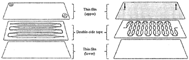
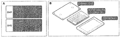
- FIG. 2 is an exploded view showing the configuration of a thin film device.
- DMS dimethyl suberimidate
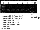
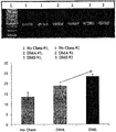
- FIG. 5 is a diagram showing the PCR amplification efficiency of the DNA extracted according to the analysis using DMA similar to DMS used in DTS analysis, DTS analysis according to the present invention.
- FIG. 6 is a view specifically showing a microfluidic chamber.
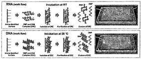
- Figure 7 shows the results of RNA and DNA extraction applied to the HINT system.
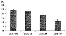
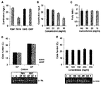
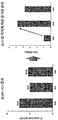
- the recovery rate of the injected DNA (1 ⁇ g human genomic DNA) with or without HIs [dimethyl suberimidate (DMS) and dimethyl pimelimidate (DMP)] was measured (A) and DMS concentration. (100, 50, 20, and 10 mg / ml), the amount of DNA extracted from cancer cells (B) and purity (C), two concentrations extracted from the system (1 ⁇ 10 3 and 1 ⁇ 10 6 )
- DMS concentration concentration
- E Actin gene amplification with DNA extracted from the system according to DMS concentration (50-250 mg / ml)
- L DNA size marker
- Q RNA extracted with Qiagen kit
- N negative control.
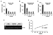
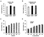
- Figure 8 shows the result of applying the HINT system to RNA extraction of cancer cells.
- AGS gastric cancer cell line
- B HCT116 (colon cancer cell line)
- C MCF7 (breast cancer cell line) results.
- D It is the result of PCR amplifying 18S gene from extracted RNA.
- E In single-stage reverse transcription RT-PCR, the cycle number (C T ) according to the concentration of HCT116 cells was confirmed.
- FIG. 9 shows the results of applying the HINT system to DNA extraction of cancer cells.
- AGS gastric cancer cell line
- B HCT116 (colon cancer cell line) result.
- C According to the concentration of HCT116 cells, the result of confirming the RT-PCR cycle number of the DNA extracted from the HINT system by applying DMS.
- D The results of RT-PCR analysis of DNA extracted from the HINT system with different E. coli concentrations are shown.
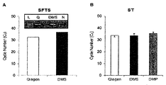
- FIG. 10 shows the results of applying a clinical sample to the HINT system.
- A Results of viral RNA extraction from the plasma of patients with Severe fever with thrombocytopenia syndrome (SFTS).
- SFTS Severe fever with thrombocytopenia syndrome
- B As a result of extracting bacterial DNA from the blood serum of a patient with scub typhus (ST).
- the inventors of the present invention have developed an extraction method for separating and extracting nucleic acids from a nucleic acid sample, forming a complex between the nucleic acid sample and a compound represented by the following formula (1), and simpler and lower cost than the conventional nucleic acid extraction method.
- the present invention has been completed by discovering that nucleic acids of high purity can be isolated and that immediate on-site diagnosis can be performed without using large equipment.
- the present invention comprises the steps of modifying by introducing an amine group to the object (first step); Injecting a nucleic acid sample and a compound represented by Chemical Formula 1 onto the modified object and forming a complex between the nucleic acid and the compound (second step); And extracting the nucleic acid by treating the object in which the complex is formed with the elution buffer (third step).
- n is an integer from 5 to 10.
- n is an integer of 5 to 7.
- the object may be any one of a thin film device, a magnetic bead (magnetic bead) or nanoparticles (nanoparticle), but is not limited thereto.
- the nucleic acid may include any one of DNA or RNA, but is not limited thereto.
- the nucleic acid may include methylated DNA, but is not limited thereto.
- the modification may be modified by introducing a silane compound in the object, but is not limited thereto.
- the method may further include washing the object through a plasma treatment before the first step, but is not limited thereto.
- the method may further include washing the object in which the complex is formed between the second and third steps, but is not limited thereto.
- the nucleic acid sample may be a sample derived from eukaryotic cells, bacterial cells, virus cells, whole blood or urine, but is not limited thereto.
- Protease and elution buffer may be further included on the object modified in the second step, but is not limited thereto.
- the present invention the upper thin film formed through the inlet and outlet holes respectively; A lower thin film spaced apart from the upper thin film; The inlet end and the outlet end are formed so that the microchannels communicating in correspondence with the inlet and outlet holes of the upper thin film are formed inside, and the injection passage communicating with the inlet of the microchannel is adjacent to the inlet end.
- a micro channel chamber disposed between the upper thin film and the lower thin film; And sealing means for sealing each side of the upper thin film and the lower thin film to seal the micro channel chamber.
- a first tubing adapter communicating the inlet hole of the upper thin film and the inlet end of the microchannel; And a second tubing adapter communicating the outlet hole of the upper thin film and the outlet end of the microchannel.
- the compound represented by Chemical Formula 1 may be injected into the inlet end of the microchannel through the inlet hole of the upper thin film, and the nucleic acid sample may be injected through the injection path of the microchamber.
- n is an integer from 5 to 10.
- n is an integer of 5 to 7.
- the microchannel may be patterned to be bent a plurality of times.
- the microchannel may include a plurality of expansion portions having an extended cross section and a plurality of reduction portions having a smaller cross section than the expansion portion, and the expansion portion and the reduction portion may be alternately disposed.
- Dimethyl suberimidate / thin film sample (DTS) analysis which is a nucleic acid analysis using DMS in a thin film device, includes three steps of sample elution / culture, washing and elution, and is performed without centrifugation.
- the thin film device is modified via 3-aminopropyltriethoxysilane (APTES) as the silane compound, and this modification converts the hydrophobic thin film device to hydrophilic.
- APTES 3-aminopropyltriethoxysilane
- the nucleic acid sample, the elution buffer and the DMS solution are injected onto the modified thin film device.
- the cross-linking mechanism between the amino acid of the nucleic acid and the DMS by interaction with the di-functional amine reactor of the DMS may be used to form a complex between the nucleic acid and the DMS and extract DNA from the sample.
- the present invention provides a composition for enhancing nucleic acid extraction efficiency comprising the compound represented by the following formula (1) as an active ingredient.
- n is an integer from 5 to 10.
- n is an integer of 5 to 7.
- composition may further include a protease and an elution buffer, and the nucleic acid may be DNA or RNA, but is not limited thereto.
- the present invention provides a kit for enhancing nucleic acid extraction efficiency comprising the composition.
- the compound represented by the formula (1) used in the present invention contains di-functional imidoesters (imidoesters), it is also referred to herein as homo-functional imidoesters (HIs), the HIs is a nucleic acid And quickly and strongly mutually bond with each other to form a complex, which is captured on the surface of the modified object with an amine reactive group to enable highly efficient nucleic acid extraction.
- imidoesters di-functional imidoesters
- homo-functional imidoesters HIs
- the HIs is a nucleic acid And quickly and strongly mutually bond with each other to form a complex, which is captured on the surface of the modified object with an amine reactive group to enable highly efficient nucleic acid extraction.
- n is an integer from 5 to 10.
- n is an integer of 5 to 7.
- HIs used in the present invention are dimethyl pimelimidate (DMP) and dimethyl suberimidate (DMS), and the chemical structure is similar to dimethyl adipimidate (dimethyl adipimidate; DMA) Comparative experiments were performed.
- DMP dimethyl pimelimidate
- DMS dimethyl suberimidate
- DMA dimethyl adipimidate Comparative experiments were performed.
- Chemical structures of DMP (Formula 2), DMS (Formula 3) and DMA (Formula 4) are as follows.
- the thin film device comprises an upper thin film and a lower thin film, and a microfluidic chamber inserted between the upper thin film and the lower thin film, the microfluidic chamber being connected to each other by flow paths in the chamber to extract DNA from a nucleic acid source. It consists of slot-type microwells.
- the microfluid chamber design was cut by a laser cutting machine on a 300 ⁇ m thick double-sided tape (100 ⁇ m thick polyester film sandwiched between 100 ⁇ m thick double-sided tape) to manufacture a microfluidic chamber. It was. Thin films (top and bottom) were cut to the same dimensions as the microfluidic chamber using a laser cutting machine.
- Inlets and outlets which are through holes, were manufactured in the upper thin film.
- the laser cutting thin films top and bottom were adhered to the surfaces of the upper and lower portions of the laser cutting microfluidic chamber, respectively, using a permanent adhesive.
- the height of the microfluidic chamber was about 300 ⁇ m and the total volume was 300 ⁇ l (300 ⁇ l amount, 8.4 cm ⁇ 3.7 cm).
- a tubing adapter for injecting a nucleic acid source was prepared by attaching a cast acrylic sheet (MARGA CIPTA, Indonesia) having a thickness of 3 mm to one side of a double-sided tape, and cutting and drilling with a laser cutting machine.
- the manufactured tubing adapter was attached to the inlet and the outlet of the microfluidic chamber, respectively.
- the pre-cut Tygon tubing (AAC02548; Cole-Parmer, Vernon Hills, USA) was then placed in the hole of the adapter and sealed with epoxy.
- the thin film device manufactured as described above has the advantage of being capable of processing various nucleic acid samples (100 ⁇ l, 300 ⁇ l, and 500 ⁇ l).
- the inside of the thin film device was treated with oxygen plasma for 10 minutes, and then the plasma treated thin film device was treated with 2% 3-aminopropyltriethoxysilane at 65 ° C. for 10 to 60 minutes (3 It was immersed in an aqueous solution containing -aminopropyltriethoxysilane (APTES) and washed thoroughly with deionized water. After cleaning, the thin film device was quickly dried under a stream of nitrogen to modify the thin film device with amine to cure the thin film device.
- APTES -aminopropyltriethoxysilane
- the water contact angle of the amine-modified thin film device using the Drop Shape Analyzer showed that the hydrophilicity of the thin film device changed considerably with temperature and incubation time. After silanization of the thin film device with APTES for 10 minutes at 65 ° C., the thin film surface hydrophilicity was increased (about 30-40 ° C.).
- DTS dimethyl suverimidate
- an optimized assay solution was first prepared for extracting DNA using a thin film device (300 ⁇ l amount, 8.4 cm ⁇ 3.7 cm) modified with amine.
- the optimized assay solution was prepared by mixing elution buffer containing 100 mM tris-hydrochloric acid (pH 8.0), 10 mM EDTA, 1% SDS, 10% Triton X-100 with DMS (50 mg / mL), As a nucleic acid assay sample, 100 ⁇ l of each sample derived from cells, bacteria, blood or urine was mixed with 200 ⁇ l of the assay solution.
- the mixed solution of the mixed nucleic acid assay sample and the assay solution is introduced into the upper substrate inlet of the thin film device modified with amine, and the mixed solution moves into the microfluidic chamber to bind two amine groups and DNA of the DMS.
- DNA was modified by combining the modified amine group with DNA to form a complex.
- the thin film device was placed in any one of a thermoelectric cooler (TEC) including an incubator or a controller (Alpha Omega Instruments) maintained at a constant temperature (56 ° C) for 20 minutes to sufficiently extract DNA from the nucleic acid analysis sample. .
- DNA was extracted using elution buffer (10 mM sodium bicarbonate, pH 10.6). After measuring the amount and purity of the extracted DNA, the optical density ratio of the sample at 260 nm (DNA) and 280 nm (protein) was determined using Enspire Multimode Plate Reader (PerkinElmer). In order to compare the conventional DNA extraction and DTS analysis of the present invention, it was analyzed using a QIAmp DNA mini kit according to a known method (Qiagen, Hilden, Germany).
- DMEM Dulbecco's modified eagle medium
- DMEM Life Technology Dulbecco's modified eagle medium
- MCF-7 Eukaryotic cells
- NCI-H1975 lung
- CaCo-2 large intestine
- T24 blade
- U937 lymphocytes
- Jurkat peripheral blood
- End-point PCR and real time PCR were performed to confirm the amount and purity of DNA. Forward and reverse primers of some genes (HRAS, Actin and RAR ⁇ ) were synthesized to a normal length of about 24 base pairs. End-point PCR was performed at 95 ° C. for 15 minutes at the initial denaturation step; 45 cycles of 95 ° C., 45 seconds, 59 ° C., 45 seconds (RAR ⁇ ), and 72 ° C., 45 seconds; And 72 ° C., final extension for 10 minutes.
- 5-10 ⁇ l of DNA was mixed with 4 ⁇ l of LightCycler FastStart DNA Master, 25 pmol of each primer, 2 ⁇ l of 1 ⁇ PCR buffer (Qiagen), 2.5 mM magnesium chloride (MgCl 2 ), 0.25 mM deoxynucleotide triphosphate ( deoxynucleotide triphosphate), and amplified in a total volume of 20 ⁇ l containing distilled water. 50 cycles of 95 ° C., 10 seconds, 58 ° C., 30 seconds (for HRAS and actin genes), and 72 ° C. for 10 seconds, followed by initial pretreatment at 95 ° C. for 10 minutes, and Cooling step in Amplified product with SYBR green signal was performed in LightCycler 2.0 (Roche Diagnostics).
- the DNA was digested by mixing either MspI or HpaII solution (150 ⁇ l) at 37 ° C. for 20 minutes in a single reaction tube. After the digestion process, the single reaction tube was left at 80 ° C. for 10 minutes to inactivate the restriction enzyme. According to the inactivation process, the digested DNA was used as a template for epigenetic analysis of the RAR ⁇ gene obtained in both assays using conventional PCR.
- DTS analysis according to the present invention and analysis using DMA, a compound similar to DMS used in DTS analysis, using breast cancer cells, as shown in FIG. 4.
- DNA could be extracted from breast cancer cells by binding to DNA.
- analysis using DMA dimethyl adipimidate
- PCR amplification of the DNA extracted using DMS resulted in a 25% improvement in amplification efficiency compared to the analysis using DMA.
- PCR-based DNA amplification was performed using the DNA extracted using the DTS assay. All primers are Escherichia coli, Mycobacterium abscessus, Mycobacterium gordonae and Salmonella Strains (Salmonella Typhimurium, Salmonella Typhimurium) Commercial primers from Salmonella Newport, and Salmonella Saintpaul were used.
- elution buffer containing 100 mM tris-hydrochloric acid (pH 8.0), 10 mM EDTA, 1% SDS, 10% Triton X-100 and 20 mg / mL lysozyme were combined with DMS (50 mg / mL).
- DMS 50 mg / mL
- General PCR was performed to validate the DTS method of the present invention.
- Escherichia coli E.
- coli XL1 Blue strain were the cultured for a day at 37 °C with shaking conditions (shaking condition), and inoculated in a 50 ⁇ g / ml tetracycline (tetracycline) and Luria-Bertani (LB) medium, 3 to 10 A sample of 10 7 colony forming units (CFUs) was used for the test.
- PCR amplifications were visualized by gel electrophoresis to separate PCR products on 2% agarose gels containing ethidium bromide (EtBr) (Sigma-Aldrich). The gel was visualized using the Gel Doc System (Bio-Rad). Measurement of DNA concentration and purity was performed with a UV spectrometer (Perkin-Elmer).
- the elution buffer containing proteinase K and DMS and the bodily fluid sample were introduced into the previously prepared thin film device, and then moved to the microchannel chamber to form a complex between the DNA and the DMA in the bodily fluid sample.
- DNA was extracted in the same manner as.
- the elution buffer and the bodily fluid sample were introduced into two different inlets at a flow rate of 1.5 ml / hr for 10 minutes using a syringe pump (KD Scientific, MA), and the cartridge was extracted for 20 minutes to extract and purify DNA.
- the inventors analyzed DNA by extracting DNA from various samples including eukaryotic cells or prokaryotic cells through DTS (Dimethyl suberimidate / Thin film Sample) analysis, which is a nucleic acid analysis using DMS in a thin film device.
- DTS Dimethyl suberimidate / Thin film Sample
- the present inventors have film-using-based microfluidic platform of RNA and homozygous second functional yimido ester for which can extract all of the DNA, extracted nucleic acids using thin film [h omobifunctional i midoesters (HIs) for n ucleic acids extraction using t hin films; HINT] system was developed (FIG. 6).
- HIs h omobifunctional i midoesters
- DMS dimethyl suberimidate
- DMP dimethyl pimelimidate
- 6A bifunctional imidoester group
- sample lysis, washing and elution were performed on a single channel.
- sample mixtures, lysis buffers and HIs (DMS or DMP) were injected into the system by pipette, which system used an amine surface to capture nucleic acid and HIs complexes. It was previously activated with a reactive group (FIG. 6B). Thereafter, 10-20 minutes of reaction was performed at room temperature for RNA extraction, and 20 minutes at 56 ° C for DNA extraction. After the reaction, nucleic acid (RNA or DNA) could be extracted through washing and elution.
- RNA and DNA nucleic acid
- FIG. 7A the recovery of injected DNA (1 ⁇ g of human genomic DNA) with and without HIs (DMS and DMP) was measured. More than 95% DNA was recovered in both groups containing DMS (black) and DMP (grey), and ⁇ 50% DNA was recovered in the experimental group without HIs (FIG. 7A).
- cancer cell lines [1 ⁇ 10 6 cells of breast cancer cell line (MCF7) or colon cancer cell line (HCT116) were used.
- the amount of DNA extracted from cancer cells (FIG. 7B) and purity (FIG. 7C) were measured by varying the DMS concentration (100, 50, 20, and 10 mg / ml). It was. Unlike DNA extraction, RNA is more easily degraded and, in general, RNA extraction is more difficult.
- DMS or DMP was applied to the HINT system.
- 18S gene amplification was performed with two concentrations of RNA extracted from the system, followed by single step reverse transcription end-point PCR and single step reverse transcription RT-PCR.
- PCR analysis was performed by extracting DNA from MCF7, AGS and HCT116 cells.
- the HINT system was compared with the Qiagen kit using the same concentration of cells ((1 ⁇ 10 6 ).
- the DNA extracted from the HINT system using AGS and HCT116 cells and DMS or DMP was obtained from the DNA extracted from the Qiagen kit.
- Equivalent to amplification efficiency FIGS. 9A and 9B
- DNA extracted from the HINT system using DMS or DMP was shown to be dependent on cell number. 9C, meanwhile, in order to confirm the applicability of the system in various samples, bacterial DNA was extracted and analyzed from E.
- the HINT system can also be used to extract viral or bacterial nucleic acids (DNA and RNA) from tick-borne disease samples such as Severe fever with thrombocytopenia syndrome (SFTS) and scub typhus (ST). It was checked if it could be applied. Viral RNA was extracted from the plasma of SFTS patients using a Qiagen kit and HINT system (using DMS). RT-PCR amplification efficiency of the HINT system was not significantly different from the Qiagen kit (Fig. 10A).
- SFTS Severe fever with thrombocytopenia syndrome
- ST scub typhus
Landscapes
- Chemical & Material Sciences (AREA)
- Health & Medical Sciences (AREA)
- Life Sciences & Earth Sciences (AREA)
- Organic Chemistry (AREA)
- Engineering & Computer Science (AREA)
- Genetics & Genomics (AREA)
- Zoology (AREA)
- Wood Science & Technology (AREA)
- General Engineering & Computer Science (AREA)
- Bioinformatics & Cheminformatics (AREA)
- Biotechnology (AREA)
- Molecular Biology (AREA)
- Biophysics (AREA)
- Microbiology (AREA)
- Biomedical Technology (AREA)
- Physics & Mathematics (AREA)
- Proteomics, Peptides & Aminoacids (AREA)
- Biochemistry (AREA)
- General Health & Medical Sciences (AREA)
- Analytical Chemistry (AREA)
- Clinical Laboratory Science (AREA)
- Chemical Kinetics & Catalysis (AREA)
- Crystallography & Structural Chemistry (AREA)
- Immunology (AREA)
- Plant Pathology (AREA)
- Measuring Or Testing Involving Enzymes Or Micro-Organisms (AREA)
Abstract
La présente invention concerne un procédé d'extraction d'acide nucléique utilisant un sujet solide. En formant un complexe entre un échantillon d'acide nucléique et un composé imidoester à l'intérieur du sujet, il est possible de séparer les acides nucléiques en grande quantité et en une pureté élevée plus simplement et à un coût plus faible que les procédés d'extraction d'acide nucléique classiques. En particulier, un dispositif à film mince, utilisé pour l'extraction d'un acide nucléique, présente des propriétés hydrophiles améliorées comparé aux substrats de silicium classiques, et ainsi peut extraire plus efficacement l'acide nucléique.
Priority Applications (4)
| Application Number | Priority Date | Filing Date | Title |
|---|---|---|---|
| EP17799607.1A EP3460072B1 (fr) | 2016-05-17 | 2017-05-15 | Procédé d'extraction d'acide nucléique utilisant un sujet solide |
| US16/302,093 US10907146B2 (en) | 2016-05-17 | 2017-05-15 | Nucleic acid extraction method using solid subject |
| CN201780043894.4A CN109804078B (zh) | 2016-05-17 | 2017-05-15 | 使用固体对象的核酸提取方法 |
| JP2018560639A JP6691978B2 (ja) | 2016-05-17 | 2017-05-15 | 固相対象物を利用した核酸抽出方法 |
Applications Claiming Priority (4)
| Application Number | Priority Date | Filing Date | Title |
|---|---|---|---|
| KR20160060310 | 2016-05-17 | ||
| KR10-2016-0060310 | 2016-05-17 | ||
| KR10-2016-0175710 | 2016-12-21 | ||
| KR1020160175710A KR101913208B1 (ko) | 2016-05-17 | 2016-12-21 | 고형상 대상물을 이용한 핵산 추출방법 |
Publications (1)
| Publication Number | Publication Date |
|---|---|
| WO2017200249A1 true WO2017200249A1 (fr) | 2017-11-23 |
Family
ID=60325380
Family Applications (1)
| Application Number | Title | Priority Date | Filing Date |
|---|---|---|---|
| PCT/KR2017/005019 Ceased WO2017200249A1 (fr) | 2016-05-17 | 2017-05-15 | Procédé d'extraction d'acide nucléique utilisant un sujet solide |
Country Status (1)
| Country | Link |
|---|---|
| WO (1) | WO2017200249A1 (fr) |
Cited By (2)
| Publication number | Priority date | Publication date | Assignee | Title |
|---|---|---|---|---|
| WO2020150781A1 (fr) * | 2019-01-23 | 2020-07-30 | Haplomic Technologies Pty Ltd | Dispositif microfluidique |
| WO2024075559A1 (fr) | 2022-10-06 | 2024-04-11 | 国立研究開発法人物質・材料研究機構 | Procédé d'extraction d'acide nucléique, procédé d'amplification d'acide nucléique, kit d'extraction d'acide nucléique et kit de test de pcr |
Citations (2)
| Publication number | Priority date | Publication date | Assignee | Title |
|---|---|---|---|---|
| US20070196833A1 (en) * | 2005-04-21 | 2007-08-23 | Gjerde Douglas T | Open channel solid phase extraction systems and methods |
| KR20150096444A (ko) * | 2012-12-13 | 2015-08-24 | 에이전시 포 사이언스, 테크놀로지 앤드 리서치 | 고형상 장치 상에서 핵산을 분리 및 분석하는 무표지 방법 |
-
2017
- 2017-05-15 WO PCT/KR2017/005019 patent/WO2017200249A1/fr not_active Ceased
Patent Citations (2)
| Publication number | Priority date | Publication date | Assignee | Title |
|---|---|---|---|---|
| US20070196833A1 (en) * | 2005-04-21 | 2007-08-23 | Gjerde Douglas T | Open channel solid phase extraction systems and methods |
| KR20150096444A (ko) * | 2012-12-13 | 2015-08-24 | 에이전시 포 사이언스, 테크놀로지 앤드 리서치 | 고형상 장치 상에서 핵산을 분리 및 분석하는 무표지 방법 |
Non-Patent Citations (3)
| Title |
|---|
| SHIN, Y. ET AL.: "Dimethyl Adipimidate/Thin Film Sample processing (DTS); A Simple, Low-cost, and Versatile Nucleic Acid Extraction Assay for Downstream Analysis", SCIENTIFIC REPORTS, vol. 5, no. 14127, 2015, pages 1 - 11, XP055556165 * |
| SHIN, Y. ET AL.: "Solid Phase Nucleic Acid extraction Technique in a Microfluidic Chip Using a Novel Non-chaotropic Agent: Dimethyl Adipimidate", LAB ON A CHIP, vol. 14, no. 2, 2014, pages 359 - 368, XP055261226 * |
| SUTER, J. D. ET AL.: "Label-free DNA Methylation Analysis using Opto-fluidicring Resonators", BIOSENSORS AND BIOELECTRONICS, vol. 26, no. 3, 2010, pages 1016 - 1020, XP027393251 * |
Cited By (4)
| Publication number | Priority date | Publication date | Assignee | Title |
|---|---|---|---|---|
| WO2020150781A1 (fr) * | 2019-01-23 | 2020-07-30 | Haplomic Technologies Pty Ltd | Dispositif microfluidique |
| JP2022518798A (ja) * | 2019-01-23 | 2022-03-16 | ハプロミック テクノロジーズ プロプライアタリー リミティド | マイクロ流体デバイス |
| KR102933843B1 (ko) | 2019-01-23 | 2026-03-03 | 하플로믹 테크놀로지즈 피티와이 엘티디 | 미세유체 장치 |
| WO2024075559A1 (fr) | 2022-10-06 | 2024-04-11 | 国立研究開発法人物質・材料研究機構 | Procédé d'extraction d'acide nucléique, procédé d'amplification d'acide nucléique, kit d'extraction d'acide nucléique et kit de test de pcr |
Similar Documents
| Publication | Publication Date | Title |
|---|---|---|
| KR101913208B1 (ko) | 고형상 대상물을 이용한 핵산 추출방법 | |
| EP3505628B1 (fr) | Procédé de concentration de micro-organismes ou d'extraction d'acide nucléique à l'aide de dtbp | |
| US20050221314A1 (en) | Method and device for determination of tissue specificity of free floating dna in bodily fluids | |
| KR20020026457A (ko) | 게놈 프로파일링: 복잡한 생물학적 샘플을 다양한 유형의생물체들의 존재에 대하여 검사하기 위한 신속한 방법 | |
| US20220325324A1 (en) | Systems and methods for the detection of infectious diseases | |
| WO2017099445A1 (fr) | Marqueur génétique permettant de distinguer et de détecter des bactéries à l'origine de l'infection à edwardsiella et de la streptococcocie chez le poisson, et procédé de différenciation et de détection de telles bactéries utilisant ledit marqueur | |
| WO2019221537A1 (fr) | Procédé de détection de mycoplasme à l'aide d'adn mitochondrial en tant qu'échantillon témoin interne | |
| CN105378108A (zh) | 用于分离核酸的系统和方法 | |
| WO2017200249A1 (fr) | Procédé d'extraction d'acide nucléique utilisant un sujet solide | |
| WO2009093856A2 (fr) | Ensemble d'amorces de pcr pour la détection de mycoplasme dans des produits médicinaux biologiques, et kit pour la détection de mycoplasme comprenant ledit ensemble | |
| CN103014135B (zh) | 一种鉴定结核分枝杆菌的方法 | |
| CN105219847B (zh) | 一种军团菌快速检测与分型试剂盒及其检测方法 | |
| CN105154538B (zh) | 一种军团菌快速检测与分型的引物及方法 | |
| WO2018038549A1 (fr) | Procédé de concentration de micro-organismes ou d'extraction d'acide nucléique à l'aide de dtbp | |
| JP5980332B2 (ja) | 分子検出アッセイ | |
| WO2011004957A2 (fr) | Sonde de détection de la résistance du bacille de koch à des médicaments et kit la contenant | |
| WO2019088429A1 (fr) | Procédé d'extraction d'un biomatériau issu d'une biopsie liquide à l'aide d'imidoester homobifonctionnel | |
| ES2253279T3 (es) | Metodos y composiciones para la deteccion de especies del complejo de mycobacterium avium. | |
| WO2018048194A1 (fr) | Composition et procédé pour améliorer la sensibilité et la spécificité de la détection d'acides nucléiques à l'aide d'une protéine dcas9 et de la liaison d'arng à une séquence d'acide nucléique cible | |
| WO2017051939A1 (fr) | Procédé pour extraire et amplifier un acide nucléique à l'aide de particules magnétiques | |
| Alleman | Molecular tools for the diagnosis of animal diseases | |
| WO2020256438A1 (fr) | Procédé de concentration d'un sujet et d'extraction d'acide nucléique à l'aide d'acide bis(sulfosuccinimidyl) subérique ou d'un dérivé de ce dernier | |
| WO2024262933A1 (fr) | Composition d'extraction d'acide nucléique et procédé d'extraction d'acide nucléique l'utilisant | |
| WO2019078638A2 (fr) | Méthode d'enrichissement d'un pathogène à l'aide d'un imidoester homobifonctionnel | |
| WO2017217700A1 (fr) | Utilisation d'adn acellulaire dans le diagnostic de maladies cancéreuses |
Legal Events
| Date | Code | Title | Description |
|---|---|---|---|
| ENP | Entry into the national phase |
Ref document number: 2018560639 Country of ref document: JP Kind code of ref document: A |
|
| NENP | Non-entry into the national phase |
Ref country code: DE |
|
| 121 | Ep: the epo has been informed by wipo that ep was designated in this application |
Ref document number: 17799607 Country of ref document: EP Kind code of ref document: A1 |
|
| ENP | Entry into the national phase |
Ref document number: 2017799607 Country of ref document: EP Effective date: 20181217 |