WO2017204187A1 - Procédé de récupération de vésicules extracellulaires et récipient destiné à des vésicules extracellulaires - Google Patents
Procédé de récupération de vésicules extracellulaires et récipient destiné à des vésicules extracellulaires Download PDFInfo
- Publication number
- WO2017204187A1 WO2017204187A1 PCT/JP2017/019121 JP2017019121W WO2017204187A1 WO 2017204187 A1 WO2017204187 A1 WO 2017204187A1 JP 2017019121 W JP2017019121 W JP 2017019121W WO 2017204187 A1 WO2017204187 A1 WO 2017204187A1
- Authority
- WO
- WIPO (PCT)
- Prior art keywords
- exosomes
- coating agent
- exosome
- extracellular vesicles
- adsorption
- Prior art date
- Legal status (The legal status is an assumption and is not a legal conclusion. Google has not performed a legal analysis and makes no representation as to the accuracy of the status listed.)
- Ceased
Links
Images


Classifications
-
- C—CHEMISTRY; METALLURGY
- C08—ORGANIC MACROMOLECULAR COMPOUNDS; THEIR PREPARATION OR CHEMICAL WORKING-UP; COMPOSITIONS BASED THEREON
- C08F—MACROMOLECULAR COMPOUNDS OBTAINED BY REACTIONS ONLY INVOLVING CARBON-TO-CARBON UNSATURATED BONDS
- C08F230/00—Copolymers of compounds having one or more unsaturated aliphatic radicals, each having only one carbon-to-carbon double bond, and containing phosphorus, selenium, tellurium or a metal
- C08F230/02—Copolymers of compounds having one or more unsaturated aliphatic radicals, each having only one carbon-to-carbon double bond, and containing phosphorus, selenium, tellurium or a metal containing phosphorus
-
- G—PHYSICS
- G01—MEASURING; TESTING
- G01N—INVESTIGATING OR ANALYSING MATERIALS BY DETERMINING THEIR CHEMICAL OR PHYSICAL PROPERTIES
- G01N33/00—Investigating or analysing materials by specific methods not covered by groups G01N1/00 - G01N31/00
- G01N33/48—Biological material, e.g. blood, urine; Haemocytometers
- G01N33/50—Chemical analysis of biological material, e.g. blood, urine; Testing involving biospecific ligand binding methods; Immunological testing
- G01N33/53—Immunoassay; Biospecific binding assay; Materials therefor
- G01N33/543—Immunoassay; Biospecific binding assay; Materials therefor with an insoluble carrier for immobilising immunochemicals
-
- C—CHEMISTRY; METALLURGY
- C08—ORGANIC MACROMOLECULAR COMPOUNDS; THEIR PREPARATION OR CHEMICAL WORKING-UP; COMPOSITIONS BASED THEREON
- C08F—MACROMOLECULAR COMPOUNDS OBTAINED BY REACTIONS ONLY INVOLVING CARBON-TO-CARBON UNSATURATED BONDS
- C08F120/00—Homopolymers of compounds having one or more unsaturated aliphatic radicals, each having only one carbon-to-carbon double bond, and only one being terminated by only one carboxyl radical or a salt, anhydride, ester, amide, imide or nitrile thereof
- C08F120/02—Monocarboxylic acids having less than ten carbon atoms; Derivatives thereof
- C08F120/10—Esters
- C08F120/20—Esters of polyhydric alcohols or polyhydric phenols, e.g. 2-hydroxyethyl (meth)acrylate or glycerol mono-(meth)acrylate
-
- C—CHEMISTRY; METALLURGY
- C08—ORGANIC MACROMOLECULAR COMPOUNDS; THEIR PREPARATION OR CHEMICAL WORKING-UP; COMPOSITIONS BASED THEREON
- C08F—MACROMOLECULAR COMPOUNDS OBTAINED BY REACTIONS ONLY INVOLVING CARBON-TO-CARBON UNSATURATED BONDS
- C08F20/00—Homopolymers and copolymers of compounds having one or more unsaturated aliphatic radicals, each having only one carbon-to-carbon double bond, and only one being terminated by only one carboxyl radical or a salt, anhydride, ester, amide, imide or nitrile thereof
- C08F20/02—Monocarboxylic acids having less than ten carbon atoms, Derivatives thereof
- C08F20/10—Esters
- C08F20/26—Esters containing oxygen in addition to the carboxy oxygen
- C08F20/28—Esters containing oxygen in addition to the carboxy oxygen containing no aromatic rings in the alcohol moiety
-
- C—CHEMISTRY; METALLURGY
- C08—ORGANIC MACROMOLECULAR COMPOUNDS; THEIR PREPARATION OR CHEMICAL WORKING-UP; COMPOSITIONS BASED THEREON
- C08F—MACROMOLECULAR COMPOUNDS OBTAINED BY REACTIONS ONLY INVOLVING CARBON-TO-CARBON UNSATURATED BONDS
- C08F220/00—Copolymers of compounds having one or more unsaturated aliphatic radicals, each having only one carbon-to-carbon double bond, and only one being terminated by only one carboxyl radical or a salt, anhydride ester, amide, imide or nitrile thereof
- C08F220/02—Monocarboxylic acids having less than ten carbon atoms; Derivatives thereof
- C08F220/10—Esters
- C08F220/12—Esters of monohydric alcohols or phenols
- C08F220/16—Esters of monohydric alcohols or phenols of phenols or of alcohols containing two or more carbon atoms
- C08F220/18—Esters of monohydric alcohols or phenols of phenols or of alcohols containing two or more carbon atoms with acrylic or methacrylic acids
- C08F220/1804—C4-(meth)acrylate, e.g. butyl (meth)acrylate, isobutyl (meth)acrylate or tert-butyl (meth)acrylate
-
- C—CHEMISTRY; METALLURGY
- C09—DYES; PAINTS; POLISHES; NATURAL RESINS; ADHESIVES; COMPOSITIONS NOT OTHERWISE PROVIDED FOR; APPLICATIONS OF MATERIALS NOT OTHERWISE PROVIDED FOR
- C09D—COATING COMPOSITIONS, e.g. PAINTS, VARNISHES OR LACQUERS; FILLING PASTES; CHEMICAL PAINT OR INK REMOVERS; INKS; CORRECTING FLUIDS; WOODSTAINS; PASTES OR SOLIDS FOR COLOURING OR PRINTING; USE OF MATERIALS THEREFOR
- C09D133/00—Coating compositions based on homopolymers or copolymers of compounds having one or more unsaturated aliphatic radicals, each having only one carbon-to-carbon double bond, and at least one being terminated by only one carboxyl radical, or of salts, anhydrides, esters, amides, imides, or nitriles thereof; Coating compositions based on derivatives of such polymers
- C09D133/04—Homopolymers or copolymers of esters
- C09D133/06—Homopolymers or copolymers of esters of esters containing only carbon, hydrogen and oxygen, the oxygen atom being present only as part of the carboxyl radical
- C09D133/062—Copolymers with monomers not covered by C09D133/06
- C09D133/066—Copolymers with monomers not covered by C09D133/06 containing -OH groups
-
- C—CHEMISTRY; METALLURGY
- C09—DYES; PAINTS; POLISHES; NATURAL RESINS; ADHESIVES; COMPOSITIONS NOT OTHERWISE PROVIDED FOR; APPLICATIONS OF MATERIALS NOT OTHERWISE PROVIDED FOR
- C09D—COATING COMPOSITIONS, e.g. PAINTS, VARNISHES OR LACQUERS; FILLING PASTES; CHEMICAL PAINT OR INK REMOVERS; INKS; CORRECTING FLUIDS; WOODSTAINS; PASTES OR SOLIDS FOR COLOURING OR PRINTING; USE OF MATERIALS THEREFOR
- C09D133/00—Coating compositions based on homopolymers or copolymers of compounds having one or more unsaturated aliphatic radicals, each having only one carbon-to-carbon double bond, and at least one being terminated by only one carboxyl radical, or of salts, anhydrides, esters, amides, imides, or nitriles thereof; Coating compositions based on derivatives of such polymers
- C09D133/04—Homopolymers or copolymers of esters
- C09D133/14—Homopolymers or copolymers of esters of esters containing halogen, nitrogen, sulfur or oxygen atoms in addition to the carboxy oxygen
-
- C—CHEMISTRY; METALLURGY
- C09—DYES; PAINTS; POLISHES; NATURAL RESINS; ADHESIVES; COMPOSITIONS NOT OTHERWISE PROVIDED FOR; APPLICATIONS OF MATERIALS NOT OTHERWISE PROVIDED FOR
- C09D—COATING COMPOSITIONS, e.g. PAINTS, VARNISHES OR LACQUERS; FILLING PASTES; CHEMICAL PAINT OR INK REMOVERS; INKS; CORRECTING FLUIDS; WOODSTAINS; PASTES OR SOLIDS FOR COLOURING OR PRINTING; USE OF MATERIALS THEREFOR
- C09D143/00—Coating compositions based on homopolymers or copolymers of compounds having one or more unsaturated aliphatic radicals, each having only one carbon-to-carbon double bond, and containing boron, silicon, phosphorus, selenium, tellurium, or a metal; Coating compositions based on derivatives of such polymers
- C09D143/02—Homopolymers or copolymers of monomers containing phosphorus
-
- C—CHEMISTRY; METALLURGY
- C09—DYES; PAINTS; POLISHES; NATURAL RESINS; ADHESIVES; COMPOSITIONS NOT OTHERWISE PROVIDED FOR; APPLICATIONS OF MATERIALS NOT OTHERWISE PROVIDED FOR
- C09D—COATING COMPOSITIONS, e.g. PAINTS, VARNISHES OR LACQUERS; FILLING PASTES; CHEMICAL PAINT OR INK REMOVERS; INKS; CORRECTING FLUIDS; WOODSTAINS; PASTES OR SOLIDS FOR COLOURING OR PRINTING; USE OF MATERIALS THEREFOR
- C09D5/00—Coating compositions, e.g. paints, varnishes or lacquers, characterised by their physical nature or the effects produced; Filling pastes
- C09D5/16—Antifouling paints; Underwater paints
- C09D5/1656—Antifouling paints; Underwater paints characterised by the film-forming substance
- C09D5/1662—Synthetic film-forming substance
- C09D5/1668—Vinyl-type polymers
-
- C—CHEMISTRY; METALLURGY
- C09—DYES; PAINTS; POLISHES; NATURAL RESINS; ADHESIVES; COMPOSITIONS NOT OTHERWISE PROVIDED FOR; APPLICATIONS OF MATERIALS NOT OTHERWISE PROVIDED FOR
- C09D—COATING COMPOSITIONS, e.g. PAINTS, VARNISHES OR LACQUERS; FILLING PASTES; CHEMICAL PAINT OR INK REMOVERS; INKS; CORRECTING FLUIDS; WOODSTAINS; PASTES OR SOLIDS FOR COLOURING OR PRINTING; USE OF MATERIALS THEREFOR
- C09D129/00—Coating compositions based on homopolymers or copolymers of compounds having one or more unsaturated aliphatic radicals, each having only one carbon-to-carbon double bond, and at least one being terminated by an alcohol, ether, aldehydo, ketonic, acetal, or ketal radical; Coating compositions based on hydrolysed polymers of esters of unsaturated alcohols with saturated carboxylic acids; Coating compositions based on derivatives of such polymers
- C09D129/02—Homopolymers or copolymers of unsaturated alcohols
- C09D129/04—Polyvinyl alcohol; Partially hydrolysed homopolymers or copolymers of esters of unsaturated alcohols with saturated carboxylic acids
Definitions
- the present invention relates to a method for efficiently recovering cell-derived vesicles represented by exosomes, and a container and instrument used for the recovery. Further, the present invention relates to a substrate, a flow path, and the like for use in a method for inspecting with exosomes.
- Extracellular vesicles are extracellular vesicles derived from cells and classified into exosomes, microvesicles, and apoptotic bodies based on their sizes. Exosomes and microvesicles contain nucleic acids such as proteins, mRNA and miRNA, and apoptotic bodies contain fragmented nuclei and organelles. Among extracellular vesicles, since exosomes and microvesicles contain proteins and nucleic acids, they contain information derived from released cells and have the potential to be used as biomarkers. In particular, exosomes have attracted attention in recent years because they have the potential to be used for diagnostic markers and treatment.
- An exosome is an endoplasmic reticulum with a diameter of 30 to 100 nm formed by a lipid bilayer secreted from cells. Exosomes are known to be secreted from most cells, and their presence has been confirmed in body fluids such as blood, saliva, urine and breast milk. Exosomes contain secreted RNA and proteins derived from cells, and it is suggested that information may be transmitted to distant cells by circulating in body fluid (Non-patent Document 1). The possibility that cancer progression is promoted by nucleic acids and proteins contained in exosomes derived from cancer cells was suggested, and this suggested the possibility of cancer diagnosis systems using exosomes (Non-Patent Documents) 2).
- exosome-derived nucleic acids and proteins are useful as diagnostic markers.
- exosomes are also contained in saliva and urine, if a highly accurate test method using exosomes can be established, it can be a noninvasive diagnostic method.
- attempts have been made to use exosomes collected and recovered from healthy cells by using the exosome information transmission function to recover damaged cells or diseased cells (Patent Document 1).
- exosomes in the case of recovering exosomes from the cell culture supernatant, can be recovered as pellets by ultracentrifugation for about 2 hours after removing cell debris from the medium containing exosomes by low-speed centrifugation.
- the exosomes thus obtained can be redispersed in a buffer solution such as PBS and further purified, or used as it is for analysis such as inspection and research.
- exosomes are non-specifically adsorbed on the plastic surface of the collection container to be used, and a stable collection rate cannot be obtained. It was.
- the adsorption to the surface of the container used for purification and the analysis instrument affects not only the efficiency of the analysis but also the analysis result.
- an effective method for preventing the adsorption of extracellular vesicles such as exosomes has not been developed so far.
- Patent Documents 4 and 5 disclose methods for preventing nonspecific adsorption of proteins and cells, which are biological substances, to a container with a hydrophilic polymer.
- Patent Document 4 discloses that protein adsorption can be prevented by coating a polystyrene container with a PBS solution containing a 2-methacryloyloxyethyl phosphorylcholine polymer or a copolymer of 2-methacryloyloxyethyl phosphorylcholine-containing components.
- Patent Document 5 discloses that by coating a culture vessel with a compound containing a phosphorylcholine-like group, adhesion of ES cells to the culture vessel can be prevented, and an embryoid body can be formed efficiently.
- Patent Document 4 is a method specialized for proteins, and completely prevents adsorption of extracellular vesicles formed by lipid bilayers, even though proteins are exposed on the surface of exosomes and the like. It was difficult to do. Further, the method of Patent Document 5 prevents adsorption of cells formed of lipid bilayers, but the size of cells and extracellular vesicles, particularly exosomes, is completely different, so that they can be transferred to a container. The degree of adsorption of exosomes was greatly different, and adsorption of exosomes to the container could not be completely prevented.
- the present invention prevents the adsorption of cell-derived vesicles to a container used for purification such as a centrifuge tube or a pipette tip when purifying cell-derived vesicles typified by exosomes, and improves the recovery rate. It is an object of the present invention to provide a coating agent for improvement. It is another object of the present invention to provide a coating agent that can be used in the purification process and can improve the accuracy of exosome analysis by coating a substrate, a flow path, and the like used for exosome analysis. It is another object of the present invention to provide analytical instruments such as containers, chips, and substrates coated with these coating agents.
- the present invention relates to the following coating agents, instruments, purification methods, and analysis methods.
- a coating agent for preventing the adsorption of extracellular vesicles comprising a hydrophilic polymer having a weight average molecular weight of 10,000 to 1,000,000, and the contact angle of the resulting coating film being 0 degrees to 30 degrees.
- the hydrophilic polymer includes one or more hydrophilic monomers composed of 2-methacryloyloxyethyl phosphorylcholine, vinyl alcohol, vinyl pyrrolidone, methoxyalkylene glycol monomethacrylate or 2-hydroxyethyl methacrylate. Coating agent.
- the hydrophilic monomer constituting the hydrophilic polymer is 2-methacryloyloxyethyl phosphorylcholine, and contains 30 mol% to 50 mol% of a structural unit based on 2-methacryloyloxyethyl phosphorylcholine.
- the coating agent according to (2), wherein the remainder is a copolymer comprising a hydrophobic group-containing monomer.
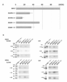
- FIG. 4A shows the results of examining the amount of exosome recovered by the coating agent by the number of particles
- FIG. 4B shows the results of examining by Western blotting
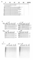
- FIG. 5A is a diagram showing the results of examining the amount of exosome recovered by the film thickness of the coating agent according to the number of particles
- FIG. 5C is a diagram in which a collected sample and an adsorbed sample are electrophoresed and analyzed by silver staining.
- an extracellular vesicle refers to a vesicle derived from a cell, and specifically refers to an exosome, a microvesicle, and an apoptotic body.
- exosomes are mainly described, but it goes without saying that the present invention can also be applied to extracellular vesicles other than exosomes.
- the coating agent of the present invention coats an instrument used when purifying extracellular vesicles such as exosomes, such as centrifuge tubes, tubes, and pipette tips, thereby preventing its adsorption and improving the recovery rate. be able to. Also, non-specific adsorption can be prevented and analysis accuracy can be improved by coating the analytical instruments and analytical instruments used in this field, such as beads, microtiter plates, slide glasses, microtass, and lab-on-chip. be able to.
- the coating agent of the present invention can be coated by any material.
- any material for example, polycarbonate, polypropylene, polyallomer, which are frequently used as centrifuge tubes, and vinyl chloride, polystyrene, polyethylene terephthalate, silicon, hydrophilic polydimethylsiloxane, hydrophobic polydimethylsiloxane, glass, stainless steel, which are used as base materials for analytical instruments
- a metal such as aluminum or aluminum
- adsorption of extracellular vesicles such as exosomes can be prevented.
- the contact angle of the coating film obtained by the coating agent of the present invention is 0 degree or more and 30 or less, preferably 0 degree or more and 25 degrees or less.
- the hydrophilic polymer of the present invention has a weight average molecular weight of 10,000 or more and 1,000,000 or less, preferably 100,000 or more from the viewpoint of the durability of the coating film, and has a high viscosity at the time of coating. In order to suppress smears caused by the above, it is more preferably 700,000 or less.
- the hydrophilic monomer of the hydrophilic polymer is not particularly limited as long as it can synthesize a hydrophilic polymer or copolymer. Among them, 2-methacryloyloxyethyl phosphorylcholine, vinyl alcohol, vinyl pyrrolidone, methoxy Alkylene glycol monomethacrylate or 2-hydroxyethyl methacrylate is preferred. These hydrophilic monomers may be used as a polymer or may be used as a copolymer together with other monomers.
- a copolymer of 2-methacryloyloxyethyl phosphorylcholine (MPC) and a hydrophobic monomer can be preferably used.
- the hydrophobic monomer methyl methacrylate (MMA), n-butyl methacrylate (BMA), 2-ethylhexyl methacrylate (EhMA), dodecyl methacrylate (DMA), stearyl methacrylate (SMA), etc.
- MMA methyl methacrylate
- BMA n-butyl methacrylate
- EhMA 2-ethylhexyl methacrylate
- DMA dodecyl methacrylate
- SMA stearyl methacrylate
- a copolymer of methacrylic acid-n-butyl is particularly preferable from the viewpoint of availability and film forming property.
- the composition ratio of MPC and hydrophobic monomer is preferably 20/80 to 50/50.
- the coating agent of the present invention can be used as a solution containing the above-mentioned polymer or copolymer at a concentration of 0.05% to 5.0%. If the amount is less than 0.05% by weight, the desired effect cannot be exhibited. If the amount is more than 5% by weight, the effect due to coating spots of the coating agent may be deteriorated or the coating agent may be physically separated.
- the solvent alcohols such as ethanol and methanol, or buffers usually used in the art such as phosphate buffer, Tris buffer, PBS, TBS, and the like can be used.
- the film thickness is preferably 50 nm or more and 2000 nm or less, and more preferably 90 nm or more and 900 nm or less.
- Polymer 1 23.5 g of MPC and 26.5 g of BMA are dissolved in 50 g of ethanol, put into a four-necked flask, blown with nitrogen for 30 minutes, and then added with 0.1 g of t-butylperoxyneodecanoate at 45 ° C. for 24 hours. I let you. The polymerization solution was added dropwise to 3 L of diethyl ether with stirring, and the deposited precipitate was filtered and dried in vacuo at 45 ° C. for 48 hours to obtain 36.8 g of powder.
- a 0.5% by weight solution was prepared by dissolving 0.5 g of the above polymer in 100 g of ethanol. This was coated on a PET film and then air-dried and washed with water. This was used as a sample for contact angle measurement. The contact angle was measured by the bubble method using a drop master (manufactured by Kyowa Interface Science Co., Ltd.), and the contact angle value was determined as the contact angle of water. Specifically, it was calculated as “180 ° ⁇ bubble angle”. The contact angle value of the obtained coating film was 25 degrees.
- Polymer 3 As the polymer 3, poly (2-hydroxyethyl methacrylate) (manufactured by Sigma-Aldrich) having a molecular weight of 300,000 was used. The contact angle value of the obtained coating film was 30 degrees.
- the coating agent containing the hydrophilic polymer Using the coating agent containing the hydrophilic polymer, a container used for purification was coated, and the amount of exosome adsorbed was analyzed. The coating was performed by wetting the container surface with an ethanol solution containing 0.5 wt% of each of the polymers 1 to 3 shown in Table 1 and the polymer 4 as a comparative example, and discharging the excess liquid followed by drying. Using.
- Exosomes were obtained from the culture supernatant of human liver cancer-derived cell line HuH-7 cells or colon cancer-derived cell line HT-29 cells.
- HuH-7 cells the procedure was as follows. 1 ⁇ 10 6 HuH-7 cells were supplemented with 10% fetal bovine serum (manufactured by Thermo Fisher Scientific), 1/100 amount of penicillin streptomycin (penicillin streptomycin solution ⁇ 100, manufactured by Wako Pure Chemical Industries).
- the suspension was suspended in 25 ml of RPMI 1640 medium, seeded in a 150 mm petri dish (cell culture dishes 150 mm, manufactured by Iwaki Co., Ltd.), and cultured at 5% CO 2 , 37 ° C. for 72 hours. Thereafter, the medium was removed by suction, 25 ml of RPMI 1640 medium not containing fetal calf serum was added, and the mixture was cultured at 5% CO 2 , 37 ° C. for 66 hours.
- the culture supernatant was collected, subjected to low-speed centrifugation at 8,900 ⁇ g for 10 minutes, and the supernatant was passed through a 0.22 ⁇ m filter (millex-GS ⁇ SLGV033RS, manufactured by Merck).
- the procedure was as follows. 2 ⁇ 10 8 HT-29 cells were seeded in 500 ml of McCoy's 5A medium supplemented with 10% fetal calf serum and penicillin streptomycin, and 5% CO 2 , 37 ° C. using a Belocell (CESCO) incubator. And cultured for 96 hours. Next, the medium was removed by decantation, and 500 ml of McCoy's 5A medium not containing fetal calf serum was added, followed by further culturing at 5% CO 2 and 37 ° C. for 72 hours. After 72 hours, the culture supernatant was collected and centrifuged at a low speed in the same manner as described above, and the supernatant was passed through a filter to obtain a culture supernatant containing exosomes.
- Exosome adsorption experiment 1 480 ml of the culture supernatant of HuH-7 cells prepared above was centrifuged at 30,700 rpm for 70 minutes to concentrate exosomes. The precipitate was resuspended in 12 ml of culture supernatant, and further 60 ml of culture supernatant was added to obtain a sample in which exosomes were concentrated.
- the following ultracentrifugation steps are all ultracentrifugation tubes (UC tubes, Ultra-Clear Tubes, manufactured by Beckman Coulter) coated with a coating agent containing the hydrophilic polymer of polymer 1 or uncoated centrifuge tubes
- UC tubes Ultra-Clear Tubes, manufactured by Beckman Coulter
- the sample containing exosome was dispensed in equal amounts and purified, and the exosome recovery rate with and without the coating agent was compared.
- the film thickness of the coating agent used in the following experiments was approximately 90 nm unless otherwise specified.
- the sample containing the concentrated exosome was centrifuged at 30,700 rpm for 120 minutes.
- the obtained precipitate was suspended by pipetting in 3 ml of 45% OptiPrep (manufactured by Cosmo Bio), centrifuged at 24,200 rpm for 20 minutes using an 8-40% gradient, and then divided into 10 fractions. Collected. Each collected fraction was centrifuged at 30,700 rpm for 120 minutes, and the precipitate was suspended and collected in 250 ⁇ l PBS.
- the exosomes collected in each fraction and the amount of exosomes adsorbed on the centrifuge tube were analyzed by Western blotting. According to a standard method, Western blotting was performed to detect CD9 and CD63 known as exosome markers.
- Anti-CD9 antibody and anti-CD63 antibody are used as primary antibodies, and HRP-labeled anti-mouse IgG antibody (Bio-Rad Laboratories Co., Ltd.) is reacted as a secondary antibody to perform chemiluminescence detection It was.
- Example 2 Exosome adsorption experiment 2 Into a centrifuge tube coated with a hydrophilic polymer of polymers 1 to 3, or an untreated centrifuge tube, spin-rotated in the same manner as described above, and filtered through a 0.22 ⁇ m filter, to 12 ml of HuH-7 cell culture supernatant. A sample to which 23 ml of PBS was added was placed, and ultracentrifugation was performed at 37,000 rpm for 70 minutes. The precipitate was collected by suspending in 250 ⁇ l of PBS. The centrifuge tube after collecting the precipitate was eluted by adding 150 ⁇ l, 1 ⁇ SDS sample buffer (first centrifuge tube adsorption sample).
- the recovered exosome was added with 30 ml of PBS, and centrifuged again at 37,000 rpm for 120 minutes using a new centrifuge tube coated with hydrophilic polymers 1 to 3 or a new untreated centrifuge tube.
- the exosome was recovered by suspending in PBS so as to be 150 ⁇ l.
- the centrifuge tube after exosome recovery was eluted by adding 150 ⁇ l of 1 ⁇ SDS sample buffer (second centrifuge tube adsorption sample).
- FIG. 2 shows the results of Western blotting of the same experiment performed three times.
- the supernatant is the culture supernatant
- 1-3 are centrifuge tubes coated with a coating agent containing a hydrophilic polymer of polymers 1-3. .
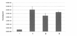
- Example 3 Exosome adsorption experiment 3 The number of exosome particles recovered in Example 2 was measured with Nanosite LM10 (Malvern). Calibration was performed using Silica Microspheres: 100 nm (Polysciences, Inc. # 24041) diluted with MilliQ water through GE plastic disk 13 (NYL 0.1 ⁇ m). Next, the exosome was diluted to about 10 8 with PBS through the GE plastic disk 13, the sample solution was injected into the measurement cell with a syringe, waited for 10 seconds, and then data measurement was performed for 30 seconds. By pushing the syringe and moving the sample solution in the chamber, the measurement was performed five times while replacing the inside of the cell with a new sample. FIG. 3 shows average values and standard deviations. The detection threshold was 4, and the camera level was 14.
- the exosome recovery rate is greatly improved compared to the case of purification using an untreated centrifuge tube. Is clear.
- Example 4 Centrifugation was performed once or twice as described below, and the amount of exosome recovered and the amount adsorbed on the tube were examined. Exosomes were purified from the culture supernatant of HT-29 cells as follows. The culture supernatant of 500 ml of HT-29 cells prepared as described above is centrifuged at 160,000 ⁇ g for 70 minutes to precipitate exosomes, and then the precipitate is resuspended in 180 ml of PBS, and further at 160,000 ⁇ g for 70 minutes. Washed by centrifugation. The resulting precipitate is resuspended in 1.3 ml of PBS to obtain a crude exosome fraction.
- fraction 2 (density 1.36 g / cm 3 ) was used as a purified exosome sample in this experiment.
- FIG. 4A shows the analysis result of the number of recovered exosome particles.
- the experiment shows the average of three times as a percentage of the input, and the number in parentheses shows the number of times of centrifugation.
- the polymer 1 when centrifugation is performed once, 50% or more of the input, and even when centrifugation is performed twice, 30% or more of the particles can be recovered.
- the comparative example of the polymer 4 when the comparative example of the polymer 4 was used, the recovery rate was only about 30% when the centrifugation was performed once and about 15% when the centrifugation was performed twice.
- FIG. 4B shows the analysis result by Western blotting.
- Each panel analyzes the collected sample and the sample adsorbed on the tube in each of the first, second, and third experiments, and shows the analysis results from the first to the third in order from the top. Detection was performed using an anti-EpCAM antibody (R & D Systems) and an anti-CD9 antibody. In the case where the polymer 4 was used, the sample collected by performing centrifugation twice was hardly detected even when using either the anti-EpCAM antibody or the anti-CD9 antibody. When 1 was used, detection was possible in any case.
- Example 5 The amount of adsorption by the film thickness of the coating agent was compared.
- Polymer 1 is dissolved in ethanol to 0.5, 1.0, 2.0, and 4.0 wt%, and coated on a microtube (manufactured by Treff) with a film thickness varying from 90 nm to 1400 nm for analysis. Used for.
- the film thickness was measured using a reflection spectral film thickness meter (FE-3000, manufactured by Otsuka Electronics Co., Ltd.).
- FE-3000 reflection spectral film thickness meter
- FIG. 5A shows the analysis result of the number of recovered exosome particles.
- 90, 140, 370, and 1400 shown on the left side indicate the film thickness (nm) coated with the microtube.
- the experiment shows the average of three times as a percentage of the input, and the number in parentheses shows the number of times of centrifugation.
- the film thickness is 50 nm or more and 2000 nm or less in consideration of variations in film formation and workability. If the film thickness is within this range, it is considered that exosome adsorption is prevented and a stable measurement result can be obtained.
- FIG. 5B shows the analysis result by Western blotting.
- the result of having detected by the anti- EpCAM antibody and the anti-CD9 antibody similarly to Example 4 is shown.
- the exosome adsorption was clearly suppressed compared to the uncoated tube at any film thickness.
- FIG. 5C shows the result of SDS electrophoresis of the collected sample and the sample adsorbed on the tube and analysis by silver staining.
- an untreated tube When an untreated tube is used, the recovered amount is clearly reduced.
- coating when coating is performed at any film thickness, it is clear that the number of adsorbed samples decreases and the number of recovered samples increases.
- the use of the coating agent containing the polymers 1 to 3 reduces the adsorption of exosomes to the container and significantly improves the recovery rate.
- These coating agents can not only improve the exosome recovery rate, but can also perform quantitative analysis with high accuracy.
- the coating agent of the present invention it is possible to prevent exosomes and other extracellular vesicles from adsorbing to a container or a substrate used for analysis. As a result, the recovery rate of extracellular vesicles can be remarkably improved, and the accuracy of analysis can be further improved.
Landscapes
- Chemical & Material Sciences (AREA)
- Health & Medical Sciences (AREA)
- Organic Chemistry (AREA)
- Life Sciences & Earth Sciences (AREA)
- Engineering & Computer Science (AREA)
- Medicinal Chemistry (AREA)
- Materials Engineering (AREA)
- Wood Science & Technology (AREA)
- Chemical Kinetics & Catalysis (AREA)
- Polymers & Plastics (AREA)
- Immunology (AREA)
- Hematology (AREA)
- Molecular Biology (AREA)
- Urology & Nephrology (AREA)
- Biomedical Technology (AREA)
- Analytical Chemistry (AREA)
- Microbiology (AREA)
- Cell Biology (AREA)
- Food Science & Technology (AREA)
- Physics & Mathematics (AREA)
- Biotechnology (AREA)
- Biochemistry (AREA)
- General Health & Medical Sciences (AREA)
- General Physics & Mathematics (AREA)
- Pathology (AREA)
- Apparatus Associated With Microorganisms And Enzymes (AREA)
- Solid-Sorbent Or Filter-Aiding Compositions (AREA)
- Paints Or Removers (AREA)
Abstract
La présente invention concerne un agent de revêtement, destiné à empêcher les vésicules extracellulaires représentées par les exosomes d'être adsorbées sur un outil. On peut empêcher que les vésicules extracellulaires soient adsorbées sur un outil à l'aide d'un agent de revêtement qui comporte un polymère hydrophile possédant un poids moléculaire moyen en poids de 10 000 à 1 000 000 et qui forme un film de revêtement possédant un angle de contact de 0 à 30 degrés.
Priority Applications (4)
| Application Number | Priority Date | Filing Date | Title |
|---|---|---|---|
| CN201780032132.4A CN109153882B (zh) | 2016-05-24 | 2017-05-23 | 细胞外小泡回收方法及细胞外小泡用容器 |
| US16/303,798 US10955410B2 (en) | 2016-05-24 | 2017-05-23 | Method of recovering extracellular vesicles and container for extracellular vesicles |
| JP2018519547A JP6970665B2 (ja) | 2016-05-24 | 2017-05-23 | 細胞外小胞回収方法及び細胞外小胞用容器 |
| EP17802773.6A EP3467059B1 (fr) | 2016-05-24 | 2017-05-23 | Procédé de récupération de vésicules extracellulaires et récipient destiné à des vésicules extracellulaires |
Applications Claiming Priority (2)
| Application Number | Priority Date | Filing Date | Title |
|---|---|---|---|
| JP2016-103091 | 2016-05-24 | ||
| JP2016103091 | 2016-05-24 |
Publications (1)
| Publication Number | Publication Date |
|---|---|
| WO2017204187A1 true WO2017204187A1 (fr) | 2017-11-30 |
Family
ID=60411962
Family Applications (1)
| Application Number | Title | Priority Date | Filing Date |
|---|---|---|---|
| PCT/JP2017/019121 Ceased WO2017204187A1 (fr) | 2016-05-24 | 2017-05-23 | Procédé de récupération de vésicules extracellulaires et récipient destiné à des vésicules extracellulaires |
Country Status (5)
| Country | Link |
|---|---|
| US (1) | US10955410B2 (fr) |
| EP (1) | EP3467059B1 (fr) |
| JP (1) | JP6970665B2 (fr) |
| CN (1) | CN109153882B (fr) |
| WO (1) | WO2017204187A1 (fr) |
Cited By (3)
| Publication number | Priority date | Publication date | Assignee | Title |
|---|---|---|---|---|
| WO2022124288A1 (fr) * | 2020-12-07 | 2022-06-16 | 大日本塗料株式会社 | Procédé de détection d'une substance granulaire par une méthode d'immunochromatographie, et kit correspondant |
| CN115887758A (zh) * | 2022-11-15 | 2023-04-04 | 西南交通大学 | 负载外泌体促进糖尿病创面修复的共聚水凝胶 |
| US12631627B2 (en) | 2018-11-30 | 2026-05-19 | Corning Incorporated | System and method for analyzing extracellular vesicles with an optical biosensor |
Families Citing this family (3)
| Publication number | Priority date | Publication date | Assignee | Title |
|---|---|---|---|---|
| EP3845566A1 (fr) | 2019-12-30 | 2021-07-07 | Industrial Technology Research Institute | Procede de separation de vesicules extracellulaires, particule colloidale et procede de preparation de celle-ci |
| US20240182698A1 (en) * | 2021-03-26 | 2024-06-06 | N-Lab Technology Center Pte. Ltd. | Water-soluble polymer to prevent non-specific adsorption |
| CN118345022A (zh) * | 2024-01-24 | 2024-07-16 | 上海市肺科医院(上海市职业病防治院) | 一种唾液中外泌体的富集提取方法及在筛查结核性潜伏感染中的应用 |
Citations (8)
| Publication number | Priority date | Publication date | Assignee | Title |
|---|---|---|---|---|
| JPH0339309A (ja) * | 1989-07-07 | 1991-02-20 | Res Dev Corp Of Japan | 生体適合性医療デバイス用材料 |
| JPH075177A (ja) * | 1993-06-15 | 1995-01-10 | Nippon Oil & Fats Co Ltd | 生化学的分析方法及び生化学的分析用反応容器 |
| JP2003014767A (ja) * | 2001-04-26 | 2003-01-15 | Nof Corp | 非特異的ハイブリダイゼーション抑制剤、臨床検査薬及び臨床検査法 |
| JP2004275862A (ja) * | 2003-03-14 | 2004-10-07 | Sumitomo Bakelite Co Ltd | 糖鎖化合物吸着制御実験器具 |
| JP2005099040A (ja) * | 1998-12-24 | 2005-04-14 | Sumitomo Bakelite Co Ltd | 免疫分析用容器 |
| JP2008058334A (ja) * | 2007-11-21 | 2008-03-13 | Towns:Kk | イムノクロマトグラフィー用展開溶媒、測定法およびキット |
| WO2015029979A1 (fr) * | 2013-08-30 | 2015-03-05 | 国立大学法人東京大学 | Procédé d'analyse d'exosome, puce d'analyse d'exosome et dispositif d'analyse d'exosome |
| WO2015068772A1 (fr) * | 2013-11-06 | 2015-05-14 | Jsr株式会社 | Procédé de séparation, procédé de détection, procédé de mesure de signal, procédé pour déterminer une maladie, procédé pour évaluer une efficacité de médicament de traitement de maladie, trousse et composition liquide |
Family Cites Families (12)
| Publication number | Priority date | Publication date | Assignee | Title |
|---|---|---|---|---|
| JP3443891B2 (ja) | 1993-09-14 | 2003-09-08 | 日本油脂株式会社 | タンパク質吸着防止剤 |
| ATE314147T1 (de) * | 1998-12-24 | 2006-01-15 | Sumitomo Bakelite Co | Behälter für immunoassays |
| FR2788780B1 (fr) | 1999-01-27 | 2001-03-30 | Ap Cells Inc | Procede de preparation de vesicules membranaires |
| US20040241176A1 (en) | 2000-04-27 | 2004-12-02 | Ap Cells. Inc. | Method of producing membrane vesicles |
| EP1657302B1 (fr) | 2003-06-25 | 2013-01-16 | Nof Corporation | Procédé de formation des corps embryoides |
| JP4931342B2 (ja) * | 2004-11-26 | 2012-05-16 | 株式会社トクヤマ | 義歯床用コーティング材 |
| CN101365501B (zh) * | 2005-12-09 | 2015-12-16 | 帝斯曼知识产权资产管理有限公司 | 包含聚电解质的亲水性涂料 |
| CA2705797A1 (fr) * | 2007-11-14 | 2009-05-22 | The Regents Of The University Of California | Lipides amphiphiles modifies par un sterol |
| JPWO2010122817A1 (ja) * | 2009-04-24 | 2012-10-25 | 株式会社ネクスト21 | 医療用樹脂製製品及び呼吸補助チューブ |
| SG183579A1 (en) | 2011-02-11 | 2012-09-27 | Agency Science Tech & Res | Methods of detecting therapeutic exosomes |
| JP6467226B2 (ja) | 2012-11-29 | 2019-02-06 | 大阪有機化学工業株式会社 | 親水性コート剤 |
| WO2016033695A1 (fr) * | 2014-09-05 | 2016-03-10 | Exerkine Corporation | Isolement d'exosomes |
-
2017
- 2017-05-23 WO PCT/JP2017/019121 patent/WO2017204187A1/fr not_active Ceased
- 2017-05-23 US US16/303,798 patent/US10955410B2/en active Active
- 2017-05-23 CN CN201780032132.4A patent/CN109153882B/zh active Active
- 2017-05-23 JP JP2018519547A patent/JP6970665B2/ja active Active
- 2017-05-23 EP EP17802773.6A patent/EP3467059B1/fr active Active
Patent Citations (8)
| Publication number | Priority date | Publication date | Assignee | Title |
|---|---|---|---|---|
| JPH0339309A (ja) * | 1989-07-07 | 1991-02-20 | Res Dev Corp Of Japan | 生体適合性医療デバイス用材料 |
| JPH075177A (ja) * | 1993-06-15 | 1995-01-10 | Nippon Oil & Fats Co Ltd | 生化学的分析方法及び生化学的分析用反応容器 |
| JP2005099040A (ja) * | 1998-12-24 | 2005-04-14 | Sumitomo Bakelite Co Ltd | 免疫分析用容器 |
| JP2003014767A (ja) * | 2001-04-26 | 2003-01-15 | Nof Corp | 非特異的ハイブリダイゼーション抑制剤、臨床検査薬及び臨床検査法 |
| JP2004275862A (ja) * | 2003-03-14 | 2004-10-07 | Sumitomo Bakelite Co Ltd | 糖鎖化合物吸着制御実験器具 |
| JP2008058334A (ja) * | 2007-11-21 | 2008-03-13 | Towns:Kk | イムノクロマトグラフィー用展開溶媒、測定法およびキット |
| WO2015029979A1 (fr) * | 2013-08-30 | 2015-03-05 | 国立大学法人東京大学 | Procédé d'analyse d'exosome, puce d'analyse d'exosome et dispositif d'analyse d'exosome |
| WO2015068772A1 (fr) * | 2013-11-06 | 2015-05-14 | Jsr株式会社 | Procédé de séparation, procédé de détection, procédé de mesure de signal, procédé pour déterminer une maladie, procédé pour évaluer une efficacité de médicament de traitement de maladie, trousse et composition liquide |
Non-Patent Citations (3)
| Title |
|---|
| KYOKO FUKAZAWA ET AL.: "Surface Modification of Various Materials Using Photoreactive MPC Polymer", FUNCTION & MATERIALS, vol. 33, no. 9, 2013, JP, pages 26 - 32, XP009513070, ISSN: 0286-4835 * |
| MADOKA TAKAI: "Evaluation of Biocompatible Polymer for Medical Devices", SCIENCE AND ENGINEERING OF MATERIALS, vol. 52, no. 1, 2015, pages 14 - 17, XP009513808 * |
| See also references of EP3467059A4 * |
Cited By (7)
| Publication number | Priority date | Publication date | Assignee | Title |
|---|---|---|---|---|
| US12631627B2 (en) | 2018-11-30 | 2026-05-19 | Corning Incorporated | System and method for analyzing extracellular vesicles with an optical biosensor |
| WO2022124288A1 (fr) * | 2020-12-07 | 2022-06-16 | 大日本塗料株式会社 | Procédé de détection d'une substance granulaire par une méthode d'immunochromatographie, et kit correspondant |
| JP2022090191A (ja) * | 2020-12-07 | 2022-06-17 | 大日本塗料株式会社 | 粒状物質をイムノクロマトグラフィー法によって検出する方法及びそのためのキット |
| JP7451385B2 (ja) | 2020-12-07 | 2024-03-18 | 大日本塗料株式会社 | 粒状物質をイムノクロマトグラフィー法によって検出する方法及びそのためのキット |
| US12669504B2 (en) | 2020-12-07 | 2026-06-30 | Dai Nippon Toryo Co., Ltd. | Method for detecting particulate substance by immunochromatography, and kit for the same |
| CN115887758A (zh) * | 2022-11-15 | 2023-04-04 | 西南交通大学 | 负载外泌体促进糖尿病创面修复的共聚水凝胶 |
| CN115887758B (zh) * | 2022-11-15 | 2024-01-26 | 西南交通大学 | 负载外泌体促进糖尿病创面修复的共聚水凝胶 |
Also Published As
| Publication number | Publication date |
|---|---|
| US10955410B2 (en) | 2021-03-23 |
| CN109153882B (zh) | 2022-03-04 |
| US20200025750A1 (en) | 2020-01-23 |
| CN109153882A (zh) | 2019-01-04 |
| EP3467059A4 (fr) | 2020-03-11 |
| JP6970665B2 (ja) | 2021-11-24 |
| EP3467059B1 (fr) | 2022-02-23 |
| JPWO2017204187A1 (ja) | 2019-03-22 |
| EP3467059A1 (fr) | 2019-04-10 |
Similar Documents
| Publication | Publication Date | Title |
|---|---|---|
| JP6970665B2 (ja) | 細胞外小胞回収方法及び細胞外小胞用容器 | |
| Gandham et al. | Technologies and standardization in research on extracellular vesicles | |
| Coumans et al. | Methodological guidelines to study extracellular vesicles | |
| Lee et al. | An integrated microfluidic chip for one-step isolation of circulating tumor cells | |
| Hou et al. | Microfluidic devices for blood fractionation | |
| Zigeuner et al. | Isolation of circulating cancer cells from whole blood by immunomagnetic cell enrichment and unenriched immunocytochemistry in vitro | |
| CN101371124B (zh) | 处理生物样品和/或化学样品的方法 | |
| Yin et al. | Detection of circulating tumor cells by fluorescence microspheres-mediated amplification | |
| CN103513021B (zh) | 含血液成分的试样的处理方法 | |
| JP2010510777A (ja) | 液滴中においてサンプルを処理するための装置、およびそれを使用する方法 | |
| Zhao et al. | Separation and single-cell analysis for free gastric cancer cells in ascites and peritoneal lavages based on microfluidic chips | |
| JP6895662B2 (ja) | 特定細胞の分画方法及び捕捉方法 | |
| US11467073B2 (en) | Method for concentrating extracellular vesicles | |
| WO2015046557A1 (fr) | Dispositif pour concentrer et séparer des cellules tumorales circulantes, et procédé associé | |
| JPWO2020158835A1 (ja) | 試薬キット、測定キットおよび測定方法 | |
| Müller | Novel tools for the study of cell type-specific exosomes and microvesicles | |
| Feng et al. | Simultaneous detection of two extracellular vesicle subpopulations in saliva assisting tumor T staging of oral squamous cell carcinoma | |
| Cao et al. | Partitioning-induced isolation of analyte and analysis via multiscaled aqueous two-phase system | |
| Sabaté del Río et al. | Microfluidic dielectrophoretic purification of extracellular vesicles from plasma lipoproteins | |
| Gribko et al. | IsoMAG—An Automated System for the Immunomagnetic Isolation of Squamous Cell Carcinoma-Derived Circulating Tumor Cells | |
| JP6272296B2 (ja) | 懸濁された細胞成分を均質に分布させるための方法と装置 | |
| US9632086B2 (en) | Method and kit for determining-antibody sensitivity and clone cell strain | |
| Genshaft et al. | Clinical implementation of single-cell RNA sequencing using liver fine needle aspirate tissue sampling and centralized processing captures compartment specific immuno-diversity | |
| JP6886668B2 (ja) | 医療用検査装置及び細胞検査方法 | |
| Hakim et al. | Assessment of the gene therapy immune response in the canine muscular dystrophy model |
Legal Events
| Date | Code | Title | Description |
|---|---|---|---|
| ENP | Entry into the national phase |
Ref document number: 2018519547 Country of ref document: JP Kind code of ref document: A |
|
| NENP | Non-entry into the national phase |
Ref country code: DE |
|
| 121 | Ep: the epo has been informed by wipo that ep was designated in this application |
Ref document number: 17802773 Country of ref document: EP Kind code of ref document: A1 |
|
| ENP | Entry into the national phase |
Ref document number: 2017802773 Country of ref document: EP Effective date: 20190102 |