WO2017204446A2 - Procédé d'analyse de l'activité de cellules tueuses naturelles à l'aide de l'activité synergique d'un récepteur, et procédé de diagnostic de maladie associée à l'activité des cellules tueuses naturelles utilisant ledit procédé d'analyse - Google Patents
Procédé d'analyse de l'activité de cellules tueuses naturelles à l'aide de l'activité synergique d'un récepteur, et procédé de diagnostic de maladie associée à l'activité des cellules tueuses naturelles utilisant ledit procédé d'analyse Download PDFInfo
- Publication number
- WO2017204446A2 WO2017204446A2 PCT/KR2017/002870 KR2017002870W WO2017204446A2 WO 2017204446 A2 WO2017204446 A2 WO 2017204446A2 KR 2017002870 W KR2017002870 W KR 2017002870W WO 2017204446 A2 WO2017204446 A2 WO 2017204446A2
- Authority
- WO
- WIPO (PCT)
- Prior art keywords
- cells
- activity
- cell
- synergistic
- disease
- Prior art date
- Legal status (The legal status is an assumption and is not a legal conclusion. Google has not performed a legal analysis and makes no representation as to the accuracy of the status listed.)
- Ceased
Links
Images


Classifications
-
- G—PHYSICS
- G01—MEASURING; TESTING
- G01N—INVESTIGATING OR ANALYSING MATERIALS BY DETERMINING THEIR CHEMICAL OR PHYSICAL PROPERTIES
- G01N33/00—Investigating or analysing materials by specific methods not covered by groups G01N1/00 - G01N31/00
- G01N33/48—Biological material, e.g. blood, urine; Haemocytometers
- G01N33/50—Chemical analysis of biological material, e.g. blood, urine; Testing involving biospecific ligand binding methods; Immunological testing
- G01N33/68—Chemical analysis of biological material, e.g. blood, urine; Testing involving biospecific ligand binding methods; Immunological testing involving proteins, peptides or amino acids
Definitions
- the present invention relates to a method for examining the activity of NK cells using receptor synergistic activity of NK cells and a method for diagnosing a disease related to the activity of NK cells using the same.
- NK cells Natural killer cells
- MHC major histocompatibility complex
- NK cells The activity of NK cells involves various activating and inhibitory receptors.
- receptors considered dominant are immunoreceptor tyrosine-based activation motifs (ITAMs) -associated signaling molecules (eg, FcR ⁇ chains or TCR ⁇ chains) associated with NKp46 and the signaling molecule DAP10.
- ITAMs immunoreceptor tyrosine-based activation motifs
- NKp46 a signaling ⁇ chains or TCR ⁇ chains
- DAP10 signaling molecule DAP10
- Receptors considered costimulatory include DNAM-1 (CD226), CD2, and NKp80 ( KLRF1 gene products, as well as members of the signaling lymphocytic activation molecule (SLAM) family, such as 2B4 (CD244).
- SLAM signaling lymphocytic activation molecule
- ITAM-related molecules contribute to the signaling by several different activating receptors of NK cells, NKp46 and NKp30, which are related to FcR ⁇ and / or TCR ⁇ , while natural cytotoxicity receptors (NCRs), while NKp44 Is associated with the signal adapter DAP12 (EMBO J. 2004; 23: 255-9).
- Hemophagocytic lymphohistiocytosis is a disease that shows the proliferation of monocytes or macrophage and shows uncontrolled hypersecretion of hemophagocytosis and inflammatory cytokines.
- immunodeficiency X-linked lymphoproliferative disease 1 is a disease with symptoms such as HLH, which is caused by Epstein-Barr virus (EBV),
- EBV Epstein-Barr virus
- SH2D1A which makes SAP proteins, forms the genetic basis of XLP1 (Annu. Rev. Immunol. 2007; 25: 337-79, Annu. Rev. Immunol. 2011; 29: 665-705) .
- 2B4 contains an ITSM motif at the cellular tail and carries activation signals through recruitment of small adapters SAP and SAP-associated tyrosine kinase Fyn ( J. Exp . Med . 202, 181-192 (2005), Immunity 36, 974-985 (2012)). Signaling of 2B4 leads to a Vav1, p38 MAPK, Erk, and the PLC-enabled g2 (Nat. Immunol. 9, 495-502 (2008). Note that in NK cells obtained from congenital XLP1 patients lacking functional SAP expression, 2B4 is inactive and can instead transmit an inhibitory signal ( Annu . Rev. Immunol . 25, 337-379 (2007)).
- NK cell defects are thought to contribute to signs of clinical XLP1, such as killing defects of Epstein Barr virus (EBV) -infected B cells, defects in IFN-g production, fulminant mononucleosis, and B-cell lymphoma do.
- EBV Epstein Barr virus
- NKG2D J. Immunol. 175, 5541-5550 (2005), Cancer Res. 69, 7775-7783 (2009), J. Immunol. 189, 1360-1371 (2012), which is an anticancer receptor in NK cells of cancer patients
- Defects have been observed in NK cell activation via DNAM-1 (Immunol. Cell Biol. 90, 109-115 (2012)).
- the expression and function of NKG2D and 2B4 are also reduced in NK cells of hepatitis B virus infected patients (PloS Pathog. 8, e1002594 (2012)).
- One aspect includes specifically stimulating two or more factors that are distinct on NK cells in a sample; Measuring the degree of synergistic activation of the NK cells induced by stimulation with the two or more factors; And it provides a method for testing the activity of NK cells comprising the step of comparing the degree of synergistic activation of the NK cells compared to normal NK cells.
- Another aspect is a stimulatory substance that produces synergistic activity of NK cells, the antibody or fragment thereof specific for two or more factors that are distinguished on the NK cell, or a target cell or polypeptide that causes specific activity, specifically
- a composition for inducing activity and a composition for testing the activity of NK cells comprising one or more selected from a reversible or irreversible effector.
- Another aspect is a stimulus substance that stimulates two or more factors distinguished on NK cells in a sample to cause synergistic activity of the NK cells, wherein the antibody or fragment thereof, specific for each or more than two factors, or a specific activity thereof One or more selected from target cells or polypeptides that cause proliferation, compositions that specifically induce activity, and reversible or irreversible agents; And it provides a kit for diagnosing a disease associated with the synergistic activity of NK cells comprising a substance for detecting the presence or absence of activation of the NK cells compared to normal NK cells.
- One aspect includes specifically stimulating two or more factors that are distinct on NK cells in a sample; Measuring the degree of synergistic activation of the NK cells induced by stimulation with the two or more factors; And it provides a method for testing the activity of NK cells comprising the step of comparing the degree of synergistic activation of the NK cells compared to normal NK cells.
- the sample may be a biological sample derived from an individual, eg, a mammal, including a human.
- the biological sample may be isolated from an individual, and may be blood, whole blood, serum, plasma, lymph, urine, feces, tissue, cells, organs, bone marrow, saliva, sputum, cerebrospinal fluid, or a combination thereof.
- the biological sample may also comprise PBMCs, purified NK cells or primary resting cells (ie, isolated directly from blood).
- the sample is blood.
- differentiation includes all cases that are not equivalent in position, structure, function, etc. in a cell. Specifically, different signaling processes are induced within the cell after stimulation, or positions or factors on the cells themselves are structurally different so that the means or methods of stimulation are different, or in a single stimulus different roles in the cell. Or insufficiently effective in effectively activating.
- factor refers to all molecular elements that cause synergistic activation of NK cells. Specifically, the factor may include intracellular and external adapters, proteins, glycoproteins, peptides, and motifs that can bind to receptors, channels, and specific ligands in and out of cells.
- the factor may be a receptor.
- receptor refers to any molecule that binds to a specific substance and changes the activity of NK cells.
- specific substances include chemical compositions, specific proteins derived from the body or artificial, peptides, cholesterol, glycoproteins, cytokines or chemokines by other immune cells or NK cells themselves, or specific receptors on target cells or NK cells themselves. Or membrane protein.
- the factor may be present on the cell surface or within the cell.
- the factor when it is a receptor, it is located on the cell surface of the NK cells and binds to a specific substance and transmits the signal into the cell to induce the activity of the NK cells, as well as through the cell membrane to the cell membrane inner surface or cytoplasm.
- Representative receptors include all receptors that can bind to a specific external stimulus and cause a signaling action.
- the term "stimulus” means acting on a factor to cause specific signaling in and out of NK cells.
- the method of causing stimulation may employ antibodies, fragments or peptides specific for one or more of the factors, compositions that specifically induce or inhibit, and reversible or irreversible agonists or antagonists.
- culture with a culture comprising various target cells capable of stimulating NK cells eg, K562, 721.221, or P815 coated with specific receptor stimulating antibodies).
- NK cell activity is not sufficient to effectively activate NK cell activity, but the effect of each of the single factors is significantly changed compared to that of each of the single factors when each distinct factor is co-stimulated simultaneously or sequentially. It means that it can be induced or effectively activate the NK cell activity effectively.
- “synergistic activity” or “synergy activation” of NK cells specifically stimulates two or more factors distinguished on NK cells, respectively, and combinations thereof, to stimulate stimulation for each of these factors and combinations thereof.
- the degree of NK cell activity is at least 1.5 times, at least 2 times, or at least 3 times, it may be determined that synergistic activity is induced, and if it is more than 2 times, it may be determined that synergistic activity is induced.
- the degree of synergistic activation is secreted by the degree of phosphorylation or expression of subfactors, degranulation activity, cytotoxic activity and NK cell stimulation using flow cytometry, immunoblotting, etc., as described herein.
- the cytokines can be quantified based on the measurement, but are not limited thereto.
- the step of comparing the synergistic activation of the NK cells compared to the normal NK cells specifically, two or more factors distinguished under the same conditions as the experimental group with normal NK cells as a control group, respectively Or when synergistic activation occurs in normal NK cells when stimulated with a specific combination, it is determined to be abnormal when the test group is markedly high, not present, or significantly less.
- it is easy to artificially generate synergistic activation includes determining by comparing the data.
- abnormal NK cell refers to NK cells that have or are derived from an individual with no disease, and the individual without the disease is at least physical, genetic, or exotic known to affect NK cell activity. There is no condition.
- the method may further comprise the step of separating the NK cells required in the sample. If necessary, the step may be performed after stimulation while being present with other blood cells or lymphocytes, and the stimulating step may be performed after the step is performed to prepare a sample containing only NK cells as lymphocytes.
- the degree of purification of the isolated NK cells and the composition of the sample may vary to the extent necessary for the experiment. NK cells can be used as it is purified from the sample as needed, and can be used after proliferation in order to secure conditions or cell mass suitable for the experiment.
- the step of separating the NK cells may not be necessary. .
- the stimulus may be made simultaneously or sequentially to the factor. Specifically, it may be stimulated by structurally binding to the factor or by modifying the factor. Such modifications may include post-translation modifications or cleavage such as, for example, phosphorylation, ubiquitination, sulfonylation, methylation, parylation or glycosylation.
- the factor is selected from ITAM-containing signaling molecules (eg, CD16, NKp46, NKp30 or NKp44, etc.), NKG2D, 2B4 (CD244), DNAM-1 (CD226), CD2 and NKp80 It may be.
- ITAM-containing signaling molecules eg, CD16, NKp46, NKp30 or NKp44, etc.
- NKG2D, 2B4 CD244
- DNAM-1 CD226)
- CD2 and NKp80 It may be. According to Bryceson Y et al. (Blood 2006; 107: 159-166) and Kim HS et al.
- NK cell synergy activation may be possible by various combinations of the factors listed above, combinations of these factors Examples may include, but are not limited to, 2B4 and DNAM-1, DNAM-1 and NKG2D, 2B4 and NKG2D, or 2B4, DNAM-1 and NKG2D.
- said stimulating in said stimulating step is an antibody or fragment thereof specific for said two or more factors, or a fragment thereof, a target cell or polypeptide that elicits specific activity, a compound that specifically induces or inhibits, and It may be caused by one or more selected from reversible or irreversible agonists or antagonists.
- Antibodies or fragments thereof specific for such factors include isotypes and may bind simultaneously to two or more distinct factors. Specifically, an example of simultaneous binding to two or more distinct factors may be P815 coated with CD244 / 2B4 or CD226 / DNAM-1.
- the agent or antagonist may be a compound having structurally specific properties for a factor of interest, and may be a polypeptide having tertiary structural specificity.
- the target cell that induces activity specific to said factor may be coated with one or more selected from antibodies, fragments and combinations thereof against two or more factors distinguished on NK cells. Two or more factors distinguished on said NK cells are as described above.
- the antibody may be an antibody against the isotype of the factors, and the fragment may comprise only a specific portion of the antibody sufficient to effect activity as a fragment of the antibody. Coating can be carried out by preincubation with target cells, as described in the Examples below, or can be coated with the antibody, fragment or combination thereof using cross-linking techniques.
- the step of measuring the degree of activation comprises the degree of phosphorylation or expression of subfactors of two or more factors distinguished on the NK cells, degranulation activity, cytotoxicity activity and NK cell stimulation. It may include one or more selected from the measurements of cytokines secreted by.
- the term “subfactor” refers to an intracellular protein or polypeptide that is affected by increased expression or phosphorylation when a factor on the NK cell is stimulated, and particularly for phosphorylation, the subfactor is a substrate. Can be used as the same meaning (substrate).
- the expression level of the subfactor may be determined by, for example, a well-known immunoblot using an antibody specific for Erk or pY174-Vav1, which is one of intracellular proteins involved in NK cell activation after lysis of stimulated NK cells. It can be measured using an immunoblot method.
- the degranulation activity may mean, for example, inducing lysis of target cells with perforin or granzyme secretion, which can be analyzed using FACS. Specifically, after stimulating PBMC or purely isolated NK cells isolated from a sample, a method of measuring CD107a expression in proportion to degranulation can be measured using a fluorochrome-conjugated antibody.
- the cytotoxic activity is determined by, for example, incubation with a culture comprising target cells capable of activating NK cells labeled with europium fluorescent dyes, and then the amount of fluorescent dyes released through target cell lysis. Measurements can be made using a microplate reader.
- the cytokine may be selected from IFN- ⁇ , TNF- ⁇ , TNF- ⁇ , MIP-1 ⁇ , MIP-1 ⁇ , PANTES, IL-8 and IL-10.
- the NK cell immunoactivator expression analysis may be performed using FACS, intracellular cytokine staining, or ELISA. Specifically, after staining the surface of NK cells with a specific fluorochrome-conjugated antibody, the cells are permeabilized and treated with another specific immunoactivator (eg, IFN- ⁇ ) with fluorochrome-conjugated antibodies. By staining the cytokines and the like can be used a method for measuring the expression of immune activator in NK cells.
- the normal person showed a more than two-fold difference in synergistic activation based on the higher of the two factors in terms of cytokine secretion or cytotoxic degranulation.
- synergistic activation there was a more than two-fold difference in the amount of synergistic activated cytokine secretion or the degree of cytotoxic degranulation.
- comparing the level of synergistic activation of the NK cells with the normal NK cells is cytokine secreted by the degree of phosphorylation or expression of the subfactor, degranulation activity, cytotoxic activity and NK cell stimulation
- the difference in the amount of secretion is more than two times, specifically two or more times based on the higher of the two factors can be determined that the activity of NK cells in the sample is normal.
- the difference in the amount of synergistic activated cytokine secretion or cytotoxic degranulation of the NK cells is 2 times or less, specifically 2 based on the amount of synergistic activated cytokine secretion or cytotoxic degranulation of normal NK cells. If it is less than 2 times, it can be determined that the activity of NK cells in the sample is abnormal.
- the method for testing the activity of NK cells of the present invention can be used to provide information on the diagnosis of a disease associated with the synergistic activity of NK cells.
- the present inventors found that although NKH activation is induced in HLH patients and pancreatic cancer patients, the normal persons have a high degree of synergistic activation, whereas the patients fail or do not have a small degree of synergistic activity.
- synergistic activation compared with simply inducing NK cell activation makes the comparison more meaningful and easier and more accurate in diagnosing HLH or pancreatic cancer. Therefore, the method for testing the activity of NK cells of the present invention may be useful for distinguishing samples showing abnormal NK cell synergistic activity, and providing information on diagnosis of a disease associated with synergistic activity of NK cells, including HLH and pancreatic cancer.
- the disease associated with the synergistic activity of the NK cell is an abnormal synergistic activity of the NK cell, for example, irritable immune disease, autoimmune disease, immune rejection, immunodeficiency disease, histiocytosis, cancer, Type 2 diabetes, parasitic infectious diseases and viral diseases.
- the irritable immune disease is at least one selected from asthma and sinusitis
- the autoimmune disease is at least one selected from lupus, multiple sclerosis, type 1 diabetes and rheumatoid arthritis
- the histiocytosis is HLH, XLP1 And may be one or more selected from XLP2.
- Hematopoietic lymphocytocytosis is associated with group 2 Langerhans' cell histiocytosis, erythroid phagocytosis (family, sporadic), infectious hematopoietic syndrome, virus-associated hematopoietic syndrome, massive lymphadenopathy May include the meaning of histiocytosis, or reticulosis.
- the term "cancer” includes a tumor, hematologic cancer, or solid cancer, and includes not impairing synergistic activity of an individual's NK cells or causing synergistic activity of NK cells as a target cell under certain conditions.
- the cancer is lung cancer, liver cancer, esophageal cancer, gastric cancer, colon cancer, small intestine cancer, pancreatic cancer, melanoma, breast cancer, oral cancer, brain tumor, thyroid cancer, parathyroid cancer, kidney cancer, cervical cancer, sarcoma, prostate cancer, urethra It may be selected from the group consisting of cancer, bladder cancer, testicular cancer, hematologic cancer, lymphoma, skin cancer, psoriasis and fibroadenoma.
- the cancer may be pancreatic cancer or B cell lymphoma.
- the viral disease may be hepatitis B.
- the immunodeficiency disease may be DiGeorge synderome or Chedial-Higashi syndrome.
- the information on the disease associated with the activity of the NK cells can be determined to be abnormal NK cells when no synergistic activation of the experimental group NK cells compared to normal NK cells, or NK cells that lost synergistic activity to a specific receptor target It can be determined that the target cell is associated with a particular disease if it causes or does not cause abnormal synergistic activity against the cell.
- the target cell may further comprise coating with one or more selected from antibodies, fragments and combinations thereof against two or more factors distinguished on the NK cells.
- Another aspect is a stimulatory substance that produces synergistic activity of NK cells, the antibody or fragment thereof specific for two or more factors that are distinguished on the NK cell, or a target cell or polypeptide that causes specific activity, specifically It provides a composition for testing the activity of NK cells comprising a compound inducing activity, and at least one selected from a reversible or irreversible effector.
- the factor on the NK cells can be selected from NKG2D, 2B4 (CD244), DNAM-1 (CD226), CD2, CD16, NKp30, NKp44, NKp46, and NKp80.
- said composition is secreted by the degree of phosphorylation or expression of subfactors of said factor, degranulation activity, cytotoxic activity and NK cell stimulation. It may further comprise an antibody or fragment thereof for detecting one or more selected of the measured cytokines.
- the agent for detecting the presence of synergistic activation of the NK cells is Perforin, Granzim, CD107a, CD107b, IFN- ⁇ , MIP-1 ⁇ , MIP-1 ⁇ , TNF- ⁇ , TNF- ⁇ , PANTES, One or more selected from antibodies, fragments or combinations thereof against one or more selected from IL-8 and IL-10.
- the composition for testing the activity of the NK cells is a substance for visually displaying the detection result of the presence of synergistic activation of the NK cells, dye (dye), fluorescent dyes, secondary antibodies, or biological or molecular It may further include an enemy probe.
- Another aspect is a stimulus substance that stimulates two or more factors distinguished on NK cells in a sample to cause synergistic activity of the NK cells, wherein the antibody or fragment thereof, specific for each or more than two factors, or a specific activity thereof One or more selected from target cells or polypeptides that cause proliferation, compounds that specifically induce activity, and reversible or irreversible agents; And it provides a kit for diagnosing a disease associated with the synergistic activity of NK cells comprising a substance for detecting the presence or absence of activation of the NK cells compared to normal NK cells.
- the agent for detecting the presence of synergistic activation of the NK cells is Perforin, Granzim, CD107a, CD107b, IFN- ⁇ , MIP-1 ⁇ , MIP-1 ⁇ , TNF- ⁇ , TNF- ⁇ , PANTES, At least one selected from antibodies, fragments or combinations thereof against at least one selected from IL-8 and IL-10, or a fluorescent dye.
- the antibody, fragment or combination thereof may be labeled or unlabeled with a fluorescent probe.
- the fluorescent dye or fluorescent probe may vary depending on conditions such as an object to be detected, a combination or number of the objects, a desired wavelength, a detector, and the like, for example, peridinin chlorophyll (PerCP), phycoerythrin (phycoerythrin; PE), fluorescent isothiocyanate (FITC), or europium (europium), but is not limited thereto.
- PerCP peridinin chlorophyll
- PE phycoerythrin
- FITC fluorescent isothiocyanate
- europium europium
- IFN- [gamma] and TNF- [alpha] are labeled with a fluorescent probe, FITC, so that the IFN- [gamma] and TNF- [alpha] can be directly detected by flow cytometry without secondary processing.
- a fluorescent dye for example, P815
- P815 fluorescent dye
- the degree of synergy activation can be measured. Therefore, using a fluorescent dye as a substance for detecting the presence of synergistic activation of the NK cells may be particularly advantageous for measuring cytotoxicity.
- the target cell that induces activity specific for a factor on the NK cell may be coated with one or more selected from antibodies, fragments, and combinations thereof against two or more factors distinguished on the NK cell.
- the NK cell activity test kit is a material for visually displaying the detection result of the presence of synergistic activation of the NK cells, a dye, fluorescent dyes, secondary antibodies, or biological or molecular probes It may further include.
- the disease associated with the synergistic activity of the NK cells may be selected from irritable immune diseases, autoimmune diseases, immune rejection, histiocytosis, cancer, type 2 diabetes, parasitic infection diseases and viral infection diseases.
- kits such as those mentioned in the method for assaying the activity of the claimed NK cells or for the composition for testing the activity of NK cells, are understood as mentioned in the method or composition claimed above.
- NK cells activity test method comprising the step of comparing the degree of synergistic activation of the NK cells compared to normal NK cells, it is possible to test the activity of NK cells in a simpler, faster and more significant than the known technology
- diseases associated with the activity of NK cells can be readily diagnosed early and prognostic.
- a stimulant that produces synergistic activity of NK cells according to another aspect, wherein the antibody or fragment thereof specific for two or more factors distinguished on the NK cells, or a target cell or polypeptide that induces specific activity, specific
- a method for early diagnosis, prognostic determination, and the like of diseases related to the activity of NK cells using a composition for inducing activity, and a composition for testing the activity of NK cells comprising one or more selected from reversible or irreversible agonists.
- Stimulus substance that stimulates two or more factors distinguished on NK cells in a sample according to another aspect, thereby causing synergistic activity of the NK cells, wherein the antibody or fragment thereof, specific for two or more factors, respectively One or more selected from target cells or polypeptides that induce activity, compositions that specifically induce activity, and reversible or irreversible agents;
- a kit for diagnosing a disease related to synergistic activity of NK cells including a substance that detects the presence or absence of activation of the NK cells compared to normal NK cells, by activating NK cells in a simpler, faster, and more meaningful manner than the known technology. Early diagnosis or prognosis can be determined.
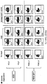
- Figure 1 shows the results of flow cytometry analysis of the degree of phosphorylation of NF- ⁇ B p65 subunit when specifically stimulating the distinct factors NKG2D and / or 2B4.
- Figure 2 shows the results of flow cytometry analysis of the frequency of cells expressing IFN- ⁇ or TNF- ⁇ when specifically stimulating the distinct factors NKG2D and / or 2B4.
- Figure 3 confirms the synergistic activity of NK cells as cytotoxic when single or co-stimulated specific receptors.
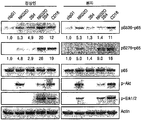
- NK cells 4 shows the expression level of factors related to NK cell activation when NK cells obtained from normal or XLP1 patient donors are stimulated with distinct factors NKG2D and / or 2B4.
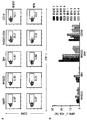
- FIG. 5 (A) When NK cells obtained from normal or XLP1 patient donors were stimulated with distinct factors NKG2D and / or 2B4, IFN- ⁇ expression was analyzed by flow cytometry. (B) The percentage of NK cells expressing IFN- [gamma] expressing IFN- [gamma] is shown when stimulation (IFN- [gamma] + NK cells) is applied to NK cells without stimulation ([Delta] IFN- [gamma] + cells).
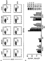
- FIG. 6 shows defective cytotoxic degranulation of XLP1 NK cells for coactivation
- A expression of CD107a when NK cells obtained from normal or XLP1 donors are stimulated with distinct factors NKG2D and / or 2B4 The degree was analyzed by flow cytometry.
- B The percentage of increase of CD107a + NK cells is shown when no stimulation is applied to NK cells ( ⁇ CD107a + cells) compared to stimulation (CD107a + NK cells).
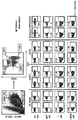
- Figure 7 shows the results of flow cytometry analysis of the degree of activity by the stimulation of the NKG2D / 2B4 combination in various blood cells.
- Figure 8 shows the results of flow cytometry analysis of the degree of activity by stimulation of the NKG2D / 2B4 combination in NK cells.
- Example 1 The distinct factor NKG2D And 2B4's Normal by joint activation NK Check for synergistic activity in cells
- PBMCs Peripheral blood mononuclear cells
- LSM lymphocyte separation medium
- NK cell separation kit StemCell Technologies
- Human NK cell host NKL (donated by M. Robertson) was cultured in RPMI1640 supplemented with 10% FBS, 1 mM sodium pyruvate, and 200 U / ml recombinant IL-2 (rIL-2).
- NKL cells were resting in RPMI1640 supplemented with 5% FBS and 0.5 mM sodium pyruvate for 24 hours without rIL-2.
- NK- ⁇ B is an important transcription factor as a regulator in the innate and acquired immune responses, resulting in increased NK cell activity upon phosphorylation of the NK- ⁇ B subunit p65 Ser536 residue (Front Immunol. 2014; 5: 662.) . Therefore, phosphorylation of NF- ⁇ B p65 subunits in primary resting NK cells was measured as NK cell activity factor.
- NK cells Purified human primary NK or NKL cells were preincubated with mAbs specific for NKG2D and / or 2B4 or isotype control mAb (cIgG1) at NK receptor (all at 10 ⁇ g / ml) on ice for 30 minutes. After washing with medium, NK cells were stimulated by receptor crosslinking with goat anti-mouse F (ab ') 2 secondary Ab at 37 ° C for 5 minutes. Cells were washed with DPBS and fixed at 4% paraformaldehyde at 37 ° C. for 10 minutes. The immobilized cells were then permeabilized on ice for 30 minutes with 90% methanol and blocked for 10 minutes at room temperature with 0.5% BSA.
- Cells were stained with Alexa Fluor 488-conjugated anti-pS536 p65 Ab (93H1) or isotype control rabbit IgG (DA1E) (Cell Signaling) and analyzed for flow analysis using a flow cytometry analyzer for p65 phosphorylation.
- the percentage of cells that respond increases synergistically with NKG2D and 2B4 coupled stimulation of primary NK cells, while the response to receptor alone is significantly less than that of coactivation. Appeared.
- NK Immune activator measurement as an indicator of synergistic activity of cells
- NK cells When NK cells are activated, they secrete IFN- ⁇ or TNF- ⁇ , which are immune activators (or cytokines). Therefore, we tested whether the secretion of cytokines increased synergistically by the coactivation of specific factors.
- NK cells were harvested as described above and PBMCs were stimulated with P815 cells (American Type Culture Collection) preincubated with mAbs specific for NKG2D and / or 2B4 or isotype control mAbs. It was then incubated for 6 hours and the cell surface markers stained with CD3 and CD56. Cytokine production by NK cells was measured as intracellular expression of IFN- ⁇ or TNF- ⁇ in CD3-CD56 + NK cells. Specifically, PBMCs or primary resting NK cells were stimulated at 37 ° C. for 1 hour with the same number of designated labeled cells. Brefeldin A (GolgiPlug; BD Bioscience) was then added and incubated for an additional 5 hours for a total of 6 hours.
- P815 cells American Type Culture Collection
- mAbs specific for NKG2D and / or 2B4 or isotype control mAbs It was then incubated for 6 hours and the cell surface markers stained with CD3 and CD56. Cytokin
- the cells were then stained with surface markers with anti-CD56-PE and / or anti-CD3-PerCP mAbs for 30 minutes in the 4 ° C. dark. After washing cells twice with FACS buffer, they were incubated in BD Cytofix / Cytoperm solution (BD Bioscience) for 20 minutes at 4 ° C. in the dark. After intracellular staining with anti-IFN- ⁇ -FITC or anti-TNF- ⁇ -FITC mAb for 30 minutes in the dark at 4 ° C, cells were washed twice with BD Perm / Wash buffer (BD Bioscience). Cells were then gated against NK cells and analyzed by flow cytometry.
- BD Cytofix / Cytoperm solution BD Bioscience
- Example 2 By coactivation of distinct factors NK Cytotoxicity measurement as an indicator of synergistic activity of cells
- P815 cells were preincubated with mAbs or isotype control mAbs specific for CD56, CD16 and NKG2D and / or 2B4, DNAM-1 and / or 2B4, and P815 coated with the NK cell specific receptor stimulating antibody was europium ) Labeled with fluorescent dye (dye). Then, as described above, the primary resting NK cells were stimulated by culturing with the P815 cells. Afterwards, the culture medium was recovered by centrifugation, and the amount of fluorescent dyes released through target cell lysis, which is an indicator of cytotoxicity, was measured. The degree of fluorescence in the culture was analyzed through a microplate reader.
- CD16 induced exceptionally strong cytotoxicity, but when stimulated with NKG2D + 2B4 and DNAM-1 + 2B4, they induce synergistic cytotoxicity compared to the stimulated cases, respectively.
- specific receptor stimulation for example, by the use of specific receptor stimulating antibodies as described above, thereby enabling synergistic measurements by specifically stimulating two distinct factors.
- synergistic activity induced by simultaneous stimulation compared to the case of single stimulation may be easy to distinguish due to a significant difference in comparing NK activity.
- Example 3 Normal and XLP1 Patient's NKG2D And 2B4's Comparison of NK Cell Activity Using Synergistic Differences in Specific Factor Expression by Coactivation
- NK cells were donated from a patient of XLP1.
- Hereditary SH2D1A as a Target for Congenital XLP1 Gene mutation patients were included.
- NK cells of XLP1 patients were expanded as described above to study molecular signaling on NF- ⁇ B activation and effector function.
- NK cells from normal donors were also proliferated and, after resting periods, stimulated with NKG2D and 2B4 to reproduce synergistic increases in effector function and signaling.
- NK cells and NK cells in XLP1 patients were compared using Western blot. Specifically, primary resting NK cells after proliferation from normal or XLP1 patient donors were preincubated with mAbs or isotype control mAb (cIgG1) specific for the designated receptor. After crosslinking the receptors for 2 minutes (p-Akt and p-Erk1 / 2) and 5 minutes (pS536-p65 and pS276-p65), the lysates were immunoblotted with the specified phosphorylation. As a result, as shown in FIG.
- diagnosing XLP1 synergistic activity caused by coactivating distinct factors of NK cells is observed because the difference is significantly observed when co-activating each distinct factor rather than measuring the activity of NK cells. It is shown that diagnosing XLP1 may be advantageous.
- Example 4 Normal and XLP1 Patient's NKG2D And 2B4's By joint activation IFN using synergistic differences in the amount of - ⁇ expression NK Cell activity comparison
- a test was performed to compare the expression level of IFN- ⁇ , one of the immune activators. Specifically, primary resting NK cells after propagation from normal or XLP1 patient donors were mixed with P815 cells preincubated with mAbs specific for the designated receptor. After incubation for 6 hours, staining with fluorochrome-conjugated mAb for CD56 and intracellular staining with IFN- ⁇ were analyzed by flow cytometry as described above.
- Example 5 Normal and XLP1 Patient's NKG2D And 2B4's By joint activation In degranulation Using synergistic differences NK Cell activity comparison
- XLP1 By measuring the expression level of CD107a on the surface of NK cells in proportion to degranulation, one of the indicators of NK cell activity, we examined whether XLP1 could be diagnosed by the synergistic effect caused by the stimulation of two distinct factors.
- NK cells Degranulation of NK cells was assessed by CD107a expression and Granzyme B release (BioLegend) on the cell surface.
- primary resting NK cells of normal and XLP1 patients were propagated as described above and then mixed with P815 preincubated with mAb specific for the designated receptor in the presence of fluorochrome-conjugated anti-CD107a mAb. . It was then incubated for 2 hours and cells analyzed using flow cytometer. Specifically, it was spun down at 30 ⁇ g for 3 minutes, incubated at 37 ° C. for 2 hours, and spun down again. Cell pellets were resuspended in FACS buffer (PBS with 2% FBS) and stained in the dark for 30 minutes at 4 ° C. with anti-CD56-PE. Lymphocytes were gated with forward scatter / side scatter and CD107a expression in NK cells was analyzed by flow cytometry and FlowJo software.
- monocytes of normal persons were mixed with P815 cells expressing ligands specific for receptors designated (ULBP1 for NKG2D, and CD48 for 2B4) in the presence of fluorochrome-conjugated anti-CD107a mAb. Then, as in Example 5, each cell pellet was obtained and resuspended and stained in the dark for 30 minutes at 4 ° C. with anti-CD3-PerCP, anti-CD56-PE.
- Lymphocytes were gated with forward scattering / lateral scattering and flow cytometry was performed for CD107a expression of NK cells (CD3-CD56 +), NKT cells (CD3 + CD56 +), T cells (CD3 + CD56-), and remaining cells (CD3-CD56-). And by FlowJo software.
- pancreatic cancer samples were used to measure the expression level of CD107a on the surface of NK cells in proportion to degranulation, one of NK cell activity indicators. .
- monocytes of normal and pancreatic cancer patients were treated with P815 cells expressing ligands specific for receptors designated (ULBP1 for NKG2D, and CD48 for 2B4) in the presence of fluorochrome-conjugated anti-CD107a mAb.
- PBMCs monocytes
- P815 cells expressing ligands specific for receptors designated (ULBP1 for NKG2D, and CD48 for 2B4)
- fluorochrome-conjugated anti-CD107a mAb As a comparison group, K562, a blood cancer cell line utilized for measuring NK cell activity, was used.
- each cell pellet was obtained and resuspended and stained in the dark for 30 minutes at 4 ° C. with anti-CD3-PerCP, anti-CD56-PE.
- Lymphocytes were gated with forward scattering / lateral scattering and CD107a expression in NK cells (CD3-CD56 +) was analyzed by flow cytometry and FlowJo software.
- NK cells were stimulated with the comparative group K562, there was no significant difference in induction of degranulation of NK cells in normal subjects and pancreatic cancer patients, unlike when specifically stimulated with NKG2D and 2B4. Therefore, it can be seen that synergistic activation by stimulation of specific factors on NK cells, such as the combined stimulation of NKG2D and 2B4, is more meaningful as a diagnostic method for NK cell activity than using K562.
Landscapes
- Life Sciences & Earth Sciences (AREA)
- Health & Medical Sciences (AREA)
- Engineering & Computer Science (AREA)
- Molecular Biology (AREA)
- Chemical & Material Sciences (AREA)
- Biomedical Technology (AREA)
- Urology & Nephrology (AREA)
- Hematology (AREA)
- Immunology (AREA)
- Biotechnology (AREA)
- Analytical Chemistry (AREA)
- Cell Biology (AREA)
- Proteomics, Peptides & Aminoacids (AREA)
- Food Science & Technology (AREA)
- Medicinal Chemistry (AREA)
- Physics & Mathematics (AREA)
- Microbiology (AREA)
- Biochemistry (AREA)
- General Health & Medical Sciences (AREA)
- General Physics & Mathematics (AREA)
- Pathology (AREA)
- Measuring Or Testing Involving Enzymes Or Micro-Organisms (AREA)
- Investigating Or Analysing Biological Materials (AREA)
Abstract
La présente invention concerne un procédé permettant d'analyser l'activité de cellules tueuses naturelles et une composition permettant d'analyser l'activité des cellules tueuses naturelles, ledit procédé permettant d'analyser l'activité des cellules tueuses naturelles comprenant les étapes consistant à : stimuler spécifiquement au moins deux facteurs distincts dans une cellule tueuse naturelle dans un échantillon; mesurer le niveau d'activation synergique de la cellule tueuse naturelle; et comparer le niveau d'activation synergique de la cellule tueuse naturelle avec celui d'une cellule tueuse naturelle normale. De plus, la présente invention concerne un kit permettant de diagnostiquer une maladie associée à l'activité synergique d'une cellule tueuse naturelle, ledit kit comprenant ladite composition.
Priority Applications (4)
| Application Number | Priority Date | Filing Date | Title |
|---|---|---|---|
| JP2019514696A JP6784833B2 (ja) | 2016-05-24 | 2017-03-17 | 受容体シナジー活性を利用したnk細胞の活性度検査方法、及びそれを利用したnk細胞の活性度が関連する疾患の診断方法 |
| CN201780042132.2A CN109477838B (zh) | 2016-05-24 | 2017-03-17 | 利用受体协同活性的nk细胞的活性检查方法及利用其的诊断方法 |
| US16/304,103 US20190302116A1 (en) | 2016-05-24 | 2017-03-17 | Method for testing nk cell activity using synergistic activity of receptor, and method for diagnosing disease associated with nk cell activity using same |
| EP17802965.8A EP3467507B1 (fr) | 2016-05-24 | 2017-03-17 | Procédé d'analyse de l'activité de cellules tueuses naturelles à l'aide de l'activité synergique d'un récepteur, et procédé de diagnostic de maladie associée à l'activité des cellules tueuses naturelles utilisant ledit procédé d'analyse |
Applications Claiming Priority (4)
| Application Number | Priority Date | Filing Date | Title |
|---|---|---|---|
| KR10-2016-0063735 | 2016-05-24 | ||
| KR20160063735 | 2016-05-24 | ||
| KR10-2017-0033207 | 2017-03-16 | ||
| KR1020170033207A KR101926166B1 (ko) | 2016-05-24 | 2017-03-16 | 수용체 시너지 활성을 이용한 nk 세포의 활성도 검사 방법 및 이를 이용한 nk 세포의 활성도가 관련된 질환의 진단 방법 |
Publications (2)
| Publication Number | Publication Date |
|---|---|
| WO2017204446A2 true WO2017204446A2 (fr) | 2017-11-30 |
| WO2017204446A3 WO2017204446A3 (fr) | 2018-09-07 |
Family
ID=60412454
Family Applications (1)
| Application Number | Title | Priority Date | Filing Date |
|---|---|---|---|
| PCT/KR2017/002870 Ceased WO2017204446A2 (fr) | 2016-05-24 | 2017-03-17 | Procédé d'analyse de l'activité de cellules tueuses naturelles à l'aide de l'activité synergique d'un récepteur, et procédé de diagnostic de maladie associée à l'activité des cellules tueuses naturelles utilisant ledit procédé d'analyse |
Country Status (1)
| Country | Link |
|---|---|
| WO (1) | WO2017204446A2 (fr) |
Cited By (2)
| Publication number | Priority date | Publication date | Assignee | Title |
|---|---|---|---|---|
| CN112961826A (zh) * | 2021-03-05 | 2021-06-15 | 山东大学 | 转录因子zhx2在nk细胞调控中的应用 |
| WO2024151126A1 (fr) * | 2023-01-12 | 2024-07-18 | 한국생명공학연구원 | Procédé de prédiction de la cytotoxicité de cellules nk |
Family Cites Families (2)
| Publication number | Priority date | Publication date | Assignee | Title |
|---|---|---|---|---|
| CA2682661C (fr) * | 2007-03-22 | 2017-03-14 | Dendreon Corporation | Procedes d'induction d'une reponse immunitaire mediee par des cellules tueuses naturelles (nk) et d'augmentation de l'activite des cellules nk |
| KR20120093002A (ko) * | 2011-02-14 | 2012-08-22 | (주)에이티젠 | Nk 세포의 활성 측정을 이용한 암 진단 방법 및 진단 키트 |
-
2017
- 2017-03-17 WO PCT/KR2017/002870 patent/WO2017204446A2/fr not_active Ceased
Cited By (2)
| Publication number | Priority date | Publication date | Assignee | Title |
|---|---|---|---|---|
| CN112961826A (zh) * | 2021-03-05 | 2021-06-15 | 山东大学 | 转录因子zhx2在nk细胞调控中的应用 |
| WO2024151126A1 (fr) * | 2023-01-12 | 2024-07-18 | 한국생명공학연구원 | Procédé de prédiction de la cytotoxicité de cellules nk |
Also Published As
| Publication number | Publication date |
|---|---|
| WO2017204446A3 (fr) | 2018-09-07 |
Similar Documents
| Publication | Publication Date | Title |
|---|---|---|
| Gimenez et al. | SARS‐CoV‐2‐reactive interferon‐γ‐producing CD8+ T cells in patients hospitalized with coronavirus disease 2019 | |
| Çebi et al. | CD4+ T cells of myasthenia gravis patients are characterized by increased IL-21, IL-4, and IL-17A productions and higher presence of PD-1 and ICOS | |
| Klein et al. | CD127low/-and FoxP3+ expression levels characterize different regulatory T-cell populations in human peripheral blood | |
| Qualai et al. | Expression of CD11c is associated with unconventional activated T cell subsets with high migratory potential | |
| Shirodaria et al. | Viral antibody titers: comparison in patients with multiple sclerosis and rheumatoid arthritis | |
| WO2013168876A1 (fr) | Kit pour surveiller l'état immunitaire après une greffe et méthode de surveillance l'utilisant | |
| WO2017204446A2 (fr) | Procédé d'analyse de l'activité de cellules tueuses naturelles à l'aide de l'activité synergique d'un récepteur, et procédé de diagnostic de maladie associée à l'activité des cellules tueuses naturelles utilisant ledit procédé d'analyse | |
| Vega-Benedetti et al. | Immune responses to oligomeric α-synuclein in Parkinson’s disease peripheral blood mononuclear cells | |
| Kared et al. | SLAMF7 defines subsets of human effector CD8 T cells | |
| Correale et al. | Regulatory B cells, helminths, and multiple sclerosis | |
| KR101926166B1 (ko) | 수용체 시너지 활성을 이용한 nk 세포의 활성도 검사 방법 및 이를 이용한 nk 세포의 활성도가 관련된 질환의 진단 방법 | |
| CN103063836B (zh) | 检测分枝杆菌感染的试剂、方法和试剂盒 | |
| US8771971B2 (en) | Methods and kits for measurement of lymphocyte function | |
| US12405267B2 (en) | Diagnosis, prognosis and treatment of a disease related to a decrease of F. prausnitzii | |
| CA2589279A1 (fr) | Methodes destinees a identifier, preparer et utiliser des emigrants thymiques recents sous forme de lymphocytes t naifs | |
| US20120040864A1 (en) | Method for Determining the Cbl-b Expression | |
| Hutchinson et al. | Mitogen‐induced T‐cell CD69 expression is a less sensitive measure of T‐cell function than [3H]‐thymidine uptake | |
| Tesfa et al. | A protocol for combining proliferation, tetramer staining and intracellular cytokine detection for the flow-cytometric analysis of antigen specific T-cells | |
| CN109187987B (zh) | Ms4a3蛋白作为标志物在诊断活动性结核病中的应用 | |
| RU2582395C1 (ru) | Способ оценки реакции бласттрансформации лимфоцитов методом проточной цитофлуориметрии при специфической активации антигеном | |
| Dung et al. | OPTIMIZATION OF THE FLOW CYTOMETRIC METHOD FOR ANALYZING NK CELL CYTOTOXIC ACTIVITY IN BREAST CANCER | |
| WO2020209469A1 (fr) | Biomarqueur pour diagnostiquer chaque sous-type de neuropathie périphérique | |
| US20070243576A1 (en) | Method to confirm immunosuppression in human patients by measuring lymphocyte activation | |
| WO2025018697A1 (fr) | Procédé de traitement d'échantillon pour le diagnostic du cancer, et procédé de fourniture d'informations concernant le diagnostic du cancer à l'aide de celui-ci | |
| WO2023121197A1 (fr) | Composition et kit d'évaluation du vieillissement immunitaire |
Legal Events
| Date | Code | Title | Description |
|---|---|---|---|
| ENP | Entry into the national phase in: |
Ref document number: 2019514696 Country of ref document: JP Kind code of ref document: A |
|
| NENP | Non-entry into the national phase in: |
Ref country code: DE |
|
| 121 | Ep: the epo has been informed by wipo that ep was designated in this application |
Ref document number: 17802965 Country of ref document: EP Kind code of ref document: A2 |
|
| ENP | Entry into the national phase in: |
Ref document number: 2017802965 Country of ref document: EP Effective date: 20190102 |