WO2018092986A2 - 태아의 자율신경계 및 심장활동 상태 진단방법 - Google Patents
태아의 자율신경계 및 심장활동 상태 진단방법 Download PDFInfo
- Publication number
- WO2018092986A2 WO2018092986A2 PCT/KR2017/001908 KR2017001908W WO2018092986A2 WO 2018092986 A2 WO2018092986 A2 WO 2018092986A2 KR 2017001908 W KR2017001908 W KR 2017001908W WO 2018092986 A2 WO2018092986 A2 WO 2018092986A2
- Authority
- WO
- WIPO (PCT)
- Prior art keywords
- fetal
- fetus
- signal
- electrocardiogram
- heart rate
- Prior art date
- Legal status (The legal status is an assumption and is not a legal conclusion. Google has not performed a legal analysis and makes no representation as to the accuracy of the status listed.)
- Ceased
Links
Images


Classifications
-
- A—HUMAN NECESSITIES
- A61—MEDICAL OR VETERINARY SCIENCE; HYGIENE
- A61B—DIAGNOSIS; SURGERY; IDENTIFICATION
- A61B5/00—Measuring for diagnostic purposes; Identification of persons
- A61B5/24—Detecting, measuring or recording bioelectric or biomagnetic signals of the body or parts thereof
- A61B5/316—Modalities, i.e. specific diagnostic methods
- A61B5/318—Heart-related electrical modalities, e.g. electrocardiography [ECG]
- A61B5/344—Foetal cardiography
-
- A—HUMAN NECESSITIES
- A61—MEDICAL OR VETERINARY SCIENCE; HYGIENE
- A61B—DIAGNOSIS; SURGERY; IDENTIFICATION
- A61B5/00—Measuring for diagnostic purposes; Identification of persons
-
- A—HUMAN NECESSITIES
- A61—MEDICAL OR VETERINARY SCIENCE; HYGIENE
- A61B—DIAGNOSIS; SURGERY; IDENTIFICATION
- A61B5/00—Measuring for diagnostic purposes; Identification of persons
- A61B5/02—Detecting, measuring or recording for evaluating the cardiovascular system, e.g. pulse, heart rate, blood pressure or blood flow
- A61B5/024—Measuring pulse rate or heart rate
-
- A—HUMAN NECESSITIES
- A61—MEDICAL OR VETERINARY SCIENCE; HYGIENE
- A61B—DIAGNOSIS; SURGERY; IDENTIFICATION
- A61B5/00—Measuring for diagnostic purposes; Identification of persons
- A61B5/02—Detecting, measuring or recording for evaluating the cardiovascular system, e.g. pulse, heart rate, blood pressure or blood flow
- A61B5/024—Measuring pulse rate or heart rate
- A61B5/02411—Measuring pulse rate or heart rate of foetuses
-
- A—HUMAN NECESSITIES
- A61—MEDICAL OR VETERINARY SCIENCE; HYGIENE
- A61B—DIAGNOSIS; SURGERY; IDENTIFICATION
- A61B5/00—Measuring for diagnostic purposes; Identification of persons
- A61B5/24—Detecting, measuring or recording bioelectric or biomagnetic signals of the body or parts thereof
- A61B5/316—Modalities, i.e. specific diagnostic methods
- A61B5/318—Heart-related electrical modalities, e.g. electrocardiography [ECG]
- A61B5/346—Analysis of electrocardiograms
- A61B5/349—Detecting specific parameters of the electrocardiograph cycle
- A61B5/352—Detecting R peaks, e.g. for synchronising diagnostic apparatus; Estimating R-R interval
-
- A—HUMAN NECESSITIES
- A61—MEDICAL OR VETERINARY SCIENCE; HYGIENE
- A61B—DIAGNOSIS; SURGERY; IDENTIFICATION
- A61B5/00—Measuring for diagnostic purposes; Identification of persons
- A61B5/72—Signal processing specially adapted for physiological signals or for diagnostic purposes
- A61B5/7225—Details of analogue processing, e.g. isolation amplifier, gain or sensitivity adjustment, filtering, baseline or drift compensation
-
- A—HUMAN NECESSITIES
- A61—MEDICAL OR VETERINARY SCIENCE; HYGIENE
- A61B—DIAGNOSIS; SURGERY; IDENTIFICATION
- A61B5/00—Measuring for diagnostic purposes; Identification of persons
- A61B5/72—Signal processing specially adapted for physiological signals or for diagnostic purposes
- A61B5/7235—Details of waveform analysis
-
- A—HUMAN NECESSITIES
- A61—MEDICAL OR VETERINARY SCIENCE; HYGIENE
- A61B—DIAGNOSIS; SURGERY; IDENTIFICATION
- A61B5/00—Measuring for diagnostic purposes; Identification of persons
- A61B5/72—Signal processing specially adapted for physiological signals or for diagnostic purposes
- A61B5/7271—Specific aspects of physiological measurement analysis
Definitions
- the present invention relates to a method for diagnosing autonomic nervous system and cardiac activity of a fetus, and more particularly, to separate fetal signals from an ECG obtained in the abdomen of a pregnant woman, Fetal autonomic nervous system and cardiac activity to separate fetal autonomic nervous system and cardiac activity by measuring fetal electrocardiogram by separating fetal ECG and EMG signals
- the present invention relates to a method for diagnosing a condition.
- Fetal heart rate variability is a major indicator of fetal cardiovascular function, which is controlled by the autonomic nervous system. Under normal physiological conditions, the beat-to-beat interval of the fetus's heart changes constantly with slight differences. These irregularly appearing beat-to-beat changes vary in size and direction with a periodicity, and thus the beat-to-beat change oscillates around the average level of fetal heart rate.
- the fetal heart rate (FHR) is normal at 155 bpm in early pregnancy and at the end of gestation at an average of 135-140 bpm. FHR rises when the sympathetic nerve is stimulated and decreases FHR when the parasympathetic nerve is stimulated.
- Fetal heart rate variability shows an increase of 3 to 5 bpm per minute when the fetal arousal or behavioral state (fetal) is present.
- the cause of the increase in FHRV is when the fetus is awakened or manifested.
- FHRV decreases in some cases, such as transient sleep conditions, fetal asphyxia, hypoxia, acidosis, analgesics, neurostabilizers, and anesthesia.
- Patent Document 1 discloses a "fetal health evaluation method and apparatus", the fetal health evaluation method according to this, the process of detecting a biological signal from the mother's abdomen And extracting fetal ECG data from the detected signal, reading a fetal heart rate variability (HRV) signal from the extracted fetal ECG data, and reading the read fetal heart rate variation signal.
- the method includes subdividing frame by frame, nonlinear analysis of the fetal heartbeat variance signal for each frame, and recognizing the fetal health state based on the nonlinear analysis result.
- the fetal heartbeat variability (HRV) signal is subdivided by frame, and by evaluating the health status of the fetus based on the entropy information obtained by nonlinear analysis by a predetermined cycle, the conventional analog It may be possible to estimate fetal ECG signals more accurately and to be able to effectively reduce unwanted noise or to improve the signal-to-noise ratio, but to recognize fetal health using only fetal ECG signals. As a result, there is a problem that it is difficult to recognize a case where there is an abnormality in another part of the fetus (for example, the autonomic nervous system).
- HRV fetal heartbeat variability
- the present invention was created in view of the above situation, and separates the fetal signal from the ECG obtained from the abdomen of the pregnant woman, and the ECG and electrocardiogram of the fetus from the acquired signal of the fetus.
- e calculating fetal heart rate (FHR 0 ) and fetal heart rate variability (FHRV) by measuring the RR peak in the normal fetal ECG based on the acquired fetal ECG;
- the method may further include processing an EMG signal using a band filter and a moving average filter (MAF) to obtain a smoother signal for the acquired EMG signal.
- a band filter and a moving average filter MAF
- the original signal in acquiring the original signal through the electrocardiograph in step a), the original signal may be acquired in real time through a single channel electrocardiograph which may be worn (mounted) on the body.
- the obtained original signal is separated into individual signals by ECG and ECG signals of the pregnant woman and the fetus, and may be separated into individual signals through a blind source separation (BSS) method.
- BSS blind source separation
- a singular value decomposition (SVD) algorithm and a moving average filter (MAF) may be applied to the extracted fetal signal to acquire and separate an ECG and an EMG signal of the fetus.
- SVD singular value decomposition
- MAF moving average filter
- the EMG signal By measuring the fetal ECG at the time of detecting the fetal womb, there is an advantage that can accurately diagnose the autonomic nervous system and heart activity of the fetus.
- FIG. 1 is a flowchart illustrating an execution process of a method for diagnosing autonomic nervous system and cardiac activity of a fetus according to an embodiment of the present invention.
- FIG. 2 is a signal processing flowchart of a pregnant woman abdominal electrocardiograph for diagnosing the fetal autonomic nervous system.
- FIG. 3 is a diagram illustrating R-R peaks and intervals in an ECG signal.
- FIG. 1 is a flowchart illustrating an execution process of a method for diagnosing autonomic nervous system and cardiac activity state of a fetus according to an embodiment of the present invention
- FIG. 2 is a signal processing flowchart of a pregnant woman abdominal electrocardiograph for diagnosing fetal autonomic nervous system.
- the method of diagnosing autonomic nervous system and cardiac activity state of the fetus according to the present invention, the electrocardiogram and electrocardiogram of the pregnant woman and the fetus are mixed from the abdomen of the pregnant woman using an electrocardiogram (not shown). Acquire a signal (steps S101 and S201).
- the original signal in acquiring the original signal through the electrocardiograph, the original signal may be acquired in real time through a single channel electrocardiograph which may be worn (mounted) on the body.
- the obtained original signal is separated into the ECG and EMG signals of the pregnant woman and the fetus, that is, the individual signals, respectively, using a predetermined signal separation method, and the ECG and ECG of the pregnant woman are removed from the separated signal.
- the signals may be separated into individual signals through a blind source separation (BSS) method.
- BSS blind source separation
- a specific filter is applied to the extracted fetal signal to acquire and separate ECG and EMG signals of the fetus (steps S104 and S204).
- an ECG and EMG signal of the fetus may be obtained and separated by applying a singular value decomposition (SVD) algorithm and a moving average filter (MAF) to the extracted fetal signal.
- the method may further include processing the EMG signal using a band pass filter and a moving average filter (MAF) to obtain a smoother signal with respect to the acquired EMG signal.
- the heart rate of the normal heart is measured through the fetus's electrocardiogram.
- the ECG also detects EMG signals in real time.
- the fetal heart rate (FHR 0 ) and the fetal heart rate variability (FHRV) are measured by measuring the RR peak (RR interval) as shown in FIG. 3 in the normal fetal ECG based on the acquired fetal ECG. It calculates (step S105, S205-S207).
- the ECG is recorded from the time of detection of the fetus, the ECG after the detection time of the fetus, likewise As shown in FIG. 3, the RR peak (RR interval) is measured to calculate fetal heart rate (FHR R ) and fetal heart rate variability (FHRV) (steps S106 and S207 to S210).
- FHR R fetal heart rate
- FHRV fetal heart rate variability
- the calculated fetal heart rate (FHR 0 ) of the normal fetus, fetal heart rate (FHR R ) after the detection of the fetus, and fetal heart rate variability (FHRV) of the fetus after the detection of the normal and fetuses are analyzed.
- the autonomic nervous system response and cardiac activity are diagnosed (steps S107, S211).
- FHRV fetal heart rate variability
- SDHR Standard Deviation of NN interval
- RMSSD Root Mean Square of the Successive Differences
- SDNN is the standard deviation of the NN intervals. This is the simplest variable and is the square root of the variance. Since variance is mathematically equivalent to the overall power of spectral analysis, SDNN reflects all the cycle factors responsible for the variation in the recording cycle. In most studies, SDNN is calculated over 24 hours, including the shortest high frequency variation, as well as the lowest frequency component that can be seen in 24 hours. Since the total variance of heart rate variability increases with the length of the record to be analyzed, it is not suitable for statistical quantification on shortly drawn ECGs. In fact, the longer the SDNN, the higher the SDNN value, so it is not appropriate to compare data with different recording times. Therefore, when comparing with each other like most heart rate variations, the recording time should be the same.
- SDNN is the average of the 5 minute standard deviations, so if you record 24 hours, it is the average of 288 NN standard deviations. Clinically low SDNNs serve as predictors of high mortality in cardiovascular disease.
- RMSSD is the sum of the squares of the differences between adjacent NN intervals, which is expressed as the square root of them.
- the difference between adjacent NN intervals is expressed as follows.
- RMSSD can be expressed by the following mathematical relationship.
- This variable is well known for short-term cardiac variability, indicates sympathetic nerve activity, and is closely associated with sudden death and atrial fibrillation in epilepsy.
- FHR does not recover but is delayed and recovered
- the method of diagnosing autonomic nervous system and cardiac activity of the fetus separates the signal of the fetus from the ECG obtained from the abdomen of the pregnant woman, and obtains the signal of the fetus from the signal of the fetus.
- ECG electrocardiogram
- the method of diagnosing autonomic nervous system and cardiac activity of the fetus separates the signal of the fetus from the ECG obtained from the abdomen of the pregnant woman, and obtains the signal of the fetus from the signal of the fetus.
Landscapes
- Health & Medical Sciences (AREA)
- Life Sciences & Earth Sciences (AREA)
- Engineering & Computer Science (AREA)
- Cardiology (AREA)
- Surgery (AREA)
- Public Health (AREA)
- Pathology (AREA)
- Physics & Mathematics (AREA)
- Biomedical Technology (AREA)
- Heart & Thoracic Surgery (AREA)
- Medical Informatics (AREA)
- Molecular Biology (AREA)
- Veterinary Medicine (AREA)
- Animal Behavior & Ethology (AREA)
- General Health & Medical Sciences (AREA)
- Biophysics (AREA)
- Physiology (AREA)
- Signal Processing (AREA)
- Artificial Intelligence (AREA)
- Computer Vision & Pattern Recognition (AREA)
- Psychiatry (AREA)
- Power Engineering (AREA)
- Gynecology & Obstetrics (AREA)
- Pregnancy & Childbirth (AREA)
- Measurement And Recording Of Electrical Phenomena And Electrical Characteristics Of The Living Body (AREA)
Abstract
본 발명에 따른 태아의 자율신경계 및 심장활동 상태 진단방법은, 심전계에 의해 임신부의 복부로부터 임신부와 태아의 심전도 및 근전도가 혼합되어 있는 원신호를 획득하는 단계; 획득된 원신호를 임신부와 태아의 심전도와 근전도 신호로 각각 개별적 신호로 분리하고, 분리된 신호로부터 임신부의 심전도와 근전도를 제거하는 단계; 원신호와 임신부의 심전도와 근전도가 제거된 신호와의 차이를 구하여 태아 신호를 추출하는 단계; 추출된 태아 신호로부터 태아의 심전도와 근전도 신호를 획득 및 분리하는 단계; 획득된 태아의 심전도를 바탕으로 평시의 태아 심전도에서 R-R 피크를 측정하여 태아의 심박수 및 태아 심박 변이율을 계산하는 단계; 획득된 태아의 근전도 신호를 바탕으로 태동 감지 시점부터 심전도를 기록하여 태아의 태동 감지 시점 이후의 심전도에서 R-R 피크를 측정하여 태아의 심박수 및 태아 심박 변이율을 계산하는 단계; 및 계산된 평시의 태아의 심박, 태동 감지 이후의 태아의 심박수, 평시 및 태동 감지 이후의 각각의 태아 심박 변이율에 대한 분석을 통해 태아의 자율신경계 반응 및 심장 활동을 진단하는 단계를 포함한다.
Description
본 발명은 태아의 자율신경계 및 심장활동 상태 진단방법에 관한 것으로서, 더 상세하게는 임신부의 복부에서 획득한 심전도(ECG)로부터 태아의 고유의 신호를 분리하고, 획득된 태아의 고유의 신호로부터 태아의 심전도와 근전도(EMG) 신호를 분리하여, 근전도 신호에서 태아의 태동을 감지할 시에 태아의 심전도를 측정함으로써, 태아의 자율신경계 및 심장활동 상태를 진단할 수 있는 태아의 자율신경계 및 심장활동 상태 진단방법에 관한 것이다.
FHRV(태아 심박 변이율, Fetal Heart Rate Variability)는 태아 심혈관 기능의 주요한 지표가 되며, 이는 자율신경계에 의해 조절된다. 정상적인 생리 상태에서 태아의 심장의 박동 대 박동 간격(beat-to-beat interval)은 일정하게 근소한 차이를 가지고 바뀐다. 불규칙하게 나타나는 이 박동 대 박동 변화는 그 크기와 방향이 어떠한 주기성을 갖고 변하며, 이에 따라 박동 대 박동 변화는 태아 심박동률의 평균수준을 중심으로 하여 진동을 이룬다.
태아의 심박수(FHR)는 정상 상태에서 임신 초기에는 평균 155bpm, 임신 말기에는 평균 135∼140bpm의 값을 보인다. 교감 신경이 자극될 때 FHR은 상승하고, 부교감 신경이 자극될 때는 FHR이 감소한다.
태아 심박 변이율(FHRV)은 태아의 각성 상태나 행동 상태(태동)일 때 분당 3∼5bpm의 상승값을 보인다. FHRV의 상승 요인으로는 태아의 각성이나 태동의 양태를 보일 때이다. FHRV가 감소하는 경우도 있는데 이는 일시적인 수면 상태, 태아질식, 저산소증, 산독증, 진통제, 신경안정제, 마취 등의 상황일 때 나타난다.
정상적으로 태아가 호흡하는 중에는 박동 대 박동 변동성이 증가하고, 이는 호흡성 심방성 부정맥에 기인한다. 이러한 호흡성 변동성은 태아의 질식에 의해 감소된다. 태동과 변동성은 연관이 있어, 40∼80분 주기로 태아는 깨어 있고, 30∼70분의 수면주기를 갖는 만삭의 태아에서 이러한 변동성과 비활동 주기가 일치하는 소견을 보인다. 또한, 정상적으로 임신 기간이 증가하면서 기초 변동성은 증가한다.
기존에는 태아의 자율신경계 사정을 위한 진단법으로 임신부의 복부에서 심전도를 측정하거나, 태아의 두피에 설치한 전극을 통하여 침습적인 방법으로 직접 태아의 심전도를 측정하면서 태아의 태동이 발생할 시에 심박수의 변화를 기록하는 방식을 사용했다. 하지만, 태아의 태동은 발생 빈도가 불규칙적이고 짧은 시간 내에 나타나는 경우가 많아, 심박수의 변화를 정확히 기록하기가 어렵고, 침습적인 방식에 의해 태아의 심전도를 측정함에 따라 전극 설치의 어려움과 기타 다른 부작용이 발생할 수 있는 문제가 있다.
한편, 공개특허공보 제10-2014-0016024호(특허문헌 1)에는 "태아 건강 평가 방법 및 장치"가 개시되어 있는바, 이에 따른 태아 건강 평가 방법은, 산모의 복부로부터 생체신호를 검출하는 과정과, 상기 검출된 신호로부터 태아의 심전도 데이터를 추출하는 과정과, 상기 추출한 태아의 심전도 데이터로부터 태아 심박동 변이(Heart Rate Variability, HRV) 신호를 독출하는 과정과, 상기 독출된 태아 심박동 변이 신호를 프레임별로 세분화하고, 상기 프레임별 태아 심박동 변이 신호를 기설정된 주기별로 비선형 분석하는 과정과, 상기 비선형 분석 결과에 기초하여 태아의 건강 상태를 인식하는 과정을 포함함을 특징으로 한다.
이상과 같은 특허문헌 1의 경우, 태아 심박동 변이(HRV) 신호를 프레임별로 세분화하고, 기설정된 주기별로 비선형 분석하여 획득된 엔트로피(entropy) 정보에 기초하여 태아의 건강 상태를 평가함으로써, 기존의 아날로그적 방식에서 벗어나 더욱 정확한 태아 심전도 신호를 추정할 수 있고, 원치 않은 노이즈를 효과적으로 줄이거나 신호 대 잡음비가 개선된 분석이 가능한 효과가 있을지는 몰라도, 태아의 심전도 신호만을 이용하여 태아의 건강 상태를 인식함에 따라 태아의 다른 부위(예를 들면, 자율신경계)에 이상이 있는 경우에 대해서는 인식하기 어렵다는 문제가 있다.
본 발명은 상기와 같은 상황을 종합적으로 감안하여 창출된 것으로서, 임신부의 복부에서 획득한 심전도(ECG)로부터 태아의 고유의 신호를 분리하고, 획득된 태아의 고유의 신호로부터 태아의 심전도와 근전도(EMG) 신호를 분리하여, 근전도 신호에서 태아의 태동을 감지할 시에 태아의 심전도를 측정함으로써, 태아의 자율신경계 및 심장활동 상태를 진단할 수 있는 태아의 자율신경계 및 심장활동 상태 진단방법을 제공함에 그 목적이 있다.
상기의 목적을 달성하기 위하여 본 발명에 따른 태아의 자율신경계 및 심장활동 상태 진단방법은,
a) 심전계에 의해 임신부의 복부로부터 임신부와 태아의 심전도 및 근전도가 혼합되어 있는 원신호를 획득하는 단계;
b) 상기 획득된 원신호를 소정의 신호 분리 방법을 이용하여 임신부와 태아의 심전도와 근전도 신호로 각각 개별적 신호로 분리하고, 분리된 신호로부터 임신부의 심전도와 근전도를 제거하는 단계;
c) 상기 원신호와 상기 임신부의 심전도와 근전도가 제거된 신호와의 차이를 구하여 태아 신호를 추출하는 단계;
d) 상기 추출된 태아 신호에 특정 필터를 적용하여 태아의 심전도와 근전도 신호를 획득 및 분리하는 단계;
e) 상기 획득된 태아의 심전도를 바탕으로 평시의 태아 심전도에서 R-R 피크를 측정하여 태아의 심박수(FHR0) 및 태아 심박 변이율(FHRV)을 계산하는 단계;
f) 상기 획득된 태아의 근전도 신호를 바탕으로 근전도 신호의 주파수가 급증할 때를 태아의 태동이 있는 경우로 판단하고, 태동 감지 시점부터 심전도를 기록하여 태아의 태동 감지 시점 이후의 심전도에서 R-R 피크를 측정하여 태아의 심박수(FHRR) 및 태아 심박 변이율(FHRV)을 계산하는 단계; 및
g) 상기 계산된 평시의 태아의 심박수(FHR0), 태동 감지 이후의 태아의 심박수(FHRR), 평시 및 태동 감지 이후의 각각의 태아 심박 변이율(FHRV)에 대한 분석을 통해 태아의 자율신경계 반응 및 심장 활동을 진단하는 단계를 포함하는 점에 그 특징이 있다.
여기서, 상기 단계 d)에서 상기 근전도 신호를 획득한 후, 획득된 근전도 신호에 대한 더욱 매끄러운 신호를 얻기 위해 대역 필터와 MAF(Moving Average Filter)를 이용하여 근전도 신호를 처리하는 단계를 더 포함할 수 있다.
또한, 상기 단계 a)에서 심전계를 통하여 원신호를 획득함에 있어서, 신체에 착용(장착)가능한 단일 채널 심전계를 통하여 실시간으로 원신호를 획득할 수 있다.
또한, 상기 단계 b)에서 상기 획득된 원신호를 임신부와 태아의 심전도와 근전도 신호로 각각 개별적 신호로 분리함에 있어서, 암묵 신호 분리(BSS, Blind Source Separation; BSS) 방법을 통하여 개별적 신호로 분리할 수 있다.
또한, 상기 단계 d)에서 상기 추출된 태아 신호에 특이값 분해(Singular Value Decomposition; SVD) 알고리즘 및 MAF(Moving Average Filter)를 적용하여 태아의 심전도와 근전도 신호를 획득 및 분리할 수 있다.
이와 같은 본 발명에 의하면, 임신부의 복부에서 획득한 심전도(ECG)로부터 태아의 고유의 신호를 분리하고, 획득된 태아의 고유의 신호로부터 태아의 심전도와 근전도(EMG) 신호를 분리하여, 근전도 신호에서 태아의 태동을 감지할 시에 태아의 심전도를 측정함으로써, 태아의 자율신경계 및 심장활동 상태를 정확하게 진단할 수 있는 장점이 있다.
도 1은 본 발명의 실시예에 따른 태아의 자율신경계 및 심장활동 상태 진단방법의 실행 과정을 나타낸 흐름도이다.
도 2는 태아 자율신경계 진단용 임신부 복부 심전계의 신호처리 흐름도이다.
도 3은 심전도 신호에서의 R-R 피크 및 인터벌을 나타낸 도면이다.
본 명세서 및 청구범위에 사용된 용어나 단어는 통상적이거나 사전적인 의미로 한정되어 해석되지 말아야 하며, 발명자는 그 자신의 발명을 가장 최선의 방법으로 설명하기 위해 용어의 개념을 적절하게 정의할 수 있다는 원칙에 입각하여 본 발명의 기술적 사상에 부합하는 의미와 개념으로 해석되어야 한다.
명세서 전체에서, 어떤 부분이 어떤 구성요소를 "포함"한다고 할 때, 이는 특별히 반대되는 기재가 없는 한 다른 구성요소를 제외하는 것이 아니라 다른 구성요소를 더 포함할 수 있다는 것을 의미한다. 또한, 명세서에 기재된 "…부", "…기", "모듈", "장치" 등의 용어는 적어도 하나의 기능이나 동작을 처리하는 단위를 의미하며, 이는 하드웨어나 소프트웨어 또는 하드웨어 및 소프트웨어의 결합으로 구현될 수 있다.
이하 첨부된 도면을 참조하여 본 발명의 실시예를 상세히 설명한다.
도 1은 본 발명의 실시예에 따른 태아의 자율신경계 및 심장활동 상태 진단방법의 실행 과정을 나타낸 흐름도이고, 도 2는 태아 자율신경계 진단용 임신부 복부 심전계의 신호처리 흐름도이다.
도 1 및 도 2를 참조하면, 본 발명에 따른 태아의 자율신경계 및 심장활동 상태 진단방법은, 먼저 심전계(미도시)를 이용하여 임신부의 복부로부터 임신부와 태아의 심전도 및 근전도가 혼합되어 있는 원신호를 획득한다(단계 S101, S201). 이때, 심전계를 통하여 원신호를 획득함에 있어서, 신체에 착용(장착)가능한 단일 채널 심전계를 통하여 실시간으로 원신호를 획득할 수 있다.
원신호의 획득이 완료되면, 획득된 원신호를 소정의 신호 분리 방법을 이용하여 임신부와 태아의 심전도와 근전도 신호로, 즉 각각 개별적 신호로 분리하고, 분리된 신호로부터 임신부의 심전도와 근전도를 제거한다(단계 S102, S202). 여기서, 상기 획득된 원신호를 임신부와 태아의 심전도와 근전도 신호로 각각 개별적 신호로 분리함에 있어서, 암묵 신호 분리(BSS, Blind Source Separation; BSS) 방법을 통하여 개별적 신호로 분리할 수 있다.
이상에 의해 분리된 신호로부터 임신부의 심전도와 근전도의 제거가 완료되면, 상기 원신호와 상기 임신부의 심전도와 근전도가 제거된 신호와의 차이를 구하여 태아 신호(태아의 고유 신호)를 추출한다(단계 S103, S203).
그런 후, 상기 추출된 태아 신호에 특정 필터를 적용하여 태아의 심전도와 근전도 신호를 획득 및 분리한다(단계 S104, S204). 이때, 상기 추출된 태아 신호에 특이값 분해(Singular Value Decomposition; SVD) 알고리즘 및 MAF(Moving Average Filter)를 적용하여 태아의 심전도와 근전도 신호를 획득 및 분리할 수 있다. 이때 또한, 상기 근전도 신호를 획득한 후, 획득된 근전도 신호에 대한 더욱 매끄러운 신호를 얻기 위해 대역 필터와 MAF(Moving Average Filter)를 이용하여 근전도 신호를 처리하는 단계를 더 포함할 수 있다.
이렇게 하여 태아의 심전도와 근전도를 획득한 이후, 임신부 복부의 심전계를 통하여 태아의 심전도와 근전도가 실시간으로 확보되고 있는 경우, 태아의 심전도를 통하여 평시의 심박수를 측정하게 된다. 또한, 심전계는 실시간으로 근전도 신호를 감지한다.
이후에, 상기 획득된 태아의 심전도를 바탕으로 평시의 태아 심전도에서 도 3에 도시된 바와 같이, R-R 피크(R-R interval)를 측정하여 태아의 심박수(FHR0) 및 태아 심박 변이율(FHRV)을 계산한다(단계 S105, S205∼S207).
또한, 상기 획득된 태아의 근전도 신호를 바탕으로 근전도 신호의 주파수가 급증할 때를 태아의 태동이 있는 경우로 판단하고, 태동 감지 시점부터 심전도를 기록하여 태아의 태동 감지 시점 이후의 심전도에서, 마찬가지로 도 3에서와 같이 R-R 피크(R-R interval)를 측정하여 태아의 심박수(FHRR) 및 태아 심박 변이율(FHRV)을 계산한다(단계 S106, S207∼S210).
그런 다음, 상기 계산된 평시의 태아의 심박수(FHR0), 태동 감지 이후의 태아의 심박수(FHRR), 평시 및 태동 감지 이후의 각각의 태아 심박 변이율(FHRV)에 대한 분석을 통해 태아의 자율신경계 반응 및 심장 활동을 진단한다(단계 S107, S211).
여기서, 상기 태아 심박 변이율(FHRV)을 정량적으로 분석하기 위해, SDNN (Standard Deviation of NN interval)(ms)과 RMSSD(Root Mean Square of the Successive Differences)(ms) 지표를 이용하고, 이를 토대로 FHR의 변화정도를 정량화하여 분석하고 심전계 내 메모리에 기록한다.
여기서, 상기 SDNN와 RMSSD에 대하여 부연 설명해 보기로 한다.
SDNN은 NN 간격의 표준편차이다. 이것은 가장 간단한 변수로서 분산의 제곱근이다. 분산은 스펙트럼 분석의 전체 파워와 수학적으로 동일하기 때문에 SDNN은 기록주기에서 변이를 책임지는 모든 순환요인들을 반영한다. 대부분의 연구들에서 SDNN은 24시간에 걸쳐서 계산되는데, 짧은 시간의 고주파 변이뿐만 아니라 24시간에서 보일 수 있는 가장 낮은 주파수 성분을 포함한다. 심박 변이의 전체 분산은 분석하고자 하는 기록의 길이에 따라 증가하기 때문에 인위적으로 뽑아낸 짧은 길이의 심전도에서는 통계적으로 정량화하기가 적당하지 않다. 실제로 SDNN은 길이가 길수록 높은 SDNN 값을 가지기 때문에 서로 다른 기록시간을 가진 자료들끼리 비교하는 것은 적당하지 않다. 따라서 대부분의 심박 변이와 같이 서로 비교할 경우에는 기록 시간을 동일하게 해야 한다. 짧게 5분 기록을 비교하거나, 길게 24시간 기록끼리 비교하는 것이 적절하다. 예를 들면, 24시간 기록에서 좋은 결과와 나쁜 결과를 나누는 값이 70-100ms이지만, 5분 기록에서는 30ms이다. SDNN는 5분 표준편차의 평균이기 때문에 24시간을 기록한다면 288개의 NN 표준편차의 평균이다. 임상적으로 낮은 SDNN은 심혈관계 질환에서 높은 사망률의 예측 인자 역할을 한다.
이상과 같은 SDNN을 수식 관계로 표현하면 다음과 같다.

RMSSD는 인접한 NN 간격의 차이에 대한 제곱의 합을 평균하여 이에 대한 제곱근으로 표현한 것이다. 도 3에서 인접한 NN 간격의 차이는 다음과 같이 표현된다.
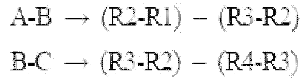
따라서, RMSSD는 다음의 수식 관계로 표현될 수 있다.

이 변수는 짧은 기간의 심박변이요소를 잘 나타내고 있으며, 부교감신경 활동정도를 표시하고, 뇌전증에서 갑작스런 사망과 심방세동에 밀접한 연관성을 가지는 것으로 알려져 있다.
이상과 같은 본 발명의 방법으로 진단 가능한 질환은 다음과 같다.
1) 태아의 태동시의 FHRV 변화에 따른 자율신경계 이상유무 판단
2) 태아 심박동수 평가에 따른 건강상태 판단
① FHRV 상승
- FHR의 일시적 상승으로 15초 이상동안 15 bpm 이상 증가 상태
- 산모 자궁수축 중 FHR 증가 : 정상, 교감신경의 자극으로 인한
상승
- 제대의 부분적 압박 : 제대정맥 일부 폐쇄 -> 태아 저혈압 ->
일시적 FHR 상승
② FHR 조기하강
- 산모 자궁수축시 태아 머리 압박으로 인한 감소
- 부교감신경(미주신경) 자극
- 주로 초기 활동기에 나타남
- FHR 하강은 최저 심박수 수준으로 삼소 (보통 100 bpm 수준으로
감소)
③ FHR 후기하강
- 산모 자궁수축 끝난 후에도 FHR이 회복되지 않고 지연되어 회복
- 자궁-태반간 순환부전 (자궁수축-> 제대정맥 혈류 감소-> 태반내
산소 공급 저하)
- 산모의 저산소증, 저/고혈압, 자궁 고긴장상태, 태반 혈류 부족
④ 분만시 FHRV 하강
- 제대 압박으로 인해 나타나는 반응으로 양수과소증 시 흔히
관찰됨
- 제대 압박 완화를 위한 분만 체위 변경
⑤ 지속성 FHR 감소
- 10분 이상 동안 FHR이 정상 범위를 벗어나 감소된 상태 (일반
적으로 정상 심박수 대비 30 bpm/min 감소)
- 산모의 저혈압, 태반조기박리, 과도한 자궁수축 or 파열, 제대압박 or 탈출, 급격한 태아 하강
⑥ 태아 tachycardia
- 태아 저산소증의 초기 증상
- 산모 고체온, 감염, 갑상선 항진증, 부교감신경 차단 약물
- 태아 빈혈, 심질환, 심부정맥
- 10분 이상 160 bpm 이상
- FHRV의 주기적 변화없이 지속적인 빈맥은 임상적 의미 부여하지
않음
⑦ 태아 bradycardia
- 태아 저산소증의 후기 증상
- 산모 국소마취, 저체온, 저혈압
- 태아 지속적인 제대압박, 선천성 심장이상
- 10분 이상 120 bpm 이하
- 80 bpm 이하인 경우를 제외하고 정상적인 FHRV의 변화를 보이거나
주기적인 변화가 없는 경우 임상적 의미 부여하지 않음
이상의 설명과 같이, 본 발명에 따른 태아의 자율신경계 및 심장활동 상태 진단방법은 임신부의 복부에서 획득한 심전도(ECG)로부터 태아의 고유의 신호를 분리하고, 획득된 태아의 고유의 신호로부터 태아의 심전도와 근전도(EMG) 신호를 분리하여, 근전도 신호에서 태아의 태동을 감지할 시에 태아의 심전도를 측정함으로써, 태아의 자율신경계 및 심장활동 상태를 정확하게 진단할 수 있는 장점이 있다.
이상, 바람직한 실시예를 통하여 본 발명에 관하여 상세히 설명하였으나, 본 발명은 이에 한정되는 것은 아니며, 본 발명의 기술적 사상을 벗어나지 않는 범위 내에서 다양하게 변경, 응용될 수 있음은 당해 기술분야의 통상의 기술자에게 자명하다. 따라서, 본 발명의 진정한 보호 범위는 다음의 청구범위에 의하여 해석되어야 하며, 그와 동등한 범위 내에 있는 모든 기술적 사상은 본 발명의 권리 범위에 포함되는 것으로 해석되어야 할 것이다.
Claims (5)
- a) 심전계에 의해 임신부의 복부로부터 임신부와 태아의 심전도 및 근전도가 혼합되어 있는 원신호를 획득하는 단계;b) 상기 획득된 원신호를 소정의 신호 분리 방법을 이용하여 임신부와 태아의 심전도와 근전도 신호로 각각 개별적 신호로 분리하고, 분리된 신호로부터 임신부의 심전도와 근전도를 제거하는 단계;c) 상기 원신호와 상기 임신부의 심전도와 근전도가 제거된 신호와의 차이를 구하여 태아 신호를 추출하는 단계;d) 상기 추출된 태아 신호에 특정 필터를 적용하여 태아의 심전도와 근전도 신호를 획득 및 분리하는 단계;e) 상기 획득된 태아의 심전도를 바탕으로 평시의 태아 심전도에서 R-R 피크를 측정하여 태아의 심박수(FHR0) 및 태아 심박 변이율(FHRV)을 계산하는 단계;f) 상기 획득된 태아의 근전도 신호를 바탕으로 근전도 신호의 주파수가 급증할 때를 태아의 태동이 있는 경우로 판단하고, 태동 감지 시점부터 심전도를 기록하여 태아의 태동 감지 시점 이후의 심전도에서 R-R 피크를 측정하여 태아의 심박수(FHRR) 및 태아 심박 변이율(FHRV)을 계산하는 단계; 및g) 상기 계산된 평시의 태아의 심박수(FHR0), 태동 감지 이후의 태아의 심박수(FHRR), 평시 및 태동 감지 이후의 각각의 태아 심박 변이율(FHRV)에 대한 분석을 통해 태아의 자율신경계 반응 및 심장 활동을 진단하는 단계를 포함하는 태아의 자율신경계 및 심장활동 상태 진단방법.
- 제1항에 있어서,상기 단계 d)에서 상기 근전도 신호를 획득한 후, 획득된 근전도 신호에 대한 더욱 매끄러운 신호를 얻기 위해 대역 필터와 MAF(Moving Average Filter)를 이용하여 근전도 신호를 처리하는 단계를 더 포함하는 태아의 자율신경계 및 심장활동 상태 진단방법.
- 제1항에 있어서,상기 단계 a)에서 심전계를 통하여 원신호를 획득함에 있어서, 신체에 착용(장착)가능한 단일 채널 심전계를 통하여 실시간으로 원신호를 획득하는 것을 특징으로 하는 태아의 자율신경계 및 심장활동 상태 진단방법.
- 제1항에 있어서,상기 단계 b)에서 상기 획득된 원신호를 임신부와 태아의 심전도와 근전도 신호로 각각 개별적 신호로 분리함에 있어서, 암묵 신호 분리(BSS, Blind Source Separation; BSS) 방법을 통하여 개별적 신호로 분리하는 것을 특징으로 하는 태아의 자율신경계 및 심장활동 상태 진단방법.
- 제1항에 있어서,상기 단계 d)에서 상기 추출된 태아 신호에 특이값 분해(Singular Value Decomposition; SVD) 알고리즘 및 MAF(Moving Average Filter)를 적용하여 태아의 심전도와 근전도 신호를 획득 및 분리하는 것을 특징으로 하는 태아의 자율신경계 및 심장활동 상태 진단방법.
Applications Claiming Priority (2)
| Application Number | Priority Date | Filing Date | Title |
|---|---|---|---|
| KR1020160152146A KR20180055019A (ko) | 2016-11-15 | 2016-11-15 | 태아의 자율신경계 및 심장활동 상태 진단방법 |
| KR10-2016-0152146 | 2016-11-15 |
Publications (2)
| Publication Number | Publication Date |
|---|---|
| WO2018092986A2 true WO2018092986A2 (ko) | 2018-05-24 |
| WO2018092986A9 WO2018092986A9 (ko) | 2018-08-16 |
Family
ID=62146462
Family Applications (1)
| Application Number | Title | Priority Date | Filing Date |
|---|---|---|---|
| PCT/KR2017/001908 Ceased WO2018092986A2 (ko) | 2016-11-15 | 2017-02-21 | 태아의 자율신경계 및 심장활동 상태 진단방법 |
Country Status (2)
| Country | Link |
|---|---|
| KR (1) | KR20180055019A (ko) |
| WO (1) | WO2018092986A2 (ko) |
Cited By (2)
| Publication number | Priority date | Publication date | Assignee | Title |
|---|---|---|---|---|
| CN114027852A (zh) * | 2021-11-11 | 2022-02-11 | 浙江智柔科技有限公司 | 宫内胎儿状况分析装置和方法 |
| CN114159040A (zh) * | 2021-12-30 | 2022-03-11 | 徐智策 | 一种具备预警功能的胎心监护仪及监护方法 |
Families Citing this family (2)
| Publication number | Priority date | Publication date | Assignee | Title |
|---|---|---|---|---|
| CN112869724B (zh) * | 2021-01-19 | 2022-04-22 | 西安交通大学 | 一种基于多通道被动式采集信号的胎儿健康监测仪 |
| KR20250108940A (ko) | 2024-01-09 | 2025-07-16 | 주식회사 클레어오디언스 | 청진신호를 통한 태아위치 확인 시스템 및 장치 |
-
2016
- 2016-11-15 KR KR1020160152146A patent/KR20180055019A/ko not_active Ceased
-
2017
- 2017-02-21 WO PCT/KR2017/001908 patent/WO2018092986A2/ko not_active Ceased
Cited By (2)
| Publication number | Priority date | Publication date | Assignee | Title |
|---|---|---|---|---|
| CN114027852A (zh) * | 2021-11-11 | 2022-02-11 | 浙江智柔科技有限公司 | 宫内胎儿状况分析装置和方法 |
| CN114159040A (zh) * | 2021-12-30 | 2022-03-11 | 徐智策 | 一种具备预警功能的胎心监护仪及监护方法 |
Also Published As
| Publication number | Publication date |
|---|---|
| KR20180055019A (ko) | 2018-05-25 |
| WO2018092986A9 (ko) | 2018-08-16 |
Similar Documents
| Publication | Publication Date | Title |
|---|---|---|
| US7949389B2 (en) | Fetal ECG monitoring | |
| Fanelli et al. | Quantitative assessment of fetal well-being through CTG recordings: a new parameter based on phase-rectified signal average | |
| US8892181B2 (en) | Non-invasive fetal monitoring | |
| US8116855B2 (en) | Fetal surveillance | |
| CA2479916C (en) | System and method of assessment of arousal, pain and stress during anesthesia and sedation | |
| US20130102856A1 (en) | Non-invasive detection of fetal or maternal illness | |
| JP4386235B2 (ja) | 心電図の順次比較のための方法及び装置 | |
| US8483810B2 (en) | Apparatus and method for monitoring fetus in maternal body | |
| Chia et al. | Cardiac time intervals of normal fetuses using noninvasive fetal electrocardiography | |
| WO2018092986A2 (ko) | 태아의 자율신경계 및 심장활동 상태 진단방법 | |
| US20130096440A1 (en) | Portable fetal eeg-recording device and method of use | |
| Kähler et al. | Fetal magnetocardiography: development of the fetal cardiac time intervals | |
| WO2016093608A2 (ko) | 심전도를 이용한 짧은 rr 시계열로 chf 및 af의 자동 감지 방법 | |
| Pegorie et al. | Antenatal noninvasive fetal electrocardiography: a literature review | |
| KR101890072B1 (ko) | 자궁 근전도 신호를 기반으로 임산부의 조산예측기능을 갖는 임산부 모니터링 시스템 | |
| WO2014021515A1 (ko) | 태아 건강 평가 방법 및 장치 | |
| RU2248745C1 (ru) | Способ исследования функционального состояния головного мозга и устройство для его реализации | |
| Echeverria et al. | Fetal QRS extraction based on wavelet analysis and pattern matching | |
| Fanelli et al. | A smart wearable prototype for fetal monitoring | |
| Tian et al. | A comprehensive evaluation of the predictive abilities of fetal electrocardiogram‐derived parameters during labor in newborn acidemia: our institutional experience | |
| JP3196871B2 (ja) | 診断装置 | |
| Schmidt et al. | Attenuated autonomic function in multiple organ dysfunction syndrome across three age groups | |
| Hon | Fetal heart rate monitoring: for evaluation of fetal wellbeing | |
| Huddleston et al. | The prediction of fetal oxygenation by an on-line computer analysis of fetal monitor output | |
| Spairani | Artificial intelligence solutions for processing, analyzing and classifying fetal heart rate variability series: a multi-parametric approach |
Legal Events
| Date | Code | Title | Description |
|---|---|---|---|
| 121 | Ep: the epo has been informed by wipo that ep was designated in this application |
Ref document number: 17871971 Country of ref document: EP Kind code of ref document: A2 |
|
| NENP | Non-entry into the national phase |
Ref country code: DE |
|
| 122 | Ep: pct application non-entry in european phase |
Ref document number: 17871971 Country of ref document: EP Kind code of ref document: A2 |