WO2018221402A1 - Procédé de production de matériau décellularisé pour transplantation et composition de greffe comprenant un matériau biocompatible comprenant ledit matériau - Google Patents
Procédé de production de matériau décellularisé pour transplantation et composition de greffe comprenant un matériau biocompatible comprenant ledit matériau Download PDFInfo
- Publication number
- WO2018221402A1 WO2018221402A1 PCT/JP2018/020141 JP2018020141W WO2018221402A1 WO 2018221402 A1 WO2018221402 A1 WO 2018221402A1 JP 2018020141 W JP2018020141 W JP 2018020141W WO 2018221402 A1 WO2018221402 A1 WO 2018221402A1
- Authority
- WO
- WIPO (PCT)
- Prior art keywords
- blood vessel
- branch
- transplantation
- decellularized
- treatment
- Prior art date
- Legal status (The legal status is an assumption and is not a legal conclusion. Google has not performed a legal analysis and makes no representation as to the accuracy of the status listed.)
- Ceased
Links
Images


Classifications
-
- A—HUMAN NECESSITIES
- A61—MEDICAL OR VETERINARY SCIENCE; HYGIENE
- A61L—METHODS OR APPARATUS FOR STERILISING MATERIALS OR OBJECTS IN GENERAL; DISINFECTION, STERILISATION OR DEODORISATION OF AIR; CHEMICAL ASPECTS OF BANDAGES, DRESSINGS, ABSORBENT PADS OR SURGICAL ARTICLES; MATERIALS FOR BANDAGES, DRESSINGS, ABSORBENT PADS OR SURGICAL ARTICLES
- A61L27/00—Materials for grafts or prostheses or for coating grafts or prostheses
- A61L27/36—Materials for grafts or prostheses or for coating grafts or prostheses containing ingredients of undetermined constitution or reaction products thereof, e.g. transplant tissue, natural bone, extracellular matrix
- A61L27/3683—Materials for grafts or prostheses or for coating grafts or prostheses containing ingredients of undetermined constitution or reaction products thereof, e.g. transplant tissue, natural bone, extracellular matrix subjected to a specific treatment prior to implantation, e.g. decellularising, demineralising, grinding, cellular disruption/non-collagenous protein removal, anti-calcification, crosslinking, supercritical fluid extraction, enzyme treatment
- A61L27/3691—Materials for grafts or prostheses or for coating grafts or prostheses containing ingredients of undetermined constitution or reaction products thereof, e.g. transplant tissue, natural bone, extracellular matrix subjected to a specific treatment prior to implantation, e.g. decellularising, demineralising, grinding, cellular disruption/non-collagenous protein removal, anti-calcification, crosslinking, supercritical fluid extraction, enzyme treatment characterised by physical conditions of the treatment, e.g. applying a compressive force to the composition, pressure cycles, ultrasonic/sonication or microwave treatment, lyophilisation
-
- A—HUMAN NECESSITIES
- A61—MEDICAL OR VETERINARY SCIENCE; HYGIENE
- A61F—FILTERS IMPLANTABLE INTO BLOOD VESSELS; PROSTHESES; DEVICES PROVIDING PATENCY TO, OR PREVENTING COLLAPSING OF, TUBULAR STRUCTURES OF THE BODY, e.g. STENTS; ORTHOPAEDIC, NURSING OR CONTRACEPTIVE DEVICES; FOMENTATION; TREATMENT OR PROTECTION OF EYES OR EARS; BANDAGES, DRESSINGS OR ABSORBENT PADS; FIRST-AID KITS
- A61F2/00—Filters implantable into blood vessels; Prostheses, i.e. artificial substitutes or replacements for parts of the body; Appliances for connecting them with the body; Devices providing patency to, or preventing collapsing of, tubular structures of the body, e.g. stents
- A61F2/02—Prostheses implantable into the body
- A61F2/04—Hollow or tubular parts of organs, e.g. bladders, tracheae, bronchi or bile ducts
- A61F2/06—Blood vessels
-
- A—HUMAN NECESSITIES
- A61—MEDICAL OR VETERINARY SCIENCE; HYGIENE
- A61L—METHODS OR APPARATUS FOR STERILISING MATERIALS OR OBJECTS IN GENERAL; DISINFECTION, STERILISATION OR DEODORISATION OF AIR; CHEMICAL ASPECTS OF BANDAGES, DRESSINGS, ABSORBENT PADS OR SURGICAL ARTICLES; MATERIALS FOR BANDAGES, DRESSINGS, ABSORBENT PADS OR SURGICAL ARTICLES
- A61L27/00—Materials for grafts or prostheses or for coating grafts or prostheses
- A61L27/36—Materials for grafts or prostheses or for coating grafts or prostheses containing ingredients of undetermined constitution or reaction products thereof, e.g. transplant tissue, natural bone, extracellular matrix
- A61L27/3604—Materials for grafts or prostheses or for coating grafts or prostheses containing ingredients of undetermined constitution or reaction products thereof, e.g. transplant tissue, natural bone, extracellular matrix characterised by the human or animal origin of the biological material, e.g. hair, fascia, fish scales, silk, shellac, pericardium, pleura, renal tissue, amniotic membrane, parenchymal tissue, fetal tissue, muscle tissue, fat tissue, enamel
- A61L27/3625—Vascular tissue, e.g. heart valves
-
- A—HUMAN NECESSITIES
- A61—MEDICAL OR VETERINARY SCIENCE; HYGIENE
- A61L—METHODS OR APPARATUS FOR STERILISING MATERIALS OR OBJECTS IN GENERAL; DISINFECTION, STERILISATION OR DEODORISATION OF AIR; CHEMICAL ASPECTS OF BANDAGES, DRESSINGS, ABSORBENT PADS OR SURGICAL ARTICLES; MATERIALS FOR BANDAGES, DRESSINGS, ABSORBENT PADS OR SURGICAL ARTICLES
- A61L27/00—Materials for grafts or prostheses or for coating grafts or prostheses
- A61L27/50—Materials characterised by their function or physical properties, e.g. injectable or lubricating compositions, shape-memory materials, surface modified materials
- A61L27/507—Materials characterised by their function or physical properties, e.g. injectable or lubricating compositions, shape-memory materials, surface modified materials for artificial blood vessels
-
- A—HUMAN NECESSITIES
- A61—MEDICAL OR VETERINARY SCIENCE; HYGIENE
- A61F—FILTERS IMPLANTABLE INTO BLOOD VESSELS; PROSTHESES; DEVICES PROVIDING PATENCY TO, OR PREVENTING COLLAPSING OF, TUBULAR STRUCTURES OF THE BODY, e.g. STENTS; ORTHOPAEDIC, NURSING OR CONTRACEPTIVE DEVICES; FOMENTATION; TREATMENT OR PROTECTION OF EYES OR EARS; BANDAGES, DRESSINGS OR ABSORBENT PADS; FIRST-AID KITS
- A61F2/00—Filters implantable into blood vessels; Prostheses, i.e. artificial substitutes or replacements for parts of the body; Appliances for connecting them with the body; Devices providing patency to, or preventing collapsing of, tubular structures of the body, e.g. stents
- A61F2/02—Prostheses implantable into the body
- A61F2/04—Hollow or tubular parts of organs, e.g. bladders, tracheae, bronchi or bile ducts
- A61F2/06—Blood vessels
- A61F2/062—Apparatus for the production of blood vessels made from natural tissue or with layers of living cells
-
- A—HUMAN NECESSITIES
- A61—MEDICAL OR VETERINARY SCIENCE; HYGIENE
- A61L—METHODS OR APPARATUS FOR STERILISING MATERIALS OR OBJECTS IN GENERAL; DISINFECTION, STERILISATION OR DEODORISATION OF AIR; CHEMICAL ASPECTS OF BANDAGES, DRESSINGS, ABSORBENT PADS OR SURGICAL ARTICLES; MATERIALS FOR BANDAGES, DRESSINGS, ABSORBENT PADS OR SURGICAL ARTICLES
- A61L2430/00—Materials or treatment for tissue regeneration
- A61L2430/40—Preparation and treatment of biological tissue for implantation, e.g. decellularisation, cross-linking
Definitions
- the present invention relates to a method for producing a decellularized material that can be used for transplantation, and a graft composition comprising a biocompatible material containing the material.
- Properties required for decellularized materials include (i) strength as a graft, (ii) removal of DNA in a decellularized tissue that causes rejection, and (iii) infiltration of autologous cells after transplantation. It is easy to do. In order to obtain a decellularized material that satisfies these requirements, various production methods have been studied.
- a method using a surfactant for example, see Patent Documents 1 and 2), a method using an enzyme (for example, see Patent Document 3), a method using an oxidizing agent (for example, see Patent Document 4) ), A method by high hydrostatic pressure treatment (for example, see Patent Documents 5 to 7), a method by freeze-thaw treatment (for example, see Patent Documents 8 to 9), and a method for treatment with a hypertonic electrolyte solution (for example, Patent Document 10) For example).
- a surfactant for example, see Patent Documents 1 and 2
- a method using an enzyme for example, see Patent Document 3
- a method using an oxidizing agent for example, see Patent Document 4
- a method by high hydrostatic pressure treatment for example, see Patent Documents 5 to 7
- a method by freeze-thaw treatment for example, see Patent Documents 8 to 9
- a method for treatment with a hypertonic electrolyte solution for example, Patent Document 10.
- JP-A-60-501540 Special table 2003-518981 gazette Japanese translation of PCT publication No. 2002-507907
- Table 2003-52562 gazette Japanese Patent Laid-Open No. 2004-094552 International Publication No. 2008/111530
- Table 2013-502275 gazette JP-A-2005-185507 JP 2005-21480 A JP 2010-2221012 A
- the present inventor has found that, when processing by suturing, which is considered to cause less damage to the decellularized tissue, prevents infiltration of self cells. Based on such results, it is considered important for the graft composition derived from the living tissue to contain the sutured portion within the necessary minimum.
- a decellularized tissue obtained from a blood vessel having a branch (branch vessel)
- a branch branch vessel
- the tube is closed by performing protein denaturation treatment instead of suturing as a means for closing the excision part of the branch, the tissue of the transplanted part is regenerated without hindering the infiltration of the self-cells of the decellularized tissue. I found out.
- the present invention (A) collecting a blood vessel having a branch from a vertebrate (donor); There is provided a method for producing a decellularized material for transplantation, comprising: (b) decellularizing the blood vessel; and (c) adhering a portion where the branch has been excised by protein denaturation treatment and closing the tube.
- the method for producing a decellularized material for transplantation of the present invention may further include a step of excising a branch portion of the blood vessel collected in the step (a).
- the present invention also relates to a biocompatible material comprising a decellularized material for transplantation, comprising a vertebrate (donor) blood vessel in which a branch is excised and having at least one branch excision closed by protein denaturation.
- a biocompatible material comprising a decellularized material for transplantation, comprising a vertebrate (donor) blood vessel in which a branch is excised and having at least one branch excision closed by protein denaturation.
- An implant composition is also provided.
- the production method of the present invention can provide a decellularized material for transplantation for a graft composition in which autologous cells tend to infiltrate after transplantation.
- the graft composition containing at least a portion of the transplanted decellularized material obtained by the production method of the present invention functions as a normal living tissue even after transplantation because the autologous cells after transplantation are easily wetted. be able to.
- FIG. 1 shows a schematic view of a blood vessel with branches.
- 1 is a blood vessel having a branch
- 2 is a branch portion (branch blood vessel)
- a broken line is an example of a position where the branch portion is excised.
- This broken line is preferably 1 mm to 2 mm away from the blood vessel.
- the position of the broken line may be 5 mm to several cm away from the blood vessel.
- 3 of (ii) shows a protein denaturation part.
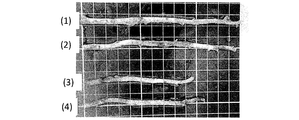
- FIG. 2 is a diagram illustrating an internal thoracic artery fragment in which a branch portion collected from a pig is excised in the embodiment.
- the internal thoracic artery piece (3) in the figure was used as a comparative example.
- ( ⁇ ) is an enlarged photograph of a cross-sectional view of the collected porcine internal thoracic artery piece stained with hematoxylin and eosin. Further, ( ⁇ ) is an enlarged photograph of a sectional view of the transplanted decellularized material obtained by decellularizing the collected porcine inner thoracic artery piece and staining it with hematoxylin and eosin. It can be confirmed that no nucleic acid is present and decellularized. Each scale bar represents 1000 ⁇ m. In the following examples, the internal thoracic artery piece (3) in the figure was used as a comparative example. FIG.
- the figure on the left side is an enlarged photograph of a section of a branch portion obtained by HE staining of a decellularized porcine internal thoracic artery piece which is a decellularization material for transplantation of the present invention before transplantation.
- the figure on the right side is an enlarged photograph of a section of a branch portion stained with HE 1 month after transplantation. Cell infiltration is observed after transplantation.
- Each scale bar represents 500 ⁇ m.
- a blood vessel having a branch is collected from a vertebrate (donor). Collecting here means separating a blood vessel having a branch from a donor.
- the “blood vessel having a branch” means a blood vessel having a branch in the donor, and includes those in which the branch is excised at the time of collection.
- the vertebrate is not particularly limited. However, since it is preferable that blood vessels are easily available, animals other than humans are preferable, and mammal livestock and avian livestock are particularly preferable.
- Mammalian livestock includes cattle, horses, camels, llamas, donkeys, yaks, sheep, pigs, goats, deer, alpaca, dogs, raccoon dogs, weasels, foxes, cats, rabbits, hamsters, guinea pigs, rats, squirrels and raccoons, etc. Is mentioned.
- avian livestock examples include parakeets, parrots, chickens, ducks, turkeys, geese, guinea fowls, pheasants, ostriches, emu and quail. Of these, pigs, rabbits or cows are preferred because of their availability.
- a blood vessel having a branch refers to a blood vessel having at least one branch portion (branch blood vessel) as shown in FIG.
- a blood vessel having a blood vessel with a branched structure has a complicated shape, so it is said that it is not suitable for processing even if it is decellularized, and since it has a branched structure, it is mostly used as a blood vessel for transplantation. It never happened.
- the blood vessel can be used as a blood vessel without a branch by adhering the branch portions of the blood vessel and closing the tube. The significance that a blood vessel having a branch that has been difficult to use in the past can be used as a graft composition is extremely large.
- blood vessels having branches include the internal thoracic artery, abdominal wall artery, gastroepiploic artery, carotid artery, radial artery, intercostal artery, muscular phrenic artery, femoral artery, deep femoral artery, aorta, ulnar artery, and upper arm Artery, anterior tibial artery, posterior tibial artery, mesenteric artery, splenic artery, internal thoracic vein, anterior intercostal vein, odd vein, semi-even vein, jugular vein, intestinal vein, femoral vein, saphenous vein, mesenteric vein, spleen Examples include veins. In consideration of physical properties (elongation, biocompatibility, strength, etc.) as the graft composition, an artery is preferable, and an internal thoracic artery is more preferable.
- a part of the body such as the chest, abdomen, or leg is incised, and the blood vessels having branches to be collected are excised.
- anesthesia method and the slaughter method methods conventionally used by those skilled in the art can be used as they are.
- a scalpel, scissors, etc. which are usually used in animal experiments and surgical operations.
- a scalpel, scissors, or the like that is usually used in animal experiments or surgical operations.
- an ultrasonic scalpel or an electric scalpel These can excise the blood vessel while coagulating the blood by ultrasonic vibration or high-frequency current at the place where the blood vessel is excised.
- an ultrasonic knife since the degree of protein denaturation at the excised portion is moderate, it is preferable to use an ultrasonic knife.
- “ultrasonic knife” and “ultrasonic vibratory knife” are used synonymously.
- Examples of the electrosurgical knife that can be used in the present invention include the Bio series manufactured by Elbe and the SHAPPER series manufactured by Izumi Engineering & Medical Co., Ltd.
- Examples of the ultrasonic scalpel that can be used in the present invention include SONOpet UST-2001 manufactured by Stryker Medtech, and Harmonic Scalpel manufactured by Ethicon End Surgery.
- the blood vessel collected in the step (a) may be in a state where the branch portion has already been excised, or in a state where the branch portion remains with a sufficient length (for example, 5 mm to several cm). If the branch part remains with a sufficient length, it is excised appropriately.
- the length of the branching portion for performing protein denaturation treatment is preferably a position 1 mm to 10 mm away from the branching portion of the blood vessel, more preferably a position 2 mm to 7 mm away, and a position 3 to 5 mm away Is most preferred. Performing protein denaturation treatment within the above range minimizes the effects of denaturation treatment (such as changes in physical properties and blood flow prevention and antithrombogenicity) when using the decellularized material for transplantation of the present invention. Can be suppressed and reorganized.
- Decellularization treatment is performed using a surfactant treatment (Singelyn JM, et al., Biomaterials, 2009, 30, 5409-5416; Singelyn JM, et al., J. Am. Coll. Cardiol., 2012, 59, 751. -763; Sonya B., et al., Sci. Transl. Med., 2013, 5, 173ra25), enzyme treatment, osmotic pressure treatment, freeze-thaw treatment, oxidant treatment, high hydrostatic pressure treatment (Sasaki S., et al. al., Mol.
- the cell is decellularized by high hydrostatic pressure treatment.
- the pressure at which high hydrostatic pressure treatment is performed may be a pressure that can destroy cells and pathogens derived from a vertebrate donor, and can be appropriately selected according to the animal species and blood vessel type of the donor. .
- the hydrostatic pressure is exemplified by 2 to 1,500 MPa.
- the applied hydrostatic pressure is higher than 50 MPa, decellularization from the blood vessel is sufficiently performed. Therefore, it is preferably 50 to 1,500 MPa, more preferably 80 to 1,300 MPa, still more preferably 90 to 1,200 MPa, and most preferably 95 to 1,100 MPa.
- Examples of the medium used for applying the high hydrostatic pressure include water, physiological saline, buffer solution, propylene glycol or an aqueous solution thereof, glycerin or an aqueous solution thereof, and an aqueous saccharide solution.
- Examples of the buffer solution include acetate buffer solution, phosphate buffer solution, citrate buffer solution, borate buffer solution, tartaric acid buffer solution, Tris buffer solution, HEPES buffer solution, and MES buffer solution.
- saccharide in the aqueous saccharide solution examples include erythrose, xylose, arabinose, allose, talose, glucose, mannose, galactose, erythritol, xylitol, mannitol, sorbitol, galactitol, sucrose, lactose, maltose, trehalose, dextran, alginic acid, and hyaluronic acid. Is mentioned.
- the temperature of the medium for high hydrostatic pressure treatment is not particularly limited as long as it does not generate ice and does not damage the tissue due to heat. It is preferably 0 to 45 ° C., more preferably 4 to 40 ° C., still more preferably 10 to 37 ° C., and most preferably 15 to 35 ° C., because the decellularization treatment is performed smoothly and the influence on the tissue is small. is there.
- the time for the high hydrostatic pressure treatment is too short, the decellularization treatment is not sufficiently performed, and if it is long, energy is wasted, so 5 minutes to 12 hours is preferable, and 7 minutes to 5 hours is more preferable. More preferably, min-3 hours.
- the suitability of decellularization can be confirmed by histological staining (hematoxylin-eosin staining) or residual DNA quantification.
- the cleaning liquid may be the same liquid as the medium used for applying the high hydrostatic pressure, may be a different cleaning liquid, or a combination of a plurality of types of cleaning liquids.
- the cleaning liquid preferably contains a nucleolytic enzyme, an organic solvent, or a chelating agent. Nucleolytic enzymes can improve the efficiency of removing nucleic acid components from blood vessels to which hydrostatic pressure is applied, organic solvents are lipids, and chelating agents are calcium ions and magnesium ions in decellularized tissues. By inactivating, calcification can be prevented when decellularized tissue is applied to the affected area.
- the portion where the branch is excised is subjected to protein denaturation treatment and closed.
- the means for denaturing the protein is not particularly limited, but from the viewpoint of ease of work and efficiency, and less damage to the tissue, it is preferable to use an ultrasonic scalpel or an electric scalpel, and damage to the tissue will be reduced. It is preferable to use an ultrasonic scalpel from the viewpoint of reducing the branch stump closing property.
- the ultrasonic scalpel has a structure in which the blade edge is mechanically ultrasonically vibrated, and frictional heat is generated near the living tissue contacted by the blade edge, so that proteins in the living tissue are denatured.
- the part where the branch blood vessel is excised and opened is adhered and closed when the protein is denatured.
- the frequency at the time of performing the treatment using ultrasonic waves as described above is preferably 20 kHz to 100 kHz, more preferably about 30 kHz to about 60 kHz.
- the output is preferably 50 to 500 mA, more preferably 100 to 400 mA, and still more preferably 200 to 300 mA (when 100 VAC is used).
- the sonication time varies depending on the frequency and output and is not particularly limited, but is preferably 0.1 second to 10 minutes, more preferably 1 second to 5 minutes, and still more preferably 3 to 60 seconds.
- the steps (a), (b), and (c) are not limited to the above order.
- the step (c) may be performed after the step (a), and then the step (b) may be performed.
- a branch portion of a blood vessel having a branch is excised before being collected from a vertebrate animal as a donor, and the portion where the branch is excised is subjected to protein denaturation treatment and collected from a donor. Later, it is also possible to decellularize the step (b). That is, the aspect included in order of a process (c), a process (a), and a process (b) is also included by this invention.
- the method may further include a step (d) of cutting the branch portion.
- this step (d) is not particularly limited. For example, it is possible to carry out in the order of step (a), step (d), step (b) and step (c), step (a), step (b), step (d) and step (c). It is.
- step (c) it is possible to close the tube without overlooking the branch vessel, and in the point that the operation becomes more efficient, in the step (c), “removing the branch” and “closing the tube by performing protein denaturation treatment” More preferably, the steps (c) and (a) are performed continuously. Even in this case, step (c) may be performed again before and after step (b).
- a branch of a vertebrate is composed of a biocompatible material including a decellularized material for transplantation which includes a resected blood vessel and has at least one branch resection part closed by protein denaturation.
- An implant composition is also provided.
- the vertebrate in the graft composition of the present invention is not particularly limited, but is preferably an animal other than a human because blood vessels are easily available, and in particular, a mammal livestock and an avian livestock are preferred.
- the mammal livestock and the avian livestock here are the same as the livestock mentioned in the specific examples of the mammalian livestock and the avian livestock used in the method for producing the decellularized material for transplantation of the present invention. Of these, pigs, rabbits or cows are preferred because of their availability.
- the blood vessel from which the branch has been excised means that a branch portion is excised from the blood vessel having the branch used for producing the decellularized material for transplantation of the present invention. It is a blood vessel.
- the blood vessel having a branch the same blood vessel as that used in the above production method can be used, and considering the physical properties (elongation, biocompatibility, strength, etc.) as the graft composition, an artery is preferable. The thoracic artery is more preferred.
- the decellularized material for transplantation used in the graft composition of the present invention has a protein-denatured part.
- the excision site of the branch portion is in an open tube state, and the open tube portion can be blocked by performing protein denaturation treatment.
- the specific method of protein denaturation treatment is not particularly limited, as with the above-described decellularized material for transplantation, but an ultrasonic scalpel or an electric scalpel can be used. It is preferable to use an ultrasonic scalpel because there is little damage to the tissue.
- the conditions regarding the frequency of ultrasonic waves and the treatment time for denaturing proteins are the same as the conditions for the frequency of ultrasonic waves and the treatment time in the above-described method for producing a decellularized material for transplantation. It is.
- the decellularization treatment can be carried out in the same manner as the decellularization treatment described above, and the decellularization treatment can be carried out by a high hydrostatic pressure treatment. It is preferable that it is processed.
- any pressure may be used as long as the cells and pathogens derived from the vertebrate donor are destroyed, and can be performed under the same conditions as the hydrostatic pressure described above.
- Examples of the medium used for applying the high hydrostatic pressure include water, physiological saline, buffer solution, propylene glycol or an aqueous solution thereof, glycerin or an aqueous solution thereof, and an aqueous saccharide solution.
- Specific examples of each solvent include The same solvent as the specific example of said solvent can be used.
- the graft composition of the present invention is made of a specific biocompatible material.
- biocompatible means that the graft is accepted by the transplanted tissue and does not induce toxicity or significant immune rejection.
- the biocompatible material is not particularly limited as long as it has no fluidity and is solid, and examples thereof include non-resorbable polymers, absorbent polymers, metals, glasses, and ceramics.
- non-resorbable polymer examples include polyethylene, polyethylene terephthalate, polybutylene, polybutylene terephthalate, polypropylene, acrylic, polyamide-imide, polyether ether ketone, polyaryl ether ketone, polycarbonate, polyamide, polyvinyl fluoride, and polyvinylidene fluoride. , Polymethyl methacrylate, and combinations and equivalents thereof, but are not limited thereto.
- the absorbent polymer may be a synthetic polymer or a natural polymer.
- Polyamino acids, polyamides, fatty acid polyesters, and natural polymers include collagen, elastin, hyaluronic acid, laminin, gelatin, keratin, chondroitin sulfate And decellularized tissue.
- metals examples include tantalum, tantalum alloy, stainless steel, titanium, titanium alloy, cobalt-chromium alloy, and the like, and biocompatible metals conventionally used in medical devices and the like can be used.
- glass or ceramic examples include tetracalcium phosphate, alpha- and beta-tricalcium phosphate, octacalcium phosphate, hydroxyapatite, substituted apatite, monetite, metaphosphate, pyrophosphate, phosphate glass, etc. Phosphates, calcium and magnesium carbonates, sulfates, and oxides, and combinations thereof, but are not limited thereto.
- the graft composition of the present invention is composed of a biocompatible material containing at least a part of a specific decellularized material for transplantation.
- “included in at least a part” means that the decellularized material for transplantation can be present in one or more and a plurality of biocompatible materials. It is more preferable that the biocompatible material is composed only of a decellularized material in that self cell infiltration is more effectively achieved.
- the graft composition of the present invention can function as a part of living tissue after transplantation.
- it can function as a blood vessel substitute for transplantation.
- FIG. 2 shows the porcine internal thoracic arteries (1) to (4).
- FIG. 3 shows a cross-sectional view (HE staining).
- the porcine internal thoracic artery was treated with an ultrahigh hydrostatic apparatus (Dr. Chef, manufactured by Kobe Steel) using water as a medium.
- the ultra-high hydrostatic pressure treatment was performed under the conditions of an applied pressure of 600 MPa, a pressure increase time of 9 minutes, a pressure maintenance time of 120 minutes, a pressure decrease time of 9 minutes, and a pressure medium temperature of 30 ° C.
- washing with 50 mg / L DNase I solution was performed for 4 days, EtOH solution for 3 days, and citric acid solution for 4 days to complete decellularization.
- HE staining is a method that uses two types of dyes, hematoxylin and eosin, to separate the cell nucleus and tissues and components other than the nucleus. Hematoxylin makes the nucleus blue-blue, and eosin removes cytoplasm, fibers, and red blood cells. Can be dyed pink.
- the pressure strength is measured by using a syringe pump (YSP-101) to feed physiological saline into the porcine internal thoracic artery from a 20 mL syringe at a rate of 3 mL / min, and the pressure at that time is a digital pressure gauge (KDM30). Measured with.
- YSP-101 syringe pump
- KDM30 digital pressure gauge
- the part of the branched blood vessel in which the leakage of physiological saline was confirmed was ligated with 6-0 proline thread. Ligation was performed by passing a suture thread through the adventitia part and then tying up the branched tubular part from the outside.
- the decellularized porcine internal thoracic artery and the peripheral part of the abdominal aorta were anastomosed in the same manner to produce a bypass blood vessel.
- the abdominal aorta between the central anastomosis and the peripheral anastomosis was ligated with 6-0 proline thread to direct blood flow to the bypass vessel.
- the abdomen was closed by suturing the peritoneum with 4-0 PDS thread, the muscle layer with 2-0 PDS, and the skin with 4-0 PDS thread.
- Vitoril (registered trademark) 0.5 mL was administered as an antibiotic, and atipamezole hydrochloride (trade name: Atipame injection 1 ml) was injected subcutaneously into the thigh as a medetomidine hydrochloride antagonist. After confirming wakefulness, the rabbit was transferred to the breeding cage.
- -Necropsy An autopsy was performed on rabbits 1 month or 3 months after transplantation of decellularized porcine internal thoracic artery.
- the abdominal aorta (transplanted decellularized porcine internal thoracic artery) was exposed by the same method as at the time of transplantation without taking an infusion line from the rabbit.
- vascular patency evaluation and thrombus formation evaluation were performed.
- Vascular patency evaluation was performed by cutting the peripheral side of the abdominal aorta and determining whether or not blood was released. All samples confirmed exsanguination and indicated that the blood vessels were patent.
- pentobarbital sodium Somnopentyl (registered trademark) 5 mL was injected into the ear vein and sacrificed. After the heart, breathing stopped, the decellularized porcine internal thoracic artery was removed. The inside of the decellularized porcine internal thoracic artery was washed with physiological saline, then incised in the longitudinal direction, and thrombus formation was visually observed. There was no adhesion of thrombus to the luminal surface of decellularized porcine internal thoracic artery, indicating high antithrombogenicity. “Patent” means that a hollow state is maintained without occluding the lumen of a blood vessel.
- the pathological specimen of the transplanted decellularized porcine internal thoracic artery was produced by the Institute for New Histological Science.
- an ultrasonic scalpel of the decellularized porcine internal thoracic artery or a portion where the tissue structure of the branched blood vessel portion occluded with 6-0 proline thread can be confirmed is an HE-stained section, Elastica van Gieson stain (Elastica van Gieson stain)
- EVG staining Elastica van Gieson stain
- a section is continuously prepared with a thickness of approximately 5 ⁇ m in the longitudinal direction of the blood vessel, and the section is cut at a portion where the tissue structure of the branch blood vessel portion blocked with an ultrasonic knife or 6-0 proline thread can be confirmed. It was created.
- EVG staining is a staining method for identifying elastic fibers, in which elastin in connective tissue can be dyed in black purple and collagen in red purple.
- FIG. 4 An HE-stained image, an EVG-stained image (3 months) of an example sample, and an HE-stained image and an EVG-stained image (3 months) of a comparative example sample are shown below (FIG. 4).
- the cells also infiltrated into the decellularized porcine internal thoracic artery tissue portion stained in dark purple by EVG staining.
- the comparative sample no cell infiltration was observed in the decellularized porcine internal thoracic artery tissue portion stained in dark purple by EVG staining.
- a finding was found in which the step of the branch blood vessel was covered with rabbit cells at one month after transplantation (FIG. 5).
- 1 a blood vessel having a branch
- 2 a branched portion (branched blood vessel)
- 3 a protein denatured portion
Landscapes
- Health & Medical Sciences (AREA)
- Life Sciences & Earth Sciences (AREA)
- Chemical & Material Sciences (AREA)
- Engineering & Computer Science (AREA)
- Biomedical Technology (AREA)
- General Health & Medical Sciences (AREA)
- Transplantation (AREA)
- Oral & Maxillofacial Surgery (AREA)
- Animal Behavior & Ethology (AREA)
- Public Health (AREA)
- Veterinary Medicine (AREA)
- Epidemiology (AREA)
- Medicinal Chemistry (AREA)
- Dermatology (AREA)
- Vascular Medicine (AREA)
- Chemical Kinetics & Catalysis (AREA)
- Botany (AREA)
- Molecular Biology (AREA)
- Cardiology (AREA)
- Heart & Thoracic Surgery (AREA)
- Urology & Nephrology (AREA)
- Zoology (AREA)
- Gastroenterology & Hepatology (AREA)
- Pulmonology (AREA)
- Materials For Medical Uses (AREA)
Abstract
Priority Applications (8)
| Application Number | Priority Date | Filing Date | Title |
|---|---|---|---|
| US16/615,177 US12403219B2 (en) | 2017-05-30 | 2018-05-25 | Method for producing decellularized material for transplantation and graft composition consisting of biocompatible material including said material |
| CA3065498A CA3065498A1 (fr) | 2017-05-30 | 2018-05-25 | Procede de production de materiau decellularise pour transplantation et composition de greffe comprenant un materiau biocompatible comprenant ledit materiau |
| KR1020247043219A KR20250007698A (ko) | 2017-05-30 | 2018-05-25 | 이식용 탈세포화 재료의 제조 방법 및 당해 재료를 포함하는 생체 적합성 재료로 이루어지는 이식편 조성물 |
| EP18809381.9A EP3632481B1 (fr) | 2017-05-30 | 2018-05-25 | Procédé de production de matériau décellularisé pour transplantation et composition de greffe comprenant un matériau biocompatible comprenant ledit matériau |
| JP2019522186A JP7658716B2 (ja) | 2017-05-30 | 2018-05-25 | 移植用脱細胞化材料の製造方法及び当該材料を含む生体適合性材料からなる移植片組成物 |
| CN201880036506.4A CN110740762A (zh) | 2017-05-30 | 2018-05-25 | 移植用脱细胞化材料制造方法及由包含该材料的生物相容性材料组成的移植片组成物 |
| KR1020197035144A KR20200016226A (ko) | 2017-05-30 | 2018-05-25 | 이식용 탈세포화 재료의 제조 방법 및 당해 재료를 포함하는 생체 적합성 재료로 이루어지는 이식편 조성물 |
| JP2023061981A JP2023076668A (ja) | 2017-05-30 | 2023-04-06 | 移植用脱細胞化材料の製造方法及び当該材料を含む生体適合性材料からなる移植片組成物 |
Applications Claiming Priority (2)
| Application Number | Priority Date | Filing Date | Title |
|---|---|---|---|
| JP2017-106400 | 2017-05-30 | ||
| JP2017106400 | 2017-05-30 |
Publications (1)
| Publication Number | Publication Date |
|---|---|
| WO2018221402A1 true WO2018221402A1 (fr) | 2018-12-06 |
Family
ID=64455364
Family Applications (1)
| Application Number | Title | Priority Date | Filing Date |
|---|---|---|---|
| PCT/JP2018/020141 Ceased WO2018221402A1 (fr) | 2017-05-30 | 2018-05-25 | Procédé de production de matériau décellularisé pour transplantation et composition de greffe comprenant un matériau biocompatible comprenant ledit matériau |
Country Status (8)
| Country | Link |
|---|---|
| US (1) | US12403219B2 (fr) |
| EP (1) | EP3632481B1 (fr) |
| JP (2) | JP7658716B2 (fr) |
| KR (2) | KR20250007698A (fr) |
| CN (1) | CN110740762A (fr) |
| CA (1) | CA3065498A1 (fr) |
| TW (1) | TWI749233B (fr) |
| WO (1) | WO2018221402A1 (fr) |
Cited By (1)
| Publication number | Priority date | Publication date | Assignee | Title |
|---|---|---|---|---|
| JPWO2022102739A1 (fr) * | 2020-11-12 | 2022-05-19 |
Families Citing this family (3)
| Publication number | Priority date | Publication date | Assignee | Title |
|---|---|---|---|---|
| CN218059021U (zh) * | 2022-09-22 | 2022-12-16 | 舩本诚一 | 一种医用材料处理用高压设备 |
| CN115554472B (zh) * | 2022-09-22 | 2023-08-18 | 舩本诚一 | 一种用于移植的生物组织处理方法 |
| CN117709441B (zh) * | 2024-02-06 | 2024-05-03 | 云南联合视觉科技有限公司 | 通过逐步迁移领域训练专业医疗大模型的方法 |
Citations (14)
| Publication number | Priority date | Publication date | Assignee | Title |
|---|---|---|---|---|
| JPS60501540A (ja) | 1983-06-10 | 1985-09-19 | ユニバ−シテイ パテンツ,インコ−ポレイテイド | 細胞外マトリクスの体移植片並びに該移植片の製造及び使用のための手段及び方法 |
| JP2000505315A (ja) * | 1996-01-24 | 2000-05-09 | オリジン・メッドシステムズ,インコーポレイテッド | 切開プローブをもつ組織分離カニューレ及び方法 |
| JP2002507907A (ja) | 1997-06-27 | 2002-03-12 | バーダー、アウグスチヌス | 生合成移植片及びその製造方法 |
| JP2003518981A (ja) | 1999-12-29 | 2003-06-17 | チルドレンズ メディカル センター コーポレーション | 臓器脱細胞化のための方法および組成物 |
| JP2003525062A (ja) | 1998-09-30 | 2003-08-26 | メドトロニック・インコーポレーテッド | 移植で使用される組織の無機質化を減少させる方法 |
| JP2004094552A (ja) | 2002-08-30 | 2004-03-25 | Sumitomo Chem Co Ltd | 病理組織画像解析方法および病理組織画像解析システム |
| JP2005185507A (ja) | 2003-12-25 | 2005-07-14 | Yoshihiro Takami | 皮膚の分離無細胞化方法、無細胞化真皮マトリックス及びその製造方法並びに無細胞化真皮マトリックスを用いた複合培養皮膚 |
| JP2005211480A (ja) | 2004-01-30 | 2005-08-11 | Yoshihiro Takami | 皮膚の分離無細胞化方法、無細胞化真皮マトリックス及びその製造方法並びに無細胞化真皮マトリックスを用いた複合培養皮膚 |
| JP4092397B2 (ja) | 2002-09-10 | 2008-05-28 | 国立循環器病センター総長 | 超高静水圧印加による移植用生体組織の処理方法 |
| WO2008111530A1 (fr) | 2007-03-09 | 2008-09-18 | National University Corporation, Tokyo Medical And Dental University | Procédé de préparation d'un tissu mou décellularisé, d'une greffe et d'un matériau de culture |
| JP2009050297A (ja) | 2007-08-23 | 2009-03-12 | Tokyo Medical & Dental Univ | 脱細胞処理液、脱細胞化組織の調製方法、移植片、及び培養部材 |
| JP2010221012A (ja) | 2009-02-25 | 2010-10-07 | Kobe Univ | 高張電解質溶液による生体組織の脱細胞化処理方法 |
| JP2013502275A (ja) | 2009-08-18 | 2013-01-24 | ライフセル コーポレーション | 組織の処理方法 |
| WO2016194895A1 (fr) * | 2015-06-02 | 2016-12-08 | 株式会社Adeka | Feuille de tissu biologique, structure tubulaire produite à partir de ladite feuille, et vaisseau sanguin artificiel comprenant ladite structure tubulaire |
Family Cites Families (16)
| Publication number | Priority date | Publication date | Assignee | Title |
|---|---|---|---|---|
| JPS63279832A (ja) * | 1987-05-12 | 1988-11-16 | Sugino Mach:Kk | 液体ジェット手術装置 |
| US5026387A (en) * | 1990-03-12 | 1991-06-25 | Ultracision Inc. | Method and apparatus for ultrasonic surgical cutting and hemostatis |
| US5336616A (en) * | 1990-09-12 | 1994-08-09 | Lifecell Corporation | Method for processing and preserving collagen-based tissues for transplantation |
| US6293970B1 (en) * | 1998-06-30 | 2001-09-25 | Lifenet | Plasticized bone and soft tissue grafts and methods of making and using same |
| US6310036B1 (en) * | 1999-01-09 | 2001-10-30 | Last Chance Tissue Adhesives Corporation | High strength, Bio-compatible tissue adhesive and methods for treating vigorously bleeding surfaces |
| IL139708A0 (en) * | 2000-11-15 | 2002-02-10 | Amiel Gilad | Process of decellularizing biological matrices and acellular biological matrices useful in tissue engineering |
| WO2005063316A1 (fr) | 2003-12-26 | 2005-07-14 | Cardio Incorporated | Biomateriau transplantable et son procede de preparation |
| CN101185770A (zh) * | 2007-12-27 | 2008-05-28 | 南京市儿童医院 | 猪带瓣血管脱细胞的支架的超高压制备方法 |
| KR101335203B1 (ko) * | 2010-03-26 | 2013-11-29 | 숙명여자대학교산학협력단 | 혈관신생촉진용 펩타이드 및 이의 용도 |
| US20130013083A1 (en) * | 2011-01-06 | 2013-01-10 | Humacyte | Tissue-Engineered Constructs |
| CA2867441C (fr) | 2012-03-16 | 2019-01-15 | Novahep Ab | Vaisseau sanguin allogeneique forme par genie biologique |
| US9498246B2 (en) * | 2013-03-14 | 2016-11-22 | Saphena Medical, Inc. | Unitary endoscopic vessel harvesting devices |
| CN203802502U (zh) | 2013-09-06 | 2014-09-03 | 姚建新 | 超声切割止血手术仪 |
| CN104287869B (zh) * | 2014-09-19 | 2017-03-29 | 上海市肺科医院 | 一种用于气管移植的新型纳米纤维膜/纱支架及其制备方法 |
| CN204293229U (zh) * | 2014-11-28 | 2015-04-29 | 徐胜前 | 一种超声手术刀 |
| TWI711455B (zh) * | 2015-05-13 | 2020-12-01 | 臺北榮民總醫院 | 多層視網膜細胞移植物 |
-
2018
- 2018-05-25 KR KR1020247043219A patent/KR20250007698A/ko active Pending
- 2018-05-25 EP EP18809381.9A patent/EP3632481B1/fr active Active
- 2018-05-25 CA CA3065498A patent/CA3065498A1/fr active Pending
- 2018-05-25 KR KR1020197035144A patent/KR20200016226A/ko not_active Ceased
- 2018-05-25 JP JP2019522186A patent/JP7658716B2/ja active Active
- 2018-05-25 WO PCT/JP2018/020141 patent/WO2018221402A1/fr not_active Ceased
- 2018-05-25 CN CN201880036506.4A patent/CN110740762A/zh active Pending
- 2018-05-25 US US16/615,177 patent/US12403219B2/en active Active
- 2018-05-29 TW TW107118285A patent/TWI749233B/zh active
-
2023
- 2023-04-06 JP JP2023061981A patent/JP2023076668A/ja active Pending
Patent Citations (14)
| Publication number | Priority date | Publication date | Assignee | Title |
|---|---|---|---|---|
| JPS60501540A (ja) | 1983-06-10 | 1985-09-19 | ユニバ−シテイ パテンツ,インコ−ポレイテイド | 細胞外マトリクスの体移植片並びに該移植片の製造及び使用のための手段及び方法 |
| JP2000505315A (ja) * | 1996-01-24 | 2000-05-09 | オリジン・メッドシステムズ,インコーポレイテッド | 切開プローブをもつ組織分離カニューレ及び方法 |
| JP2002507907A (ja) | 1997-06-27 | 2002-03-12 | バーダー、アウグスチヌス | 生合成移植片及びその製造方法 |
| JP2003525062A (ja) | 1998-09-30 | 2003-08-26 | メドトロニック・インコーポレーテッド | 移植で使用される組織の無機質化を減少させる方法 |
| JP2003518981A (ja) | 1999-12-29 | 2003-06-17 | チルドレンズ メディカル センター コーポレーション | 臓器脱細胞化のための方法および組成物 |
| JP2004094552A (ja) | 2002-08-30 | 2004-03-25 | Sumitomo Chem Co Ltd | 病理組織画像解析方法および病理組織画像解析システム |
| JP4092397B2 (ja) | 2002-09-10 | 2008-05-28 | 国立循環器病センター総長 | 超高静水圧印加による移植用生体組織の処理方法 |
| JP2005185507A (ja) | 2003-12-25 | 2005-07-14 | Yoshihiro Takami | 皮膚の分離無細胞化方法、無細胞化真皮マトリックス及びその製造方法並びに無細胞化真皮マトリックスを用いた複合培養皮膚 |
| JP2005211480A (ja) | 2004-01-30 | 2005-08-11 | Yoshihiro Takami | 皮膚の分離無細胞化方法、無細胞化真皮マトリックス及びその製造方法並びに無細胞化真皮マトリックスを用いた複合培養皮膚 |
| WO2008111530A1 (fr) | 2007-03-09 | 2008-09-18 | National University Corporation, Tokyo Medical And Dental University | Procédé de préparation d'un tissu mou décellularisé, d'une greffe et d'un matériau de culture |
| JP2009050297A (ja) | 2007-08-23 | 2009-03-12 | Tokyo Medical & Dental Univ | 脱細胞処理液、脱細胞化組織の調製方法、移植片、及び培養部材 |
| JP2010221012A (ja) | 2009-02-25 | 2010-10-07 | Kobe Univ | 高張電解質溶液による生体組織の脱細胞化処理方法 |
| JP2013502275A (ja) | 2009-08-18 | 2013-01-24 | ライフセル コーポレーション | 組織の処理方法 |
| WO2016194895A1 (fr) * | 2015-06-02 | 2016-12-08 | 株式会社Adeka | Feuille de tissu biologique, structure tubulaire produite à partir de ladite feuille, et vaisseau sanguin artificiel comprenant ladite structure tubulaire |
Non-Patent Citations (7)
| Title |
|---|
| JOURNAL OF BIOMEDICAL MATERIALS RESEARCH A, vol. 103A, October 2015 (2015-10-01), pages 10 |
| NEGISHI J. ET AL., J. ARTIF. ORGANS, vol. 14, 2011, pages 223 - 231 |
| SASAKI S. ET AL., MOL. VIS., vol. 15, 2009, pages 2022 - 2028 |
| SINGELYN J. M. ET AL., BIOMATERIALS, vol. 30, 2009, pages 5409 - 5416 |
| SINGELYN J. M. ET AL., J. AM. COLL. CARDIOL., vol. 59, 2012, pages 751 - 763 |
| SONYA B. ET AL., SCI. TRANSL. MED., vol. 5, 2013, pages 173ra25 |
| YOSHIHIDE H. ET AL., BIOMATERIALS, vol. 31, 2010, pages 3590 - 3595 |
Cited By (1)
| Publication number | Priority date | Publication date | Assignee | Title |
|---|---|---|---|---|
| JPWO2022102739A1 (fr) * | 2020-11-12 | 2022-05-19 |
Also Published As
| Publication number | Publication date |
|---|---|
| TW201906586A (zh) | 2019-02-16 |
| JP7658716B2 (ja) | 2025-04-08 |
| KR20200016226A (ko) | 2020-02-14 |
| US12403219B2 (en) | 2025-09-02 |
| JP2023076668A (ja) | 2023-06-01 |
| EP3632481A1 (fr) | 2020-04-08 |
| EP3632481B1 (fr) | 2023-08-23 |
| JPWO2018221402A1 (ja) | 2020-04-30 |
| TWI749233B (zh) | 2021-12-11 |
| CN110740762A (zh) | 2020-01-31 |
| EP3632481A4 (fr) | 2021-03-31 |
| CA3065498A1 (fr) | 2018-12-06 |
| KR20250007698A (ko) | 2025-01-14 |
| US20200222589A1 (en) | 2020-07-16 |
Similar Documents
| Publication | Publication Date | Title |
|---|---|---|
| JP2023076668A (ja) | 移植用脱細胞化材料の製造方法及び当該材料を含む生体適合性材料からなる移植片組成物 | |
| Pennel et al. | The performance of cross-linked acellular arterial scaffolds as vascular grafts; pre-clinical testing in direct and isolation loop circulatory models | |
| US20120226218A1 (en) | Extracellular matrix material created using non-thermal irreversible electroporation | |
| Chaouat et al. | The evaluation of a small-diameter polysaccharide-based arterial graft in rats | |
| AU2013373262B2 (en) | Artificial blood vessel using decellularized blood vessel sheet | |
| US20220323647A1 (en) | Sheet of biological tissue, tubular structure obtained from said sheet, and artificial blood vessel comprising said tubular structure | |
| EP3705141B1 (fr) | Matière décellularisée de type feuille et vaisseau sanguin artificiel l'utilisant | |
| JPS60501540A (ja) | 細胞外マトリクスの体移植片並びに該移植片の製造及び使用のための手段及び方法 | |
| JP2010221012A (ja) | 高張電解質溶液による生体組織の脱細胞化処理方法 | |
| Wang | Evaluation of Amnion Membrane Made Vascular Graft in Rat Model and Porcine Model | |
| JP6515429B2 (ja) | 人工血管、および、人工血管の製造方法 | |
| JP2011130989A (ja) | 抗血栓性修飾剤、医療用具、及び、多孔質コラーゲン | |
| HK40036680A (en) | Sheet-like decellularized material and artificial blood vessel employing said material | |
| HK1247077B (zh) | 源自生物体的组织的片材、由该片材得到的管状结构体以及包含该管状结构体的人工血管 |
Legal Events
| Date | Code | Title | Description |
|---|---|---|---|
| 121 | Ep: the epo has been informed by wipo that ep was designated in this application |
Ref document number: 18809381 Country of ref document: EP Kind code of ref document: A1 |
|
| WWE | Wipo information: entry into national phase |
Ref document number: 2019522186 Country of ref document: JP |
|
| ENP | Entry into the national phase |
Ref document number: 20197035144 Country of ref document: KR Kind code of ref document: A |
|
| ENP | Entry into the national phase |
Ref document number: 3065498 Country of ref document: CA |
|
| NENP | Non-entry into the national phase |
Ref country code: DE |
|
| ENP | Entry into the national phase |
Ref document number: 2018809381 Country of ref document: EP Effective date: 20200102 |
|
| WWG | Wipo information: grant in national office |
Ref document number: 16615177 Country of ref document: US |