WO2019069955A1 - Dispositif de différenciation, procédé de différenciation de symptômes de dépression, procédé de détermination du niveau de symptômes de dépression, procédé de stratification de patients de dépression, procédé de détermination des effets d'un traitement de symptômes de dépression, et dispositif d'apprentissage d'activité cérébrale - Google Patents
Dispositif de différenciation, procédé de différenciation de symptômes de dépression, procédé de détermination du niveau de symptômes de dépression, procédé de stratification de patients de dépression, procédé de détermination des effets d'un traitement de symptômes de dépression, et dispositif d'apprentissage d'activité cérébrale Download PDFInfo
- Publication number
- WO2019069955A1 WO2019069955A1 PCT/JP2018/036952 JP2018036952W WO2019069955A1 WO 2019069955 A1 WO2019069955 A1 WO 2019069955A1 JP 2018036952 W JP2018036952 W JP 2018036952W WO 2019069955 A1 WO2019069955 A1 WO 2019069955A1
- Authority
- WO
- WIPO (PCT)
- Prior art keywords
- functional
- classifier
- subject
- correlation
- brain activity
- Prior art date
- Legal status (The legal status is an assumption and is not a legal conclusion. Google has not performed a legal analysis and makes no representation as to the accuracy of the status listed.)
- Ceased
Links
Images













Classifications
-
- G—PHYSICS
- G16—INFORMATION AND COMMUNICATION TECHNOLOGY [ICT] SPECIALLY ADAPTED FOR SPECIFIC APPLICATION FIELDS
- G16H—HEALTHCARE INFORMATICS, i.e. INFORMATION AND COMMUNICATION TECHNOLOGY [ICT] SPECIALLY ADAPTED FOR THE HANDLING OR PROCESSING OF MEDICAL OR HEALTHCARE DATA
- G16H30/00—ICT specially adapted for the handling or processing of medical images
- G16H30/20—ICT specially adapted for the handling or processing of medical images for handling medical images, e.g. DICOM, HL7 or PACS
-
- A—HUMAN NECESSITIES
- A61—MEDICAL OR VETERINARY SCIENCE; HYGIENE
- A61B—DIAGNOSIS; SURGERY; IDENTIFICATION
- A61B5/00—Measuring for diagnostic purposes; Identification of persons
- A61B5/05—Detecting, measuring or recording for diagnosis by means of electric currents or magnetic fields; Measuring using microwaves or radio waves
- A61B5/055—Detecting, measuring or recording for diagnosis by means of electric currents or magnetic fields; Measuring using microwaves or radio waves involving electronic [EMR] or nuclear [NMR] magnetic resonance, e.g. magnetic resonance imaging
-
- G—PHYSICS
- G06—COMPUTING OR CALCULATING; COUNTING
- G06F—ELECTRIC DIGITAL DATA PROCESSING
- G06F18/00—Pattern recognition
- G06F18/20—Analysing
- G06F18/21—Design or setup of recognition systems or techniques; Extraction of features in feature space; Blind source separation
- G06F18/214—Generating training patterns; Bootstrap methods, e.g. bagging or boosting
-
- G—PHYSICS
- G06—COMPUTING OR CALCULATING; COUNTING
- G06F—ELECTRIC DIGITAL DATA PROCESSING
- G06F18/00—Pattern recognition
- G06F18/20—Analysing
- G06F18/24—Classification techniques
- G06F18/243—Classification techniques relating to the number of classes
- G06F18/2431—Multiple classes
-
- G—PHYSICS
- G06—COMPUTING OR CALCULATING; COUNTING
- G06N—COMPUTING ARRANGEMENTS BASED ON SPECIFIC COMPUTATIONAL MODELS
- G06N20/00—Machine learning
-
- G—PHYSICS
- G06—COMPUTING OR CALCULATING; COUNTING
- G06T—IMAGE DATA PROCESSING OR GENERATION, IN GENERAL
- G06T7/00—Image analysis
- G06T7/0002—Inspection of images, e.g. flaw detection
- G06T7/0012—Biomedical image inspection
-
- G—PHYSICS
- G06—COMPUTING OR CALCULATING; COUNTING
- G06T—IMAGE DATA PROCESSING OR GENERATION, IN GENERAL
- G06T7/00—Image analysis
- G06T7/0002—Inspection of images, e.g. flaw detection
- G06T7/0012—Biomedical image inspection
- G06T7/0014—Biomedical image inspection using an image reference approach
-
- G—PHYSICS
- G16—INFORMATION AND COMMUNICATION TECHNOLOGY [ICT] SPECIALLY ADAPTED FOR SPECIFIC APPLICATION FIELDS
- G16H—HEALTHCARE INFORMATICS, i.e. INFORMATION AND COMMUNICATION TECHNOLOGY [ICT] SPECIALLY ADAPTED FOR THE HANDLING OR PROCESSING OF MEDICAL OR HEALTHCARE DATA
- G16H20/00—ICT specially adapted for therapies or health-improving plans, e.g. for handling prescriptions, for steering therapy or for monitoring patient compliance
- G16H20/70—ICT specially adapted for therapies or health-improving plans, e.g. for handling prescriptions, for steering therapy or for monitoring patient compliance relating to mental therapies, e.g. psychological therapy or autogenous training
-
- G—PHYSICS
- G16—INFORMATION AND COMMUNICATION TECHNOLOGY [ICT] SPECIALLY ADAPTED FOR SPECIFIC APPLICATION FIELDS
- G16H—HEALTHCARE INFORMATICS, i.e. INFORMATION AND COMMUNICATION TECHNOLOGY [ICT] SPECIALLY ADAPTED FOR THE HANDLING OR PROCESSING OF MEDICAL OR HEALTHCARE DATA
- G16H50/00—ICT specially adapted for medical diagnosis, medical simulation or medical data mining; ICT specially adapted for detecting, monitoring or modelling epidemics or pandemics
- G16H50/30—ICT specially adapted for medical diagnosis, medical simulation or medical data mining; ICT specially adapted for detecting, monitoring or modelling epidemics or pandemics for calculating health indices; for individual health risk assessment
-
- G—PHYSICS
- G16—INFORMATION AND COMMUNICATION TECHNOLOGY [ICT] SPECIALLY ADAPTED FOR SPECIFIC APPLICATION FIELDS
- G16H—HEALTHCARE INFORMATICS, i.e. INFORMATION AND COMMUNICATION TECHNOLOGY [ICT] SPECIALLY ADAPTED FOR THE HANDLING OR PROCESSING OF MEDICAL OR HEALTHCARE DATA
- G16H50/00—ICT specially adapted for medical diagnosis, medical simulation or medical data mining; ICT specially adapted for detecting, monitoring or modelling epidemics or pandemics
- G16H50/70—ICT specially adapted for medical diagnosis, medical simulation or medical data mining; ICT specially adapted for detecting, monitoring or modelling epidemics or pandemics for mining of medical data, e.g. analysing previous cases of other patients
-
- G—PHYSICS
- G16—INFORMATION AND COMMUNICATION TECHNOLOGY [ICT] SPECIALLY ADAPTED FOR SPECIFIC APPLICATION FIELDS
- G16H—HEALTHCARE INFORMATICS, i.e. INFORMATION AND COMMUNICATION TECHNOLOGY [ICT] SPECIALLY ADAPTED FOR THE HANDLING OR PROCESSING OF MEDICAL OR HEALTHCARE DATA
- G16H70/00—ICT specially adapted for the handling or processing of medical references
- G16H70/60—ICT specially adapted for the handling or processing of medical references relating to pathologies
-
- G—PHYSICS
- G06—COMPUTING OR CALCULATING; COUNTING
- G06T—IMAGE DATA PROCESSING OR GENERATION, IN GENERAL
- G06T2207/00—Indexing scheme for image analysis or image enhancement
- G06T2207/10—Image acquisition modality
- G06T2207/10072—Tomographic images
- G06T2207/10088—Magnetic resonance imaging [MRI]
-
- G—PHYSICS
- G06—COMPUTING OR CALCULATING; COUNTING
- G06T—IMAGE DATA PROCESSING OR GENERATION, IN GENERAL
- G06T2207/00—Indexing scheme for image analysis or image enhancement
- G06T2207/20—Special algorithmic details
- G06T2207/20081—Training; Learning
-
- G—PHYSICS
- G06—COMPUTING OR CALCULATING; COUNTING
- G06T—IMAGE DATA PROCESSING OR GENERATION, IN GENERAL
- G06T2207/00—Indexing scheme for image analysis or image enhancement
- G06T2207/30—Subject of image; Context of image processing
- G06T2207/30004—Biomedical image processing
- G06T2207/30016—Brain
Definitions
- the present invention relates to a discriminating apparatus, a method of discriminating depressive symptoms, a method of discriminating levels of depressive symptoms, a method of stratification of depressive patients, a method of discriminating therapeutic effects of depressive symptoms, and a brain activity training apparatus.
- Biomarker An indicator that quantifies and quantifies biological information in order to quantitatively grasp biological changes in a living body is called a “biomarker”.
- biomarker As “an item that can be objectively measured and evaluated as an indicator of normal processes, pathological processes, or pharmacological responses to treatment.”
- biomarkers that characterize the disease state, changes, and degree of cure are used as surrogate markers (alternative markers) to confirm the efficacy of new drugs in clinical trials.
- Blood glucose level and cholesterol level are representative biomarkers as indicators of lifestyle-related diseases. Not only substances of biological origin contained in urine and blood but also electrocardiogram, blood pressure, PET images, bone density, lung function etc. are included.
- advances in genome analysis and proteome analysis have led to the discovery of various biomarkers related to DNA, RNA, biological proteins and the like.
- Biomarkers are not only the measurement of the treatment effect after having a disease, but also as a routine index for preventing the disease, individualized medicine that selects an effective treatment that prevents the disease and avoids side effects. Its application to the future is expected.
- Biomarkers are used not only in routine medical care but also in drug development.
- pharmacodynamic markers will be used in the exploratory research stage, and toxicity markers that show the toxicity of candidate compounds will also be used in the non-clinical stage.
- markers are used as endpoints, and various markers such as diagnostic markers, prognostic markers, predictive markers, monitoring markers and safety markers are used.
- Alternate marker refers to a marker whose scientific relationship to the true endpoint has been demonstrated.
- diagnostic markers There are two types of “diagnostic markers”: a diagnostic marker that identifies the presence or absence of a disease and the type of disease, and a diagnostic marker that determines the severity and progression of a disease.
- a “prognostic marker” is a biomarker that distinguishes populations susceptible to a particular drug.
- a “prognostic marker” is a biomarker that predicts the progression or recovery of the disease regardless of the particular treatment.
- the “monitoring marker” is also called a “drug effect marker”, and is a marker for examining the efficacy of a drug and the therapeutic effect. By monitoring this marker for a long time, it can be a hint for elucidating the cause of drug resistance.
- the target molecule when the target molecule is clear, etc., for example, the target molecule may be available as a patient stratification marker in clinical trials to identify and select a population of patients susceptible to a particular drug.
- a diagnostic reagent for measuring the patient stratification marker when a drug using a patient stratification marker is approved, a diagnostic reagent for measuring the patient stratification marker may be called a "companion diagnostic drug".
- a diagnosis performed to select a treatment method for a patient by such a patient stratification marker may be referred to as a "companion diagnosis”.
- the trait marker that reflects behavioral or biological processes related to the cause of the disease, and the clinical status image in the patient It is called state marker that reflects.
- the trait marker is also called a genetic regularity index, and the state marker is also called a state dependency index.
- OCD obsessive-compulsive disorder
- drug therapy and behavioral therapy are known as the treatment methods.
- a drug therapy for example, a selective serotonin reuptake inhibitor is used, and as a behavioral therapy, an exposure response interference method combining an exposure method and a response interference method is known.
- Patent Document 6 a real-time neurofeedback method has been studied as a therapeutic possibility in the case of neuropsychiatric disease.
- Functional Magnetic Resonance Imaging a method of visualizing hemodynamic responses related to human brain activity using Magnetic Resonance Imaging (MRI)
- Functional brain function imaging methods including brain activity detection areas corresponding to components of brain functions of interest by detecting differences in brain activity due to sensory stimulation or cognitive task execution and brain activity due to rest or control task execution. It has been used to identify the brain, that is, to clarify the functional localization of the brain.
- Non-Patent Document 1 a report on a real-time neurofeedback technique using brain function imaging such as functional magnetic resonance imaging (fMRI) (Non-Patent Document 1).
- fMRI functional magnetic resonance imaging
- Neurofeedback is a type of biofeedback, in which a subject learns how to manipulate brain activity by receiving feedback on his or her brain activity.
- the resting brain is also active. That is, there are nerve cells in the brain that are sedated during active activity and actively excited at rest.
- the main site is the medial aspect where the left and right cerebrum meet, such as the medial aspect of the frontal lobe, posterior cingulate gyrus, anterior wing, posterior half of parietal joint area, medial temporal gyrus, etc.
- a region indicating a baseline of resting brain activity is named Default Mode Network (DMN), and operates in synchronization as one network (see Non-Patent Document 3).
- brain activity in a default mode network may be mentioned as an example in which brain activity is different between healthy and mental patients.
- the default mode network refers to a site where brain activity is more active in resting state than when performing goal-directed tasks. It has been reported that patients with diseases such as schizophrenia and Alzheimer's disease show abnormalities in this default mode network when compared with healthy people. For example, in the resting state, there is a report that, in the resting state, the activity of the posterior cingulate cortex belonging to the default mode network and the activity of the lateral parietal region, the medial prefrontal cortex, and the cerebellar cortex is lowered.
- rs-fcMRI resting-state functional connectivity MRI
- clinical research for various neurological and psychiatric disorders is also conducted. It's getting better.
- the conventional rs-fcMRI method is to observe the activity of a global neural network such as the default mode network as described above, and it is considered that a more detailed functional connection is sufficiently studied. It can not be said the situation.
- Nuclear Magnetic Resonance Imaging A brief description of such a nuclear magnetic resonance imaging method is as follows.
- nuclear magnetic resonance imaging When applied to the human body, nuclear magnetic resonance imaging has, for example, the following features, as compared to “X-ray CT” which is a similar in-body tomographic imaging method.
- Such nuclear magnetic resonance imaging utilizes the magnetic properties of hydrogen nuclei (protons), which are most abundant in each cell of the human body and have the greatest magnetism.
- the motion in the magnetic field of the spin angular momentum responsible for the magnetism of hydrogen nuclei is classically compared to the precession of coma.
- the direction of the spin angular momentum of the hydrogen nucleus (direction of the rotation axis of the coma) as described above is randomly directed in an environment without a magnetic field, but is directed in the direction of magnetic lines when a static magnetic field is applied.
- Such a resonance frequency f 0 is 42.6 ⁇ B 0 (MHz) in a hydrogen atom when the intensity of the static magnetic field is B 0 (T).
- nuclear magnetic resonance imaging it is also possible to visualize the active site of the brain in response to an external stimulus or the like by using the appearance of a change in the detected signal in response to a change in blood flow.
- Such nuclear magnetic resonance imaging is particularly referred to as functional magnetic resonance imaging.
- fMRI In fMRI, an apparatus is used which is equipped with a normal MRI apparatus and further with hardware and software necessary for fMRI measurement.
- the reason that the change in blood flow causes a change in the NMR signal intensity is that oxygenated and deoxygenated hemoglobin in blood has different magnetic properties. While oxygenated hemoglobin is diamagnetic in nature and does not affect the relaxation time of the hydrogen atoms of the water present around it, deoxygenated hemoglobin is a paramagnetic material and changes the surrounding magnetic field. Therefore, when the brain is stimulated to increase local blood flow and oxygenated hemoglobin is increased, the change can be detected as an MRI signal.
- For the stimulation of the subject for example, visual stimulation or auditory stimulation or execution of a predetermined task (task) is used (for example, Patent Document 2).
- the increase of the nuclear magnetic resonance signal (MRI signal) of the hydrogen atom corresponding to the phenomenon (BOLD effect) in which the concentration of deoxygenated hemoglobin in the red blood cell in the microveins and capillaries is decreased The measurement of brain activity is done by doing.
- Non-Patent Document 4 fMRI analyzes brain activity on a voxel basis (volumetric pixel: voxel) in the brain, making it possible to estimate a stimulus input and a recognition state from a spatial pattern of brain activity.
- Patent Document 3 discloses a brain activity analysis method for realizing a biomarker by brain function imaging for neurological and psychiatric disorders.
- the correlation matrix of activity between predetermined brain regions is derived for each subject from the data of resting functional combined MRI measured in the healthy group and the patient group.
- Feature extraction is performed by regularization canonical correlation analysis on the subject's attributes including the subject's disease / healthy label and the correlation matrix.
- a classifier that functions as a biomarker is generated by discriminant analysis by sparse logistic regression (SLR).
- biotypes neurophysiological subtypes
- Non-Patent Document 5 classification of the above-mentioned subtypes is performed by functional connection in the brain based on BOLD signals from 258 regions in the brain.
- biomarker generated by machine learning for brain activity measured in one facility It is necessary to improve the prediction accuracy of It is also necessary that the biomarkers generated in this way can be generalized to brain activity measured in other institutions.
- the first issue is the small sample size issue.
- Classifier parameters are easily over-fit to training data ("over-adapted” or “over-learning") as the amount of data N, the number of participants, is much smaller than the dimension M of the measured brain activity measurement data Will be called).
- the constructed classifier will perform very poorly on newly sampled test data. This is because these test data are not used to train the classifier.
- Non-Patent Document 5 it can not be said that such regularization is sufficiently considered.
- the second issue is that the classifier is clinically useful only if the constructed classifier maintains good performance even for MRI data scanned at an imaging site different from the site from which the training data was collected. It is useful and scientifically reliable.
- the present invention has been made to solve the above-mentioned problems, and one object of the present invention is to realize objective discrimination of disease labels of depressive symptoms with respect to brain activity. It is. Another object of the present invention is to realize discrimination of information indicating the degree of therapeutic effect on depressive symptoms. Another object of the present invention is to provide a classifier that outputs an index value as a biomarker for objectively determining the state of depressive symptoms. Another object of the invention is to realize the determination of the level of depressive symptoms. Another object of the invention is to achieve stratification of depressed patients.
- One embodiment of the present invention relates to a determination apparatus for assisting determination of the presence or absence of depressive symptoms in a subject.
- the discrimination device of the present embodiment when resting, uses signals representing brain activity in a plurality of predetermined regions in the brain of each of a plurality of participants including a healthy person and a depressed patient in advance in time series by the brain activity detection device
- a storage device is provided to store information for identifying a classifier generated by the classifier generation process from the measured signal.
- the classifier is based on a weighted sum of a plurality of functional combinations selected by feature selection as related to a disease label of depressive symptoms by machine learning out of functional connections among the plurality of predetermined areas. It is generated to determine the disease label of depressive symptoms.
- the selected plurality of functional connections are a first functional connection between the left dorsolateral prefrontal cortex and the anterior left and posterior cingulate cortex, and the lower left forehead valvular and the right back At least one selected from the second functional connection between the medial prefrontal cortex and the right supplemental motor area.
- the discrimination device of the present embodiment further includes an arithmetic device, and the arithmetic device executes discrimination processing for generating a classification result for depressive symptoms of a subject by the classifier.
- One embodiment of the present invention relates to a device for determining the level of depressive symptoms.
- the discrimination device of the present embodiment when resting, uses signals representing brain activity in a plurality of predetermined regions in the brain of each of a plurality of participants including a healthy person and a depressed patient in advance in time series by the brain activity detection device
- a storage device is provided for storing information identifying the classifier generated by the classifier generation process from the measured signal.
- the classifier is based on a weighted sum of a plurality of functional combinations selected by feature selection as related to a disease label of depressive symptoms by machine learning out of functional connections among the plurality of predetermined areas. It is generated to determine the disease label of depressive symptoms.
- the selected plurality of functional connections are a first functional connection between the left dorsolateral prefrontal cortex and the anterior left and posterior cingulate cortex, and the lower left forehead valvular and the right back At least one selected from the second functional connection between the medial prefrontal cortex and the right supplemental motor area.
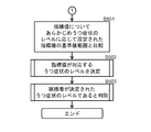
- the discrimination apparatus of the present embodiment further includes an arithmetic unit, and the arithmetic unit is used to evaluate depression symptoms of the elements of the correlation matrix of the functional connection measured in the subject at rest by the classifier. Generate index values for The computing device compares the index value to a reference range of index values set in advance according to the level of depression symptoms for each functional connection. The arithmetic device determines that the subject is the level of depression symptoms corresponding to a reference range in which the index value is included.
- One embodiment of the present invention relates to a determination apparatus for determining a therapeutic effect in a subject.
- the discrimination device of the present embodiment when resting, uses signals representing brain activity in a plurality of predetermined regions in the brain of each of a plurality of participants including a healthy person and a depressed patient in advance in time series by the brain activity detection device
- a storage device is provided for storing information identifying the classifier generated by the classifier generation process from the measured signal.
- the classifier is based on a weighted sum of a plurality of functional combinations selected by feature selection as related to a disease label of depressive symptoms by machine learning out of functional connections among the plurality of predetermined areas. It is generated to determine the disease label of depressive symptoms.
- the selected plurality of functional connections are a first functional connection between the left dorsolateral prefrontal cortex and the anterior left and posterior cingulate cortex, and the lower left forehead valvular and the right back At least one selected from the second functional connection between the medial prefrontal cortex and the right supplemental motor area.
- the determination apparatus of the present embodiment further includes an arithmetic unit, and the arithmetic unit is configured to calculate an element of the functional combination correlation matrix measured at rest in the subject at the first time point by the classifier. Generate a first value to assess the symptoms.
- the arithmetic device is configured to cause the classifier to perform the functional connection in the resting brain of the same subject as the subject at a second time point after the start of the treatment and after the first time point.
- the arithmetic device compares the first value with the second value.
- the determination apparatus determines that the treatment is effective for the improvement of the depressive symptoms of the subject when the second value is improved compared to the first value, and / or If the second value does not improve more than the first value, it is determined that the treatment is not effective for the improvement of the depressive symptoms of the subject.
- One embodiment of the present invention relates to a discrimination apparatus for stratification of depressed patients when depressive symptoms are stratified into a plurality of preset subclasses.
- the determination apparatus of the present embodiment includes an arithmetic unit, and the arithmetic unit generates an index value for evaluating depression symptoms for an element of a correlation matrix of functional connections measured in a subject at rest. .
- the arithmetic device compares the index value with a reference range of index values set in advance according to the subclass for each functional connection.
- the arithmetic device determines that the subject is the subclass corresponding to a reference range in which the index value is included.
- One embodiment of the present invention relates to a computer program for causing the determination device to execute the above process.
- the determination method of the present embodiment includes the step of generating an index value for evaluating depressive symptoms for an element of the functional connection correlation matrix measured in the subject at rest.
- the functional connection is the first functional connection between the left dorsal lateral prefrontal cortex and the left anterior and left posterior cingulate cortex, and the lower left frontal valvular portion, the right dorsal medial prefrontal cortex and At least one selected from the second functional connection between the right supplementary motor area.
- the determination method of the present embodiment includes the step of determining that the subject has a depressive symptom when the index value exceeds a reference value.
- One embodiment of the present invention relates to a determination method to assist in determining the level of depressive symptoms in a subject.
- the determination method of the present embodiment includes the step of generating an index value for evaluating depressive symptoms for the elements of the correlation matrix of the functional connection measured in the subject at rest.
- the functional connection is the first functional connection between the left dorsal lateral prefrontal cortex and the left anterior and left posterior cingulate cortex, and the lower left frontal valvular portion, the right dorsal medial prefrontal cortex and At least one selected from the second functional connection between the right supplementary motor area.
- One embodiment of the present invention relates to a determination method for assisting determination of a treatment effect in a subject.
- the determination method of the present embodiment includes the step of generating a first value for evaluating depressive symptoms for an element of the functional connection correlation matrix measured at rest in the subject at a first time point.
- the functional connection is the first functional connection between the left dorsal lateral prefrontal cortex and the left anterior and left posterior cingulate cortex, and the lower left frontal valvular portion, the right dorsal medial prefrontal cortex and At least one selected from the second functional connection between the right supplementary motor area.
- the functional coupling in the brain of the same subject as the subject at the second time point after the start of the treatment and after the first time point is at rest Generating a second value for evaluating depressive symptoms for an element of the same functional connection correlation matrix.
- the treatment in the step of comparing the first value with the second value, and when the second value is better than the first value, the treatment is not Determining that it is effective to improve the depressive symptoms of the examiner.
- One embodiment of the present invention relates to a determination method for assisting determination of a treatment effect in a subject.
- the determination method of the present embodiment includes the step of generating a first value for evaluating depressive symptoms for an element of the functional connection correlation matrix measured at rest in the subject at a first time point.
- the functional connection is the first functional connection between the left dorsal lateral prefrontal cortex and the left anterior and left posterior cingulate cortex, and the lower left frontal valvular portion, the right dorsal medial prefrontal cortex and At least one selected from the second functional connection between the right supplementary motor area.
- the discrimination method of this embodiment is the same as the functional coupling in the resting brain of the same subject as the subject at the second time point after the start of the treatment and after the first time point.
- Generating a second value for assessing depressive symptoms for an element of the correlation matrix of functional connections of In the determination method of the present embodiment, in the step of comparing the first value and the second value, and in the case where the second value is not better than the first value, the treatment is not performed. Determining that it is not effective in improving the depressive symptoms of the examiner.
- One embodiment of the present invention relates to a discrimination method for assisting the stratification of depressed patients.
- the determination method of the present embodiment when depression is stratified into a plurality of preset subclasses, depression symptoms of an element of a correlation matrix of functional connections measured in a subject at rest Generating an index value for evaluating.
- the functional connection is the first functional connection between the left dorsal lateral prefrontal cortex and the left anterior and left posterior cingulate cortex, and the lower left frontal valvular portion, the right dorsal medial prefrontal cortex and At least one selected from the second functional connection between the right supplementary motor area.
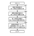
- the determination method of the present embodiment includes the step of comparing the index value with a reference range of the index value set in advance according to the subclass for each functional connection, and the subject includes the index value. Determining the subclass corresponding to the reference range to be determined.
- One embodiment of the present invention relates to a discriminator for assisting the stratification of depressed patients.
- the discrimination device of the present embodiment when resting, uses signals representing brain activity in a plurality of predetermined regions in the brain of each of a plurality of participants including a healthy person and a depressed patient in advance in time series by the brain activity detection device
- a storage device for storing information for identifying a classifier generated by a classifier generation process from the measured signal, and an arithmetic device for executing a stratification process.
- the classifier when the depressive symptoms are stratified into a plurality of preset subclasses, the classifier performs machine learning from among functional connections among the plurality of predetermined areas.
- the selected plurality of functional connections are a first functional connection between the left dorsolateral prefrontal cortex and the anterior left and posterior cingulate cortex, and the lower left forehead valvular and the right back At least one selected from the second functional connection between the medial prefrontal cortex and the right supplemental motor area.
- the arithmetic unit executes processing for determining the subclass from the weighted sum and the element of the correlation matrix to be determined.
- One embodiment of the present invention relates to a discriminating apparatus for assisting in the determination of the therapeutic effect in a depressed patient.
- the determination apparatus of the present embodiment includes an arithmetic unit, and the arithmetic unit is the first of a plurality of functional combinations selected from functional combination identification numbers 1 to 12 shown in Table 1 below for a plurality of subjects.
- a classifier generation process is performed to generate in advance a classifier that identifies a group in which the therapeutic effect is observed and a group in which the therapeutic effect is not observed in the plurality of subjects.
- the computing device measures a first correlation of the plurality of functional connections in the resting brain of a subject at a first time, and the same subject as the subject at a second time. Measuring a second correlation of the plurality of functional connections in the resting brain of the person.
- the arithmetic device determines the treatment effect on the subject by the classifier based on a difference between the second correlation and the first correlation of the plurality of functional combinations of the subject.
- One embodiment of the present invention relates to a determination method for assisting determination of treatment effect in depressed patients.
- the discrimination method of the present embodiment is the correlation between the first time point of the plurality of functional combinations selected from the functional combination identification numbers 1 to 12 shown in the following Table 1 and the start of the treatment for a plurality of subjects.
- the method may further include the step of generating in advance a classifier that identifies a group in which the therapeutic effect is observed and a group in which the therapeutic effect is not observed in the plurality of subjects.
- the determination method of the present embodiment includes the steps of: measuring a first correlation of the plurality of functional connections in the brain at rest of the subject at a first time; and the subject at a second time Measuring a second correlation of the plurality of functional connections in the resting brain of the same subject.
- the classifier performs the treatment effect on the subject based on the difference between the second correlation and the first correlation of the plurality of functional combinations of the subject. And a determining step.
- a signal indicating brain activity in a plurality of predetermined regions in a brain of each of a plurality of participants including a healthy person and a depressed patient is time-series by a brain activity detection device
- the present invention relates to a method of using a classifier generated by classifier generation processing from a signal measured in advance to determine the presence or absence of depressive symptoms or to determine the level of depressive symptoms in a subject.
- the method of using is based on a weighted sum of a plurality of functional combinations selected by feature selection as being related to a disease label of depressive symptoms by machine learning out of functional connections among the plurality of predetermined areas. Generating the classifier for determining the disease label of the depressive condition.
- the selected plurality of functional connections are a first functional connection between the left dorsolateral prefrontal cortex and the anterior left and posterior cingulate cortex, and the lower left forehead valvular and the right back At least one selected from the second functional connection between the medial prefrontal cortex and the right supplemental motor area.
- the use method may further comprise the step of inputting into the classifier an index value generated from an element of the correlation matrix of the plurality of functional combinations of the subject.
- a signal indicating brain activity in a plurality of predetermined regions in a brain of each of a plurality of participants including a healthy person and a depressed patient is time-series by a brain activity detection device
- the present invention relates to a method of using a classifier generated by a classifier generation process from previously measured signals to assist in the determination of the effect of treatment of depressive symptoms.
- the method of using is based on a weighted sum of a plurality of functional combinations selected by feature selection as being related to a disease label of depressive symptoms by machine learning out of functional connections among the plurality of predetermined areas. Generating the classifier for determining the disease label of the depressive condition.
- the selected plurality of functional connections are a first functional connection between the left dorsolateral prefrontal cortex and the anterior left and posterior cingulate cortex, and the lower left forehead valvular and the right back At least one selected from the second functional connection between the medial prefrontal cortex and the right supplemental motor area.
- the use method may further comprise the step of inputting into the classifier an index value generated from an element of the correlation matrix of the plurality of functional combinations of the subject.
- the brains of each of a plurality of participants including a healthy person and a depressed patient.
- the classifier is based on a weighted sum of a plurality of functional combinations selected by feature selection as related to a disease label of depressive symptoms by machine learning out of functional connections among the plurality of predetermined areas. It is generated to determine the disease label of depressive symptoms.
- the selected plurality of functional connections are a first functional connection between the left dorsolateral prefrontal cortex and the anterior left and posterior cingulate cortex, and the lower left forehead valvular and the right back At least one selected from the second functional connection between the medial prefrontal cortex and the right supplemental motor area.
- the method of use comprises the step of inputting into the classifier index values generated from elements of the correlation matrix of the plurality of functional combinations of the subject.
- the discrimination device is a signal obtained by measuring in advance a signal indicating brain activity in a plurality of predetermined regions in the brain of each of a plurality of participants including a healthy person and a depressed patient at rest at a time series.
- a classifier generating device including a first computing device for generating a first classifier.
- the first classifier is based on a weighted sum of a plurality of functional combinations selected by feature selection as related to a disease label of depressive symptoms by machine learning out of functional connections among the plurality of predetermined areas. Are generated to determine the disease label of the depressive symptoms.
- the selected plurality of functional connections are a first functional connection between the left dorsolateral prefrontal cortex and the anterior left and posterior cingulate cortex, and the lower left forehead valvular and the right back At least one selected from the second functional connection between the medial prefrontal cortex and the right supplemental motor area.
- the first arithmetic unit is for the plurality of subjects, after correlation of the first time point of the plurality of functional combinations selected from the functional combination identification numbers 1 to 12 shown in Table 5 below and after the start of the treatment In the correlation state space spanned by each of the differences in the correlation between the first and second time points of the plurality of functional connections, respectively measuring the correlation at the second time point after the first time point Generating a second classifier that identifies a group with therapeutic effects and a group without therapeutic effects in the plurality of subjects.
- the determining device further comprises a stratification device including a second computing device and a storage device.
- the storage device stores information of the first and second classifiers and information for stratification of depressive symptoms into a plurality of preset subclasses.
- the second arithmetic unit performs a stratification process on the subject into the subclasses using the first classifier.
- the second arithmetic unit measures a first correlation of the plurality of functional connections at rest at a first time point, for the subject stratified into a specific subclass by the stratification process.
- Execute processing for The second arithmetic unit executes a process for measuring a second correlation of the plurality of functional connections at rest of the same subject as the subject at a second time point.
- the second computing device is configured to send the subject to the subject by the second classifier based on a difference between the first correlation and the second correlation of the plurality of functional combinations of the subject. Determine the treatment effect.
- One embodiment of the present invention relates to a determination method for assisting determination of a treatment effect in a subject.
- the determination method of the present embodiment in the case where depression symptoms are stratified into a plurality of preset subclasses, at rest, in the brain of each of a plurality of participants including a healthy person and a depressed patient.
- the subject is detected using a first classifier generated by a first classifier generation process from signals measured beforehand in time series by the brain activity detection device as signals indicating brain activity in a plurality of predetermined regions. Includes stratification steps to stratify into subclasses.
- the first classifier is selected from a plurality of functional connections among the plurality of predetermined regions by machine learning as being related to a disease label of depressive symptoms by a plurality of feature selections. Based on the weighted sum of functional binding, it is generated to determine the disease label of the depressive symptoms.
- the selected plurality of functional connections are a first functional connection between the left dorsolateral prefrontal cortex and the left anterior and left posterior cingulate cortex, and the lower left frontal fornix and the right dorsomedia And at least one selected from a second functional connection between the prefrontal cortex and the right supplementary motor area.
- the determination method of the present embodiment measures, for a subject stratified into a specific subclass by the stratification step, a first method of measuring a first correlation of the plurality of functional connections at rest before administration. And a second correlation measurement step of measuring a second correlation of the plurality of functional connections at rest of the same subject as the subject after a predetermined period has elapsed from the start of dosing. .
- the determination method of the present embodiment is a determination step of determining the efficacy of the subject classified into a specific subclass using a second classifier, wherein the second classifier includes a plurality of subjects.
- the correlation between the first time point of the plurality of functional bindings selected from the functional binding identification numbers 1 to 12 shown in Table 1 below and the second time point after administration and after the first time point Measure each correlation. Then, in the correlation state space spanned by each of the differences in the correlation at the first time point and the second time point of the plurality of functional connections, the group and the treatment effect in which the treatment effect is observed in the plurality of subjects.
- the object based on the difference between the second correlation and the first correlation of the plurality of functional combinations of the subject generated in advance by a second classifier generation process for identifying an unidentified group. And a discrimination step of discriminating a medicinal effect on an examiner.
- One embodiment of the present invention relates to a first classifier generation device including an arithmetic device and a storage device.
- the arithmetic device is a signal obtained by measuring in advance a signal indicating brain activity in a plurality of predetermined regions in the brain of each of a plurality of participants including a healthy person and a depressed patient at rest at a time series.
- the classifier is based on a weighted sum of a plurality of functional combinations selected by feature selection as related to a disease label of depressive symptoms by machine learning out of functional connections among the plurality of predetermined areas. It is generated to determine the disease label of depressive symptoms.
- the selected plurality of functional connections are a first functional connection between the left dorsolateral prefrontal cortex and the anterior left and posterior cingulate cortex, and the lower left forehead valvular and the right back At least one selected from the second functional connection between the medial prefrontal cortex and the right supplemental motor area.
- the storage device stores information identifying the first classifier generated by the arithmetic device.
- One embodiment of the present invention relates to a method of generating a first classifier.
- signals indicating brain activity in a plurality of predetermined regions in the brain of each of a plurality of participants including a healthy person and a depressed patient are measured in advance by a brain activity detection device in time series.
- the first classifier is based on a weighted sum of a plurality of functional combinations selected by feature selection as related to a disease label of depressive symptoms by machine learning out of functional connections among the plurality of predetermined areas. Are generated to determine the disease label of the depressive symptoms.
- the functional connection is the first functional connection between the left dorsal lateral prefrontal cortex and the left anterior and left posterior cingulate cortex, and the lower left frontal valvular portion, the right dorsal medial prefrontal cortex and At least one selected from the second functional connection between the right supplementary motor area.
- One embodiment of the present invention relates to a second classifier generation device including an arithmetic device and a storage device.
- the computing device is configured to compare the correlation between the first points of time of the plurality of functional combinations selected from the functional combination identification numbers 1 to 12 shown in the following Table 3 and the start of the treatment for the plurality of subjects. The correlations at the second point in time after the point in time are respectively measured.
- the present embodiment is a group in which a plurality of subjects show therapeutic effects in a correlation state space spanned by each of the differences in the correlation between the first time point and the second time point of the plurality of functional connections.
- a second classifier is generated that distinguishes the group from which no therapeutic effect is seen.
- the storage device stores information for identifying the second classifier generated by the arithmetic device.
- One embodiment of the present invention relates to a second classifier generation method.
- the correlation between the first point of time of the plurality of functional bindings selected from the functional binding identification numbers 1 to 12 shown in the following Table 4 and the start of the treatment for the plurality of subjects is a first
- the correlations at the second point in time after the point in time are respectively measured.
- the present embodiment is a group in which a plurality of subjects show therapeutic effects in a correlation state space spanned by each of the differences in the correlation between the first time point and the second time point of the plurality of functional connections.
- One embodiment of the present invention relates to a brain activity training device provided with a brain activity detection device, a presentation device, a computing device, and a storage device.
- the brain activity detection device detects signals indicating brain activity in a plurality of predetermined regions in the brain of the trainee of the neuro feedback training in time series
- the storage device is healthy at rest From the functional connection between the plurality of predetermined areas, based on the signal indicating the brain activity in a plurality of predetermined areas in the brain of each of the plurality of participants including the person and the depressed patient in advance in time series
- Information identifying the functional connection of the training object among the plurality of functional connections selected by feature selection to distinguish the disease label of depressive symptoms by machine learning, and the functional connection of the training object in neurofeedback training Storing the target pattern of the target object, and the arithmetic unit generates temporal correlation of the functional combination of the training object for a predetermined period from the signal detected by the brain activity detector.
- the functional connection of the training object includes a first functional connection between the left dorsolateral prefrontal cortex and the anterior left and posterior caudal cortex, and in the brain of the trainee
- Each of the plurality of predetermined regions corresponds to a plurality of predetermined regions in the brain of the plurality of participants.
- One embodiment of the present invention relates to a control method of a brain activity training device including a brain activity detection device, a presentation device, an arithmetic device, and a storage device.
- the brain activity detection device detects signals indicating brain activity in a plurality of predetermined regions in the brain of the trainee of the neuro feedback training in time series
- the storage device is healthy at rest From the functional connection between the plurality of predetermined areas, based on the signal indicating the brain activity in a plurality of predetermined areas in the brain of each of the plurality of participants including the person and the depressed patient in advance in time series Information identifying the functional connection of the training object among the plurality of functional connections selected by feature selection to distinguish the disease label of depressive symptoms by machine learning, and the functional connection of the training object in neurofeedback training And storing the target pattern of the target and the brain activity detecting device to indicate brain activity in a plurality of predetermined regions in the brain of the trainee of the neuro feedback training.
- the functional connection of the training object includes a first functional connection between the left dorsolateral prefrontal cortex and the anterior left and posterior caudal cortex, and in the brain of the trainee
- Each of the plurality of predetermined regions corresponds to a plurality of predetermined regions in the brain of the plurality of participants.
- the present invention it is possible to provide a discrimination apparatus and discrimination method for objectively discriminating an index for the state of depression symptoms with respect to the brain activity state. According to the present invention, it is possible to objectively determine information representing the degree of therapeutic effect on depressive symptoms. According to the present invention, it is possible to provide a classifier that outputs an index value as a biomarker for objectively determining depressive symptoms. According to this invention, the level of depressive symptoms can be determined. According to the present invention, a depressed patient can be objectively stratified. According to the present invention, neuro feedback training can be performed.
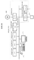
- FIG. 1 is a schematic view showing an entire configuration of an MRI apparatus 10;
- FIG. 2 is a hardware block diagram of a data processing unit 32. It is a conceptual diagram which shows the procedure which extracts the correlation matrix which shows the correlation of the functional connection at rest about a region of interest. It is a conceptual diagram explaining the process of producing
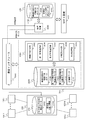
- FIG. 5 is a functional block diagram for performing a generation process of the first classifier as described in FIG. 4 and a determination process by the generated first classifier. It is a flowchart for demonstrating the process which the data processing part 32 performs in order to produce
- FIG. 20 is a functional block diagram showing an example of processing data collection, estimation processing and brain activity measurement of a subject in a distributed manner shown in FIG. 19; 5 is a flow chart for generating a second classifier.
- FIG. 20 is a functional block diagram showing an example of processing data collection, estimation processing and brain activity measurement of a subject in a distributed manner shown in FIG. 19; 5 is a flow chart for generating a second classifier.
- FIG. 7 is a flow chart for determining drug efficacy using a second classifier.
- FIG. It is a functional block diagram for performing the treatment effect determination process by the production
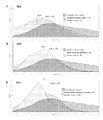
- d shows the smoothed histogram of related weighted sums of melancholic MDDs.
- e shows the smoothed histogram of the associated weighted sum of non-Melancholy MDDs.
- f shows the smoothed histogram of the associated weighted sum of treatment resistant MDDs.
- g denotes the smoothed histogram of the associated weighted sum of ASDs.
- h shows the smoothed histogram of the related weighted sum of SSDs.
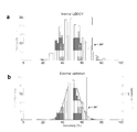
- a shows a histogram of LOOCV rearrangement test (1000 iterations) of training data.
- b shows the accuracy of the out-of-sample test data set, showing the binomial distribution as a curve.
- the accuracy of the melancholic MDD classifier trained and verified without reordering is shown as a vertical line.
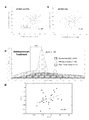
- a shows the correlation between the BDI score and the discrimination result using the first classifier (all MDD and whole healthy subject).
- b shows the correlation (the whole MDD whole) of the discrimination
- c shows the smoothed histogram of the related weighted sum before and after treatment.
- d shows the correlation between the change in BDI score after administration and the change in discrimination result using the first classifier.
- a shows the difference in correlation between the healthy control group and the MDD group in each functional binding and the difference in correlation before and after dosing.
- b shows the difference between pre-dose and post-dose correlations in FC1 and FC2 for the Hiroshima cohort.
- c shows the difference between pre-dose and post-dose correlations in FC1 and FC2 for the Chiba cohort.
- d shows the difference in correlation between pre-dose and post-dose in FC1 and FC2 for a cohort of healthy individuals.
- e shows the difference in correlation before and after dosing in FC1 and FC2 of the post-dosing remission group and the non-post-dosing group.
- f shows the distribution of signed ⁇ FC1 and ⁇ FC2 in the dose group.
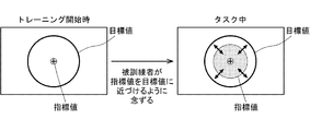
- It is a figure which shows the concept of a structure of a brain activity training apparatus. It is an example of the monitor display for showing the closeness of an index value and a target value.
- a shows neurofeedback scores during training of MDD participants.
- b shows the HDRS score before and after training of the MDD participant.
- c shows neurofeedback scores during training of participants of subclinical depression.
- d shows the correlation between BDI score after training and rs-fcMRI variation.
- Fig. 6 shows a flowchart of neurofeed
- depressive symptoms include: depressive mood; decreased interest or concern; decreased vigor; frustration; inhibition; diminished thinking ability, concentration ability or determination ability; sense of worthlessness or guilt; , Suicidal ideation or suicide planning; morbid thinking content; delusions; physical symptoms (such as general fatigue, headache, headache, back pain, etc. pain in the body, palpitations, shortness of breath, loss of appetite, weight loss etc.), sleep At least one type selected from faults is included.
- the depressive symptoms include symptoms associated with Major Depressive Disorder (MDD) in the Diagnostic and Statistical Manual of Mental Disorders (DSM) -IV criteria.
- MDD Major Depressive Disorder
- DSM Diagnostic and Statistical Manual of Mental Disorders
- depression is not limited as long as it has the above-mentioned depressive symptoms, but is preferably MDD.
- MDDs include melancholic MDD, non-melancholic MDD and refractory MDD. In the present specification, MDD may be referred to simply as depression.
- HAM-D For the evaluation of the degree of depression, for example, an evaluation by the self-reported Beck Depression Inventory (Beck Depression Survey Table) is conventionally used as a supplementary material at screening and interviewing at the time of medical examination.
- the Hamilton Depression Rating Scale (HDRS) which is used as a rating scale for physicians, is a multi-item questionnaire that provides an index of depression, and serves as an index for evaluating recovery.
- the abbreviation is HAM-D.
- the subject is preferably, but not limited to, a person having the depressive symptoms. Age and gender are not limited. The subject may be one who has not received treatment for improving the depressive symptoms or one who has received the treatment.
- Treatment to ameliorate depressive symptoms includes at least one selected from medication, neurofeedback therapy, modified electroconvulsive therapy and repetitive transcranial magnetic stimulation therapy.
- Drugs to be administered include tricyclic antidepressants (imipramine, trimipramine, clomipramine, amitriptyline, nortriptyline, amoxapine, lofepramine, and doslepin), tetracyclic antidepressants (maprotiline, aminserine, cetiptiline, and the like), trazodone Selective serotonin reuptake inhibitors (such as escitalopram, fluvoxamine, paroxetine, and sertrilarin), serotonin-norepinephrine reuptake inhibitors (such as milnacipran and duloxetine), noradrenaline / serotonergic antidepressants (such as mirtazapine), And at least one selected from benzamide drugs (such as sulpiride).
- the improvement of depressive symptoms means that the symptom is better than the previous state, as seen from clinical findings such as BDI, or It means that it will be in the state that can be said to be "remitted” from the viewpoint of
- the determination of the level of depressive symptoms in the subject is assisted, the determination of the presence or absence of depressive symptoms in the subject is assisted, and the determination of the treatment effect in the subject Assisting and aiding in subject stratification into disease subclasses are included.
- drug reprofiling is a drug that has already been shown to have an effect on humans against other diseases or conditions, or that it is weak against humans but has no significant effect. It is intended to detect drug effects on depressive symptoms for drugs that are known and whose effects on depressive symptoms are unknown.
- the imaging device of resting fMRI is not limited.
- the imaging conditions are also not limited as long as the fMRI image can be acquired.
- magnetic filed is about 3.0 T
- field of view is about 192 mm to 256 mm
- matrix is about 64 ⁇ 64
- number of slices is about 30 to 40
- number of volumes is about 112 to 244
- slice thickness is 3 .0 mm to about 4.0 mm
- slice gap about 0 mm to about 0.8 mm
- TR about 2,000 ms to about 2,700 ms
- TE about 25 ms to about 31 ms
- total imaging time about 5 minutes to about 10 minutes
- flip angle about about 75 deg to 90 deg
- slice acquisition order is Ascending (Interleaved).
- Imaging is preferably performed under dark illumination. Further, it is preferable that the subject thinks nothing during imaging and does not sleep. Furthermore, it is preferable that the subject keep looking at the cross mark in the center of the monitor screen during imaging.
- the imaged fMRI image can be processed by the method described in Yahata et al. (NATURE COMMUNICATIONS
- the imaged fMRI image data is not particularly limited, for example, T1 weighted structure image and resting state function using SPM 8 (Wellcome Trust Center for Neuroimaging, University College London, UK) of Matlab R 2014a (Mathworks inc., USA) Standard pre-processing on the image can be applied.
- SPM 8 Wellcome Trust Center for Neuroimaging, University College London, UK
- an image determined to have moved by 0.5 mm or more from the previous image may be excluded from the analysis.
- FC functional connectivity
- ROI regions of interest
- FC functional connectivity
- the functional connection is a feature value (element of correlation matrix) generally used in resting brain activity analysis, and is defined by Pearson's correlation coefficient between time-series signals of different regions of interest.
- the binding strength can be represented by an average value of Pearson's correlation coefficients from values measured over a predetermined time.
- the coupling strength may be represented by another statistic about Pearson's correlation coefficient within a predetermined time.
- Extraction of the elements of the correlation matrix is not particularly limited, but can be performed, for example, in the following procedure.
- a time series of average signals of all voxels (Voxel) included in each region of interest is extracted.
- a band pass filter (0.008 to 0.1 Hz) is applied to remove noise from the signal value.
- regression is performed with nine explanatory variables (average signal of whole brain, white matter, cerebrospinal fluid and six motion correction parameters).
- the correlation coefficient is a value representing the coupling strength of the functional coupling, and the coupling strength corresponding to each pair is determined.
- an index value for determining the disease label of depressive symptoms for functional binding is calculated.
- association weighted sums for a plurality of functional combinations are calculated as index values for determining a disease label of depressive symptoms.
- the “relevant weighted sum” refers to the sum of multiple functional combinations multiplied by the corresponding weights.
- the index value is not simply data obtained by measuring the brain activity of the subject, but is a value artificially calculated in consideration of the weight of each functional connection.
- the index value is used to determine the label of depressive symptoms, to determine the level of depressive symptoms, to determine the therapeutic effect, or for stratification of depressed patients.
- the highest contribution to depressive symptoms is the functional connection indicated by the functional connection identification number 1.
- the next highest degree of contribution is the functional binding indicated by the functional binding identification number 2. Therefore, in each of the embodiments described below, at least one or both of the functional binding identification number 1 and the functional binding identification number 2 may be selected and used.
- the 12 pairs of functional connections are particularly suitable for discriminating between melancholic MDD patient groups and healthy control groups.
- the first classifier at rest, displays signals indicative of brain activity in a plurality of predetermined regions in the brain of each of a plurality of participants including normal persons and depressed patients. It is generated by a classifier generation process from signals measured beforehand in time series by the brain activity detection device.
- a depressed patient means a participant who has been diagnosed with depression in advance by diagnosis by a doctor and associated with the "disease label" of depression.
- the classifier is based on weights of functional combinations selected by feature selection as related to a disease label of depressive symptoms by machine learning out of functional connections between the plurality of predetermined areas. It is generated to determine the disease label.
- the sparse logistic Feature selection by regression selects the functional binding used to determine the disease label for depression. Then, from the functional combination selected in this manner, the above-mentioned associated weighted sum is calculated.
- Discrimination device 1 For example, the first classifier is generated from fMRI image data acquired from the MRI apparatus 10 shown in FIG.
- FIG. 1 is a schematic view showing an entire configuration of an MRI apparatus 10 as a discrimination apparatus 1 according to a first embodiment of the present invention.
- the MRI apparatus 10 comprises an MRI imaging unit 25 for imaging MRI, and a data processing unit 32 for setting control sequences of the MRI imaging unit 25 and processing various data signals to generate an image.
- the MRI imaging unit 25 applies a controlled magnetic field to the region of interest of the subject 2 to apply a controlled magnetic field and applies an RF wave, and a response wave (NMR) from the subject (or the subject)
- a drive unit 21 that controls the magnetic field applied to the subject 2 and controls transmission and reception of the RF wave.
- the central axis of the cylindrical bore on which the subject 2 is placed is taken as the Z axis
- the X axis is defined in the horizontal direction perpendicular to the Z axis and the Y axis in the vertical direction.
- the nuclear spins of the nuclei constituting the subject 2 are oriented in the magnetic field direction (Z-axis) by the static magnetic field applied by the magnetic field applying mechanism 11, and It performs precession motion around this magnetic field direction at the Larmor frequency specific to the nucleus.
- the atom when the same RF pulse as the Larmor frequency is irradiated, the atom resonates, absorbs energy and is excited, and a nuclear magnetic resonance phenomenon (NMR phenomenon; Nuclear Magnetic Resonance) occurs. After this resonance, when the RF pulse irradiation is stopped, the atom emits energy and in the relaxation process returns to the original steady state, it outputs an electromagnetic wave (NMR signal) of the same frequency as the Larmor frequency.
- NMR phenomenon Nuclear Magnetic Resonance
- the output NMR signal is received by the receiving coil 20 as a response wave from the subject 2, and the data processing unit 32 images the region of interest of the subject 2.
- the magnetic field application mechanism 11 includes a static magnetic field generating coil 12, a gradient magnetic field generating coil 14, an RF irradiation unit 16, and a bed 18 on which the subject 2 is placed in the bore.
- the subject 2 lies, for example, on the bed 18.
- the prism glasses 4 can view a screen displayed on the presentation device 6 (for example, a display) installed perpendicularly to the Z axis.
- a visual stimulus is given to the subject 2 by the image of the presentation device 6.
- the visual stimulus to the subject 2 may be configured such that an image is projected by the projector in front of the subject 2.
- Such visual stimulation corresponds to presentation of feedback information in the above-described neurofeedback.
- the drive unit 21 includes a static magnetic field power supply 22, a gradient magnetic field power supply 24, a signal transmission unit 26, a signal reception unit 28, and a bed drive unit 30 for moving the bed 18 to an arbitrary position in the Z-axis direction.
- the data processing unit 32 includes an input unit 40 for receiving various operations and information input from an operator (not shown), a display unit 38 for displaying various images and information on the region of interest of the subject 2 and various processes.
- a control unit that controls the operation of each function unit such as a storage unit 36 that stores programs, control parameters, image data (structured images, etc.) to be executed and other electronic data, and generates a control sequence that drives the drive unit 21 42, an interface unit 44 for executing transmission and reception of various signals between the drive unit 21, a data collection unit 46 for collecting data consisting of a group of NMR signals derived from a region of interest, and data of the NMR signals.
- an image processing unit 48 for forming an image
- a network interface unit 50 for performing communication with the network.
- the data processing unit 32 is a general-purpose computer that executes functions to operate the respective functional units in addition to the dedicated computer, and the operation and data specified based on the program installed in the storage unit 36 It also includes cases where processing or control sequences are generated.
- the data processing unit 32 will be described as being a general purpose computer.
- the static magnetic field generating coil 12 generates an induction magnetic field by supplying a current supplied from the static magnetic field power supply 22 to a helical coil wound around the Z axis to generate a static magnetic field in the Z axis direction in the bore. .
- the region of interest of the subject 2 is set in the region of high uniformity of the static magnetic field formed in the bore.
- the static magnetic field coil 12 is more specifically composed of, for example, four air core coils, and by combining them, a uniform magnetic field is generated inside, and predetermined nuclei in the body of the subject 2 are more specific. Gives orientation to the spins of hydrogen nuclei.
- the gradient magnetic field generating coil 14 is composed of an X coil, a Y coil, and a Z coil (not shown), and is provided on the inner circumferential surface of the static magnetic field generating coil 12 having a cylindrical shape.
- These X coil, Y coil and Z coil superimpose a gradient magnetic field on the uniform magnetic field in the bore while switching the X-axis direction, Y-axis direction and Z-axis direction in order to give an intensity gradient to the static magnetic field .
- the Z coil inclines the magnetic field strength in the Z direction to define the resonance surface
- the Y coil applies a short time inclination immediately after the application of the magnetic field in the Z direction to modulate the phase proportional to the Y coordinate to the detection signal Is added (phase encoding)
- the X coil is subsequently inclined at the time of data acquisition to give the detection signal a frequency modulation proportional to the X coordinate (frequency encoding).
- the switching of the superimposed gradient magnetic field is realized by outputting different pulse signals from the transmitting unit 24 to the X coil, the Y coil, and the Z coil in accordance with the control sequence.
- the position of the subject 2 at which the NMR phenomenon occurs can be specified, and positional information on three-dimensional coordinates necessary to form an image of the subject 2 can be given.
- imaging can be performed from various angles by assigning slice directions, phase encoding directions, and frequency encoding directions to three sets of orthogonal gradient magnetic fields, respectively, and combining them.
- the sagittal slice or coronal slice orthogonal thereto and further, the direction perpendicular to the plane is parallel to the axis of three orthogonal gradient magnetic field It is possible to image not an oblique slice or the like.
- the RF irradiation unit 16 irradiates an RF (Radio Frequency) pulse to a region of interest of the subject 2 based on the high frequency signal transmitted from the signal transmission unit 33 according to the control sequence.
- RF Radio Frequency
- the RF irradiation unit 16 is built in the magnetic field application mechanism 11 in FIG. 1, it may be provided on the bed 18 or may be integrated with the receiving coil 20.
- the receiving coil 20 detects a response wave (NMR signal) from the subject 2 and is disposed close to the subject 2 in order to detect the NMR signal with high sensitivity.
- NMR signal response wave
- a weak current is generated in the receiving coil 20 based on electromagnetic induction.
- the weak current is amplified by the signal receiving unit 28, and further converted from an analog signal into a digital signal and sent to the data processing unit 32.
- a given nucleus for example a hydrogen nucleus
- a predetermined nucleus in a portion matching the resonance condition for example, a slice of a predetermined thickness of the subject 2
- spins rotate at a time.
- an electromagnetic wave emitted by rotating spins induces a signal in the receiving coil 20, and this signal is detected for a while.
- a tissue containing a predetermined atom in the body of the subject 2 is observed.
- the X and Y gradient magnetic fields are applied to detect the signal.
- the image processing unit 48 measures the detection signal while repeatedly giving the excitation signal based on the data constructed in the storage unit 36, reduces the frequency of the resonance to the X coordinate by the first Fourier transform calculation, and the second time The Y coordinate is restored by the Fourier transform of to obtain an image, and the image corresponding to the display unit 38 is displayed.
- the above-described BOLD signal is imaged in real time by such an MRI system, and the control unit 42 performs an analysis process as described later on images captured in time series, thereby achieving resting functional coupling It becomes possible to perform imaging of MRI (rs-fcMRI).
- FIG. 2 is a hardware block diagram of the data processing unit 32. As shown in FIG.
- the hardware of the data processing unit 32 is not particularly limited as described above, but a general-purpose computer can be used.
- a computer main body 2010 of the data processing unit 32 is connected to a processing unit (central processing unit: CPU; also referred to as processor) 2040, a disk drive 2030 and a memory drive 2020 in addition to a memory drive 2020 and a disk drive 2030.
- CPU central processing unit
- disk drive 2030 and a memory drive 2020 in addition to a memory drive 2020 and a disk drive 2030.
- RAM random access memory
- RAM random access memory
- the communication interface 2090 corresponds to an interface unit 44 for exchanging signals with the drive unit 21 and the like, and a network interface 50 for communicating with another computer via a network (not shown).
- a hard disk (HDD), a solid state drive (SSD: Solid State Drive), or the like can be used as the nonvolatile storage device 2080.
- the non-volatile storage device 2080 corresponds to the storage unit 36.
- the computer main body 2010 of the data processing unit 32 also functions as a first classifier generation device and / or a second classifier generation device.
- Each function of the data processing unit 32 for example, each function of the control unit 42, the data collection unit 46, and the image processing unit 48 is realized by arithmetic processing that the CPU 2040 executes based on a program.
- a program that causes the data processing unit 32 to execute the functions of the above-described embodiment is stored in the CD-ROM 2200 or the memory medium 2210, inserted into the disk drive 2030 or the memory drive 2020, and further stored in the non-volatile storage device 2080. It may be forwarded. The program is loaded into the RAM 2070 upon execution.
- the data processing unit 32 further includes a keyboard 2100 and a mouse 2110 as an input device, and a display 2120 as an output device.
- the keyboard 2100 and the mouse 2110 correspond to the input unit 40
- the display 2120 corresponds to the display unit 38.
- the program for functioning as the data processing unit 32 as described above may not necessarily include the operating system (OS) that causes the computer main body 2010 to execute the function of the information processing apparatus or the like.
- the program needs to call only an appropriate function (module) in a controlled manner, and include only a part of an instruction that makes it possible to obtain a desired result. It is well known how the data processing unit 32 operates, and the detailed description will be omitted.
- the computer that executes the program may be singular or plural. That is, centralized processing may be performed, or distributed processing may be performed.
- FIG. 3 is a conceptual diagram showing a procedure for extracting a correlation matrix indicating correlation of functional connection at rest associated with a disease label of depressive symptoms.
- the average “activity” of each region of interest is calculated from the fMRI data for n (n: natural number) times of resting fMRI measured in real time, and the brain regions are Calculate the correlation value of the degree of activity).
- the correlation shows only 34 ⁇ 34 on the diagram.
- the calculation of the elements of such a correlation matrix is not particularly limited, but can be performed, for example, as follows.
- Functional connection is a feature that is generally used in resting brain activity analysis, and is defined by Pearson's correlation coefficient between time-series signals of different regions of interest.
- a band pass filter is applied, and then regression is performed using nine explanatory variables (average signals of whole brain, white matter, brain back, cerebrospinal fluid and six motion correction parameters).
- 137 regions of interest included in Brain Sulci Atlas are used as the region of interest.
- a functional connection FC between these 137 regions of interest is used as a feature.
- FIG. 4 is a conceptual diagram for explaining the process of generating a first classifier serving as a biomarker from the correlation matrix as described in FIG.
- the first classifier functions as, for example, an index as a diagnostic marker for "depression", and is used for assisting a doctor in diagnosing the severity or progression of the disease. be able to.
- the device that performs the operations of the first classifier and the output of the results can also be called a diagnostic aid device.
- the data processing unit 32 derives the elements of the correlation matrix of the degree of activity between the brain regions (between the regions of interest).
- the data processing unit 32 performs feature extraction by regularization canonical correlation analysis on the attributes of the participant including the participant's disease / healthy label and the correlation matrix.
- regular learning in machine learning and statistics, a regularization term weighted by a hyperparameter is added to an error function to add suppression to the complexity and freedom of the model to prevent over-learning. It is a method.
- regularization canonical correlation analysis in the case where sparsification is also realized for the explanatory variables, it is particularly referred to as sparse canonical correlation analysis (SCCA). Below, as a specific example, it demonstrates as what performs sparse canonical correlation analysis.
- SCCA sparse canonical correlation analysis
- the union of functional combinations FC extracted as explanatory variables is called the “second union” over all cross validations.
- LOCV one-out-one-out cross validation
- FIG. 5 is a functional block diagram for performing the generation process of the first classifier as described in FIG. 4 and the determination process by the generated first classifier.
- a signal indicating brain activity in a plurality of predetermined regions in the brain of each of a plurality of participants including a healthy person and a depressed patient is measured in advance in time series by an MRI device.
- a participant rs-fc MRI measurement data 3102 which is information and a plurality of human attribute information 3104 associated with each participant who measured the MRI measurement data are stored.
- the “person attribute information” includes “person characteristic information” for identifying an individual participant and “measurement condition information” for identifying a measurement condition for each participant.
- Human characteristic information means information such as disease label, age, gender, and medication history for participants.
- Measurement condition information refers to measurement site information (including information for specifying a measured facility and / or a measured device) that measured a participant, and whether it was measured by eye open / closed eye during measurement, or It is a condition for specifying measurement conditions such as magnetic field strength.
- the computing device 2040 performs processing to generate a classifier for the disease label based on the participant rs-fc MRI measurement data 3102 and the corresponding human attribute information 3104.
- the correlation matrix calculation unit 3002 calculates, from the participant rs-fc MRI measurement data 3102, a correlation matrix of functional connections of brain activity among a plurality of predetermined regions for each participant.
- the functional matrix correlation matrix data thus calculated is stored in the non-volatile storage device 2080 as functional matrix correlation matrix data 3106 for each participant.
- the first feature selection unit 3004 sequentially selects one subset for K different subsets (K: a natural number of 2 or more) extracted from a plurality of participants, and removes the selected subset. Sparse canonical correlation analysis on a plurality of attribute information and an element of a correlation row example for (K-1) subsets, and specific attribute information of a plurality of human attribute information, for example, only a disease label Extract elements of the correlation row example connected to the canonical variable corresponding to. Furthermore, the first feature selection unit 3004 acquires a first union, which is a union of elements of the extracted correlation row example, over the subsets selected sequentially, and causes the non-volatile storage device 2080 to execute the first function. It stores as the dynamic combination union data 3108.
- the “first functional combination union data set” may be an index for specifying an element corresponding to the first union among the data 3106 of the functional connection correlation matrix.
- the second feature selection unit 3006 selects N participants from the plurality of participants when the remaining participants excluding the subset of the K among the plurality of participants are set as the test set and the test set is divided into N different groups.
- Sparse logistic test classifier for estimating the specific attribute information (for example, disease label) based on the first union, targeting a set of participants excluding one of the selected groups It calculates by regression and extracts the element of the correlation line example which becomes an explanatory variable of a test classifier with sparsification.
- the second feature selection unit 3006 further repeats feature extraction while sequentially selecting one group out of N groups, and is a union set of elements of correlated row examples extracted as an explanatory variable of the test classifier.
- a certain second union is acquired and stored in the non-volatile storage device 2080 as second functional combined union data 3110.
- the “second functional combination sum set data” may also be an index for specifying an element corresponding to the second sum set among the data 3106 of the functional connection correlation matrix.
- the number of elements in each of the N groups may be one.
- the classifier generation unit 3008 calculates a first classifier for estimating specific attribute information (for example, a disease label) by sparse logistic regression, using the second union as an explanatory variable.
- the classifier generation unit 3008 stores the generated information for identifying the first classifier in the non-volatile storage device 2080 as classifier data 3112.
- the discrimination processing unit 3010 performs discrimination processing on input data based on the classifier specified by the classifier data 3112.
- the classifier generation unit 3008 is described as generating a classifier using the second union as an explanatory variable, but, for example, the classifier generation unit 3008 directly transmits the first union.
- a first classifier may be generated as an explanatory variable.
- regularized logistic regression that is logistic regression using regularization (for example, L1 regularization or L2 regularization) can be used, and more specifically, for example, , "Sparse Logistic Regression" described above can also be used.
- a support vector machine, LDA (Linear discriminant analysis), or the like may be used to generate the first classifier.
- LDA Linear discriminant analysis
- the test calculated by the second feature selection unit 3006 while sequentially selecting one group to be removed from N groups.
- the cross validation may be performed by the classifier to calculate the discrimination result with the excluded groups as test samples.
- test set in the processing of the second feature selection unit 3006 is independent of the data set used to reduce the number of dimensions.
- FIG. 6 is a flowchart for explaining the processing performed by the data processing unit 32 to generate a classifier to be a biomarker.
- feature selection a process performed to explain observation data with a smaller number of explanatory variables.
- a first classifier for predicting a target disease label a process performed to explain observation data with a smaller number of explanatory variables.
- feature selection feature extraction
- regularization is used as a feature extraction method.
- canonical correlation analysis such processing is called “sparse canonical correlation analysis” in the sense that processing is carried out along with regularization and processing is performed so as to leave something more important as an explanatory variable.
- a penalty is imposed on the magnitude of the absolute value of the parameter of canonical correlation analysis called “L1 regularization” as described below. It is possible to use the imposing method.
- the data processing unit 32 when the data processing unit 32 starts the processing of classifier generation by the input of the processing start from the input unit 40 or the like (S100), the data processing unit 32 receives the information about each participant
- the MRI measurement data is read (S102), and the first feature extraction process is performed by sparse canonical correlation analysis (SCCA) (S104).
- SCCA sparse canonical correlation analysis
- step S104 the process of step S104 is referred to as “inner loop feature selection”.
- each variable x 1 and x 2 is standardized so as to have an average of 0 and a standard deviation of 1. Be done.
- CCA canonical correlation analysis
- CCA canonical correlation analysis
- L1-SCCA can be formulated as the following formula (1).
- hyperparameters ⁇ 1 and ⁇ 2 are the degree of sparsity of weighting parameters w 1 and w 2 (referred to as “sparse projection vector” because corresponding variables are sparseized by w 1 and w 2 ) It shows each.
- two data matrices corresponding to the above variables are constructed in order to identify the latent relationship between human attribute information and functional connection FC. .
- the next three columns show the measured institutions (locations) of brain activity, in this case corresponding to having been measured at three different sites [100] (site A), [010] (Site B) or [001] (Site C) is included.
- the fifth column contains the participant's age value
- the sixth column is the participant's gender information, including either 1 (male) or 0 (female) values
- the seventh column indicates, of the measurement conditions, whether the participant had opened or closed his eyes during measurement, and includes a value of 1 (open eye) or 0 (closed eye).
- the last three columns contain status information about the medication history of the three drugs, and each column contains a value of 1 (with drug treatment) or 0 (without drug treatment history).
- the human characteristic information and the measurement condition information are not limited to those described above, for example, other characteristics such as information on the medication history of other agents of the participant, and other measurement conditions, for example, an MRI apparatus
- the information of conditions, such as the magnitude of the applied magnetic field, may be included.
- Second data matrix X 2 is assumed to be a representation of the elements of the lower triangular portion of the non-diagonal of the correlation matrix representing a correlation of the functional binding of the participants (FC) in a row vector format.
- the sparse projection vector w 2 is used to specify the index (element) for identifying the correlation matrix element of the functional combination associated only with the disease label in the corresponding second data matrix X 2 Do.
- hyperparameters ⁇ 1 and ⁇ 2 of L 1 -SCCA are independently modified between 0.1 and 0.9, for example in steps of size 0.1.
- the size of the step of ranges and varying hyperparameters lambda 1 and lambda 2 and the variable is not intended to be limited to such an example.
- variable i k to indicate the index of the element is not a k-th zero of the projection vector w 2.
- 1 ⁇ k ⁇ m, and m indicates the number of non-zero elements.
- correlation matrix elements that correspond to canonical variables that are only relevant to the diagnostic label correlation matrix elements that are essential for classification are selected.
- FIG. 7 is a conceptual diagram for explaining inner loop feature selection.
- the set of participants is divided into K (K: two or more natural numbers), and (K-1) subsets Among the remaining subsets excluding one set, performing L1-SCCA described above is repeated (K-1) times.
- L 1 -SCCA is executed by changing ⁇ 2 by a predetermined step at a predetermined range.
- a functional combination element FC related to a canonical variable related only to the “diagnosis” label is extracted as a feature value.
- This procedure constructs the first classifier, which is generated based on, for example, MRI measurement data from a plurality of imaging sites in Japan, as described later, robustly generalizes to foreign countries, for example, the United States It will help.
- the data processing unit 32 executes the second feature extraction process using sparse logistic regression based on the result of the inner loop feature selection (S106).
- step S106 the process of step S106 is referred to as “outer loop feature selection”.
- Sparse logistic regression is a method in which logistic regression analysis is extended to the framework of Bayesian estimation, and dimension compression of feature vectors is performed simultaneously with weight estimation for discrimination. It is useful when the number of dimensions of feature vectors of data is very high and a large number of unnecessary feature quantities are included. For unnecessary feature quantities, weight parameters in linear discriminant analysis are made zero (ie, feature selection is performed), and only a very small number of feature quantities related to discrimination are extracted (sparseness).
- the probability p belonging to the class to be classified is obtained for each class with respect to the obtained feature data, and the maximum value is assigned to the output class.
- p is output by logistic regression.
- Weight estimation is performed by ARD (Automatic Relevance Determination), and feature quantities that contribute little to class discrimination are excluded from calculation as the weight approaches zero.
- the disease label is predicted using the first classifier based on the next hierarchical Bayesian estimation with the first union of the feature values extracted using the L1 regularization CCA described above as an input.
- z-hat (a letter with a ⁇ attached to it is called a "hat") is a feature vector with an expanded input.
- the feature vector z is extracted from the joint correlation matrix of resting MRI samples of one participant according to equation (2).
- ⁇ is a parameter vector of the logistic function.
- the distribution of the parameter ⁇ at this time is set to the following normal distribution.
- the distribution of each parameter is estimated by performing hierarchical Bayesian estimation by setting the distribution of the hyper parameter ⁇ of the distribution of the parameter w as follows.
- a 0 , b 0 are parameters for determining the hyperparameter gamma distribution.
- ⁇ is a parameter vector that represents the variance of the normal distribution of the vector ⁇ , and the ith element of the vector is ⁇ i .
- FIG. 11 is a conceptual diagram for explaining outer loop feature selection.
- the set of participants is divided into K (K: a natural number of 2 or more) during the inner loop feature selection as described in FIG. 11.
- K a natural number of 2 or more
- the remaining one subset excluding (K-1) subsets used for loop feature selection is used as a "test pool" containing test data to be trained.
- this test pool is divided into L groups, and one group (indicated by hatching in a dotted square in FIG. 11) is sequentially selected from the L groups, A set of participants For the participants other than the selected group, a first classifier that predicts a disease label is sparse logistic regression, with the element FC of the functional union correlation matrix that is the first union set as an explanatory variable. Generate by At this time, “element FC of correlation matrix of functional connection” is further selected by sparse logistic regression. This is called "the element FC of the correlation matrix of the functional connection selected in the outer loop".
- test set used in outer loop feature extraction is always independent of the data set used to reduce the dimensionality in inner loop feature extraction.
- each of the L groups includes one participant.
- prediction processing is performed with the first classifier generated for one excluded participant, and the disease label and the prediction result for the excluded one participant
- Cumulative processing of the error of is equivalent to performing so-called one-out cross validation (LOOCV). Therefore, in the process of FIG. 11, the above-mentioned error is calculated in each iterative process, and this is accumulated and averaged for L times to perform cross validation of the first classifier generated, in other words, generalization It is also possible to evaluate the ability.
- the data processing unit 32 performs a classifier generation process by sparse logistic regression based on the result (second union) of outer loop feature selection (S108).
- FIG. 13 is a diagram showing the concept of the classifier generation process in step S108.
- step S108 a classifier is generated by sparse logistic regression for all the participants, using the second union set extracted from the first union set by outer loop feature selection as an explanatory variable. Be done.
- Classifier data (data relating to function types and parameters) 3112 for identifying the generated classifier is stored in the non-volatile storage device 2080 and is different from the MRI measurement used for the training as described above. When data (test data) is input, it is used for discrimination processing when performing estimation of a disease label on test data.
- a classifier generated to determine whether test data for new participants different from these correspond to depression symptoms or health functions as a biomarker for depression symptoms.
- the “disease label” which is the output of the biomarker may include the probability of being a disease (or the probability of being healthy) since the classifier is generated by logistic regression. For example, it is output as a display such as “probability of disease is ⁇ %”. Such a probability can be used as a "disease marker”.
- a discrete determination result may be output as to which attribute the attribute belongs, or continuous values belonging to a certain class, for example, a probability may be output.
- feature extraction is performed by the above-mentioned inner loop feature selection and outer loop feature selection using rs-fcMRI data as an input, and the extracted features Depressive symptoms / healthy label discrimination is performed by a classifier generated using the amount as an explanatory variable.
- FIG. 8 is a diagram for explaining the concept of inner loop feature selection processing.
- FIG. 9 is a diagram showing the results for specific hyperparameters ⁇ 1 and ⁇ 2 in the iterative process when inner loop feature selection is performed as an example.
- the symbol c j on the dotted line connecting the canonical variable w 1 T x 1 and the canonical variable w 2 T x 2 represents the correlation coefficient between the respective canonical variables.
- FIG. 9 shows, by way of example, the smallest combination of hyperparameters that generates at least one canonical correlation for each person attribute information in the first outer loop.
- Circles in the left column indicate canonical variables w 1 T x 1 derived from human characteristic information and measurement conditions.
- the circles in the right column show the canonical variable w 2 T x 2 derived from the functional binding (FC).
- the numbers above the dotted line connecting the canonical variables represent the correlation coefficient between the canonical variables w 1 T x 1 and w 2 T x 2 .
- connection between the label of human attribute information and the canonical variable w 2 T x 2 is represented by a solid line or a dotted line.
- FIG. 10 is a flowchart for explaining in more detail the process of inner loop feature selection.
- the set of participants is divided into K subsets as shown in FIG. 7 (a), and one specific subset of them is excluded (K-1) Subsets are used.
- the inner loop training is performed. Perform sparse regular correlation analysis based on the participant's human attribute information 3104 and functional combination correlation matrix data 3106 stored in the non-volatile storage device 2080 except the i-th data block among the data (S206).
- the CPU 2040 When there is a canonical variable that binds only to the disease label for the current value of ( ⁇ 1 , ⁇ 2 ) (Y in S208), the CPU 2040 has a function corresponding to the canonical variable that binds only to the disease label
- the elements (FC) of the correlation matrix of the dynamic coupling are extracted and stored in the non-volatile storage device 2080 (S210). After the process of step S210, or when there is no canonical variable binding only to the disease label for the current ( ⁇ 1 , ⁇ 2 ) value pair (N in S208), only the value of i is 1 After incrementing, the process returns to step S204.
- the CPU 2040 repeats the processing of steps S206 to S212 (K-1) times.
- step S204 when the value of i exceeds the number of iterations of inner loop feature selection (K-1) (Y in S204), the CPU 2040 can not make the predetermined rule for ( ⁇ 1 , ⁇ 2 ) , ⁇ 1 and ⁇ 2 are changed by a predetermined step amount (S 214).
- step S216 when the value of ( ⁇ 1 , ⁇ 2 ) is within the variable range (Y in S216), the CPU 2040 returns the process to step S202.
- the CPU 2040 sums the elements (FC) of the correlation matrix of the functional combination extracted in the previous processing.
- the process is ended by obtaining a set and storing it as the first functional combined sum set data 3108 in the non-volatile storage device 2080.
- FIG. 12 is a flowchart for explaining the process of outer loop feature selection in more detail.
- the set of participants is divided into K subsets as shown in FIG. 11, and one subset not used in inner loop feature selection is a test pool. Used as The number of participants included in this test pool is NT.
- arithmetic processing unit (CPU) 2040 sets the value of variable i to 1 (S300), and CPU 2040 determines that the value of i is outside. If the number of iterations NT of loop feature selection is not exceeded (N in S302), the participant's personal attribute information 3104 and function stored in the non-volatile storage device 2080 except the i-th data in the test pool Sparse logistic regression (SLR) is performed based on the correlation matrix data 3106 of the specific connection to generate a test classifier (S304).
- SLR Sparse logistic regression
- the CPU 2040 uses the generated test classifier to calculate an SLR predicted value for the excluded i-th data (S306).
- the CPU 2040 stores the functional combination FC extracted by sparsification at the time of generating the test classifier in the non-volatile storage device 2080 as the selected feature amount, and increments the value of the variable i by 1 ( S310).
- the CPU 2040 obtains the union (the second union) of the extracted functional combinations FC by NT repetitive processing, and the non-volatile storage device 2080 is obtained as the second functional union-sum data 3110. Is stored in, and the process ends.
- FIG. 13 is a conceptual diagram for explaining the procedure of finally generating the first classifier.
- the second functional combination sum set data 3110 finally extracted by the inner loop feature selection process and the outer loop feature selection process is used as an explanatory variable, and all the participants are , Generate a first classifier by sparse logistic regression.
- Information (a parameter vector ⁇ of the logistic function, etc.) for identifying the generated first classifier is stored in the non-volatile storage device 2080.
- FIG. 14 is a flowchart for explaining the process performed by the data processing unit 32 in order to determine the disease label from the data of the subject's rs-fc MRI using the generated first classifier.
- the CPU 2040 acquires the subject rs-fc MRI measurement data 3113 at rest from the MRI imaging unit 25 via the interface unit 44 in step S401.
- the CPU 2040 executes steps S402 to S403 to execute the above-described 2. And extract the elements of the correlation matrix for all or part of the functional combinations described in Table 1.
- step S404 the CPU 2040 obtains a Pearson correlation coefficient with respect to the elements of the extracted correlation matrix.
- steps S405 to S406 the CPU 2040 inputs the correlation coefficient into the classifier, and generates an index value for determining a disease label of depressive symptoms for at least one functional combination.
- the CPU 2040 inputs the correlation coefficient into a classifier, and generates an association weighting sum for a plurality of functional combinations as an index value for determining a disease label of depressive symptoms.
- the CPU 2040 compares the index value with the reference value in step S407.
- the threshold value of the disease label of the depressive condition and the label of the healthy person is set to 0, so the reference value is 0.
- the reference value may be set to a value different from 0 in order to adjust the sensitivity and specificity of the first classifier. For example, if the reference value is smaller than 0, the sensitivity that is determined to be the disease level of "depression" is improved, but the specificity is decreased. If it is intended to use the discrimination result of the classifier described above as reference information (support information) for diagnosis by a doctor based on other subsequent information, setting may be made to give priority to improvement in sensitivity.
- step S408 when the index value is higher than 0 ("YES: Y"), the CPU 2040 can determine that the subject is a label of depression symptoms (S409). In addition, when the index value is lower than 0 ("NO: N"), it can be determined that the subject corresponds to a label of depressive symptoms (S410).
- the brain activity measurement device measures brain activity data measured at one measurement location, and based on the brain activity data, generates a classifier and a disease label by the classifier.
- estimation prediction
- the data of the healthy group and the depressed patient group is not limited to the case where it is measured by the MRI apparatus 10 itself, and the data generated by other MRI apparatuses may be integrated to generate a classifier.
- the data processing unit 32 does not necessarily have to be a computer for executing control of the MRI apparatus, and receives measurement data from one or more MRI apparatuses to generate a classifier and The generated classifier may be a computer specialized for performing discrimination processing.
- FIG. 15 is a functional block diagram showing an example of processing data collection, estimation processing and brain activity measurement of a subject in a distributed manner, which is another embodiment of the first embodiment of the present invention.
- sites 100.1 to 100. N is a facility that measures data of participants including a depressed patient group and a healthy person group using a brain activity measurement apparatus, and the management server 200 includes sites 100.1 to 100.N. Manage measurement data from N.
- the computer 300 corresponding to the determination device 2 generates a classifier from the data stored in the management server 200.
- the MRI apparatus 410 is provided at another site using the result of the classifier on the computer 300, and measures brain activity data for a specific subject.
- the computer 400 is installed at another site where the MRI apparatus 410 is provided, calculates correlation data of the functional connection of the brain of a specific subject from measurement data of the MRI apparatus 410, and transmits correlation data of the functional connection to the computer 300. It transmits and uses the judgment result by the returned classifier.
- the server 200 has sites 100.1 to 100. Participant FMRI measurement data 3102 of depression patient group and healthy people group transmitted from N, and human attribute information 3104 of participant associated with participant FMRI measurement data 3102 are stored, and access from computer 300 These data are transmitted to the computer 300 according to
- the computer 300 receives the participant FMRI measurement data 3102 and the human attribute information 3104 of the subject from the server 200 via the communication interface 2090.
- server 200 The hardware configuration of server 200, computer 300, and computer 400 is basically the same as the configuration of "data processing unit 32" described with reference to FIG. 2, and thus the description thereof will not be repeated.
- the correlation matrix calculation unit 3002, the first feature selection unit 3004, the second feature selection unit 3006, the classifier generation unit 3008 and the discrimination processing unit 3010, and data 3106 of the functional connection correlation matrix The one functional combination sum set data 3108, the second functional combination sum set data 3110 and the classifier data 3112 are the same as those described in the first embodiment, and therefore the description thereof will not be repeated.
- the MRI apparatus 410 measures brain activity data of a subject whose diagnostic label is to be estimated, and the processing device 4040 of the computer 400 stores the measured MRI measurement data 4102 in the non-volatile storage device 4100.
- the processing device 4040 of the computer 400 performs functional coupling correlation matrix data in the same manner as the correlation matrix calculation unit 3002. 4106 is calculated and stored in the non-volatile storage device 4100.
- the disease to be diagnosed is specified by the user of the computer 400, and the computer 400 transmits the functional connection correlation matrix data 4106 to the computer 300 according to the user's instruction of transmission.
- the determination processing unit 3010 calculates the determination result for the designated diagnostic label, and the computer 300 transmits the determination result to the computer 400 via the communication interface 2090.
- the computer 400 notifies the user of the determination result via a display device (not shown) or the like.
- the server 200 and the computer 300 may be managed by different administrators, in which case the security of the information on the subject stored in the server 200 can be obtained by restricting the computer that can access the server 200. It is also possible to improve
- the discrimination devices 1 and 2 discriminate the disease label for depressive symptoms and, in addition, generate information in which the degree of effect of the treatment of depressive symptoms is discriminated in order to discriminate the level of depressive symptoms. Or when depression symptoms are stratified into a plurality of preset subclasses, or can be used to generate support information when stratifiing a subject into said subclasses . Each usage will be described later.
- the first classifier can be used as a diagnostic biomarker to determine depressive symptoms. Also, the first classifier can be used as a diagnostic biomarker to determine the level of depressive symptoms. The first classifier can be used as a medicinal marker for determining the effect of treatment for depressive symptoms. The first classifier can be used as a biomarker to stratify a subject into subclasses of depression.
- steps S100 to S108 includes steps S100 to S108 (specifically, including S200 to S218 and S300 to S314). Including a program for causing the computer to execute the processing of the classifier generation processing and a program for causing the computer to execute the processing of steps S401 to S410 and performing the processing of steps S401 to S410. . Further, in another mode of the first embodiment, the processing as steps S100 to S108 (specifically, including S200 to S218 and S300 to S314) and steps S401 to S410 are executed to function as a determination apparatus.
- a program that causes a computer to execute may be stored in a storage medium such as a hard disk, a semiconductor memory device such as a flash memory, or an optical disk.
- the storage format of the program on the storage medium is not limited as long as the CPU 2040 can read the program.
- Storage in the storage medium is preferably non-volatile.
- Embodiment 2 of the present invention relates to a method of discriminating depression symptoms.
- the determination method includes the steps of: generating an index value for evaluating depressive symptoms with respect to an element of a correlation matrix of functional connections measured in a subject at rest; and when the index value exceeds a reference value And determining that the subject is depressive.
- a specific process conforms to the above-described S401 to S410, but in the process, the human may perform all or a part of the process excluding the process of generating the index value.
- the discrimination device 1 and the discrimination device 2 can be used to discriminate the level of depressive symptoms. That is, the apparatus for determining the level of depression symptoms, which is another mode of the first embodiment, includes an arithmetic unit and a storage unit. This computing device is for evaluating the depressive symptoms for the elements of the correlation matrix of the functional connection measured in the subject at rest by the first classifier based on the program stored in the storage device. And the subject is compared with a reference range of an index value set in advance according to the level of depressive symptoms for each functional connection, and the subject includes the index value. The process of determining that it is the level of depression symptoms corresponding to the range is performed.
- the discrimination processing unit 3010 performs discrimination processing on input data based on the classifier specified by the classifier data 3112 and discriminates the level of depression symptoms of the subject.
- the possible values of the “association weighting sum”, which is an index value may be classified in advance into a plurality of reference ranges. It is possible to output the discrimination result corresponding to the "depressive symptom level”.
- the machine learning of the classifier may be performed by using “disease labels” used to generate the classifier as a plurality of “level disease labels” according to the level of depression in advance.
- the third embodiment relates to a determination method of the level of depressive symptoms.
- the discrimination method comprises the steps of: generating an index value for evaluating depressive symptoms with respect to elements of a correlation matrix of functional connectivity measured in a subject at rest;
- the method includes the steps of comparing with a reference range of index values set according to the level, and determining that the subject is the level of depressive symptoms corresponding to the reference range in which the index value is included.
- FIG. 16 is a flowchart for explaining the process performed by the data processing unit 32 in order to determine the level of depression symptoms from the data of rs-fc MRI of the subject using the generated classifier.
- the CPU 2040 generates an index value by performing steps S401 to S406 described in FIG. 15 by an input of process start from the input unit 40 or the like.
- step S501 shown in FIG. 16 the CPU 2040 compares the index value with a reference range of index values set in advance according to the depression symptom level. Since the 12 pairs of functional connections are correlated with, for example, the severity of depression symptoms of BDI, the reference range of the index value for determining the level of depression symptoms is from the correlation with the severity of depression symptoms of BDI It can be determined in advance.
- step S502 the CPU 2040 determines which level of depression symptoms the index value generated in step S406 of FIG. 15 corresponds to (S502).
- step S503 the CPU 2040 determines that the subject is the level of the depressive symptom determined in step S502.
- a human may perform all or part of the steps excluding the step of generating the index value.
- Another aspect of the third embodiment includes a program that causes the computer to execute the function as the determination device by executing the process including steps S401 to S406 and steps S501 to S503.
- These programs may be stored in a storage medium such as a hard disk, a semiconductor memory device such as a flash memory, or an optical disk.
- the storage format of the program on the storage medium is not limited as long as the CPU 2040 can read the program. Storage in the storage medium is preferably non-volatile.
- the discrimination device 1 and the discrimination device 2 can be used to generate information for discriminating the therapeutic effect. That is, the determination apparatus for determining the treatment effect in the subject, which is another form of the first embodiment, includes the arithmetic device and the storage device. The arithmetic unit is configured to, based on the program stored in the storage unit, depress the symptom of the functional connection correlation matrix measured at rest in the subject at the first time point by the first classifier.
- a first value for evaluating the subject and the classifier causes the subject to rest at the same time as the subject at a second time point after the start of the treatment and after the first time point
- the treatment is effective for ameliorating the depressive symptoms of the subject when not improving more than the first value.
- the configurations of the determination device 1 and the determination device 2 are the same as those described in 3.
- the discrimination processing unit 3010 performs discrimination processing on input data based on the classifier specified by the classifier data 3112 and generates information for discriminating the therapeutic effect of the subject.
- Another aspect of this embodiment involves using the discriminator for drug reprofiling.
- Discrimination method Embodiment 4 relates to the correlation matrix for functional combination described in Table 1 obtained from the resting fMRI data of the subject imaged at at least two time points in the first classifier. Input elements and generate information to determine the treatment effect. Specifically, for the elements of the functional connection correlation matrix measured at rest in the subject at the first time point, generating a first value for evaluating depressive symptoms; Generating a second value for evaluating depressive symptoms with respect to an element of a correlation matrix of the same functional connection as the functional connection in the resting brain of the same subject as the subject at the time point; The step of comparing the first value and the second value, and in the case where the second value is improved than the first value, the treatment is effective for the improvement of the depressive symptoms of the subject And generating information indicating the possibility of being valid.
- the first time point may be before treatment or a predetermined time may have elapsed from the start of treatment, but is preferably before the start of treatment. In addition, even if the treatment is started again after the treatment is ended, it may be "before treatment” from the end of the treatment to the time before the resumption of the treatment.
- the second time point is not limited as long as it is after initiation of treatment and after the first time point.
- FIG. 17 is a flowchart for explaining the process for determining the therapeutic effect from the fMRI data of the subject using the first classifier.
- the CPU 2040 is a subject's fMRI measurement data at rest captured by the MRI imaging unit 25 at the first time point in step S601 described in FIG. 17 by the input of start of processing from the input unit 40 or the like. 3113 is acquired via the interface unit 44.
- steps S602 to S603 the above 2.
- the pre-processing described in is performed to extract the elements of the correlation matrix at the first time point for all or some of the functional combinations selected from the 12 pairs of functional combinations described in Table 1.
- step S604 the CPU 2040 calculates a Pearson correlation coefficient with respect to the elements of the correlation matrix at the first time point extracted in step S603.
- the CPU 2040 inputs the correlation coefficient calculated in step S604 to the first classifier, and generates a first value for the selected functional combination.
- the first value is, for example, the value of 2. above.
- the index values mentioned in and are calculated.
- the CPU 2040 acquires, via the interface unit 44, the subject fMRI measurement data 3113 at rest which is imaged from the subject by the MRI imaging unit 25 shown in step S607.
- steps S608 to S609 the above 2. And perform the pre-processing described in Table 1 to extract the elements of the correlation matrix at the second time point for all or some of the functional combinations selected from the 12 pairs of functional combinations described in Table 1.
- step S610 the CPU 2040 calculates a Pearson correlation coefficient with respect to the elements of the correlation matrix at the first time point extracted in step S609.
- the CPU 2040 inputs the correlation coefficient calculated in step S610 to the first classifier to generate a first value for the selected functional combination.
- the second value is, for example, the value of 2. above. It is generated as the index value described in.
- the CPU 2040 compares the first value generated in step S606 with the second value generated in step S612 (S613).
- step S614 determines in step S614 that the second value is improved more than the first value (“YES: Y”), the CPU 2040 performs the treatment for the improvement of the depressive symptoms of the subject.
- Information indicating the state is generated (S615).
- the CPU 2040 may then proceed to step S616 and present information indicating the possibility of continuing the treatment. The doctor determines whether to continue the treatment based on the information indicating the possibility of the continuation of the treatment.
- information about the region set in advance as the effectiveness of the treatment is distinguishably displayed, and the “information indicating the state of the treatment” is a classifier
- the "presentation of information indicating the possibility of continuing the treatment” may be to display this index value together with the above-mentioned region being displayed.
- step S614 When the CPU 2040 determines in step S614 that the second value is not improved more than the first value (“NO: N”), the CPU 2040 performs the above-mentioned for the improvement of the depressive symptoms of the subject. Information indicating the state of treatment is generated (S617). In this case, the CPU 2040 may then proceed to step S618 and present information indicating the possibility of the end of the treatment.
- the doctor determines whether to end the treatment based on the information indicating the possibility of the end of the treatment.
- information about a preset area is displayed in a distinguishable manner if there is no effectiveness of the treatment, and the “information indicating the state of the treatment” is a classifier
- the "presentation of information indicating the possibility of the end of the treatment” may be to display this index value together with the above-mentioned area being displayed.
- the CPU 2040 may proceed to step S619 and present information indicating the possibility of changing to another treatment method.
- the possibility of more specific treatment may be presented.
- the doctor can determine the treatment method to be changed from the current treatment method among the possibilities of the displayed treatment method.
- the threshold value of the disease label of the depressive condition and the label of the healthy person is set to 0, so the reference value is 0.
- step S614 in step S614, as the depressive symptoms become heavier, the index value increases in the positive direction. Therefore, when the second value is smaller than the first value, the CPU 2040 can determine that the second value is improved more than the first value. In addition, when the second value is larger than the first value, or when no significant difference is recognized between the first value and the second value, the CPU 2040 is more than the first value. Also, it can be determined that the second value has not improved.
- the first value and the second value may be index values, but the correlation coefficients calculated in steps S604 and S610 may be first values and second values, respectively.
- step S601 needs to be performed before step S607, but steps S602 to S606 do not need to be performed before step S607. Steps S602 to S606 may be performed at least somewhere before step S613 after step S601.
- the human may carry out all or part of steps S601 to S619.
- Embodiment 3 can also be used to determine the effect of medication treatment.
- the first classifier is used to determine the dose effect, it is preferable to use at least the functional combination identification numbers 1 and 2 as an index.
- Another aspect of the embodiment 4 includes using the said determination method for drug re-profiling.
- a program which causes the computer to execute the function as the determination device by executing the process including steps S601 to S619.
- the program may be stored in a storage medium such as a hard disk, a semiconductor memory device such as a flash memory, or an optical disk.
- the storage format of the program on the storage medium is not limited as long as the CPU 2040 can read the program. Storage in the storage medium is preferably non-volatile.
- Embodiment 1 Stratification device and stratification method of depression patients 6-1.
- Stratification Device Another form of Embodiment 1 relates to a stratification device for depressed patients.
- the discrimination device 1 and the discrimination device 2 respectively perform the stratification device 1 for assisting the doctor to stratify a depressed patient And the stratification device 2 (hereinafter collectively referred to as a "stratification device").
- the stratification apparatus includes an arithmetic unit and a storage unit.
- the arithmetic device of the discrimination device for stratification of depressed patients is based on the program stored in the storage device, An index value for evaluating depressive symptoms is generated for an element of the correlation matrix of the functional connection measured in the subject at rest, and the index value is previously determined according to the subclass for each functional connection.
- a process is performed to determine that the subject is the subclass corresponding to the reference range in which the index value is included, in comparison with the set reference range of the index value.
- the configurations of the stratification device 1 and the stratification device 2 are the same as those described in 3. above.
- the configuration is the same as that of the discrimination device 1 and the discrimination device 2 described in the above, and thus the description will be omitted.
- FIG. 19 and FIG. 20 the functional block diagram of the stratification apparatus 1 and the stratification apparatus 2 is shown. 19 and FIG. 20 are the same as FIG. 5 and FIG. 15 except that the discrimination processing unit 3010 is a stratification processing unit 3020.
- the stratification processing unit 3020 performs stratification processing on input data based on the classifier specified by the classifier data 3112, performs discrimination for stratification of depression patients, and outputs the result Do.
- the doctor performs stratification of the patient by using the determination result for stratification of the depressed patient output from the stratification device 1 or the stratification device 2 as support information.
- a stratification support device such a device can also be called a stratification support device.
- Stratification Method Embodiment 5 relates to a stratification method for stratification of depression patients.
- the discrimination method evaluates depressive symptoms with respect to elements of a correlation matrix of functional connections measured in a subject at rest when depression is stratified into a plurality of preset subclasses. Generating an index value for the purpose, comparing with a reference range of the index value set in advance according to the subclass for each functional connection, and a reference range in which the subject includes the index value Determining that it is the subclass corresponding to.
- FIG. 18 is a flowchart for explaining processing performed by the data processing unit 32 that generates information for determining a subclass of depressive symptoms from fMRI data of a subject using the generated classifier.
- the CPU 2040 generates an index value by performing steps S401 to S406 described in FIG. 14 by an input of process start from the input unit 40 or the like.
- step S701 shown in FIG. 18 the CPU 2040 compares the index value with a reference range of index values set in advance according to the subclass of depression.
- Subclasses of depression are classified into melancholic MDD, non-melancholic MDD, and refractory MDD based on clinical findings. Therefore, for example, by using a classifier in which melancholic MDD is generated as a "disease label", it is determined whether measurement data from a subject who has measured brain activity corresponds to a disease label of melancholic MDD. it can.
- non-melancholic MDD or treatment-resistant MDD is generated as a "disease label"
- measurement data from a subject who measures brain activity is non-melancholy MDD or treatment-resistant MDD. It can be determined whether it corresponds to the disease label of
- step S702 the CPU 2040 determines which subclass the index value generated in step S406 in FIG. 15 corresponds to. Then, in step S703, the CPU 2040 determines that the subject belongs to the subclass of depression determined in step S702.
- the doctor performs stratification of the patient as the support information for the stratification of the depressed patient as the information of the subclass output in this way. In this sense, such a method can also be called a stratification support method.
- Each step performed by the CPU 2040 may be performed entirely or partially by a human except for the step of generating the index value.
- a program that causes the computer to execute the function as the stratification apparatus by executing the processing including the above steps S701 to S703 is included.
- Another aspect of the fifth embodiment includes a program that causes the computer to execute the function as a stratification apparatus by executing the processing including the above steps S401 to S406 and steps S701 to S703.
- These programs may be stored in a storage medium such as a hard disk, a semiconductor memory device such as a flash memory, or an optical disk.
- the storage format of the program on the storage medium is not limited as long as the CPU 2040 can read the program. Storage in the storage medium is preferably non-volatile.
- the sixth embodiment is a classifier generation process for generating a second classifier for determining, for a plurality of subjects, a group in which the therapeutic effect administered for treatment can be seen and a group in which the therapeutic effect is not observed.
- the present invention relates to a therapeutic effect determination device or a therapeutic support device, which executes S. and generates information serving as an index for assisting a doctor in judging a therapeutic effect.
- the configuration of the therapeutic effect discriminating apparatus is basically the same as that of FIG. 1, but the data processor 32 in FIG. 1 is replaced with the data processor 62 in the sixth embodiment.
- the configuration of the data processing unit 62 is basically the same as in FIGS. 1 and 2, but in the sixth embodiment, the storage unit 36 in FIG. 42, a control unit 62, an input unit 40, an input unit 70, an interface unit 44, an interface unit 74, a data collection unit 46, a data collection unit 76, an image processing unit 48, an image processing unit 78, and a display unit 38.
- the display control unit 34 as the display control unit 64. Further, in FIG.
- a computer main body 2010 of the data processing unit 32 is a computer main body 6010
- a memory drive 2020 is a memory drive 6020
- a disk drive 2030 is a disk drive 6030
- an arithmetic unit (CPU) 2040 is an arithmetic unit (CPU) 6040.
- the communication interface 2090 is replaced with the communication interface 6090.
- the hardware of the data processing unit 62 is not particularly limited as described above, but a general-purpose computer can be used.
- FIG. 23 is a functional block diagram for performing generation processing of a second classifier and discrimination processing using the generated second classifier.
- Non-volatile storage device 6080 is a signal, which indicates brain activity in a plurality of predetermined areas in the brain of a plurality of subjects including a healthy person and a depressed patient who has been treated, measured in advance in time series by an MRI apparatus Rs-fc MRI measurement data 6102 of the subject measured at the first time point, rs-fc MRI measurement data 6104 of the subject measured at the second time point, and the rs-fc MRI measurement data A plurality of treatment data 6105 associated with each subject is stored.
- the treatment data 6105 includes information on the subject's disease label and treatment history (treatment method, medicine name, dosage, administration period, etc.).
- the CPU 6040 Process to generate a classifier to distinguish between the group with therapeutic effect ("remitted") and the group without therapeutic effect (“non-remitted") .
- the disease label in this case includes the "remission label” and the "non-remission label”.
- the first correlation measurement unit 6002 calculates, for each subject, a correlation matrix of functional coupling of brain activity between a plurality of predetermined areas from the rs-fc MRI measurement data 6102 of the subject at the first time point.
- the calculated data of the functional connection correlation matrix is stored in the non-volatile storage device 6080 for each subject as first correlation measurement data 6106 of the functional connection correlation matrix.
- the second correlation measurement unit 6004 calculates, for each subject, a correlation matrix of functional coupling of brain activity between a plurality of predetermined areas from the rs-fc MRI measurement data 6104 of the subject at the second time point.
- the calculated data of the functional connection correlation matrix is stored in the non-volatile storage device 6080 as the second correlation measurement data 6108 of the functional connection correlation matrix for each subject.
- the second classifier generation unit 6008 obtains a regression equation based on the difference between the first and second time points of the plurality of functional combinations.
- the regression equation is stored in the non-volatile storage device 6080 as second classifier data 6020 for identifying a second classifier.
- the therapeutic effect determination processing unit 6100 performs a therapeutic effect determination process on the input data based on the second classifier specified by the second classifier data 6020.
- FIG. 21 shows a flowchart for explaining the process performed by the data processing unit 62 to generate the second classifier.
- the data processing unit 62 reads the rs-fc MRI measurement data 6104 of each subject at the second time point from the storage unit 66, and for each of the plurality of functional combinations shown in Table 1 for each subject, the second The correlation of time points is measured (S803). The measurement of this correlation is performed by calculating the Pearson correlation coefficient of each functional combination.
- the data processing unit 62 determines, for example, each of the first functional connection and the second functional connection among the plurality of functional connections shown in Table 1 of each subject, at the first time point.
- the difference between the correlation and the correlation at the second time point is calculated (S804).
- the difference is calculated, for example, based on the following equation (4).
- FC1 shows the 1st bond strength in Table 1
- FC2 shows the 2nd bond strength in Table 1. That is, as a result of the calculation of the related weighted sum, the combination with the largest absolute value of the weight is FC1, and the second largest combination is FC2.
- the superscript "post” indicates a second point in time
- pre indicates a first point in time.
- the first time point is a time point before treatment (pre-dosing)
- the second time point is a predetermined time point after the start of treatment (after the start of dosing).
- sign (w 1 ) indicates the sign of the weight of FC 1
- sign (w 2 ) indicates the sign of the weight of FC 2.
- this classifier created in FIG. 33f as described below, has an accuracy of 0.75, AUC: 0.79, a specificity of 0.88, and a sensitivity of 0.43.
- the data processing unit 62 determines the relationship between the difference between the two correlations in each functional connection of each subject and the explanatory variable (labeled as “remitting group” or “non-remitting group”) included in the treatment data 6105. Find a regression equation to explain The regression equation is not particularly limited, but can be determined by linear regression analysis. Based on this regression equation, it is possible to generate a classifier that identifies the group with therapeutic effect and the group without therapeutic effect in the correlated state space (S804).
- correlated state space means the difference in intensity between the first and second points in time of the plurality of functional connections (the difference between the difference in intensity and the sign of weight if necessary) in this way.
- Each refers to a space that is stretched as an axis of space. In the above example, it is a two-dimensional space.
- the difference between the first time point and the second time point in the functional connection identification number 1 shown in Table 1 of each subject (referred to as ⁇ sign (W) FC1); If the correlation state of the said difference (it is set as (DELTA) sign (W) FC2) of the 1st time of a functional connection identification number 2 and a 2nd time is set, it will become like FIG. 33 f.
- ⁇ sign (W) FC1 and ⁇ sign (W) FC2 From the relationship between the correlation between ⁇ sign (W) FC1 and ⁇ sign (W) FC2 and the presence or absence of the therapeutic effect in each subject, a linear regression equation having a negative slope can be determined. Then, based on the regression line based on this linear regression equation, a group having no therapeutic effect on subjects distributed above the regression line and a group having therapeutic effects on subjects distributed below the regression line Can be identified as
- the doctor uses the information on the therapeutic effect outputted in this way as support information to judge the therapeutic effect on the depressed patient.
- FIG. 22 is a flowchart for explaining a process performed by the data processing unit 62 for determining the therapeutic effect from the data of rs-fc MRI of the subject using the second classifier generated in the seventh embodiment.
- the CPU 6040 acquires the subject rs-fc MRI measurement data 6113 at rest at the first time point from the MRI imaging unit 25 via the interface unit 64 in step S 901 by the input of process start from the input unit 70 or the like. .
- the CPU 6040 executes steps S902 to S903 to execute the above-described 2.
- the elements of the correlation matrix are extracted for all or part of the functional combinations listed in Table 1, and the correlation of each functional combination at the first time point is measured.
- step S 904 the CPU 6040 acquires the subject rs-fc MRI measurement data 6113 at rest at the second time point from the MRI imaging unit 25 via the interface unit 64.
- the CPU 6040 executes steps S905 to S906 to execute the above-described 2.
- the elements of the correlation matrix are extracted for all or part of the functional combinations listed in Table 1 and the correlation of each functional combination at the second time point is measured.
- step S 907 the CPU 6040 correlates each functional connection at the first point of time measured in step S 903 and each of the second point in time measured in step S 906 for each of the plurality of functional connections. Calculate the functional association correlation difference.
- the CPU 6040 inputs the difference calculated in step S 907 to the second classifier (S 908), and determines whether the therapeutic effect is recognized or not recognized in the subject by the second classifier. (S908).
- the method of measuring each correlation, the method of calculating the difference between the correlations, and the method of determining the therapeutic effect are the same as the contents described in the method of generating the second classifier.
- the first time point may be before the start of the treatment or a predetermined time may have elapsed from the start of the treatment, but is preferably before the start of the treatment. In addition, even after the treatment is restarted, it may be “before the start of treatment” from the end of treatment to the time before the resumption of treatment after the treatment is ended once.
- the second time point is not limited as long as it is after initiation of treatment and after the first time point.
- step S901 needs to be performed before step S606, but steps S902 to S903 do not have to be performed before step S904. Steps S902 to S903 may be performed at least somewhere before step S907 after step S901.
- Embodiment 6 includes using the said 2nd classifier, a therapeutic effect determination apparatus, or a treatment assistance apparatus for drug re-profiling.
- Embodiment 7 relates to a method of discriminating a therapeutic effect using a second classifier.
- the determination method is identical to the subject at the second time point with the step of measuring the first correlation of the plurality of functional connections in the brain at rest of the subject at the first time point.
- Measuring a second correlation of the plurality of functional connections in the resting brain of the subject, the second correlation of the plurality of functional connections of the subject, and the first correlation Determining the therapeutic effect on the subject by the classifier based on the difference between
- the specific steps conform to the above-described S901 to S909, but all or part of the steps may be performed by human.
- the determination information on the therapeutic effect outputted in this manner is used as the support information to judge the therapeutic effect on the depressed patient.
- Embodiment 7 includes using the said therapeutic effect determination method for drug re-profiling.
- a program that causes the computer to execute the function as the second classifier by executing the processing including the above steps S801 to S804 is included.
- Another aspect of the seventh embodiment includes a program that causes the computer to execute the function as a therapeutic effect determination device by executing the process including steps S801 to S804 and steps S901 to S909.
- Another aspect of the seventh embodiment includes a program that causes the computer to execute the function as the therapeutic effect determination device by executing the process including steps S901 to S909.
- These programs may be stored in a storage medium such as a hard disk, a semiconductor memory device such as a flash memory, or an optical disk.
- the storage format of the program on the storage medium is not limited as long as the CPU 6040 can read the program. Storage in the storage medium is preferably non-volatile.
- the eighth embodiment relates to the stratification device for depression patients according to the fourth embodiment, which uses rs-fc MRI data from stratified depression patients according to the sixth embodiment.
- the therapeutic effect determination device is a device that determines the therapeutic effect.
- the eighth aspect of the embodiment is the same as the third aspect above.
- Embodiment 8 includes using the therapeutic effect determining apparatus for drug reprofiling.
- Embodiment 9 relates to a method of determining therapeutic effect using a second classifier in a stratified depression patient.
- FIG. 24 shows a flowchart for determining the treatment effect using the second classifier in stratified depression patients.
- Steps S701 to S703 in FIG. 24 are the same as in FIG. Therefore, the above 6-2.
- the description of is incorporated herein by reference.
- the CPU 6040 performs steps S921 to S929 for the subject whose subclass is determined in step S703 shown in FIG.
- Each step of steps S921 to S929 is the same as the above 7-2. And correspond to each step of steps S901 to S909 described in FIG. Therefore, the above 7-2. Is incorporated herein by reference.
- the doctor determines that the treatment method (medication of a specific drug, etc.) is not effective, with the support of the treatment effect determination device,
- the doctor may use another treatment method (administration of another specific drug, neurofeedback, modified electroconvulsive therapy, repeated transcranial magnetic stimulation treatment, etc.) in combination with the treatment method, or to another treatment method. It is possible to make a decision to change the treatment method.
- Embodiment 9 includes using the said therapeutic effect determination method for drug re-profiling.
- a program which causes the computer to execute the function as the second classifier by executing the processing including the above steps S921 to S929 is included.
- These programs may be stored in a storage medium such as a hard disk, a semiconductor memory device such as a flash memory, or an optical disk.
- a storage medium such as a hard disk, a semiconductor memory device such as a flash memory, or an optical disk.
- the storage format of the program on the storage medium is not limited as long as the CPU 6040 can read the program.
- Storage in the storage medium is preferably non-volatile.
- Brain Activity Training Device Embodiment 10 provides brain activity training for giving feedback to the brain activity of a trainee and bringing it close to the correlated (combined) state of a healthy person (also referred to as neurofeedback training or simply training). It relates to the device. More generally, such a brain activity training apparatus relates to the functional connection state of the brain activity of the trainee with the functional activity of the brain activity of the healthy person in relation to the brain activity of the healthy group and the patient group. It is also possible to use for training in the sense of bringing the functional connection state of the current brain activity of the trainee closer to the functional connection state of the target brain activity as well as the training for bringing the state closer.
- the functional connection state of the brain activity of a healthy person and the connection state of the target brain activity are called "target pattern" as described later.
- the target pattern is stored in the storage device 10080 or the memory drive 10020.
- a subject labeled “depressive symptoms” by the depression symptom determination device 1 described above, the depression symptom determination device 2 or the depression symptom determination method preferably MDD, more preferably a melancholic-type MDD subject
- a subject whose level of depressive symptoms has been determined by the apparatus and method for discriminating the level of depressive symptoms described in (a subject of MDD, more preferably a subject of melancholy type MDD);
- a stratified subject preferably a subject stratified into MDD, more preferably a melancholy type MDD) according to the stratification apparatus and stratification method for depression patients described in It is preferable that it is a subject). Therefore, the above 3.
- FIG. 34 is a diagram showing the concept of the configuration of such a brain activity training apparatus.
- the configuration of the MRI apparatus 10 shown in FIG. 1 described above can be used.
- the hardware configuration of the brain activity training apparatus is basically the same as that of FIG. 1, but the data processor 32 in FIG. 1 is replaced with the data processor 102 in the tenth embodiment.
- the configuration of the data processing unit 102 is basically the same as in FIGS. 1 and 2, but in the tenth embodiment, the storage unit 36 in FIG. 42, a control unit 112, an input unit 40, an input unit 110, an interface unit 44, an interface unit 114, a data collection unit 46, a data collection unit 116, an image processing unit 48, an image processing unit 118, and a display unit 38.
- a computer main body 2010 of the data processing unit 32 is a computer main body 10010
- a memory drive 2020 is a memory drive 10020
- a disk drive 2030 is a disk drive 10030
- an arithmetic unit (CPU) 2040 is an arithmetic unit (CPU) 10040.
- the communication interface 2090 is replaced with the communication interface 10090.
- the hardware of the data processing unit 62 is not particularly limited as described above, but a general-purpose computer can be used.
- the driving method of the brain activity training device described below by way of example will be described as using a real time fMRI as a brain activity detecting device for measuring signals indicating brain activity in time series by brain function imaging.
- brain activity of the trainee is detected by the MRI apparatus 10 in time series for a predetermined time as a signal indicating brain activity in a plurality of predetermined regions in the brain (FIG. 38, step S1001). During this time, it is preferable that the trainee make an effort to increase a reward value described later.
- Echo-planar imaging (EPI) imaging is performed to image as fMRI.
- the CPU 10040 of the data processing unit 102 reconstructs the captured image in real time.
- the functional connection between the regions of interest 1 to 12 shown in Table 1 is for determining the label of "depressive symptom" Was selected by feature selection.
- At least one specific functional connection of the 12 functional connections is further selected and extracted as “functional connection of training object”.
- functional connection of training object although not particularly limited, for example, among such functional connections shown in Table 1, the “left dorsolateral frontal lobe that contributes the most to the discrimination of the depression label among such functional connections shown in Table 1 is not particularly limited.
- We select the "functional connection between the anterior cortex and the anterior left and posterior cingulate cortex” (the ID 1 connection in Table 1).
- “the degree of contribution” means “the absolute value of the weight in the related weighted sum”.
- this binding is the binding having the largest contribution among the binding that changes in the opposite direction to the healthy person by the administration of the “antidepressant” as described above.
- the data processing unit 102 performs a process of calculating temporal correlation within a predetermined time of functional combination with respect to the region of interest corresponding to “functional combination of training object” (FIG. 38, step S1002).
- the data processing unit 102 first determines the activity of each region of interest from the fMRI data (signal) for n consecutive (n: natural number, n.gtoreq.1) measurement times of the fMRI at rest measured in real time Calculate the degree.
- This activity degree can be, for example, an average value of measurement values of n consecutive measurement times.
- a period consisting of n continuous measurement times will be referred to as a "sample step".
- the data processing unit 102 determines the correlation (hereinafter referred to as “time correlation”) of the activity on the time axis by the following formula for the measurement window of a predetermined period. calculate.
- the measurement window is a period that includes the activity (for example, m (m: natural number, m ⁇ 2)) of a plurality of continuous sample steps on the time axis.
- x means the "activity" of one region of interest of "functional combination of training objects"
- y is "of the other region of interest of” functional connection of training objects
- x (bar) (hereinafter, a symbol representing "average” by adding-(bar) to the head of the letter x is called “x (bar)”. The same applies to other letters.) Represents the average of activity x within the measurement window as described above. The same applies to "y (bar)".
- ⁇ means, for example, summing up the activity of m sample steps in the measurement window.
- the measurement window is not particularly limited, but can be set to, for example, about 10 seconds to 20 seconds.
- the temporal correlation of the functional combination can take positive or negative values as a code.
- the sign of the time correlation of the functional combination is, for example, negative in the case of “healthy person”, but positive in the subject determined to have “depression symptom”. It is possible. Of course, the opposite is also possible, or both may have the same code.
- pattern of temporal correlation The sign of the temporal correlation of the functional combination and the magnitude of the absolute value will be collectively referred to as “pattern of temporal correlation”.
- the time correlation of the functional combination of the training object of the trainee can be calculated as the following function F1. i) If the sign of the time correlation of the functional combination of the trainee to be trained is positive, the constant value V1 is taken, and if the sign is negative, the value becomes V2 (> V1 ⁇ 0), and the time correlation A function F1 such that the value V2 increases with the increase of the absolute value of.
- the time correlation of functional subject of training object of trainee is The "degree of approximation" to the time correlation of a healthy subject can be calculated as the following function F2. ii) If the sign of the time correlation of the functional combination of the trainee's training is negative, take a constant value V1; if the sign is positive, the value becomes V2 (> V1 ⁇ 0), and the time correlation A function F2 such that the value V2 increases with the increase of the absolute value of.
- the “degree of approximation” is not limited to the above-described example, and it may be a function that the degree of “approximation” becomes higher as the pattern approximates the time correlation of the functional connection of a healthy person including the code. , Other functions may be used.
- a value obtained by averaging the respective degrees of approximation may be used as “degree of approximation”, or each degree of approximation may be selected.
- a value obtained by weighting and averaging the degree of contribution of coupling may be used as the “degree of approximation”.
- the data processing unit 102 calculates a reward value according to the degree of approximation calculated as described above (FIG. 38, step S1003).
- the reward value is a higher score, the higher the degree of approximation, according to the degree of approximation to the joint strength of the target brain activity.
- the data processing unit 102 provides feedback to the trainee 2 by displaying the calculated score SC on the presentation device 6 (FIG. 38, step S1004).
- the information to be fed back may be the numerical value of the score itself, or a graphic whose size changes according to the value of the numerical value, as shown in FIG. 35, for example. Other configurations may be used as long as the trainee can recognize the size of the score.
- processing of score feedback is repeated in real time from EPI imaging for a predetermined period.
- FIG. 36 is a diagram showing an example of a neuro feedback training sequence.
- the trainee measures brain activity at rest in advance.
- neurofeedback training will be conducted on the second and third days.
- the number of days spent on training may be at least or more than this.
- measurement of brain activity at rest is performed, for example, two months after the elapse of a predetermined period.
- the therapeutic effect of neurofeedback training is described above in 5.
- the computer program includes a computer program for driving the brain activity training device by executing the processes from step S1001 to S1004.
- These programs may be stored in a storage medium such as a hard disk, a semiconductor memory device such as a flash memory, or an optical disk.
- the storage format of the program on the storage medium is not limited as long as the CPU 10040 can read the program.
- Storage in the storage medium is preferably non-volatile.
- Example II Data collection and evaluation method Data collection target For the screening of patients with major depressive disorder (MDD) based on the Diagnosis and Statistics Manual for Mental Disorders-IV for 105 patients recruited at Hiroshima University Hospital and clinics in Hiroshima city, MINI Screening was performed using (the mental illness simplified structured interview method). Depression criteria, psychiatric symptoms, alcoholism or abuse, substance addiction or abuse, antisocial personality disorder were used as the exclusion criteria for current and past. And finally, 93 patients with self-reported depressive symptoms were selected as training datasets for the MDD classifier. Patients obtained fMRI data under the conditions described below before starting dosing or within 0 to 2 weeks after starting dosing.
- MDD major depressive disorder
- the MDD includes three subtypes of melancholic MDD, non-melancholic MDD, and treatment-resistant MDD.
- the group including all MDD patients is all MDD group
- the group including melancholic MDD patients is melancholic MDD group
- the group including non-melancholic depression patients is non-melancholic MDD group
- treatment resistant type A group including MDD patients is a treatment-resistant MDD group
- a group of healthy persons is a healthy control group.
- the training data set for generating the melancholic MDD classifier was based on the Beck Depression Inventory (BDI), in which the age and gender were matched with the healthy control group, for depression with moderate depressive symptoms (MINI Based) to be a subtype.
- BDI Beck Depression Inventory
- MINI moderate depressive symptoms
- Table 2b shows the breakdown of test data used in this example.
- the independent validation cohort (Chiba cohort) was created at the Japan Radiological Institute. Participants were assessed for lifelong history of mental illness based on the MINI.
- MDD patients had no psychiatric disorder of co-morbidity, healthy controls had no physical, neurological or psychiatric disorders, and had no history of current or past drug abuse.
- Written informed consent was given to all participants prior to the experiment. The experiment was approved by the Radiological Drug Safety Committee and the Institutional Review Committee of the Institute of Radiological Sciences in accordance with the ethical standards set forth in the Helsinki Declaration of 1964 and subsequent amendments.
- BDI Beck Depression Inventory
- HAMD Hamilton Rating Scale for Depression
- Non-melancholic MDD and treatment resistant MDD The non-melancholic MDD group includes all MDDs with a BDI score of 17 or more.
- Treatment-resistant MDD is a person who has been diagnosed with MDD and does not improve depressive symptoms even if two or more antidepressant drugs are administered.
- the fMRI data was then normalized and resampled at 2 ⁇ 2 ⁇ 2 mm 3 voxels, using normalization parameters obtained by segmentation of the structural image tuned by the average functional image. Finally, the functional image was smoothed with an isotropic 6 mm full width half value Gaussian kernel. After these pre-processing steps, based on the relative change between frames of time series data, a scrubbing procedure was performed to exclude any volume with excess head motion (ie functional images). (See Table 4 for a summary of head movements).
- fMRI data was extracted over time for each of the 137 regions of interest (ROI) covering the entire cerebral cortex as anatomically defined in Brainvisa Sulci Atlas (BSA; http: // brainvisa).
- ROI regions of interest
- BSA Brainvisa Sulci Atlas
- L1-SCCA L1 normalized sparse canonical correlation analysis
- SLR sparse logistic regression
- L1-SCCA L1 normalized sparse canonical correlation analysis
- SLR sparse logistic regression
- N is the number of LOOCVs (i.e., the number of subjects), and w i k is a weight associated with the k-th functional connection FC of the i-th LOOCV.
- the larger value of the cumulative weight c i k means that the kth functional combination FC makes a more important contribution to the classification to MDD and HC throughout the LOOCV.
- L and R of Lat Indicate the distinction between the left brain and the right brain.
- BSA indicates a Brodmann region
- BA indicates a Brodmann region number.
- rControl shows the correlation coefficient with respect to the normal control group.
- rMDD indicates the correlation coefficient for melancholic MDDs.
- Weight indicates the weight of the associated weighted sum.
- FIG. 25 shows selected 12 functional combinations and their weights.
- DLPFC left dorsalfrontal cortex
- PCC posterior cingulate cortex
- IFG ocular Precuneus
- FOG ocular Precuneus
- FOG ocular Precuneus
- FOG ocular Precuneus
- FOG ocular Precuneus
- FOG ocular Precuneus
- FOG ocular Precuneus
- FOG ocular Precuneus
- FOG ocular left inferior frontal gyrus
- SMA supplementary motor area
- a regression equation, or a classifier for classifying melancholic MDD patients and healthy subjects was created. Furthermore, the associated weighted sum (WLS) was calculated as an index for determining whether each subject is melancholic MDD or healthy.
- the distribution of WLS in the melancholic MDD group and the healthy control group is shown in FIG. 27a.
- Black bars represent melancholic MDD groups and white bars represent healthy controls.
- the first classifier according to the present invention can distinguish melancholic MDD from healthy persons. .
- the cohort collected in Chiba was also examined using the classifier obtained by the above method. The results are shown in FIG. 27b.
- the classifier of the present invention was able to classify melancholic MDD and healthy subjects even on a cohort independent of training data.
- the classifier of the present invention was considered to be able to classify melancholy type MDD and to be generalized.
- FIG. 27 c shows training data (longitudinal direction) for each group of all MDD group, melancholic MDD group, and non-melancholy MDD group.
- Inventive classifiers are created, and each group is used as test data (horizontal direction) to show data whose accuracy has been examined.
- the classifier generated in the melancholic MDD group had an accuracy of 54% (sensitivity 42%, specificity 67%, AUC 0.65) in non-melancholic MDD.
- the accuracy in LOOCV was 66% (sensitivity 58%, specificity 74%, AUC 0.74).
- the classifier that uses the entire MDD group as training data is inferior in classification accuracy to the classifier using the melancholy type MDD group as training data and the classifier using the melancholy type MDD group as training data. It shows. And it was considered that the whole MDD group contained more diverse MDD subtypes.
- Example 3 Evaluation of the severity of depression and treatment using the WLS score
- the correlation between the BD score and the WLS score was examined.
- Example 5 Changes before and after treatment of 12 pairs of functional bindings
- SSRI escitalopram
- N represents the number of patients receiving treatment.
- W i represents the weight of the classifier.
- the same contribution score is also expressed between the MDD and the healthy control group:
- s i indicates the difference between the average combined strength of the healthy control group and the MDD group weighted by the weight of the classifier.
- MD is always positive (positive score) and normal is negative (negative score), so si is always negative (negative score).
- I the decrement (negative sign in subtraction).
- the large negative values of c i and s i indicate that the contribution of FC i is large in the post-treatment-pre-treatment healthy person-MDD.
- the c i is negative indicating that the FC i after treatment approaches by the corresponding FC healthy individuals.
- the fact that c i becomes positive indicates that FC i after treatment has become MDD. Because the numbers of healthy control and MDD groups differ, the significant difference between c i and s i for each FC i was evaluated by Welch's t-test. Welch's t-test is:
- FC # (Left DLPFC-Left Precuneus / PCC FC) is the functional combination with the highest degree of contribution among the 12 pairs.
- the significant difference in change before and after treatment with FC # 1 in the classification of the melancholic MDD group and the healthy control group was p 0.009 in a pairwise comparison (FIG. 32a).
- the transition in the positive direction indicates that the direction is toward the melancholy MDD
- the transition in the negative direction indicates that the direction is toward the healthy person. This is because the sign of the weight of the Melancholy MDD classifier is multiplied by the difference of FC or FC.
- FC # 1 we were positively directed at FC # 1 after escitalopram treatment, so we examined whether the results would be the same in a completely independent cohort.
- SNRI serotonin-norepinephrine reuptake inhibitor
- FC # 1 and FC # 2 were obtained by the following formula.
- FC1 post indicates after FC1 administration
- FC1 pre indicates before FC1 administration
- FC2 post indicates after FC2 administration
- FC2 pre indicates before FC2 administration
- sign indicates each functional binding Indicates the sign of the weight of. The accuracy of this equation was: Accuracy: 0.75, AUC: 0.79, Specificity: 0.88, Sensitivity: 0.43.
- FC # 1 changes and FC # 2 changes appear to contribute to remission.
- Example 4 Neurofeedback V-1. FC Selection of Target Functional Binding Used for Neurofeedback Training The first functional binding shown in Table 5 above was selected as a target of neuro feedback according to the following steps.
- biomarker for rs-fcMRI a biomarker for predicting the severity of depressive symptoms based on the classifier of the present invention and BDI score (Yamashita A, Hayasaka S, Lisi G, Ichikawa N, Takamura M, Okada G Common functional connectivity between depression and depressed mood. The 37th annual meeting of the Japanese society of biological psychiatry; 24-26 September; Tokyo, Japan.), And Morimoto J, Yahata N, Okamoto Y, Kawato M, Imamizu H (2015) Common functional connectivity. did.
- Target functional binding included the biomarkers described above.
- the training included a total of 10 people, three with MDD and seven with subclinical depression. Prior to neurofeedback training, participants with subclinical depression at two different time points conducted had a mean BDI-II value of> 10.
- the neuro feedback training is to have the participant face up on the fMRI device, show the monitor in the device through the prism glasses, and image the connection index of the functional connection shown in FIG. 35 to move in the negative direction. Went by.
- Neurofeedback scores were calculated according to known methods.
- FIG. 37a shows a summary of neurofeedback scores for each training day of three MDD participants.
- the score tended to increase over the 4 days of training.
- a multiple regression model including two explanatory variables (each training day and one training item) and one response variable (neuro feedback score)
- CI confidence interval
- the functional connections shown in Table 5 were used to improve the neuropathy to improve depressive symptoms. It has been shown that it can be used as a target functional connection for feedback training. Furthermore, it was shown that the functional binding shown in Table 5 of the present invention can be used as a therapeutic method biomarker for developing a neurofeedback training method for ameliorating depressive symptoms.
- test subject 6 display, 10 MRI apparatus, 11 magnetic field application mechanism, 12 static magnetic field generating coil, 14 gradient magnetic field generating coil, 16 RF irradiation unit, 18 bed, 20 receiving coil, 21 drive unit, 22 static magnetic field power supply, 24 tilt Magnetic field power supply, 26 signal transmitting unit, 28 signal receiving unit, 30 bed driving unit, 32 data processing unit, 36 storage unit, 38 display unit, 40 input unit, 42 control unit, 44 interface unit, 46 data collecting unit, 48 images Processing unit, 50 network interface.
Landscapes
- Engineering & Computer Science (AREA)
- Health & Medical Sciences (AREA)
- Medical Informatics (AREA)
- Data Mining & Analysis (AREA)
- Public Health (AREA)
- Theoretical Computer Science (AREA)
- General Health & Medical Sciences (AREA)
- Physics & Mathematics (AREA)
- Primary Health Care (AREA)
- Epidemiology (AREA)
- General Physics & Mathematics (AREA)
- Computer Vision & Pattern Recognition (AREA)
- Life Sciences & Earth Sciences (AREA)
- Nuclear Medicine, Radiotherapy & Molecular Imaging (AREA)
- Radiology & Medical Imaging (AREA)
- Evolutionary Computation (AREA)
- General Engineering & Computer Science (AREA)
- Artificial Intelligence (AREA)
- Biomedical Technology (AREA)
- Pathology (AREA)
- Software Systems (AREA)
- Evolutionary Biology (AREA)
- Databases & Information Systems (AREA)
- Bioinformatics & Cheminformatics (AREA)
- Bioinformatics & Computational Biology (AREA)
- Quality & Reliability (AREA)
- Computing Systems (AREA)
- Psychiatry (AREA)
- Social Psychology (AREA)
- Psychology (AREA)
- Hospice & Palliative Care (AREA)
- Mathematical Physics (AREA)
- Child & Adolescent Psychology (AREA)
- Developmental Disabilities (AREA)
- Molecular Biology (AREA)
- Veterinary Medicine (AREA)
- Surgery (AREA)
- Animal Behavior & Ethology (AREA)
- Heart & Thoracic Surgery (AREA)
- Biophysics (AREA)
Abstract
La présente invention différencie des marqueurs de maladie objectifs destinés à des symptômes de dépression par rapport à des états d'activité cérébrale. En tant que moyen de résoudre le problème à résoudre par l'invention, la présente invention concerne un dispositif de différenciation qui est destiné à aider à déterminer si un sujet est atteint de symptômes de dépression. Le dispositif de différenciation comprend un dispositif de stockage, qui est destiné à stocker des informations qui spécifient un classificateur qui a été généré par un traitement de génération de classificateur à partir de signaux obtenus à l'aide d'un dispositif de détection d'activité cérébrale, en vue de prémesurer chronologiquement des signaux qui indiquent une activité cérébrale dans une pluralité de régions prescrites des cerveaux de repos de chaque participant d'une pluralité de participants, qui comportent des patients sains et dépressifs. Le classificateur est généré sur la base d'une somme pondérée d'une pluralité de connectivités fonctionnelles qui, parmi des connectivités fonctionnelles entre la pluralité de régions prescrites, ont, au moyen d'un apprentissage automatique, été sélectionnées par sélection de caractéristiques comme étant associées à des marqueurs de maladie destinés à des symptômes de dépression ; et est généré en vue de différencier les marqueurs de maladie destinés à des symptômes de dépression. Le dispositif de différenciation comprend également un dispositif informatique. À l'aide du classificateur, le dispositif informatique exécute un traitement de différenciation qui génère des résultats de classification destinés aux symptômes de dépression du sujet.
Priority Applications (4)
| Application Number | Priority Date | Filing Date | Title |
|---|---|---|---|
| US16/753,291 US12020427B2 (en) | 2017-10-03 | 2018-10-02 | Differentiation device, differentiation method for depression symptoms, determination method for level of depression symptoms, stratification method for depression patients, determination method for effects of treatment of depression symptoms, and brain activity training device |
| CN201880078144.5A CN111447874B (zh) | 2017-10-03 | 2018-10-02 | 判别装置、抑郁症状的判别方法、抑郁症状的水平的判断方法、抑郁症患者的分类方法、抑郁症状的治疗效果的判断方法和脑活动训练装置 |
| EP18864859.6A EP3692909A4 (fr) | 2017-10-03 | 2018-10-02 | Dispositif de différenciation, procédé de différenciation de symptômes de dépression, procédé de détermination du niveau de symptômes de dépression, procédé de stratification de patients de dépression, procédé de détermination des effets d'un traitement de symptômes de dépression, et dispositif d'apprentissage d'activité cérébrale |
| US18/642,566 US12573042B2 (en) | 2017-10-03 | 2024-04-22 | Differentiation device, differentiation method for depression symptoms, determination method for level of depression symptoms, stratification method for depression patients, determination method for effects of treatment of depression symptoms, and brain activity training device |
Applications Claiming Priority (4)
| Application Number | Priority Date | Filing Date | Title |
|---|---|---|---|
| JP2017193897 | 2017-10-03 | ||
| JP2017-193897 | 2017-10-03 | ||
| JP2018-005110 | 2018-01-16 | ||
| JP2018005110A JP6722845B2 (ja) | 2017-10-03 | 2018-01-16 | 判別装置、うつ症状の判別方法、うつ症状のレベルの判定方法、うつ病患者の層別化方法、うつ症状の治療効果の判定方法及び脳活動訓練装置 |
Related Child Applications (2)
| Application Number | Title | Priority Date | Filing Date |
|---|---|---|---|
| US16/753,291 A-371-Of-International US12020427B2 (en) | 2017-10-03 | 2018-10-02 | Differentiation device, differentiation method for depression symptoms, determination method for level of depression symptoms, stratification method for depression patients, determination method for effects of treatment of depression symptoms, and brain activity training device |
| US18/642,566 Continuation US12573042B2 (en) | 2017-10-03 | 2024-04-22 | Differentiation device, differentiation method for depression symptoms, determination method for level of depression symptoms, stratification method for depression patients, determination method for effects of treatment of depression symptoms, and brain activity training device |
Publications (1)
| Publication Number | Publication Date |
|---|---|
| WO2019069955A1 true WO2019069955A1 (fr) | 2019-04-11 |
Family
ID=65994914
Family Applications (1)
| Application Number | Title | Priority Date | Filing Date |
|---|---|---|---|
| PCT/JP2018/036952 Ceased WO2019069955A1 (fr) | 2017-10-03 | 2018-10-02 | Dispositif de différenciation, procédé de différenciation de symptômes de dépression, procédé de détermination du niveau de symptômes de dépression, procédé de stratification de patients de dépression, procédé de détermination des effets d'un traitement de symptômes de dépression, et dispositif d'apprentissage d'activité cérébrale |
Country Status (2)
| Country | Link |
|---|---|
| US (1) | US12573042B2 (fr) |
| WO (1) | WO2019069955A1 (fr) |
Cited By (9)
| Publication number | Priority date | Publication date | Assignee | Title |
|---|---|---|---|---|
| EP3381361A4 (fr) * | 2015-11-24 | 2019-12-04 | Advanced Telecommunications Research Institute International | Dispositif d'analyse d'activité cérébrale, procédé d'analyse d'activité cérébrale, et dispositif de biomarqueur |
| US10748644B2 (en) | 2018-06-19 | 2020-08-18 | Ellipsis Health, Inc. | Systems and methods for mental health assessment |
| CN112971781A (zh) * | 2020-12-30 | 2021-06-18 | 杭州电子科技大学 | 一种基于脑功能网络抑郁对比方法 |
| CN113017650A (zh) * | 2021-03-12 | 2021-06-25 | 南昌航空大学 | 一种基于功率谱密度图像的脑电特征提取方法和系统 |
| US11120895B2 (en) | 2018-06-19 | 2021-09-14 | Ellipsis Health, Inc. | Systems and methods for mental health assessment |
| CN115147417A (zh) * | 2022-09-02 | 2022-10-04 | 之江实验室 | 一种基于过滤法特征选择的功能连接矩阵处理系统及装置 |
| TWI780396B (zh) * | 2020-01-22 | 2022-10-11 | 長庚醫療財團法人嘉義長庚紀念醫院 | 基於多特徵磁振造影與人工智慧之自殺意念的評估方法及評估系統 |
| US12020427B2 (en) | 2017-10-03 | 2024-06-25 | Advanced Telecommunications Research Institute International | Differentiation device, differentiation method for depression symptoms, determination method for level of depression symptoms, stratification method for depression patients, determination method for effects of treatment of depression symptoms, and brain activity training device |
| US12573042B2 (en) | 2017-10-03 | 2026-03-10 | Advanced Telecommunications Research Institute International | Differentiation device, differentiation method for depression symptoms, determination method for level of depression symptoms, stratification method for depression patients, determination method for effects of treatment of depression symptoms, and brain activity training device |
Citations (5)
| Publication number | Priority date | Publication date | Assignee | Title |
|---|---|---|---|---|
| JPS5641531B2 (fr) | 1974-03-20 | 1981-09-29 | ||
| JP2006132313A (ja) | 2004-10-07 | 2006-05-25 | Yashima:Kk | 基礎と架台の接合構造 |
| JP2011000184A (ja) | 2009-06-16 | 2011-01-06 | Advanced Telecommunication Research Institute International | 磁気共鳴画像化装置および磁気共鳴画像化方法 |
| WO2014178323A1 (fr) * | 2013-05-01 | 2014-11-06 | 株式会社国際電気通信基礎技術研究所 | Dispositif d'analyse de l'activité cérébrale, procédé d'analyse de l'activité cérébrale, et dispositif de marquage biologique |
| WO2017090590A1 (fr) | 2015-11-24 | 2017-06-01 | 株式会社国際電気通信基礎技術研究所 | Dispositif d'analyse d'activité cérébrale, procédé d'analyse d'activité cérébrale, et dispositif de biomarqueur |
Family Cites Families (62)
| Publication number | Priority date | Publication date | Assignee | Title |
|---|---|---|---|---|
| US20020103429A1 (en) | 2001-01-30 | 2002-08-01 | Decharms R. Christopher | Methods for physiological monitoring, training, exercise and regulation |
| US7236830B2 (en) | 2002-12-10 | 2007-06-26 | Northstar Neuroscience, Inc. | Systems and methods for enhancing or optimizing neural stimulation therapy for treating symptoms of Parkinson's disease and/or other movement disorders |
| US20040116798A1 (en) | 2002-12-11 | 2004-06-17 | Robert Cancro | Method and system for investigation of central nervous system drugs using 3-D brain source localization |
| JP4555230B2 (ja) | 2003-09-11 | 2010-09-29 | 株式会社日立メディコ | 生体光計測装置 |
| US20050215884A1 (en) | 2004-02-27 | 2005-09-29 | Greicius Michael D | Evaluation of Alzheimer's disease using an independent component analysis of an individual's resting-state functional MRI |
| US7577472B2 (en) | 2005-03-18 | 2009-08-18 | The Mcw Research Foundation Inc. | MRI method for producing an index indicative of brain disorders |
| WO2006102370A2 (fr) | 2005-03-22 | 2006-09-28 | University Of Massachusetts | Cartographie irm fonctionnelle cerebrale utilisee en tant que marqueur dans des maladies et des troubles du snc |
| JP4518281B2 (ja) | 2005-06-09 | 2010-08-04 | 株式会社日立メディコ | 疾患判定支援システム |
| WO2007014467A1 (fr) | 2005-08-03 | 2007-02-08 | Centre For Addiction And Mental Health | Diagnostic et traitement de troubles de l'humeur |
| AU2007269608A1 (en) | 2006-07-06 | 2008-01-10 | Regents Of The University Of Minnesota | Analysis of brain patterns using temporal measures |
| JP4978860B2 (ja) | 2007-01-24 | 2012-07-18 | 株式会社国際電気通信基礎技術研究所 | 行動予測方法及び行動予測装置 |
| US9119549B2 (en) | 2007-11-12 | 2015-09-01 | Siemens Aktiengesellschaft | Method for developing test for neuropsychiatric disease |
| EP2245568A4 (fr) | 2008-02-20 | 2012-12-05 | Univ Mcmaster | Système expert pour déterminer une réponse d'un patient à un traitement |
| US20100249573A1 (en) | 2009-03-30 | 2010-09-30 | Marks Donald H | Brain function decoding process and system |
| WO2011038124A2 (fr) | 2009-09-24 | 2011-03-31 | President And Fellows Of Harvard College | Systèmes et méthodes pour traiter la dépression, comprenant le recours à des tâches neurocomportementales |
| WO2011115956A1 (fr) | 2010-03-15 | 2011-09-22 | Mcw Research Foundation, Inc. | Systèmes et méthodes permettant de détecter et de prédire des troubles du cerveau basés sur l'interaction des réseaux de neurones |
| US9612306B2 (en) | 2010-11-26 | 2017-04-04 | Brigham And Women's Hospital, Inc. | Method for assessing repetitive head injuries with two-dimensional magnetic resonance spectroscopy |
| US10238329B2 (en) | 2010-12-17 | 2019-03-26 | The Trustees Of Columbia University In The City Of New York | Apparatus, method and computer-accessible medium for diagnosing and subtyping psychiatric diseases |
| US8451930B2 (en) | 2011-02-08 | 2013-05-28 | Mitsubishi Electric Research Laboratories, Inc. | Sparse channel estimation using order extension for frequency and time selective MIMO wireless communication networks |
| WO2012165602A1 (fr) | 2011-05-31 | 2012-12-06 | 国立大学法人名古屋工業大学 | Equipement de détermination de dysfonctionnement cognitif, système de détermination de dysfonctionnement cognitif et programme |
| US8861815B2 (en) | 2011-08-03 | 2014-10-14 | International Business Machines Corporation | Systems and methods for modeling and processing functional magnetic resonance image data using full-brain vector auto-regressive model |
| US20130102918A1 (en) | 2011-08-16 | 2013-04-25 | Amit Etkin | System and method for diagnosing and treating psychiatric disorders |
| JP5320543B2 (ja) | 2011-11-08 | 2013-10-23 | 株式会社国際電気通信基礎技術研究所 | 脳機能亢進支援装置および脳機能亢進支援方法 |
| US9480402B2 (en) | 2011-11-11 | 2016-11-01 | Washington University | System and method for task-less mapping of brain activity |
| US9072496B2 (en) | 2012-02-02 | 2015-07-07 | International Business Machines Corporation | Method and system for modeling and processing fMRI image data using a bag-of-words approach |
| US20140058189A1 (en) * | 2012-02-20 | 2014-02-27 | William F. Stubbeman | Systems and methods using brain stimulation for treating disorders |
| US9510756B2 (en) | 2012-03-05 | 2016-12-06 | Siemens Healthcare Gmbh | Method and system for diagnosis of attention deficit hyperactivity disorder from magnetic resonance images |
| US20150248615A1 (en) | 2012-10-11 | 2015-09-03 | The Research Foundation Of The City University Of New York | Predicting Response to Stimulus |
| JP5200311B1 (ja) | 2012-10-19 | 2013-06-05 | 株式会社神島組 | 破砕方法 |
| WO2014121146A1 (fr) * | 2013-01-31 | 2014-08-07 | The Regents Of The University Of California | Système et procédé permettant de modéliser les dynamiques du cerveau en bonne santé et malade |
| WO2014178322A1 (fr) | 2013-05-01 | 2014-11-06 | 株式会社国際電気通信基礎技術研究所 | Dispositif d'entraînement cérébral ainsi que système d'entraînement cérébral |
| US20140343399A1 (en) * | 2013-05-20 | 2014-11-20 | Stefan Posse | Seed-Based Connectivity Analysis in Functional MRI |
| JP2015000100A (ja) * | 2013-06-13 | 2015-01-05 | キヤノン株式会社 | 脳の感覚野における領野の空間分布を同定する方法、プログラム、及びのその記録媒体 |
| JP5538597B2 (ja) | 2013-06-19 | 2014-07-02 | 株式会社日立製作所 | 異常検知方法及び異常検知システム |
| US9265441B2 (en) | 2013-07-12 | 2016-02-23 | Siemens Aktiengesellschaft | Assessment of traumatic brain injury |
| US9782590B2 (en) * | 2013-10-18 | 2017-10-10 | Functional Neuromodulation, Inc. | Brain stimulation system including diagnostic tool |
| US9632162B2 (en) | 2013-12-06 | 2017-04-25 | Toshiba Medical Systems Corporation | Method of, and apparatus for, correcting distortion in medical images |
| US9694155B2 (en) | 2013-12-17 | 2017-07-04 | Juliana Stoianova Panova | Adjuvant method for the interface of psychosomatic approaches and technology for improving medical outcomes |
| WO2015134762A1 (fr) | 2014-03-07 | 2015-09-11 | The Research Foundation For The State University Of New York | Procédé de diagnostic de la dépression par imagerie tep |
| WO2015149170A1 (fr) * | 2014-03-31 | 2015-10-08 | Functional Neuromodulation, Inc. | Systèmes et méthodes de détermination de trajectoire pour dérivation de stimulation cérébrale |
| WO2015187247A2 (fr) * | 2014-04-14 | 2015-12-10 | Research Foundation For The State University Of New York | Neurostimulation multicanaux biomimétique |
| WO2016069058A1 (fr) | 2014-04-25 | 2016-05-06 | The General Hospital Corporation | Procédé d'identification de diagnostic croisé et de traitement de caractéristiques neurologiques sous-tendant des troubles mentaux et émotionnels |
| US9940712B2 (en) * | 2014-04-25 | 2018-04-10 | The Regents Of The University Of California | Quantitating disease progression from the MRI images of multiple sclerosis patients |
| US9773308B2 (en) * | 2014-07-15 | 2017-09-26 | The Brigham And Women's Hospital | Systems and methods for generating biomarkers based on multivariate classification of functional imaging and associated data |
| EP3180080B1 (fr) * | 2014-08-14 | 2021-01-06 | Functional Neuromodulation, Inc. | Système de stimulation du cerveau comprenant de multiples modes de stimulation |
| DE102015109853A1 (de) | 2015-06-19 | 2016-12-22 | Nclogics Ag | Assistenz- und Entscheidungssystem und Verfahren zur Auswertung von Elektroenzephalogrammen |
| JP7023857B2 (ja) | 2016-03-23 | 2022-02-22 | センソシェン アクティエ ボラーグ | 解析方法およびそれに用いるアレイ |
| US10580129B2 (en) | 2016-05-27 | 2020-03-03 | The Florida International University Board Of Trustees | Hybrid spectroscopy imaging system for intraoperative epileptic cortex detection |
| US11666219B2 (en) | 2016-07-29 | 2023-06-06 | Beth Israel Deaconess Medical Center, Inc. | Methods and apparatus for network localization of neurological symptoms from focal brain lesions |
| JP6661511B2 (ja) | 2016-10-12 | 2020-03-11 | 株式会社日立製作所 | 脳結合性分析システム、及び脳結合性分析方法 |
| JP2018089142A (ja) | 2016-12-05 | 2018-06-14 | 学校法人同志社 | 脳機能イメージングデータからヒトの脳活動状態を推定する方法 |
| CN106407733A (zh) | 2016-12-12 | 2017-02-15 | 兰州大学 | 基于虚拟现实场景脑电信号的抑郁症风险筛查系统和方法 |
| JP6780033B2 (ja) | 2017-02-08 | 2020-11-04 | 日本電信電話株式会社 | モデル学習装置、推定装置、それらの方法、およびプログラム |
| FR3063378A1 (fr) | 2017-02-27 | 2018-08-31 | Univ Rennes | |
| US11040068B2 (en) | 2017-04-26 | 2021-06-22 | Alkahest, Inc. | Dosing regimen for treatment of cognitive and motor impairments with blood plasma and blood plasma products |
| US11704790B2 (en) | 2017-09-26 | 2023-07-18 | Washington University | Supervised classifier for optimizing target for neuromodulation, implant localization, and ablation |
| WO2019069955A1 (fr) | 2017-10-03 | 2019-04-11 | 株式会社国際電気通信基礎技術研究所 | Dispositif de différenciation, procédé de différenciation de symptômes de dépression, procédé de détermination du niveau de symptômes de dépression, procédé de stratification de patients de dépression, procédé de détermination des effets d'un traitement de symptômes de dépression, et dispositif d'apprentissage d'activité cérébrale |
| JP6722845B2 (ja) | 2017-10-03 | 2020-07-15 | 株式会社国際電気通信基礎技術研究所 | 判別装置、うつ症状の判別方法、うつ症状のレベルの判定方法、うつ病患者の層別化方法、うつ症状の治療効果の判定方法及び脳活動訓練装置 |
| JP2019198376A (ja) | 2018-05-14 | 2019-11-21 | キヤノンメディカルシステムズ株式会社 | 医用画像処理装置、医用画像処理方法、及び医用画像処理システム |
| JP7148311B2 (ja) | 2018-08-07 | 2022-10-05 | ファナック株式会社 | 製品検査装置 |
| US11490856B2 (en) | 2018-09-11 | 2022-11-08 | City University Of Hong Kong | Method and a system for analyzing neuropharmacology of a drug |
| US11412933B2 (en) | 2019-07-19 | 2022-08-16 | Brainsight Technology Private Limited | System for analysing an activity of brain using magnetic resonance imaging (MRI) data |
-
2018
- 2018-10-02 WO PCT/JP2018/036952 patent/WO2019069955A1/fr not_active Ceased
-
2024
- 2024-04-22 US US18/642,566 patent/US12573042B2/en active Active
Patent Citations (7)
| Publication number | Priority date | Publication date | Assignee | Title |
|---|---|---|---|---|
| JPS5641531B2 (fr) | 1974-03-20 | 1981-09-29 | ||
| JP2006132313A (ja) | 2004-10-07 | 2006-05-25 | Yashima:Kk | 基礎と架台の接合構造 |
| JP2011000184A (ja) | 2009-06-16 | 2011-01-06 | Advanced Telecommunication Research Institute International | 磁気共鳴画像化装置および磁気共鳴画像化方法 |
| WO2014178323A1 (fr) * | 2013-05-01 | 2014-11-06 | 株式会社国際電気通信基礎技術研究所 | Dispositif d'analyse de l'activité cérébrale, procédé d'analyse de l'activité cérébrale, et dispositif de marquage biologique |
| JP2015062817A (ja) | 2013-05-01 | 2015-04-09 | 株式会社国際電気通信基礎技術研究所 | 脳活動解析装置、脳活動解析方法およびバイオマーカー装置 |
| WO2017090590A1 (fr) | 2015-11-24 | 2017-06-01 | 株式会社国際電気通信基礎技術研究所 | Dispositif d'analyse d'activité cérébrale, procédé d'analyse d'activité cérébrale, et dispositif de biomarqueur |
| JP6195329B1 (ja) | 2015-11-24 | 2017-09-13 | 株式会社国際電気通信基礎技術研究所 | 脳活動解析装置、脳活動解析方法、プログラムおよびバイオマーカー装置 |
Non-Patent Citations (17)
| Title |
|---|
| DECHARMS RCMAEDA FGLOVER GH ET AL.: "Control over brain activation and pain learned by using real-time functional MRI", PROC NATL ACAD SCI USA, vol. 102, no. 51, 2005, pages 18626 - 18631, XP055191120, DOI: 10.1073/pnas.0505210102 |
| DRYSDALE ATGROSENICK LDOWNAR JDUNLOP KMANSOURI FMENG YFETCHO RNZEBLEY BOATHES DJETKIN A: "Resting-state connectivity biomarkers define neurophysiological subtypes of depression", NAT MED., vol. 23, no. 1, 2017, pages 28 - 38, XP055453891, DOI: 10.1038/nm.4246 |
| DRYSDALE, A T ET AL.: "Resting-state connectivity biomarkers define neurophysiological subtypes of depression", NAT MED., vol. 23, no. 1, January 2017 (2017-01-01), pages 28 - 38, XP055453891, DOI: 10.1038/nm.4246 * |
| ICHIKAWA, N ET AL.: "Identifying melancholic depression biomarker using whole-brain functional connectivity", IDENTIFIED 12 FCS IN THE MELANCHOLIC MDD, 3 April 2017 (2017-04-03), XP055637860, Retrieved from the Internet <URL:https://arxiv.org/abs/1704.01039v1> * |
| KAMITANI YTONG F: "Decoding the visual and subjective contents of the human brain", NAT NEUROSCI., vol. 8, 2005, pages 679 - 85 |
| NIKOLAUS WEISKOPF: "Real-time fMRI and its application to neurofeedback", NEUROIMAGE, vol. 62, 2012, pages 682 - 692, XP028928475, DOI: 10.1016/j.neuroimage.2011.10.009 |
| NORIAKI YAHATAJUN MORIMOTORYUICHIRO HASHIMOTOGIUSEPPE LISIKAZUHISA SHIBATAYUKI KAWAKUBOHITOSHI KUWABARAMIHO KURODATAKASHI YAMADAFU: "Nature Communications", MATHWORKS INC., article "A small number of abnormal brain connections predicts adult autism spectrum disorder" |
| OKITO YAMASHITAMASA AKI SATOTAKU YOSHIOKAFRANK TONGYUKIYASU KAMITANI: "Sparse Estimation automatically selects voxels relevant for the decoding of fMRI activity patterns", NEUROLMAGE, vol. 42, no. 4, 2008, pages 1414 - 1429 |
| PERROT ET AL., MED IMAGE ANAL, vol. 15, no. 4, 2011 |
| RAICHLE MEMACLEOD AMSNYDER AZ ET AL.: "A default mode of brain function", PROC NATL ACAD SCI USA, vol. 98, no. 2, 2001, pages 676 - 682 |
| SHIBASAKI CHIYO ET AL.: "MRI biomarker of mental illness", JAPANESE JOURNAL OF MOLECULAR PSYCHIATRY, vol. 17, no. 3, July 2017 (2017-07-01), pages 2 (154) - 7 (159), XP009520445, ISSN: 1345-9082 * |
| SHIMIZU, Y ET AL.: "Toward Probabilistic Diagnosis and Understanding of Depression Based on Functional MRI Data Analysis with Logistic Group LASSO", PLOS ONE, vol. 10, no. 5, 1 May 2015 (2015-05-01), pages e0123524, XP055589974 * |
| TZOURIO-MAZOYER ET AL., NEUROIMAGE, vol. 15, no. 1, 2002 |
| WITTEN DMTIBSHIRANI RT HASTIE: "A penalized matrix decomposition, with applications to sparse principal components and canonical correlation analysis", BIOSTATISTICS, vol. 10, no. 3, 2009, pages 515 - 534 |
| YAHATA, NATURE COMMUNICATIONS, vol. 7, pages 11254 |
| YAMADA, T ET AL.: "Resting-State Functional Connectivity-Based Biomarkers and Functional MRI-BasedNeurofeedback for Psychiatric Disorders: A Challengefor Developing Theranostic Biomarkers", INTERNATIONAL JOURNAL OF NEUROPSYCHOPHARMACOLOGY, vol. 20, no. 10, 17 July 2017 (2017-07-17), pages 769 - 781, XP055589968, ISSN: 1461-1457, DOI: 10.1093/ijnp/pyx059 * |
| YAMASHITA AHAYASAKA SLISI GICHIKAWA NTAKAMURA MOKADA GMORIMOTO JYAHATA NOKAMOTO YKAWATO M: "Common functional connectivity between depression and depressed mood", THE 37TH ANNUAL MEETING OF THE JAPANESE SOCIETY OF BIOLOGICAL PSYCHIATRY, 24 September 2015 (2015-09-24) |
Cited By (15)
| Publication number | Priority date | Publication date | Assignee | Title |
|---|---|---|---|---|
| US11382556B2 (en) | 2015-11-24 | 2022-07-12 | Advanced Telecommunications Research Institute International | Brain activity analyzing apparatus, brain activity analyzing method, program and biomarker apparatus |
| EP3381361A4 (fr) * | 2015-11-24 | 2019-12-04 | Advanced Telecommunications Research Institute International | Dispositif d'analyse d'activité cérébrale, procédé d'analyse d'activité cérébrale, et dispositif de biomarqueur |
| US12020427B2 (en) | 2017-10-03 | 2024-06-25 | Advanced Telecommunications Research Institute International | Differentiation device, differentiation method for depression symptoms, determination method for level of depression symptoms, stratification method for depression patients, determination method for effects of treatment of depression symptoms, and brain activity training device |
| US12573042B2 (en) | 2017-10-03 | 2026-03-10 | Advanced Telecommunications Research Institute International | Differentiation device, differentiation method for depression symptoms, determination method for level of depression symptoms, stratification method for depression patients, determination method for effects of treatment of depression symptoms, and brain activity training device |
| US10748644B2 (en) | 2018-06-19 | 2020-08-18 | Ellipsis Health, Inc. | Systems and methods for mental health assessment |
| US11120895B2 (en) | 2018-06-19 | 2021-09-14 | Ellipsis Health, Inc. | Systems and methods for mental health assessment |
| US11942194B2 (en) | 2018-06-19 | 2024-03-26 | Ellipsis Health, Inc. | Systems and methods for mental health assessment |
| US12230369B2 (en) | 2018-06-19 | 2025-02-18 | Ellipsis Health, Inc. | Systems and methods for mental health assessment |
| TWI780396B (zh) * | 2020-01-22 | 2022-10-11 | 長庚醫療財團法人嘉義長庚紀念醫院 | 基於多特徵磁振造影與人工智慧之自殺意念的評估方法及評估系統 |
| CN112971781A (zh) * | 2020-12-30 | 2021-06-18 | 杭州电子科技大学 | 一种基于脑功能网络抑郁对比方法 |
| CN112971781B (zh) * | 2020-12-30 | 2022-10-28 | 杭州电子科技大学 | 一种基于脑功能网络抑郁对比装置 |
| CN113017650A (zh) * | 2021-03-12 | 2021-06-25 | 南昌航空大学 | 一种基于功率谱密度图像的脑电特征提取方法和系统 |
| CN115147417A (zh) * | 2022-09-02 | 2022-10-04 | 之江实验室 | 一种基于过滤法特征选择的功能连接矩阵处理系统及装置 |
| US11989883B2 (en) | 2022-09-02 | 2024-05-21 | Zhejiang Lab | Functional connectivity matrix processing system and device based on feature selection using filtering method |
| CN115147417B (zh) * | 2022-09-02 | 2022-11-15 | 之江实验室 | 一种基于过滤法特征选择的功能连接矩阵处理系统及装置 |
Also Published As
| Publication number | Publication date |
|---|---|
| US20240354947A1 (en) | 2024-10-24 |
| US12573042B2 (en) | 2026-03-10 |
Similar Documents
| Publication | Publication Date | Title |
|---|---|---|
| JP7140348B2 (ja) | 判別装置、うつ症状の判別方法、うつ症状のレベルの判定方法、うつ病患者の層別化方法、うつ症状の治療効果の判定方法及び脳活動訓練装置 | |
| JP7247159B2 (ja) | 脳活動分類器のハーモナイズシステム、及び脳活動分類器プログラム | |
| US12573042B2 (en) | Differentiation device, differentiation method for depression symptoms, determination method for level of depression symptoms, stratification method for depression patients, determination method for effects of treatment of depression symptoms, and brain activity training device | |
| JP6903518B2 (ja) | 脳活動解析システム、脳活動解析装置、及び脳活動解析プログラム | |
| JP6497687B2 (ja) | 脳活動解析システム及び方法 | |
| CN105188528B (zh) | 脑活动训练装置以及脑活动训练系统 | |
| JP7057995B2 (ja) | 哺乳動物におけるうつ病の病態プロファイルを検出するバイオマーカーとその利用 | |
| WO2020075737A1 (fr) | Procédé d'ajustement de valeur de corrélation de connectivité fonctionnelle cérébrale, système d'ajustement de valeur de corrélation de connectivité fonctionnelle cérébrale, procédé d'harmonisation de classificateur d'activité cérébrale, système d'harmonisation de classificateur d'activité cérébrale et système de biomarqueur d'activité cérébrale | |
| JP6875054B2 (ja) | 脳活動訓練装置、脳活動訓練方法および脳活動訓練プログラム | |
| WO2022014682A1 (fr) | Dispositif de regroupement de valeurs de corrélation de connectivité fonctionnelle cérébrale, système de regroupement de valeurs de corrélation de connectivité fonctionnelle cérébrale, procédé de regroupement de valeurs de corrélation de connectivité fonctionnelle cérébrale, programme de classification de valeurs de corrélation de connectivité fonctionnelle cérébrale, et système de classification de marqueur d'activité cérébrale | |
| JP2024003782A (ja) | 統合失調スペクトラム障害に関する情報を提供するための診断支援システム、診断支援装置、診断支援方法、及び支援プログラム | |
| Abdulkadir | Brain MRI analysis and machine learning for diagnosis of neurodegeneration |
Legal Events
| Date | Code | Title | Description |
|---|---|---|---|
| 121 | Ep: the epo has been informed by wipo that ep was designated in this application |
Ref document number: 18864859 Country of ref document: EP Kind code of ref document: A1 |
|
| NENP | Non-entry into the national phase |
Ref country code: DE |
|
| ENP | Entry into the national phase |
Ref document number: 2018864859 Country of ref document: EP Effective date: 20200504 |
|
| WWG | Wipo information: grant in national office |
Ref document number: 201880078144.5 Country of ref document: CN |