WO2019073902A1 - Cell proliferation detection method using fluorescence in water-in-oil emulsion culturing - Google Patents
Cell proliferation detection method using fluorescence in water-in-oil emulsion culturing Download PDFInfo
- Publication number
- WO2019073902A1 WO2019073902A1 PCT/JP2018/037257 JP2018037257W WO2019073902A1 WO 2019073902 A1 WO2019073902 A1 WO 2019073902A1 JP 2018037257 W JP2018037257 W JP 2018037257W WO 2019073902 A1 WO2019073902 A1 WO 2019073902A1
- Authority
- WO
- WIPO (PCT)
- Prior art keywords
- fluorescence
- microorganism
- droplet
- nucleic acid
- detection method
- Prior art date
- Legal status (The legal status is an assumption and is not a legal conclusion. Google has not performed a legal analysis and makes no representation as to the accuracy of the status listed.)
- Ceased
Links
Images





Classifications
-
- C—CHEMISTRY; METALLURGY
- C12—BIOCHEMISTRY; BEER; SPIRITS; WINE; VINEGAR; MICROBIOLOGY; ENZYMOLOGY; MUTATION OR GENETIC ENGINEERING
- C12Q—MEASURING OR TESTING PROCESSES INVOLVING ENZYMES, NUCLEIC ACIDS OR MICROORGANISMS; COMPOSITIONS OR TEST PAPERS THEREFOR; PROCESSES OF PREPARING SUCH COMPOSITIONS; CONDITION-RESPONSIVE CONTROL IN MICROBIOLOGICAL OR ENZYMOLOGICAL PROCESSES
- C12Q1/00—Measuring or testing processes involving enzymes, nucleic acids or microorganisms; Compositions therefor; Processes of preparing such compositions
- C12Q1/02—Measuring or testing processes involving enzymes, nucleic acids or microorganisms; Compositions therefor; Processes of preparing such compositions involving viable microorganisms
-
- C—CHEMISTRY; METALLURGY
- C12—BIOCHEMISTRY; BEER; SPIRITS; WINE; VINEGAR; MICROBIOLOGY; ENZYMOLOGY; MUTATION OR GENETIC ENGINEERING
- C12Q—MEASURING OR TESTING PROCESSES INVOLVING ENZYMES, NUCLEIC ACIDS OR MICROORGANISMS; COMPOSITIONS OR TEST PAPERS THEREFOR; PROCESSES OF PREPARING SUCH COMPOSITIONS; CONDITION-RESPONSIVE CONTROL IN MICROBIOLOGICAL OR ENZYMOLOGICAL PROCESSES
- C12Q1/00—Measuring or testing processes involving enzymes, nucleic acids or microorganisms; Compositions therefor; Processes of preparing such compositions
- C12Q1/68—Measuring or testing processes involving enzymes, nucleic acids or microorganisms; Compositions therefor; Processes of preparing such compositions involving nucleic acids
- C12Q1/6806—Preparing nucleic acids for analysis, e.g. for polymerase chain reaction [PCR] assay
-
- C—CHEMISTRY; METALLURGY
- C12—BIOCHEMISTRY; BEER; SPIRITS; WINE; VINEGAR; MICROBIOLOGY; ENZYMOLOGY; MUTATION OR GENETIC ENGINEERING
- C12N—MICROORGANISMS OR ENZYMES; COMPOSITIONS THEREOF; PROPAGATING, PRESERVING, OR MAINTAINING MICROORGANISMS; MUTATION OR GENETIC ENGINEERING; CULTURE MEDIA
- C12N15/00—Mutation or genetic engineering; DNA or RNA concerning genetic engineering, vectors, e.g. plasmids, or their isolation, preparation or purification; Use of hosts therefor
- C12N15/09—Recombinant DNA-technology
- C12N15/11—DNA or RNA fragments; Modified forms thereof; Non-coding nucleic acids having a biological activity
Definitions
- the present invention relates to a method of culturing, detecting and fractionating microorganisms using a water-in-oil (W / O) emulsion.
- the present invention relates to a method using autofluorescence by a microorganism and a method using a fluorescence-modified nucleic acid as a reporter molecule for signal detection as a method of finding droplets in which a microorganism has grown.
- Non-Patent Document 1 and Non-Patent Document 2 a method using a W / O emulsion has been attracting attention as one of the methods for separation and culture of microorganisms.
- An object of the present invention is to provide a method which enables non-invasive detection and separation of droplets in which microorganisms have grown.
- the present inventors have conceived of a method using non-invasive detection of a microorganism in a droplet, that is, a method using autofluorescence by the microorganism and a method using a fluorescence-modified nucleic acid as a reporter molecule for signal detection.
- a method using autofluorescence detection of autofluorescent molecules such as NADH and flavin metabolized by microbial cells in a label free state was tried.
- fluorescently modified nucleic acid was degraded by the nuclease which was secreted and released in the droplet without invading the microbial cells, and an attempt was made to detect the signal generated.
- the present invention has been completed based on the above findings. That is, the present invention is as follows.
- the present invention in one aspect, comprises [1] A method for detecting the growth of microorganisms in droplets in a W / O emulsion, (A) preparing a droplet containing a microorganism, (b) culturing the microorganism in the droplet, and (c) detecting the growth of the microorganism in the droplet Measuring the fluorescence produced by said fluorescence-modified nucleic acid probe, using a fluorescence-modified nucleic acid probe that acts on the substance secreted or released to produce a change in fluorescence properties.
- the present invention relates to a detection method that indicates that the droplet in which a change in fluorescence characteristics has been detected is a droplet containing an intact and grown microorganism.
- [2] The detection method according to [1] above,
- the fluorescence-modified nucleic acid probe in the detection step (c) is characterized in that the fluorescence property is changed by the action of RNase or DNase secreted or released from the microorganism outside the body.
- [3] The detection method according to [1] or [2] above, The detection step (c) is performed on a microchannel.
- the microorganism is characterized in that it is derived from the environment. In one embodiment of the detection method of the present invention, [5] The detection method according to any one of [1] to [4] above, The microorganism is characterized in that it is not artificially produced. In one embodiment of the detection method of the present invention, [6] The detection method according to any one of the above [1] to [3], The microorganism is characterized in that it is a genetically modified microorganism.
- the step (a) of producing the droplet is a step of producing a droplet containing two or more kinds of microorganisms.
- the step of producing the (a) droplet is a step of producing a droplet containing a microorganism in one cell unit,
- the droplet in which a change in fluorescence property is detected in the detection step (c) is a microorganism which is intact and grown, and includes a single cell-derived microorganism.
- the fluorescence-modified nucleic acid probe is (A) In the process of producing the droplet of (a), it is enclosed in the droplet, or (A) Before the detection step of (c), it is characterized in that it is enclosed in a droplet containing a microorganism by combining a droplet containing a microorganism and a droplet containing a fluorescence modified nucleic acid probe. Do.
- the fluorescence modified nucleic acid probe is a FRET type fluorescence modified nucleic acid probe.
- the present invention provides: [11] A method for recovering droplets containing intact and grown microorganisms from a W / O emulsion, comprising: (I) preparing a droplet containing a microorganism, (ii) culturing the microorganism in the droplet, and (iii) detecting the growth of the microorganism in the pre-droplet, Measuring the fluorescence produced by the fluorescence-modified nucleic acid probe using a fluorescence-modified nucleic acid probe that acts on a substance secreted or released from the microorganism to cause a change in fluorescence properties, or
- the present invention relates to a recovery method comprising the steps of: measuring the autofluorescence of a microorganism; and (iv)
- the present invention provides: [14] A kit for use in the detection method according to any one of [1] to [10] above or the recovery method according to any one of [11] to [13] above,
- the present invention relates to a kit comprising a culture solution for W / O emulsion aqueous phase, an oil component for W / O emulsion oil phase, a surfactant for stabilizing W / O emulsion, and a fluorescence modified nucleic acid probe.
- the detection method of the present invention it is possible to non-invasively detect the microorganism grown in the droplet. Moreover, the microorganisms contained in the detected droplet can be fractionated separately from the microorganisms contained in other droplets.
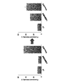
- FIG. 1 is a schematic view showing the fluorescence principle of the fluorescence modified nucleic acid used in the present invention.
- FIG. 2 is a graph showing the change in fluorescence intensity by E. coli culture solution. The graph on the left of FIG. 2 shows the state immediately after mixing, and the graph on the right of FIG. 2 shows the value of fluorescence intensity when incubation was carried out at 37 ° C. for 4 hours.
- FIG. 3 shows the reaction of fluorescence modified nucleic acid in the droplet.
- 3 (A) to 3 (F) respectively show (A) LB + fluorescence modified nucleic acid, (B) LB + fluorescence modified nucleic acid, (C) LB + fluorescence modified nucleic acid, and (D) LB + fluorescence modified nucleic acid. + RNase A section, (E) LB + fluorescence modified nucleic acid + RNase A section, (F) LB + fluorescence modified nucleic acid + RNase A section.
- 3 (A) and 3 (D) show phase contrast observation images
- FIGS. 3 (B) and 3 (E) show dark field observation images
- FIGS. 3 (C) and 3 (F) show fluorescence observation images, respectively.
- G) is a mixed phase difference image of (A) and (D), FIG.
- FIG. 3 (H) is a mixed dark field image of (A) and (D), and FIG. 3 (I) is a mixture of (A) and (D) The fluorescence image is shown.
- FIG. 4 shows an image of E. coli cultured W / O emulsion containing fluorescence modified nucleic acid. 4 (A) to 4 (F) respectively show (A) phase difference image immediately after preparation, (B) dark field image immediately after preparation, (C) fluorescence image immediately after preparation, and (D) phase difference after culture for 24 h. Images are shown (E) dark field image after 24 h culture and (F) fluorescence image after 24 h culture.
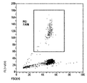
- FIG. 5 shows dot plots obtained by measurement using On-chip Sort after culturing an E.
- FIG. 6 shows images ((a) phase difference, (b) dark field, (c) fluorescence field) of droplets after culturing and sorting E. coli culture W / O emulsion containing fluorescence modified nucleic acid for 24 hours
- FIG. 7 shows an image of E. coli cultured W / O emulsion.
- 4 (A) to 4 (F) respectively show (A) phase difference image immediately after preparation, (B) dark field image immediately after preparation, (C) fluorescence image immediately after preparation, and (D) phase difference after culture for 24 h.
- FIG. 8 shows dot plots obtained by measurement using On-chip Sort after culturing E. coli culture W / O emulsion for 24 hours.
- FIG. 9 shows an image of a Bacillus subtilis cultured W / O emulsion containing a fluorescence modified nucleic acid.
- 9 (A) to 9 (F) respectively show (A) bright field image immediately after preparation, (B) dark field image immediately after preparation, (C) fluorescence image immediately after preparation, (D) bright field after culture for 48 h.
- the image, (E) dark field image after 48 h culture, and (F) fluorescence image after 48 h culture are shown.
- FIG. 10 shows a fluorescence histogram obtained by measurement using On-chip Sort after culturing a B. subtilis culture W / O emulsion containing a fluorescence-modified nucleic acid for 48 hours.
- FIG. 11 is an image of droplets after culturing and sorting a B. subtilis culture W / O emulsion containing a fluorescence-modified nucleic acid for 48 hours ((a) bright field, (b) dark field, (c) fluorescence field) Indicates
- FIG. 12 shows an image of Streptomyces aureofaciens cultured W / O emulsion containing fluorescence modified nucleic acid.
- FIG. 12 (A) to 12 (F) respectively show (A) bright field image immediately after preparation, (B) dark field image immediately after preparation, (C) fluorescence image immediately after preparation, (D) bright field after culture for 48 h.
- the image, (E) dark field image after 48 h culture, and (F) fluorescence image after 48 h culture are shown.
- FIG. 13 shows a fluorescence histogram obtained by measurement using On-chip Sort after culturing a S. aureofaciens cultured W / O emulsion containing a fluorescent modified nucleic acid for 48 hours.
- FIG. 14 shows images of droplets after culturing and sorting S.
- FIG. 15 shows an image of a Bradyrhizobium japonicum cultured W / O emulsion containing a fluorescent modified nucleic acid.
- 15 (A) to 15 (F) respectively show (A) bright field image immediately after preparation, (B) dark field image immediately after preparation, (C) fluorescence image immediately after preparation, (D) bright field after 6 d culture. Images are shown: (E) dark field image after 6 d culture; and (F) fluorescence image after 6 d culture.
- FIG. 16 shows a fluorescence histogram obtained by measurement using On-chip Sort after culturing a B. japonicum culture W / O emulsion containing a fluorescence-modified nucleic acid for 6 days.
- FIG. 17 shows images of droplets after culturing and sorting a B. japonicum cultured W / O emulsion containing a fluorescence-modified nucleic acid for 6 days ((a) bright field, (b) dark field, (c) fluorescent field) Indicates
- the invention relates in one aspect to a method of detecting the growth of microorganisms in droplets in a W / O emulsion, (A) preparing a droplet containing a microorganism, (b) culturing the microorganism in the droplet, and (c) detecting the growth of the microorganism in the droplet, (C1) measuring the fluorescence generated by the fluorescence-modified nucleic acid probe using a fluorescence-modified nucleic acid probe that generates fluorescence by acting on a substance secreted or released from the microorganism outside the body, or (C2) measuring the autofluorescence of the microorganism;
- the present invention relates to a detection method that indicates that the droplet in which a change in fluorescence characteristics has been detected is a droplet containing an intact and grown microorganism.
- the W / O emulsion refers to a state in which fine water droplets (droplets) are present as a dispersed phase in an oil phase which is a continuous phase.
- droplet refers to compartmentalized water droplets in an emulsion.
- the water phase constituting the W / O emulsion may be a hydrophilic liquid immiscible with the oil phase. Examples of the solution that can be used for such an aqueous phase include, but are not limited to, LB and R2A. Also, lake water or seawater can be used as it is as the water phase.
- the oil phase constituting the W / O emulsion may be a hydrophobic liquid immiscible with the water phase.
- Such oil phases are known and may include, but are not limited to, for example, FC40, Novec 7500, mineral oil, etc., or combinations thereof.
- a surfactant to the aqueous phase or the oil phase or both.
- surfactants include Span 80 and Tween 20 as surfactants for the aqueous phase, Pico-surf 1, Krytox and the like as surfactants for the oil phase, or combinations thereof.
- concentration of surfactant added to the aqueous phase or oil phase can be appropriately adjusted according to the conditions such as the type of surfactant used and the desired droplet size.
- the W / O emulsion used in the present invention is not limited as long as the microorganism can grow in the compartmentalized droplet in the emulsion and the growth of the microorganism can be detected.
- the size of droplets contained in the W / O emulsion is not particularly limited as long as the microorganism can grow and the growth of the microorganism can be detected.
- the method for producing a W / O emulsion consisting of the above aqueous phase and oil phase is known, and can be produced, for example, using a commercially available apparatus such as QX100 (Bio-RAD).
- the microorganism to which the method of the present invention can be applied is not particularly limited as long as it is a microorganism that can grow in droplets and can detect secretions with a fluorescence modified nucleic acid probe or can detect autofluorescence.
- Escherichia coli, Bacillus subtilis, actinomycetes and the like can be mentioned.
- the microorganism may be an environment-derived microorganism, or may be artificially produced, such as a recombinant microorganism to which gene transfer or the like has been performed.
- the microorganism is of environmental origin.
- the method of the present invention can be directed to, for example, a group of microorganisms contained in an environment such as soil.
- a group of microorganisms contained in an environment such as soil.
- By enclosing the microbes contained in the environment in droplets it is possible to detect the growth according to the properties of each microbe contained in each droplet, and to isolate or separate every droplet that exhibits different growth potential Can.
- By encasing the microorganism in droplets for each cell it becomes possible to isolate each droplet containing a microorganism exhibiting different growth potential.
- the microorganism is not artificially produced. That is, in one embodiment of the detection method of the present invention, A method of detecting the growth of microorganisms in droplets in a W / O emulsion, comprising (A) preparing a droplet containing a microorganism, (b) culturing the microorganism in the droplet, and (c) detecting the growth of the microorganism in the droplet, (C1) measuring the fluorescence generated by the fluorescence-modified nucleic acid probe using a fluorescence-modified nucleic acid probe that generates fluorescence by acting on a substance secreted or released from the microorganism outside the body, or (C2) measuring the autofluorescence of the microorganism; A detection method indicating that the droplet in which a change in fluorescence property has been detected is a droplet containing an intact and grown microorganism (except the detection method in which the microorganism is artificially produced) It is.
- the detection method of the present invention includes the step of (a) producing a droplet containing a microorganism.
- step (a) droplets containing microorganisms are produced.
- the microorganism encapsulated in the droplet may be one derived from one species, or two or more kinds of microorganisms may be encapsulated.
- the step of (a) producing a droplet can be a step of producing a droplet containing two or more kinds of microorganisms.
- the number of microorganisms encapsulated in the droplet may be encapsulated in one cell unit, or may be encapsulated in multiple cell units of two or more cells. That is, in the detection method of the present invention, in one embodiment, the step (a) of producing the droplet can be a step of producing a droplet containing a microorganism in a unit of one cell. As a result, in the droplet in which the change of the fluorescence property is detected, a microorganism which is an intact and grown microorganism and is derived from a single cell is included.
- the microorganism when it is intended to isolate a specific microorganism species from a sample containing a plurality of microorganisms, it is preferable to include the microorganism in a single cell unit in the droplet.
- the method of producing a W / O emulsion is known so that a microbe may be contained one cell at a time in one droplet (for example, nonpatent literature 3).
- the production method is not limited as long as the microorganism contains one cell unit in the droplet, but, for example, one cell is encapsulated by adjusting the number of microorganisms and the number of droplets present in the aqueous phase according to Poisson distribution. Can be made.
- the microorganisms contained in such a droplet are microorganisms derived from a single cell. Therefore, when a microbe group including a plurality of microbe species is used as a sample, it can contribute to analysis and scale-up of each microbe by selectively separating every droplet showing different growth ability. Also, even when two or more types of microorganisms are enclosed in the droplet, the microorganisms can be enclosed in the droplet in units of one cell for each type.
- the step of (a) producing a droplet can be a step of producing a droplet containing a microorganism in two or more cells.
- the culture time required for growth to the number of detectable microorganisms can be suppressed, which is preferable.
- an effect is also expected that the proliferation is promoted by the interaction between microorganisms and the like.
- the detection method of the present invention comprises the step of (b) culturing the microorganism in the droplet prepared in the above step (a).
- a method for culturing a microorganism using droplets in a W / O emulsion is known, and a person skilled in the art can appropriately select and culture a device necessary for culture, such as on a microchannel.
- the culture conditions are also not particularly limited as long as the microorganism contained in the droplet can grow, and those skilled in the art should appropriately set preferable culture conditions according to the purpose of the culture and the microorganism to be cultured.
- the temperature condition can be 4 to 95 ° C.
- the culture time can be performed up to 85 days.
- the detection method of the present invention comprises the step of (c) detecting the growth of the microorganism in the droplet. Also, in the detection step (c), is it possible to measure the fluorescence generated by the fluorescence-modified nucleic acid probe by using a fluorescence-modified nucleic acid probe that generates fluorescence by acting on a substance secreted or released from the microorganism (c1) outside the body? Or (c2) by measuring the autofluorescence of the microorganism.
- a substance secreted or released from the microorganism in vitro as used herein means a nucleic acid-related enzyme and the like, and specific examples include RNase and DNase.
- a fluorescence-modified nucleic acid probe that produces fluorescence by acting on a substance secreted or released from the above-mentioned microorganism from the outside of the microorganism is a fluorescence-modified nucleic acid, and is not limited to the following.
- Type fluorescent modified nucleic acid probes and the like can be mentioned.
- FRET type fluorescence modified nucleic acid probes are used as fluorescence modified nucleic acid probes, commercially available ones can be used. For example, those described in Table 1 below can be used.
- the FRET type fluorescent modified nucleic acid probe described in Table 1 has a fluorescent group and a quenching group at the 5 'end and the 3' end, respectively.
- the FRET type fluorescent modified nucleic acid probe described in Table 1 has Alexa 488 at the 5 'end and BHQ1 modification at the 3' end.
- known fluorescent groups, quenching groups, and combinations thereof that can be used for FRET-type fluorescent modified nucleic acid probes can be used.
- fluorescent groups such as FAM, TET, HEX and the like, quenching groups such as Dabcyl, Eclipse, BHQ2 etc.
- the base sequence portion of the FRET type fluorescent modified nucleic acid probe is a sequence capable of maintaining the quenching state by FRET, and is designed so as to include an internal sequence such that the enzyme to be detected recognizes and cleaves or binds. be able to.
- FRET-type fluorescently modified nucleic acid probes can be designed to include internal sequences that bind. It is known that microorganisms such as E.
- RNases such as RNase I, RNase Y, and RNase E, and the patterns of RNases held by each microorganism are different.
- RNases differ in their sequence specificities, for example, RNase I possessed by E. coli has no sequence specificity, and RNase Y possessed by Bacillus (Bacillus) preferentially degrades the A / U portion of ssRNA RNase E possessed by Bradyrhizobium preferentially degrades the A / U portion of ssRNA.
- the RNase to be targeted is selected for each microorganism encapsulated in the droplet, and the FRET type fluorescent modified nucleic acid probe is in the quenching state by FRET in the absence of RNase I And may be designed to include any ribonucleotide sequence capable of being cleaved by the RNase in the presence of the targeted and selected RNase.
- the FRET type fluorescent modified nucleic acid probe may contain recognition sequences of multiple RNases, or may be designed to contain only recognition sequences of specific RNases. Those skilled in the art can appropriately select a target RNase and its recognition sequence, and design of a FRET type fluorescently modified nucleic acid probe based on known information.
- the fluorescence-modified nucleic acid probe is one that increases in fluorescence intensity when RNA cleavage by a microorganism-derived RNase occurs.
- the advantage of targeting RNase produced by a microorganism in the present invention is fivefold. First, RNases are conserved in all organisms and do not limit the species of microorganisms that can be detected. Second, RNases that do not require a special environment (temperature, pH, salt concentration, etc.) or buffers for cleaving RNA are also present, so they can usually be detected in a culture environment. Thirdly, RNase is a very stable enzyme, and once included in the reaction system, it is possible to detect the activity with high sensitivity.
- the cleavage reaction of the fluorescence-modified nucleic acid probe occurs extracellularly, which enables non-invasive detection of cells.
- the nucleic acid probe used in the present invention is water-soluble, and is maintained in the enclosed droplet without dissolving in oil or moving to the nearby droplet, and therefore, it can be used for a long time Detection is possible.
- a method of measuring the fluorescence generated by the fluorescence modified nucleic acid probe can be appropriately performed by those skilled in the art using a commercially available apparatus.
- the inside of the droplet is measured by measuring the fluorescence generated by the fluorescence modified nucleic acid probe using the fluorescence modified nucleic acid probe which generates fluorescence by acting on a substance secreted or released from the microorganism outside the body.
- the growth of microorganisms can be detected.
- the fluorescently modified nucleic acid probe is encapsulated in the droplet before the measurement of fluorescence.
- the timing of the encapsulation of the fluorescence modified nucleic acid probe is not limited as long as the growth of the microorganism in the droplet can be detected, and specifically, it can be encapsulated at the following timing (a) or (b): (A) In the step of producing the droplet of (a), the fluorescence modified nucleic acid probe is enclosed in the droplet, or (B) Before the detection step of (c), the fluorescence modified nucleic acid probe is enclosed in the droplet containing the microorganism by combining the droplet containing the microorganism with the droplet containing the fluorescence modified nucleic acid probe .
- the fluorescence modified nucleic acid probe in the case where the fluorescence modified nucleic acid probe is enclosed in the droplet, the fluorescence modified nucleic acid probe may be mixed with the solution used as the aqueous phase just before the droplet production.
- the method of combining a droplet containing a microorganism and a droplet containing a fluorescence modified nucleic acid probe is, for example, by associating these two droplets in the presence of a W / O emulsion destabilizer. It can be carried out.
- the autofluorescence of the microorganism refers to fluorescence which can be detected when the microorganism and the medium are irradiated with excitation light in a state where they are not labeled with a specific fluorescent dye.
- the autofluorescence of microorganisms is known and refers to, for example but not limited to, fluorescence from NADH, flavins, amino acids.
- the growth of the microorganism in the droplet can be detected by measuring the autofluorescence of the microorganism.
- those skilled in the art can appropriately measure the autofluorescence of the target microorganism.
- the droplets whose fluorescence is detected by the detection method of the present invention detect the growth of microorganisms in a non-invasive manner, droplets containing microorganisms in an intact and grown state can be detected.
- intact microorganism refers to a microorganism in which the cell membrane of the present microorganism is maintained, capable of growth, and exhibiting physiological activity. That is, from the “intact microorganism”, the microorganism in a state in which the cell membrane is dissolved is removed by the treatment with a cell lysate or the like.
- the “proliferated microorganism” refers to a microorganism that has grown beyond the detection limit in the detection step (c). The number of microorganisms encapsulated at the time of droplet production is below the detection limit, and the growth of the microorganisms in the droplet by the culture process exceeds the detection limit.
- step of detecting the growth of the microorganism in the droplet can be performed on the microchannel in one embodiment. In this way, by combining with a commercially available cell sorter etc., it becomes possible to carry out high-throughput detection of fluorescence and sorting according to the detection result.
- the present invention provides: A method of recovering droplets containing intact and grown microorganisms from a W / O emulsion, comprising: (I) preparing a droplet containing a microorganism, (ii) culturing the microorganism in the droplet, and (iii) detecting the growth of the microorganism in the droplet, Measuring fluorescence generated by the fluorescence-modified nucleic acid probe using a fluorescence-modified nucleic acid probe that generates fluorescence by acting on a substance secreted or released from the microorganism, or There is provided a recovery method comprising the steps of: measuring the autofluorescence of a microorganism; and (iv) recovering the droplets from which the fluorescence has been detected.
- steps (i) to (iii) can be performed according to steps (a) to (c) of the above detection method.
- the recovery method of the present invention includes the step of (iv) recovering the droplet in which the fluorescence is detected after detecting the fluorescence indicating the growth of the microorganism.
- Techniques for fractionating or recovering droplets from which fluorescence has been detected or fluorescence has not been detected from droplet groups are known. Those skilled in the art can appropriately select a preferable recovery method according to the device (microchannel, culture dish, etc.) which is providing the droplet group for culture and fluorescence detection.
- a commercially available chip-type cell sorter can be used to sort droplets with increased fluorescence intensity.
- the present invention also provides, in another aspect, a kit used for the above detection method or the above recovery method.
- the kit of the present invention comprises a culture solution for the W / O emulsion aqueous phase, an oil component for the W / O emulsion oil phase, a surfactant for stabilizing the W / O emulsion, and a fluorescence modified nucleic acid probe. It is characterized by The kit of the present invention may include ones other than those described above, and may include microchannels, culture dishes and the like used for culture and the like.
- nucleic acid aptamers of the present invention are not limited to those disclosed below. Also, the contents of the documents cited herein are incorporated herein by reference.
- Test Example 2 Reaction of Fluorescently Modified Nucleic Acid in Droplet Changes in fluorescence intensity due to cleavage of fluorescence modified nucleic acid (R-Ale-UCUCG) (Japan Bioservices) were investigated in the droplet.
- the W / O emulsion manufacturing apparatus QX100 Bio-RAD
- droplets having a diameter of about 130 ⁇ m could be manufactured.
- Test Example 3 Detection and Preparation of Microbial Growth Using Fluorescently Modified Nucleic Acid Attempts were made to detect microbial growth within the droplet by measuring the fluorescence of the fluorescently modified nucleic acid.
- An E. coli culture solution (LB culture solution) mixed with 1 ⁇ M of a fluorescence modified nucleic acid (R-Ale-UCUCG) was used for the aqueous phase of the W / O emulsion.
- As oil phase DG oil (Bio-RAD) and FC-40 (3 M) were used.
- Pico-surf 1 Dolomite, final 1%) was used as a surfactant.
- the E. coli culture solution was adjusted in concentration so that E.
- coli contained 0 cells in 90% or more droplets in Poisson distribution and one or more cells in the remaining droplets.
- the W / O emulsion manufacturing apparatus QX100 Bio-RAD
- droplets having a diameter of about 130 ⁇ m could be manufactured.
- the produced W / O emulsion was collected in a 1.5 mL tube, and cultured stationary at 37 ° C. for 24 hours. Microscopic observation was performed immediately after preparation of the W / O emulsion and after culture for 24 hours, and it was confirmed that E. coli growth and fluorescence intensity rose in some droplets (FIG. 4).
- Test Example 4 Detection and Preparation of Microbial Growth Using Autofluorescence It was verified that the growth of the microorganism could be detected by measuring the autofluorescence of the microorganism in the droplet.
- E. coli culture solution was used in which the concentration was adjusted so that E. coli contained 0 cells in 90% or more droplets in Poisson distribution and one or more cells in the remaining droplets.
- oil phase DG oil (Bio-RAD) and FC-40 (3 M) were used.
- Pico-surf 1 Dolomite, final 1%) was used as a surfactant.
- Test Example 5 Detection and Preparation of Microbial Growth Using Fluorescently Modified Nucleic Acid Attempts were made to detect microbial growth within the droplet by measuring the fluorescence of the fluorescently modified nucleic acid.
- a Bacillus subtilis culture solution (LB culture solution) mixed with 200 nM of a fluorescent modified nucleic acid (R-Ale-UCUCG) was used.
- Novec 7500 was used for the oil phase.
- Pico-surf 1 Dolomite, final 1%) was used as a surfactant. The concentration of B. subtilis culture solution was adjusted so that B.
- subtilis 0 cells were contained in about 50% of droplets in Poisson distribution, and one or more cells were contained in the remaining droplets.
- the W / O emulsion manufacturing apparatus QX100 Bio-RAD
- droplets having a diameter of about 130 ⁇ m could be manufactured.
- the produced W / O emulsion was collected in a 1.5 mL tube, and cultured stationary at 30 ° C. for 48 hours. Microscopic observation was performed immediately after preparation of the W / O emulsion and after culture for 48 hours, and it was confirmed that growth of B. subtilis and an increase in fluorescence intensity were observed in some droplets (FIG. 9).
- aureofaciens contained 0 cells in about 80% droplets in Poisson distribution, and one or more cells were contained in the remaining droplets.
- a droplet letlet having a diameter of about 130 ⁇ m could be manufactured.
- the produced W / O emulsion was collected in a 1.5 mL tube, and cultured stationary at 28 ° C. for 48 hours. Microscopic observation was performed immediately after preparation of the W / O emulsion and after 48 hours of culture, and it was confirmed that S. aureofaciens growth and an increase in fluorescence intensity were observed in some droplets (FIG. 12).
- Test Example 7 Detection and Preparation of Microbial Growth Using Fluorescently Modified Nucleic Acid Attempts were made to detect microbial growth within the droplet by measuring the fluorescence of the fluorescently modified nucleic acid.
- Bradyrhizobium japonicum culture solution (NBRC 805 culture solution) mixed with 200 nM of fluorescence modified nucleic acid (R-Ale-UCUCG) was used for the aqueous phase of the W / O emulsion.
- Novec 7500 was used for the oil phase.
- Pico-surf 1 Dolomite, final 1%) was used as a surfactant. The concentration of B. japonicum culture solution was adjusted so that B.
- japonicum contained 0 cells in about 95% droplets in Poisson distribution, and the remaining droplets contained 1 or more cells.
- the W / O emulsion manufacturing apparatus QX100 Bio-RAD
- droplets having a diameter of about 130 ⁇ m could be manufactured.
- the produced W / O emulsion was collected in a 1.5 mL tube, and cultured stationary at 28 ° C. for 6 days. Microscopic observation was performed immediately after preparation of the W / O emulsion and after 6 days of culture, and it was possible to confirm growth of B. japonicum and an increase in fluorescence intensity in some droplets (FIG. 15).
- environmental microorganisms can be separated and cultured at different growth rates.
- the same substrate is used in environmental microorganisms, and in the group of microorganisms having different growth rates, microorganisms having a slow growth rate are infected with microorganisms having a high growth rate.
- One application of this technique is to alleviate the competition between microorganisms using the same substrate, and provide an approach for separating and culturing microorganisms that have been trapped.
- microorganisms in the environment that perform growth control utilizing interactions between different microorganisms. It is expected that multiple types of microorganisms will be enclosed in the same droplet, and the interaction between microorganisms will promote the growth of each type of microorganism.
Landscapes
- Chemical & Material Sciences (AREA)
- Organic Chemistry (AREA)
- Life Sciences & Earth Sciences (AREA)
- Zoology (AREA)
- Wood Science & Technology (AREA)
- Proteomics, Peptides & Aminoacids (AREA)
- Health & Medical Sciences (AREA)
- Engineering & Computer Science (AREA)
- Analytical Chemistry (AREA)
- Biophysics (AREA)
- Biochemistry (AREA)
- Microbiology (AREA)
- Molecular Biology (AREA)
- Biotechnology (AREA)
- Physics & Mathematics (AREA)
- Genetics & Genomics (AREA)
- Immunology (AREA)
- Bioinformatics & Cheminformatics (AREA)
- General Engineering & Computer Science (AREA)
- General Health & Medical Sciences (AREA)
- Chemical Kinetics & Catalysis (AREA)
- Measuring Or Testing Involving Enzymes Or Micro-Organisms (AREA)
- Micro-Organisms Or Cultivation Processes Thereof (AREA)
Abstract
Description
本発明は、water-in-oil(W/O)エマルションを用いた微生物の培養・検出・分取の方法に関する。特に、微生物が増殖したドロップレットを見出す手法として、微生物による自家蛍光を用いた手法およびシグナル検出用レポーター分子として蛍光修飾核酸を用いた方法に関する。 The present invention relates to a method of culturing, detecting and fractionating microorganisms using a water-in-oil (W / O) emulsion. In particular, the present invention relates to a method using autofluorescence by a microorganism and a method using a fluorescence-modified nucleic acid as a reporter molecule for signal detection as a method of finding droplets in which a microorganism has grown.
環境中には様々な微生物が生息しており、私たちはこれら微生物の活性や微生物が産生する物質を利用してきた。しかしながら、環境中の微生物の99%は未だ培養に成功しておらず、生物資源の多くは手付かずのままとなっている。これら未知微生物を培養することで、環境中での微生物の生理学的特性を明らかにするだけでなく、医学・産業の発展にも応用されることが期待される。
近年、微生物の分離・培養法の一つとして、W/Oエマルションを用いた手法が注目されつつある(非特許文献1および非特許文献2)。本手法では、油相中に水滴(微生物培養培地を使用)を分散させ、各水滴(ドロップレット)を一つの培養場として用いる。マイクロ流路を用いることにより、数分で数十万~数百万単位のドロップレットを作製することが可能となりハイスループット化が見込まれる。また、水相中に存在する微生物数とドロップレット数の調整により、一細胞が封入されたドロップレットを作ることができれば、シングルセルからの培養が可能となる(非特許文献3)。
本手法を用いて微生物を培養する場合、1つのドロップレットあたりの細胞数はポワソン分布に従う。1つのドロップレットに一細胞が入る確率を高くしていくにつれ、微生物が存在しないドロップレットが生じる確率も高くなる。この時、微生物の増殖が見られるドロップレットを選択的に分取することができれば各微生物の解析、スケールアップ等に活用できる。微生物が存在するドロップレットの検出には細胞内分子と反応する蛍光基質が使用されてきたが、これらは同時に細胞破砕が必要となるので、微生物を生きたまま分取することができない(非特許文献4、非特許文献5)。
Various microorganisms live in the environment, and we have used the activity of these microorganisms and the substances produced by them. However, 99% of environmental microorganisms have not yet been successfully cultured, and many of the biological resources remain untouched. By cultivating these unknown microorganisms, it is expected to be applied not only to clarify the physiological characteristics of microorganisms in the environment but also to the development of medicine and industry.
In recent years, a method using a W / O emulsion has been attracting attention as one of the methods for separation and culture of microorganisms (Non-Patent Document 1 and Non-Patent Document 2). In this method, water droplets (using a microorganism culture medium) are dispersed in the oil phase, and each water droplet (droplet) is used as one culture site. By using a microchannel, it is possible to produce hundreds of thousands to millions of droplets in a few minutes, and high throughput is expected. In addition, if a droplet in which one cell is enclosed can be formed by adjusting the number of microorganisms present in the aqueous phase and the number of droplets, culture from a single cell becomes possible (Non-patent Document 3).
When culturing the microorganism using this method, the number of cells per droplet follows Poisson distribution. As the probability of one cell entering one droplet increases, the probability of the occurrence of a droplet without a microorganism also increases. At this time, if droplets in which the growth of microorganisms can be observed can be selectively separated, it can be used for analysis, scale-up, etc. of each microorganism. Although fluorescent substrates that react with intracellular molecules have been used for detection of droplets in the presence of microorganisms, they also require cell disruption at the same time, and therefore the microorganisms can not be separated alive (non-patented) Document 4, non-patent document 5).
本発明の課題は、微生物が増殖したドロップレットを非侵襲的に検出・分取を可能とする方法の提供である。 An object of the present invention is to provide a method which enables non-invasive detection and separation of droplets in which microorganisms have grown.
本発明者らは、ドロップレット内の微生物を非侵襲的に検出する手法として、微生物による自家蛍光を用いた手法およびシグナル検出用レポーター分子として蛍光修飾核酸を用いた手法について着想した。自家蛍光を用いた手法では、ラベルフリーの状態で微生物細胞によって代謝されたNADHやフラビンといった自家蛍光分子の検出を試みた。一方で蛍光修飾核酸は微生物細胞を侵襲せずに、ドロップレット内で微生物の分泌・放出したヌクレアーゼによって分解を受け、生じたシグナルの検出を試みた。上記手法について鋭意検討したところ、二つの手法ともに、微生物を含むドロップレット内の微生物の増殖を検出できることを見出した。さらに微生物の増殖を検出できたドロップレットをシグナルの違いにより回収できた。本発明は上記知見により完成されたものである。
すなわち、本発明は以下のとおりである。
本発明は、一態様において、
〔1〕W/Oエマルションにおけるドロップレット内の微生物の増殖を検出する方法であって、
(a)微生物を含むドロップレットを作製する工程と
(b)前記ドロップレット中で微生物を培養する工程と
(c)前記ドロップレット内の微生物の増殖を検出する工程であって、前記微生物より体外に分泌または放出された物質に作用して蛍光特性の変化を生じる蛍光修飾核酸プローブを用いて、前記蛍光修飾核酸プローブが生じる蛍光を測定する工程と
を含み、
蛍光特性の変化が検出された前記ドロップレットが、インタクトかつ増殖した微生物を含むドロップレットであることを示す、検出方法に関する。
また、本発明の検出方法は、一実施の形態において、
〔2〕上記〔1〕に記載の検出方法であって、
前記(c)の検出工程における前記蛍光修飾核酸プローブが前記微生物より体外に分泌または放出されたRNaseまたはDNaseの作用により蛍光特性に変化を生じるものであることを特徴とする。
また、本発明の検出方法は、一実施の形態において、
〔3〕上記〔1〕または〔2〕に記載の検出方法であって、
前記(c)の検出工程がマイクロ流路上で行われることを特徴とする。
また、本発明の検出方法は、一実施の形態において、
〔4〕上記〔1〕~〔3〕のいずれかに記載の検出方法であって、
前記微生物が環境由来であることを特徴とする。
また、本発明の検出方法は、一実施の形態において、
〔5〕上記〔1〕~〔4〕のいずれかに記載の検出方法であって、
前記微生物が人工的に作製されたものではないことを特徴とする。
また、本発明の検出方法は、一実施の形態において、
〔6〕上記〔1〕~〔3〕のいずれかに記載の検出方法であって、
前記微生物が遺伝子組換え微生物であることを特徴とする。
また、本発明の検出方法は、一実施の形態において、
〔7〕上記〔1〕~〔6〕のいずれかに記載の検出方法であって、
前記(a)ドロップレットを作製する工程が、二種以上の微生物を含むドロップレットを作製する工程であることを特徴とする。
また、本発明の検出方法は、一実施の形態において、
〔8〕上記〔1〕~〔7〕のいずれかに記載の検出方法であって、
前記(a)ドロップレットを作製する工程が、微生物を一細胞単位で含むドロップレットを作製する工程であって、
前記(c)の検出工程において蛍光特性の変化が検出された前記ドロップレットが、インタクトかつ増殖した微生物であって、単一の細胞由来の微生物を含むものであることを特徴とする。
また、本発明の検出方法は、一実施の形態において、
〔9〕上記〔1〕~〔8〕のいずれかに記載の検出方法であって、
前記(c)の検出工程において前記蛍光修飾核酸プローブは、
(ア)前記(a)のドロップレットの作製工程において、ドロップレット中に封入されるか、または、
(イ)前記(c)の検出工程の前に、微生物を含むドロップレットと蛍光修飾核酸プローブを含むドロップレットとを合一させることにより、微生物を含むドロップレット中に封入されることを特徴とする。
また、本発明の検出方法は、一実施の形態において、
〔10〕上記〔9〕に記載の検出方法であって、
前記蛍光修飾核酸プローブがFRET型蛍光修飾核酸プローブであることを特徴とする。
また、本発明は、別の態様において、
〔11〕 W/Oエマルションからインタクトかつ増殖した微生物を含むドロップレットを回収する方法であって、
(i)微生物を含むドロップレットを作製する工程と
(ii)前記ドロップレット中で微生物を培養する工程と
(iii)前ドロップレット内の微生物の増殖を検出する工程であって、
前記微生物より分泌または放出された物質に作用して蛍光特性に変化を生じる蛍光修飾核酸プローブを用いて、前記蛍光修飾核酸プローブが生じる蛍光を測定するか、または、
微生物の自家蛍光を測定する工程と
(iv)蛍光特性の変化を検出したドロップレットを回収する工程と
を含む、回収方法に関する。
また、本発明の回収方法は、一実施の形態において、
〔12〕上記〔11〕に記載の回収方法であって、
前記(iii)および(iv)の工程をさらに1回または複数回繰り返すことを特徴とする。
また、本発明の回収方法は、一実施の形態において、
〔13〕上記〔11〕または〔12〕に記載の回収方法であって、前記(iv)の回収工程の後に
(v)前記ドロップレットから微生物を回収することを含むことを特徴とする。
また、本発明は、別の態様において、
〔14〕上記〔1〕~〔10〕のいずれかに記載の検出方法、または、上記〔11〕~〔13〕のいずれかに記載の回収方法に使用されるキットであって、
W/Oエマルション水相用の培養液と
W/Oエマルション油相用の油成分と
W/Oエマルションを安定化するための界面活性剤と
蛍光修飾核酸プローブと
を含む、キットに関する。
The present inventors have conceived of a method using non-invasive detection of a microorganism in a droplet, that is, a method using autofluorescence by the microorganism and a method using a fluorescence-modified nucleic acid as a reporter molecule for signal detection. In the method using autofluorescence, detection of autofluorescent molecules such as NADH and flavin metabolized by microbial cells in a label free state was tried. On the other hand, fluorescently modified nucleic acid was degraded by the nuclease which was secreted and released in the droplet without invading the microbial cells, and an attempt was made to detect the signal generated. As a result of intensive studies on the above methods, it was found that the two methods were able to detect the growth of microorganisms in droplet containing microorganisms. Furthermore, the droplet which could detect the growth of the microorganism could be recovered by the difference of the signal. The present invention has been completed based on the above findings.
That is, the present invention is as follows.
The present invention, in one aspect, comprises
[1] A method for detecting the growth of microorganisms in droplets in a W / O emulsion,
(A) preparing a droplet containing a microorganism, (b) culturing the microorganism in the droplet, and (c) detecting the growth of the microorganism in the droplet Measuring the fluorescence produced by said fluorescence-modified nucleic acid probe, using a fluorescence-modified nucleic acid probe that acts on the substance secreted or released to produce a change in fluorescence properties.
The present invention relates to a detection method that indicates that the droplet in which a change in fluorescence characteristics has been detected is a droplet containing an intact and grown microorganism.
In one embodiment of the detection method of the present invention,
[2] The detection method according to [1] above,
The fluorescence-modified nucleic acid probe in the detection step (c) is characterized in that the fluorescence property is changed by the action of RNase or DNase secreted or released from the microorganism outside the body.
In one embodiment of the detection method of the present invention,
[3] The detection method according to [1] or [2] above,
The detection step (c) is performed on a microchannel.
In one embodiment of the detection method of the present invention,
[4] The detection method according to any one of [1] to [3] above,
The microorganism is characterized in that it is derived from the environment.
In one embodiment of the detection method of the present invention,
[5] The detection method according to any one of [1] to [4] above,
The microorganism is characterized in that it is not artificially produced.
In one embodiment of the detection method of the present invention,
[6] The detection method according to any one of the above [1] to [3],
The microorganism is characterized in that it is a genetically modified microorganism.
In one embodiment of the detection method of the present invention,
[7] The detection method according to any one of [1] to [6] above,
The step (a) of producing the droplet is a step of producing a droplet containing two or more kinds of microorganisms.
In one embodiment of the detection method of the present invention,
[8] The detection method according to any one of [1] to [7] above,
The step of producing the (a) droplet is a step of producing a droplet containing a microorganism in one cell unit,
The droplet in which a change in fluorescence property is detected in the detection step (c) is a microorganism which is intact and grown, and includes a single cell-derived microorganism.
In one embodiment of the detection method of the present invention,
[9] The detection method according to any one of [1] to [8] above,
In the detection step (c), the fluorescence-modified nucleic acid probe is
(A) In the process of producing the droplet of (a), it is enclosed in the droplet, or
(A) Before the detection step of (c), it is characterized in that it is enclosed in a droplet containing a microorganism by combining a droplet containing a microorganism and a droplet containing a fluorescence modified nucleic acid probe. Do.
In one embodiment of the detection method of the present invention,
[10] The detection method according to [9] above,
The fluorescence modified nucleic acid probe is a FRET type fluorescence modified nucleic acid probe.
Also, in another aspect, the present invention provides:
[11] A method for recovering droplets containing intact and grown microorganisms from a W / O emulsion, comprising:
(I) preparing a droplet containing a microorganism, (ii) culturing the microorganism in the droplet, and (iii) detecting the growth of the microorganism in the pre-droplet,
Measuring the fluorescence produced by the fluorescence-modified nucleic acid probe using a fluorescence-modified nucleic acid probe that acts on a substance secreted or released from the microorganism to cause a change in fluorescence properties, or
The present invention relates to a recovery method comprising the steps of: measuring the autofluorescence of a microorganism; and (iv) recovering a droplet in which a change in fluorescence characteristics has been detected.
In one embodiment of the recovery method of the present invention,
[12] The recovery method according to [11] above,
The steps (iii) and (iv) may be repeated one or more times.
In one embodiment of the recovery method of the present invention,
[13] The recovery method according to the above [11] or [12], comprising (v) recovering the microorganism from the droplet after the recovery step of (iv).
Also, in another aspect, the present invention provides:
[14] A kit for use in the detection method according to any one of [1] to [10] above or the recovery method according to any one of [11] to [13] above,
The present invention relates to a kit comprising a culture solution for W / O emulsion aqueous phase, an oil component for W / O emulsion oil phase, a surfactant for stabilizing W / O emulsion, and a fluorescence modified nucleic acid probe.
本発明の検出方法によれば、ドロップレット内で増殖した微生物を非侵襲的に検出することが可能となる。また、検出したドロップレットに含まれる微生物は、他のドロップレットに含まれる微生物と区別して分取することができる。 According to the detection method of the present invention, it is possible to non-invasively detect the microorganism grown in the droplet. Moreover, the microorganisms contained in the detected droplet can be fractionated separately from the microorganisms contained in other droplets.
本発明は、一態様において、W/Oエマルションにおけるドロップレット内の微生物の増殖を検出する方法であって、
(a)微生物を含むドロップレットを作製する工程と
(b)前記ドロップレット中で微生物を培養する工程と
(c)前記ドロップレット内の微生物の増殖を検出する工程であって、
(c1)前記微生物より体外に分泌または放出された物質に作用して蛍光を生じる蛍光修飾核酸プローブを用いて、前記蛍光修飾核酸プローブが生じる蛍光を測定するか、または、
(c2)微生物の自家蛍光を測定する工程と
を含み、
蛍光特性の変化が検出された前記ドロップレットが、インタクトかつ増殖した微生物を含むドロップレットであることを示す、検出方法に関する。
The invention relates in one aspect to a method of detecting the growth of microorganisms in droplets in a W / O emulsion,
(A) preparing a droplet containing a microorganism, (b) culturing the microorganism in the droplet, and (c) detecting the growth of the microorganism in the droplet,
(C1) measuring the fluorescence generated by the fluorescence-modified nucleic acid probe using a fluorescence-modified nucleic acid probe that generates fluorescence by acting on a substance secreted or released from the microorganism outside the body, or
(C2) measuring the autofluorescence of the microorganism;
The present invention relates to a detection method that indicates that the droplet in which a change in fluorescence characteristics has been detected is a droplet containing an intact and grown microorganism.
ここで、本明細書において、W/Oエマルションとは連続相である油相中に、分散相として微粒子状の水滴(ドロップレット)が存在している状態をいう。
また、本明細書において「ドロップレット」とは、エマルション中の区画化された水滴をいう。
W/Oエマルションを構成する水相は、油相と混和しない親水性の液体であればよい。このような水相に用いることのできる溶液としては、以下に限定されないが、例えば、LB、R2Aなどを挙げることができる。また、湖沼水や海水をそのまま水相として用いることもできる。
W/Oエマルションを構成する油相は、水相と混和しない疎水性の液体であればよい。このような油相は公知であり、以下に限定されないが、例えば、FC40、Novec7500、ミネラルオイルなど、またはこれらの組み合わせを挙げることができる。
W/Oエマルションを安定化させるために、界面活性剤を水相あるいは油相、もしくはその両方に添加する必要がある。用いられる界面活性剤には、例えば、水相用の界面活性剤としてはSpan80、Tween20など、油相用の界面活性剤としてはPico-surf1、Krytoxなど、またはこれらの組み合わせを挙げることができる。水相または油相に添加される界面活性剤の濃度は、用いる界面活性剤の種類や所望するドロップレットの大きさ等の条件により適宜調整することができる。
本発明に用いられるW/Oエマルションは、エマルション中に区画化されたドロップレット内で微生物が増殖でき、かつ、微生物の増殖を検出可能なものであれば制限されない。また、W/Oエマルション中に含まれるドロップレットの大きさも、微生物が増殖でき、かつ、微生物の増殖を検出可能なものである限り特に制限されない。
上記の水相および油相からなるW/Oエマルションの作製方法は公知であり、例えば、QX100(Bio-RAD)などの市販の装置を用いて作成することができる。
Here, in the present specification, the W / O emulsion refers to a state in which fine water droplets (droplets) are present as a dispersed phase in an oil phase which is a continuous phase.
Also, as used herein, “droplet” refers to compartmentalized water droplets in an emulsion.
The water phase constituting the W / O emulsion may be a hydrophilic liquid immiscible with the oil phase. Examples of the solution that can be used for such an aqueous phase include, but are not limited to, LB and R2A. Also, lake water or seawater can be used as it is as the water phase.
The oil phase constituting the W / O emulsion may be a hydrophobic liquid immiscible with the water phase. Such oil phases are known and may include, but are not limited to, for example, FC40, Novec 7500, mineral oil, etc., or combinations thereof.
In order to stabilize the W / O emulsion, it is necessary to add a surfactant to the aqueous phase or the oil phase or both. Examples of surfactants that can be used include Span 80 and
The W / O emulsion used in the present invention is not limited as long as the microorganism can grow in the compartmentalized droplet in the emulsion and the growth of the microorganism can be detected. In addition, the size of droplets contained in the W / O emulsion is not particularly limited as long as the microorganism can grow and the growth of the microorganism can be detected.
The method for producing a W / O emulsion consisting of the above aqueous phase and oil phase is known, and can be produced, for example, using a commercially available apparatus such as QX100 (Bio-RAD).
本発明の方法を適用可能な微生物としては、ドロップレット内で増殖可能であり、かつ、分泌物を蛍光修飾核酸プローブで検出可能、または、自家蛍光を検出可能な微生物であれば特に制限されず、例えば、大腸菌、枯草菌、放線菌などを挙げることができる。微生物は、環境由来の微生物であってもよく、または遺伝子導入などが施された組換え微生物など、人工的に作製されたものであってもよい。 The microorganism to which the method of the present invention can be applied is not particularly limited as long as it is a microorganism that can grow in droplets and can detect secretions with a fluorescence modified nucleic acid probe or can detect autofluorescence. For example, Escherichia coli, Bacillus subtilis, actinomycetes and the like can be mentioned. The microorganism may be an environment-derived microorganism, or may be artificially produced, such as a recombinant microorganism to which gene transfer or the like has been performed.
一実施の形態においては、微生物は環境由来のものである。本発明の方法は、例えば、土壌等の環境中に含まれる微生物群を対象とすることができる。環境中に含まれる微生物群をドロップレットに封入させることで、各ドロップレットに含まれる微生物ごとの性質に応じた増殖を検出でき、異なる増殖能を示すドロップレットごとに単離や分取することができる。特に、一細胞ごとに微生物をドロップレット中に封入することで、異なる増殖能を示す微生物を含むドロップレットごとに単離が可能となる。 In one embodiment, the microorganism is of environmental origin. The method of the present invention can be directed to, for example, a group of microorganisms contained in an environment such as soil. By enclosing the microbes contained in the environment in droplets, it is possible to detect the growth according to the properties of each microbe contained in each droplet, and to isolate or separate every droplet that exhibits different growth potential Can. In particular, by encasing the microorganism in droplets for each cell, it becomes possible to isolate each droplet containing a microorganism exhibiting different growth potential.
また、別の実施の形態においては、微生物は人工的に作製されたものではない。すなわち、本発明の検出方法は、一実施の形態において、
W/Oエマルションにおけるドロップレット内の微生物の増殖を検出する方法であって、
(a)微生物を含むドロップレットを作製する工程と
(b)前記ドロップレット中で微生物を培養する工程と
(c)前記ドロップレット内の微生物の増殖を検出する工程であって、
(c1)前記微生物より体外に分泌または放出された物質に作用して蛍光を生じる蛍光修飾核酸プローブを用いて、前記蛍光修飾核酸プローブが生じる蛍光を測定するか、または、
(c2)微生物の自家蛍光を測定する工程と
を含み、
蛍光特性の変化が検出された前記ドロップレットが、インタクトかつ増殖した微生物を含むドロップレットであることを示す、検出方法(ただし、前記微生物が人工的に作製されたものである検出方法を除く)である。
In another embodiment, the microorganism is not artificially produced. That is, in one embodiment of the detection method of the present invention,
A method of detecting the growth of microorganisms in droplets in a W / O emulsion, comprising
(A) preparing a droplet containing a microorganism, (b) culturing the microorganism in the droplet, and (c) detecting the growth of the microorganism in the droplet,
(C1) measuring the fluorescence generated by the fluorescence-modified nucleic acid probe using a fluorescence-modified nucleic acid probe that generates fluorescence by acting on a substance secreted or released from the microorganism outside the body, or
(C2) measuring the autofluorescence of the microorganism;
A detection method indicating that the droplet in which a change in fluorescence property has been detected is a droplet containing an intact and grown microorganism (except the detection method in which the microorganism is artificially produced) It is.
本発明の検出方法は、(a)微生物を含むドロップレットを作製する工程を含む。
上記工程(a)において、微生物を含むドロップレットが作製される。
このとき、ドロップレット内に封入される微生物は、一つの種に由来するものであってもよく、また、二種以上の微生物が封入されていてもよい。
すなわち、本発明の検出方法は、一実施の形態において、(a)ドロップレットを作製する工程を、二種以上の微生物を含むドロップレットを作製する工程とすることができる。一つのドロップレット内に二種以上の微生物を含むことにより、一つのドロップレット内に封入された微生物間の相互作用の影響について検証することが可能となる。
The detection method of the present invention includes the step of (a) producing a droplet containing a microorganism.
In the step (a), droplets containing microorganisms are produced.
At this time, the microorganism encapsulated in the droplet may be one derived from one species, or two or more kinds of microorganisms may be encapsulated.
That is, in one embodiment of the detection method of the present invention, the step of (a) producing a droplet can be a step of producing a droplet containing two or more kinds of microorganisms. By including two or more kinds of microorganisms in one droplet, it is possible to verify the influence of the interaction between the microorganisms enclosed in one droplet.
また、ドロップレット内に封入される微生物の数は、一細胞単位で封入されてもよく、または、二細胞以上の複数の細胞単位で封入されても良い。
すなわち、本発明の検出方法は、一実施の形態において、(a)ドロップレットを作製する工程が、微生物を一細胞単位で含むドロップレットを作製する工程とすることができる。これにより、蛍光特性の変化が検出されたドロップレット内には、インタクトかつ増殖した微生物であって、単一の細胞由来の微生物が含まれることになる。特に、複数種の微生物を含む試料から特定の微生物種の単離を目的とする場合、ドロップレット内には一細胞単位で微生物を含むことが好ましい。なお、1つのドロップレット内に微生物が一細胞ずつ含まれるように、W/Oエマルションを作製する方法は公知である(例えば、非特許文献3)。ドロップレット内に微生物が一細胞単位で含まれる限り製造方法は制限されないが、例えば、ポワソン分布に従い、水相中に存在する微生物数とドロップレット数の調整をすることで、一細胞が封入されたドロップレットを作製することができる。このようなドロップレット内に含まれる微生物は、単一の細胞由来の微生物である。よって、複数の微生物種を含む微生物群を試料とした場合、異なる増殖能を示すドロップレットごとに選択的に分取することで、各微生物の解析、スケールアップに寄与することができる。
また、ドロップレット内に二種以上の微生物を封入する際にも、各種ごとに一細胞単位でドロップレット内に微生物を封入させることもできる。
また、上述のように、本発明の検出方法は、一実施の形態において、(a)ドロップレットを作製する工程が、微生物を二細胞以上で含むドロップレットを作製する工程とすることができる。各ドロップレット中に複数の細胞を封入することで、検出可能な微生物数までの増殖に必要な培養時間を抑えることができ好ましい。また、共生関係を持つ微生物同士が同一ドロップレットに封入された場合、微生物相互間作用等によって増殖が促進されるという効果も期待される。
Also, the number of microorganisms encapsulated in the droplet may be encapsulated in one cell unit, or may be encapsulated in multiple cell units of two or more cells.
That is, in the detection method of the present invention, in one embodiment, the step (a) of producing the droplet can be a step of producing a droplet containing a microorganism in a unit of one cell. As a result, in the droplet in which the change of the fluorescence property is detected, a microorganism which is an intact and grown microorganism and is derived from a single cell is included. In particular, when it is intended to isolate a specific microorganism species from a sample containing a plurality of microorganisms, it is preferable to include the microorganism in a single cell unit in the droplet. In addition, the method of producing a W / O emulsion is known so that a microbe may be contained one cell at a time in one droplet (for example, nonpatent literature 3). The production method is not limited as long as the microorganism contains one cell unit in the droplet, but, for example, one cell is encapsulated by adjusting the number of microorganisms and the number of droplets present in the aqueous phase according to Poisson distribution. Can be made. The microorganisms contained in such a droplet are microorganisms derived from a single cell. Therefore, when a microbe group including a plurality of microbe species is used as a sample, it can contribute to analysis and scale-up of each microbe by selectively separating every droplet showing different growth ability.
Also, even when two or more types of microorganisms are enclosed in the droplet, the microorganisms can be enclosed in the droplet in units of one cell for each type.
In addition, as described above, in one embodiment of the detection method of the present invention, the step of (a) producing a droplet can be a step of producing a droplet containing a microorganism in two or more cells. By encapsulating a plurality of cells in each droplet, the culture time required for growth to the number of detectable microorganisms can be suppressed, which is preferable. In addition, when microorganisms having a symbiotic relationship are enclosed in the same droplet, an effect is also expected that the proliferation is promoted by the interaction between microorganisms and the like.
本発明の検出方法は、(b)上記工程(a)において作製されたドロップレット中で微生物を培養する工程を含む。
W/Oエマルション中のドロップレットを用いて微生物を培養する方法は公知であり、マイクロ流路上など当業者であれば適宜培養に必要な装置を選択して培養することができる。培養条件についても、ドロップレットに含まれる微生物が増殖できる条件であれば特に制限されず、当業者であれば、培養の目的や培養の対象となる微生物に応じて適宜好ましい培養条件を設定することができる。以下に限定されないが、例えば、温度条件は4~95℃とすることができ、培養時間は最大85日間行うことができる。
The detection method of the present invention comprises the step of (b) culturing the microorganism in the droplet prepared in the above step (a).
A method for culturing a microorganism using droplets in a W / O emulsion is known, and a person skilled in the art can appropriately select and culture a device necessary for culture, such as on a microchannel. The culture conditions are also not particularly limited as long as the microorganism contained in the droplet can grow, and those skilled in the art should appropriately set preferable culture conditions according to the purpose of the culture and the microorganism to be cultured. Can. Although not limited to the following, for example, the temperature condition can be 4 to 95 ° C., and the culture time can be performed up to 85 days.
本発明の検出方法は、(c)ドロップレット内の微生物の増殖を検出する工程を含む。また、工程(c)の検出工程は、(c1)微生物より体外に分泌または放出された物質に作用して蛍光を生じる蛍光修飾核酸プローブを用いて、蛍光修飾核酸プローブが生じる蛍光を測定するか、または、(c2)微生物の自家蛍光を測定することにより行われる。
ここで、本明細書における「微生物より体外に分泌または放出された物質」とは、核酸関連酵素などを意味し、具体的にはRNase、DNaseなどを挙げることができる。
また、「上記微生物より体外に分泌または放出された物質に作用して蛍光を生じる蛍光修飾核酸プローブ」とは蛍光修飾核酸であり、以下に制限されないが、例えば、FRET型蛍光修飾核酸プローブ、PET型蛍光修飾核酸プローブなどを挙げることができる。
FRET型蛍光修飾核酸プローブを蛍光修飾核酸プローブとして用いる際には、市販のものを用いることができ、例えば、下記表1に記載のものを使用することができる。
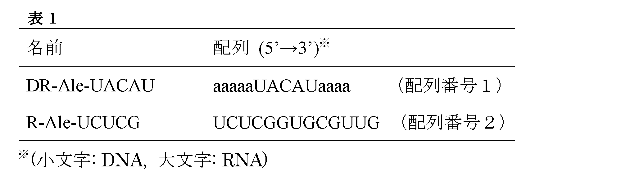
Here, "a substance secreted or released from the microorganism in vitro" as used herein means a nucleic acid-related enzyme and the like, and specific examples include RNase and DNase.
Also, “a fluorescence-modified nucleic acid probe that produces fluorescence by acting on a substance secreted or released from the above-mentioned microorganism from the outside of the microorganism” is a fluorescence-modified nucleic acid, and is not limited to the following. Type fluorescent modified nucleic acid probes and the like can be mentioned.
When FRET type fluorescence modified nucleic acid probes are used as fluorescence modified nucleic acid probes, commercially available ones can be used. For example, those described in Table 1 below can be used.
表1に記載のFRET型蛍光修飾核酸プローブは、5’末端および3’末端にそれぞれ蛍光基および消光基を有する。例えば、一実施の形態において、表1に記載のFRET型蛍光修飾核酸プローブは5’末端にAlexa488、3’末端にBHQ1の修飾を有する。その他、FRET型蛍光修飾核酸プローブに用いることのできる蛍光基、消光基、および、それらの組み合わせは公知のものを使用することができる。以下に限定されないが、蛍光基としてはFAM、TET、HEXなど、消光基としてはDabcyl、Eclipse、BHQ2などを挙げることができ、代表的な組み合わせの使用例としては、FAM/Dabcyl(蛍光基/消光基)などを挙げることができる。通常はFRETにより消光状態が維持されているが、微生物により体外に放出された酵素により核酸の切断が生じるとFRETが解消され、蛍光強度が上昇する(図1)(Kelemen B. R. et al., “Hypersensitive substrate for ribonucleases” Nucleic Acids Research, 1999; 27, 3696-3701、Sato S. and Takenaka S., “Highly Sensitive Nuclease Assays Based on Chemically Modified DNA or RNA” Sensors, 2014; 14, 12437-12450)。また、FRET型蛍光修飾核酸プローブの塩基配列部分は、FRETによる消光状態を維持することが可能な配列であって、検出対象酵素が認識して切断あるいは結合するような内部配列を含むようにして設計することができる。微生物が産生するRNaseまたはDNaseを標的とする際、当業者であればドロップレット内に封入する微生物に応じて標的とする具体的なRNaseまたはDNaseを選択し、当該RNaseまたはDNaseが認識して切断あるいは結合する内部配列を含むようにFRET型蛍光修飾核酸プローブを設計することができる。
なお、大腸菌、枯草菌、放線菌などの微生物はRNaseI、RNaseY、RNaseEなどのRNaseを複数種有していることが知られており、各微生物により保有するRNaseのパターンが異なる。またRNaseはその種類ごとに配列特異性が異なり、例えば、大腸菌が保有するRNaseIは配列特異性を有さず、バチルス属(Bacillus)が保有するRNaseYはssRNAのA/U部分を優先的に分解し、ブラディリゾビウム属(Bradyrhizobium)が保有するRNaseEはssRNAのA/U部分を優先的に分解する。このように微生物が体外に放出するRNaseを標的とする場合、ドロップレット内に封入する微生物ごとに標的とするRNaseを選択し、FRET型蛍光修飾核酸プローブがRNaseIの非存在下ではFRETによる消光状態を維持し、標的とし選択したRNase存在下では当該RNaseによって切断されることが可能な任意のリボヌクレオチド配列を含むように設計すればよい。FRET型蛍光修飾核酸プローブは、複数種のRNaseの認識配列を含んでいてもよいし、特定のRNaseの認識配列のみを含むように設計してもよい。当業者であれば、標的とするRNaseおよびその認識配列の選択とFRET型蛍光修飾核酸プローブの設計は公知の情報に基づいて適宜行うことができる。
本発明の好ましい一実施の形態において、蛍光修飾核酸プローブは微生物由来RNaseによるRNA切断が生じると蛍光強度が増大するものである。本発明で微生物が産生するRNaseを標的とする利点は5つあげられる。まず、RNaseはあらゆる生物に保存されており、検出できる微生物種を限定しない。2つ目として、RNAを切断する際に特殊な環境(温度、pH、塩濃度等)やバッファーを必要としないRNaseも存在するため、通常培養環境下で検出可能である。3つ目として、RNaseは非常に安定した酵素であり、一度反応系に含まれれば高感度に活性を検出することが可能である。4つ目として、蛍光修飾核酸プローブの切断反応は細胞外で生じるため、細胞非侵襲的な検出が可能となる。最後に5つ目として、本発明で使用する核酸プローブは水溶性であり、オイルに溶け出すことや近傍のドロップレットに移動することなく、封入されたドロップレット内に維持されるため長期間の検出が可能である。
蛍光修飾核酸プローブが生じる蛍光を測定する方法は、当業者であれば市販の装置を用いて適宜行うことができる。本発明の検出方法においては、微生物より体外に分泌または放出された物質に作用して蛍光を生じる蛍光修飾核酸プローブを用いて、蛍光修飾核酸プローブが生じる蛍光を測定することにより、ドロップレット内の微生物の増殖を検出することできる。
The FRET type fluorescent modified nucleic acid probe described in Table 1 has a fluorescent group and a quenching group at the 5 'end and the 3' end, respectively. For example, in one embodiment, the FRET type fluorescent modified nucleic acid probe described in Table 1 has Alexa 488 at the 5 'end and BHQ1 modification at the 3' end. In addition, known fluorescent groups, quenching groups, and combinations thereof that can be used for FRET-type fluorescent modified nucleic acid probes can be used. Although not limited to the following, fluorescent groups such as FAM, TET, HEX and the like, quenching groups such as Dabcyl, Eclipse, BHQ2 etc. can be mentioned, and typical examples of use of combinations are FAM / Dabcyl (fluorescent group / fluorescent group / And the like. Normally, the quenching state is maintained by FRET, but when cleavage of the nucleic acid is caused by an enzyme released from the body by the microorganism, FRET is eliminated and the fluorescence intensity is increased (Fig. 1) (Kelemen BR et al., " Hypersensitive substrate for ribonucleases "Nucleic Acids Research, 1999; 27, 3696-3701, Sato S. and Takenaka S.," Highly Sensitive Nuclease Assays Based on Chemically Modified DNA or RNA "Sensors, 2014; 14, 12437-12450). In addition, the base sequence portion of the FRET type fluorescent modified nucleic acid probe is a sequence capable of maintaining the quenching state by FRET, and is designed so as to include an internal sequence such that the enzyme to be detected recognizes and cleaves or binds. be able to. When targeting RNase or DNase produced by a microorganism, one skilled in the art selects a specific RNase or DNase to be targeted depending on the microorganism encapsulated in the droplet, and the RNase or DNase recognizes and cleaves Alternatively, FRET-type fluorescently modified nucleic acid probes can be designed to include internal sequences that bind.
It is known that microorganisms such as E. coli, Bacillus subtilis, and actinomycetes have a plurality of RNases such as RNase I, RNase Y, and RNase E, and the patterns of RNases held by each microorganism are different. Furthermore, RNases differ in their sequence specificities, for example, RNase I possessed by E. coli has no sequence specificity, and RNase Y possessed by Bacillus (Bacillus) preferentially degrades the A / U portion of ssRNA RNase E possessed by Bradyrhizobium preferentially degrades the A / U portion of ssRNA. Thus, when targeting the RNase released by the microorganism outside the body, the RNase to be targeted is selected for each microorganism encapsulated in the droplet, and the FRET type fluorescent modified nucleic acid probe is in the quenching state by FRET in the absence of RNase I And may be designed to include any ribonucleotide sequence capable of being cleaved by the RNase in the presence of the targeted and selected RNase. The FRET type fluorescent modified nucleic acid probe may contain recognition sequences of multiple RNases, or may be designed to contain only recognition sequences of specific RNases. Those skilled in the art can appropriately select a target RNase and its recognition sequence, and design of a FRET type fluorescently modified nucleic acid probe based on known information.
In a preferred embodiment of the present invention, the fluorescence-modified nucleic acid probe is one that increases in fluorescence intensity when RNA cleavage by a microorganism-derived RNase occurs. The advantage of targeting RNase produced by a microorganism in the present invention is fivefold. First, RNases are conserved in all organisms and do not limit the species of microorganisms that can be detected. Second, RNases that do not require a special environment (temperature, pH, salt concentration, etc.) or buffers for cleaving RNA are also present, so they can usually be detected in a culture environment. Thirdly, RNase is a very stable enzyme, and once included in the reaction system, it is possible to detect the activity with high sensitivity. Fourth, the cleavage reaction of the fluorescence-modified nucleic acid probe occurs extracellularly, which enables non-invasive detection of cells. Finally, as the fifth point, the nucleic acid probe used in the present invention is water-soluble, and is maintained in the enclosed droplet without dissolving in oil or moving to the nearby droplet, and therefore, it can be used for a long time Detection is possible.
A method of measuring the fluorescence generated by the fluorescence modified nucleic acid probe can be appropriately performed by those skilled in the art using a commercially available apparatus. In the detection method of the present invention, the inside of the droplet is measured by measuring the fluorescence generated by the fluorescence modified nucleic acid probe using the fluorescence modified nucleic acid probe which generates fluorescence by acting on a substance secreted or released from the microorganism outside the body. The growth of microorganisms can be detected.
(c)の検出工程において蛍光修飾核酸プローブを使用する場合、蛍光の測定前に、ドロップレット内に蛍光修飾核酸プローブを封入する。蛍光修飾核酸プローブの封入のタイミングは、ドロップレット内の微生物の増殖を検出できる限り制限されず、具体的には、下記(ア)または(イ)のタイミングで封入することができる:
(ア)(a)のドロップレットの作製工程において、蛍光修飾核酸プローブをドロップレット中に封入する、または、
(イ)(c)の検出工程の前に、微生物を含むドロップレットと蛍光修飾核酸プローブを含むドロップレットとを合一させることにより、蛍光修飾核酸プローブを、微生物を含むドロップレット中に封入する。
(ア)ドロップレットの作製工程において、蛍光修飾核酸プローブをドロップレット中に封入する場合には、ドロップレット作製直前に、水相として使用する溶液に蛍光修飾核酸プローブを混合するようにすればよい。
また、(イ)微生物を含むドロップレットと蛍光修飾核酸プローブを含むドロップレットとを合一させる手法は、例えば、W/Oエマルションディスタビライザー存在下でこれら2つのドロップレット同士を会合させるようにして行うことができる。
When a fluorescently modified nucleic acid probe is used in the detection step (c), the fluorescently modified nucleic acid probe is encapsulated in the droplet before the measurement of fluorescence. The timing of the encapsulation of the fluorescence modified nucleic acid probe is not limited as long as the growth of the microorganism in the droplet can be detected, and specifically, it can be encapsulated at the following timing (a) or (b):
(A) In the step of producing the droplet of (a), the fluorescence modified nucleic acid probe is enclosed in the droplet, or
(B) Before the detection step of (c), the fluorescence modified nucleic acid probe is enclosed in the droplet containing the microorganism by combining the droplet containing the microorganism with the droplet containing the fluorescence modified nucleic acid probe .
(A) In the process of producing the droplet, in the case where the fluorescence modified nucleic acid probe is enclosed in the droplet, the fluorescence modified nucleic acid probe may be mixed with the solution used as the aqueous phase just before the droplet production. .
In addition, (a) the method of combining a droplet containing a microorganism and a droplet containing a fluorescence modified nucleic acid probe is, for example, by associating these two droplets in the presence of a W / O emulsion destabilizer. It can be carried out.
また、本明細書において「微生物の自家蛍光」とは微生物および培地が特定の蛍光色素で標識されていない状態で励起光を照射した時に検出できる蛍光をいう。微生物の自家蛍光は公知であり、以下に制限されないが、例えば、NADH、フラビン、アミノ酸からの蛍光をいう。本発明の検出方法においては、微生物の自家蛍光を測定することにより、ドロップレット内の微生物の増殖を検出することできる。また、当業者であれば適宜対象とする微生物の自家蛍光を測定することができる。 Further, in the present specification, "the autofluorescence of the microorganism" refers to fluorescence which can be detected when the microorganism and the medium are irradiated with excitation light in a state where they are not labeled with a specific fluorescent dye. The autofluorescence of microorganisms is known and refers to, for example but not limited to, fluorescence from NADH, flavins, amino acids. In the detection method of the present invention, the growth of the microorganism in the droplet can be detected by measuring the autofluorescence of the microorganism. In addition, those skilled in the art can appropriately measure the autofluorescence of the target microorganism.
本発明の検出方法により蛍光が検出されたドロップレットは、非侵襲的な手法により微生物の増殖を検出するため、インタクトかつ増殖した状態の微生物を含むドロップレットを検出することができる。
ここで、本明細書において「インタクトの微生物」というとき、本微生物の細胞膜が維持されており、増殖が可能であり、生理活性を示す状態にある微生物をいう。すなわち、「インタクトの微生物」からは、細胞溶解液等の処理により細胞膜が溶解した状態の微生物は除かれる。また、本明細書において「増殖した微生物」とは、上記(c)の検出工程における検出限界を超えて増殖した微生物をいう。ドロップレット作製時に封入される微生物数は検出限界未満であり、培養工程によりドロップレット内の微生物が増殖することで検出限界を超える。
Since the droplets whose fluorescence is detected by the detection method of the present invention detect the growth of microorganisms in a non-invasive manner, droplets containing microorganisms in an intact and grown state can be detected.
Here, when the term "intact microorganism" is used in the present specification, it refers to a microorganism in which the cell membrane of the present microorganism is maintained, capable of growth, and exhibiting physiological activity. That is, from the “intact microorganism”, the microorganism in a state in which the cell membrane is dissolved is removed by the treatment with a cell lysate or the like. Moreover, in the present specification, the “proliferated microorganism” refers to a microorganism that has grown beyond the detection limit in the detection step (c). The number of microorganisms encapsulated at the time of droplet production is below the detection limit, and the growth of the microorganisms in the droplet by the culture process exceeds the detection limit.
また、(c)ドロップレット内の微生物の増殖を検出する工程は、一実施の形態において、マイクロ流路上で行うことができる。これにより市販のセルソーターなどと組み合わせることで、ハイスループットに蛍光の検出と検出結果に応じた分取が可能になる。 In addition, (c) the step of detecting the growth of the microorganism in the droplet can be performed on the microchannel in one embodiment. In this way, by combining with a commercially available cell sorter etc., it becomes possible to carry out high-throughput detection of fluorescence and sorting according to the detection result.
また、本発明は、別の態様において、
W/Oエマルションからインタクトかつ増殖した微生物を含むドロップレットを回収する方法であって、
(i)微生物を含むドロップレットを作製する工程と
(ii)前記ドロップレット中で微生物を培養する工程と
(iii)前記ドロップレット内の微生物の増殖を検出する工程であって、
前記微生物より分泌または放出された物質に作用して蛍光を生じる蛍光修飾核酸プローブを用いて、前記蛍光修飾核酸プローブが生じる蛍光を測定するか、または、
微生物の自家蛍光を測定する工程と
(iv)蛍光を検出したドロップレットを回収する工程と
を含む、回収方法を提供する。
本発明の回収方法において、工程(i)~(iii)は、上記検出方法の工程(a)~(c)に準じて行うことができる。
Also, in another aspect, the present invention provides:
A method of recovering droplets containing intact and grown microorganisms from a W / O emulsion, comprising:
(I) preparing a droplet containing a microorganism, (ii) culturing the microorganism in the droplet, and (iii) detecting the growth of the microorganism in the droplet,
Measuring fluorescence generated by the fluorescence-modified nucleic acid probe using a fluorescence-modified nucleic acid probe that generates fluorescence by acting on a substance secreted or released from the microorganism, or
There is provided a recovery method comprising the steps of: measuring the autofluorescence of a microorganism; and (iv) recovering the droplets from which the fluorescence has been detected.
In the recovery method of the present invention, steps (i) to (iii) can be performed according to steps (a) to (c) of the above detection method.
また、本発明の回収方法は、微生物の増殖を示す蛍光を検出した後、(iv)蛍光を検出したドロップレットを回収する工程を含む。
ドロップレット群より、蛍光が検出された、または、蛍光が検出されなかったドロップレットを分取または回収する手法は公知である。当業者であれば、ドロップレット群を培養および蛍光検出に供している器具(マイクロ流路や培養ディッシュなど)に応じて、適宜好ましい回収方法を選択することができる。以下に制限されないが、例えば、市販のチップ式セルソーターを利用して、蛍光強度が上昇したドロップレットをソーティングすることができる。
In addition, the recovery method of the present invention includes the step of (iv) recovering the droplet in which the fluorescence is detected after detecting the fluorescence indicating the growth of the microorganism.
Techniques for fractionating or recovering droplets from which fluorescence has been detected or fluorescence has not been detected from droplet groups are known. Those skilled in the art can appropriately select a preferable recovery method according to the device (microchannel, culture dish, etc.) which is providing the droplet group for culture and fluorescence detection. Although not limited to the following, for example, a commercially available chip-type cell sorter can be used to sort droplets with increased fluorescence intensity.
また、本発明は、別の態様において、上記検出方法または上記回収方法に使用されるキットを提供する。
本発明のキットは、W/Oエマルション水相用の培養液と、W/Oエマルション油相用の油成分と、W/Oエマルション安定化のための界面活性剤と蛍光修飾核酸プローブとを含むことを特徴とする。本発明のキットには、上記の構成以外のものも含まれてよく、培養等に用いるマイクロ流路、培養皿などが含まれていても良い。
The present invention also provides, in another aspect, a kit used for the above detection method or the above recovery method.
The kit of the present invention comprises a culture solution for the W / O emulsion aqueous phase, an oil component for the W / O emulsion oil phase, a surfactant for stabilizing the W / O emulsion, and a fluorescence modified nucleic acid probe. It is characterized by The kit of the present invention may include ones other than those described above, and may include microchannels, culture dishes and the like used for culture and the like.
以下に、具体的な実施例を示すが、本発明の核酸アプタマーは以下に開示するものに限定されない。また、本明細書中で引用する文献の内容は、本明細書に参照として組み込まれる。 Specific examples will be shown below, but the nucleic acid aptamers of the present invention are not limited to those disclosed below. Also, the contents of the documents cited herein are incorporated herein by reference.
(試験例1:大腸菌培養液による蛍光修飾核酸への影響評価)
蛍光修飾核酸(DR-Ale-UACAU)(日本バイオサービス)1μMと、(i)大腸菌を含まないLB培養液、(ii)大腸菌を含むLB培養液(OD600=2.77)、または、(iii)大腸菌を含まないLB培養液にRNaseA 2.5ng/μLを添加したものをそれぞれ混合しトータル20μLとした。混合直後および37℃で4時間インキュベーション後の蛍光強度をLight Cycler480(Roche)を用いて測定した。(i)LBと蛍光修飾核酸(DR-Ale-UACAU)を混合した時には蛍光強度は低いまま維持されていた。一方、(ii)大腸菌培養液と蛍光修飾核酸(DR-Ale-UACAU)を混合した場合には時間が経つと蛍光強度の上昇が見られた。(iii)RNaseAを添加した時には高い蛍光強度が維持されていた(図2)。このことから大腸菌培養液によって蛍光修飾核酸の切断、続く蛍光強度の上昇が生じることが明らかとなった。
(Test Example 1: Evaluation of the influence of fluorescently modified nucleic acid by E. coli culture solution)
Fluorescently modified nucleic acid (DR-Ale-UACAU) (Japan Bioservices) 1 μM and (i) LB broth without E. coli, (ii) LB broth with E. coli (OD 600 = 2.77), or (iii) What added RNaseA 2.5 ng / microliter to LB culture solution which does not contain E. coli was mixed, respectively, and it was set as 20 microliters in total. The fluorescence intensities immediately after mixing and after incubation for 4 hours at 37 ° C. were measured using a Light Cycler 480 (Roche). (i) The fluorescence intensity was kept low when LB and a fluorescence-modified nucleic acid (DR-Ale-UACAU) were mixed. On the other hand, (ii) when the E. coli culture solution and the fluorescence-modified nucleic acid (DR-Ale-UACAU) were mixed, an increase in fluorescence intensity was observed with time. (iii) High fluorescence intensity was maintained when RNase A was added (FIG. 2). From this, it was revealed that the E. coli culture solution causes cleavage of the fluorescence-modified nucleic acid and the subsequent increase in fluorescence intensity.
(試験例2:ドロップレット内における蛍光修飾核酸の反応)
ドロップレット内において蛍光修飾核酸(R-Ale-UCUCG)(日本バイオサービス)の切断による蛍光強度の変化を調べた。(i)LB培養液に蛍光修飾核酸(R-Ale-UCUCG)1μMのみを添加した溶液、または、(ii)LB培養液に蛍光修飾核酸(R-Ale-UCUCG)1μMとRNaseA 5ng/μLをそれぞれ添加した溶液を水相として、DG oil(Bio-RAD)とFC-40(3M)を油相として、Pico-surf1(Dolomite, final 1%)を界面活性剤として使用してW/Oエマルションを作製した。W/Oエマルション作製装置QX100(Bio-RAD)を使用したところ、直径130μm程度の大きさのドロップレットが作製できた。それぞれのW/Oエマルションを顕微鏡によって蛍光観察をしたところ、蛍光修飾核酸のみの系において蛍光強度は上昇せず、RNaseAを添加した系において蛍光強度の上昇が見られ、これら2系のW/Oエマルションを蛍光の強さで区別することができた(図3)。
Test Example 2 Reaction of Fluorescently Modified Nucleic Acid in Droplet
Changes in fluorescence intensity due to cleavage of fluorescence modified nucleic acid (R-Ale-UCUCG) (Japan Bioservices) were investigated in the droplet. (i) A solution obtained by adding only 1 μM of fluorescence-modified nucleic acid (R-Ale-UCUCG) to LB culture solution, or (ii) 1 μM of fluorescence-modified nucleic acid (R-Ale-UCUCG) and 5 ng / μL of RNaseA in LB culture solution W / O emulsion using each added solution as aqueous phase, DG oil (Bio-RAD) and FC-40 (3 M) as oil phase, and Pico-surf 1 (Dolomite, final 1%) as surfactant Was produced. When the W / O emulsion manufacturing apparatus QX100 (Bio-RAD) was used, droplets having a diameter of about 130 μm could be manufactured. When the fluorescence of each W / O emulsion was observed by a microscope, the fluorescence intensity did not increase in the system of only the fluorescence modified nucleic acid, but the increase of the fluorescence intensity was observed in the system to which RNase A was added. The emulsions could be distinguished by the intensity of the fluorescence (Figure 3).
(試験例3:蛍光修飾核酸を用いた微生物増殖の検出および分取)
蛍光修飾核酸の蛍光を測定することによりドロップレット内における微生物増殖の検出を試みた。W/Oエマルションの水相には蛍光修飾核酸(R-Ale-UCUCG)1μMを混入した大腸菌培養液(LB培養液)を用いた。また、油相にはDG oil(Bio-RAD)とFC-40(3M)を使用した。界面活性剤としてPico-surf1(Dolomite, final 1%)を使用した。大腸菌培養液は、ポワソン分布で90%以上のドロップレットに大腸菌が0細胞、残りのドロップレットに一細胞以上が含まれるように濃度調整を行った。W/Oエマルション作製装置QX100(Bio-RAD)を使用したところ、直径130μm程度の大きさのドロップレットが作製できた。作製したW/Oエマルションは1.5mLチューブに回収し、37℃で24時間静置培養した。W/Oエマルション作製直後および24時間培養後に顕微鏡観察を行なったところ、一部のドロップレットにて大腸菌の増殖および蛍光強度の上昇が確認できた(図4)。On-chip Sort(株式会社オンチップ・バイオテクノロジーズ)を用いた蛍光検出を行なったところ、一部蛍光強度の高いドロップレット集団が生じていた(図5)。蛍光強度の高いドロップレット集団を分取し、顕微鏡観察を行なった結果、大腸菌が増殖したドロップレットが濃縮された(図6)。
Test Example 3: Detection and Preparation of Microbial Growth Using Fluorescently Modified Nucleic Acid
Attempts were made to detect microbial growth within the droplet by measuring the fluorescence of the fluorescently modified nucleic acid. An E. coli culture solution (LB culture solution) mixed with 1 μM of a fluorescence modified nucleic acid (R-Ale-UCUCG) was used for the aqueous phase of the W / O emulsion. As oil phase, DG oil (Bio-RAD) and FC-40 (3 M) were used. Pico-surf 1 (Dolomite, final 1%) was used as a surfactant. The E. coli culture solution was adjusted in concentration so that E. coli contained 0 cells in 90% or more droplets in Poisson distribution and one or more cells in the remaining droplets. When the W / O emulsion manufacturing apparatus QX100 (Bio-RAD) was used, droplets having a diameter of about 130 μm could be manufactured. The produced W / O emulsion was collected in a 1.5 mL tube, and cultured stationary at 37 ° C. for 24 hours. Microscopic observation was performed immediately after preparation of the W / O emulsion and after culture for 24 hours, and it was confirmed that E. coli growth and fluorescence intensity rose in some droplets (FIG. 4). When fluorescence detection was carried out using On-chip Sort (on-chip biotechnologies, Inc.), a droplet population with high fluorescence intensity was partially generated (FIG. 5). As a result of separating the droplet population with high fluorescence intensity and performing microscopic observation, the droplet which E. coli grew was concentrated (FIG. 6).
(試験例4:自家蛍光を用いた微生物増殖の検出および分取)
ドロップレット内の微生物の自家蛍光を測定することで微生物の増殖を検出可能か検証した。W/Oエマルションの水相には、ポワソン分布で90%以上のドロップレットに大腸菌が0細胞、残りのドロップレットに一細胞以上が含まれるように濃度調整を行った大腸菌培養液を用いた。油相にはDG oil (Bio-RAD)とFC-40 (3M)を使用した。界面活性剤としてPico-surf1(Dolomite, final 1%)を使用した。W/Oエマルション作製装置QX100(Bio-RAD)を使用したところ、直径130μm程度の大きさのドロップレットが作製できた。作製したW/Oエマルションは1.5mLチューブに回収し、37℃で24時間静置培養した。W/Oエマルション作製直後および24時間培養後に顕微鏡観察を行なったところ、一部のドロップレットにて大腸菌の増殖および蛍光強度の上昇が確認できた(図7)。On-chip Sort(株式会社オンチップ・バイオテクノロジーズ)を用いた蛍光測定を行なったところ、一部蛍光強度の高いドロップレット集団が生じていた(図8)ことから蛍光強度の差による分取が可能であると示唆される。
Test Example 4: Detection and Preparation of Microbial Growth Using Autofluorescence
It was verified that the growth of the microorganism could be detected by measuring the autofluorescence of the microorganism in the droplet. In the aqueous phase of the W / O emulsion, an E. coli culture solution was used in which the concentration was adjusted so that E. coli contained 0 cells in 90% or more droplets in Poisson distribution and one or more cells in the remaining droplets. As oil phase, DG oil (Bio-RAD) and FC-40 (3 M) were used. Pico-surf 1 (Dolomite, final 1%) was used as a surfactant. When the W / O emulsion manufacturing apparatus QX100 (Bio-RAD) was used, droplets having a diameter of about 130 μm could be manufactured. The produced W / O emulsion was collected in a 1.5 mL tube, and cultured stationary at 37 ° C. for 24 hours. Microscopic observation was performed immediately after preparation of the W / O emulsion and after culture for 24 hours, and it was confirmed that E. coli growth and fluorescence intensity rose in some droplets (FIG. 7). When fluorescence measurement was performed using On-chip Sort (On-chip Biotechnologies Corporation), some high fluorescence intensity droplet populations were generated (Fig. 8), and therefore separation by difference in fluorescence intensity was observed. It is suggested that it is possible.
(試験例5:蛍光修飾核酸を用いた微生物増殖の検出および分取)
蛍光修飾核酸の蛍光を測定することによりドロップレット内における微生物増殖の検出を試みた。W/Oエマルションの水相には蛍光修飾核酸(R-Ale-UCUCG) 200nMを混入したBacillus subtilis培養液(LB培養液)を用いた。また、油相にはNovec7500を使用した。界面活性剤としてPico-surf1(Dolomite, final 1%)を使用した。B.subtilis培養液は、ポワソン分布で50%程度のドロップレットにB.subtilisが0細胞、残りのドロップレットに一細胞以上が含まれるように濃度調整を行った。W/Oエマルション作製装置QX100(Bio-RAD)を使用したところ、直径130μm程度の大きさのドロップレットが作製できた。作製したW/Oエマルションは1.5mLチューブに回収し、30℃で48時間静置培養した。W/Oエマルション作製直後および48時間培養後に顕微鏡観察を行ったところ、一部のドロップレットにてB.subtilisの増殖および蛍光強度の上昇が確認できた(図9)。48時間培養後にOn-chip Sortを用いた蛍光検出を行なったところ、一部蛍光強度の高いドロップレット集団が生じていた(図10)。蛍光強度の高いドロップレット集団を分取し、顕微鏡観察を行なった結果、B.subtilisが増殖したドロップレットが濃縮された(図11)。
Test Example 5 Detection and Preparation of Microbial Growth Using Fluorescently Modified Nucleic Acid
Attempts were made to detect microbial growth within the droplet by measuring the fluorescence of the fluorescently modified nucleic acid. For the aqueous phase of the W / O emulsion, a Bacillus subtilis culture solution (LB culture solution) mixed with 200 nM of a fluorescent modified nucleic acid (R-Ale-UCUCG) was used. Also, Novec 7500 was used for the oil phase. Pico-surf 1 (Dolomite, final 1%) was used as a surfactant. The concentration of B. subtilis culture solution was adjusted so that
(試験例6:蛍光修飾核酸を用いた微生物増殖の検出および分取)
蛍光修飾核酸の蛍光を測定することによりドロップレット内における微生物増殖の検出を試みた。W/Oエマルションの水相には蛍光修飾核酸(R-Ale-UCUCG) 200nMを混入したStreptomyces aureofaciens培養液(LB培養液)を用いた。また、油相にはNovec7500を使用した。界面活性剤としてPico-surf1(Dolomite, final 1%)を使用した。S.aureofaciens培養液は、ポワソン分布で80%程度のドロップレットにS.aureofaciensが0細胞、残りのドロップレットに一細胞以上が含まれるように濃度調整を行った。W/Oエマルション作製装置QX100を使用したところ、直径130μm程度の大きさのドロップレットレットが作製できた。作製したW/Oエマルションは1.5mLチューブに回収し、28℃で48時間静置培養した。W/Oエマルション作製直後および48時間培養後に顕微鏡観察を行なったところ、一部のドロップレットにてS.aureofaciensの増殖および蛍光強度の上昇が確認できた(図12)。48時間培養後にOn-chip Sort(株式会社オンチップ・バイオテクノロジーズ)を用いた蛍光検出を行なったところ、一部蛍光強度の高いドロップレット集団が生じていた(図13)。蛍光強度の高いドロップレット集団を分取し、顕微鏡観察を行なった結果、S.aureofaciensが増殖したドロップレットが濃縮された(図14)。
(Test Example 6: Detection and Preparation of Microbial Growth Using Fluorescently Modified Nucleic Acid)
Attempts were made to detect microbial growth within the droplet by measuring the fluorescence of the fluorescently modified nucleic acid. Streptomyces aureofaciens culture solution (LB culture solution) mixed with 200 nM of fluorescence modified nucleic acid (R-Ale-UCUCG) was used for the water phase of the W / O emulsion. Also, Novec 7500 was used for the oil phase. Pico-surf 1 (Dolomite, final 1%) was used as a surfactant. The concentration of S. aureofaciens culture solution was adjusted so that S. aureofaciens contained 0 cells in about 80% droplets in Poisson distribution, and one or more cells were contained in the remaining droplets. When the W / O emulsion manufacturing apparatus QX100 was used, a droplet letlet having a diameter of about 130 μm could be manufactured. The produced W / O emulsion was collected in a 1.5 mL tube, and cultured stationary at 28 ° C. for 48 hours. Microscopic observation was performed immediately after preparation of the W / O emulsion and after 48 hours of culture, and it was confirmed that S. aureofaciens growth and an increase in fluorescence intensity were observed in some droplets (FIG. 12). After 48 hours of culture, fluorescence detection was performed using On-chip Sort (On-chip Biotechnologies Corporation), and some high-fluorescent droplet populations were generated (FIG. 13). As a result of separating the droplet population with high fluorescence intensity and performing microscopic observation, the droplets grown by S. aureofaciens were concentrated (FIG. 14).
(試験例7:蛍光修飾核酸を用いた微生物増殖の検出および分取)
蛍光修飾核酸の蛍光を測定することによりドロップレット内における微生物増殖の検出を試みた。W/Oエマルションの水相には蛍光修飾核酸(R-Ale-UCUCG)200nMを混入したBradyrhizobium japonicum培養液(NBRC805培養液)を用いた。また、油相にはNovec7500を使用した。界面活性剤としてPico-surf1(Dolomite,final 1%)を使用した。B.japonicum培養液は、ポワソン分布で95%程度のドロップレットにB.japonicumが0細胞、残りのドロップレットに一細胞以上が含まれるように濃度調整を行った。W/Oエマルション作製装置QX100(Bio-RAD)を使用したところ、直径130μm程度の大きさのドロップレットが作製できた。作製したW/Oエマルションは1.5mLチューブに回収し、28℃で6日間静置培養した。W/Oエマルション作製直後および6日間培養後に顕微鏡観察を行なったところ、一部のドロップレットにてB.japonicumの増殖および蛍光強度の上昇が確認できた(図15)。On-chip Sortを用いた蛍光検出を行なったところ、一部蛍光強度の高いドロップレット集団が生じていた(図16)。蛍光強度の高いドロップレット集団を分取し、顕微鏡観察を行なった結果、B.japonicumが増殖したドロップレットが濃縮された(図17)。
Test Example 7: Detection and Preparation of Microbial Growth Using Fluorescently Modified Nucleic Acid
Attempts were made to detect microbial growth within the droplet by measuring the fluorescence of the fluorescently modified nucleic acid. Bradyrhizobium japonicum culture solution (NBRC 805 culture solution) mixed with 200 nM of fluorescence modified nucleic acid (R-Ale-UCUCG) was used for the aqueous phase of the W / O emulsion. Also, Novec 7500 was used for the oil phase. Pico-surf 1 (Dolomite, final 1%) was used as a surfactant. The concentration of B. japonicum culture solution was adjusted so that B. japonicum contained 0 cells in about 95% droplets in Poisson distribution, and the remaining droplets contained 1 or more cells. When the W / O emulsion manufacturing apparatus QX100 (Bio-RAD) was used, droplets having a diameter of about 130 μm could be manufactured. The produced W / O emulsion was collected in a 1.5 mL tube, and cultured stationary at 28 ° C. for 6 days. Microscopic observation was performed immediately after preparation of the W / O emulsion and after 6 days of culture, and it was possible to confirm growth of B. japonicum and an increase in fluorescence intensity in some droplets (FIG. 15). When fluorescence detection was performed using On-chip Sort, a droplet population with high fluorescence intensity was partially generated (FIG. 16). As a result of separating the droplet population with high fluorescence intensity and performing microscopic observation, the droplet which B. japonicum grew was concentrated (FIG. 17).
本技術を環境サンプルに応用できれば、環境微生物を増殖速度ごとに分離培養することが可能になる。環境微生物中で同一の基質を使用し、増殖速度の異なる微生物群においては、増植速度の遅い微生物は増殖速度が速い微生物に淘汰される。本手法の一応用例としては、同一基質を使用する微生物間の競合を緩和させ、これまで淘汰されてきた微生物を分離培養するアプローチを提供する。また環境中には異種微生物間相互作用を利用した増殖制御を行う微生物も数多く存在する。同一ドロップレット内に複数種の微生物が封入され、微生物間相互作用が働くことによって、それぞれの微生物の増殖が促進されることが期待される。 If this technology can be applied to environmental samples, environmental microorganisms can be separated and cultured at different growth rates. The same substrate is used in environmental microorganisms, and in the group of microorganisms having different growth rates, microorganisms having a slow growth rate are infected with microorganisms having a high growth rate. One application of this technique is to alleviate the competition between microorganisms using the same substrate, and provide an approach for separating and culturing microorganisms that have been trapped. There are also many microorganisms in the environment that perform growth control utilizing interactions between different microorganisms. It is expected that multiple types of microorganisms will be enclosed in the same droplet, and the interaction between microorganisms will promote the growth of each type of microorganism.
Claims (14)
(a)微生物を含むドロップレットを作製する工程と
(b)前記ドロップレット中で微生物を培養する工程と
(c)前記ドロップレット内の微生物の増殖を検出する工程であって、前記微生物より体外に分泌または放出された物質に作用して蛍光特性の変化を生じる蛍光修飾核酸プローブを用いて、前記蛍光修飾核酸プローブが生じる蛍光を測定する工程と
を含み、
蛍光特性の変化が検出された前記ドロップレットが、インタクトかつ増殖した微生物を含むドロップレットであることを示す、検出方法。 A method of detecting the growth of microorganisms in droplets in a W / O emulsion, comprising
(A) preparing a droplet containing a microorganism, (b) culturing the microorganism in the droplet, and (c) detecting the growth of the microorganism in the droplet Measuring the fluorescence produced by said fluorescence-modified nucleic acid probe, using a fluorescence-modified nucleic acid probe that acts on the substance secreted or released to produce a change in fluorescence properties.
A detection method, which indicates that the droplet in which a change in fluorescence property is detected is a droplet containing an intact and grown microorganism.
前記(c)の検出工程における前記蛍光修飾核酸プローブが前記微生物より体外に分泌または放出されたRNaseまたはDNaseの作用により蛍光特性に変化を生じるものである、検出方法。 The detection method according to claim 1, wherein
The detection method, wherein the fluorescence-modified nucleic acid probe in the detection step of (c) produces a change in fluorescence characteristics by the action of RNase or DNase secreted or released from the microorganism outside the body.
前記(c)の検出工程がマイクロ流路上で行われる、検出方法。 The detection method according to claim 1 or 2,
The detection method whose detection process of said (c) is performed on a microchannel.
前記微生物が環境由来である、検出方法。 The detection method according to any one of claims 1 to 3, wherein
The detection method, wherein the microorganism is derived from the environment.
前記微生物が人工的に作製されたものではない、検出方法。 The detection method according to any one of claims 1 to 4, wherein
The detection method, wherein the microorganism is not artificially produced.
前記微生物が遺伝子組換え微生物である、検出方法。 The detection method according to any one of claims 1 to 3, wherein
The detection method, wherein the microorganism is a genetically modified microorganism.
前記(a)ドロップレットを作製する工程が、二種以上の微生物を含むドロップレットを作製する工程である、検出方法。 The detection method according to any one of claims 1 to 6,
The detection method, wherein the step (a) of producing the droplet is a step of producing a droplet containing two or more kinds of microorganisms.
前記(a)ドロップレットを作製する工程が、微生物を一細胞単位で含むドロップレットを作製する工程であって、
前記(c)の検出工程において蛍光特性の変化が検出された前記ドロップレットが、インタクトかつ増殖した微生物であって、単一の細胞由来の微生物を含むものである、検出方法。 The detection method according to any one of claims 1 to 7, wherein
The step of producing the (a) droplet is a step of producing a droplet containing a microorganism in one cell unit,
The detection method, wherein the droplet in which a change in fluorescence property is detected in the detection step (c) is an intact and grown microorganism, which is a single cell-derived microorganism.
前記(c)の検出工程において前記蛍光修飾核酸プローブは、
(ア)前記(a)のドロップレットの作製工程において、ドロップレット中に封入されるか、または、
(イ)前記(c)の検出工程の前に、微生物を含むドロップレットと蛍光修飾核酸プローブを含むドロップレットとを合一させることにより、微生物を含むドロップレット中に封入される、検出方法。 The detection method according to any one of claims 1 to 8, wherein
In the detection step (c), the fluorescence-modified nucleic acid probe is
(A) In the process of producing the droplet of (a), it is enclosed in the droplet, or
(A) A detection method, wherein the droplet containing the microorganism and the droplet containing the fluorescence-modified nucleic acid probe are integrated in the droplet containing the microorganism before the detection step (c).
前記蛍光修飾核酸プローブがFRET型蛍光修飾核酸プローブである、検出方法。 The detection method according to claim 9,
The detection method, wherein the fluorescence modified nucleic acid probe is a FRET type fluorescence modified nucleic acid probe.
(i)微生物を含むドロップレットを作製する工程と
(ii)前記ドロップレット中で微生物を培養する工程と
(iii)前記ドロップレット内の微生物の増殖を検出する工程であって、
前記微生物より分泌または放出された物質に作用して蛍光特性に変化を生じる蛍光修飾核酸プローブを用いて、前記蛍光修飾核酸プローブが生じる蛍光を測定するか、または、
微生物の自家蛍光を測定する工程と
(iv)蛍光特性の変化を検出したドロップレットを回収する工程と
を含む、回収方法。 A method of recovering droplets containing intact and grown microorganisms from a W / O emulsion, comprising:
(I) preparing a droplet containing a microorganism, (ii) culturing the microorganism in the droplet, and (iii) detecting the growth of the microorganism in the droplet,
Measuring the fluorescence produced by the fluorescence-modified nucleic acid probe using a fluorescence-modified nucleic acid probe that acts on a substance secreted or released from the microorganism to cause a change in fluorescence properties, or
A method of recovery, comprising the steps of: measuring the autofluorescence of the microorganism; and (iv) recovering the droplet in which the change in the fluorescence characteristics has been detected.
前記(iii)および(iv)の工程をさらに1回または複数回繰り返す、回収方法。 The recovery method according to claim 11, wherein
The method of recovery, wherein the steps (iii) and (iv) are repeated one or more times.
(v)前記ドロップレットから微生物を回収することを含む、回収方法。 The method according to claim 11 or 12, further comprising: (v) recovering the microorganism from the droplet after the recovery step of (iv).
W/Oエマルション水相用の培養液と
W/Oエマルション油相用の油成分と
W/Oエマルションを安定化するための界面活性剤と
蛍光修飾核酸プローブと
を含む、キット。 A kit for use in the detection method according to any one of claims 1 to 10 or the collection method according to any one of claims 11 to 13,
A kit comprising a culture solution for the W / O emulsion aqueous phase, an oil component for the W / O emulsion oil phase, a surfactant for stabilizing the W / O emulsion, and a fluorescence modified nucleic acid probe.
Priority Applications (1)
| Application Number | Priority Date | Filing Date | Title |
|---|---|---|---|
| JP2019548165A JP6942381B2 (en) | 2017-10-11 | 2018-10-04 | Method for detecting cell proliferation using fluorescence in water-in-oil emulsion culture |
Applications Claiming Priority (2)
| Application Number | Priority Date | Filing Date | Title |
|---|---|---|---|
| JP2017197980 | 2017-10-11 | ||
| JP2017-197980 | 2017-10-11 |
Publications (1)
| Publication Number | Publication Date |
|---|---|
| WO2019073902A1 true WO2019073902A1 (en) | 2019-04-18 |
Family
ID=66100779
Family Applications (1)
| Application Number | Title | Priority Date | Filing Date |
|---|---|---|---|
| PCT/JP2018/037257 Ceased WO2019073902A1 (en) | 2017-10-11 | 2018-10-04 | Cell proliferation detection method using fluorescence in water-in-oil emulsion culturing |
Country Status (2)
| Country | Link |
|---|---|
| JP (1) | JP6942381B2 (en) |
| WO (1) | WO2019073902A1 (en) |
Cited By (3)
| Publication number | Priority date | Publication date | Assignee | Title |
|---|---|---|---|---|
| JP2022130156A (en) * | 2021-02-25 | 2022-09-06 | 国立大学法人 筑波大学 | Corresponding data between cell phenotype and autofluorescence and data usage |
| WO2024024989A1 (en) * | 2022-07-29 | 2024-02-01 | 国立研究開発法人産業技術総合研究所 | Micro-organism growth detection method, micro-organism acquisition method, micro-organism growth detection kit, micro-organism acquisition kit, and micro-organism growth evaluation method |
| TWI834999B (en) * | 2020-09-22 | 2024-03-11 | 美商應用材料股份有限公司 | Processing of images from fluorescent in-situ hybridization using multiplexed fluorescent switching |
Families Citing this family (1)
| Publication number | Priority date | Publication date | Assignee | Title |
|---|---|---|---|---|
| WO2023222765A1 (en) * | 2022-05-20 | 2023-11-23 | Institut National De Recherche Pour L'agriculture, L'alimentation Et L'environnement | New milli-to-micro-fluidics-based process to screen microbial growth in droplets |
Citations (3)
| Publication number | Priority date | Publication date | Assignee | Title |
|---|---|---|---|---|
| WO2006109588A1 (en) * | 2005-04-12 | 2006-10-19 | Denso Corporation | Novel microalgae and process for producing hydrocarbon |
| US20140199245A1 (en) * | 2011-09-01 | 2014-07-17 | Integrated Dna Technologies, Inc. | Oligonucleotide-based probes for detection of bacterial nucleases |
| JP2015031517A (en) * | 2013-07-31 | 2015-02-16 | 浜松ホトニクス株式会社 | Method and device for diagnosing pathogen infection of plant |
-
2018
- 2018-10-04 WO PCT/JP2018/037257 patent/WO2019073902A1/en not_active Ceased
- 2018-10-04 JP JP2019548165A patent/JP6942381B2/en active Active
Patent Citations (3)
| Publication number | Priority date | Publication date | Assignee | Title |
|---|---|---|---|---|
| WO2006109588A1 (en) * | 2005-04-12 | 2006-10-19 | Denso Corporation | Novel microalgae and process for producing hydrocarbon |
| US20140199245A1 (en) * | 2011-09-01 | 2014-07-17 | Integrated Dna Technologies, Inc. | Oligonucleotide-based probes for detection of bacterial nucleases |
| JP2015031517A (en) * | 2013-07-31 | 2015-02-16 | 浜松ホトニクス株式会社 | Method and device for diagnosing pathogen infection of plant |
Non-Patent Citations (4)
| Title |
|---|
| HERNANDEZ, FRANK J. ET AL.: "Non-invasive Imaging of Staphylococcus aureus Infections with a Nuclease-Activated Probe", NAT. MED., vol. 20, no. 3, 2 February 2014 (2014-02-02), pages 301 - 306, XP055182592, ISSN: 1546-170X * |
| ITO, MANAMI ET AL.: "A Bacterial Continuous Culture System Based on a Microfluidic Droplet Open Reactor", ANALYTICAL SCIENCES, vol. 32, 2016, pages 61 - 66, XP055593408, ISSN: 1348-2246 * |
| NG, EE XIEN ET AL.: "Single cell multiplexed assay for proteolytic activity using droplet microfluidics", BIOSENSORS AND BIOELECTRONICS, vol. 81, 2016, pages 408 - 414, XP029494778, ISSN: 0956-5663, DOI: doi:10.1016/j.bios.2016.03.002 * |
| OOTA, YURI ET AL.: "Establishment of techniques of cultivating and detecting microorganism using water-in-oil emulsion", LECTURE ABSTRACTS OF THE 70TH ANNUAL MEETING OF THE SOCIETY FOR BIOTECHNOLOGY, August 2018 (2018-08-01), Japan, pages 182 * |
Cited By (4)
| Publication number | Priority date | Publication date | Assignee | Title |
|---|---|---|---|---|
| TWI834999B (en) * | 2020-09-22 | 2024-03-11 | 美商應用材料股份有限公司 | Processing of images from fluorescent in-situ hybridization using multiplexed fluorescent switching |
| JP2022130156A (en) * | 2021-02-25 | 2022-09-06 | 国立大学法人 筑波大学 | Corresponding data between cell phenotype and autofluorescence and data usage |
| WO2024024989A1 (en) * | 2022-07-29 | 2024-02-01 | 国立研究開発法人産業技術総合研究所 | Micro-organism growth detection method, micro-organism acquisition method, micro-organism growth detection kit, micro-organism acquisition kit, and micro-organism growth evaluation method |
| JP2024018130A (en) * | 2022-07-29 | 2024-02-08 | 国立研究開発法人産業技術総合研究所 | Microbial growth detection method, microorganism acquisition method, microbial growth detection kit, microorganism acquisition kit, and use of dye as a microbial growth reporter |
Also Published As
| Publication number | Publication date |
|---|---|
| JP6942381B2 (en) | 2021-09-29 |
| JPWO2019073902A1 (en) | 2020-11-12 |
Similar Documents
| Publication | Publication Date | Title |
|---|---|---|
| ES2998086T3 (en) | Method of generating monodisperse emulsions | |
| Bergquist et al. | Applications of flow cytometry in environmental microbiology and biotechnology | |
| Ishii et al. | Single-cell analysis and isolation for microbiology and biotechnology: methods and applications | |
| JP6942381B2 (en) | Method for detecting cell proliferation using fluorescence in water-in-oil emulsion culture | |
| JPS60501339A (en) | Methods and kits for detecting, identifying or quantifying organisms | |
| US20200032324A1 (en) | Specific detection of ribonucleic acid sequences using novel crispr enzyme-mediated detection strategies | |
| CN110177882A (en) | The composition and method of medical diagnosis on disease are carried out using single cell analysis | |
| Smith et al. | Determination of Cryptosporidium parvum oocyst viability by fluorescence in situ hybridization using a ribosomal RNA‐directed probe | |
| Rivera et al. | RNA fluorescence in situ hybridization (FISH) to visualize microbial colonization and infection in Caenorhabditis elegans intestines | |
| US20220228150A1 (en) | Crispr system high throughput diagnostic systems and methods | |
| Yang et al. | Rapid, easy, sensitive, low‐cost and on‐site detection of environmental DNA and RNA using CRISPR‐Cas13 | |
| Shi et al. | Measuring gene expression in single bacterial cells: recent advances in methods and micro-devices | |
| Chi et al. | Single bacteria detection by droplet DNAzyme-coupled rolling circle amplification | |
| Dudek et al. | Previously uncharacterized rectangular bacterial structures in the dolphin mouth | |
| Handelsman et al. | Techniques: Conversations with the silent majority | |
| DE102020103971B4 (en) | Method for detecting living microorganisms and a fluidic channel system | |
| Chandarana et al. | Culture-independent and culture-dependent approaches in symbiont analysis | |
| JP2010213724A (en) | Composition for increasing permeability of microorganism cell wall, and method for detecting the microorganism on membrane | |
| Ferrari et al. | Flow cytometry in environmental microbiology: a rapid approach for the isolation of single cells for advanced molecular biology analysis | |
| US11268135B2 (en) | Compositions and methods for identifying, quantifying, and/or characterizing an analyte | |
| WO2020218551A1 (en) | Sequence-screening method from single-cell genome library using gel encapsulation technique | |
| JP2026501234A (en) | Sorting method using barcoded chambers for single cell workflow | |
| Cao et al. | Microsegmented flow-assisted miniaturized culturing for isolation and characterization of heavy metal-tolerant bacteria | |
| Zhang et al. | CRISPR-Cas12a-based DNA detection for fast pathogen diagnosis and GMO test in plants | |
| WO2023278834A1 (en) | Kinetic barcoding to enhance specificity of crispr/cas reactions |
Legal Events
| Date | Code | Title | Description |
|---|---|---|---|
| 121 | Ep: the epo has been informed by wipo that ep was designated in this application |
Ref document number: 18866793 Country of ref document: EP Kind code of ref document: A1 |
|
| ENP | Entry into the national phase |
Ref document number: 2019548165 Country of ref document: JP Kind code of ref document: A |
|
| NENP | Non-entry into the national phase |
Ref country code: DE |
|
| 122 | Ep: pct application non-entry in european phase |
Ref document number: 18866793 Country of ref document: EP Kind code of ref document: A1 |