WO2019107566A1 - Cellules souches mésenchymateuses ectodermiques et leur procédé de production - Google Patents
Cellules souches mésenchymateuses ectodermiques et leur procédé de production Download PDFInfo
- Publication number
- WO2019107566A1 WO2019107566A1 PCT/JP2018/044282 JP2018044282W WO2019107566A1 WO 2019107566 A1 WO2019107566 A1 WO 2019107566A1 JP 2018044282 W JP2018044282 W JP 2018044282W WO 2019107566 A1 WO2019107566 A1 WO 2019107566A1
- Authority
- WO
- WIPO (PCT)
- Prior art keywords
- positive
- cells
- lin
- negative
- subject
- Prior art date
- Legal status (The legal status is an assumption and is not a legal conclusion. Google has not performed a legal analysis and makes no representation as to the accuracy of the status listed.)
- Ceased
Links
Images












Classifications
-
- C—CHEMISTRY; METALLURGY
- C12—BIOCHEMISTRY; BEER; SPIRITS; WINE; VINEGAR; MICROBIOLOGY; ENZYMOLOGY; MUTATION OR GENETIC ENGINEERING
- C12N—MICROORGANISMS OR ENZYMES; COMPOSITIONS THEREOF; PROPAGATING, PRESERVING, OR MAINTAINING MICROORGANISMS; MUTATION OR GENETIC ENGINEERING; CULTURE MEDIA
- C12N5/00—Undifferentiated human, animal or plant cells, e.g. cell lines; Tissues; Cultivation or maintenance thereof; Culture media therefor
- C12N5/06—Animal cells or tissues; Human cells or tissues
- C12N5/0602—Vertebrate cells
- C12N5/0652—Cells of skeletal and connective tissues; Mesenchyme
- C12N5/0662—Stem cells
- C12N5/0663—Bone marrow mesenchymal stem cells (BM-MSC)
-
- A—HUMAN NECESSITIES
- A61—MEDICAL OR VETERINARY SCIENCE; HYGIENE
- A61K—PREPARATIONS FOR MEDICAL, DENTAL OR TOILETRY PURPOSES
- A61K35/00—Medicinal preparations containing materials or reaction products thereof with undetermined constitution
- A61K35/12—Materials from mammals; Compositions comprising non-specified tissues or cells; Compositions comprising non-embryonic stem cells; Genetically modified cells
- A61K35/28—Bone marrow; Haematopoietic stem cells; Mesenchymal stem cells of any origin, e.g. adipose-derived stem cells
-
- C—CHEMISTRY; METALLURGY
- C12—BIOCHEMISTRY; BEER; SPIRITS; WINE; VINEGAR; MICROBIOLOGY; ENZYMOLOGY; MUTATION OR GENETIC ENGINEERING
- C12N—MICROORGANISMS OR ENZYMES; COMPOSITIONS THEREOF; PROPAGATING, PRESERVING, OR MAINTAINING MICROORGANISMS; MUTATION OR GENETIC ENGINEERING; CULTURE MEDIA
- C12N5/00—Undifferentiated human, animal or plant cells, e.g. cell lines; Tissues; Cultivation or maintenance thereof; Culture media therefor
- C12N5/06—Animal cells or tissues; Human cells or tissues
- C12N5/0602—Vertebrate cells
- C12N5/0652—Cells of skeletal and connective tissues; Mesenchyme
- C12N5/0662—Stem cells
- C12N5/0665—Blood-borne mesenchymal stem cells, e.g. from umbilical cord blood
-
- C—CHEMISTRY; METALLURGY
- C12—BIOCHEMISTRY; BEER; SPIRITS; WINE; VINEGAR; MICROBIOLOGY; ENZYMOLOGY; MUTATION OR GENETIC ENGINEERING
- C12Q—MEASURING OR TESTING PROCESSES INVOLVING ENZYMES, NUCLEIC ACIDS OR MICROORGANISMS; COMPOSITIONS OR TEST PAPERS THEREFOR; PROCESSES OF PREPARING SUCH COMPOSITIONS; CONDITION-RESPONSIVE CONTROL IN MICROBIOLOGICAL OR ENZYMOLOGICAL PROCESSES
- C12Q1/00—Measuring or testing processes involving enzymes, nucleic acids or microorganisms; Compositions therefor; Processes of preparing such compositions
- C12Q1/02—Measuring or testing processes involving enzymes, nucleic acids or microorganisms; Compositions therefor; Processes of preparing such compositions involving viable microorganisms
-
- C—CHEMISTRY; METALLURGY
- C12—BIOCHEMISTRY; BEER; SPIRITS; WINE; VINEGAR; MICROBIOLOGY; ENZYMOLOGY; MUTATION OR GENETIC ENGINEERING
- C12Q—MEASURING OR TESTING PROCESSES INVOLVING ENZYMES, NUCLEIC ACIDS OR MICROORGANISMS; COMPOSITIONS OR TEST PAPERS THEREFOR; PROCESSES OF PREPARING SUCH COMPOSITIONS; CONDITION-RESPONSIVE CONTROL IN MICROBIOLOGICAL OR ENZYMOLOGICAL PROCESSES
- C12Q1/00—Measuring or testing processes involving enzymes, nucleic acids or microorganisms; Compositions therefor; Processes of preparing such compositions
- C12Q1/68—Measuring or testing processes involving enzymes, nucleic acids or microorganisms; Compositions therefor; Processes of preparing such compositions involving nucleic acids
- C12Q1/6869—Methods for sequencing
-
- G—PHYSICS
- G01—MEASURING; TESTING
- G01N—INVESTIGATING OR ANALYSING MATERIALS BY DETERMINING THEIR CHEMICAL OR PHYSICAL PROPERTIES
- G01N33/00—Investigating or analysing materials by specific methods not covered by groups G01N1/00 - G01N31/00
- G01N33/48—Biological material, e.g. blood, urine; Haemocytometers
- G01N33/483—Physical analysis of biological material
- G01N33/487—Physical analysis of biological material of liquid biological material
- G01N33/49—Blood
-
- G—PHYSICS
- G01—MEASURING; TESTING
- G01N—INVESTIGATING OR ANALYSING MATERIALS BY DETERMINING THEIR CHEMICAL OR PHYSICAL PROPERTIES
- G01N33/00—Investigating or analysing materials by specific methods not covered by groups G01N1/00 - G01N31/00
- G01N33/48—Biological material, e.g. blood, urine; Haemocytometers
- G01N33/50—Chemical analysis of biological material, e.g. blood, urine; Testing involving biospecific ligand binding methods; Immunological testing
- G01N33/5005—Chemical analysis of biological material, e.g. blood, urine; Testing involving biospecific ligand binding methods; Immunological testing involving human or animal cells
- G01N33/5008—Chemical analysis of biological material, e.g. blood, urine; Testing involving biospecific ligand binding methods; Immunological testing involving human or animal cells for testing or evaluating the effect of chemical or biological compounds, e.g. drugs, cosmetics
- G01N33/5044—Chemical analysis of biological material, e.g. blood, urine; Testing involving biospecific ligand binding methods; Immunological testing involving human or animal cells for testing or evaluating the effect of chemical or biological compounds, e.g. drugs, cosmetics involving specific cell types
- G01N33/5073—Stem cells
-
- A—HUMAN NECESSITIES
- A61—MEDICAL OR VETERINARY SCIENCE; HYGIENE
- A61K—PREPARATIONS FOR MEDICAL, DENTAL OR TOILETRY PURPOSES
- A61K38/00—Medicinal preparations containing peptides
-
- C—CHEMISTRY; METALLURGY
- C07—ORGANIC CHEMISTRY
- C07K—PEPTIDES
- C07K14/00—Peptides having more than 20 amino acids; Gastrins; Somatostatins; Melanotropins; Derivatives thereof
- C07K14/435—Peptides having more than 20 amino acids; Gastrins; Somatostatins; Melanotropins; Derivatives thereof from animals; from humans
- C07K14/46—Peptides having more than 20 amino acids; Gastrins; Somatostatins; Melanotropins; Derivatives thereof from animals; from humans from vertebrates
- C07K14/47—Peptides having more than 20 amino acids; Gastrins; Somatostatins; Melanotropins; Derivatives thereof from animals; from humans from vertebrates from mammals
-
- C—CHEMISTRY; METALLURGY
- C12—BIOCHEMISTRY; BEER; SPIRITS; WINE; VINEGAR; MICROBIOLOGY; ENZYMOLOGY; MUTATION OR GENETIC ENGINEERING
- C12N—MICROORGANISMS OR ENZYMES; COMPOSITIONS THEREOF; PROPAGATING, PRESERVING, OR MAINTAINING MICROORGANISMS; MUTATION OR GENETIC ENGINEERING; CULTURE MEDIA
- C12N2501/00—Active agents used in cell culture processes, e.g. differentation
- C12N2501/20—Cytokines; Chemokines
-
- C—CHEMISTRY; METALLURGY
- C12—BIOCHEMISTRY; BEER; SPIRITS; WINE; VINEGAR; MICROBIOLOGY; ENZYMOLOGY; MUTATION OR GENETIC ENGINEERING
- C12N—MICROORGANISMS OR ENZYMES; COMPOSITIONS THEREOF; PROPAGATING, PRESERVING, OR MAINTAINING MICROORGANISMS; MUTATION OR GENETIC ENGINEERING; CULTURE MEDIA
- C12N2503/00—Use of cells in diagnostics
- C12N2503/02—Drug screening
-
- C—CHEMISTRY; METALLURGY
- C12—BIOCHEMISTRY; BEER; SPIRITS; WINE; VINEGAR; MICROBIOLOGY; ENZYMOLOGY; MUTATION OR GENETIC ENGINEERING
- C12N—MICROORGANISMS OR ENZYMES; COMPOSITIONS THEREOF; PROPAGATING, PRESERVING, OR MAINTAINING MICROORGANISMS; MUTATION OR GENETIC ENGINEERING; CULTURE MEDIA
- C12N2509/00—Methods for the dissociation of cells, e.g. specific use of enzymes
- C12N2509/10—Mechanical dissociation
-
- C—CHEMISTRY; METALLURGY
- C12—BIOCHEMISTRY; BEER; SPIRITS; WINE; VINEGAR; MICROBIOLOGY; ENZYMOLOGY; MUTATION OR GENETIC ENGINEERING
- C12Q—MEASURING OR TESTING PROCESSES INVOLVING ENZYMES, NUCLEIC ACIDS OR MICROORGANISMS; COMPOSITIONS OR TEST PAPERS THEREFOR; PROCESSES OF PREPARING SUCH COMPOSITIONS; CONDITION-RESPONSIVE CONTROL IN MICROBIOLOGICAL OR ENZYMOLOGICAL PROCESSES
- C12Q2600/00—Oligonucleotides characterized by their use
- C12Q2600/136—Screening for pharmacological compounds
-
- C—CHEMISTRY; METALLURGY
- C12—BIOCHEMISTRY; BEER; SPIRITS; WINE; VINEGAR; MICROBIOLOGY; ENZYMOLOGY; MUTATION OR GENETIC ENGINEERING
- C12Q—MEASURING OR TESTING PROCESSES INVOLVING ENZYMES, NUCLEIC ACIDS OR MICROORGANISMS; COMPOSITIONS OR TEST PAPERS THEREFOR; PROCESSES OF PREPARING SUCH COMPOSITIONS; CONDITION-RESPONSIVE CONTROL IN MICROBIOLOGICAL OR ENZYMOLOGICAL PROCESSES
- C12Q2600/00—Oligonucleotides characterized by their use
- C12Q2600/158—Expression markers
-
- G—PHYSICS
- G01—MEASURING; TESTING
- G01N—INVESTIGATING OR ANALYSING MATERIALS BY DETERMINING THEIR CHEMICAL OR PHYSICAL PROPERTIES
- G01N2333/00—Assays involving biological materials from specific organisms or of a specific nature
- G01N2333/435—Assays involving biological materials from specific organisms or of a specific nature from animals; from humans
- G01N2333/705—Assays involving receptors, cell surface antigens or cell surface determinants
- G01N2333/70596—Molecules with a "CD"-designation not provided for elsewhere in G01N2333/705
-
- G—PHYSICS
- G01—MEASURING; TESTING
- G01N—INVESTIGATING OR ANALYSING MATERIALS BY DETERMINING THEIR CHEMICAL OR PHYSICAL PROPERTIES
- G01N2333/00—Assays involving biological materials from specific organisms or of a specific nature
- G01N2333/435—Assays involving biological materials from specific organisms or of a specific nature from animals; from humans
- G01N2333/705—Assays involving receptors, cell surface antigens or cell surface determinants
- G01N2333/71—Assays involving receptors, cell surface antigens or cell surface determinants for growth factors; for growth regulators
Definitions
- the present invention relates to ectodermal mesenchymal stem cells and methods for producing the same.
- the present invention also relates to methods for screening for multipotent stem cell inducers.
- MSC Mesenchymal stem cells contained in bone marrow fluid and the like have the ability (multipotency) to differentiate into various tissues such as bone, cartilage, fat, muscle, nerve, epithelium, etc.
- regenerative medicine cell transplantation treatment
- MSCs used so far for regenerative medicine gradually lose their proliferative ability and pluripotency when they are continuously subcultured outside the body. Therefore, we have found cells with a higher ability to promote tissue regeneration than general MSCs, and substances that have the effect of activating / inducing the cells in vivo, and a treatment method that is more effective than conventional regenerative medicine. It is required to provide.
- An object of the present invention is to provide ectodermal mesenchymal stem cells and a method for producing the same.
- An object of the present invention is also to provide a method of screening for pluripotent stem cell inducer.
- HMGB1 released from necrotic tissue in exfoliated epidermis causes bone marrow mesenchymal stem cells to accumulate in the exfoliated epidermal area through peripheral blood circulation, causing damaged skin.
- the source of EMSC in peripheral blood may be a specific PDGFR ⁇ -positive cell in bone marrow, and the ectodermal mesenchymal system in which EMSC in peripheral blood is generated from the epidermal side of cranial nerve fold.
- the present inventors have induced the induction activity of pluripotent stem cells by using ectodermal mesenchymal stem cells and a method for producing the same, and cells in peripheral blood induced by necrotic tissue damage as an index. We completed the invention of the method of screening the substances possessed.
- HA1-44 peptide consisting of the amino acid sequence (MGKGDPKKPRGKMSSYAFFVQTCREEHKKKHPDASVNFSEFSKK) at the N-terminal position of A-box of HMGB1 protein as mesenchymal stem cells (hereinafter referred to as "HA1-44 peptide")
- MSCs pluripotent stem cells
- the present inventors have found that the response of the living body caused by administration of the HA1-44 peptide ((i) the composition of the PDGFR ⁇ -positive cell population in the peripheral blood changes, (ii) the number of PDGFR ⁇ -positive cells in the peripheral blood is (Iii) gene expression of PDGFR ⁇ -positive cells in the vertebral bone marrow is increased. Then, a method of screening for a substance having an activity similar to that of HA1-44 peptide, that is, a pluripotent stem cell inducer, using these reactions as an index was found, and the present invention was completed.
- test substance is administered to a subject such as an animal, and the test substance has the same activity as the HA 1-44 peptide, using whether the same reaction as any of the above (i) to (iii) occurs as an indicator
- a method of determining whether to promote the induction and / or mobilization of pluripotent stem cells, and / or tissue regeneration is provided.
- the present inventors (i) contribute to tissue regeneration promotion by HA1-44 peptide.
- the cells in the peripheral blood being treated are vertebral bone marrow-derived PDGFR ⁇ -positive cells, and (ii) vertebral bone marrow-derived PDGFR ⁇ -positive cells have more bone, cartilage, and / or than PDGFR ⁇ -positive cells derived from bone marrow of other bones.
- the invention relates in particular to the following.
- G A method for obtaining, separating and / or concentrating cells having high tissue regeneration promoting ability similar to PDGFR ⁇ -positive cells in vertebral bone marrow from a living tissue containing mesenchymal stem cells (MSC).
- H A cell or cell population obtained by the method according to item G).
- I) A composition used for promoting tissue regeneration, which comprises ectodermal mesenchymal stem cells.
- J A method of screening a substance having a pluripotent stem cell induction activity, using cells in peripheral blood induced by necrotic tissue damage as an index.
- K) A method of screening for a substance having pluripotent stem cell induction activity, using HA1-44 peptide as a positive control and using the reaction of pluripotent stem cells contributing to tissue regeneration in vivo as an index.
- L) A method of determining a tissue regeneration promoting effect expected in a subject to which a MSC blood mobilization agent is administered, using colony forming P ⁇ cells in peripheral blood as an index.
- the invention relates more particularly to the following.
- a) colony forming PDGFR positive cells having the following features i) and ii) and / or iii): i) capable of differentiating into osteoblasts, adipocytes and chondrocytes; ii) capable of differentiating into epidermal cells; iii) P0 lineage positive.
- a method of producing colony forming PDGFR positive cells comprising the steps of any one of the following 1) to 4): 1) collecting peripheral blood from a subject having necrotic tissue damage and culturing on a solid phase; 2) Peripheral blood is collected from a subject having necrotic tissue damage and cultured on a solid phase, then P ⁇ + , P ⁇ lin + , P0 lin + , Prx1 lin- , Sox1 lin- , LepR lin- , CD34 + , and Sca1 - the step of selectively recovering cells having one or more features selected from; 3) Peripheral blood was collected from subjects with necrotic tissue damage, P.alpha + from the peripheral blood, P ⁇ lin +, P0 lin + , Prx1 lin-, Sox1 lin-, LepR lin
- a method for producing colony forming PDGFR positive cells comprising the steps of any one of the following 1) to 4): 1) culturing peripheral blood collected from a subject having necrotic tissue damage on a solid phase; 2) Peripheral blood collected from a subject having necrotic tissue damage is cultured on a solid phase, and then P ⁇ + , P ⁇ lin + , P0 lin + , Prx1 lin- , Sox1 lin- , LepR lin- , CD34 + , and Sca1 - the step of selectively recovering cells having one or more features selected from; 3) At least one selected from P ⁇ + , P ⁇ lin + , P0 lin + , Prx1 lin- , Sox1 lin- , LepR lin- , CD34 + , and Sca1 - from peripheral blood collected from a subject having necrotic tissue damage Selectively recovering cells having the characteristics of 4) At least one selected from P ⁇
- a method of producing colony forming PDGFR positive cells comprising the steps of any one of the following 1) to 4): 1) collecting vertebral bone marrow from a subject and culturing on a solid phase; 2) Collect vertebral bone marrow from a subject, culture on a solid phase, and then selectively recover cells having one or more features selected from P ⁇ + , P0 lin + , Prx1 lin- , and Sox1 lin- Process; 3) vertebral bone marrow taken from a subject, P.alpha + from bone marrow, P0 lin +, Prx1 lin-, and the step of selectively recovering cells having one or more features selected from Sox1 lin-; 4) Collect vertebral bone marrow from the subject, selectively collect cells having one or more characteristics selected from P ⁇ + , P 0 lin + , Prx 1 lin ⁇ , and Sox 1 lin ⁇ from the bone marrow, and solid
- a method for producing colony forming PDGFR positive cells comprising the steps of any one of the following 1) to 4): 1) culturing a vertebral bone marrow collected from a subject on a solid phase; 2) Culture the vertebral bone marrow collected from the subject on a solid phase, and then selectively recover cells having one or more features selected from P ⁇ + , P0 lin + , Prx1 lin- , and Sox1 lin- Process; 3) P.alpha from vertebral bone marrow taken from a subject +, P0 lin +, Prx1 lin- , and the step of selectively recovering cells having one or more features selected from Sox1 lin-; 4) P.alpha + from vertebral bone marrow taken from a subject, P0 lin +, Prx1 lin-, and Sox1 cells having one or more features selected selectively recovered from lin-, the cells on a solid phase by A
- the MSC blood mobilization agent is HA1-44 peptide.
- a method for producing cells comprising the steps of administering a MSC blood mobilization substance to a subject, collecting peripheral blood from the subject, and culturing the collected peripheral blood on a solid phase.
- a method of producing cells which comprises the step of culturing peripheral blood collected from a subject to which MSC blood mobilization substance has been administered on a solid phase.
- m The method according to item k) or l), wherein the MSC blood mobilizing agent is HA1-44 peptide.
- a method of producing cells comprising the step of sorting the cells.
- a method for producing cells comprising the steps of culturing on a solid phase the bone marrow of vertebrae collected from a subject to which HA1-44 peptide has been administered, or sorting PDGFR ⁇ -positive cells from the collected bone marrow.
- a method of producing a cell population comprising the following steps: 1) culturing a cell population derived from a living tissue containing MSCs on a solid phase; 2) Subcloning the colony obtained in step 1; 3) culturing a part of the cells obtained by subcloning in a bone, cartilage and / or fat differentiation induction medium, and measuring expression levels of bone, cartilage and / or fat differentiation markers; and 4 ) MSCs obtained by culturing the bone marrow of the femur on a solid phase are a differentiation marker of bone, cartilage and / or fat when cultured in a bone, cartilage and / or fat differentiation induction medium Selecting a cell clone that showed a high expression level as compared to the expression level.
- a method of producing a cell population comprising the following steps: 1) culturing a cell population derived from a living body tissue including a MSC on the solid phase; and 2) P ⁇ +, P ⁇ lin + , P0 lin +, Prx1 lin-, Sox1 lin-, LepR lin-, CD34 +, and Sca1 - Selecting a colony having one or more features selected from s)
- step 2 is a step of selecting colonies of Prx1 lineage negative.
- a method of producing a cell population comprising the following steps: 1) One or more characteristics selected from P ⁇ + , P ⁇ lin + , P0 lin + , Prx1 lin- , Sox1 lin- , LepR lin- , CD34 + , and Sca1 - from cell populations derived from living tissues including MSCs And 2. 2) culturing the cells recovered in step 1) on a solid phase.
- a composition for promoting tissue regeneration comprising colony forming PDGFR positive cells having the following features i) and ii) and / or iii): i) capable of differentiating into osteoblasts, adipocytes and chondrocytes; ii) capable of differentiating into epidermal cells; iii) P0 lineage positive.
- the composition according to claim * which is used for promoting regeneration of tissue derived from mesoderm or ectoderm.
- a screening method for pluripotent stem cell inducer comprising the following steps: 1) Peripheral blood was collected from a subject, P.alpha + contained in the peripheral blood, P ⁇ lin +, P0 lin + , Prx1 lin-, Sox1 lin-, LepR lin-, CD34 + and Sca1 - 1 or more selected from Counting cells having features; 2) Peripheral blood was collected from a subject administered a test substance, P.alpha + contained in the peripheral blood, P ⁇ lin +, P0 lin + , Prx1 lin-, Sox1 lin-, LepR lin-, CD34 + and Sca1 - is selected from And 3) counting the cells having one or more characteristics; and 3) if the number of cells counted in step 2) is larger than the number of cells counted in step 1), Selecting as a candidate for a substance having pluripotent stem cell inducing activity.
- a screening method for pluripotent stem cell inducer which comprises the following steps: Cells with one or more features selected from - 1) P.alpha included in the peripheral blood collected from a subject +, P ⁇ lin +, P0 lin +, Prx1 lin-, Sox1 lin-, LepR lin-, CD34 + and Sca1 Counting steps; One selected from - 2) P.alpha contained in the peripheral blood collected from a subject the test substance has been administered +, P ⁇ lin +, P0 lin +, Prx1 lin-, Sox1 lin-, LepR lin-, CD34 + and Sca1 And 3) counting the cells having the above characteristics; and 3) when the number of cells counted in step 2) is larger than the number of cells counted in step 1), the test substance is subjected to pluripotency Selecting as a candidate for a substance having stem cell inducing activity.
- a screening method for pluripotent stem cell inducer comprising the following steps: 1) obtaining an adherent cell population by collecting peripheral blood from a subject and culturing it on a solid phase; 2) performing comprehensive gene expression analysis of the cell population obtained in step 1 on a colony or single cell basis; 3) administering a peptide (HA1-44 peptide) consisting of the amino acid sequence of SEQ ID NO: 1 to a subject, collecting peripheral blood, and culturing on a solid phase to obtain an adherent cell population; 4) performing comprehensive gene expression analysis on a colony or single cell basis for the cell population obtained in step 3; 5) administering a test substance to a subject, collecting peripheral blood, and culturing on a solid phase to obtain an adherent cell population; 6) performing comprehensive gene expression analysis on a colony or single cell basis for the cell population obtained in step 5; 7) pooling gene expression data obtained in steps 2 and 4 and performing clustering analysis; 8) pooling the gene expression data obtained in steps 2 and 6;
- a screening method for pluripotent stem cell inducer comprising the following steps: 1) obtaining an adherent cell population by culturing peripheral blood collected from a subject on a solid phase; 2) performing comprehensive gene expression analysis on a colony or single cell basis for the cell population obtained in step 1); 3) obtaining an adherent cell population by culturing on a solid phase peripheral blood collected from a subject to which a peptide (HA1-44 peptide) consisting of the amino acid sequence of SEQ ID NO: 1 has been administered; 4) performing comprehensive gene expression analysis on a colony or single cell basis for the cell population obtained in step 3); 5) obtaining an adherent cell population by culturing on a solid phase peripheral blood collected from a subject to which a test substance has been administered; 6) performing comprehensive gene expression analysis on a colony or single cell basis for the cell population obtained in step 5); 7) pooling gene expression data obtained in steps 2) and 4) and performing clustering analysis; 8) pooling the gene expression data obtained in steps 2) and 6
- a screening method for pluripotent stem cell inducer comprising the following steps: 1) obtaining an adherent cell population by collecting peripheral blood from a subject and culturing it on a solid phase; 2) counting the number of colonies obtained in step 1); 3) a test substance is administered to a subject, peripheral blood is collected, and an adherent cell population is obtained by culturing on a solid phase; 4) a step of counting the number of colonies obtained in step 3); and 5) if the number of colonies counted in step 4) is larger than the number of colonies counted in step 2), the test substance Selecting as a candidate for a substance having pluripotent stem cell inducing activity.
- a screening method for pluripotent stem cell inducer comprising the following steps: 1) obtaining an adherent cell population by culturing peripheral blood collected from a subject on a solid phase; 2) counting the number of colonies obtained in step 1); 3) obtaining an adherent cell population by culturing on a solid phase peripheral blood collected from a subject to which a test substance is administered; 4) a step of counting the number of colonies obtained in step 3); and 5) if the number of colonies counted in step 4) is larger than the number of colonies counted in step 2), the test substance Selecting as a candidate for a substance having pluripotent stem cell inducing activity.
- a screening method for pluripotent stem cell inducer comprising the following steps: 1) harvesting bone marrow from a vertebra of a subject, and obtaining a PDGFR ⁇ -positive cell population by culture on solid phase or cell sorting; 2) performing comprehensive gene expression analysis of the cell population obtained in step 1 on a colony or single cell basis; 3) administering a test substance to a subject, collecting bone marrow from vertebrae, and obtaining a PDGFR ⁇ -positive cell population by culture on solid phase or cell sorting; 4) performing comprehensive gene expression analysis on a colony or single cell basis for the cell population obtained in step 3; 5) pooling the gene expression data obtained in steps 2 and 4 and performing pathway analysis; and 6) as a result of analysis of step 5, comparing with the cell population (untreated group) obtained in step 1 In the cell population obtained in step 3 (test substance administration group): (i) a pathway associated with EIF2 signaling, modulation of eIF4 and p70S6K signaling, and / or
- a screening method for pluripotent stem cell inducer comprising the following steps: 1) obtaining a PDGFR ⁇ -positive cell population from a bone marrow collected from a vertebra of a subject by culture on solid phase or cell sorting; 2) performing comprehensive gene expression analysis on a colony or single cell basis for the cell population obtained in step 1); 3) obtaining a PDGFR ⁇ -positive cell population from a bone marrow collected from a vertebra of a subject to which a test substance has been administered, by culture on solid phase or cell sorting; 4) performing comprehensive gene expression analysis on a colony or single cell basis for the cell population obtained in step 3); 5) pooling the gene expression data obtained in steps 2) and 4) and performing pathway analysis; and 6) as a result of analysis in step 5), comparing with the cell population obtained in step 1), Pathways related to (i) modulation of EIF2 signaling, eIF4 and p70S6K signaling, and / or mTOR signaling are activated in the cell population obtained in
- RNA sequencing RNA sequencing (RNA-seq). al) the following steps: 1) P.alpha contained in peripheral blood taken from the subject prior to administering the MSC blood mobilized material +, P ⁇ lin +, P0 lin +, Prx1 lin-, Sox1 lin-, LepR lin-, CD34 + and Sca1 - from 2) counting cells having one or more features selected; and 2) P ⁇ + , P ⁇ lin + , P 0 lin + contained in peripheral blood collected from the subject after administration of MSC blood mobilization agent a step of counting the cells with one or more features selected from - Prx1 lin-, Sox1 lin-, LepR lin-, CD34 + and Sca1; MSC blood mobilization substance which is suggested to promote tissue regeneration in said subject when the number of cells counted in step 2) is greater than the number of cells counted in step 1), Method for
- ectodermal mesenchymal stem cells and a method for producing the same.
- a screening method for pluripotent stem cell inducer it becomes possible to provide a screening method for pluripotent stem cell inducer.
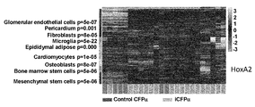
- Osteoblasts were detected by ALP staining, adipocytes by Oil Red-O staining, chondrocytes by Toluidine blue staining, and keratin 5 expressing cells by fluorescence of the reporter protein tdTomato. It is a figure which shows the result of having performed single cell transcriptome analysis about iCFP alpha cell, and having performed clustering analysis based on the obtained data. Shown on the left are cell types predicted based on gene expression profiles (such as high expression of a specific set of genes). It is a figure which shows the result of having performed transcriptome analysis of iCFP alpha cell per colony, and performing clustering analysis.
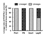
- P ⁇ expression and cells for cells present in the bone marrow tissue of the femur, vertebrae, sternum, ilia, hip joint (femoral head and acetabulum) and skull of P ⁇ -H2B-GFP :: Prx1-Cre :: Rosa26-tdTomato mice It is a photograph which shows the result of having detected Prx1 genealogy. It is a figure which shows the result of having analyzed the bone marrow cells of the femur of a P (alpha) -H2B-GFP :: Prx1-Cre :: Rosa26-tdTomato mouse, a vertebra, a sternum, and an iliac by FACS.
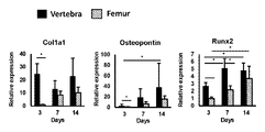
- FIG. 6 shows photographs and (c) the weight of cartilage pellets. It is a photograph of the K5 expression cell obtained by culture
- FIG. 5 shows expression of Procr in cells of intramedullary S34-MSC cluster.
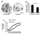
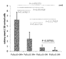
- FIG. 16 is a graph showing the abundance of Sca1 + CD34 + cells (proportion in PDGFR ⁇ + CD45 ⁇ viable cells) in the bone marrow of cervical vertebrae, thoracic vertebrae, lumbar vertebrae and femurs of P ⁇ -H2B-GFP mice. It is a photograph (a) of the colony formation cell obtained by culture
- P ⁇ cells were detected by YFP fluorescence, and type 7 collagen was detected by antibody.
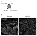
- Figure showing outline of parabiosis model (a), photograph showing observation results of transplanted skin tissue after HA1-44 peptide administration (b), graph showing percentage of PDGFR ⁇ + cells in the transplanted skin tissue (c) ).
- the expression of PDGFR ⁇ was detected by the fluorescence of GFP, and the Prx1 lineage was detected by the fluorescence of the reporter protein tdTomato.
- FIG. 16 is a graph showing the results of pathway analysis performed based on transcriptome analysis data of vertebral P ⁇ cells in mice in the HA1-44 peptide administration group and the saline administration group.
- 16 is a graph showing the results of pathway analysis (function analysis) performed based on transcriptome analysis data of vertebral P ⁇ cells in mice in the HA1-44 peptide administration group and the saline administration group.
- 6 is a graph showing the percentage of CD45-negative, TER-119-negative, and PDGFR ⁇ -positive cells in human peripheral blood mononuclear cell fractions collected before, 8 hours after, and 24 hours after administration of HA1-44 peptide.
- cell means one cell or a plurality of cells, depending on the context.
- the cells in the present application may be a cell population consisting of one type of cells or may be a cell population containing multiple types of cells.
- the expression "cells capable of differentiating into osteoblasts, adipocytes and chondrocytes” means that 1 / type of cells (homogeneous cell population derived from them) is differentiated into these 3 cell types. Not only in the case of having an activity, but also in the case of a cell population containing multiple types of cells exerting the ability to differentiate into the three cell types as a whole cell population.
- ectodermal mesenchymal stem cells have the ability to form colonies and to differentiate into three lineages of mesenchymal cells (osteoblasts, adipocytes, chondrocytes), and ectoderm It means PDGFR positive cells which are suggested to be derived. It is suggested that the cells are derived from ectoderm, for example, by being P0 lin + .
- EMSCs also have the ability to differentiate into epidermal cells (specifically, K5 positive keratinocytes).
- Epidermal cells which can differentiate EMSCs include, but are not limited to, keratinocytes, cells expressing keratin 5 (K5) (K5 positive cells), and keratinocytes expressing K5 (K5 positive keratinocytes). For example, whether it has differentiation ability to K5 positive keratinocytes can be determined by whether it can be differentiated to cells expressing K5 when cultured under keratinocyte differentiation induction conditions.
- Markers that characterizes the EMSC including lineage marker, P ⁇ +, P ⁇ lin + , P0 lin +, Prx1 lin-, Sox1 lin-, LepR lin-, CD34 +, and Sca1 - the like.
- EMSC Colony forming P ⁇ cells whose abundance in peripheral blood increases in response to necrotic tissue damage (skin flap etc.)
- iCFP ⁇ cells Colony-forming P ⁇ cells contained in vertebral bone marrow
- vertebral CFP ⁇ cells Colony-forming P ⁇ cells contained in vertebral bone marrow
- iCFP ⁇ cells have i) colony forming ability and ii) differentiation ability to osteoblasts, adipocytes and chondrocytes. Therefore, it can be said that iCFP ⁇ cells have properties as mesenchymal stem cells. Furthermore, iCFP ⁇ cells have the ability to differentiate into epidermal cells (K5 positive keratinocytes) and are P0 lin + . Therefore, iCFP ⁇ cells can be said to be ectodermal mesenchymal stem cells.
- Markers that characterize iCFP ⁇ cells P ⁇ +, P ⁇ lin +, P0 lin +, Prx1 lin-, Sox1 lin-, LepR lin-, CD34 +, and Sca1 - the like.
- Vertebrate-derived CFP ⁇ cells have i) colony forming ability and ii) differentiation ability to osteoblasts, adipocytes and chondrocytes. Therefore, it can be said that vertebral-derived CFP ⁇ cells have the property as mesenchymal stem cells. Furthermore, vertebral-derived CFP ⁇ cells have the ability to differentiate into epidermal cells (K5 positive keratinocytes) and are P0 lin + . Therefore, vertebra-derived CFP ⁇ cells can be said to be ectodermal mesenchymal stem cells.
- the vertebra from CFP ⁇ cells include Lepr lin + cells and Lepr lin- cells. Among these, LepR lin- cells are considered to exhibit properties closer to iCFP ⁇ cells in peripheral blood.
- the present application provides, as one aspect of EMSC, colony forming PDGFR positive cells having the following features i) and ii) and / or iii): i) capable of differentiating into osteoblasts, adipocytes and chondrocytes; ii) capable of differentiating into epidermal cells; iii) P0 lineage positive.
- the colony forming PDGFR positive cells are PDGFR ⁇ positive cells.
- the colonizing PDGFR-positive cells P ⁇ +, P ⁇ lin +, P0 lin +, Prx1 lin-, Sox1 lin-, LepR lin-, CD34 +, and Sca1 - 1 or more selected from It is a cell having a feature.
- Examples of additional features of the above colony forming PDGFR positive cells include: -It is P ⁇ + ; Is CD34 + ; ⁇ Sca1 - is; ⁇ CD34 +, and Sca1 - is; CD34 + and having one or more features selected from P ⁇ lin + , P 0 lin + , Prxl lin- , Soxl lin- , and LepR lin- ; ⁇ Sca1 - a and, and, P ⁇ lin +, P0 lin + , Prx1 lin-, Sox1 lin-, and having one or more features selected from Lepr lin-; ⁇ CD34 +, and Sca1 - a and, and, P ⁇ lin +, P0 lin + , Prx1 lin-, Sox1 lin-, and having one or more features selected from Lepr lin-; With one or more features selected from P ⁇ lin + ,
- the present invention relates to vertebral bone marrow derived cells that are PDGFR ⁇ positive, CD34 positive, and Sca1 negative.
- the cells are presumed to be cells corresponding to iCFP ⁇ cells in peripheral blood from the commonality of the markers. Therefore, it is expected to exhibit the same properties as iCFP ⁇ cells.
- the vertebral bone marrow-derived cells can be obtained, for example, by collecting vertebral bone marrow from a subject and selectively recovering P ⁇ + , CD34 + , and Sca 1 ⁇ cells.
- the present application also provides bone marrow-derived P ⁇ + CD34 + Sca1 + cells. Furthermore, the present application provides a method for producing cells, which comprises the step of selectively recovering P ⁇ + , CD34 + , and Sca1 + cells from bone marrow.
- Bone marrow that may be used as a source of P ⁇ + CD34 + Sca1 + cells includes vertebral bone (cervical, thoracic, lumbar) and femoral bone marrow.
- the bone marrow that may be used as a source of P ⁇ + CD34 + Sca1 + cells is vertebral bone marrow.
- the bone marrow that may be used as a source of P ⁇ + CD34 + Sca1 + cells is the bone marrow of the cervical spine.
- the present application provides P ⁇ + CD34 + Sca1 + cells derived from bone marrow of bone of ectomesenchyme as developmental origin.
- the present application also provides a method for producing a cell, which comprises the step of selectively recovering P ⁇ + , CD34 + and Sca1 + cells from bone marrow of ectomesenchyme as a developmental source.
- bones of ectomesenchyme as a developmental origin include frontal bones, nasal bones, zygomatic bones, maxillary bones, palatal bones, mandibles and the like.
- the present invention relates to a method of producing colony forming PDGFR positive cells, comprising the steps of any one of the following 1) to 4): 1) collecting peripheral blood from a subject having necrotic tissue damage and culturing on a solid phase; 2) Peripheral blood is collected from a subject having necrotic tissue damage and cultured on a solid phase, then P ⁇ + , P ⁇ lin + , P0 lin + , Prx1 lin- , Sox1 lin- , LepR lin- , CD34 + , and Sca1 - the step of selectively recovering cells having one or more features selected from; 3) Peripheral blood was collected from subjects with necrotic tissue damage, P.alpha + from the peripheral blood, P ⁇ lin +, P0 lin + , Prx1 lin-, Sox1 lin-, LepR lin-, CD34 +, and Sca1 - is selected from Selectively recovering cells having one or more
- the present invention relates to a method of producing colony forming PDGFR positive cells, comprising the steps of any one of the following 1) to 4): 1) culturing peripheral blood collected from a subject having necrotic tissue damage on a solid phase; 2) Peripheral blood collected from a subject having necrotic tissue damage is cultured on a solid phase, and then P ⁇ + , P ⁇ lin + , P0 lin + , Prx1 lin- , Sox1 lin- , LepR lin- , CD34 + , and Sca1 - the step of selectively recovering cells having one or more features selected from; 3) At least one selected from P ⁇ + , P ⁇ lin + , P0 lin + , Prx1 lin- , Sox1 lin- , LepR lin- , CD34 + , and Sca1 - from peripheral blood collected from a subject having necrotic tissue damage Selectively recovering cells having the characteristics of the following 1) to
- Necrotizing tissue damage includes, but is not limited to, skin flap, epidermal exfoliation in epidermolysis bullosa, and the like.
- skin flap the blood supply to the tip of the flap is insufficient, and the ischemic state causes cell / tissue necrosis.
- necrosis occurs in the exfoliated epidermal tissue.
- erythrocytes When peripheral blood is cultured on a solid phase, erythrocytes may be removed from peripheral blood prior to culture. Red blood cells can be removed by a method using a hemolytic reagent known to those skilled in the art, a method of treating peripheral blood with hetastarch, and recovering a supernatant containing nucleated cells.
- Examples of cells selectively recovered in step 2), 3) or 4) of the method for producing colony forming PDGFR positive cells include the following: ⁇ P.alpha cells ⁇ CD34 + cells ⁇ Sca1 of + - cells ⁇ CD34 + of, and Sca1 - a cell ⁇ CD34 +, and, P ⁇ lin +, P0 lin + , Prx1 lin-, Sox1 lin-, and Lepr lin- cells have one or more features selected from the ⁇ Sca1 - a and, and, P ⁇ lin +, P0 lin + , Prx1 lin-, Sox1 lin-, and cells having one or more features selected from Lepr lin- - CD34 +, and Sca1 - a and, and, P ⁇ lin +, P0 lin + , Prx1 lin-, Sox1 lin-, and LepR cells, having one or more features selected from lin-
- a method of “selectively recovering” cells for example, the following methods can be mentioned: (1) A method of “sorting” cells expressing a desired marker molecule using a cell sorter or the like. (2) A method of “recovering”, “selecting”, “separating”, “isolating” or “enriching” cells / colony expressing a desired marker molecule visually or based on the result of gene expression analysis.
- the marker molecules referred to here include surface markers (cell surface antigens), reporter proteins of lineage marker genes and the like.
- the present invention relates to a method of producing colony forming PDGFR positive cells, comprising the steps of any one of the following 1) to 4): 1) collecting vertebral bone marrow from a subject and culturing on a solid phase; 2) Collect vertebral bone marrow from a subject, culture on a solid phase, and then selectively recover cells having one or more features selected from P ⁇ + , P0 lin + , Prx1 lin- , and Sox1 lin- Process; 3) vertebral bone marrow taken from a subject, P.alpha + from bone marrow, P0 lin +, Prx1 lin-, and the step of selectively recovering cells having one or more features selected from Sox1 lin-; 4) Collect vertebral bone marrow from the subject, selectively collect cells having one or more characteristics selected from P ⁇ + , P 0 lin + , Prx 1 lin ⁇ , and Sox 1 lin ⁇ from
- the present invention relates to a method of producing colony forming PDGFR positive cells, comprising the steps of any one of the following 1) to 4): 1) culturing a vertebral bone marrow collected from a subject on a solid phase; 2) Culture the vertebral bone marrow collected from the subject on a solid phase, and then selectively recover cells having one or more features selected from P ⁇ + , P0 lin + , Prx1 lin- , and Sox1 lin- Process; 3) P.alpha from vertebral bone marrow taken from a subject +, P0 lin +, Prx1 lin- , and the step of selectively recovering cells having one or more features selected from Sox1 lin-; 4) P.alpha + from vertebral bone marrow taken from a subject, P0 lin +, Prx1 lin-, and Sox1 cells having one or more features selected selectively recovered from lin-, the cells on a solid phase by A
- step 2 of the method for producing the colonizing PDGFR-positive cells), 3) or 4 in), P ⁇ +, P0 lin + , Prx1 lin-, 1 selected from Sox1 lin- and Lepr lin- Cells having one or more features may be selectively recovered.
- Examples of cells selectively recovered in step 2), 3) or 4) of the method for producing colony forming PDGFR positive cells include the following: P0 lin + cells Prx1 lin- cells Sox1 lin- cells P0 lin + cells Prx1 lin- cells P0 lin + cells Sox1 lin- cells Prx1 lin- cells Sox1 lin- cells P0 lin +, Prx1 lin-, and Sox1 lin- cells ⁇ P ⁇ +, and P0 lin + cells ⁇ P ⁇ +, and Prx1 lin- cells ⁇ P ⁇ +, and Sox1 lin- cells ⁇ P ⁇ +, and Sox1 lin- cells ⁇ P ⁇ +, P0 lin +, and Prx1 lin- cells ⁇ P ⁇ +, P0 lin +, and Sox1 lin- cells ⁇ P ⁇ +, Prx1 lin-, and Sox1 lin- cells ⁇
- Vertebras that can be used as a source of colony forming PDGFR positive cells include cervical spine, thoracic spine, and lumbar spine.
- the vertebra used as a source of colony forming PDGFR positive cells is the cervical spine.
- bone marrow collected from a subject means bone marrow tissue containing various bone marrow cells.
- the present invention relates to a method of screening for a substance having a pluripotent stem cell inducing activity, using cells in peripheral blood induced by necrotic tissue damage as an index.
- iCFP ⁇ cells in peripheral blood are increased by necrotic tissue damage (eg, Skin flap), and P ⁇ + P 0 lin + Prx 1 lin ⁇ cells (that is, peripheral cells by the administration of HA 1-44 peptide) , ICFP ⁇ cell-containing cell population) was found to increase.
- necrotic tissue damage eg, Skin flap
- P ⁇ + P 0 lin + Prx 1 lin ⁇ cells that is, peripheral cells by the administration of HA 1-44 peptide
- pluripotent stem cells for example, MSC
- proliferative ability colony forming ability
- multipotency multi-lineage differentiation ability
- the present invention relates to a method of screening for a pluripotent stem cell inducer comprising the following steps: 1) Peripheral blood was collected from a subject, P.alpha + contained in the peripheral blood, P ⁇ lin +, P0 lin + , Prx1 lin-, Sox1 lin-, LepR lin-, CD34 + and Sca1 - 1 or more selected from Counting cells having features; 2) Peripheral blood was collected from a subject administered a test substance, P.alpha + contained in the peripheral blood, P ⁇ lin +, P0 lin + , Prx1 lin-, Sox1 lin-, LepR lin-, CD34 + and Sca1 - is selected from And 3) counting the cells having one or more characteristics; and 3) if the number of cells counted in step 2) is larger than the number of cells counted in step 1), Selecting as a candidate for a substance having pluripotent stem cell in
- the present invention relates to a method of screening for a pluripotent stem cell inducer comprising the following steps: Cells with one or more features selected from - 1) P.alpha included in the peripheral blood collected from a subject +, P ⁇ lin +, P0 lin +, Prx1 lin-, Sox1 lin-, LepR lin-, CD34 + and Sca1 Counting steps; One selected from - 2) P.alpha contained in the peripheral blood collected from a subject the test substance has been administered +, P ⁇ lin +, P0 lin +, Prx1 lin-, Sox1 lin-, LepR lin-, CD34 + and Sca1 And 3) counting the cells having the above characteristics; and 3) when the number of cells counted in step 2) is larger than the number of cells counted in step 1), the test substance is subjected to pluripotency Selecting as a candidate for a substance having stem cell inducing activity.
- the surface markers (P ⁇ , CD34, Sca1) can be detected using an antibody or the like with respect to the characteristics of the cells counted in the above screening method.
- the product (fluorescent protein etc.) of the reporter gene can be detected as an indicator.
- Genealogy markers (P ⁇ , P0, Prx1, Sox1, LepR) can be detected by using transgenic animals with DNA structures / constructs (Cre-loxP etc.) that allow lineage tracing of the gene of interest .
- Examples of cells counted in steps 1) and 2) of the above screening method include the following: ⁇ P.alpha cells ⁇ CD34 + cells ⁇ Sca1 of + - cells ⁇ CD34 + of, and Sca1 - a cell ⁇ CD34 +, and, P ⁇ lin +, P0 lin + , Prx1 lin-, Sox1 lin-, and Lepr lin- cells have one or more features selected from the ⁇ Sca1 - a and, and, P ⁇ lin +, P0 lin + , Prx1 lin-, Sox1 lin-, and cells having one or more features selected from Lepr lin- - CD34 +, and Sca1 - a and, and, P ⁇ lin +, P0 lin + , Prx1 lin-, Sox1 lin-, and LepR cells, having one or more features selected from lin- P ⁇ lin +, P0 lin +
- the present invention provides a substance having pluripotent stem cell induction activity, using HA1-44 peptide as a positive control and using the reaction of pluripotent stem cells contributing to tissue regeneration in vivo as an index.
- the present invention relates to a method of screening for a pluripotent stem cell inducer comprising the following steps: 1) obtaining an adherent cell population by collecting peripheral blood from a subject and culturing it on a solid phase; 2) performing comprehensive gene expression analysis of the cell population obtained in step 1 on a colony or single cell basis; 3) administering a peptide (HA1-44 peptide) consisting of the amino acid sequence of SEQ ID NO: 1 to a subject, collecting peripheral blood, and culturing on a solid phase to obtain an adherent cell population; 4) performing comprehensive gene expression analysis on a colony or single cell basis for the cell population obtained in step 3; 5) administering a test substance to a subject, collecting peripheral blood, and culturing on a solid phase to obtain an adherent cell population; 6) performing comprehensive gene expression analysis on a colony or single cell basis for the cell population obtained in step 5; 7) pooling gene expression data obtained in steps 2 and 4 and performing clustering analysis; 8) pooling the gene
- a test substance is administered instead of the HA1-44 peptide in step 3, and an HA1-44 peptide is administered instead of the test substance in step 5. That is, either the HA1-44 peptide or the test substance may be administered to the subject first.
- the subjects in steps 1, 3 and 5 may be the same individual or different individuals. For example, three animals of the same strain (strain) are prepared, one animal is not administered the substance (or only the solvent is administered), the other one is administered the HA1-44 peptide, and the remaining one is the test substance. Then, peripheral blood may be collected from each individual to obtain an adherent cell population, comprehensive gene expression analysis and clustering analysis.
- the subject in step 1 may be a subject in which only the same solvent as that used in administering the HA1-44 peptide and the test substance in steps 3 and 5, respectively, has been administered.
- a variant, modified form or tagged HA1-44 peptide of HA1-44 peptide is used.
- the variant is several, for example, 1 to 5, preferably 1 to 4, preferably 1 to 3, more preferably 1 to 2, more preferably 1 in the amino acid sequence of the HA1-44 peptide.
- Amino acids have amino acid sequences which have been substituted, inserted, deleted and / or added.
- the variant is 50% or more, preferably 60% or more, more preferably 70% or more, more preferably 80% when the local alignment (Local Alignment) is performed on the amino acid sequence of the HA1-44 peptide.
- Amino acids having homology of the above even more preferably 85% or more, particularly preferably 90% or more (eg, 91%, 92%, 93%, 94%, 95%, 96%, 97% or 98%) It is a peptide having a sequence.
- the homology of amino acid sequences can be measured, for example, using FASTA, BLAST, DNASIS (manufactured by Hitachi Software Engineering Co., Ltd.), and GENETYX (manufactured by Genetics Co., Ltd.). Alternatively, sequences can simply be compared and calculated.
- the modified form is several, for example, 1 to 5, preferably 1 to 4, preferably 1 to 3, more preferably 1 to 2, more preferably 1 in the amino acid sequence of the HA1-44 peptide.
- HA1-44 peptide it has an amino acid sequence in which the amino acid residue of the amino acid is modified.
- exemplary tags include, but are not limited to, His 6 -tag, FLAG tag, myc tag, and GST tag. The tag may be added to the N-terminus or C-terminus of the amino acid sequence.
- the global gene expression analysis may be RNA sequencing (RNA-seq).
- clustering analysis may be performed using an iterative clustering and guide-gene selection (ICGS) algorithm.
- the present invention relates to a method of screening for a pluripotent stem cell inducer comprising the following steps: 1) obtaining an adherent cell population by culturing peripheral blood collected from a subject on a solid phase; 2) performing comprehensive gene expression analysis on a colony or single cell basis for the cell population obtained in step 1); 3) obtaining an adherent cell population by culturing on a solid phase peripheral blood collected from a subject to which a peptide (HA1-44 peptide) consisting of the amino acid sequence of SEQ ID NO: 1 has been administered; 4) performing comprehensive gene expression analysis on a colony or single cell basis for the cell population obtained in step 3); 5) obtaining an adherent cell population by culturing on a solid phase peripheral blood collected from a subject to which a test substance has been administered; 6) performing comprehensive gene expression analysis on a colony or single cell basis for the cell population obtained in step 5); 7) pooling gene expression data obtained in steps 2) and 4) and performing clustering analysis; 8) pooling the
- the present invention relates to a method of screening for a pluripotent stem cell inducer comprising the following steps: 1) obtaining an adherent cell population by collecting peripheral blood from a subject and culturing it on a solid phase; 2) counting the number of colonies obtained in step 1); 3) a test substance is administered to a subject, peripheral blood is collected, and an adherent cell population is obtained by culturing on a solid phase; 4) a step of counting the number of colonies obtained in step 3); and 5) if the number of colonies counted in step 4) is larger than the number of colonies counted in step 2), the test substance Selecting as a candidate for a substance having pluripotent stem cell inducing activity.
- the subject in step 1) may be a subject to which only the same solvent as that used in administering the test substance in step 3) has been administered.
- Step 2) and 4) colonies counted in, P ⁇ +, P ⁇ lin +, P0 lin +, Prx1 lin-, Sox1 lin-, LepR lin-, CD34 + and Sca1 - colonies having one or more features selected from It may be
- the present invention relates to a method of screening for a pluripotent stem cell inducer comprising the following steps: 1) obtaining an adherent cell population by culturing peripheral blood collected from a subject on a solid phase; 2) counting the number of colonies obtained in step 1); 3) obtaining an adherent cell population by culturing on a solid phase peripheral blood collected from a subject to which a test substance is administered; 4) a step of counting the number of colonies obtained in step 3); and 5) if the number of colonies counted in step 4) is larger than the number of colonies counted in step 2), the test substance Selecting as a candidate for a substance having pluripotent stem cell inducing activity.
- the subject in step 1) may be a subject to which only the same solvent as that used when the test substance is administered in step 3) is administered.
- Step 2) and 4) colonies counted in, P ⁇ +, P ⁇ lin +, P0 lin +, Prx1 lin-, Sox1 lin-, LepR lin-, CD34 + and Sca1 - colonies having one or more features selected from It may be
- the present invention relates to a method of screening for a pluripotent stem cell inducer comprising the following steps: 1) harvesting bone marrow from a vertebra of a subject, and obtaining a PDGFR ⁇ -positive cell population by culture on solid phase or cell sorting; 2) performing comprehensive gene expression analysis of the cell population obtained in step 1 on a colony or single cell basis; 3) administering a test substance to a subject, collecting bone marrow from vertebrae, and obtaining a PDGFR ⁇ -positive cell population by culture on solid phase or cell sorting; 4) performing comprehensive gene expression analysis on a colony or single cell basis for the cell population obtained in step 3; 5) pooling the gene expression data obtained in steps 2 and 4 and performing pathway analysis; and 6) as a result of analysis of step 5, comparing with the cell population (untreated group) obtained in step 1 In the cell population obtained in step 3 (test substance administration group): (i) a pathway associated with EIF2 signaling, modulation of eIF4 and p70S6
- the global gene expression analysis may be RNA sequencing (RNA-seq).
- pathway analysis may be performed using Ingenuity Pathway Analysis (IPA) software (https://www.qiagenbioinformatics.com).
- IPA Ingenuity Pathway Analysis
- the present invention relates to a method of screening for a pluripotent stem cell inducer comprising the following steps: 1) obtaining a PDGFR ⁇ -positive cell population from a bone marrow collected from a vertebra of a subject by culture on solid phase or cell sorting; 2) performing comprehensive gene expression analysis on a colony or single cell basis for the cell population obtained in step 1); 3) obtaining a PDGFR ⁇ -positive cell population from a bone marrow collected from a vertebra of a subject to which a test substance has been administered, by culture on solid phase or cell sorting; 4) performing comprehensive gene expression analysis on a colony or single cell basis for the cell population obtained in step 3); 5) pooling the gene expression data obtained in steps 2) and 4) and performing pathway analysis; and 6) as a result of analysis in step 5), comparing with the cell population obtained in step 1), Pathways related to (i) modulation of EIF2 signaling, eIF4 and p70S6K signaling, and / or mTOR signaling
- the global gene expression analysis may be RNA sequencing (RNA-seq).
- pathway analysis may be performed using Ingenuity Pathway Analysis (IPA) software (https://www.qiagenbioinformatics.com).
- IPA Ingenuity Pathway Analysis
- the invention in another aspect, relates to a cell or cell population in peripheral blood induced by a MSC blood mobilization agent.
- the present invention also relates to cells or cell populations in the bone marrow of vertebrae induced by HA1-44 peptide.
- MSC blood mobilization substance means a substance having an activity of mobilizing mesenchymal stem cells (MSCs) into peripheral blood or an activity of increasing the abundance of MSCs in peripheral blood.
- MSC blood mobilization substances include HMGB1 protein, HMGB2 protein and HMGB3 protein (for example, described in WO2008 / 053892 and WO2009 / 133939), S100A8 protein and S100A9 protein (for example, described in WO2009 / 133940 and WO2011 / 052668), and Examples include various HMGB1 peptides described in international application WO2012 / 147470 by the present inventors (for example, a peptide consisting of amino acid residues 1-44 of HMGB1 protein (HA1-44 peptide in the present application)) and the like, but not limited thereto .
- the present invention relates to a cell population obtained by administering a MSC blood mobilization substance to a subject, collecting peripheral blood from the subject, and culturing the collected peripheral blood on a solid phase .
- the MSC blood mobilization agent may be an HA 1-44 peptide.
- the present invention comprises: 1) administering HA1-44 peptide to a subject, 2) harvesting vertebral bone marrow from the subject, and 3) culturing the harvested bone marrow on a solid phase, or
- the present invention relates to a cell population obtained by sorting PDGFR ⁇ -positive cells from the collected bone marrow.
- the invention relates to a method of producing a cell or cell population as described above.
- the present invention relates to a method for producing cells, comprising the steps of: administering a MSC blood mobilization substance to a subject; collecting peripheral blood from the subject; and culturing the collected peripheral blood on a solid phase About.
- the present invention relates to a method for producing cells, which comprises the step of culturing peripheral blood collected from a subject to which MSC blood mobilization substance has been administered on a solid phase.
- the MSC blood mobilization agent may be an HA1-44 peptide.
- the present invention comprises: 1) administering HA1-44 peptide to a subject, 2) harvesting vertebral bone marrow from the subject, and 3) culturing the harvested bone marrow on a solid phase, or
- the present invention relates to a method for producing cells, which comprises the step of sorting PDGFR ⁇ -positive cells from the collected bone marrow.
- the present invention obtains, separates and / or concentrates from living tissues including mesenchymal stem cells (MSCs) cells having the same high tissue regeneration promoting ability as PDGFR ⁇ -positive cells in vertebral bone marrow. On the way.
- the present invention relates to a cell or cell population obtained by the method for obtaining, separating and / or concentrating as described above.
- the present invention relates to a method of producing a cell population, comprising the steps of: 1) culturing a cell population derived from a living tissue containing mesenchymal stem cells (MSC) on a solid phase; 2) Subcloning the colony obtained in step 1; 3) culturing a part of the cells obtained by subcloning in a bone, cartilage and / or fat differentiation induction medium, and measuring expression levels of bone, cartilage and / or fat differentiation markers; and 4 ) MSCs obtained by culturing the bone marrow of the femur on a solid phase are a differentiation marker of bone, cartilage and / or fat when cultured in a bone, cartilage and / or fat differentiation induction medium Selecting a cell clone that showed a high expression level as compared to the expression level.
- MSC mesenchymal stem cells
- the present invention relates to a method of producing a cell population, comprising the steps of: 1) culturing a cell population derived from a living body tissue including a MSC on the solid phase; and 2) P ⁇ +, P ⁇ lin + , P0 lin +, Prx1 lin-, Sox1 lin-, LepR lin-, CD34 +, and Sca1 - Selecting a colony having one or more features selected from
- step 2 may be a step of selecting Prx1 lineage negative colonies.
- the present invention is selected from P ⁇ + , P ⁇ lin + , P0 lin + , Prx1 lin- , Sox1 lin- , LepR lin- , CD34 + , and Sca1 - from cell populations derived from living tissues including MSCs
- a method of producing a cell population comprising the step of selectively recovering cells having one or more of the characteristics described above.
- the present invention relates to a method of producing a cell population, comprising the steps of: 1) One or more characteristics selected from P ⁇ + , P ⁇ lin + , P0 lin + , Prx1 lin- , Sox1 lin- , LepR lin- , CD34 + , and Sca1 - from cell populations derived from living tissues including MSCs And 2. 2) culturing the cells recovered in step 1) on a solid phase.
- examples of biological tissues containing MSCs include, but are not limited to, bone marrow, umbilical cord, cord blood, placenta, adipose tissue, dental pulp, periosteum, synovium, oomen, peripheral blood, etc. I will not.
- bone marrow include, but are not limited to, bone marrow such as femur, vertebra, sternum, ilium and skull.
- the present invention relates to a composition for promoting tissue regeneration, which comprises ectodermal mesenchymal stem cells.
- the present invention relates to a composition for promoting tissue regeneration comprising colony forming PDGFR positive cells having the following features i) and ii) and / or iii): i) capable of differentiating into osteoblasts, adipocytes and chondrocytes; ii) capable of differentiating into epidermal cells; iii) P0 lineage positive.
- the composition is used to promote regeneration of tissue derived from mesoderm or ectoderm.
- Tissues derived from mesoderm include, but are not limited to, bone, cartilage, muscle, vascular endothelium and the like.
- Tissues derived from ectoderm include, but are not limited to, epithelial tissue (eg, epidermis), neural tissue, and the like.
- the composition used for promoting tissue regeneration of the present invention may contain a pharmaceutically acceptable carrier, diluent and / or excipient.
- a pharmaceutically acceptable carrier diluent and / or excipient.
- the amount of ectodermal mesenchymal stem cells contained in the composition used for promoting tissue regeneration according to the present invention, the dosage form of the composition, the frequency of administration, etc. depend on the type of tissue to be regenerated and / or It can be selected appropriately according to the conditions such as the state of the object.
- the invention relates to a method of promoting tissue regeneration in a subject comprising administering ectodermal mesenchymal stem cells.
- the present invention promotes tissue regeneration of a subject comprising administering to the subject colony forming PDGFR positive cells having the following features i) and / or ii) and / or iii): On the method: i) capable of differentiating into osteoblasts, adipocytes and chondrocytes; ii) capable of differentiating into epidermal cells; iii) P0 lineage positive.
- the administration method of the above-mentioned cells can be appropriately selected according to the conditions such as the type of tissue promoting regeneration and / or the condition of the subject to be administered.
- the administration method includes, but is not limited to, intradermal administration, subcutaneous administration, intramuscular administration, intravenous administration, nasal administration, oral administration, suppository and the like.
- the invention relates to ectodermal mesenchymal stem cells for use in promoting tissue regeneration in a subject.
- the invention relates to the use of ectodermal mesenchymal stem cells for the manufacture of a medicament for promoting tissue regeneration in a subject.
- ICFP ⁇ cells in peripheral blood are considered to be useful biomarkers for evaluating EMSC-mediated tissue regeneration activity, such as in cases where necrotic tissue damage has occurred. Accordingly, the present application provides a method for determining the tissue regeneration promoting effect expected in a subject who has been administered a MSC blood mobilization agent, using iCFP ⁇ cells in peripheral blood as an indicator.
- the present invention comprises the steps of: 1) P.alpha contained in peripheral blood taken from the subject prior to administering the MSC blood mobilized material +, P ⁇ lin +, P0 lin +, Prx1 lin-, Sox1 lin-, LepR lin-, CD34 + and Sca1 - from 2) counting cells having one or more features selected; and 2) P ⁇ + , P ⁇ lin + , P 0 lin + contained in peripheral blood collected from the subject after administration of MSC blood mobilization agent a step of counting the cells with one or more features selected from - Prx1 lin-, Sox1 lin-, LepR lin-, CD34 + and Sca1; MSC blood mobilization substance which is suggested to promote tissue regeneration in said subject when the number of cells counted in step 2) is greater than the number of cells counted in step 1), Method for determining the tissue regeneration promoting effect of
- the "subject” may be any of human and non-human animals.
- the subject is a non-human animal.
- Non-human animals include, but are not limited to, mice, rats, monkeys, pigs, dogs, rabbits, hamsters, guinea pigs, horses, sheep and the like.
- the timing of collecting peripheral blood from the subject is not particularly limited with regard to the method for producing cells (or cell population), screening method, and method for determining the tissue regeneration promoting effect of MSC blood mobilization substances provided by the present application.
- peripheral blood is collected 2 to 24 hours after creation of necrotic tissue injury or administration of MSC blood mobilization agent
- the timing of collecting peripheral blood from the subject is from generation of necrotic tissue damage or administration of a MSC blood mobilizing agent 2, 3, 4, 5, 6, 7, 8, 9, 10, 11, 12. , 13, 14, 15, 16, 17, 18, 19, 20, 21, 22, 23 or 24 hours later.
- the timing of collecting peripheral blood from the subject is 4-24 hours, 8-24 hours, 8-16 hours, or 10-24 hours after the creation of the necrotic tissue damage or administration of the MSC blood mobilization agent. It can be after ⁇ 14 hours.
- PDGFR positive cells are PDGFR ⁇ positive cells.
- mice Materials and Methods mouse P ⁇ -H2B-GFP mice (The Jackson Laboratory, Stock No: 007669), in which the expression of PDGFR ⁇ can be confirmed by the fluorescence of GFP, and various cell lineage tracking mice using the Cre-loxP system were used for the experiments.
- Cell lineage tracking mice utilizing the Cre-loxP system can be generated by crossing Cre driver mice with Cre reporter mice.
- the Cre driver mouse is a transgenic mouse having a DNA structure in which the coding sequence of Cre recombinase is introduced downstream of the promoter sequence of the desired gene.
- a Cre reporter mouse is a DNA sequence of the structure “promoter (CAG promoter etc.)-LoxP-stop cassette-loxP-desired reporter gene (EYFP or tdTomato in this example)” introduced at a locus such as ROSA26. Transgenic mice.
- mice were prepared as Cre driver mice.
- ⁇ P ⁇ -Cre mouse The Jackson Laboratory, Stock No: 013148
- P0-Cre mouse The Jackson Laboratory, Stock No: 017927
- Prx1-Cre mouse The Jackson Laboratory, Stock No: 005584
- Sox1-Cre mouse RIKEN BioResource Research Center, Accession No. CDB0525K
- LepR-Cre mouse The Jackson Laboratory, Stock No: 008320
- Krt 5-Cre mouse MMI ID: 1926815, K5 Cre transgenic mouse described in Proc Natl Acad Sci USA A. 1997 Jul 8; 94 (14): 7400-5)
- mice were prepared as Cre reporter mice. ⁇ Rosa26-EYFP mouse (The Jackson Laboratory, Stock No: 006148) ⁇ Rosa26-tdTomato mouse (The Jackson Laboratory, Stock No: 007909)
- P ⁇ -Cre :: Rosa26-EYFP mice were created by crossing P ⁇ -Cre driver mice with Rosa26-EYFP reporter mice.
- the P ⁇ -H2B-GFP mouse is a mouse in which a sequence encoding a fusion protein of histone H2B and eGFP is knocked in downstream of the promoter of the PDGFR ⁇ gene.
- a P ⁇ -H2B-GFP :: Prx1-Cre :: Rosa26-tdTomato mouse was created.
- skin flaps were prepared as a method for causing necrotic tissue damage.
- the specific method of creating a skin flap is as follows. 8-10 week-old male mice (20-25 g) were shaved on the back under 1.5-2.0% (v / v) isoflurane inhalation anesthesia. In order to maintain the continuity of the skin only on the caudal side, a skin flap of 2.0 x 4.0 cm wide was created at the center of the back using scissors (this causes the tip far from the root of the flap to become ischemic, Necrotic damage of skin tissue occurs). The affected area after skin flap preparation was protected with a bandage of sufficient size. At 12 hours after skin flap preparation, cardiac blood collection, cell separation of vertebrae and femur bone marrow, and cryosectioning of vertebrae and femur were performed.
- parabiosis model was created using 6 week old male wild type mice and 6 week old male cell lineage tracking mice did. Two animals were simultaneously induced with general anesthesia with isoflurane and positioned on a heat pad. Two opposing sides of the body were shaved and the skin was resected approximately 5 mm wide from the elbow joint to the knee joint. The dorsal skin was continuously sutured with 5-0 bricryl. The opposing elbow and knee joints were sutured with 3-0 bikryl. Similarly, the ventral skin was continuously sutured with 5-0 bricryl. I waited on the heat pad for awakening from anesthesia. Because activity is limited, up to 1 pair was kept in 1 cage and the bait was given.
- the vertebra and femur taken under general anesthesia were immersed in 0.2% collagenase A solution (Sigma, Cat # 10103580001) and incubated at 37 ° C. for 1 hour.
- Bone marrow cells were extruded with a mortar, collected by pipetting, and passed through a 40 ⁇ m cell strainer. The supernatant was removed by centrifugation at 300 G for 5 minutes, hemolyzed with RBC Lysis buffer (BioLegend, cat # 420301) to remove erythrocytes, and subjected to the next experiment as bone marrow cells.
- Osteoblast differentiation induction was performed. After induction of each differentiation, fix with 4% PFA, adipocytes are stained with oil red-O, osteoblasts are stained with ALP using ALP activity assay kit (TAKARA BIO Inc., Kusatsu, Japan) and observed with a microscope respectively did.
- ALP activity assay kit (TAKARA BIO Inc., Kusatsu, Japan) and observed with a microscope respectively did.
- mice 8. Induction of differentiation into keratinocytes, animals Krt5-Cre :: Rosa26-tdTomato mice were bred to 8-10 weeks of age and used for experiments. Sampling A skin flap was made on the back of the mouse the day before and 12 hours after preparation, peripheral blood was collected by heart blood collection under anesthesia. Peripheral blood was similarly collected from mice for which skin flaps were not generated. Femurs and vertebrae were collected after blood collection. ⁇ Cell preparation Peripheral blood removes red blood cells using HetaSep, and one minute of blood is seeded on a 6-well plate of collagen I-coated plate in 1 well under conditions of 5% O 2 , 5% CO 2 , 37 ° C. Cultured.
- RNA-seq Transcriptome analysis (cell population RNA-seq) RNA was extracted from the cell population, and RNA-seq library was created according to Smart-seq2 protocol (Nature Protocols 9, 171-181 (2014) doi: 10.1038 / nprot.2014.006). The obtained library was sequenced on Nextseq 500 (Illumina) using nextseq high output kit (37 bp pair end reading). Base call conversion to fastq format and demultiplexing were performed using Illumina's bcl2fastq v2.17.1.14 with default parameters and the --no-lane-splitting option.
- Trim the lead using TrimGalore http://www.bioinformatics.babraham.ac.uk/projects/trim_galore/
- RSEM Li et al., BMC Bioinformatics 12, 323 (2011); version is STAR-2.5 Mapping and counting (quantification) were performed using 2b).
- Expression variation analysis between samples was performed using DESeq2 (Love et al., Genome Biol. 15, 550 (2014)).
- Expression variation genes (DEGs) were uploaded to Ingenuity Pathway Analysis (IPA) software (https://www.qiagenbioinformatics.com) to extract the biological pathways and functions most relevant to DEGs.
- IPA Ingenuity Pathway Analysis
- a clustering analysis was performed using the AltAnalyze_v.2.1.0-Py ICGS algorithm based on the log2 converted value (log 2 (TPM + 1)) obtained by adding 1 to the TPM (Transcripts Per kilobase Million) count obtained by RSEM .
- RNA-seq Single cell suspensions were prepared and cell survival assessed using an automated cell counter TC20 (BioRad). Single cell RNA-seq library was created using ddSEQ Single-Cell Isolator and SureCell WTA 3 'Library prep kit according to the manufacturer's protocol. The obtained library was sequenced with Nextseq 500 (Illumina) using Nextseq high output kit (Read 1: 68 bp, Read 2: 75 bp). Base call conversion to fastq format and demultiplexing were performed using Illumina's bcl2fastq v2.17.1.14 with default parameters and the --no-lane-splitting option.
- FACS Analysis For bone marrow cells collected from vertebrae and femurs, FACS analysis was performed using fluorescently labeled antibodies against various surface molecules. A series of processes such as fluorescence detection and sorting were performed using BD FACS Aria III system, and analysis of the obtained data was performed using Flow Jo software Ver. 6.3.3 (Tree Star, Ashland, OR).
- a parabiosis model was created using 6-week-old male wild-type mice and 6-week-old male P0-Cre :: Rosa26-tdTomato mice according to the method described in. After completion of the blood chimera four weeks after the operation, a 0.5 mm diameter cartilage defect was created in the knee joint of a wild type mouse using a 0.5 mm diameter hand drill (manufactured by MEISINGER). Immediately after creating the cartilage defect, 100 ⁇ g of HA1-44 peptide diluted with 100 ⁇ L of physiological saline was administered from the tail vein, and thereafter, the same dose was also administered twice a week for 4 weeks after surgery at the same dose.
- a cartilage defect is created in the knee joint in the same manner as described above using 8-week-old male wild-type mice, and the HA1-44 peptide is administered at the same dose and schedule as described above. Knee joints were harvested 8 and 12 weeks later and Safranin O staining of tissue sections was performed.
- XX + XX positive XX -: XX negative YY lin + : YY line positive YY lin- : YY line negative
- PDGFR platelet-derived growth factor receptor
- P ⁇ cells PDGFR ⁇ -positive cells
- MSC mesenchymal stem cells
- EMSC ectomesenchymal stem cell
- CFP ⁇ cells colony-forming P ⁇ cells
- iCFP ⁇ cells necrotic injury-induced colony forming P ⁇ cells (necrotic injury-induced colony-forming P ⁇ cells)
- CFU colony-forming unit
- LepR Leptin receptor (Leptin receptor)
- Example 1 Properties of iCFP ⁇ Cells in Peripheral Blood
- a skin flap was created on the back of P ⁇ -H2B-GFP mice, and the number of P ⁇ cells contained in peripheral blood collected 12 hours later was examined.
- P ⁇ cells were significantly increased in the skin flap group as compared with the control group in which the skin flap was not generated, and the increase in P ⁇ cells was correlated with an increase in HMGB1 concentration in peripheral blood.
- Figure 1 In addition, as a result of culturing the peripheral blood of P ⁇ -H2B-GFP mice and performing colony assay, significantly more colonies are obtained in the skin flap group than in the control group (all P ⁇ -positive cells) and CFU activity is high. Was shown ( Figure 2).
- colony-forming P ⁇ cells obtained by culturing peripheral blood of mice in which skin flaps have been produced show differentiation ability to osteoblasts, adipocytes and chondrocytes, and keratinocytes It showed the ability to differentiate into cells expressing keratin 5 (Krt5, K5) under conditions for inducing differentiation into (FIG. 3).
- iCFP ⁇ cells Single cell transcriptome analysis of peripheral blood-derived CFP ⁇ cells (iCFP ⁇ cells) after skin flap formation showed that most cells had high expression of gene groups corresponding to cell types such as MSC (FIG. 4) .
- cells expressing genes characteristic of epidermal cells such as Krt8 and Krt18 were also included.
- transcriptome analysis of peripheral blood-derived CFP ⁇ cells in the control group and the skin flap group was performed on a colony basis, and clustering analysis was performed using the ICGS algorithm.
- Example 2 In all of the surface marker control group and skin flap group of iCFP ⁇ cells in peripheral blood, colonies obtained by culturing peripheral blood were all P ⁇ positive (FIG. 6). In addition, as a result of transcriptome analysis of peripheral blood CFP ⁇ cells (iCFP ⁇ cells) of the skin flap group in a single cell, it was CD34 positive and Sca 1 negative.
- Example 3 Cell lineage marker for iCFP ⁇ cells in peripheral blood
- CFP ⁇ cells iCFP ⁇ cells derived from peripheral blood of mice are transated using P- ⁇ lineage, P0 lineage, Prx1 lineage, Sox1 lineage and LepR lineage using Cre-loxP system
- P- ⁇ lineage P0 lineage
- Prx1 lineage Prx1 lineage
- Sox1 lineage Sox1 lineage
- LepR lineage negative FIG. 7
- Prxl lineage 93% of iCFP ⁇ cells were negative ( Figure 7).
- Prx1 lineage positive and negative colony forming cells were slightly present in the blood in the normal blood, cells that increased with skin flaps were exclusively Prx1 lineage negative cells (FIG. 8).
- iCFP ⁇ cells can be defined as Prx1 lineage negative.
- ICFP ⁇ cells in peripheral blood are considered because it is P0 lin + and Prx 1 lin- is derived from ectoderm, further, considering that and HoxA2 gene it is Sox1 lin- is highly expressed, the head It is suggested that it is a cell that originates from neural crest.
- Example 5 Lineage Markers of Vertebrate and Femur CFP ⁇ Cells
- P0 lineage, Prx1 lineage, Sox1 lineage and LepR lineage were examined using cell lineage tracking mice.
- CFParufa cells vertebrae, P0 lin +, Prx 1 lin-, and Sox1 lin- a, with respect Lepr lin about 60% were negative (Fig. 11).
- CFP ⁇ cells of the femur, P0 lin +, Prx1 lin + , and Sox1 lin- a, with respect Lepr lin about 80% were positive ( Figure 12).
- peripheral blood of iCFP ⁇ cells (P ⁇ lin +, P0 lin + , Prx1 lin-, Sox1 lin-, LepR lin-) sources is suggested to be present in the vertebrae.
- Example 6 Properties of CFP ⁇ cells of vertebra and femur CFP ⁇ cells obtained by culturing the bone marrow of vertebra and femur were compared with colony forming ability and ability to differentiate into osteoblasts, adipocytes and chondrocytes. CFP ⁇ cells showed higher potency (FIGS. 13-16). Moreover, as a result of differentiation induction of CFP ⁇ cells of vertebra and femur with all-trans retinoic acid (ATRA) and BMP-4, colonies expressing keratin 5 were observed (FIGS. 17 and 18) Were confirmed to contain cells capable of differentiating into K5 positive cells.
- ATRA all-trans retinoic acid
- Example 7 Transcriptome analysis of vertebrae and femur P ⁇ cells
- P ⁇ cells were sorted from vertebral bone and femur bone marrow cells, and single cell transcriptome analysis was performed.
- bone marrow P ⁇ cells were divided into six clusters (FIG. 19).
- the six clusters are (1) S34-MSC (Sca1 and CD34-expressing), (2) osteoprogenitor (Osteomodulin and Wnt16- expressing), based on the gene specifically expressed by the cells of each cluster.
- vertebral P ⁇ cells were separated by FACS using the expression of Sca1 and CD34 as an index, and a CFU assay was performed.
- CFU activity was higher in the order of Sca1 + CD34 + cells> Sca1 + CD34 - cells and Sca1 - CD34 + cells> Sca1 - CD34 - cells (FIG. 20).
- Example 8 Correspondence between P ⁇ cells in peripheral blood and P ⁇ cells in vertebral bodies Based on transcriptome analysis data of iCFP ⁇ cells in peripheral blood, clustering analysis was performed with P ⁇ cells in vertebral bones and femurs. As a result, peripheral blood iCFP ⁇ cells were located near the S34-MSC cluster (FIG. 21). Moreover, the peripheral blood iCFP ⁇ cells, CD34 + Sca1 - was. Considering this result in combination with lineage markers, it is suggested that peripheral blood iCFP ⁇ cells correspond to CD34 + Sca 1 - cells contained in the S34-MSC cluster of vertebral bone.
- Procr can be a marker for Sca1 + CD34 + cells, which are considered to be the cells with the highest proliferation potential and the highest hierarchy among bone marrow P ⁇ cells.
- cervical vertebral> thoracic vertebra> lumbar spine> femur was in the order (FIG. 23).
- Example 10 Contribution to Circulating Tissue Regeneration of Circulating Cells Induced by HMGB1 Administration
- HMGB1 protein As a domain with the activity of mobilizing bone marrow-derived P ⁇ -positive mesenchymal stem cells into peripheral blood.
- a peptide (HA1-44 peptide) consisting of the amino acid sequence 1 to 44 of SEQ ID NO: 1 (SEQ ID NO: 1) has been identified.
- SEQ ID NO: 1 SEQ ID NO: 1
- P.alpha cells in peripheral blood induced by HA1-44 peptide (P ⁇ + P0 lin + Prx1 lin- cells) is the same as iCFP ⁇ cells induced by necrotic tissue damage, or at least iCFP ⁇ It contains cells and is considered to play a role in repairing tissue damage such as epidermis and cartilage.
- Example 11 Changes in cells induced by HMGB1 administration
- Peripheral blood was collected from mice administered HA1-44 peptide and mice administered saline and cultured on a plastic plate to obtain colonies of adherent cells .
- the transcriptome analysis was performed on a colony basis for the cells, and clustering was performed using the ICGS algorithm based on the obtained data.
- results shown in FIG. 29 were obtained.
- FIG. 30 which simplified and represented the result of the said clustering.
- the result is such that the number is “cluster 1: live food group ⁇ ⁇ test substance group, cluster 2: live food group ⁇ test substance group, cluster 3: live food group> test substance group, cluster 4: live food group> test substance group”
- the substance can be evaluated as a candidate for a substance having pluripotent stem cell inducing activity.
- Transcriptome analysis was performed on P ⁇ cells of vertebrae collected from mice to which HA1-44 peptide was administered and to mice to which physiological saline was administered, and pathway analysis was performed by IPA based on the obtained data.
- Example 12 Activity of HMGB1 Peptide in Human Body In Phase I clinical trials, intravenous administration of HA1-44 peptide was shown to increase CD45 negative, TER-119 negative, and PDGFR beta positive cells in circulating blood (FIG. 33) .
- PDGFR ⁇ is a marker for human mesenchymal stem cells
- PDGFR ⁇ is a “PDGFR ⁇ ” in the markers (including a combination of multiple markers) for defining iCFP ⁇ cells and vertebral CFP ⁇ cells in the peripheral blood described herein. It is considered that what was read in the above will be a marker (including a combination of a plurality of markers) defining EMSC (colony forming PDGFR positive cells in peripheral blood or vertebra) in human.
- EMSC colony forming PDGFR positive cells in peripheral blood or vertebra
- the ectodermal mesenchymal stem cells in the peripheral blood according to the present invention are superior in proliferation ability and pluripotency to bone marrow-derived mesenchymal stem cells conventionally used for regenerative medicine, and more than bone marrow aspiration.
- a therapeutic cell which can be obtained by a method of minimally invasive peripheral blood collection, it can be used for cell transplantation therapy and the like.
- the pluripotent stem cells in the body are induced using the cells as an index. It is possible to efficiently screen for substances having the desired activity.
Landscapes
- Health & Medical Sciences (AREA)
- Life Sciences & Earth Sciences (AREA)
- Engineering & Computer Science (AREA)
- Chemical & Material Sciences (AREA)
- Biomedical Technology (AREA)
- Organic Chemistry (AREA)
- Immunology (AREA)
- Zoology (AREA)
- Biotechnology (AREA)
- Developmental Biology & Embryology (AREA)
- Bioinformatics & Cheminformatics (AREA)
- Hematology (AREA)
- Wood Science & Technology (AREA)
- Cell Biology (AREA)
- General Health & Medical Sciences (AREA)
- Genetics & Genomics (AREA)
- Biochemistry (AREA)
- Molecular Biology (AREA)
- Microbiology (AREA)
- Urology & Nephrology (AREA)
- Physics & Mathematics (AREA)
- General Engineering & Computer Science (AREA)
- Medicinal Chemistry (AREA)
- Proteomics, Peptides & Aminoacids (AREA)
- Analytical Chemistry (AREA)
- Rheumatology (AREA)
- Biophysics (AREA)
- Toxicology (AREA)
- General Physics & Mathematics (AREA)
- Pathology (AREA)
- Food Science & Technology (AREA)
- Tropical Medicine & Parasitology (AREA)
- Veterinary Medicine (AREA)
- Animal Behavior & Ethology (AREA)
- Public Health (AREA)
- Gastroenterology & Hepatology (AREA)
- Epidemiology (AREA)
- Pharmacology & Pharmacy (AREA)
- Virology (AREA)
- Ecology (AREA)
Abstract
Priority Applications (11)
| Application Number | Priority Date | Filing Date | Title |
|---|---|---|---|
| AU2018376452A AU2018376452A1 (en) | 2017-12-01 | 2018-11-30 | Ectodermal mesenchymal stem cells and method for producing same |
| CN201880088377.3A CN111868231B (zh) | 2017-12-01 | 2018-11-30 | 外胚层间充质干细胞及其产生方法 |
| EP18883298.4A EP3719117A4 (fr) | 2017-12-01 | 2018-11-30 | Cellules souches mésenchymateuses ectodermiques et leur procédé de production |
| US16/768,203 US12421498B2 (en) | 2017-12-01 | 2018-11-30 | Ectodermal mesenchymal stem cells and method for producing same |
| JP2019556765A JPWO2019107566A1 (ja) | 2017-12-01 | 2018-11-30 | 外胚葉性間葉系幹細胞およびその製造方法 |
| CA3084013A CA3084013A1 (fr) | 2017-12-01 | 2018-11-30 | Cellules souches mesenchymateuses ectodermiques et leur procede de production |
| CN202410671511.XA CN118460462A (zh) | 2017-12-01 | 2018-11-30 | 外胚层间充质干细胞及其产生方法 |
| KR1020207018355A KR20200091894A (ko) | 2017-12-01 | 2018-11-30 | 외배엽성 간엽계 줄기 세포 및 그의 제조 방법 |
| JP2023007554A JP7506363B2 (ja) | 2017-12-01 | 2023-01-20 | 外胚葉性間葉系幹細胞およびその製造方法 |
| JP2024090935A JP7842417B2 (ja) | 2017-12-01 | 2024-06-04 | 外胚葉性間葉系幹細胞およびその製造方法 |
| US19/305,142 US20260062677A1 (en) | 2017-12-01 | 2025-08-20 | Ectodermal mesenchymal stem cells and method for producing same |
Applications Claiming Priority (2)
| Application Number | Priority Date | Filing Date | Title |
|---|---|---|---|
| US201762593310P | 2017-12-01 | 2017-12-01 | |
| US62/593,310 | 2017-12-01 |
Related Child Applications (2)
| Application Number | Title | Priority Date | Filing Date |
|---|---|---|---|
| US16/768,203 A-371-Of-International US12421498B2 (en) | 2017-12-01 | 2018-11-30 | Ectodermal mesenchymal stem cells and method for producing same |
| US19/305,142 Continuation US20260062677A1 (en) | 2017-12-01 | 2025-08-20 | Ectodermal mesenchymal stem cells and method for producing same |
Publications (1)
| Publication Number | Publication Date |
|---|---|
| WO2019107566A1 true WO2019107566A1 (fr) | 2019-06-06 |
Family
ID=66664955
Family Applications (1)
| Application Number | Title | Priority Date | Filing Date |
|---|---|---|---|
| PCT/JP2018/044282 Ceased WO2019107566A1 (fr) | 2017-12-01 | 2018-11-30 | Cellules souches mésenchymateuses ectodermiques et leur procédé de production |
Country Status (8)
| Country | Link |
|---|---|
| US (2) | US12421498B2 (fr) |
| EP (1) | EP3719117A4 (fr) |
| JP (3) | JPWO2019107566A1 (fr) |
| KR (1) | KR20200091894A (fr) |
| CN (2) | CN111868231B (fr) |
| AU (1) | AU2018376452A1 (fr) |
| CA (1) | CA3084013A1 (fr) |
| WO (1) | WO2019107566A1 (fr) |
Cited By (5)
| Publication number | Priority date | Publication date | Assignee | Title |
|---|---|---|---|---|
| US11298403B2 (en) | 2017-12-01 | 2022-04-12 | StemRIM Inc. | Therapeutic agent for inflammatory bowel disease |
| JP2023033613A (ja) * | 2017-12-01 | 2023-03-10 | 株式会社ステムリム | 外胚葉性間葉系幹細胞およびその製造方法 |
| US11969459B2 (en) | 2017-01-27 | 2024-04-30 | StemRIM Inc. | Therapeutic agent for cardiomyopathy, old myocardial infarction and chronic heart failure |
| US12304933B2 (en) | 2018-10-05 | 2025-05-20 | StemRIM Inc. | Disease treatment drug based on mesenchymal-stem-cell mobilization |
| US12428458B2 (en) | 2018-02-08 | 2025-09-30 | StemRIM Inc. | Therapeutic agent for psoriasis |
Families Citing this family (4)
| Publication number | Priority date | Publication date | Assignee | Title |
|---|---|---|---|---|
| JP5676253B2 (ja) | 2008-04-30 | 2015-02-25 | 株式会社ジェノミックス | 生体内機能的細胞の高効率採取法 |
| JP5865703B2 (ja) | 2009-10-28 | 2016-02-17 | 株式会社ジェノミックス | 骨髄間葉系および/または多能性幹細胞の血中動員による組織再生促進剤 |
| US20200038486A1 (en) * | 2017-04-07 | 2020-02-06 | StemRIM Inc. | Therapeutic medicine for fibrous disease |
| CA3117107A1 (fr) * | 2018-10-25 | 2020-04-30 | Osaka University | Medicament therapeutique destine a un trouble du cartilage |
Citations (5)
| Publication number | Priority date | Publication date | Assignee | Title |
|---|---|---|---|---|
| WO2008053892A1 (fr) | 2006-10-30 | 2008-05-08 | Genomix Co., Ltd. | Produit pharmaceutique servant à favoriser la régénération fonctionnelle d'un tissu endommagé |
| WO2009133940A1 (fr) | 2008-04-30 | 2009-11-05 | 株式会社ジェノミックス | Agent pharmaceutique pour favoriser la régénération fonctionnelle d'un tissu lésé |
| WO2009133939A1 (fr) | 2008-04-30 | 2009-11-05 | 株式会社ジェノミックス | Agent de recrutement d'une cellule souche pluripotente issue de la moelle osseuse dans la circulation périphérique |
| WO2011052668A1 (fr) | 2009-10-28 | 2011-05-05 | 株式会社ジェノミックス | Promoteur de régénération tissulaire utilisant le recrutement de cellules souches mésenchymateuses de moelle osseuse ou de cellules souches pluripotentes dans le sang |
| WO2012147470A1 (fr) | 2011-04-26 | 2012-11-01 | 株式会社ジェノミックス | Peptide permettant d'induire la régénération d'un tissu et son utilisation |
Family Cites Families (85)
| Publication number | Priority date | Publication date | Assignee | Title |
|---|---|---|---|---|
| JPS5238680B2 (fr) | 1972-12-27 | 1977-09-30 | ||
| US4732155A (en) | 1985-08-27 | 1988-03-22 | The Children's Medical Center Corporation | Implantable chemoattractant system |
| US5133755A (en) | 1986-01-28 | 1992-07-28 | Thm Biomedical, Inc. | Method and apparatus for diodegradable, osteogenic, bone graft substitute device |
| US5658894A (en) | 1989-04-23 | 1997-08-19 | The Trustees Of The University Of Pennsylvania | Compositions for inhibiting restenosis |
| US5760261A (en) | 1990-02-28 | 1998-06-02 | Guttag; Alvin | Higher fatty acid derivatives of salicylic acid and salts thereof |
| US5661127A (en) | 1995-05-01 | 1997-08-26 | The Regents Of The University Of California | Peptide compositions with growth factor-like activity |
| JP3018313B2 (ja) | 1996-02-23 | 2000-03-13 | 雪印乳業株式会社 | 骨形成促進及び骨吸収防止剤 |
| US6875753B1 (en) | 1996-03-14 | 2005-04-05 | The Governors Of The University Of Alberta | Methods for cell mobilization using in vivo treatment with hyaluronan (HA) |
| US20050101564A1 (en) | 1996-04-02 | 2005-05-12 | Pilarski Linda M. | Methods for cell mobilization using in vivo treatment with hyaluronan (HA) |
| CN1100971C (zh) | 1997-03-08 | 2003-02-05 | 中国科学院金属腐蚀与防护研究所 | 一种高效积能电热水装置 |
| US20040031072A1 (en) | 1999-05-06 | 2004-02-12 | La Rosa Thomas J. | Soy nucleic acid molecules and other molecules associated with transcription plants and uses thereof for plant improvement |
| ATE440602T1 (de) | 1999-07-28 | 2009-09-15 | Univ Leland Stanford Junior | Nicotinrezeptoragonisten in stammzellen und vorlaüferzellinduktion |
| ES2313900T3 (es) | 1999-07-28 | 2009-03-16 | The Board Of Trustees Of The Leland Stanford Junior University | Nicotina en angiogenesis y vasculogenesis terapeutica. |
| EP1114862A3 (fr) | 1999-11-17 | 2003-08-06 | Switch Biotech Aktiengesellschaft | Utilitsation des polypeptides ou leurs acids nucléiques pour la diagnose ou traitement des maladies de la peau et leurs utilisation pour l'identification des substances pharmacologiquement actives |
| KR20030032922A (ko) | 2000-02-24 | 2003-04-26 | 싸이트 테라피스 인코포레이티드 | 세포의 동시 자극 및 농축 |
| JP3421741B2 (ja) | 2000-05-18 | 2003-06-30 | 筑波大学長 | 人工骨髄、血球細胞の増殖方法 |
| EP1176200A3 (fr) | 2000-06-20 | 2005-01-12 | Switch Biotech Aktiengesellschaft | Utilisation des polypeptides ou leurs acides nucléiques pour le diagnose ou traitement des maladies de la peau ou de la cicatrisation de blessures et leurs utilisations pour l'identification des substances pharmacologiquement actives |
| US7754217B2 (en) | 2001-03-16 | 2010-07-13 | Bio3 Research Srl | HMGB1 protein inhibitors and/or antagonists for the treatment of vascular diseases |
| DE10121254A1 (de) | 2001-04-30 | 2002-11-07 | Switch Biotech Ag | MRP8/MRP14 Heterodimers oder seiner Einzelkomponenten alleine oder in Kombination zur Behandlung und/oder Prävention von Hauterkrankungen, Wunden und/oder Wundheilungsstörungen, die durch eine verringerte Menge an MRP8/MRP14 Heterodimeren gekennzeichnet sind |
| EP1390058A2 (fr) | 2001-04-30 | 2004-02-25 | Switch Biotech Aktiengesellschaft | Heterodimere mrp8/mrp14 ou ses composants individuels combines pour le traitement et/ou la prevention de maladies cutanees, de blessures et/ou de troubles de la cicatrisation des blessures caracterises par une quantite reduite d'heterodimeres mrp8/mrp14 |
| US7220723B2 (en) | 2001-05-15 | 2007-05-22 | The Feinstein Institute For Medical Research | Inhibitors of the interaction between HMGB polypeptides and toll-like receptor 2 as anti-inflammatory agents |
| CN100447154C (zh) | 2001-05-15 | 2008-12-31 | 费因斯坦医学研究学院 | 利用hmg片段作为抗炎症试剂 |
| US7304034B2 (en) | 2001-05-15 | 2007-12-04 | The Feinstein Institute For Medical Research | Use of HMGB fragments as anti-inflammatory agents |
| ITMI20011986A1 (it) | 2001-09-25 | 2003-03-25 | San Raffaele Centro Fond | Metodo e composizione per l'attivazione di cellule presentanti l'antigene |
| CA2467557A1 (fr) | 2001-11-19 | 2003-05-30 | Kyowa Hakko Kogyo Co., Ltd. | Agent mobilisant les cellules souches multipotentes d'un tissu dans le sang peripherique |
| KR100930139B1 (ko) | 2001-12-07 | 2009-12-07 | 사이토리 테라퓨틱스, 인크. | 처리된 리포애스퍼레이트 세포로 환자를 치료하기 위한시스템 및 방법 |
| FR2833609B1 (fr) | 2001-12-19 | 2004-12-03 | Natural Implant | Dispositif de prelevement cellulaire ou tissulaire en phase active et utilisations |
| AU2003228099A1 (en) | 2002-07-03 | 2004-01-23 | Centro Cardiologico Monzino S.P.A.-Irccs | Use of hmgb1 in the treatment of tissue damage and/or to promote tissue repair |
| US7553488B2 (en) | 2002-07-05 | 2009-06-30 | UNIVERSITé LAVAL | Antibodies against S100A8 and S100A9 proteins for modulating inflammatory reactions |
| GB0226251D0 (en) | 2002-11-11 | 2002-12-18 | San Raffaele Centro Fond | Acetylated protein |
| US20040156851A1 (en) | 2002-11-20 | 2004-08-12 | Critical Therapeutics, Inc. | HMGB1 combination therapies |
| JP2006510619A (ja) | 2002-11-20 | 2006-03-30 | クリティカル セラピューティクス,インコーポレイテッド | 抗炎症剤としてのhmgb断片の使用 |
| US7470538B2 (en) | 2002-12-05 | 2008-12-30 | Case Western Reserve University | Cell-based therapies for ischemia |
| WO2004061456A2 (fr) | 2003-01-03 | 2004-07-22 | Alcedo Biotech Gmbh | Utilisations de proteines de liaison a l'adn |
| US7772188B2 (en) | 2003-01-28 | 2010-08-10 | Ironwood Pharmaceuticals, Inc. | Methods and compositions for the treatment of gastrointestinal disorders |
| WO2004075855A2 (fr) | 2003-02-26 | 2004-09-10 | Biomed Solutions, Llc | Procede de traitement in vivo de cibles biologiques specifiques dans un fluide corporel |
| EP1608397B1 (fr) | 2003-03-28 | 2008-10-15 | Universite Laval | Proteine s100 utilisee en tant qu'activateur des neutrophiles pour attenuer la neutropenie dans le traitement du cancer |
| US20050014255A1 (en) | 2003-05-29 | 2005-01-20 | Board Of Regents, The University Of Texas System | Stem cells for clinical and commercial uses |
| WO2005025604A2 (fr) | 2003-09-10 | 2005-03-24 | The General Hospital Corporation | Utilisation de hmgb et fragments de hmgb pour faire diminuer la reponse immunitaire specifique |
| AU2004272607B2 (en) | 2003-09-11 | 2008-11-06 | Cornerstone Therapeutics Inc. | Monoclonal antibodies against HMGB1 |
| ITRM20040058A1 (it) | 2004-02-03 | 2004-05-03 | Marco E Bianchi | Inibitori ed antagonisti di hmgb1 in grado di regolare la proliferazione delle cellule muscolari lisce ed endoteliali. |
| JP2008507505A (ja) | 2004-07-20 | 2008-03-13 | プロヴィンシア・イタリアーナ・デッラ・コングレガチォーネ・フィリ・デッリマコラータ・コンセチォーネ−イスティトゥト・デルモパティコ・デッリマコラータ−アイ・アール・シー・シー・エス | 創傷治癒のためのhmgb1の使用 |
| MX2007001155A (es) | 2004-07-29 | 2007-08-14 | Creabilis Therapeutics Spa | Uso de inhibidores de k-252a y de quinasa para la prevencion o el tratamiento de patologias asociadas con hmgb1. |
| JP5622351B2 (ja) | 2004-09-03 | 2014-11-12 | クレアビリス・セラピューティクス・エスピーエー | プロテアーゼ抵抗性ヒトおよび非ヒトHMGB1Box−A変異体、ならびにそれらの治療/診断への使用 |
| KR100593397B1 (ko) | 2004-10-27 | 2006-06-28 | 한국원자력연구소 | 중배엽 줄기세포 및/또는 p 물질을 함유하는 상처 치유또는 상처 치유 촉진제, 또는 세포 치료제 |
| WO2006047820A1 (fr) | 2004-11-01 | 2006-05-11 | Newsouth Innovations Pty Limited | Utilisation de la calgranuline a et b pour la stimulation et l'inhibition de la formation de tissus mineralises |
| ITRM20050032A1 (it) | 2005-01-21 | 2006-07-22 | Marco E Bianchi | Inibitori ed antagonisti di hmgb1 in grado di inibire la patogenesi e la progressione della malattia aterosclerotica. |
| JP5103626B2 (ja) | 2005-01-27 | 2012-12-19 | 財団法人ヒューマンサイエンス振興財団 | 間葉系幹細胞を含む細胞シート |
| ES2345837T3 (es) | 2005-03-23 | 2010-10-04 | Biosafe S.A. | Sistema integrado para la recogida, el procesamiento y el trasplante de subgrupos de celulas, incluyendo celulas madre adultas, para medicina regenerativa. |
| WO2006114805A2 (fr) | 2005-04-28 | 2006-11-02 | Fondazione Centro San Raffaele Del Monte Tabor | Utilisation des proteines hmgb2 et hmgb3 pour applications medicales |
| WO2007011606A2 (fr) | 2005-07-18 | 2007-01-25 | Critical Therapeutics, Inc. | Hmgb1 et etats inflammatoires de la peau |
| WO2007015546A1 (fr) | 2005-08-04 | 2007-02-08 | Genomix Co., Ltd | Inducteur de cellules souches mésenchymales, promoteur de régénération tissulaire et méthode de préparation de cellules souches mésenchymales |
| WO2007031100A1 (fr) | 2005-09-14 | 2007-03-22 | Ostini, Marco | Immunothérapie active pour inflammation systémique potentiellement mortelle |
| US20090155853A1 (en) | 2005-11-18 | 2009-06-18 | Gary Nabel | Non-viral gene delivery complex |
| WO2007076200A2 (fr) | 2005-11-28 | 2007-07-05 | Medimmune, Inc. | Antagonistes de hmgb1 et/ou rage et leurs procedes d'utilisation |
| CN101341254A (zh) | 2005-12-24 | 2009-01-07 | 蒂加斯克乳制品研究中心 | 反-10,顺-12十八碳二烯酸的生产方法 |
| CA2640087A1 (fr) | 2006-01-25 | 2007-08-02 | Mount Sinai School Of Medicine | Procedes et compositions pour la modulation de la mobilisation de cellules souches |
| US7829097B2 (en) | 2006-02-06 | 2010-11-09 | University Of Pittsburgh - Of The Commonwealth System Of Higher Education | Use of HMGB1 for protection against ischemia reperfusion injury |
| JP4982739B2 (ja) | 2006-06-01 | 2012-07-25 | 国立大学法人 東京医科歯科大学 | ポリグルタミン病の予防・治療剤 |
| US20090028832A1 (en) | 2006-08-10 | 2009-01-29 | Chung Leland F | Compositions and methods for targeted tumor therapy |
| JP5244785B2 (ja) | 2006-08-11 | 2013-07-24 | 小野薬品工業株式会社 | ストローマ由来因子−1(sdf−1)に対するモノクローナル抗体 |
| CA2663300C (fr) | 2006-09-15 | 2014-10-07 | Creabilis Therapeutics S.P.A. | Conjugues polymeres d'une boite a de hmgb1 et variantes de la boite a de hmgb1 |
| CA2629652A1 (fr) | 2007-04-24 | 2008-10-24 | Yaojiong Wu | Compositions permettant la prevention ou le traitement des defauts de la peau et methodes d'utilisation connexes |
| JP5676253B2 (ja) | 2008-04-30 | 2015-02-25 | 株式会社ジェノミックス | 生体内機能的細胞の高効率採取法 |
| ES2609006T3 (es) | 2009-10-16 | 2017-04-18 | Rutgers, The State University Of New Jersey | Procedimeinto de tratamiento de lesión crónica de tejido nervioso usando una estrategia de terapia celular |
| CA2773356A1 (fr) | 2009-10-20 | 2011-04-28 | Maine Medical Center | Compositions et methodes pour traiter l'inflammation et la fibrose |
| EP2529235B1 (fr) | 2010-01-29 | 2019-03-06 | Metanomics GmbH | Moyens et méthodes de diagnostic d'une insuffisance cardiaque chez un sujet |
| KR20130115086A (ko) | 2010-05-17 | 2013-10-21 | 세빅스 인코포레이티드 | 페길화된 c-펩티드 |
| CN102247392A (zh) | 2010-05-21 | 2011-11-23 | 中国医学科学院药物研究所 | 甘草酸治疗扩张型心肌病心脏重构和心功能障碍的用途 |
| EP2613797B1 (fr) | 2010-09-09 | 2015-11-04 | University Of Southern California | Compositions et procédés pour éliminer des biofilms |
| CN102443064A (zh) | 2011-11-03 | 2012-05-09 | 刘誉 | 一种基于凝血酶活性的双靶向作用的嵌合多肽及其应用 |
| ES2661500T3 (es) | 2012-07-26 | 2018-04-02 | Ospedale San Raffaele S.R.L. | Variantes de HMGB1 y sus usos |
| US9688733B2 (en) | 2012-10-25 | 2017-06-27 | Genomix Co., Ltd. | Method for treating spinal cord injury using HMGB1 fragment |
| ES2660420T3 (es) | 2012-10-25 | 2018-03-22 | Genomix Co., Ltd. | Método novedoso para tratar infarto cardiaco utilizando fragmento de HMGB1 |
| AU2014231709A1 (en) | 2013-03-15 | 2015-10-15 | Stemcell Technologies Inc. | Compositions and methods for obtaining enriched mesenchymal stem cell cultures |
| GB2514424A (en) | 2013-05-25 | 2014-11-26 | Univ Dublin | Therapies for Cardiomyopathy |
| GB201508337D0 (en) | 2015-05-15 | 2015-06-24 | Hmgbiotech S R L | Novel peptides |
| EP3556378B1 (fr) | 2017-01-27 | 2025-03-12 | StemRIM Inc. | Agent thérapeutique pour les cardiomyopathies, l'infarctus du myocarde ancien et l'insuffisance cardiaque chronique |
| US20200038486A1 (en) | 2017-04-07 | 2020-02-06 | StemRIM Inc. | Therapeutic medicine for fibrous disease |
| CA3059544A1 (fr) | 2017-04-25 | 2018-11-01 | Shionogi & Co., Ltd. | Peptide permettant d'induire la regeneration d'un tissu et utilisation associee |
| CN111542335A (zh) | 2017-12-01 | 2020-08-14 | 斯特姆里姆有限公司 | 炎症性肠病的治疗药 |
| CN111868231B (zh) | 2017-12-01 | 2024-05-31 | 斯特姆里姆有限公司 | 外胚层间充质干细胞及其产生方法 |
| US20210024594A1 (en) | 2018-02-08 | 2021-01-28 | StemRIM Inc. | Therapeutic Agent for Psoriasis |
| US12304933B2 (en) | 2018-10-05 | 2025-05-20 | StemRIM Inc. | Disease treatment drug based on mesenchymal-stem-cell mobilization |
| WO2020071520A1 (fr) | 2018-10-05 | 2020-04-09 | 株式会社ステムリム | Peptide possédant une activité de mobilisation des cellules souches mésenchymateuses |
-
2018
- 2018-11-30 CN CN201880088377.3A patent/CN111868231B/zh active Active
- 2018-11-30 CN CN202410671511.XA patent/CN118460462A/zh active Pending
- 2018-11-30 KR KR1020207018355A patent/KR20200091894A/ko not_active Withdrawn
- 2018-11-30 WO PCT/JP2018/044282 patent/WO2019107566A1/fr not_active Ceased
- 2018-11-30 CA CA3084013A patent/CA3084013A1/fr active Pending
- 2018-11-30 JP JP2019556765A patent/JPWO2019107566A1/ja active Pending
- 2018-11-30 EP EP18883298.4A patent/EP3719117A4/fr active Pending
- 2018-11-30 AU AU2018376452A patent/AU2018376452A1/en not_active Abandoned
- 2018-11-30 US US16/768,203 patent/US12421498B2/en active Active
-
2023
- 2023-01-20 JP JP2023007554A patent/JP7506363B2/ja active Active
-
2024
- 2024-06-04 JP JP2024090935A patent/JP7842417B2/ja active Active
-
2025
- 2025-08-20 US US19/305,142 patent/US20260062677A1/en active Pending
Patent Citations (5)
| Publication number | Priority date | Publication date | Assignee | Title |
|---|---|---|---|---|
| WO2008053892A1 (fr) | 2006-10-30 | 2008-05-08 | Genomix Co., Ltd. | Produit pharmaceutique servant à favoriser la régénération fonctionnelle d'un tissu endommagé |
| WO2009133940A1 (fr) | 2008-04-30 | 2009-11-05 | 株式会社ジェノミックス | Agent pharmaceutique pour favoriser la régénération fonctionnelle d'un tissu lésé |
| WO2009133939A1 (fr) | 2008-04-30 | 2009-11-05 | 株式会社ジェノミックス | Agent de recrutement d'une cellule souche pluripotente issue de la moelle osseuse dans la circulation périphérique |
| WO2011052668A1 (fr) | 2009-10-28 | 2011-05-05 | 株式会社ジェノミックス | Promoteur de régénération tissulaire utilisant le recrutement de cellules souches mésenchymateuses de moelle osseuse ou de cellules souches pluripotentes dans le sang |
| WO2012147470A1 (fr) | 2011-04-26 | 2012-11-01 | 株式会社ジェノミックス | Peptide permettant d'induire la régénération d'un tissu et son utilisation |
Non-Patent Citations (11)
| Title |
|---|
| KAMRAN P ET AL., J VIS EXP., vol. 80, 6 October 2013 (2013-10-06) |
| LI ET AL., BMC BIOINFORMATICS, vol. 12, 2011, pages 323 |
| LOVE ET AL., GENOME BIOL., vol. 15, 2014, pages 550 |
| NATURE PROTOCOLS, vol. 9, 2014, pages 171 - 181 |
| PNAS, vol. 108, no. 16, 19 April 2011 (2011-04-19), pages 6609 - 14 |
| PROC NATL ACAD SCI U S A., vol. 94, no. 14, 8 July 1997 (1997-07-08), pages 7400 - 5 |
| SAWA YOSIKI: "Drug discovery research for corresponding to unmet needs in the brain and cardiovascular area", 2012 RESEARCH REPORT ON GENERALIZATION AND ALLOTMENT, MHLW GRANTS SYSTEM, 11 March 2014 (2014-03-11), pages 1 - 2, XP055710643, Retrieved from the Internet <URL:https;//mhlw-grants.niph.go.jp/niph/search/NIDDOO.do?resrchNum=201243002A> [retrieved on 20190205] * |
| SHIMBO TAKASHI ET AL.: "Characterization analysis of peripheral circulating PDGFRa-positive mesenchymal stem cells by comprehensive gene expression analysis", THE JAPANSES SOCIETY FOR REGENERATIVE MEDICINE, 23 February 2018 (2018-02-23), XP055710647, Retrieved from the Internet <URL:http://www2.convention.co.jp/17jsrm/> [retrieved on 20180326] * |
| TAMAI KATSUTO: "Development of regenaration inducing medicine using biological damaged tissue regeneration mechanism of peripheral circulatory mesenchymal cells", BIO CLINICA,20160910HOKURYUKAN, TOKYO, JP, vol. 31, no. 10, 10 September 2016 (2016-09-10), pages 34 - 38, XP009517322, ISSN: 0919-8237 * |
| TAMAI KATSUTO: "Study on the practical application of new drugs for epidermolysis bullosa", 2014 OUTSOURCING PERFORMANCE REPORT, MHLW GRANTS SYSTEM, 13 May 2016 (2016-05-13), pages 1 - 2, XP055710638, Retrieved from the Internet <URL:https;//mhlw-grants.niph.go.jp/niph/search/NIDDOO.do?resrchNum=201442002A> [retrieved on 20190205] * |
| TAMAI, K. ET AL.: "179 Systemic administration of HMGB1 peptide drastically improves survival of the RDEB model mice by mobilizing multipotent stem/progenitor cells from bone marrow", JOURNAL OF INVESTIGATIVE DERMATOLOGY, vol. 137, no. 10, 8 September 2017 (2017-09-08), pages S223, XP055615181 * |
Cited By (7)
| Publication number | Priority date | Publication date | Assignee | Title |
|---|---|---|---|---|
| US11969459B2 (en) | 2017-01-27 | 2024-04-30 | StemRIM Inc. | Therapeutic agent for cardiomyopathy, old myocardial infarction and chronic heart failure |
| US11298403B2 (en) | 2017-12-01 | 2022-04-12 | StemRIM Inc. | Therapeutic agent for inflammatory bowel disease |
| JP2023033613A (ja) * | 2017-12-01 | 2023-03-10 | 株式会社ステムリム | 外胚葉性間葉系幹細胞およびその製造方法 |
| JP7506363B2 (ja) | 2017-12-01 | 2024-06-26 | 株式会社ステムリム | 外胚葉性間葉系幹細胞およびその製造方法 |
| US12421498B2 (en) | 2017-12-01 | 2025-09-23 | StemRIM Inc. | Ectodermal mesenchymal stem cells and method for producing same |
| US12428458B2 (en) | 2018-02-08 | 2025-09-30 | StemRIM Inc. | Therapeutic agent for psoriasis |
| US12304933B2 (en) | 2018-10-05 | 2025-05-20 | StemRIM Inc. | Disease treatment drug based on mesenchymal-stem-cell mobilization |
Also Published As
| Publication number | Publication date |
|---|---|
| JP2024107099A (ja) | 2024-08-08 |
| JP2023033613A (ja) | 2023-03-10 |
| AU2018376452A1 (en) | 2020-06-25 |
| US20260062677A1 (en) | 2026-03-05 |
| CA3084013A1 (fr) | 2019-06-06 |
| CN118460462A (zh) | 2024-08-09 |
| EP3719117A1 (fr) | 2020-10-07 |
| EP3719117A4 (fr) | 2021-11-03 |
| CN111868231B (zh) | 2024-05-31 |
| JPWO2019107566A1 (ja) | 2020-12-03 |
| JP7842417B2 (ja) | 2026-04-08 |
| KR20200091894A (ko) | 2020-07-31 |
| JP7506363B2 (ja) | 2024-06-26 |
| US12421498B2 (en) | 2025-09-23 |
| CN111868231A (zh) | 2020-10-30 |
| US20200291359A1 (en) | 2020-09-17 |
Similar Documents
| Publication | Publication Date | Title |
|---|---|---|
| JP7506363B2 (ja) | 外胚葉性間葉系幹細胞およびその製造方法 | |
| EP3397294B1 (fr) | Procédé de préparation de bloc organoïde cartilagineux 3d | |
| EP2554660A1 (fr) | Cellule progénitrice/souche du noyau gélatineux de disque intervertébral, procédé pour sa mise en culture et son application | |
| KR101697141B1 (ko) | 연골 재생용 세포 치료제 | |
| Sanjurjo-Rodríguez et al. | Ovine mesenchymal stromal cells: morphologic, phenotypic and functional characterization for osteochondral tissue engineering | |
| JP6713412B2 (ja) | 哺乳類筋肉由来の幹細胞 | |
| Xing et al. | Itm2a expression marks periosteal skeletal stem cells that contribute to bone fracture healing | |
| JP7706824B2 (ja) | 移植可能な軟骨組織およびその選択方法 | |
| Nair et al. | Identification of p63+ keratinocyte progenitor cells in circulation and their matrix-directed differentiation to epithelial cells | |
| Linon et al. | Engraftment of autologous bone marrow cells into the injured cranial cruciate ligament in dogs | |
| Halleux et al. | Isolation of mouse osteocytes using cell fractionation for gene expression analysis | |
| WO2024138982A1 (fr) | Cellule souche, son procédé de préparation et son utilisation, et procédé de traitement des défauts osseux | |
| Gadomski et al. | Time-and cell-specific activation of BMP signaling restrains chondrocyte hypertrophy | |
| JP6207299B2 (ja) | 間葉系幹細胞の分離方法 | |
| JP2020516241A (ja) | 筋肉由来の前駆細胞分化した細胞を取得する方法 | |
| KR20130028294A (ko) | 개과 동물의 와튼 젤리-유래 중간엽 줄기세포의 분리 및 특성화 | |
| Maiti et al. | Bone Marrow-Derived Mesenchymal Stem Cells from Different Species of Animals | |
| TW202224691A (zh) | 用於治療骨骼缺損之體幹細胞 | |
| WO2025168108A1 (fr) | Utilisation de cellules souches squelettiques/tendineuses itm2a-positives dans le traitement de lésions squelettiques/tendineuses | |
| Lozito et al. | Cartilage and Muscle Cell Fate and Origins during Lizard Tail Regeneration | |
| WO2021039882A1 (fr) | Procédé de culture d'une population cellulaire contenant des cellules souches/progénitrices positives tie2 à l'aide d'un substrat de culture et utilisation correspondante | |
| Kjærgaard et al. | Research Article Bone Formation by Sheep Stem Cells in an Ectopic Mouse Model: Comparison of Adipose and Bone Marrow Derived Cells and Identification of Donor-Derived Bone by Antibody Staining | |
| Pellegrini | Myoblast Transplantation: Murine Studies | |
| WO2003027281A2 (fr) | Cellules souches totipotentes provenant des tissus intestinaux de muscle squelettique |
Legal Events
| Date | Code | Title | Description |
|---|---|---|---|
| 121 | Ep: the epo has been informed by wipo that ep was designated in this application |
Ref document number: 18883298 Country of ref document: EP Kind code of ref document: A1 |
|
| ENP | Entry into the national phase |
Ref document number: 3084013 Country of ref document: CA |
|
| WWE | Wipo information: entry into national phase |
Ref document number: 2019556765 Country of ref document: JP |
|
| NENP | Non-entry into the national phase |
Ref country code: DE |
|
| ENP | Entry into the national phase |
Ref document number: 2018376452 Country of ref document: AU Date of ref document: 20181130 Kind code of ref document: A Ref document number: 20207018355 Country of ref document: KR Kind code of ref document: A |
|
| ENP | Entry into the national phase |
Ref document number: 2018883298 Country of ref document: EP Effective date: 20200701 |
|
| WWG | Wipo information: grant in national office |
Ref document number: 16768203 Country of ref document: US |