WO2019132008A1 - Dispositif de perfusion de tissu artificiel et méthode d'évaluation de médicament utilisant un tissu artificiel - Google Patents
Dispositif de perfusion de tissu artificiel et méthode d'évaluation de médicament utilisant un tissu artificiel Download PDFInfo
- Publication number
- WO2019132008A1 WO2019132008A1 PCT/JP2018/048493 JP2018048493W WO2019132008A1 WO 2019132008 A1 WO2019132008 A1 WO 2019132008A1 JP 2018048493 W JP2018048493 W JP 2018048493W WO 2019132008 A1 WO2019132008 A1 WO 2019132008A1
- Authority
- WO
- WIPO (PCT)
- Prior art keywords
- perfusion
- artificial tissue
- culture
- cells
- perfusion device
- Prior art date
- Legal status (The legal status is an assumption and is not a legal conclusion. Google has not performed a legal analysis and makes no representation as to the accuracy of the status listed.)
- Ceased
Links
Images





Classifications
-
- C—CHEMISTRY; METALLURGY
- C12—BIOCHEMISTRY; BEER; SPIRITS; WINE; VINEGAR; MICROBIOLOGY; ENZYMOLOGY; MUTATION OR GENETIC ENGINEERING
- C12M—APPARATUS FOR ENZYMOLOGY OR MICROBIOLOGY; APPARATUS FOR CULTURING MICROORGANISMS FOR PRODUCING BIOMASS, FOR GROWING CELLS OR FOR OBTAINING FERMENTATION OR METABOLIC PRODUCTS, i.e. BIOREACTORS OR FERMENTERS
- C12M25/00—Means for supporting, enclosing or fixing the microorganisms, e.g. immunocoatings
- C12M25/02—Membranes; Filters
- C12M25/04—Membranes; Filters in combination with well or multiwell plates, i.e. culture inserts
-
- C—CHEMISTRY; METALLURGY
- C12—BIOCHEMISTRY; BEER; SPIRITS; WINE; VINEGAR; MICROBIOLOGY; ENZYMOLOGY; MUTATION OR GENETIC ENGINEERING
- C12M—APPARATUS FOR ENZYMOLOGY OR MICROBIOLOGY; APPARATUS FOR CULTURING MICROORGANISMS FOR PRODUCING BIOMASS, FOR GROWING CELLS OR FOR OBTAINING FERMENTATION OR METABOLIC PRODUCTS, i.e. BIOREACTORS OR FERMENTERS
- C12M29/00—Means for introduction, extraction or recirculation of materials, e.g. pumps
- C12M29/10—Perfusion
-
- C—CHEMISTRY; METALLURGY
- C12—BIOCHEMISTRY; BEER; SPIRITS; WINE; VINEGAR; MICROBIOLOGY; ENZYMOLOGY; MUTATION OR GENETIC ENGINEERING
- C12M—APPARATUS FOR ENZYMOLOGY OR MICROBIOLOGY; APPARATUS FOR CULTURING MICROORGANISMS FOR PRODUCING BIOMASS, FOR GROWING CELLS OR FOR OBTAINING FERMENTATION OR METABOLIC PRODUCTS, i.e. BIOREACTORS OR FERMENTERS
- C12M21/00—Bioreactors or fermenters specially adapted for specific uses
- C12M21/08—Bioreactors or fermenters specially adapted for specific uses for producing artificial tissue or for ex-vivo cultivation of tissue
-
- C—CHEMISTRY; METALLURGY
- C12—BIOCHEMISTRY; BEER; SPIRITS; WINE; VINEGAR; MICROBIOLOGY; ENZYMOLOGY; MUTATION OR GENETIC ENGINEERING
- C12M—APPARATUS FOR ENZYMOLOGY OR MICROBIOLOGY; APPARATUS FOR CULTURING MICROORGANISMS FOR PRODUCING BIOMASS, FOR GROWING CELLS OR FOR OBTAINING FERMENTATION OR METABOLIC PRODUCTS, i.e. BIOREACTORS OR FERMENTERS
- C12M23/00—Constructional details, e.g. recesses, hinges
- C12M23/02—Form or structure of the vessel
- C12M23/06—Tubular
-
- C—CHEMISTRY; METALLURGY
- C12—BIOCHEMISTRY; BEER; SPIRITS; WINE; VINEGAR; MICROBIOLOGY; ENZYMOLOGY; MUTATION OR GENETIC ENGINEERING
- C12M—APPARATUS FOR ENZYMOLOGY OR MICROBIOLOGY; APPARATUS FOR CULTURING MICROORGANISMS FOR PRODUCING BIOMASS, FOR GROWING CELLS OR FOR OBTAINING FERMENTATION OR METABOLIC PRODUCTS, i.e. BIOREACTORS OR FERMENTERS
- C12M23/00—Constructional details, e.g. recesses, hinges
- C12M23/02—Form or structure of the vessel
- C12M23/16—Microfluidic devices; Capillary tubes
-
- C—CHEMISTRY; METALLURGY
- C12—BIOCHEMISTRY; BEER; SPIRITS; WINE; VINEGAR; MICROBIOLOGY; ENZYMOLOGY; MUTATION OR GENETIC ENGINEERING
- C12M—APPARATUS FOR ENZYMOLOGY OR MICROBIOLOGY; APPARATUS FOR CULTURING MICROORGANISMS FOR PRODUCING BIOMASS, FOR GROWING CELLS OR FOR OBTAINING FERMENTATION OR METABOLIC PRODUCTS, i.e. BIOREACTORS OR FERMENTERS
- C12M23/00—Constructional details, e.g. recesses, hinges
- C12M23/38—Caps; Covers; Plugs; Pouring means
-
- C—CHEMISTRY; METALLURGY
- C12—BIOCHEMISTRY; BEER; SPIRITS; WINE; VINEGAR; MICROBIOLOGY; ENZYMOLOGY; MUTATION OR GENETIC ENGINEERING
- C12M—APPARATUS FOR ENZYMOLOGY OR MICROBIOLOGY; APPARATUS FOR CULTURING MICROORGANISMS FOR PRODUCING BIOMASS, FOR GROWING CELLS OR FOR OBTAINING FERMENTATION OR METABOLIC PRODUCTS, i.e. BIOREACTORS OR FERMENTERS
- C12M25/00—Means for supporting, enclosing or fixing the microorganisms, e.g. immunocoatings
- C12M25/02—Membranes; Filters
-
- C—CHEMISTRY; METALLURGY
- C12—BIOCHEMISTRY; BEER; SPIRITS; WINE; VINEGAR; MICROBIOLOGY; ENZYMOLOGY; MUTATION OR GENETIC ENGINEERING
- C12M—APPARATUS FOR ENZYMOLOGY OR MICROBIOLOGY; APPARATUS FOR CULTURING MICROORGANISMS FOR PRODUCING BIOMASS, FOR GROWING CELLS OR FOR OBTAINING FERMENTATION OR METABOLIC PRODUCTS, i.e. BIOREACTORS OR FERMENTERS
- C12M35/00—Means for application of stress for stimulating the growth of microorganisms or the generation of fermentation or metabolic products; Means for electroporation or cell fusion
- C12M35/08—Chemical, biochemical or biological means, e.g. plasma jet, co-culture
-
- C—CHEMISTRY; METALLURGY
- C12—BIOCHEMISTRY; BEER; SPIRITS; WINE; VINEGAR; MICROBIOLOGY; ENZYMOLOGY; MUTATION OR GENETIC ENGINEERING
- C12N—MICROORGANISMS OR ENZYMES; COMPOSITIONS THEREOF; PROPAGATING, PRESERVING, OR MAINTAINING MICROORGANISMS; MUTATION OR GENETIC ENGINEERING; CULTURE MEDIA
- C12N5/00—Undifferentiated human, animal or plant cells, e.g. cell lines; Tissues; Cultivation or maintenance thereof; Culture media therefor
- C12N5/06—Animal cells or tissues; Human cells or tissues
- C12N5/0602—Vertebrate cells
- C12N5/0618—Cells of the nervous system
-
- C—CHEMISTRY; METALLURGY
- C12—BIOCHEMISTRY; BEER; SPIRITS; WINE; VINEGAR; MICROBIOLOGY; ENZYMOLOGY; MUTATION OR GENETIC ENGINEERING
- C12N—MICROORGANISMS OR ENZYMES; COMPOSITIONS THEREOF; PROPAGATING, PRESERVING, OR MAINTAINING MICROORGANISMS; MUTATION OR GENETIC ENGINEERING; CULTURE MEDIA
- C12N5/00—Undifferentiated human, animal or plant cells, e.g. cell lines; Tissues; Cultivation or maintenance thereof; Culture media therefor
- C12N5/06—Animal cells or tissues; Human cells or tissues
- C12N5/0602—Vertebrate cells
- C12N5/069—Vascular Endothelial cells
-
- C—CHEMISTRY; METALLURGY
- C12—BIOCHEMISTRY; BEER; SPIRITS; WINE; VINEGAR; MICROBIOLOGY; ENZYMOLOGY; MUTATION OR GENETIC ENGINEERING
- C12N—MICROORGANISMS OR ENZYMES; COMPOSITIONS THEREOF; PROPAGATING, PRESERVING, OR MAINTAINING MICROORGANISMS; MUTATION OR GENETIC ENGINEERING; CULTURE MEDIA
- C12N5/00—Undifferentiated human, animal or plant cells, e.g. cell lines; Tissues; Cultivation or maintenance thereof; Culture media therefor
- C12N5/06—Animal cells or tissues; Human cells or tissues
- C12N5/0697—Artificial constructs associating cells of different lineages, e.g. tissue equivalents
-
- G—PHYSICS
- G01—MEASURING; TESTING
- G01N—INVESTIGATING OR ANALYSING MATERIALS BY DETERMINING THEIR CHEMICAL OR PHYSICAL PROPERTIES
- G01N33/00—Investigating or analysing materials by specific methods not covered by groups G01N1/00 - G01N31/00
- G01N33/48—Biological material, e.g. blood, urine; Haemocytometers
- G01N33/50—Chemical analysis of biological material, e.g. blood, urine; Testing involving biospecific ligand binding methods; Immunological testing
- G01N33/5005—Chemical analysis of biological material, e.g. blood, urine; Testing involving biospecific ligand binding methods; Immunological testing involving human or animal cells
- G01N33/5008—Chemical analysis of biological material, e.g. blood, urine; Testing involving biospecific ligand binding methods; Immunological testing involving human or animal cells for testing or evaluating the effect of chemical or biological compounds, e.g. drugs, cosmetics
- G01N33/5082—Supracellular entities, e.g. tissue, organisms
- G01N33/5088—Supracellular entities, e.g. tissue, organisms of vertebrates
-
- C—CHEMISTRY; METALLURGY
- C12—BIOCHEMISTRY; BEER; SPIRITS; WINE; VINEGAR; MICROBIOLOGY; ENZYMOLOGY; MUTATION OR GENETIC ENGINEERING
- C12N—MICROORGANISMS OR ENZYMES; COMPOSITIONS THEREOF; PROPAGATING, PRESERVING, OR MAINTAINING MICROORGANISMS; MUTATION OR GENETIC ENGINEERING; CULTURE MEDIA
- C12N2503/00—Use of cells in diagnostics
- C12N2503/02—Drug screening
-
- C—CHEMISTRY; METALLURGY
- C12—BIOCHEMISTRY; BEER; SPIRITS; WINE; VINEGAR; MICROBIOLOGY; ENZYMOLOGY; MUTATION OR GENETIC ENGINEERING
- C12N—MICROORGANISMS OR ENZYMES; COMPOSITIONS THEREOF; PROPAGATING, PRESERVING, OR MAINTAINING MICROORGANISMS; MUTATION OR GENETIC ENGINEERING; CULTURE MEDIA
- C12N2533/00—Supports or coatings for cell culture, characterised by material
- C12N2533/90—Substrates of biological origin, e.g. extracellular matrix, decellularised tissue
Definitions
- the present invention relates to an artificial tissue perfusion device and a drug evaluation method using an artificial tissue.
- Transwell registered trademark
- a porous membrane for evaluating the interaction between the vascular cell layer and the parenchymal cell layer in the state where flow stimulation by blood flow is added.
- nonpatent literature 1 a technique using Transwell (registered trademark) including a porous membrane is known as a system for evaluating the interaction between the vascular cell layer and the parenchymal cell layer in the state where flow stimulation by blood flow is added.
- the present invention has been made in view of the above problems, and provides an artificial tissue perfusion device capable of analyzing the interaction between a vascular cell layer and a parenchymal cell layer with high accuracy, and a drug evaluation method using the artificial tissue. To aim.
- the present invention has a co-culture system in which a plurality of types of cells are cultured, and the co-culture system extends in a predetermined direction with a tubular well having a culture space inside, and a culture medium A substrate having a perfusion channel which can be perfused, and a holding unit which is open to the perfusion channel and detachably holds the well, and when held by the holding unit, the base A porous membrane-like gel membrane, which is disposed at an end of the well portion facing the membrane, and the gel membrane is cultured with tissue-based cells on the side facing the culture space, and faces the perfusion channel
- An artificial tissue perfusion device is provided, characterized in that luminal cells are cultured on the side of the surface.
- an artificial tissue manufactured using the artificial tissue perfusion device according to the first aspect of the present invention, contacting a drug with said artificial tissue, and contacting said drug.
- a drug evaluation method using an artificial tissue comprising: measuring the response of the artificial tissue to a stimulus or the permeability of the drug to the artificial tissue.
- the artificial tissue perfusion device capable of analyzing the interaction between the vascular cell layer and the parenchymal cell layer with high accuracy, and the interaction between the vascular cell layer and the parenchymal layer for the drug can be evaluated with high accuracy
- a drug evaluation method can be provided.
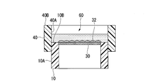
- FIG. 7 is a cross-sectional view in which the inner peripheral surface of the cap member 40 is fitted to the outer peripheral surface of the well portion 10;
- FIG. 5 shows the relationship between flow stimulation and gene expression of neural stem cells (upper) and vascular endothelial cells (lower).
- FIG. 5 shows the measurement result of the albumin output for every gel membrane.
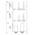
- A) shows the relationship between flow stimulation and albumin production
- B) shows the relationship between flow stimulation and albumin permeation
- C shows the relationship between flow stimulation and barrier function
- FIGS. 1 to 8 show one aspect of the present invention, does not limit the present invention, and can be arbitrarily changed within the scope of the technical idea of the present invention. Moreover, in the following drawings, in order to make each structure intelligible, the scale, the number, etc. in an actual structure and each structure may be varied.
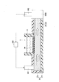
- FIG. 1 is a schematic block diagram of a perfusion system SYS having a perfusion device (artificial tissue perfusion device) 1 according to the present invention.
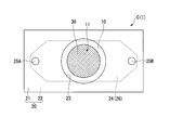
- FIG. 2 is a plan view of the perfusion device 1 as viewed from the well 10 side.
- the perfusion system SYS includes a perfusion device 1, a culture medium supply unit 100, a supply tube 101, a discharge tube 102 and a control unit 110.
- the medium supply unit 100 stores the medium and supplies the medium to the perfusion device 1.
- the supply pipe 101 is connected to the perfusion device 1 at one end.
- the supply pipe 101 introduces the culture medium supplied from the culture medium supply unit 100 into the perfusion device 1.
- the drain tube 102 drains the culture medium from the perfusion device 1.
- the adjustment unit 110 adjusts the flow rate of the culture medium in the perfusion device 1 by adjusting the introduction amount of the culture medium supplied from the culture medium supply unit 100 and introduced into the perfusion device 1.
- the adjustment unit 110 is, for example, a pump provided midway in the supply pipe 101.
- the flow rate of the culture medium in the perfusion device 1 can be controlled by adjusting the driving amount of the pump as the adjustment unit 110 and adjusting the introduction amount of the culture medium introduced into the perfusion device 1.
- the adjusting unit 110 may be configured to adjust the amount of culture medium supplied from the culture medium supplying unit 100.
- the perfusion device 1 has a co-culture system C in which cells of a plurality of types are cultured.
- the co-culture system C includes a well 10, a base 20, a gel film 30, and a cap member (pre-cultured part) 40 (see FIG. 3).
- the well portion 10 is formed in a tubular shape.
- the well portion 10 is formed in a cylindrical shape, for example, of a transparent material.
- the well has a culture space 11 inside.
- the material of the well portion 10 is not particularly limited, and, for example, polystyrene (PS), polycarbonate (PC), polyethylene terephthalate (PET), polytetrafluoroethylene (PTFE) or the like can be used.
- the base 20 has an upper base 21 and a lower base 22. As shown in FIG. 2, the upper base 21 and the lower base 22 are both formed in a rectangular shape in plan view. The upper base 21 and the lower base 22 are joined in the vertical direction, for example, by an adhesive or the like. Although it does not specifically limit as a raw material of the upper base material 21, For example, It is an elastomer and silicone rubber, PDMS (polydimethylsiloxane), etc. are mentioned.
- the material of the lower base material 22 is not particularly limited, and examples thereof include elastomers, silicone rubber, PDMS (polydimethylsiloxane), glass, and the like.
- the lower base 22 is formed in the shape of a flat plate having a rectangular shape in plan view.
- the upper base 21 has a groove 24 opened in the surface (lower surface) opposite to the lower base 22.
- the groove 24 extends in the longitudinal direction (left and right direction in FIGS. 1 and 2).
- the groove portion 24 is a recess whose periphery is surrounded by the lower surface of the upper base 21. Both end portions in the longitudinal direction of the groove portion 24 are each formed in a tapered shape at the tip. In addition.
- the groove portion 24 is not limited to a tapered shape, and may have the same width.
- the upper base 21 has a holding portion 23 which is circular in plan view and located at the center in the longitudinal direction and at the center in the lateral direction.
- the holding portion 23 penetrates the upper substrate 21 in the thickness direction (vertical direction in FIG. 1).
- the holding portion 23 is liquid-tightly fitted to the outer peripheral surface of the well portion 10, and holds the well portion 10 detachably to the upper base 21.
- the lower end surface of the well portion 10 when held by the holding portion 23 is, for example, flush with the bottom surface of the groove portion 24.
- the upper base 21 has connection holes 25A and 25B at the center in the short direction and at the outer sides in the longitudinal direction.
- the connection holes 25A, 25B respectively penetrate the upper base 21 in the thickness direction.
- the lower ends of the connection holes 25A and 25B are open in the groove 24 respectively.
- the supply pipe 101 is connected to the connection hole 25A.
- the discharge pipe 102 is connected to the connection hole 25B.
- a perfusion channel 26 which is surrounded by the groove 24 and the upper surface of the lower base 22 and extends in the longitudinal direction is formed.
- the supply pipe 101 is connected to one end side in the longitudinal direction, and the discharge pipe 102 is connected to the other end side.
- the gel film 30 is disposed on the end of the well 10 facing the perfusion channel 26 when the well 10 is held by the holder 23.
- the gel membrane 30 has a first surface 31 facing the culture space 11 of the well 10 and a second surface 32 facing the perfusion channel 26.
- the second surface 32 is, for example, flush with the lower end surface of the well 10 and the bottom of the groove 24.
- the gel membrane 30 is formed in the shape of a porous membrane.
- the gel membrane 30 contains a vitrified extracellular matrix component.
- the gel film 30 is produced, for example, by rehydration after drying and vitrification of the hydrogel.
- the gel film 30 is, for example, a collagen vitrified gel film, and is formed in a porous film shape in which collagen fibers are arranged at high density.
- Tissue-based cells are cultured after being seeded to form a tissue layer 41 on the first surface 31 of the gel membrane 30 (details will be described later).
- luminal cells are cultured after being seeded to form a planar luminal layer 42 (details will be described later).
- the width of the perfusion channel 26 is set so that the flow stimulation by the medium that perfuses the perfusion channel 26 is applied to the luminal cells seeded on the second surface 32 of the gel membrane 30.
- it is larger than the maximum diameter of
- the thickness (depth) of the perfusion channel 26 is preferably 1 mm or less.
- FIG. 3 is a cross-sectional view in which the inner peripheral surface of the cap member 40 is fitted to the outer peripheral surface of the well portion 10. Before the cap member 40 holds the well 10 on the holding portion 23 of the upper base 21 and co-cultures on the first surface 31 and the second surface 32 of the gel film 30, pre-culture is performed on the second surface 32. It is used when doing.
- the cap member 40 is formed in a cylindrical shape, for example, with the same material as the upper base 21 and the lower base 22.
- the inner circumferential surface 40A of the cap member 40 is fitted to at least the upper side of the outer circumferential surface 10A of the well portion 10 with the gel film 30 directed upward.
- the upper end surface 40B of the cap member 40 is provided on the upper side than the end surface 10B of the well 10 on the side on which the gel film 30 is provided.
- a preculture space 60 surrounded by the inner circumferential surface 40A of the cap member 40, the end face 10B of the well 10 and the second surface 32 of the gel film 30 is formed. ing.
- luminal cells (details will be described later) are seeded on the second surface 32 of the gel film 30, and the medium is supplied. By doing so, a planar luminal layer can be pre-incubated on the second surface 32 of the gel membrane 30.
- the well 10 in which the luminal layer is pre-incubated on the second surface 32 of the gel film 30 is fitted to the holding portion 23 of the upper base 21 after the cap member 40 is detached. Match.
- the luminal layer pre-incubated on the second surface 32 of the gel membrane 30 is disposed facing the groove 24 (perfusion channel 26).
- the tissue layer 41 using the tissue-based cells formed on the first surface 31 of the gel film 30 is not particularly limited.
- the tissue layer 41 for example, nerve tissue using nerve cells, muscle tissue using skeletal muscle cells or cardiomyocytes, liver tissue using liver cells, pancreas tissue using pancreas cells, pancreas cells using dermal cells are used. And dermal tissue.
- the tissue layer is not limited to a single layer, and a plurality of tissue layers may be stacked.
- the tissue layer may be a three-dimensional tissue layer in which a plurality of cells are mixed.
- a variety of media and extracellular matrix components for culturing tissue-based cells can be selected.
- Examples of the luminal layer 42 using luminal cells formed on the second surface 32 of the gel film 30 include a blood vessel layer using vascular cells, and a tubular layer using tubular cells.
- vascular cells vascular endothelial cells are preferable.
- examples of vascular endothelial cells include cells derived from mammals such as human, mouse, rat and the like, and human-derived vascular endothelial cells are preferable.
- human-derived vascular endothelial cells human umbilical vein endothelial cells (HUVEC), human umbilical artery endothelial cells (HUAEC), human coronary artery endothelial cells (Human Coronary Artery Endothelial Cells: HCAEC), Human Saphenous Vein Endothelial Cells (HSaVEC), Human Pulmonary Artery Endothelial Cells (HPAEC), Human Aortic Endothelial Cells (Human Aortic Endothelial Cells: HAoEC), Human skin microvessels Endothelial cells (Human Dermal Microvascular Endothelial Cells: HDMEC), Human Dermal Blood Endothelial Cells (Human Dermal Blood Endothelial Cells: HDBEC), Human Dermal Lymphatic Endothelial Cells (Human Dermal Lymphatic Endothelial Cells: HDLEC), Human Lung Microvascular Endothelial Cells (Human Dermal Lymph
- the upper base 21 and the lower base 22 described above are connected in the vertical direction using an adhesive or the like.
- a longitudinally extending perfusion channel 26 is formed.
- the outer peripheral surface 10A of the well 10 is fitted and held in the holding portion 23 of the upper base 21 and the second surface 32 of the gel film 30 is faced to the perfusion channel 26, whereby the perfusion device 1 is Manufactured.
- an adhesive may be applied between the outer peripheral surface 10 A and the holding portion 23 and fixed in a liquid tight manner.
- the adhesive it is possible to use the photocurable (UV curable) PDMS or the like.
- the supply pipe 101 connected to the culture medium supply unit 100 through the adjustment unit 110 is connected to the connection hole 25A, and the discharge pipe 102 is connected to the connection hole 25B, whereby the perfusion system SYS is manufactured.
- the co-culture method using the perfusion system SYS which has the said perfusion device 1 is demonstrated.
- the inner peripheral surface 40A of the cap member 40 is fitted to the outer peripheral surface 10A of the well portion 10 with the gel film 30 directed upward.
- the second surface 32 of the gel membrane 30 is coated with an extracellular matrix component, and the luminal cells are seeded, and the medium is introduced into the preculture space 60 to preculture.
- the cap member 40 When luminal cells adhere to the second surface 32 of the gel membrane 30 by pre-culture, the cap member 40 is removed from the well 10. Next, the well portion 10 from which the cap member 40 is removed is vertically inverted, and as shown in FIG. 1, the gel film 30 is inserted into the holding portion 23 of the upper base material 21 with the gel film 30 facing downward Let Thereby, the second surface 32 of the gel membrane 30 to which the luminal cells adhere is exposed to the perfusion channel 26.
- tissue-based cells are seeded on the first surface 31 of the gel film 30 exposed at the bottom of the culture space 11 of the well section 10, and a culture medium is introduced into the culture space 11 to culture the tissue-based cells.
- the well unit 10 may be held by the holding unit 23.
- the culture medium in an amount supplied from the culture medium supply unit 100 and adjusted by the adjustment unit 110 is introduced from the supply tube 101 into the perfusion channel 26.
- the perfusion channel 26 gradually extends in width from the longitudinal end to the center side, the culture medium introduced from the supply tube 101 into the perfusion channel 26 smoothly fills the perfusion channel 26 and perfuses. Media perfusing the perfusion channel 26 is drained from the drain tube 102.
- the tissue-based cells seeded on the first surface 31 of the gel membrane 30 are cultured by the medium introduced into the culture space 11, and the luminal cells adhered to the second surface 32 of the gel membrane 30 are: It is cultured by the culture medium introduced into the perfusion channel 26 and perfused. That is, in the perfusion device 1, the tissue-based cells and the luminal cells are co-cultured in a state where flow stimulation is applied, and it is possible to reproduce, for example, the actual environment such as the periphery of blood vessels.
- the medium is not perfused in the perfusion channel 26 without being perfused when the medium is co-cultured in a state where the culture is perfused in the perfusion channel 26 It is also possible to compare and evaluate the case of co-culturing in the state. Further, even in the case where co-culture is performed in a state where the culture medium is perfused in the perfusion channel 26, the adjusting unit 110 is adjusted to make the introduction amount of the culture medium into the perfusion channel 26 different, and the flow rate of the culture medium in the perfusion channel 26 It is also possible to compare and evaluate different cases.
- the perfusion channel 26, the holding unit 23, and the well unit 10 in the perfusion device 1 in a state in which a plurality of each are separated from one another.
- the supply channel 101 can be branched into a plurality of portions so that the culture medium can be individually introduced into the plurality of perfusion channels 26, and the above-described perfusion channel 26 can be provided by providing the adjusting unit 110 in each of the branched supply channels 101. It is also possible to simultaneously compare / evaluate the presence / absence of perfusion of the medium in the case, the case where the flow rate of the medium is different, etc.
- the present invention relates to a method for evaluating the irritation due to contact of a drug in an artificial tissue manufactured using the perfusion device 1 according to the present invention.
- the drug in the present invention includes drugs such as pharmaceuticals, cosmetics, quasi-drugs and the like.
- evaluation method of the present invention for example, evaluation of a drug can be performed in an environment near the actual lumen layer and the like as compared with the conventional method.
- the evaluation method of the present invention is extremely useful, for example, in the evaluation of the dynamics of drugs of various molecular weights in the creation (screening) of new drugs and the like, and in the evaluation in the development of cosmetics and quasi drugs.
- the evaluation method of the present invention can be performed, for example, by bringing a drug into contact with an artificial tissue, and measuring the response of the artificial tissue to stimulation due to the contact of the drug, or the permeability of the drug to the artificial tissue.
- the response can be measured, for example, by measuring the percutaneous and transendothelial electrical resistance (barrier function).
- the drug is a substance to be evaluated, and examples thereof include inorganic compounds and organic compounds.
- the drug is applied to the tissue layer 41, the uptake of the drug into the perfusion channel 26 is evaluated, or the drug is introduced into the medium introduced into the perfusion channel 26.
- the agent can be mixed to evaluate the diffusion / permeation of the drug from the perfusion channel 26 to the tissue layer 41.
- a vitrigel membrane is used as the gel membrane 30.
- Vascular endothelial cells (HUVECs) were attached to and cultured on the second surface 32 on the perfusion channel 26 side of Vitrigel membrane, and neural stem cells (NSCs) were attached to and cultured on the first surface 31 on the culture space 11 side of Vitrigel membrane. The one was made.
- the expression of each gene was examined by real-time PCR.
- FIG. 4 is a view showing the relationship between flow stimulation and gene expression of neural stem cells (upper row) and vascular endothelial cells (lower row).
- a flow stimulus-dependent increase in expression of CYP1B1 or DECR1 in which gene expression was reported to increase in a flow stimulus-dependent manner was confirmed.
- the flow stimulation-dependent increase in expression of the NSC marker was confirmed, and thus the vascular endothelial cells that received the flow stimulation are stem cells of neural stem cells ( It has been suggested to enhance stemness.
- vascular endothelial cells are attached and cultured on the second surface 32 on the perfusion channel 26 side of the gel membrane 30, and the first surface 31 on the culture space 11 side of the gel membrane 30 , Hepatic carcinoma cells (HepG2) were adherently cultured.
- Hepatic carcinoma cells HepG2
- the gel film 30 three types of PET film, a collagen-coated PET film, and a vitrigel film were used. Based on the finding that albumin was released from hepatocytes and released into blood vessels, the amount of albumin produced in the culture space (upper part) and perfusion channel (lower part) in the perfusion culture was measured for each gel membrane 30.
- FIG. 5 shows the measurement results of albumin production for each gel membrane. As shown in FIG. 5, it was confirmed that the amount of albumin production was increased by the culture on the vitrigel membrane. It was also confirmed that albumin permeated through the membrane and diffused to the lower part.
- the amount of production of albumin in stationary culture and perfusion culture the amount of albumin permeation from the upper part (culture space 11) to the lower part (perfusion flow path 26), TEER (transepithelial electricity) Resistance) was measured.
- FIG. 6 (A) and FIG. 6 (B) it was confirmed that the albumin production amount and the permeation amount are increased in the perfusion culture.
- FIG. 6 (C) it was confirmed that the barrier function is rising by perfusion.
- FIG. 7 is a view showing fluorescence images in response to flow stimulation of cells cultured in stationary culture and in perfusion culture. As shown in FIG. 7, under perfusion culture, the orientation of cells by flow stimulation was confirmed.
- luminal cells are disposed on the side facing the perfusion channel 26 of the gel membrane 30, and tissue cells are disposed on the side facing the culture space 11 of the gel membrane 30,
- the above-mentioned embodiment illustrated the composition which uses cylindrical cap member 40 as a preculture part, it is not limited to this composition.
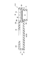
- the base 20 has an inner circumferential surface 28A fitted with the outer circumferential surface 10A of the well 10 at a position not interfering with the perfusion channel 26 in the base 20
- the through-hole 28 which penetrates may be provided.
- the well portion 10 with the gel film 30 directed upward is inserted from the lower opening of the through hole 28 so that a space is formed above the gel film 30 Match.
- a preculture space 60 surrounded by the inner circumferential surface 28A, the end surface 10B of the well 10 and the second surface 32 of the gel film 30 is formed.
- the base material 20 and the cap member 40 do not need to be separately circulated and stored. It becomes possible to facilitate storage, distribution and management.
- the configuration in which the well portion 10 is cylindrical is exemplified, but the configuration is not limited to this as long as it is cylindrical, and a cylinder such as an angular cylinder having a polygonal cross section or an elliptical cross section It may be a letter.
- a flange portion may be provided which protrudes outward in the radial direction from the outer peripheral surface 10A.
- the gel film is engaged by the flange portion engaging the upper surface of the first base material 21 as the position of the flange portion in the vertical direction
- the position of the 30 second surface 32 that is, the position of the luminal cell relative to the perfusion channel 26
- the position of the second surface 32 of the gel film 30 is defined by the flange portion engaging with the lower surface of the second base material 22.
- a medium storage portion 100A at least a part of which communicates with the upstream side of the perfusion channel 26 is provided integrally with the base material 20 as a medium storage portion for storing the culture medium.
- the supply pipe 101 is not connected to the base material 20 and is connected to the culture medium storage unit 100A.
- the adjustment unit 110 By driving the adjustment unit 110, the culture medium stored in the culture medium storage unit 100A is introduced into the perfusion channel 26. Then, the culture medium perfused through the perfusion channel 26 returns from the discharge pipe 102 to the adjustment unit 110 and is supplied again to the culture medium storage unit 100A via the supply pipe 101.
- the culture medium storage unit 100A at least a part of which communicates with the downstream side of the perfusion channel 26 It may be configured integrally.
- the supply pipe 101 is connected to the base material 20, and the discharge pipe 102 is not connected to the base material 20 and is connected to the culture medium reservoir 100A.
- the culture medium stored in the culture medium storage unit 100A returns to the adjustment unit 110 via the pipe 102 and is introduced to the perfusion channel 26 via the supply pipe 101. Then, the culture medium perfused through the perfusion channel 26 is stored again in the culture medium storage unit 100A.
- the usability of the perfusion device 1 is improved.
- a configuration in which a plurality of well portions 10 are provided in series with respect to one perfusion channel 26 it is also possible to culture a tissue layer in a plurality of wells 10 in line with the flow direction of the liquid flowing through the luminal layer in the living body.
- the hepatoma cells described in the above-mentioned Experimental Example 2 are cultured in the culture space of the well 10 disposed upstream of the perfusion channel 26 and the culture of the well 10 disposed downstream of the perfusion channel 26
- the pancreatic tissue may be cultured in space.
- analysis and evaluation such as the influence of albumin diffused in the perfusion channel 26 through the membrane after being produced in the upstream well 10 on the pancreatic tissue cultured in the downstream well 10 is carried out. It will be possible.
- the present invention can be applied to artificial tissue perfusion devices and drug evaluation methods using artificial tissue perfusion devices.
Landscapes
- Health & Medical Sciences (AREA)
- Engineering & Computer Science (AREA)
- Life Sciences & Earth Sciences (AREA)
- Chemical & Material Sciences (AREA)
- Biomedical Technology (AREA)
- Bioinformatics & Cheminformatics (AREA)
- Wood Science & Technology (AREA)
- Zoology (AREA)
- Organic Chemistry (AREA)
- Genetics & Genomics (AREA)
- Biotechnology (AREA)
- Microbiology (AREA)
- Biochemistry (AREA)
- General Health & Medical Sciences (AREA)
- General Engineering & Computer Science (AREA)
- Sustainable Development (AREA)
- Cell Biology (AREA)
- Immunology (AREA)
- Molecular Biology (AREA)
- Clinical Laboratory Science (AREA)
- Urology & Nephrology (AREA)
- Hematology (AREA)
- General Physics & Mathematics (AREA)
- Neurosurgery (AREA)
- Medicinal Chemistry (AREA)
- Physics & Mathematics (AREA)
- Analytical Chemistry (AREA)
- Tropical Medicine & Parasitology (AREA)
- Pathology (AREA)
- Vascular Medicine (AREA)
- Toxicology (AREA)
- Neurology (AREA)
- Food Science & Technology (AREA)
- General Chemical & Material Sciences (AREA)
- Physiology (AREA)
- Dispersion Chemistry (AREA)
- Apparatus Associated With Microorganisms And Enzymes (AREA)
- Measuring Or Testing Involving Enzymes Or Micro-Organisms (AREA)
Abstract
Le but de la présente invention est de fournir un dispositif de perfusion de tissu artificiel permettant une analyse de haute précision d'interactions entre une couche de cellules vasculaires et une couche de cellules parenchymateuses. La présente invention comprend un système de co-culture (C) dans lequel une pluralité de types de cellules sont cultivées. Le système de co-culture comprend : une partie de puits cylindrique (10) ayant un espace de culture (11) à l'intérieur de celle-ci ; un matériau de base (20) qui a un canal de perfusion (26) qui s'étend dans une direction prédéterminée et qui peut être perfusé avec le milieu, et qui a une section de retenue (23) qui s'ouvre dans le canal de perfusion et retient de manière amovible la partie de puits ; et un film de gel membraneux poreux (30) disposé sur une section d'extrémité de la partie de puits faisant face au canal de perfusion lorsque la partie de puits est retenue par la section de retenue. Dans le film de gel, des cellules de type tissu sont cultivées sur un côté de surface opposé à l'espace de culture, et des cellules de type lumen sont cultivées sur un côté de surface opposé au canal de perfusion.
Priority Applications (3)
| Application Number | Priority Date | Filing Date | Title |
|---|---|---|---|
| US16/958,013 US20210371792A1 (en) | 2017-12-28 | 2018-12-28 | Artificial tissue perfusion device and method of drug assessment using artificial tissue |
| JP2019562508A JP7280618B2 (ja) | 2017-12-28 | 2018-12-28 | 人工組織灌流デバイス、人工組織を用いた薬剤評価方法 |
| EP18895199.0A EP3733831A4 (fr) | 2017-12-28 | 2018-12-28 | Dispositif de perfusion de tissu artificiel et méthode d'évaluation de médicament utilisant un tissu artificiel |
Applications Claiming Priority (2)
| Application Number | Priority Date | Filing Date | Title |
|---|---|---|---|
| US201762611338P | 2017-12-28 | 2017-12-28 | |
| US62/611,338 | 2017-12-28 |
Publications (1)
| Publication Number | Publication Date |
|---|---|
| WO2019132008A1 true WO2019132008A1 (fr) | 2019-07-04 |
Family
ID=67067686
Family Applications (1)
| Application Number | Title | Priority Date | Filing Date |
|---|---|---|---|
| PCT/JP2018/048493 Ceased WO2019132008A1 (fr) | 2017-12-28 | 2018-12-28 | Dispositif de perfusion de tissu artificiel et méthode d'évaluation de médicament utilisant un tissu artificiel |
Country Status (4)
| Country | Link |
|---|---|
| US (1) | US20210371792A1 (fr) |
| EP (1) | EP3733831A4 (fr) |
| JP (1) | JP7280618B2 (fr) |
| WO (1) | WO2019132008A1 (fr) |
Cited By (1)
| Publication number | Priority date | Publication date | Assignee | Title |
|---|---|---|---|---|
| JP2024524769A (ja) * | 2021-06-28 | 2024-07-05 | マットテック コーポレーション | 組織モデル培養のための積み重ね式プレート |
Families Citing this family (3)
| Publication number | Priority date | Publication date | Assignee | Title |
|---|---|---|---|---|
| WO2019033096A1 (fr) * | 2017-08-11 | 2019-02-14 | The Trustees Of The University Of Pennsylvania | Inserts de membrane dérivés de matrice extracellulaire native pour des organes sur puces, des microdispositifs microfluidiques multicouches et des systèmes pour culture cellulaire tridimensionnelle |
| CN114317272B (zh) * | 2021-12-07 | 2024-01-12 | 上海前瞻创新研究院有限公司 | 一种多细胞共培养的培养装置及细胞培养方法 |
| WO2025255123A1 (fr) * | 2024-06-03 | 2025-12-11 | The Charles Stark Draper Laboratory, Inc. | Système microphysiologique pour modélisation neuronale |
Citations (2)
| Publication number | Priority date | Publication date | Assignee | Title |
|---|---|---|---|---|
| WO2016069892A1 (fr) * | 2014-10-29 | 2016-05-06 | Corning Incorporated | Dispositifs et procédés pour la génération et la culture d'agrégats cellulaires 3d |
| WO2017126532A1 (fr) * | 2016-01-20 | 2017-07-27 | 国立大学法人東京大学 | Tissu tridimensionnel artificiel et son procédé de fabrication, dispositif de perfusion pour le tissu tridimensionnel artificiel, et procédé d'évaluation de médicaments utilisant le tissu tridimensionnel artificiel |
Family Cites Families (3)
| Publication number | Priority date | Publication date | Assignee | Title |
|---|---|---|---|---|
| US20130280807A1 (en) * | 2010-11-12 | 2013-10-24 | Toshiaki Takezawa | Cell culture chamber, method for producing same, tissue model using cell culture chamber, and method for producing same |
| EP2788119A1 (fr) * | 2011-12-05 | 2014-10-15 | Research Triangle Institute | Modèle humain des voies respiratoires comprenant de multiples passages fluidiques |
| CA3006063A1 (fr) * | 2015-11-24 | 2017-06-01 | Vanderbilt University | Bioreacteurs micro-fluidiques empilables et superposes a compartiments multiples et leurs applications |
-
2018
- 2018-12-28 EP EP18895199.0A patent/EP3733831A4/fr not_active Withdrawn
- 2018-12-28 WO PCT/JP2018/048493 patent/WO2019132008A1/fr not_active Ceased
- 2018-12-28 JP JP2019562508A patent/JP7280618B2/ja active Active
- 2018-12-28 US US16/958,013 patent/US20210371792A1/en not_active Abandoned
Patent Citations (2)
| Publication number | Priority date | Publication date | Assignee | Title |
|---|---|---|---|---|
| WO2016069892A1 (fr) * | 2014-10-29 | 2016-05-06 | Corning Incorporated | Dispositifs et procédés pour la génération et la culture d'agrégats cellulaires 3d |
| WO2017126532A1 (fr) * | 2016-01-20 | 2017-07-27 | 国立大学法人東京大学 | Tissu tridimensionnel artificiel et son procédé de fabrication, dispositif de perfusion pour le tissu tridimensionnel artificiel, et procédé d'évaluation de médicaments utilisant le tissu tridimensionnel artificiel |
Non-Patent Citations (3)
| Title |
|---|
| LAB CHIP, vol. 13, 2013, pages 3538 - 3547 |
| SASAKI, NAOKI ET AL.: "Analytical Applications of Microfluidic Vascular Models", BUNSEKI KAGAKU, vol. 65, 2016, pages 241 - 247 , p. 242-244, XP055624030 * |
| See also references of EP3733831A4 |
Cited By (1)
| Publication number | Priority date | Publication date | Assignee | Title |
|---|---|---|---|---|
| JP2024524769A (ja) * | 2021-06-28 | 2024-07-05 | マットテック コーポレーション | 組織モデル培養のための積み重ね式プレート |
Also Published As
| Publication number | Publication date |
|---|---|
| JP7280618B2 (ja) | 2023-05-24 |
| JPWO2019132008A1 (ja) | 2020-12-10 |
| EP3733831A4 (fr) | 2021-09-29 |
| US20210371792A1 (en) | 2021-12-02 |
| EP3733831A1 (fr) | 2020-11-04 |
Similar Documents
| Publication | Publication Date | Title |
|---|---|---|
| Chaicharoenaudomrung et al. | Three-dimensional cell culture systems as an in vitro platform for cancer and stem cell modeling | |
| Tehranirokh et al. | Microfluidic devices for cell cultivation and proliferation | |
| Kaisar et al. | New experimental models of the blood-brain barrier for CNS drug discovery | |
| Xie et al. | h-FIBER: microfluidic topographical hollow fiber for studies of glomerular filtration barrier | |
| JP7280618B2 (ja) | 人工組織灌流デバイス、人工組織を用いた薬剤評価方法 | |
| JP6041872B2 (ja) | バイオ人工腎臓 | |
| Park et al. | 3D miniaturization of human organs for drug discovery | |
| Berdichevsky et al. | Microfluidics and multielectrode array-compatible organotypic slice culture method | |
| US20110082563A1 (en) | Microscale multiple-fluid-stream bioreactor for cell culture | |
| Musafargani et al. | Blood brain barrier: A tissue engineered microfluidic chip | |
| NL2011895C2 (en) | Fluidic device and perfusion system for in vitro tissue reconstruction. | |
| US10624991B2 (en) | Three-dimensional artificial tissue, method for producing the same, three-dimensional artificial tissue perfusion device, and drug evaluation method using three-dimensional artificial tissue | |
| Illa et al. | A novel modular bioreactor to in vitro study the hepatic sinusoid | |
| Paz et al. | Challenges and opportunities for tissue-engineering polarized epithelium | |
| US10030219B2 (en) | Neurovascular unit(NVU)-on-a-chip and method of fabricating the same | |
| US11459542B2 (en) | Micro-physiological organoid culture | |
| KR20170087837A (ko) | 신경혈관단위-온-칩 및 그 칩의 제조방법 | |
| Kreutzer et al. | Cell culture chamber with gas supply for prolonged recording of human neuronal cells on microelectrode array | |
| Hosseinkhani et al. | Engineering of the embryonic and adult stem cell niches | |
| CN117813186A (zh) | 一种形成微流体凝胶结构的方法和装置 | |
| Marimuthu et al. | Microfluidic cell coculture methods for understanding cell biology, analyzing bio/pharmaceuticals, and developing tissue constructs | |
| Wang et al. | Current progress of in vitro vascular models on microfluidic chips | |
| US10837002B2 (en) | Artificial peritoneal tissue and method for producing same | |
| Mantecón-Oria et al. | On the quest of reliable 3D dynamic in vitro blood-brain barrier models using polymer hollow fiber membranes: Pitfalls, progress, and future perspectives | |
| EP3987014A1 (fr) | Dispositif de culture |
Legal Events
| Date | Code | Title | Description |
|---|---|---|---|
| 121 | Ep: the epo has been informed by wipo that ep was designated in this application |
Ref document number: 18895199 Country of ref document: EP Kind code of ref document: A1 |
|
| ENP | Entry into the national phase |
Ref document number: 2019562508 Country of ref document: JP Kind code of ref document: A |
|
| NENP | Non-entry into the national phase |
Ref country code: DE |
|
| ENP | Entry into the national phase |
Ref document number: 2018895199 Country of ref document: EP Effective date: 20200728 |