WO2019144568A1 - Procédé de criblage d'anticorps monoclonal à efficacité élevée contre des cellules cancéreuses - Google Patents
Procédé de criblage d'anticorps monoclonal à efficacité élevée contre des cellules cancéreuses Download PDFInfo
- Publication number
- WO2019144568A1 WO2019144568A1 PCT/CN2018/093245 CN2018093245W WO2019144568A1 WO 2019144568 A1 WO2019144568 A1 WO 2019144568A1 CN 2018093245 W CN2018093245 W CN 2018093245W WO 2019144568 A1 WO2019144568 A1 WO 2019144568A1
- Authority
- WO
- WIPO (PCT)
- Prior art keywords
- cells
- cell
- cancer
- monoclonal antibody
- antibody
- Prior art date
- Legal status (The legal status is an assumption and is not a legal conclusion. Google has not performed a legal analysis and makes no representation as to the accuracy of the status listed.)
- Ceased
Links
Images




Classifications
-
- A—HUMAN NECESSITIES
- A61—MEDICAL OR VETERINARY SCIENCE; HYGIENE
- A61K—PREPARATIONS FOR MEDICAL, DENTAL OR TOILETRY PURPOSES
- A61K39/00—Medicinal preparations containing antigens or antibodies
- A61K39/395—Antibodies; Immunoglobulins; Immune serum, e.g. antilymphocytic serum
-
- C—CHEMISTRY; METALLURGY
- C07—ORGANIC CHEMISTRY
- C07K—PEPTIDES
- C07K16/00—Immunoglobulins [IG], e.g. monoclonal or polyclonal antibodies
- C07K16/18—Immunoglobulins [IG], e.g. monoclonal or polyclonal antibodies against material from animals or humans
- C07K16/28—Immunoglobulins [IG], e.g. monoclonal or polyclonal antibodies against material from animals or humans against receptors, cell surface antigens or cell surface determinants
- C07K16/30—Immunoglobulins [IG], e.g. monoclonal or polyclonal antibodies against material from animals or humans against receptors, cell surface antigens or cell surface determinants from tumour cells
-
- A—HUMAN NECESSITIES
- A61—MEDICAL OR VETERINARY SCIENCE; HYGIENE
- A61P—SPECIFIC THERAPEUTIC ACTIVITY OF CHEMICAL COMPOUNDS OR MEDICINAL PREPARATIONS
- A61P35/00—Antineoplastic agents
Definitions
- the invention relates to the technical field of molecular biological antigen antibody screening, in particular to a screening method for high-efficiency anti-cancer monoclonal antibody.
- cancer cells are malignant proliferating cells that escape the body's immune surveillance. They "camouflage" into normal cells as much as possible to avoid being recognized and cleared by the body's immune system. Therefore, it is very difficult for the immune system to recognize cancer cells.
- tumor antibody therapy the most critical step is to obtain cancer-specific monoclonal antibodies.
- mice or other animals directly with certain cancer cells or their extracts, and obtain monoclonal antibodies against the cancer cells, and then negatively screen these antibodies by using control cells (such as corresponding normal cells). , finally obtaining specific antibodies;
- a cancer-specific protein especially a membrane protein
- the protein is used to deimmunize and obtain a monoclonal antibody.
- both methods have many problems.
- cancer cells escape the cells monitored by the immune system, so although on a molecular scale, cancer cells do differ a lot from normal cells, this difference is not enough to activate the body's immune system. Therefore, if the antibody is obtained by the first method, it is often necessary to screen a cancer cell-specific antibody from thousands or even tens of thousands of clones. Because of this defect, this method has been basically eliminated.
- the second method is also the current mainstream method, and some antibodies have been obtained and obtained by the prior art.
- cancer cells that are not a tumor must express this antigen, and not all cancer cells express a specific antigen even in a tumor tissue. Therefore, this antibody can only recognize and kill cancer cells expressing this antigen, but cancer cells that do not express this antigen are therefore screened out and malignantly hyperplasia. This high variability of cancer cells is also one of the reasons why cancer is difficult to treat.
- the technical problem to be solved by the present invention is to provide a screening method for highly effective anti-cancer monoclonal antibodies.
- the method of the present invention obtained monoclonal antibodies against 11 anti-hepatocarcinoma cell lines HepG2. These mAbs specifically recognize HepG2 without recognizing or substantially recognizing normal human hepatocyte HH cells. At the same time, these monoclonal antibodies are specifically recognized for HepG2 as compared to other tissue-derived cancer cells.
- the present invention provides a screening method for highly effective anti-cancer monoclonal antibodies, comprising the steps of directly removing cancer cells or cancerous tissues and normal in vivo by using antibodies against corresponding normal cells or normal tissues.
- the method does not perform large-scale screening of antigens in vitro.
- the method can further include:
- step B treating the animal with the antiserum obtained in step A or the purified polyclonal antibody;
- step B treated animals with corresponding cancer cells or cancerous tissues
- the step A further comprises: immunizing the animal with a whole cell of a normal cell or a normal tissue, a whole cell lysate or an extracted membrane protein as an antigen to obtain an anti-normal cell serum.
- step B further comprises treating the new unimmunized animal with the antiserum obtained in step A or the purified polyclonal antibody.
- the step D further comprises: separating the spleen cells of the animal treated in the step C, and fusing with the myeloma cells to obtain a monoclonal antibody cell line.
- the step A may further comprise: immunizing the animal with whole cells, whole cell lysate or extracted membrane protein of normal cells or normal tissues as an antigen, immunizing 1 to 5 times; collecting blood after collecting blood for 8 to 12 days after the last immunization , obtain anti-normal cell serum.
- the present invention also provides a method for screening a highly effective anti-cancer monoclonal antibody according to any one of the preceding claims for the preparation of an antitumor drug.
- the present invention further provides the use of a screening method for a highly effective anti-cancer cell monoclonal antibody according to any of the preceding claims for cell-specific recognition.
- the present invention further provides a method for screening a highly effective anti-cancer cell monoclonal antibody according to any one of the preceding claims in combination with a humanization technique or a drug coupling technique.
- the present invention provides a method for obtaining cancer cell-specific antibodies of unknown specific antigen, comprising the following steps:
- step B treating the animal with the antiserum obtained in step A or the purified polyclonal antibody;
- step B treated animals with corresponding cancer cells or cancerous tissues
- the isolated B lymphocytes are fused with SP 2/0 cells to obtain a monoclonal antibody cell line;
- a monoclonal antibody is prepared using the monoclonal antibody cell line.
- the cancer cells are preferably HepG2 cells.
- the normal cell is preferably an HH cell.
- the step A may further comprise: immunizing the mouse with 80-120 million cells, the immunization interval is 10-20 days, and immunizing 1 to 5 times; after the last immunization 8-12 days, the blood is collected and serum is collected.
- the step B further comprises: treating the new unimmunized animal with the antiserum obtained in step A or the purified polyclonal antibody; treating at intervals of 10 to 20 days, and treating 1 to 5 times.
- the present invention further provides a method for obtaining a cancer cell-specific antibody of an unknown specific antigen, comprising the steps of directly removing cancer cells or cancer cells in vivo by using antibodies against the corresponding normal cells or normal tissues.
- Tissues are antigens shared with normal or normal tissues.
- the method does not allow for large-scale screening of antigens in vitro.
- the present invention also provides an antibody prepared by the method of obtaining an antibody specific for an unknown specific antigen cancer cell according to any of the preceding claims.
- the present invention further provides an application of the aforementioned antibody in the preparation of an antitumor drug.
- the present invention further provides an application of the aforementioned antibody for finding a cell-specific antigen.
- the present invention provides a simple method for finding a cell-specific antigen, comprising the following steps:
- step B treating the animal with the antiserum obtained in step A or the purified polyclonal antibody;
- step B treated animals with corresponding cancer cells or cancerous tissues
- the isolated B cells are fused with the myeloma cells to obtain a monoclonal antibody cell strain; and the monoclonal antibody cell strain is used to prepare a monoclonal antibody;
- the monoclonal antibody obtained in step E affinity-purifies the cell lysate of the cancer cell to obtain an antigenic protein bound thereto.
- the cancer cells are preferably liver cancer cells.
- the method for obtaining a cancer cell-specific antibody of an unknown specific antigen preferably further comprises:
- step F Mass spectrometric analysis of the antigenic protein obtained in step F.
- the step A may further include:
- the sub-step A1 may further include:
- the culture solution is aspirated; the residual medium is washed away with PBS, and then the cells are treated with PBS/8-12 mM EDTA, the cells are detached and finally blown to collect the cells.
- the sub-step A2 may further include:
- the sub-step A3 may further include:
- the step C preferably further comprises:
- B cells are obtained after immunization.
- the sub-step B1 further comprises: when the cancer cell is cultured and expanded to a density of about 78-82%, the culture solution is aspirated; the residual medium is washed away with PBS, and then the cells are treated with PBS/7-13 mM EDTA. , the cells are shed and eventually blown to collect the cells.
- the sub-step B2 preferably further comprises:
- the 11 antibodies screened by the present invention were directly screened by HepG2 cells, that is, without negative screening by HH cell or other cells. With the new method of the invention, no subsequent negative screening process is used or substantially eliminated. This undoubtedly greatly improves the efficiency of screening.
- the method of the present invention does not require immunization with a known antigen, and is particularly suitable for obtaining a specific antibody against cancer cells of unknown specific antigen.
- the method of the present invention provides a simple method for finding a cell-specific antigen, and the 11 monoclonal antibodies can be used to affinity-purify the cell lysate of HepG2, so that the present invention obtains a protein which can bind to them. Then by mass spectrometry or other methods to analyze these proteins, you can know what these proteins are, which is convenient for further research.
- FIG. 1 is a schematic diagram of removing a common antigen from a cancer cell and a corresponding normal cell in vivo and obtaining an anti-cancer monoclonal antibody according to an embodiment of the present invention
- FIG. 2 is a comparison diagram of the recognition ability of the anti-HH cell serum to HH cell and HepG2 according to an embodiment of the present invention
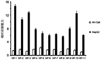
- 3 is a bar graph of specific recognition ability of 11 monoclonal antibodies according to an embodiment of the present invention.
- FIG. 5 is a schematic diagram showing the in vitro removal of a common antigen from a cancer cell and a corresponding normal cell and finally obtaining an anti-cancer monoclonal antibody according to an embodiment of the present invention
- Figure 6 is a bar graph of the ability of a monoclonal antibody to specifically recognize HepG2 obtained by an in vitro method according to an embodiment of the present invention.
- the acquisition of monoclonal antibodies specific for anti-cancer cells has been a key factor in the immunotherapy of numerous tumors. But unfortunately, so far, there is no way to efficiently screen the antibody.
- the present invention provides a method for efficiently obtaining an anti-cancer monoclonal antibody.
- the core design idea of the method of the present invention is to directly remove the antigen shared by the cancer cells and the normal cells in the body by using the anti-corresponding normal cell antibody, instead of performing large-scale screening in vitro, the present invention refers to this method as an antibody. Filter".
- hepatoma cell HepG2 was used as a model example, and 11 monoclonal cell lines against HepG2 were obtained.
- FIG. 1 it is a schematic diagram of removing the common antigen of cancer cells and corresponding normal cells in vivo and obtaining anti-cancer monoclonal antibody according to an embodiment of the present invention.
- the steps are as follows: 1. immunize the mouse with normal cells; 2, collect anti-normal cell serum; 3: inject antiserum into another mouse; 4, simultaneously immunize the mouse with the corresponding cancer cells; 5, collect The mouse spleen cells; 6, cell fusion; 7, screening monoclonal antibody cell lines against the cancer cells.
- the novel method of the present invention obtained 11 monoclonal antibodies against HepG2. These mAbs specifically recognize HepG2 without recognizing or substantially recognizing HH cells. At the same time, these monoclonal antibodies are specifically recognized for HepG2 as compared to other tissue-derived cancer cells.
- the present invention immunizes a mouse with a whole cell of a normal cell (e.g., HH cell), or a whole cell lysate or a membrane protein extracted as an antigen, so that the present invention obtains an anti-HH cell serum.
- the present invention then injects the antiserum or purified polyclonal antibody into a new unimmunized mouse while simultaneously immunizing the mouse with the corresponding cancer cells (e.g., HepG2). Since the mouse has an antibody against HH cell in this time, the antigen shared with HH cell in HepG2 cells is recognized by the anti-HH cell antibody and eventually phagocytosed by macrophages or other phagocytic cells.
- the remaining HepG2 antigen is the specific antigen of the cell. These antigens activate the humoral immune system of the mouse and cause proliferation and differentiation of the corresponding B lymphocytes. Finally, the present invention separates the mouse spleen cells (B lymphocytes account for a large proportion) and fuses with SP 2/0 cells to finally obtain a monoclonal antibody cell strain. As described above, since the injected anti-HH cell serum has cleared the antigen shared by HepG2 and HH cells, this part of the antigen does not cause the body's immune response and there is no corresponding differentiation of B lymphocytes, so the present invention is finally obtained.
- the monoclonal antibody cell line is only anti-HepG2-specific antigen.
- the method of the present invention which is a method of "antibody filter" is to remove a common antigen from a normal cell and a cancer cell by a serum or an antibody against a normal cell, and finally obtain a cancer cell-specific antibody.
- FIG. 2 it is a comparison chart of the recognition ability of the anti-HH cell serum to HH cell and HepG2 according to an embodiment of the present invention.
- the results of this figure show that anti-HH cell serum has the same recognition ability for HH cell and HepG2.
- mice were immunized with paraformaldehyde-immobilized HH cells and finally anti-HH cell antiserum was obtained, and then the serum was diluted 1000-fold with PBS/1% BSA and diluted with a PBS/1% BSA multiple gradient. Serum, the highest dilution factor was 2,048,000 times.
- the present invention used a HH cell and a HepG2 cell to plate a 96-well cell culture plate, respectively, and examined whether the antiserum had a difference in their ability to recognize. The results showed that even if the dilution factor reached 2 million times, the serum did not differ in the ability to recognize the two cells.
- FIG. 3 it is a histogram of the specific recognition ability of 11 monoclonal antibodies according to an embodiment of the present invention.
- the results of this figure show that the 11 monoclonal antibodies obtained by the method of the present invention specifically recognize HepG2 and do not substantially recognize HH cells.
- anti-HH cell serum was injected (tail vein injection or intraperitoneal injection) into 5 new BALB/c mice, and the mice were immunized with paraformaldehyde-fixed HepG2 cells.
- the method of the present invention maintains the concentration of anti-HH cell antibody in vivo by periodically injecting anti-HH cell serum.
- the spleen of the mouse was obtained by the method of the present invention and fused with SP2/0 cells.
- the obtained monoclonal cell strain was screened by HepG2 cells, and finally 11 anti-HepG2 monoclonal antibodies were obtained. After testing, these 11 monoclonal antibodies did not recognize or substantially recognize HH cells, but strongly recognized HepG2, indicating the specificity of these monoclonal antibodies.
- the monoclonal antibodies obtained by the present invention only HP1, HP2 and HP3 are IgG1 subtypes, and the remaining 8 are all IgM subtypes.
- the monoclonal antibodies of the three IgG1 subtypes generally have a better recognition ability for HepG2 than the remaining monoclonal antibodies.
- FIG. 4 a comparison diagram of specific recognition of HepG2 by other types of cells and the monoclonal antibody of the present invention is shown in the examples of the present invention.
- Figure 4 shows that these monoclonal antibodies have a more specific recognition of HepG2 compared to other cell types. Among them: 1, HepG2; 2,786-O; 3, A375; 4, DLD-1; 5, ScaBER; 6, A549; 7, H441; 8, WI-38. Its ordinate is the relative recognition ability, and the abscissa is the above various cells.
- the present invention also detects other tissue-derived cancer cells and immortalized cells, including: 786-O, A375, DLD-1, ScaBER, A549, H441, and WI-38.
- tissue-derived cancer cells and immortalized cells including: 786-O, A375, DLD-1, ScaBER, A549, H441, and WI-38.
- the present inventors have found that these 11 antibodies have better recognition ability for HepG2 than these cells.
- the experimental results of the present invention showed that there was no difference in the ability of anti-normal hepatocyte (HH cell) serum to recognize HH cell and liver cancer cells (HepG2). This result can indirectly explain why cancer cells can escape immune system monitoring, because cancer cells are simulating normal cells as much as possible; it can also explain why traditional methods of directly immunizing cancer cells to obtain monoclonal antibodies are inefficient because This means that people need to eliminate a large number of monoclonal antibody cell lines that cross-react with normal cells.
- HH cell anti-normal hepatocyte
- HepG2 liver cancer cells
- IgM subtypes Of the 11 monoclonal antibodies obtained by the present invention, 8 are IgM subtypes. It is generally believed that IgM subtypes are mainly found in primary or weak immune processes. This result indicates on the one hand that the cell surface antigen of HH cell is highly consistent with the cell surface antigen of HepG2, such that the injected anti-HH cell antibody clears the antigen shared by the two cells, and the remaining amount of HepG2 antigen is too low to be effective. Activate the humoral immune system. On the other hand, even in this case, the eight IgM subtype monoclonal antibodies have a more specific recognition of HepG2, which further proves the rationality of the design of the method.
- the present inventors have also found that three IgG1 subtype monoclonal antibodies (HP1, HP2, HP3) have stronger recognition ability for HepG2 than most IgM subtypes. This result indicates that the amount of cell surface antigen of HepG2 corresponding to these three antibodies is sufficient, so they induce a sufficient immune response. Then, after clonal selection of B lymphocytes, the body produces an IgG1 subtype antibody with stronger antigen recognition ability.
- this also suggests the present invention, if this method is further improved, for example, to further optimize the ratio of the amount of anti-HH cell antibody injected and the amount of immunized HepG2, that is, the anti-HH cell antibody is sufficient to remove the antigen shared by the two cells.
- the amount of HepG2-specific antigen is increased as much as possible, so that it is possible to obtain more antibodies with higher affinity subtypes such as IgG1, IgG2a and IgG2b.
- the present invention uses whole cell immunization, which means that many intracellular proteins are also involved in immunization, which may further explain why the antibodies obtained in the present invention are less and most are IgM subtypes. Therefore, in the next step, the subsequent study of the present invention will only extract membrane proteins for immunization, which theoretically can further increase the quantity and quality of specific antibodies.
- the present invention used these 11 monoclonal antibodies to detect clear cell renal cell carcinoma cells (786-O), melanoma cells (A375), colon cancer cells (DLD-1), and bladder. Cancer cells (ScaBER), lung cancer cells (A549, H441) and human normal fibroblasts (WI-38).
- liver cancer cells HepG2
- 11 antibodies were not recognized or recognized weakly.
- HP-5 can also recognize cells from other sources, which may suggest that HP-5 recognized antigens are also expressed on other cell types.
- the present invention obtained 11 monoclonal antibodies. Although 8 of them are of the IgM subtype, these 11 antibodies have strong specific binding to HepG2 compared to HH cells and other cells. It is important to note here that these 11 antibodies were screened directly by HepG2 cells, that is to say without negative screening by HH cells or other cells.
- the method of the invention provides a simple method of finding cell-specific antigens.
- the present invention can be used to affinity-purify a cell lysate of HepG2 with these 11 monoclonal antibodies, so that the present invention obtains a protein which can bind to them. Then by mass spectrometry or other methods to analyze these proteins, you can know what these proteins are, which is no doubt convenient for further research.
- a screening method for a highly effective anti-cancer monoclonal antibody comprises the steps of directly removing cancer cells or cancerous tissues and normal in vivo by using antibodies against corresponding normal cells or normal tissues.
- the method does not allow for large-scale screening of antigens in vitro.
- the method can further include:
- step B treating the animal with the antiserum obtained in step A or the purified polyclonal antibody;
- step B treated animals with corresponding cancer cells or cancerous tissues
- the step A further comprises: immunizing the animal with a whole cell of a normal cell or a normal tissue, a whole cell lysate or an extracted membrane protein as an antigen to obtain an anti-normal cell serum.
- step B further comprises treating the new unimmunized animal with the antiserum obtained in step A or the purified polyclonal antibody.
- the step D further comprises: separating the spleen cells of the animal treated in the step C, and fusing with the myeloma cells to obtain a monoclonal antibody cell line.
- the step A further comprises: immunizing the animal with the whole cell of the normal cell or the normal tissue, the whole cell lysate or the extracted membrane protein as an antigen, and immunizing the animal 1 to 5 times; collecting blood after collecting the blood for 8 to 12 days after the last immunization, Obtain anti-normal cell serum.
- the present invention provides a method for obtaining cancer cell-specific antibodies of unknown specific antigen, comprising the following steps:
- step B treating the animal with the antiserum obtained in step A or the purified polyclonal antibody;
- step B treated animals with corresponding cancer cells or cancerous tissues
- the isolated B lymphocytes are fused with SP 2/0 cells to obtain a monoclonal antibody cell line;
- a monoclonal antibody is prepared using the monoclonal antibody cell line.
- the cancer cells are preferably HepG2 cells.
- the normal cell is preferably an HH cell.
- the step A may further comprise: immunizing the mouse with 80-120 million cells, the immunization interval is 10-20 days, and immunizing 1 to 5 times; after the last immunization 8-12 days, the blood is collected and serum is collected.
- the step B further comprises: treating the new unimmunized animal with the antiserum obtained in step A or the purified polyclonal antibody; treating at intervals of 10 to 20 days, and treating 1 to 5 times.
- the present invention further provides a method for obtaining a cancer cell-specific antibody of an unknown specific antigen, comprising the steps of directly removing cancer cells or cancer cells in vivo by using antibodies against the corresponding normal cells or normal tissues.
- Tissues are antigens shared with normal or normal tissues.
- the method does not allow for large-scale screening of antigens in vitro.
- the present invention also provides an antibody prepared by the method of obtaining an antibody specific for an unknown specific antigen cancer cell according to any of the preceding claims.
- the present invention further provides an application of the aforementioned antibody in the preparation of an antitumor drug.
- the present invention further provides an application of the aforementioned antibody for finding a cell-specific antigen.
- the present invention provides a simple method for finding a cell-specific antigen, comprising the following steps:
- step B treating the animal with the antiserum obtained in step A or the purified polyclonal antibody;
- step B treated animals with corresponding cancer cells or cancerous tissues
- the isolated B cells are fused with the myeloma cells to obtain a monoclonal antibody cell strain; and the monoclonal antibody cell strain is used to prepare a monoclonal antibody;
- the monoclonal antibody obtained in step E affinity-purifies the cell lysate of the cancer cell to obtain an antigenic protein bound thereto.
- the cancer cells are preferably liver cancer cells.
- the method for obtaining a cancer cell-specific antibody of an unknown specific antigen preferably further comprises:
- step F Mass spectrometric analysis of the antigenic protein obtained in step F.
- the step A may further include:
- the sub-step A1 may further include:
- the culture solution is aspirated; the residual medium is washed away with PBS, and then the cells are treated with PBS/8-12 mM EDTA, the cells are detached and finally blown to collect the cells.
- the sub-step A2 may further include:
- the sub-step A3 may further include:
- the step C preferably further comprises:
- B cells are obtained after immunization.
- the sub-step B 1 further comprises: when the cancer cell is cultured and expanded to a density of about 78-82%, the culture solution is aspirated; the residual medium is washed away with PBS, and then treated with PBS/7-13 mM EDTA. Cells, which cause cells to shed and eventually blow up collect cells.
- the sub-step B2 preferably further comprises:
- single antibody therapy is increasingly showing limitations in antibody therapy for tumors. This is because the tumor itself has high variability, and the treatment of a single antibody is highly likely to screen out cancer cells that do not express a specific antigen, and these cancer cells may be more malignant. Therefore, more and more scientists believe that the simultaneous administration of several or more antibodies, the so-called “cocktail therapy", can minimize the proportion of escape and kill cancer cells. But how to efficiently obtain tumor-specific antibodies is the key to the problem.
- HepG2 was purchased from the American Type Culture Collection (ATCC). Normal human liver cells were purchased from ScienCell. DMEM medium, fetal calf serum, penicillin, trypsin/EDTA, and the like were purchased from Thermo. Mouse monoclonal antibody subtype identification kit, dimethyl sulfoxide (DMSO), paraformaldehyde, HAT additive, HT additive and other chemical reagents were purchased from Sigma.
- ATCC American Type Culture Collection
- DMEM medium fetal calf serum
- penicillin trypsin/EDTA, and the like were purchased from Thermo.
- Mouse monoclonal antibody subtype identification kit, dimethyl sulfoxide (DMSO), paraformaldehyde, HAT additive, HT additive and other chemical reagents were purchased from Sigma.
- HepG2 was cultured in DMEM medium (containing 10% fetal bovine serum plus 50 IU/ml penicillin, 50 ⁇ g/ml).
- DMEM medium containing 10% fetal bovine serum plus 50 IU/ml penicillin, 50 ⁇ g/ml.
- Normal human hepatocytes (Cat. No. 5200, ScienCell Research Laboratories, Inc.) were cultured with hepatocyte culture solution (Cat. No. 5201, Scien Cell Research Laboratories, Inc.).
- the culture environment of the cells was 37 ° C, 5% CO 2 , and humidity greater than 95%.
- the culture solution was gently aspirated, the residual medium was washed away with PBS, and then the cells were treated with PBS/10 mM EDTA, and the cells were detached and finally blown to collect the cells.
- the obtained cells were collected and centrifuged at 1000 rpm for 4 minutes.
- the cells were blasted with 1% paraformaldehyde (dissolved in PBS) and fixed at room temperature for 0.5 hours. Then, paraformaldehyde was washed away by centrifugation and reselected with PBS.
- the cells, after counting the cells, were finally stored at -20 °C.
- the culture solution was gently aspirated, and the remaining medium was washed away with PBS, and then the cells were treated with PBS/10 mM EDTA, and the cells were detached and finally blown to collect the cells.
- the obtained cells were collected and centrifuged at 1000 rpm for 4 minutes.
- the cells were blasted with 1% paraformaldehyde (dissolved in PBS) and fixed at room temperature for 0.5 hours. Then, paraformaldehyde was washed away by centrifugation and reselected with PBS.
- the cells, after counting the cells, were finally stored at -20 °C.
- mice Five 6-week-old female BALB/c mice were first injected (tail vein injection or intraperitoneal injection) with anti-HH cell pure serum or polyclonal antibody purified from the corresponding serum (injection is sufficient to remove HH cell and HepG2 common antigen). Antiserum or polyclonal antibody, only 0.3 ml of serum was used in this example. During the implementation of step 3, the same dose of serum is administered weekly to maintain sufficient anti-HH cell antibody concentration in the mouse.
- mice After the first injection of anti-HH cell serum, the above mice were immunized with anti-HH cell by subcutaneous immunization with the amount of 1 million HepG2 cells per mouse according to the above-mentioned immunization method (the same method as human hepatocyte immunization). Serum-treated mice. In particular, as described in 2, during the entire HepG2 cell immunization (ie, the immune interval is 2 weeks, a total of three immunizations, the first with complete Freund's adjuvant, the second with incomplete Freund's adjuvant, For a total of 6 weeks), anti-HH cell serum should be injected once a week, 0.3ml each time.
- the well-fused cells were plated into 8 96-well cell culture plates and selectively cultured in HAT medium (containing: 100 ⁇ M hypoxanthine, 0.4 ⁇ M aminopterin, 16 ⁇ M thymidine). After about 10 days, the cells were cultured. The cells that survived are hybridoma cells.
- HepG2 cells were cultured in 96-well cell culture plates. When the cell density reached 90-100%, the medium was gently removed and washed once with PBS, then fixed in PBS/1% paraformaldehyde for 30 minutes at room temperature, and finally washed gently with PBS for 3 times. . Then, 100 ⁇ l of the cell culture supernatant of the monoclonal cell strain was added, and the mixture was incubated at 37 ° C for 1 hour. After gentle washing of the primary antibody, HRP-labeled goat anti-mouse secondary antibody was added, diluted with PBS/1% BSA, and incubated at 37 ° C for 1 hour. Finally, the secondary antibody is gently washed again and finally developed with TMB coloring solution.
- the growth of the hybridoma cell culture plate was compared with the results of the TMB color reaction, and the monoclonal antibody cell strain to be selected was determined.
- These cell lines were then subcloned, and the medium required for subcloning was HT medium (containing: 100 ⁇ M hypoxanthine, 16 ⁇ M thymidine), and the method for screening positive clones was as described above.
- the subcloning screen was performed for a total of three rounds and finally a stable monoclonal antibody cell line was obtained.
- cells (HH cell, HepG2, etc.) were cultured in 96-well cell culture plates. When the cell density reached 90-100%, the medium was gently removed and washed once with PBS, and then fixed in PBS/1% paraformaldehyde for 30 minutes at room temperature. Finally, gently wash 3 times with PBS. Then, a monoclonal cell strain cell culture supernatant or a control thereof, or a serum diluted with PBS/1% BSA or the like was added, and the volume was 100 ⁇ l/well, and the mixture was incubated at 37 ° C for 1 hour.
- HRP-labeled goat anti-mouse secondary antibody was added, diluted with PBS/1% BSA, and incubated at 37 ° C for 1 hour. Finally, the secondary antibody is gently washed again and finally developed with TMB coloring solution. The entire reaction system was 100 ⁇ l.
- the negative control was SP2/0 cell culture supernatant or PBS/1% BSA.
- Relative recognition ability absorbance of the monoclonal antibody cell culture supernatant / absorbance of the SP2/0 cell culture supernatant.
- an anti-cancer monoclonal antibody by removing a common antigen from a cancer cell and a corresponding normal cell in vitro.
- FIG. 5 it is a schematic diagram of removing the common antigen of cancer cells and corresponding normal cells in vitro according to an embodiment of the present invention and finally obtaining an anti-cancer monoclonal antibody.
- the mouse is first immunized with normal cells (such as HH cell) to obtain anti-HH cell serum, and the polyclonal antibody against HH cell is purified, and the polyclonal antibody is coupled to the agarose gel microsphere.
- normal cells such as HH cell
- the present invention lyses a corresponding cancer cell (such as HepG2) to obtain its lysate.
- the same fraction as the HH cell in HepG2 cells was removed using an affinity column coupled with an anti-HH cell polyclonal antibody.
- the unbound part was used as an antigen to immunize mice.
- spleen cells were taken and fused with SP2/0 cells, and finally, monoclonal antibodies against HepG2 were obtained.
- Anti-HH cell serum was obtained as described above, and the obtained antiserum was sufficiently incubated with protein A/G (GE) agarose gel microspheres, and then washed thoroughly with ice PBS until the hybrid protein was completely washed, and then 50 mM acetic acid was used.
- the antibody in the serum was purified by sodium/acetic acid buffer (pH 3.0), thereby obtaining an anti-HH cell polyclonal antibody, and finally dialyzed into PBS buffer. Finally, the antibody was coupled to an Affi gel (Bio-Rad) column according to the instructions provided by Bio-Rad.
- HepG2 was lysed with PBS/1% Triton X-100 plus "cocktail" protease inhibitor (containing: 100 nM Aprotinin, 50 ⁇ M Leupeptin, 10 ⁇ M Pepstatin A, 0.2 mM PMSF, 2 mM Benzamidine) for 1 hour on ice, centrifuged at 15,000 rpm for 10 minutes and cleaved. Liquid supernatant. The lysate is then added to the anti-HH cell polyclonal antibody affinity column and fully incubated, whereby the antigen shared with the HH cell in HepG2 cells is adsorbed by the affinity column, while the HepG2-specific antigen is not. Will bind the affinity column. The remaining HepG2 lysate that was not bound to the affinity column was finally dialyzed into PBS buffer to obtain a HepG2-specific antigen. After the protein concentration was measured, it was stored at -20 °C.
- "cocktail" protease inhibitor
- mice Female 6-week-old BALB/c mice were immunized with the obtained HepG2-specific antigen at a dose of 50 ⁇ g per immunization, 5 co-immunized, and immunized at intervals of 2 weeks for a total of three times. After the last immunization, the spleen cells were fused with SP2/0 cells to obtain a monoclonal cell line.
- FIG. 6 it is a histogram of the specific recognition of HepG2 ability of the monoclonal antibody obtained by the in vitro method according to the embodiment of the present invention.
- the results in the figure show that, using this method, the present invention obtained a total of five monoclonal antibody cell lines, and two subtypes were determined to be IgG1 (PY1 and PY2), and three were IGM (PY3, PY4 and PY5). In comparison with HH cell, the five antibodies were found to have better recognition ability for HepG2.
- the method of the invention can rapidly obtain tumor-specific antibodies, and it provides a possibility for rapid application to tumor treatment.
- the present invention can first obtain a patient's tumor cells and corresponding normal cells, and use this method to quickly obtain the patient-specific anti-tumor antibody and apply its individualized treatment.
- the present technology can be applied not only to obtaining tumor-specific antibodies, but also to cells that are of interest to other research projects, such as terminally differentiated cells and stem cells. In addition to distinguishing membrane proteins, it can also be applied to the differentiation of intracellular proteins.
Landscapes
- Health & Medical Sciences (AREA)
- Chemical & Material Sciences (AREA)
- Life Sciences & Earth Sciences (AREA)
- Immunology (AREA)
- Medicinal Chemistry (AREA)
- General Health & Medical Sciences (AREA)
- Organic Chemistry (AREA)
- Biophysics (AREA)
- Mycology (AREA)
- Biochemistry (AREA)
- Molecular Biology (AREA)
- Proteomics, Peptides & Aminoacids (AREA)
- Cell Biology (AREA)
- Engineering & Computer Science (AREA)
- Bioinformatics & Cheminformatics (AREA)
- Microbiology (AREA)
- Genetics & Genomics (AREA)
- Pharmacology & Pharmacy (AREA)
- Epidemiology (AREA)
- Animal Behavior & Ethology (AREA)
- Public Health (AREA)
- Veterinary Medicine (AREA)
- Peptides Or Proteins (AREA)
- Preparation Of Compounds By Using Micro-Organisms (AREA)
Abstract
La présente invention concerne un procédé de criblage d'anticorps monoclonal à efficacité élevée contre des cellules cancéreuses, comprenant les étapes suivantes consistant à : éliminer de manière directe des antigènes communs à des cellules cancéreuses ou des tissus cancéreux et des cellules normales ou des tissus normaux in vivo à l'aide d'anticorps dirigés contre des cellules normales ou des tissus normaux correspondants. Le procédé fourni par la présente invention ne réalise pas de criblage à grande échelle d'antigènes in vitro. Le procédé selon la présente invention obtient 11 anticorps monoclonaux dirigés contre une souche de cellule de HepG2 anti-hépatome. Lesdits anticorps monoclonaux reconnaissent de manière spécifique HepG2, et ne reconnaissent pas ou reconnaissent de manière fondamentale des cellules HH hépatocytaires humaines normales. Lesdits anticorps monoclonaux ont également une reconnaissance spécifique de HepG2 par rapport à des cellules cancéreuses provenant d'autres sources de tissus.
Applications Claiming Priority (12)
| Application Number | Priority Date | Filing Date | Title |
|---|---|---|---|
| CN201810081241.1A CN107987165A (zh) | 2018-01-26 | 2018-01-26 | 一种高效抗癌细胞单克隆抗体的筛选方法 |
| CN201810081205.5A CN108264556A (zh) | 2018-01-26 | 2018-01-26 | 一种分化细胞和干细胞的特异性抗体筛选方法 |
| CN201810081205.5 | 2018-01-26 | ||
| CN201810081247.9 | 2018-01-26 | ||
| CN201810081245.X | 2018-01-26 | ||
| CN201810081249.8 | 2018-01-26 | ||
| CN201810081249.8A CN108250299A (zh) | 2018-01-26 | 2018-01-26 | 一种针对不同抗原的特异性抗体筛选方法 |
| CN201810081247.9A CN108084266A (zh) | 2018-01-26 | 2018-01-26 | 一种个体化快速获得肿瘤特异性抗体的方法 |
| CN201810081244.5 | 2018-01-26 | ||
| CN201810081245.XA CN108250287A (zh) | 2018-01-26 | 2018-01-26 | 一种简便寻找细胞特异性抗原的方法 |
| CN201810081244.5A CN108250298A (zh) | 2018-01-26 | 2018-01-26 | 一种获得未知特异性抗原癌细胞特异性抗体的方法 |
| CN201810081241.1 | 2018-01-26 |
Publications (1)
| Publication Number | Publication Date |
|---|---|
| WO2019144568A1 true WO2019144568A1 (fr) | 2019-08-01 |
Family
ID=67395226
Family Applications (2)
| Application Number | Title | Priority Date | Filing Date |
|---|---|---|---|
| PCT/CN2018/093245 Ceased WO2019144568A1 (fr) | 2018-01-26 | 2018-06-28 | Procédé de criblage d'anticorps monoclonal à efficacité élevée contre des cellules cancéreuses |
| PCT/CN2018/093283 Ceased WO2019144569A1 (fr) | 2018-01-26 | 2018-06-28 | Procédé de criblage d'anticorps spécifiques dirigés contre différents antigènes |
Family Applications After (1)
| Application Number | Title | Priority Date | Filing Date |
|---|---|---|---|
| PCT/CN2018/093283 Ceased WO2019144569A1 (fr) | 2018-01-26 | 2018-06-28 | Procédé de criblage d'anticorps spécifiques dirigés contre différents antigènes |
Country Status (1)
| Country | Link |
|---|---|
| WO (2) | WO2019144568A1 (fr) |
Cited By (1)
| Publication number | Priority date | Publication date | Assignee | Title |
|---|---|---|---|---|
| CN115728493A (zh) * | 2022-11-11 | 2023-03-03 | 武汉华联科生物技术有限公司 | 一种用于微流体抗体筛选的试剂制作方法及装置 |
Citations (9)
| Publication number | Priority date | Publication date | Assignee | Title |
|---|---|---|---|---|
| CN1195773A (zh) * | 1997-04-04 | 1998-10-14 | 王胜军 | 制备能全方位选择性识别癌细胞的多克隆抗癌抗体的方法 |
| CN1250782A (zh) * | 1999-10-29 | 2000-04-19 | 王胜军 | 能精确识别细胞间差异的新型抗体的制备方法 |
| CN1344746A (zh) * | 2001-09-07 | 2002-04-17 | 刘文泰 | 一种个体化抗肿瘤特异性抗体的制备方法及应用 |
| CN107987165A (zh) * | 2018-01-26 | 2018-05-04 | 裴新辉 | 一种高效抗癌细胞单克隆抗体的筛选方法 |
| CN108084266A (zh) * | 2018-01-26 | 2018-05-29 | 裴新辉 | 一种个体化快速获得肿瘤特异性抗体的方法 |
| CN108250287A (zh) * | 2018-01-26 | 2018-07-06 | 裴新辉 | 一种简便寻找细胞特异性抗原的方法 |
| CN108250298A (zh) * | 2018-01-26 | 2018-07-06 | 裴新辉 | 一种获得未知特异性抗原癌细胞特异性抗体的方法 |
| CN108250299A (zh) * | 2018-01-26 | 2018-07-06 | 裴新辉 | 一种针对不同抗原的特异性抗体筛选方法 |
| CN108264556A (zh) * | 2018-01-26 | 2018-07-10 | 裴新辉 | 一种分化细胞和干细胞的特异性抗体筛选方法 |
Family Cites Families (1)
| Publication number | Priority date | Publication date | Assignee | Title |
|---|---|---|---|---|
| JP2008505921A (ja) * | 2004-07-10 | 2008-02-28 | アレクシオン ファーマシューティカルズ, インコーポレイテッド | 免疫原としてマスクされたガン細胞を用いて産生された、ガンに対する抗体 |
-
2018
- 2018-06-28 WO PCT/CN2018/093245 patent/WO2019144568A1/fr not_active Ceased
- 2018-06-28 WO PCT/CN2018/093283 patent/WO2019144569A1/fr not_active Ceased
Patent Citations (9)
| Publication number | Priority date | Publication date | Assignee | Title |
|---|---|---|---|---|
| CN1195773A (zh) * | 1997-04-04 | 1998-10-14 | 王胜军 | 制备能全方位选择性识别癌细胞的多克隆抗癌抗体的方法 |
| CN1250782A (zh) * | 1999-10-29 | 2000-04-19 | 王胜军 | 能精确识别细胞间差异的新型抗体的制备方法 |
| CN1344746A (zh) * | 2001-09-07 | 2002-04-17 | 刘文泰 | 一种个体化抗肿瘤特异性抗体的制备方法及应用 |
| CN107987165A (zh) * | 2018-01-26 | 2018-05-04 | 裴新辉 | 一种高效抗癌细胞单克隆抗体的筛选方法 |
| CN108084266A (zh) * | 2018-01-26 | 2018-05-29 | 裴新辉 | 一种个体化快速获得肿瘤特异性抗体的方法 |
| CN108250287A (zh) * | 2018-01-26 | 2018-07-06 | 裴新辉 | 一种简便寻找细胞特异性抗原的方法 |
| CN108250298A (zh) * | 2018-01-26 | 2018-07-06 | 裴新辉 | 一种获得未知特异性抗原癌细胞特异性抗体的方法 |
| CN108250299A (zh) * | 2018-01-26 | 2018-07-06 | 裴新辉 | 一种针对不同抗原的特异性抗体筛选方法 |
| CN108264556A (zh) * | 2018-01-26 | 2018-07-10 | 裴新辉 | 一种分化细胞和干细胞的特异性抗体筛选方法 |
Cited By (1)
| Publication number | Priority date | Publication date | Assignee | Title |
|---|---|---|---|---|
| CN115728493A (zh) * | 2022-11-11 | 2023-03-03 | 武汉华联科生物技术有限公司 | 一种用于微流体抗体筛选的试剂制作方法及装置 |
Also Published As
| Publication number | Publication date |
|---|---|
| WO2019144569A1 (fr) | 2019-08-01 |
Similar Documents
| Publication | Publication Date | Title |
|---|---|---|
| CN104894074B (zh) | 分泌s100a9单克隆抗体的杂交瘤细胞株、单克隆抗体及其应用 | |
| WO2001068709A1 (fr) | Anticorps se liant la phosphatidyl serine et methode d'utilisation desdits anticorps | |
| JP2024514855A (ja) | Dll3に対する結合分子及びその使用 | |
| CN114075552B (zh) | 分泌抗fgl1单克隆抗体的杂交瘤细胞株及其应用 | |
| CN108250299A (zh) | 一种针对不同抗原的特异性抗体筛选方法 | |
| WO2019144568A1 (fr) | Procédé de criblage d'anticorps monoclonal à efficacité élevée contre des cellules cancéreuses | |
| CN113710273A (zh) | 通过靶向驻留在癌细胞上的角蛋白14(krt14)的细胞外部分治疗或预防癌症的方法 | |
| WO2022179535A1 (fr) | Anticorps monoclonal contre la protéine nucléocapsidique du sars-cov-2, son procédé de préparation et son utilisation | |
| CN119661697B (zh) | 一种抗人乙肝e抗原的单克隆抗体及其应用 | |
| JP3154724B2 (ja) | 下痢性貝毒に特異的なモノクローナル抗体、ハイブリドーマ及び下痢性貝毒の検出方法 | |
| CN118717967A (zh) | 一种具有抗血小板凝集作用的功能性p2x1单克隆抗体的应用 | |
| WO2015156734A1 (fr) | Enrichissement et caractérisation de cellules endothéliales cornéennes humaines (hcenc) par un nouvel anticorps monoclonal | |
| AU690756B2 (en) | Antibodies against allogenic and xenogenic proteins, their use in diagnosis and therapy and methods for the determination thereof | |
| JP5716257B2 (ja) | 胆管癌特異的糖鎖エピトープを認識するモノクローナル抗体 | |
| CN108250298A (zh) | 一种获得未知特异性抗原癌细胞特异性抗体的方法 | |
| TW202246332A (zh) | 抗trpv6單株抗體及其應用 | |
| CN114149503A (zh) | 抗tsh蛋白单克隆抗体、细胞系及其制备方法和应用 | |
| CN101654668B (zh) | 杂交瘤细胞株及其产生抗广州管圆线虫v期幼虫55kd抗原单克隆抗体 | |
| RU2196178C2 (ru) | Способ получения моноклонального антитела против антигена поверхности т-клеток, моноклональное антитело, фармацевтическая композиция и диагностический реагент | |
| WO2021258114A1 (fr) | Procédé de lecture directe d'immunoglobulines | |
| CN114276456A (zh) | 分泌鼠抗人IgG单克隆抗体的杂交瘤细胞株及其分泌的单克隆抗体和应用 | |
| CN113603773B (zh) | 靶向淀粉样蛋白的单克隆抗体7b8、分泌该抗体的杂交瘤细胞株及应用 | |
| US20240329048A1 (en) | Antibodies targeting intracellular tumor antigens and methods for identifying both | |
| CN108250287A (zh) | 一种简便寻找细胞特异性抗原的方法 | |
| CN107987165A (zh) | 一种高效抗癌细胞单克隆抗体的筛选方法 |
Legal Events
| Date | Code | Title | Description |
|---|---|---|---|
| 121 | Ep: the epo has been informed by wipo that ep was designated in this application |
Ref document number: 18902300 Country of ref document: EP Kind code of ref document: A1 |
|
| NENP | Non-entry into the national phase |
Ref country code: DE |
|
| 122 | Ep: pct application non-entry in european phase |
Ref document number: 18902300 Country of ref document: EP Kind code of ref document: A1 |