WO2019172181A1 - Dispositif d'aide au diagnostic, programme, modèle appris et dispositif d'apprentissage - Google Patents
Dispositif d'aide au diagnostic, programme, modèle appris et dispositif d'apprentissage Download PDFInfo
- Publication number
- WO2019172181A1 WO2019172181A1 PCT/JP2019/008378 JP2019008378W WO2019172181A1 WO 2019172181 A1 WO2019172181 A1 WO 2019172181A1 JP 2019008378 W JP2019008378 W JP 2019008378W WO 2019172181 A1 WO2019172181 A1 WO 2019172181A1
- Authority
- WO
- WIPO (PCT)
- Prior art keywords
- image
- brain
- unit
- data
- input
- Prior art date
- Legal status (The legal status is an assumption and is not a legal conclusion. Google has not performed a legal analysis and makes no representation as to the accuracy of the status listed.)
- Ceased
Links
Images


Classifications
-
- G—PHYSICS
- G16—INFORMATION AND COMMUNICATION TECHNOLOGY [ICT] SPECIALLY ADAPTED FOR SPECIFIC APPLICATION FIELDS
- G16H—HEALTHCARE INFORMATICS, i.e. INFORMATION AND COMMUNICATION TECHNOLOGY [ICT] SPECIALLY ADAPTED FOR THE HANDLING OR PROCESSING OF MEDICAL OR HEALTHCARE DATA
- G16H50/00—ICT specially adapted for medical diagnosis, medical simulation or medical data mining; ICT specially adapted for detecting, monitoring or modelling epidemics or pandemics
- G16H50/20—ICT specially adapted for medical diagnosis, medical simulation or medical data mining; ICT specially adapted for detecting, monitoring or modelling epidemics or pandemics for computer-aided diagnosis, e.g. based on medical expert systems
-
- A—HUMAN NECESSITIES
- A61—MEDICAL OR VETERINARY SCIENCE; HYGIENE
- A61B—DIAGNOSIS; SURGERY; IDENTIFICATION
- A61B10/00—Instruments for taking body samples for diagnostic purposes; Other methods or instruments for diagnosis, e.g. for vaccination diagnosis, sex determination or ovulation-period determination; Throat striking implements
-
- A—HUMAN NECESSITIES
- A61—MEDICAL OR VETERINARY SCIENCE; HYGIENE
- A61B—DIAGNOSIS; SURGERY; IDENTIFICATION
- A61B5/00—Measuring for diagnostic purposes; Identification of persons
-
- A—HUMAN NECESSITIES
- A61—MEDICAL OR VETERINARY SCIENCE; HYGIENE
- A61B—DIAGNOSIS; SURGERY; IDENTIFICATION
- A61B5/00—Measuring for diagnostic purposes; Identification of persons
- A61B5/05—Detecting, measuring or recording for diagnosis by means of electric currents or magnetic fields; Measuring using microwaves or radio waves
- A61B5/055—Detecting, measuring or recording for diagnosis by means of electric currents or magnetic fields; Measuring using microwaves or radio waves involving electronic [EMR] or nuclear [NMR] magnetic resonance, e.g. magnetic resonance imaging
-
- G—PHYSICS
- G01—MEASURING; TESTING
- G01T—MEASUREMENT OF NUCLEAR OR X-RADIATION
- G01T1/00—Measuring X-radiation, gamma radiation, corpuscular radiation, or cosmic radiation
- G01T1/16—Measuring radiation intensity
- G01T1/161—Applications in the field of nuclear medicine, e.g. in vivo counting
-
- G—PHYSICS
- G06—COMPUTING OR CALCULATING; COUNTING
- G06N—COMPUTING ARRANGEMENTS BASED ON SPECIFIC COMPUTATIONAL MODELS
- G06N3/00—Computing arrangements based on biological models
- G06N3/02—Neural networks
- G06N3/04—Architecture, e.g. interconnection topology
-
- G—PHYSICS
- G06—COMPUTING OR CALCULATING; COUNTING
- G06N—COMPUTING ARRANGEMENTS BASED ON SPECIFIC COMPUTATIONAL MODELS
- G06N3/00—Computing arrangements based on biological models
- G06N3/02—Neural networks
- G06N3/04—Architecture, e.g. interconnection topology
- G06N3/0464—Convolutional networks [CNN, ConvNet]
-
- G—PHYSICS
- G06—COMPUTING OR CALCULATING; COUNTING
- G06N—COMPUTING ARRANGEMENTS BASED ON SPECIFIC COMPUTATIONAL MODELS
- G06N3/00—Computing arrangements based on biological models
- G06N3/02—Neural networks
- G06N3/08—Learning methods
- G06N3/09—Supervised learning
-
- G—PHYSICS
- G06—COMPUTING OR CALCULATING; COUNTING
- G06T—IMAGE DATA PROCESSING OR GENERATION, IN GENERAL
- G06T7/00—Image analysis
Definitions
- the present invention relates to a diagnosis support apparatus, a program, a learned model, and a learning apparatus.
- a nuclear medicine examination has been performed in which a radiopharmaceutical that emits a minute amount of radiation is administered into the body and the state of the body is captured by an image.
- radiopharmaceuticals administered into the body are imaged as they gather in organs, tissues, etc., and useful information can be obtained for diagnosing diseases, confirming stage and prognosis, and determining therapeutic effects.
- Dementia diseases include Alzheimer-type dementia, Lewy body dementia, frontotemporal dementia, and the like. Diagnosis using nuclear medicine images is also performed for classification of dementia. Conventionally, doctors interpret nuclear medicine images and identify diseases based on other clinical information.
- the present disclosure aims to provide a diagnosis support apparatus that can classify a disease based on an image.
- the diagnosis support apparatus includes a storage unit that stores a learned model configured by learning weighting coefficients of a neural network using data on brain images of a large number of subjects and disease data of the subjects as teacher data. And an input unit for inputting a brain image of a subject to be diagnosed, a standardization unit for anatomical standardization and normalization of the brain image input at the input unit, and an anatomical standardized and normalized An inference unit that infers the diagnosis subject's disease by applying data relating to brain images to the learned model, and an output unit that outputs data of the disease inferred by the inference unit.
- the brain image may be a nuclear medicine image, in which case, a brain surface image or a tomographic image may be used as an input to the learned model, and instead of or in addition to the brain surface image, An image that has been subjected to statistical analysis including a Z score image of the person to be diagnosed may be used, and further clinical information may be used.
- the data relating to the brain image includes images subjected to statistical analysis including the brain surface image, the tomographic image, and the Z score image of the diagnosis target person described here.
- the brain image may be an MRI image. In that case, as an input to the learned model, statistical images including a gray matter brain image extracted from the MRI image and a Z score image of the diagnosis subject are included. You may use the image which analyzed and also clinical information.
- a program according to the present disclosure is a program for assisting in distinguishing a disease of a diagnosis subject based on data related to a brain image of the subject of diagnosis, and the computer relates to data related to brain images of a large number of subjects and Using the subject's disease data as teacher data, storing a learned model that has been constructed by learning weighting coefficients of a neural network, inputting a brain image of a person to be diagnosed, and inputting the brain image Anatomical normalization and normalization; applying anatomical standardized and normalized brain image data to the learned model to infer the diagnosis subject's disease; inference And outputting the data of the disease that has been performed.
- the brain image may be a nuclear medicine image, and in that case, a brain surface image or a tomographic image may be used as an input to the learned model, and instead of or in addition to the brain surface image, An image subjected to statistical analysis including a Z score image of the person to be diagnosed may be used, and further clinical information may be used.
- the brain image may be an MRI image. In that case, as an input to the learned model, statistical images including a gray matter brain image extracted from the MRI image and a Z score image of the diagnosis subject are included. You may use the image which analyzed and also clinical information.
- a learned model according to the present disclosure is a learned model for causing a computer to function to support discrimination of a disease affected by a diagnosis subject based on data relating to a brain image of the diagnosis subject,
- a neural network weighting coefficient is learned and composed of data related to brain images that have undergone anatomical standardization and signal strength normalization for brain images of a large number of subjects, and disease data of the subjects.
- the data on the brain image of the person being diagnosed input to the input layer of the neural network is calculated based on the learned weighting coefficients and the data on the disease affected by the person being diagnosed is output. It is a learned model for causing a computer to function.
- the brain image may be a nuclear medicine image, in which case the learned model may be configured by learning using a brain surface image or a tomographic image, and instead of the brain surface image, or In addition, learning may be performed using an image that has been subjected to statistical analysis including a Z score image of the person to be diagnosed, or may be learned and configured using clinical information.
- the brain image may be an MRI image. In that case, the learned model is subjected to statistical analysis including a gray matter brain image extracted from the MRI image and a Z score image of the person to be diagnosed. Further, the image may be learned and configured using clinical information.
- a learning apparatus includes an input unit that inputs data relating to brain images of a large number of subjects and disease data of the subjects as teacher data, and anatomical standardization and normalization of brain images input by the input unit
- a standardization unit that performs normalization, and anatomically standardized and normalized data relating to many brain images are sequentially input to the input layer of the neural network, and the subject's disease data corresponding to the input brain images Is input to the output layer of the neural network, and a learning unit that repeatedly performs the process of updating the weighting coefficient of the neural network by the inverse error propagation method, and a storage unit that stores the neural network learned by the learning unit.
- the brain image may be a nuclear medicine image, in which case the learning device may perform learning using a brain surface image or a tomographic image, and instead of or in addition to the brain surface image, Learning may be performed using an image that has been subjected to statistical analysis including a Z score image of the person to be diagnosed, and further learning may be performed using clinical information.
- the brain image may be an MRI image.
- the learning apparatus performs a statistical analysis including a gray matter brain image extracted from the MRI image and a Z score image of the person to be diagnosed. You may learn using an image and also clinical information.
- the diagnosis can be performed based on the brain image of the person to be diagnosed.
- the subject's disease can be appropriately differentiated.
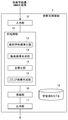
- FIG. 1 is a diagram illustrating a configuration of the diagnosis support apparatus according to the first embodiment.
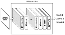
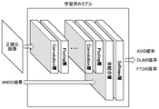
- FIG. 2 is a diagram illustrating an example of a configuration of a learned model used in the first embodiment.
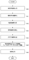
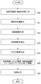
- FIG. 3 is a flowchart illustrating the operation of the diagnosis support apparatus according to the first embodiment.
- FIG. 4 is a diagram illustrating a configuration of the learning device according to the first embodiment.
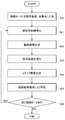
- FIG. 5 is a flowchart illustrating the operation of the learning device according to the first embodiment.
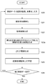
- FIG. 6 is a flowchart showing the operation of the learning device according to the modification.
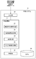
- FIG. 7 is a diagram illustrating a configuration of the diagnosis support apparatus according to the second embodiment.
- FIG. 8 is a diagram illustrating an example of a configuration of a learned model used in the second embodiment.
- FIG. 9 is a flowchart illustrating the operation of the diagnosis support apparatus according to the second embodiment.
- FIG. 10 is a diagram illustrating a configuration of the learning device according to the second embodiment.
- FIG. 1 is a diagram illustrating a configuration of a diagnosis support apparatus 1 according to the first embodiment.
- the diagnosis support apparatus 1 includes an input unit 10, a preprocessing unit 11, an inference unit 16, and an output unit 17.
- the input unit 10 has a function of receiving an input of a nuclear medicine image of the brain of a diagnosis subject.
- an image taken by the nuclear medicine examination apparatus may be input from the nuclear medicine examination apparatus, or a nuclear medicine image taken by another facility may be received via a network.
- Examples of the nuclear medicine examination apparatus include a PET apparatus and a SPECT apparatus. Further, the nuclear medicine image may be a PET image captured by a PET apparatus or a SPECT image captured by a SPECT apparatus.
- the pre-processing unit 11 has a function of performing a process such as normalization so that a disease can be appropriately identified based on a nuclear medicine image having a difference depending on imaging conditions and individual differences.
- the preprocessing unit 11 includes an anatomical standardization unit 12, a brain surface image generation unit 13, a normalization unit 14, and a Z score image generation unit 15.
- the anatomical standardization unit 12 performs a process of matching the brain image of the person to be diagnosed with a standard brain image.
- the brain surface image generation unit 13 generates a brain surface image from a tomographic image of the brain of the person to be diagnosed. Specifically, by looking at the pixel value in the cortex from the brain surface pixels determined in the stereotaxic coordinate system, extracting the pixel value peak in the cortex as the pixel value of the brain surface, and projecting it to the brain surface A brain surface image is generated.
- the normalization unit 14 is a process for normalizing signal strengths that differ depending on imaging conditions.
- the normalization unit 14 performs normalization by dividing the nuclear medicine image by the average pixel value of a predetermined reference region of the brain. For example, the whole brain, thalamus, cerebellum, bridge, and sensorimotor area can be used as the reference site.
- Z score is the degree to which the number of standard deviations is above or below the population mean, and as shown in the above equation, statistical analysis is performed on subject data and data on multiple healthy subjects Can be obtained.
- the example which uses the data of several healthy persons was given here, when the average value and standard deviation of the data of several healthy persons are calculated
- the processing by the preprocessing unit 11 described above may be performed using software “3D-SSP”.
- the inference unit 16 has a function of inferring the diagnosis subject's disease by applying the brain surface image and the Z score image of the diagnosis subject to the learned model stored in the learned model storage unit 18.
- FIG. 2 is a diagram illustrating an example of a learned model stored in the learned model storage unit 18.
- the learned model is a deep learning model that is a kind of neural network. It has a plurality of sets of Convolution layers and Pooling layers, all coupled layers in the subsequent stage, and a Softmax layer.
- the plurality of sets of the Convolution layer and the Pooling layer extract the feature amount of the input image by repeating convolution and pooling.
- the total connection layer combines the extracted feature values into one node.
- the softmax layer outputs the probability of each disease based on the output from the all connected layers.
- weight coefficients are learned in advance using data of a large number of subjects. Specifically, learning is performed on the parameters of the filter used in the Convolution layer and the parameters of the node weights in the fully connected layer. The generation of the learned model will be described later.
- the learned model of this embodiment is assumed to be used as a program module that is a part of artificial intelligence software.
- the learned model of the present embodiment is used in a computer having a CPU and a memory. Specifically, in accordance with a command from the learned model stored in the memory, the computer CPU inputs the input data (brain surface image and Z score image) input to the input layer (first Convolution layer) of the neural network.
- the feature amount of the image is obtained by performing convolution and pooling, the calculation based on the learned weighting coefficient is performed on the obtained feature amount, and the result (probability value having each disease) is obtained from the output layer (Softmax layer). Operates to output.
- the output unit 17 outputs the disease data inferred by the inference unit 16.
- the output unit 17 may display a brain image of the person to be diagnosed on the monitor together with the inferred disease name.
- FIG. 3 is a flowchart showing the operation of the diagnosis support apparatus 1.
- the diagnosis support apparatus 1 performs anatomical standardization of the input brain image of the diagnosis target person (S11). Subsequently, the diagnosis support apparatus 1 generates a brain surface image using the anatomically standardized image (S12), and normalizes the signal intensity of the generated brain surface image (S13). Next, the diagnosis support apparatus 1 generates a Z score image using the data of the healthy person (S14). Thereafter, the diagnosis support apparatus 1 applies the brain surface image and the Z score image to the learned model, infers a disease (S15), and outputs the inferred result (S16).
- FIG. 4 is a diagram illustrating a configuration of the learning device 2 that generates a learned model.
- the learning device 2 includes an input unit 20, a preprocessing unit 21, a learning unit 26, and a model storage unit 27.
- the input unit 20 has a function of accepting input of nuclear medicine images of brains of many subjects as learning teacher data.
- Subjects include healthy individuals as well as those with dementia diseases.
- the input teacher data is data of a subject having a disease to be classified using a learned model, such as Alzheimer's dementia, Lewy body dementia, and frontotemporal dementia.
- the input unit 20 accepts input of data specifying the subject's disease together with the brain image data of the subject.
- the pre-processing unit 21 performs pre-processing of the input nuclear medicine image of the subject's brain.
- the configuration and processing contents of the preprocessing unit 21 are the same as those of the preprocessing unit 11 of the diagnosis support apparatus 1.
- the learning unit 26 has a function of learning the model stored in the model storage unit 27 using the teacher data.
- the model storage unit 27 stores the model shown in FIG.
- a model that has been learned with respect to the model stored in the model storage unit 27 is a learned model.
- the model before learning and the model after learning have different parameter weights, but the basic configuration is the same.
- the learning unit 26 applies the brain surface image and the Z score image generated from the brain image data of the subject of the teacher data to the input layer of the model to be learned, and applies the disease of the subject to the Softmax layer.
- the Softmax layer has nodes of Alzheimer type dementia, Lewy body dementia, frontotemporal dementia, and healthy subjects. When inferring the disease of the person to be diagnosed, the probability value is output to the node of each disease or healthy person, but when learning is performed, the node corresponding to the disease of the subject as the teacher data "1" is applied to, and "0" is applied to other nodes.
- the learning unit 26 learns the parameter value of the model using the inverse error propagation method, and stores the learned model in the model storage unit 27.
- FIG. 5 is a flowchart showing the operation of the learning device 2.
- the learning device 2 inputs the nuclear medicine images of the brains of many subjects and the disease data of the subjects as teacher data (S20).
- the learning device 2 performs anatomical standardization of the brain image of the subject used for learning (S21), and generates a brain surface image from the anatomically standardized brain image (S22).
- the signal intensity of the brain surface image is normalized (S23), and a Z score image is generated based on the normalized brain surface image and the brain surface images of a plurality of healthy persons (S24).
- the learning device 2 applies brain surface images and Z score images of a plurality of subjects to the input layer of the model to be learned, and applies the disease data of the subjects to the output layer of the model to perform the reverse error propagation method. By doing so, the model is learned (S25).
- the learning device 2 determines whether there is other teacher data (S26). If there is other teacher data (YES in S26), the learning device 2 learns the model using the other teacher data. . Specifically, the process returns to step S21 of the anatomical standardization of the subject's brain to perform the processing. If the processing of all input teacher data is completed and there is no other teacher data (NO in S26), the learning device 2 ends the model learning process.
- diagnosis support apparatus 1 and the learning apparatus 2 The configuration of the diagnosis support apparatus 1 and the learning apparatus 2 according to the present embodiment has been described above.
- Examples of the hardware of the diagnosis support apparatus 1 and the learning apparatus 2 described above include a CPU, a RAM, a ROM, a hard disk, a display, and a keyboard. , A computer equipped with a mouse, a communication interface, and the like.
- the diagnosis support apparatus 1 and the learning apparatus 2 described above are realized by storing a program having a module for realizing each function described above in a RAM or a ROM and executing the program by the CPU. Such a program is also included in the scope of the present invention.
- the diagnosis support apparatus 1 of the present embodiment has a learned model for inferring diseases from nuclear medicine images.
- this learned model By using this learned model and inferring the subject's disease from the nuclear medicine image of the person to be diagnosed, it is possible to support the discrimination of the disease.
- the model is learned using a brain surface image obtained by normalizing the signal intensity generated from the subject's nuclear medicine image, so an appropriate model can be generated with a small amount of teacher data. .
- FIG. 6 is a flowchart showing the operation of the learning device 2 according to the modification of the first embodiment.
- the operation according to the modification is basically the same as the operation described with reference to FIG. 5, but the signal intensity normalization process (S23a) is different.
- normalization processing is performed by dividing the brain surface image of the subject by the average pixel values of the whole brain, thalamus, cerebellum, bridge, and sensorimotor area of the subject. Thereby, five normalized images are obtained from the brain image of one subject. Since these five normalized images can be used for learning, teacher data increases. Thereby, even when there are few test subject data used for learning, an appropriate model can be generated by increasing the teacher data.
- FIG. 7 is a diagram illustrating a configuration of the diagnosis support apparatus 3 according to the second embodiment.
- the basic configuration of the second diagnosis support apparatus 3 is the same as that of the first embodiment, but the second diagnosis support apparatus 3 includes the MMSE result of the subject together with the nuclear medicine image of the brain of the diagnosis target. The difference is that the input of the data is accepted and the disease is identified using the MMSE result.
- MMSE is a test for diagnosis of dementia called a mini mental state test, and diagnoses dementia by answering a plurality of question items.
- FIG. 8 is a diagram illustrating a learned model used in the diagnosis support apparatus 3 according to the second embodiment.
- the learned model of the second embodiment has a configuration in which the MMSE result is input to all the coupling layers in addition to the learned model of the first embodiment.
- FIG. 9 is a flowchart showing the operation of the diagnosis support apparatus 3.
- the diagnosis support apparatus 3 performs anatomical standardization of the input brain image of the diagnosis target person (S30). S31). Subsequently, the diagnosis support apparatus 3 generates a brain surface image using the anatomically standardized image (S32), and normalizes the signal intensity of the generated brain surface image (S33). Next, the diagnosis support apparatus 3 generates a Z score image using the data of the healthy person (S34). Thereafter, the diagnosis support apparatus 3 applies the brain surface image, the Z score image, and the MMSE result to the learned model, infers a disease (S35), and outputs the inferred result (S36).
- FIG. 10 is a diagram illustrating a configuration of the learning device 4 that generates a learned model.
- the basic configuration of the learning device 4 of the second embodiment is the same as that of the first embodiment, and includes an input unit 10, a preprocessing unit 21, a learning unit 26, and a model storage unit 27. doing.
- the model learned by the learning device 4 of the second embodiment is different from the model shown in FIG.
- the input unit 20 has a function of accepting input of nuclear medicine images of the brains of a large number of subjects and MMSE result data as learning teacher data.
- the input unit 20 receives input of data specifying the subject's disease together with the brain image of the subject and the data of the MMSE result.
- the pre-processing unit 21 performs pre-processing of the input nuclear medicine image of the subject's brain.
- the configuration and processing contents of the preprocessing unit 21 are the same as the processing of the preprocessing unit 21 of the diagnosis support apparatus 3.
- the learning unit 26 has a function of learning the model stored in the model storage unit 27 using the teacher data.
- the model storage unit 27 stores the model shown in FIG.
- the learning unit 26 applies the brain surface image, the Z score image, and the MMSE result generated from the brain image data of the subject as the teacher data to the input layer of the model to be learned, and applies the disease of the subject to the Softmax layer.
- the learning unit 26 learns the parameter value of the model using the inverse error propagation method, and stores the learned model in the model storage unit 27.
- the diagnosis support apparatus 3 of the second embodiment infers the disease using the MMSE result in addition to the nuclear medicine image of the person to be diagnosed, so that the accuracy of disease discrimination can be improved.
- MMSE is used as clinical information of a subject and a diagnosis subject
- clinical information other than MMSE may be used.
- a tomographic image of the brain may be used. That is, by generating a tomographic image from an anatomically standardized nuclear medicine image of the brain and using a tomographic image generated by normalizing the signal intensity of the tomographic image, as with the above-described embodiment, less teacher data is used. Appropriate models can be generated.
- a Z score image is given as an example of an image subjected to statistical analysis. However, an image subjected to statistical analysis processing other than the Z score image can be used as teacher data. It is.
- Alzheimer-type dementia, Lewy body dementia, and frontotemporal dementia are differentiated.
- the present invention includes other dementias such as brain tumor, chronic hard disease, and the like. It can also be used for differentiation of submembrane blood species, normal pressure hydrocephalus, sequelae of head trauma, and the like.
- a nuclear medicine image has been described as an example, but the diagnosis support apparatus of the present invention can be applied to an MRI image, an fMRI image, or the like.
- the MRI image when using the MRI image to analyze the morphological information of the medial temporal atrophy that is characteristic of early Alzheimer-type dementia (AD), the gray matter image extracted from the MRI image is anatomized.
- AD Alzheimer-type dementia
- an image obtained by statistical analysis may be used.
- an MRI image of a subject subjected to anatomical standardization and normalization may be compared with MRI images of a plurality of healthy subjects to generate a Z score image, and model learning may be performed using the Z score image. .
Landscapes
- Health & Medical Sciences (AREA)
- Engineering & Computer Science (AREA)
- Life Sciences & Earth Sciences (AREA)
- Physics & Mathematics (AREA)
- Biomedical Technology (AREA)
- General Health & Medical Sciences (AREA)
- Molecular Biology (AREA)
- Medical Informatics (AREA)
- Theoretical Computer Science (AREA)
- Public Health (AREA)
- Biophysics (AREA)
- General Physics & Mathematics (AREA)
- Pathology (AREA)
- Nuclear Medicine, Radiotherapy & Molecular Imaging (AREA)
- Veterinary Medicine (AREA)
- Animal Behavior & Ethology (AREA)
- Surgery (AREA)
- Heart & Thoracic Surgery (AREA)
- Data Mining & Analysis (AREA)
- Software Systems (AREA)
- Evolutionary Computation (AREA)
- Computational Linguistics (AREA)
- Computing Systems (AREA)
- Mathematical Physics (AREA)
- Artificial Intelligence (AREA)
- High Energy & Nuclear Physics (AREA)
- General Engineering & Computer Science (AREA)
- Radiology & Medical Imaging (AREA)
- Computer Vision & Pattern Recognition (AREA)
- Spectroscopy & Molecular Physics (AREA)
- Optics & Photonics (AREA)
- Databases & Information Systems (AREA)
- Epidemiology (AREA)
- Primary Health Care (AREA)
- Magnetic Resonance Imaging Apparatus (AREA)
- Image Analysis (AREA)
- Nuclear Medicine (AREA)
Abstract
L'invention porte sur un dispositif d'aide au diagnostic (1) comprenant : une unité de stockage de modèle appris (18) dans laquelle est stocké un modèle appris conçu par l'apprentissage de facteurs de pondération d'un réseau neuronal, à l'aide de données d'image de médecine nucléaire concernant des cerveaux de multiples sujets, et de données concernant des maladies des sujets, en tant que données d'apprentissage ; une unité d'entrée (10) permettant d'entrer une image de médecine nucléaire d'un cerveau d'une personne en cours de diagnostic ; une unité de normalisation anatomique (12) qui normalise de manière anatomique l'entrée d'image de médecine nucléaire dans l'unité d'entrée (10) ; une unité de génération d'image de surface cérébrale (13) qui génère une image de surface cérébrale à partir de l'image normalisée de manière anatomique ; une unité de normalisation (14) qui normalise l'intensité de signal de l'image de surface cérébrale ; une unité de génération d'image de score Z (15) qui génère une image de score Z pour la personne en cours de diagnostic, sur la base de l'image de surface cérébrale normalisée et des images de surface cérébrale d'une pluralité de sujets sains ; une unité de déduction (16) qui applique l'image de surface cérébrale normalisée au modèle appris pour déduire une maladie de la personne en cours de diagnostic ; et une unité de sortie (17) qui fournit des données relatives à la maladie déduite par l'unité de déduction (16).
Priority Applications (1)
| Application Number | Priority Date | Filing Date | Title |
|---|---|---|---|
| JP2020505016A JP7382306B2 (ja) | 2018-03-09 | 2019-03-04 | 診断支援装置、プログラム、学習済みモデル、および学習装置 |
Applications Claiming Priority (2)
| Application Number | Priority Date | Filing Date | Title |
|---|---|---|---|
| JP2018-043242 | 2018-03-09 | ||
| JP2018043242 | 2018-03-09 |
Publications (1)
| Publication Number | Publication Date |
|---|---|
| WO2019172181A1 true WO2019172181A1 (fr) | 2019-09-12 |
Family
ID=67846264
Family Applications (1)
| Application Number | Title | Priority Date | Filing Date |
|---|---|---|---|
| PCT/JP2019/008378 Ceased WO2019172181A1 (fr) | 2018-03-09 | 2019-03-04 | Dispositif d'aide au diagnostic, programme, modèle appris et dispositif d'apprentissage |
Country Status (3)
| Country | Link |
|---|---|
| JP (1) | JP7382306B2 (fr) |
| TW (1) | TW201941220A (fr) |
| WO (1) | WO2019172181A1 (fr) |
Cited By (6)
| Publication number | Priority date | Publication date | Assignee | Title |
|---|---|---|---|---|
| CN112185548A (zh) * | 2020-09-25 | 2021-01-05 | 广州宝荣科技应用有限公司 | 一种基于神经网络算法的智能中医诊断方法及装置 |
| JP2021076585A (ja) * | 2019-11-06 | 2021-05-20 | 日本メジフィジックス株式会社 | 疾患データベースとの関連性を用いた診断補助技術 |
| JPWO2021221008A1 (fr) * | 2020-04-28 | 2021-11-04 | ||
| JPWO2021260928A1 (fr) * | 2020-06-26 | 2021-12-30 | ||
| WO2022065061A1 (fr) * | 2020-09-28 | 2022-03-31 | 富士フイルム株式会社 | Dispositif de traitement d'images, procédé d'utilisation de dispositif de traitement d'images et programme d'utilisation de traitement d'images |
| WO2024116309A1 (fr) * | 2022-11-30 | 2024-06-06 | 日本電気株式会社 | Dispositif de génération d'image, dispositif d'apprentissage, procédé de génération d'image et programme |
Families Citing this family (1)
| Publication number | Priority date | Publication date | Assignee | Title |
|---|---|---|---|---|
| JP7824765B2 (ja) * | 2021-12-20 | 2026-03-05 | 株式会社日立製作所 | 医療画像診断支援装置、医療画像診断支援方法及びプログラム |
Citations (8)
| Publication number | Priority date | Publication date | Assignee | Title |
|---|---|---|---|---|
| JP2005230456A (ja) * | 2004-02-23 | 2005-09-02 | Kokuritsu Seishin Shinkei Center | 脳疾患の診断支援方法及び装置 |
| WO2008056638A1 (fr) * | 2006-11-06 | 2008-05-15 | Fujifilm Ri Pharma Co., Ltd. | Procédé d'aide au diagnostic à partir d'une image du cerveau, logiciel, et procédé d'enregistrement |
| JP2008157640A (ja) * | 2006-12-20 | 2008-07-10 | Fujifilm Ri Pharma Co Ltd | 脳画像データに関する時系列データの解析方法、プログラムおよび記録媒体 |
| JP2010012176A (ja) * | 2008-07-07 | 2010-01-21 | Hamamatsu Photonics Kk | 脳疾患診断システム |
| JP2014009945A (ja) * | 2012-06-27 | 2014-01-20 | Fujifilm Ri Pharma Co Ltd | 画像処理プログラム、記録媒体、画像処理装置、及び画像処理方法 |
| WO2015122524A1 (fr) * | 2014-02-14 | 2015-08-20 | 広二 柳本 | Agent de prévention ou de traitement de thésaurismose de bêta-amyloïde intracérébrale |
| JP2018038515A (ja) * | 2016-09-06 | 2018-03-15 | 国立大学法人島根大学 | アルツハイマー病の診断支援装置、診断支援プログラムおよび診断支援方法 |
| WO2019044078A1 (fr) * | 2017-08-28 | 2019-03-07 | 富士フイルム株式会社 | Programme, procédé et dispositif de normalisation d'image cérébrale |
-
2019
- 2019-03-04 WO PCT/JP2019/008378 patent/WO2019172181A1/fr not_active Ceased
- 2019-03-04 TW TW108107088A patent/TW201941220A/zh unknown
- 2019-03-04 JP JP2020505016A patent/JP7382306B2/ja active Active
Patent Citations (8)
| Publication number | Priority date | Publication date | Assignee | Title |
|---|---|---|---|---|
| JP2005230456A (ja) * | 2004-02-23 | 2005-09-02 | Kokuritsu Seishin Shinkei Center | 脳疾患の診断支援方法及び装置 |
| WO2008056638A1 (fr) * | 2006-11-06 | 2008-05-15 | Fujifilm Ri Pharma Co., Ltd. | Procédé d'aide au diagnostic à partir d'une image du cerveau, logiciel, et procédé d'enregistrement |
| JP2008157640A (ja) * | 2006-12-20 | 2008-07-10 | Fujifilm Ri Pharma Co Ltd | 脳画像データに関する時系列データの解析方法、プログラムおよび記録媒体 |
| JP2010012176A (ja) * | 2008-07-07 | 2010-01-21 | Hamamatsu Photonics Kk | 脳疾患診断システム |
| JP2014009945A (ja) * | 2012-06-27 | 2014-01-20 | Fujifilm Ri Pharma Co Ltd | 画像処理プログラム、記録媒体、画像処理装置、及び画像処理方法 |
| WO2015122524A1 (fr) * | 2014-02-14 | 2015-08-20 | 広二 柳本 | Agent de prévention ou de traitement de thésaurismose de bêta-amyloïde intracérébrale |
| JP2018038515A (ja) * | 2016-09-06 | 2018-03-15 | 国立大学法人島根大学 | アルツハイマー病の診断支援装置、診断支援プログラムおよび診断支援方法 |
| WO2019044078A1 (fr) * | 2017-08-28 | 2019-03-07 | 富士フイルム株式会社 | Programme, procédé et dispositif de normalisation d'image cérébrale |
Non-Patent Citations (4)
| Title |
|---|
| 1 December 2017 (2017-12-01), Retrieved from the Internet <URL:https://www.neurology-jp.org/guidelinem/degl/degl_2017_02.pdf> [retrieved on 20190510] * |
| 5 March 2018 (2018-03-05), Retrieved from the Internet <URL:http://www.soumu.go.jp/main_content/000542895.pdf> [retrieved on 20190510] * |
| CHOI, HONGYOON ET AL.: "Predicting Cognitive Decline with Deep Learning of Brain Metabolism and Amyloid Imaging", ARXIV: 1704. 06033, 20 April 2017 (2017-04-20), Retrieved from the Internet <URL:https://arxiv.org/abs/1704.06033> * |
| MURAKAMI TAKESHI: "New Processing Using Anatomical Standardization in Nuclear Neuroimaging", JAPANESE JOURNAL OF RADIOLOGICAL TECHNOLOGY, vol. 57, no. 8, August 2001 (2001-08-01), pages 913 - 919 * |
Cited By (14)
| Publication number | Priority date | Publication date | Assignee | Title |
|---|---|---|---|---|
| JP2021076585A (ja) * | 2019-11-06 | 2021-05-20 | 日本メジフィジックス株式会社 | 疾患データベースとの関連性を用いた診断補助技術 |
| JP7576417B2 (ja) | 2019-11-06 | 2024-10-31 | 日本メジフィジックス株式会社 | 疾患データベースとの関連性を用いた診断補助技術 |
| JPWO2021221008A1 (fr) * | 2020-04-28 | 2021-11-04 | ||
| JP7764037B2 (ja) | 2020-04-28 | 2025-11-05 | 株式会社Splink | システム、制御方法及びプログラム |
| JP7517420B2 (ja) | 2020-06-26 | 2024-07-17 | 株式会社島津製作所 | 吸収係数画像生成方法、核医学診断装置および学習済みモデルの作成方法 |
| JPWO2021260928A1 (fr) * | 2020-06-26 | 2021-12-30 | ||
| WO2021260928A1 (fr) * | 2020-06-26 | 2021-12-30 | 株式会社島津製作所 | Procédé de génération d'image de coefficient d'absorption, dispositif de diagnostic de médecine nucléaire et procédé de création de modèles entraînés |
| CN112185548A (zh) * | 2020-09-25 | 2021-01-05 | 广州宝荣科技应用有限公司 | 一种基于神经网络算法的智能中医诊断方法及装置 |
| JPWO2022065061A1 (fr) * | 2020-09-28 | 2022-03-31 | ||
| JP7440655B2 (ja) | 2020-09-28 | 2024-02-28 | 富士フイルム株式会社 | 画像処理装置、画像処理装置の作動方法、画像処理装置の作動プログラム |
| CN116209396A (zh) * | 2020-09-28 | 2023-06-02 | 富士胶片株式会社 | 图像处理装置、图像处理装置的工作方法、图像处理装置的工作程序 |
| WO2022065061A1 (fr) * | 2020-09-28 | 2022-03-31 | 富士フイルム株式会社 | Dispositif de traitement d'images, procédé d'utilisation de dispositif de traitement d'images et programme d'utilisation de traitement d'images |
| US12597130B2 (en) | 2020-09-28 | 2026-04-07 | Fujifilm Corporation | Image processing apparatus, operation method of image processing apparatus, and operation program of image processing apparatus for generating examination results of new medical examinations |
| WO2024116309A1 (fr) * | 2022-11-30 | 2024-06-06 | 日本電気株式会社 | Dispositif de génération d'image, dispositif d'apprentissage, procédé de génération d'image et programme |
Also Published As
| Publication number | Publication date |
|---|---|
| TW201941220A (zh) | 2019-10-16 |
| JP7382306B2 (ja) | 2023-11-16 |
| JPWO2019172181A1 (ja) | 2021-03-25 |
Similar Documents
| Publication | Publication Date | Title |
|---|---|---|
| JP7382306B2 (ja) | 診断支援装置、プログラム、学習済みモデル、および学習装置 | |
| Zhang et al. | Context-guided fully convolutional networks for joint craniomaxillofacial bone segmentation and landmark digitization | |
| US10565477B2 (en) | Deep learning medical systems and methods for image reconstruction and quality evaluation | |
| JP7051849B2 (ja) | ディープラーニング医療システムおよび医療処置のための方法 | |
| KR102698916B1 (ko) | 초음파 분석을 위한 시스템, 방법 및 컴퓨터-접근가능 매체 | |
| JP7309605B2 (ja) | ディープラーニング医療システムおよび画像収集のための方法 | |
| CN112950737B (zh) | 基于深度学习的眼底荧光造影图像生成方法 | |
| Chagas et al. | A new approach for the detection of pneumonia in children using CXR images based on an real-time IoT system | |
| Pallawi et al. | Study of Alzheimer’s disease brain impairment and methods for its early diagnosis: a comprehensive survey | |
| Zhao et al. | Machine learning approaches in comparative studies for Alzheimer’s diagnosis using 2D MRI slices | |
| Tounsi et al. | A comprehensive review on biomedical image classification using deep learning models | |
| Rahimiasl et al. | Random walks on B distributed resting-state functional connectivity to identify Alzheimer's disease and Mild Cognitive Impairment | |
| KR102809111B1 (ko) | 합성곱 신경망의 계층 연관성 전파를 이용한 퇴행성 뇌질환 이미지 분류 장치 및 방법 | |
| CN118414672A (zh) | 使用人工智能的直接医疗治疗方案预测 | |
| Fang et al. | Multimodal Identification of Alzheimer's Disease: A Review | |
| US20250209612A1 (en) | Method for the elaboration of functional magnetic resonance images | |
| Vidhya et al. | Advanced detection of brain disease using ML and DL algorithm | |
| WO2019131225A1 (fr) | Appareil, procédé et programme d'analyse d'image cérébrale | |
| Obeidat et al. | An MRI Based Algorithm for Detecting Multiple Sclerosis | |
| Xavier et al. | Brain Disease Classification along with Age Estimation from MRI | |
| Vijayakumar et al. | Examination of Blood Cell Images to Identify Nannal/Malaria with EfficientNet Model and Fused Features | |
| Mary et al. | Tuberculosis Detection in X-Ray with Two-Fold Training and Fused Deep Features Based Classification | |
| Sriharsha | Using Deep Learning to Classify and Diagnose Alzheimer's Disease | |
| Hussein et al. | Survey On Techniques For Cardiomegaly Prediction By Chest X-ray Images | |
| Archana et al. | Automated Classification of Chest Image Using Deep Learning |
Legal Events
| Date | Code | Title | Description |
|---|---|---|---|
| 121 | Ep: the epo has been informed by wipo that ep was designated in this application |
Ref document number: 19764032 Country of ref document: EP Kind code of ref document: A1 |
|
| ENP | Entry into the national phase |
Ref document number: 2020505016 Country of ref document: JP Kind code of ref document: A |
|
| NENP | Non-entry into the national phase |
Ref country code: DE |
|
| 122 | Ep: pct application non-entry in european phase |
Ref document number: 19764032 Country of ref document: EP Kind code of ref document: A1 |