WO2020004425A1 - Procédé de culture du virus de la grippe - Google Patents
Procédé de culture du virus de la grippe Download PDFInfo
- Publication number
- WO2020004425A1 WO2020004425A1 PCT/JP2019/025252 JP2019025252W WO2020004425A1 WO 2020004425 A1 WO2020004425 A1 WO 2020004425A1 JP 2019025252 W JP2019025252 W JP 2019025252W WO 2020004425 A1 WO2020004425 A1 WO 2020004425A1
- Authority
- WO
- WIPO (PCT)
- Prior art keywords
- influenza virus
- protease
- virus
- culturing
- cells
- Prior art date
- Legal status (The legal status is an assumption and is not a legal conclusion. Google has not performed a legal analysis and makes no representation as to the accuracy of the status listed.)
- Ceased
Links
Images


Classifications
-
- C—CHEMISTRY; METALLURGY
- C12—BIOCHEMISTRY; BEER; SPIRITS; WINE; VINEGAR; MICROBIOLOGY; ENZYMOLOGY; MUTATION OR GENETIC ENGINEERING
- C12N—MICROORGANISMS OR ENZYMES; COMPOSITIONS THEREOF; PROPAGATING, PRESERVING, OR MAINTAINING MICROORGANISMS; MUTATION OR GENETIC ENGINEERING; CULTURE MEDIA
- C12N5/00—Undifferentiated human, animal or plant cells, e.g. cell lines; Tissues; Cultivation or maintenance thereof; Culture media therefor
- C12N5/10—Cells modified by introduction of foreign genetic material
-
- C—CHEMISTRY; METALLURGY
- C12—BIOCHEMISTRY; BEER; SPIRITS; WINE; VINEGAR; MICROBIOLOGY; ENZYMOLOGY; MUTATION OR GENETIC ENGINEERING
- C12N—MICROORGANISMS OR ENZYMES; COMPOSITIONS THEREOF; PROPAGATING, PRESERVING, OR MAINTAINING MICROORGANISMS; MUTATION OR GENETIC ENGINEERING; CULTURE MEDIA
- C12N7/00—Viruses; Bacteriophages; Compositions thereof; Preparation or purification thereof
Definitions
- the present invention relates to a method for culturing influenza virus. Specifically, the present invention relates to a method for culturing an influenza virus using MDCK cells as a host cell, and to a method for culturing an influenza virus more efficiently.
- Influenza is an infectious disease that spreads worldwide every year and is caused by the influenza virus.
- Influenza virus belongs to the family Orthomyxoviridae and has an envelope having a lipid bilayer structure. It is classified into three genera of types A, B and C, and is called influenza A virus, influenza B virus and influenza C virus, respectively. Generally, influenza virus often refers to type A or type B. The difference between types A, B and C is based on the difference in antigenicity between the M1 protein and the NP protein among the proteins constituting the virus particle.
- HA hemagglutininin
- NA neuraminidase
- Influenza A viruses are classified into 16 HA (H1-H16) subtypes and 9 NA (N1-N9) subtypes based on their HA and NA antigenicity.
- the three HA (H1, H2 and H3) subtypes of influenza A virus are particularly important pathogens.
- the H1N1 and H3N2 subtypes of influenza A virus are seasonally prevalent and cause human infection.
- the influenza vaccine is unique in that it requires a large amount of vaccine antigens because of the above-mentioned epidemic situation, inoculation targets across a wide range of age groups, and a large number of viruses required as antigen strains. That is, there is a need for a method capable of producing an influenza vaccine more efficiently.
- Influenza vaccine production includes a method of growing influenza virus using embryonated chicken eggs and a method of growing influenza virus in cultured cells.
- Cultured cells include Madin-Darby canine kidney-derived cells (hereinafter, “MDCK cells”) and African green monkey kidney-derived cells (hereinafter, “Vero cells”).
- Patent Literature 1 describes that it is preferable to culture an influenza virus using a protease when culturing the influenza virus using a host cell. Further, it is described that the substance that enhances virus activation is preferably a protease that cleaves HA.
- the protease trypsin, chymotrypsin, thermolysin, pronase, subtilisin A, elastase, pepsin, pancreatin, carboxypeptidase, furin and the like are exemplified.
- Non-Patent Document 1 describes that influenza virus is activated by elastase, chymotrypsin and thermolysin, and Non-Patent Document 2 is activated by trypsin, chymotrypsin and plasmin.
- Non-Patent Document 3 reports that when influenza virus was cultured using various MDCK cells as a host, influenza virus grew in some MDCK cells even without trypsin. However, it has been reported that in the absence of trypsin, the plaque of the virus was smaller than in the case of culturing with the addition of trypsin.
- Non-Patent Document 4 reports a MDCK mutant cell line in which an influenza virus grows without trypsin requirement.
- Non-Patent Document 5 analysis of proteases expressed in MDCK mutant cell lines identified TMPRSS2, which was classified into the TMPRSS (transmembrane @ protease / serine) subfamily, and a part of influenza virus was cleaved by TMPRSS2.
- TMPRSS2 transmembrane @ protease / serine subfamily
- An object of the present invention is to provide a method for culturing an influenza virus more efficiently in a method for culturing an influenza virus using MDCK cells as a host. Specifically, it is an object of the present invention to provide a method for more efficiently culturing an influenza virus under a protease-free medium condition.
- the present inventors have conducted intensive studies in order to solve the above-mentioned problems. As a result, after the MDCK cells, which are host cells of the influenza virus, have exceeded the logarithmic growth phase, the influenza virus cells were transferred to the MDCK cells under a protease-free medium condition. It has been found that the above-mentioned object can be achieved by inoculating.
- the present invention consists of the following. 1. A method for culturing influenza virus using MDCK cells as a host, wherein the influenza virus is inoculated after the MDCK cells have exceeded the logarithmic growth phase under medium conditions without protease. 2. 2. The method for culturing an influenza virus according to the above item 1, wherein the method for culturing an influenza virus using MDCK cells as a host exhibits a higher virus yield than inoculating the influenza virus under medium conditions containing a protease. 3. A method for culturing an influenza virus using an MDCK cell as a host, wherein the doubling time of the MDCK cell exceeds 17 hours, and then the influenza virus is inoculated under a protease-free medium condition. Method. 4. Item 4.
- the method for culturing an influenza virus according to any one of Items 1 to 3, wherein the method for culturing an influenza virus using MDCK cells as a host is a three-dimensional culturing method. 5. The method for culturing an influenza virus according to the above item 4, wherein the three-dimensional culture method is a three-dimensional culture method using a microcarrier. 6. Item 6. The method for culturing an influenza virus according to any one of Items 1 to 5, wherein the protease is trypsin. 7. 7. The method for culturing an influenza virus according to any one of the above items 1 to 6, wherein the MDCK cells are protease-independent MDCK cells. 8. 8. 8.
- influenza virus which inoculates MDCK cell to influenza virus under the culture medium condition which does not contain protease of this invention, aggregation and damage of MDCK cell by protease can be prevented, and influenza virus can be cultured efficiently. it can.
- influenza virus By inoculating the influenza virus after the MDCK cells have exceeded the logarithmic growth phase, the influenza virus can be cultured more efficiently.
- FIG. 2 is a view showing a difference in the amount of a viral protein depending on the presence or absence of a protease (Example 1-1, Comparative Example 1-1, Example 1-2).
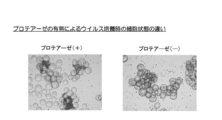
- FIG. 2 is a view showing the appearance of MDCK cells (derived from ATCC @ CCL-34) on the first day of culture after inoculation of the virus, with respect to influenza virus culture in a 2 L culture solution volume.
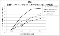
- FIG. 3 is a view showing the amounts of viral proteins of various influenza virus strains (Example 1-3). It is a figure which shows the mode of the cell line A at the time of exceeding the logarithmic growth phase on the 5th day of cell culture with 2 L culture solution volume. (Example 2)
- the present invention relates to a method for culturing an influenza virus using MDCK cells as a host.
- the method of culturing influenza virus is characterized in that after the MDCK cells have exceeded the logarithmic growth phase, the influenza virus is inoculated under a medium condition free of protease.
- the present invention relates to a method for culturing an influenza virus, which comprises inoculating an influenza virus under a medium condition containing no protease after the doubling time of the MDCK cell exceeds 17 hours.
- influenza virus growth requires cleavage and activation of influenza virus HA
- Patent Document 1 when culturing influenza virus using host cells.
- a protease it is preferable to culture an influenza virus using a protease.
- the substance that enhances the infectivity of the virus is preferably a protease that cleaves HA to cause fusion of the virus and the host cell membrane.
- the presence of a protease in the culture solution causes damage to the MDCK cells and detachment of the cells from the extracellular matrix, which tends to cause cell aggregation.
- the present inventors considered that there is a disadvantage that the number of MDCK cells infected with influenza virus is reduced due to the damage or aggregation of MDCK cells.
- the present inventors have conducted intensive studies and, surprisingly, have found that influenza virus can be efficiently propagated by inoculating MDCK cells with influenza virus under protease-free medium conditions.
- the medium containing no protease refers to a medium to which any protease conventionally added for culturing influenza virus is not added.
- Protease-free media can be used, for example, before or simultaneously with inoculation of MDCK cells with influenza virus.
- proteases not contained in the medium examples include trypsin, chymotrypsin, plasmin, thermolysin, pronase, subtilisin A, elastase, pepsin, pancreatin, carboxypeptidase, furin, and proteases classified into the TMPRSS (transmembrane @ protease / serine) subfamily. And the like.
- the protease not contained in the medium is not particularly limited as long as it causes damage or aggregation of MDCK cells, but preferably includes trypsin, chymotrypsin, plasmin, elastase, thermolysin, and more preferably trypsin.
- the protease not contained in the medium is not particularly limited as long as it exhibits various protease activities, and may be a recombinant.
- the activity of various proteases means those generally known to those skilled in the art.
- trypsin has an activity as an endopeptidase that cleaves a peptide chain at the C-terminal side of a basic residue such as lysine or arginine.
- the medium conditions without protease include proteases that do not detach cells from the microcarrier when the host cells are cultured using an extracellular matrix such as a microcarrier. Therefore, a protease that is contaminated regardless of the addition may be contained. For example, even when a cell-derived protease is contaminated, the case is included under the medium conditions that do not contain the protease of the present invention.
- the state in which the cells are not detached from the microcarrier refers to a state in which the cell suspension rate in the culture solution when the cells are cultured for a certain period of time using the microcarrier is 20% or less, preferably 15% or less. The method for calculating the cell suspension rate will be described later.
- the medium containing no protease may be a medium whose medium components do not show protease activity. Protease activity can be measured by a method known to those skilled in the art, for example, by measuring absorbance.
- the density of the MDCK cells on the microcarrier is increased.
- the high state that is, the state where the MDCK cells have exceeded the logarithmic growth phase, is efficiently maintained.
- MDCK cells are cultured in a medium containing a protease, the MDCK cells are detached from the microcarrier by the protease, and it is difficult to maintain a state in which the density of the MDCK cells on the microcarrier is high.
- the use of a protease-free medium suppresses damage and aggregation of MDCK cells, so that a state in which the density of MDCK cells on the microcarrier is high, that is, a state in which MDCK cells have exceeded the logarithmic growth phase, is maintained. It will be easier. As a result, long-term efficient virus propagation becomes possible, and a high yield of virus protein can be obtained.
- cell-cell contact on the microcarriers is strong and it is difficult for the cells to be detached, so it is considered that an environment suitable for influenza virus growth can be formed.
- the point in time at which MDCK cells reach a state beyond the logarithmic growth phase is likely to correspond to a quiescent phase of the cell cycle. It is presumed that inoculation of the virus in the cells in this state slows down the production rate of a virus resistance factor such as interferon (IFN), thereby facilitating virus infection.
- a virus resistance factor such as interferon (IFN)
- a suitable medium used for a medium containing no protease is not particularly limited.
- a serum-free medium to which no animal-derived serum is added is preferable. Any serum-free medium may be used.
- Eagle MEM medium Nisui Pharmaceutical Co., Ltd.
- Opti @ PRO @ SFM Thermo Fisher Scientific
- VP-SFM Thermo Fisher Scientific
- EX-CELL @ MDCK SAFCEnBio
- UltraMDCK Lida
- ProVero 1 Lonza
- BalanCD MDCK Irvine Scientific
- the influenza virus is inoculated after the MDCK cells have exceeded the logarithmic growth phase.
- the term “after the MDCK cells have passed the logarithmic growth phase” refers to a state where the density of the MDCK cells is high and the growth rate is reduced, that is, when the MDCK cells become confluent.
- the doubling time of MDCK cells is preferably 17 hours, more preferably after exceeding 20 hours.
- the cell doubling time refers to the time required for the cell number to double. In the present invention, it refers to the time required for doubling the number of cells from the time of cell seeding to the time of virus inoculation.
- the doubling time is prolonged when the MDCK cells exceed the logarithmic growth phase, but the time varies depending on the MDCK cells used and the culture method.
- influenza virus since influenza virus is cultured under a protease-free medium condition, it is considered that influenza virus infects MDCK cells using a cell-derived protease.
- Cell-derived proteases act on influenza virus by inoculating influenza virus into cells in a state where MDCK cells have exceeded the logarithmic growth phase, for example, the doubling time of MDCK cells exceeds 17 hours. It is possible to create an environment that is easy to do.
- the influenza virus may be inoculated into MDCK cells before scale-up or into MDCK cells after scale-up.
- the volume of the culture solution after scale-up is 1 to 10000 L, preferably 5 to 5000 L.
- Inoculating MDCK cells with influenza virus after scale-up according to the method of the present invention enables a large amount of influenza virus to be obtained, leading to efficient vaccine production.
- the container for culturing MDCK cells is not particularly limited as long as the container satisfies the target volume.
- the MDCK cells which are host cells
- the MDCK cells are inoculated with the influenza virus and then incubated, that is, maintained at a certain temperature for a certain period of time. It does not matter whether the MDCK cells proliferate or not under a constant temperature condition. Incubation can be performed under the same conditions as those for culturing MDCK cells, but preferably under the optimal conditions of the infected influenza virus.
- the optimal conditions for influenza virus culture are 31 to 37 ° C, preferably 32 to 36 ° C, and more preferably 33 to 35 ° C.
- the MDCK cells used in the method for culturing influenza virus of the present invention may be cells capable of growing influenza virus under protease-free medium conditions, and are not particularly limited.
- the cells are MDCK cells.
- Protease-independent MDCK cells during virus production refer to MDCK cells capable of growing influenza virus under protease-free medium conditions.
- the cell may be a cloned cell or a cell population containing cells that have not been cloned.
- the protease refers to a protease that is externally added, and does not refer to a protease that is expressed by cells.
- the protease-independent protease of the present invention is selected from trypsin, chymotrypsin, plasmin, thermolysin, pronase, subtilisin A, elastase, pepsin, pancreatin, carboxypeptidase, furin, and proteases classified into the TMPRSS subfamily.
- proteases classified into the TMPRSS subfamily One or more proteases.
- the protease is not particularly limited, but is preferably trypsin, chymotrypsin, plasmin, elastase, and thermolysin, and more preferably trypsin. Any MDCK cell may be used as long as it is such a protease-independent MDCK cell.
- the method for culturing influenza virus of the present invention can exert an effect particularly in three-dimensional culture using a culture tank. Furthermore, the present invention can also exert an effect when cultured under scale-up conditions for the purpose of mass culture of cells.
- the environmental load on the cells is large, and the cells are strongly damaged and aggregated by protease.
- the condition is not easily damaged by protease, but is not suitable for mass culture of cells.
- the effect of the culturing under the protease-free medium conditions of the present invention is particularly preferably confirmed under three-dimensional culturing conditions or scale-up conditions for the purpose of mass culturing of cells.
- the three-dimensional culture of MDCK cells may be performed using a microcarrier, or when no microcarrier is used, may be a suspension culture of suspended MDCK cells.
- a microcarrier or when no microcarrier is used, may be a suspension culture of suspended MDCK cells.
- Culture conditions such as the density of the microcarrier, the number of agitation, the dissolved oxygen concentration, and the culture temperature may be any as long as the cells can proliferate, and can be appropriately adjusted. In the culture method of the present invention, even with high-speed agitation, cells can adhere to the microcarrier well and can grow efficiently.
- the shape of the microcarrier used in the method for culturing influenza virus of the present invention may be spherical or disk-shaped, or may be porous.
- the size (diameter) of the spherical microcarrier is, for example, about 0.01 to 1 mm, preferably about 0.05 to 0.5 mm, and more preferably about 0.1 to 0.3 mm.
- the microcarrier may be porous. Examples of the spherical microcarriers used in the present invention include Cytodex # 1 (trade name), Cytodex # 3 (trade name), Cytopore (trade name) (above, manufactured by GE Healthcare Life Science).
- Examples of the disc-shaped microcarrier include Cytoline # 1 (trade name) and Cytoline # 2 (trade name) (all manufactured by GE Healthcare Life Science).
- Examples of porous microcarriers include Cytopore (trade name), Cytoline # 1 (trade name), Cytoline # 2 (trade name) (all manufactured by GE Helthcare Life Science), and the like.
- Other commercially available microcarriers include Biosilon (trade name) (NUNC), Hillex (trade name) (Solohill), Corning® microcarrier (Corning), and the like.
- a spherical dextran microcarrier is particularly preferable.
- Cytodex # 1 (trade name), Cytodex # 3 (trade name) and Cytopore (trade name) are preferable, Cytodex # 1 (trade name) and Cytodex $ 3 (trade name) are preferable, and especially Cytodex. 1 (trade name) is preferred.
- MDCK cells used in the method for culturing influenza virus of the present invention any cells may be used, and MDCK cells obtained from a cell bank may be used.
- MDCK cells specified by ATCC @ CCL-34 can be used.
- the adhesive force of the cells to the microcarrier can be represented by the cell floating rate when the cells are cultured in a culture solution containing the microcarrier.
- MDCK cells having a cell suspension rate of 20% or less, preferably 15% or less, more preferably 10% or less, and particularly preferably 5% or less are used.
- the cell suspension rate is a value obtained by dividing the suspension cell density after culturing MDCK cells in the presence of microcarriers for a certain period of time by the seeding cell density. The lower the cell suspension rate, the higher the adhesive strength to the microcarrier. Is shown.
- the cell suspension rate of the MDCK cells of the present invention is determined at the time when the culture time is 48 hours or less, preferably 24 hours or less, preferably 6 hours or less, more preferably 1.5 hours or less, and particularly preferably 0.5 hour or less. belongs to. More specifically, it is preferable to use MDCK cells that have a cell suspension rate of 20% or less after 0.5 hours, or a cell suspension rate of 5% or less after 1.5 hours.
- MDCK cells used in the method of culturing influenza virus of the present invention it is preferable to use MDCK cells having an expansion ratio of 4.5 or more, preferably 6.5 or more, more preferably 8.5 or more.
- the expansion ratio is a value obtained by dividing the cell density after culturing the cells for a certain period of time by the seeded cell density, and is an index indicating the ease of proliferation.
- the expansion rate of MDCK cells used in the method of culturing influenza virus of the present invention is that obtained by culturing with a microcarrier, and the culturing time is 48 hours or more, preferably 60 hours or more, more preferably 72 hours or more. The time is after 144 hours or less, preferably 120 hours or less, more preferably 96 hours or less.
- MDCK cells used in the method of culturing influenza virus of the present invention are preferably cells that can rapidly grow cells even at a low seeding density.
- MDCK cells used in the method for culturing the influenza virus of the present invention satisfying the above conditions include, specifically, MDCK cells (MDCK-F69P69) specified by the international deposit number NITE BP-022014.
- MDCK cells MDCK-F69P69
- Such cells were deposited at the National Institute of Technology and Evaluation, Patent Microorganisms Depositary Center (room No. 122, No. 122, Kazusa-Kamashita, Kisarazu-shi, Chiba) at the Patent Microorganisms Depositary Center on March 4, 2015. After being deposited in Japan as P-022014, it was requested to be transferred to the International Depositary under the Budapest Treaty on February 15, 2016 by the National Institute of Technology and Evaluation, Patent Microorganisms Depositary, under the accession number NITE @ BP-022014. It was commissioned by.
- the seeding density of the cells is not particularly limited, and can be appropriately adjusted.
- the seeding density in the present specification means the number of cells per cell surface area (cells / cm 2 ) or the number of cells per cell volume (cells / mL) when cells are seeded.
- the density of the cells in the medium can be confirmed by a method known per se, for example, by a measuring method using a hemocytometer or an automatic cell counter.
- MDCK cells before subculture or cells after subculture may be used. Passage of MDCK cells can be performed by a method known per se.
- cells that are densely adhered on the surface of the microcarrier may be moved to a new microcarrier surface.
- the influenza virus used in the culture method of the present invention which is inoculated into host cells, is called a seed virus.
- the influenza virus is isolated from a living body or produced by making some modification, and then subcultured and propagated using chicken eggs and various cells to become a seed virus.
- the seed virus used in the present invention may be subcultured in any of chicken eggs and various cells, and is not particularly limited. More preferably, it is preferable that the seed virus propagated and adapted by passage through MDCK cells is again inoculated to the MDCK cells in the culture method of the present invention, and the influenza virus is propagated.
- influenza virus cultured by the culture method of the present invention is not particularly limited, and includes, for example, all currently known subtypes and subtypes that will be isolated and identified in the future.
- Influenza A viruses are classified into subtypes (ie, 16 HA (H1-H16) subtypes and 9 NA (N1-N9) subtypes) based on the antigenicity of their HA and NA molecules.
- influenza viruses containing combinations of HA and NA subtypes are contemplated.
- influenza B virus influenza viruses including a combination of the Victoria strain and the Yamagata strain are considered.
- influenza which is said to have pandemic worldwide is called pandemic influenza, swine influenza, pandemic influenza A (H1N1), spine @ flu, A / H1N1 @ pdm, etc. ing.
- pandemic influenza swine influenza
- pandemic influenza A H1N1
- spine @ flu swine influenza
- a new type of influenza which is said to have spread from pigs directly to humans on a farm or the like after the virus that was prevalent among pigs, and subsequently spread to humans, is the seasonal A-Soviet influenza (influenza) that has existed before.
- influenza A virus H1N1 subtype and Hong Kong influenza A influenza virus (influenza A virus H3N2 subtype). Also, due to the high variability of the RNA genome, among the same subtypes of influenza A virus, virus strains are distinguished by the time and place of isolation.
- Influenza B virus continues to undergo irreversible antigenic mutation, but is relatively slower than the mutation in influenza A virus, and the epidemic cycle is about two years. Since the influenza B virus was first isolated during a medium-sized influenza pandemic in New York in 1940, the epidemic has frequently been repeated and the resulting increase in mortality has been recorded. Although infection has been confirmed only among humans, no subtype exists, and only two strains, the Yamagata strain and the Victoria strain, exist.
- the influenza virus cultured in the present invention may be attenuated, adapted for egg growth, adapted for cell culture growth, and expressed in a temperature-sensitive manner, as well as the influenza virus isolated from the living body as described above, as well as applicable to influenza vaccines. It may be a recombinant virus produced by modification such as transformation and adaptation to mucosal administration.
- a method of introducing mutations into eight RNA segments such as an antigen site and a polymerase site of influenza virus, and the reverse genetics are used to examine the RNA segments of a highly proliferative strain and the desired antigenicity.
- Various methods such as a method of recombining the RNA segments shown, a method of preparing an attenuated virus by low-temperature subculture, and a method of adding a mutagen to a virus culture system are exemplified.
- influenza virus grown by the culture method of the present invention can be used for producing an influenza vaccine.
- any method known per se or any method to be developed in the future may be used.
- the high-yield virus protein means that the virus protein amount in the culture solution after virus culture is 6.0 ⁇ g / mL or more, preferably 10 ⁇ g / mL or more, and more preferably 20 ⁇ g / mL or more. Furthermore, a higher yield of viral proteins can be obtained than when influenza virus is propagated using chicken eggs as a host. In addition, the amount of virus protein in the culture solution after virus cultivation varies greatly depending on the virus strain to be cultured.
- Example 1-1 Culture of influenza virus in 2 L culture solution
- HA titer and virus protein of influenza virus when influenza virus was cultured in a protease-free medium in 2 L culture solution volume Check the quantity.
- MDCK cells (derived from ATCC @ CCL-34) were used as host cells for culturing influenza virus.
- MDCK cells were cultured in a serum-free medium in a 2 L culture volume. Cells were seeded at a seeding density of 0.20 ⁇ 10 6 cells / mL. As the microcarrier, Cytodex 1 (GE Healthcare Life Science) was used at a density of 3.5 g / L. As a culture condition, the cells were cultured with stirring at a temperature of 37 ° C.
- the doubling time of MDCK cells at the time of influenza virus inoculation exceeded 17 hours.
- the culture volume at the time of influenza virus inoculation was 2 L, and the medium used was Eagle MEM medium (Nissui Pharmaceutical) without protease.
- FIG. 1 shows the measurement results of the viral protein amount.
- Example 1-1 virus culture was performed under medium conditions containing protease. Influenza virus was cultured under the same conditions as in Example 1-1 except that trypsin was used as a protease and a medium containing trypsin was used. For trypsin, TrypLE Select (Thermo Fisher) was used at a final concentration of 0.1 times. After the virus inoculation, the HA titer of the influenza virus on day 4 of the culture was 1024.
- FIG. 1 shows the measurement results of the viral protein amount.
- Example 1-2 Cultivation of influenza virus in 5 L culture volume
- cell line A MDCK cells cloned as host cells
- cell line A is a MDCK cell identified by accession number NITE @ BP-022014 of the International Deposit.
- the MDCK cells were selected after performing single cell cloning by limiting dilution based on MDCK cells (derived from ATCC @ CCL-34).
- Cell line A was cultured in a 5 L culture using a serum-free medium. Cells were seeded at a seeding density of 0.20 ⁇ 10 6 cells / mL. As the microcarrier, Cytodex 1 (GE Healthcare Life Science) was used at a density of 3.5 g / L. As the culture conditions, the cells were cultured while stirring at a temperature of 37 ° C.
- Eagle MEM medium (Nissui Pharmaceutical) containing no protease was used as a medium at the time of influenza virus inoculation.
- virus culture was performed while stirring at pH 7.0 ⁇ 0.2 and a temperature of 34.0 ° C. After the virus inoculation, the culture supernatant was sampled until the fourth day of the culture, and the HA titer and the amount of the virus protein were measured in the same manner as in Example 1. After inoculation of the virus, the HA titer of the influenza virus on day 4 of the culture was 2048.
- FIG. 1 shows the measurement results of the viral protein amount.
- Example 1-3 Cultivation of various influenza viruses in a 50-L culture volume
- the HA titer and virus protein of the influenza virus when the influenza virus was cultured in a protease-free medium in a 50-L culture volume Check the quantity.
- cell line A was used as a host cell for culturing influenza virus as in Example 1-2.
- Cell line A was cultured in the same manner as in Example 1-2 in a 50 L culture solution volume.
- the doubling time of MDCK cells at the time of influenza virus inoculation exceeded 17 hours.
- Eagle MEM medium (Nissui Pharmaceutical) containing no protease was used as a medium at the time of influenza virus inoculation.
- virus culture was performed while stirring at pH 7.0 ⁇ 0.2 and a temperature of 34.0 ° C. After the virus inoculation, the culture supernatant was sampled until the fourth day of the culture, and the HA titer and the amount of the virus protein were measured in the same manner as in Example 1.
- the HA titer of the influenza virus on the fourth day of culture was 128 for A / Yokohama / 50/2015, 128 for A / Sapporo / 38/2015, 512 for B / Hyogo / 3210/2015, and 512 for B / Mie / 3/2015. It was 1280.
- FIG. 3 shows the measurement results of the viral protein amount.
- Example 2 Culture of Influenza Virus in Cells Exceeding Logarithmic Growth Phase
- MDCK cells were inoculated with influenza virus to obtain proteases after and above logarithmic growth phase.
- the HA titer and virus protein content of the influenza virus when cultured in a medium without containing are confirmed.
- cell line A was used as a host cell for culturing influenza virus as in Example 1-2.
- FIG. 4 shows the state of the cells after the logarithmic growth phase on the fourth day of cell culture. It was considered that the number of cells per unit area reached the upper limit, and the intercellular adhesion became stronger.
- virus culture was performed while stirring at pH 7.0 and a temperature of 34.0 ° C. After the virus inoculation, the culture supernatant was sampled every day until the fourth day of the culture, and the amount of the virus protein was measured in the same manner as in Example 1. After the virus inoculation, the amount of the virus protein on the fourth day of the culture was 22.9 ⁇ g / mL when the virus was inoculated on the fourth day of cell culture, and 10.4 ⁇ g / mL when the virus was inoculated on the second day of the cell culture. there were.
- influenza virus was cultured when cells were cultured in three-dimensional culture using microcarriers and influenza virus was cultured in a protease-free medium. Confirm the HA titer and the amount of viral protein.
- cell line A was used as a host cell for culturing influenza virus as in Example 1-2.
- Cell line A was cultured in a 200 mL culture solution using a glass spinner flask.
- the influenza virus (B / Mie / 3/2015) was added to the cell line A at the time point beyond the logarithmic growth phase on the fifth day of cell culture.
- o. Inoculated at i 0.01.
- the doubling time of MDCK cells at the time of influenza virus inoculation exceeded 17 hours.
- Eagle MEM medium (Nissui Pharmaceutical) containing no protease was used as a medium at the time of influenza virus inoculation.
- virus culture was performed while stirring at pH 7.0 ⁇ 0.2 and a temperature of 34.0 ° C. After the virus inoculation, the culture supernatant on the fourth day of the culture was sampled, and the HA titer and the amount of the virus protein were measured in the same manner as in Example 1.
- the HA titer of the influenza virus on day 4 of culture was 2048, and the amount of the virus protein was 45.85 ⁇ g / mL.
- Example 3 As a comparative example, MDCK cells were cultured in static culture using a T225 flask, and the HA titer of the influenza virus when the influenza virus was inoculated into the cell line A beyond the logarithmic growth phase with a medium containing no protease was used. The viral protein content was measured in the same manner as in Example 1. The HA titer of the influenza virus on day 4 of the culture was 256, and the amount of the virus protein was 2.12 ⁇ g / mL. Table 1 shows a comparison between the number of cells and the amount of virus protein in Example 3 and Comparative Example 3. In this comparative example, cell line A was used as a host cell for culturing influenza virus as in Example 1-2.
- the HA titer and virus protein amount of the influenza virus were higher in yield than when the cell line A was cultured by static culture.
- the amount of the viral protein produced per cell which is the value obtained by dividing the amount of the viral protein by the number of cells at the time of influenza virus inoculation, was at least five-fold after the cells were cultured three-dimensionally (Table 1). .
- the influenza virus can be efficiently cultured.
- the virus protein of the influenza virus can be obtained at a high yield. Since the present invention can be applied even on a production scale, it is possible to efficiently produce a vaccine, which is industrially excellent.
Landscapes
- Health & Medical Sciences (AREA)
- Life Sciences & Earth Sciences (AREA)
- Chemical & Material Sciences (AREA)
- Genetics & Genomics (AREA)
- Engineering & Computer Science (AREA)
- Organic Chemistry (AREA)
- Wood Science & Technology (AREA)
- Bioinformatics & Cheminformatics (AREA)
- Zoology (AREA)
- Biomedical Technology (AREA)
- Biotechnology (AREA)
- Microbiology (AREA)
- Biochemistry (AREA)
- General Engineering & Computer Science (AREA)
- General Health & Medical Sciences (AREA)
- Cell Biology (AREA)
- Immunology (AREA)
- Medicinal Chemistry (AREA)
- Virology (AREA)
- Micro-Organisms Or Cultivation Processes Thereof (AREA)
- Medicines Containing Antibodies Or Antigens For Use As Internal Diagnostic Agents (AREA)
Abstract
L'invention concerne un procédé de culture du virus de la grippe à l'aide de cellules MDCK en tant qu'hôte, le virus de la grippe étant cultivé plus efficacement. Le virus de la grippe est inoculé dans des conditions de milieu sans protéase une fois que les cellules MDCK ont dépassé la phase de croissance logarithmique. L'agrégation et l'endommagement des cellules MDCK par la protéase peuvent être empêchés, et le virus de la grippe peut être cultivé efficacement. De plus, un rendement de protéine virale supérieur au rendement obtenu lorsque le virus de la grippe est cultivé à l'aide d'œufs de poule en tant qu'hôte est escompté.
Priority Applications (1)
| Application Number | Priority Date | Filing Date | Title |
|---|---|---|---|
| JP2020527559A JPWO2020004425A1 (ja) | 2018-06-27 | 2019-06-25 | インフルエンザウイルスの培養方法 |
Applications Claiming Priority (2)
| Application Number | Priority Date | Filing Date | Title |
|---|---|---|---|
| JP2018121739 | 2018-06-27 | ||
| JP2018-121739 | 2018-06-27 |
Publications (1)
| Publication Number | Publication Date |
|---|---|
| WO2020004425A1 true WO2020004425A1 (fr) | 2020-01-02 |
Family
ID=68985456
Family Applications (1)
| Application Number | Title | Priority Date | Filing Date |
|---|---|---|---|
| PCT/JP2019/025252 Ceased WO2020004425A1 (fr) | 2018-06-27 | 2019-06-25 | Procédé de culture du virus de la grippe |
Country Status (2)
| Country | Link |
|---|---|
| JP (1) | JPWO2020004425A1 (fr) |
| WO (1) | WO2020004425A1 (fr) |
Cited By (2)
| Publication number | Priority date | Publication date | Assignee | Title |
|---|---|---|---|---|
| CN115094023A (zh) * | 2022-07-04 | 2022-09-23 | 无锡多宁生物科技有限公司 | 一种mdck细胞微载体培养及悬浮驯化工艺 |
| JP2023003130A (ja) * | 2021-06-23 | 2023-01-11 | 花王株式会社 | インフルエンザウイルスの増殖方法 |
Citations (4)
| Publication number | Priority date | Publication date | Assignee | Title |
|---|---|---|---|---|
| JPH10503093A (ja) * | 1994-11-10 | 1998-03-24 | イムノ・アクチエンゲゼルシャフト | 無蛋白培養における生物製剤の製造法 |
| JP2007537760A (ja) * | 2004-05-20 | 2007-12-27 | アイディー バイオメディカル コーポレイション | インフルエンザワクチンを製造するためのプロセス |
| JP2008546408A (ja) * | 2005-06-21 | 2008-12-25 | メッドイミューン バクシーンズ,インコーポレイティド | 異種プロテアーゼを発現させるための方法および組成物 |
| WO2017170592A1 (fr) * | 2016-03-29 | 2017-10-05 | 一般財団法人阪大微生物病研究会 | Procédé de culture de cellules mdck |
-
2019
- 2019-06-25 WO PCT/JP2019/025252 patent/WO2020004425A1/fr not_active Ceased
- 2019-06-25 JP JP2020527559A patent/JPWO2020004425A1/ja active Pending
Patent Citations (4)
| Publication number | Priority date | Publication date | Assignee | Title |
|---|---|---|---|---|
| JPH10503093A (ja) * | 1994-11-10 | 1998-03-24 | イムノ・アクチエンゲゼルシャフト | 無蛋白培養における生物製剤の製造法 |
| JP2007537760A (ja) * | 2004-05-20 | 2007-12-27 | アイディー バイオメディカル コーポレイション | インフルエンザワクチンを製造するためのプロセス |
| JP2008546408A (ja) * | 2005-06-21 | 2008-12-25 | メッドイミューン バクシーンズ,インコーポレイティド | 異種プロテアーゼを発現させるための方法および組成物 |
| WO2017170592A1 (fr) * | 2016-03-29 | 2017-10-05 | 一般財団法人阪大微生物病研究会 | Procédé de culture de cellules mdck |
Non-Patent Citations (2)
| Title |
|---|
| KESSLER, N. ET AL.: "Suitability of MDCK cells grown in a serum-free medium for influenza virus production", DEVELOPMENTS IN BIOLOGICAL STANDARDIZATION, vol. 98, 1999, pages 13 - 21 * |
| NOMA, K. ET AL.: "Endogenous protease-dependent replication of human influenza viruses in two MDCK cell lines", ARCHIVES OF VIROLOGY, vol. 143, 1998, pages 1893 - 1909, XP002980321 * |
Cited By (3)
| Publication number | Priority date | Publication date | Assignee | Title |
|---|---|---|---|---|
| JP2023003130A (ja) * | 2021-06-23 | 2023-01-11 | 花王株式会社 | インフルエンザウイルスの増殖方法 |
| JP7695830B2 (ja) | 2021-06-23 | 2025-06-19 | 花王株式会社 | インフルエンザウイルスの増殖方法 |
| CN115094023A (zh) * | 2022-07-04 | 2022-09-23 | 无锡多宁生物科技有限公司 | 一种mdck细胞微载体培养及悬浮驯化工艺 |
Also Published As
| Publication number | Publication date |
|---|---|
| JPWO2020004425A1 (ja) | 2021-07-08 |
Similar Documents
| Publication | Publication Date | Title |
|---|---|---|
| Voeten et al. | Characterization of high-growth reassortant influenza A viruses generated in MDCK cells cultured in serum-free medium | |
| Audsley et al. | Cell-based influenza vaccines: progress to date | |
| CN1306033C (zh) | 疫苗的生产 | |
| CN103555670B (zh) | 用于病毒增殖的非致瘤性mdck细胞系 | |
| US7494659B2 (en) | Live attenuated influenza vaccine | |
| Van Wielink et al. | Adaptation of a Madin–Darby canine kidney cell line to suspension growth in serum-free media and comparison of its ability to produce avian influenza virus to Vero and BHK21 cell lines | |
| JP2018064493A (ja) | Mdck細胞を用いたインフルエンザウイルスの増殖方法 | |
| JP6166842B2 (ja) | 無タンパク質培地中で懸濁培養されたmdck由来細胞株およびそのような細胞株を使用してウイルスを増殖させる方法 | |
| Feng et al. | Development and strategies of cell-culture technology for influenza vaccine | |
| WO2020004425A1 (fr) | Procédé de culture du virus de la grippe | |
| JP4693839B2 (ja) | 動物由来の成分なしで培養可能である細胞株及びその作出方法、これを用いたウイルスの生産方法、及びワクチンの生産方法 | |
| Tzeng et al. | Development of high-growth influenza H7N9 prepandemic candidate vaccine viruses in suspension MDCK cells | |
| CN110520525A (zh) | 用于制备流感工作病毒种苗的方法、用于使用相同种苗制备流感疫苗的方法以及由该方法制备的病毒种苗 | |
| JP2010268799A (ja) | プロナーゼからのトリプシンの単離および精製の方法ならびにそれらの使用 | |
| KR20110092280A (ko) | pH에 안정한 외피보유 바이러스의 생산 방법 | |
| CN110168079A (zh) | 用于病毒生产的新型猪细胞系 | |
| JP6933643B2 (ja) | Mdck細胞の培養方法 | |
| JP6971994B2 (ja) | ワクチン生産のための無血清合成培地中のmdck浮遊細胞株 | |
| WO2020166524A1 (fr) | Procédé de prolifération | |
| TWI447226B (zh) | 用於增殖病毒之二步驟溫度型態 | |
| JPWO2005026333A1 (ja) | プロテアーゼを用いた接着性細胞の浮遊培養技術、及び該細胞を宿主として用いてウイルスを生産する方法 | |
| US8716016B2 (en) | Immortal avian cell line and methods of use | |
| Shahsavandi | Establishment of MDCK/FX Cell for Efficient Replication of Influenza Viruses | |
| Vázquez Ramírez | Process intensification for the production of MVA and influenza A virus in high density suspension cultures of AGE1. CR. pIX cells | |
| CN104178457A (zh) | 稳定共表达人源Siat7e和ST3GalIV的MDCK细胞系及用途 |
Legal Events
| Date | Code | Title | Description |
|---|---|---|---|
| 121 | Ep: the epo has been informed by wipo that ep was designated in this application |
Ref document number: 19824524 Country of ref document: EP Kind code of ref document: A1 |
|
| ENP | Entry into the national phase |
Ref document number: 2020527559 Country of ref document: JP Kind code of ref document: A |
|
| NENP | Non-entry into the national phase |
Ref country code: DE |
|
| 122 | Ep: pct application non-entry in european phase |
Ref document number: 19824524 Country of ref document: EP Kind code of ref document: A1 |