WO2020009437A1 - Traitement de lésions nerveuses par des cellules immunitaires - Google Patents
Traitement de lésions nerveuses par des cellules immunitaires Download PDFInfo
- Publication number
- WO2020009437A1 WO2020009437A1 PCT/KR2019/008066 KR2019008066W WO2020009437A1 WO 2020009437 A1 WO2020009437 A1 WO 2020009437A1 KR 2019008066 W KR2019008066 W KR 2019008066W WO 2020009437 A1 WO2020009437 A1 WO 2020009437A1
- Authority
- WO
- WIPO (PCT)
- Prior art keywords
- natural killer
- cells
- nerve
- killer cells
- neuropathy
- Prior art date
- Legal status (The legal status is an assumption and is not a legal conclusion. Google has not performed a legal analysis and makes no representation as to the accuracy of the status listed.)
- Ceased
Links
Images















Classifications
-
- A—HUMAN NECESSITIES
- A61—MEDICAL OR VETERINARY SCIENCE; HYGIENE
- A61P—SPECIFIC THERAPEUTIC ACTIVITY OF CHEMICAL COMPOUNDS OR MEDICINAL PREPARATIONS
- A61P25/00—Drugs for disorders of the nervous system
- A61P25/28—Drugs for disorders of the nervous system for treating neurodegenerative disorders of the central nervous system, e.g. nootropic agents, cognition enhancers, drugs for treating Alzheimer's disease or other forms of dementia
-
- C—CHEMISTRY; METALLURGY
- C07—ORGANIC CHEMISTRY
- C07K—PEPTIDES
- C07K14/00—Peptides having more than 20 amino acids; Gastrins; Somatostatins; Melanotropins; Derivatives thereof
- C07K14/435—Peptides having more than 20 amino acids; Gastrins; Somatostatins; Melanotropins; Derivatives thereof from animals; from humans
- C07K14/52—Cytokines; Lymphokines; Interferons
- C07K14/54—Interleukins [IL]
-
- A—HUMAN NECESSITIES
- A61—MEDICAL OR VETERINARY SCIENCE; HYGIENE
- A61K—PREPARATIONS FOR MEDICAL, DENTAL OR TOILETRY PURPOSES
- A61K35/00—Medicinal preparations containing materials or reaction products thereof with undetermined constitution
- A61K35/12—Materials from mammals; Compositions comprising non-specified tissues or cells; Compositions comprising non-embryonic stem cells; Genetically modified cells
- A61K35/14—Blood; Artificial blood
- A61K35/17—Lymphocytes; B-cells; T-cells; Natural killer cells; Interferon-activated or cytokine-activated lymphocytes
-
- A—HUMAN NECESSITIES
- A61—MEDICAL OR VETERINARY SCIENCE; HYGIENE
- A61K—PREPARATIONS FOR MEDICAL, DENTAL OR TOILETRY PURPOSES
- A61K38/00—Medicinal preparations containing peptides
- A61K38/16—Peptides having more than 20 amino acids; Gastrins; Somatostatins; Melanotropins; Derivatives thereof
- A61K38/17—Peptides having more than 20 amino acids; Gastrins; Somatostatins; Melanotropins; Derivatives thereof from animals; from humans
- A61K38/19—Cytokines; Lymphokines; Interferons
- A61K38/20—Interleukins [IL]
-
- A—HUMAN NECESSITIES
- A61—MEDICAL OR VETERINARY SCIENCE; HYGIENE
- A61K—PREPARATIONS FOR MEDICAL, DENTAL OR TOILETRY PURPOSES
- A61K38/00—Medicinal preparations containing peptides
- A61K38/16—Peptides having more than 20 amino acids; Gastrins; Somatostatins; Melanotropins; Derivatives thereof
- A61K38/17—Peptides having more than 20 amino acids; Gastrins; Somatostatins; Melanotropins; Derivatives thereof from animals; from humans
- A61K38/19—Cytokines; Lymphokines; Interferons
- A61K38/20—Interleukins [IL]
- A61K38/2013—IL-2
-
- A—HUMAN NECESSITIES
- A61—MEDICAL OR VETERINARY SCIENCE; HYGIENE
- A61K—PREPARATIONS FOR MEDICAL, DENTAL OR TOILETRY PURPOSES
- A61K38/00—Medicinal preparations containing peptides
- A61K38/16—Peptides having more than 20 amino acids; Gastrins; Somatostatins; Melanotropins; Derivatives thereof
- A61K38/17—Peptides having more than 20 amino acids; Gastrins; Somatostatins; Melanotropins; Derivatives thereof from animals; from humans
- A61K38/19—Cytokines; Lymphokines; Interferons
- A61K38/20—Interleukins [IL]
- A61K38/2033—IL-5
-
- A—HUMAN NECESSITIES
- A61—MEDICAL OR VETERINARY SCIENCE; HYGIENE
- A61K—PREPARATIONS FOR MEDICAL, DENTAL OR TOILETRY PURPOSES
- A61K38/00—Medicinal preparations containing peptides
- A61K38/16—Peptides having more than 20 amino acids; Gastrins; Somatostatins; Melanotropins; Derivatives thereof
- A61K38/17—Peptides having more than 20 amino acids; Gastrins; Somatostatins; Melanotropins; Derivatives thereof from animals; from humans
- A61K38/19—Cytokines; Lymphokines; Interferons
- A61K38/20—Interleukins [IL]
- A61K38/2053—IL-8
-
- A—HUMAN NECESSITIES
- A61—MEDICAL OR VETERINARY SCIENCE; HYGIENE
- A61K—PREPARATIONS FOR MEDICAL, DENTAL OR TOILETRY PURPOSES
- A61K38/00—Medicinal preparations containing peptides
- A61K38/16—Peptides having more than 20 amino acids; Gastrins; Somatostatins; Melanotropins; Derivatives thereof
- A61K38/17—Peptides having more than 20 amino acids; Gastrins; Somatostatins; Melanotropins; Derivatives thereof from animals; from humans
- A61K38/19—Cytokines; Lymphokines; Interferons
- A61K38/20—Interleukins [IL]
- A61K38/208—IL-12
-
- A—HUMAN NECESSITIES
- A61—MEDICAL OR VETERINARY SCIENCE; HYGIENE
- A61K—PREPARATIONS FOR MEDICAL, DENTAL OR TOILETRY PURPOSES
- A61K38/00—Medicinal preparations containing peptides
- A61K38/16—Peptides having more than 20 amino acids; Gastrins; Somatostatins; Melanotropins; Derivatives thereof
- A61K38/17—Peptides having more than 20 amino acids; Gastrins; Somatostatins; Melanotropins; Derivatives thereof from animals; from humans
- A61K38/19—Cytokines; Lymphokines; Interferons
- A61K38/20—Interleukins [IL]
- A61K38/2086—IL-13 to IL-16
-
- A—HUMAN NECESSITIES
- A61—MEDICAL OR VETERINARY SCIENCE; HYGIENE
- A61K—PREPARATIONS FOR MEDICAL, DENTAL OR TOILETRY PURPOSES
- A61K38/00—Medicinal preparations containing peptides
- A61K38/16—Peptides having more than 20 amino acids; Gastrins; Somatostatins; Melanotropins; Derivatives thereof
- A61K38/17—Peptides having more than 20 amino acids; Gastrins; Somatostatins; Melanotropins; Derivatives thereof from animals; from humans
- A61K38/19—Cytokines; Lymphokines; Interferons
- A61K38/21—Interferons [IFN]
-
- A—HUMAN NECESSITIES
- A61—MEDICAL OR VETERINARY SCIENCE; HYGIENE
- A61K—PREPARATIONS FOR MEDICAL, DENTAL OR TOILETRY PURPOSES
- A61K39/00—Medicinal preparations containing antigens or antibodies
- A61K39/395—Antibodies; Immunoglobulins; Immune serum, e.g. antilymphocytic serum
-
- A—HUMAN NECESSITIES
- A61—MEDICAL OR VETERINARY SCIENCE; HYGIENE
- A61K—PREPARATIONS FOR MEDICAL, DENTAL OR TOILETRY PURPOSES
- A61K40/00—Cellular immunotherapy
- A61K40/10—Cellular immunotherapy characterised by the cell type used
- A61K40/15—Natural-killer [NK] cells; Natural-killer T [NKT] cells
-
- A—HUMAN NECESSITIES
- A61—MEDICAL OR VETERINARY SCIENCE; HYGIENE
- A61K—PREPARATIONS FOR MEDICAL, DENTAL OR TOILETRY PURPOSES
- A61K40/00—Cellular immunotherapy
- A61K40/40—Cellular immunotherapy characterised by antigens that are targeted or presented by cells of the immune system
- A61K40/41—Vertebrate antigens
- A61K40/414—Nervous system antigens
-
- A—HUMAN NECESSITIES
- A61—MEDICAL OR VETERINARY SCIENCE; HYGIENE
- A61K—PREPARATIONS FOR MEDICAL, DENTAL OR TOILETRY PURPOSES
- A61K40/00—Cellular immunotherapy
- A61K40/40—Cellular immunotherapy characterised by antigens that are targeted or presented by cells of the immune system
- A61K40/41—Vertebrate antigens
- A61K40/42—Cancer antigens
- A61K40/4231—Cytokines
- A61K40/4234—Interleukins [IL]
-
- A—HUMAN NECESSITIES
- A61—MEDICAL OR VETERINARY SCIENCE; HYGIENE
- A61K—PREPARATIONS FOR MEDICAL, DENTAL OR TOILETRY PURPOSES
- A61K40/00—Cellular immunotherapy
- A61K40/40—Cellular immunotherapy characterised by antigens that are targeted or presented by cells of the immune system
- A61K40/41—Vertebrate antigens
- A61K40/42—Cancer antigens
- A61K40/4231—Cytokines
- A61K40/4235—Interferons [IFN]
-
- A—HUMAN NECESSITIES
- A61—MEDICAL OR VETERINARY SCIENCE; HYGIENE
- A61P—SPECIFIC THERAPEUTIC ACTIVITY OF CHEMICAL COMPOUNDS OR MEDICINAL PREPARATIONS
- A61P25/00—Drugs for disorders of the nervous system
- A61P25/02—Drugs for disorders of the nervous system for peripheral neuropathies
-
- A—HUMAN NECESSITIES
- A61—MEDICAL OR VETERINARY SCIENCE; HYGIENE
- A61P—SPECIFIC THERAPEUTIC ACTIVITY OF CHEMICAL COMPOUNDS OR MEDICINAL PREPARATIONS
- A61P35/00—Antineoplastic agents
-
- C—CHEMISTRY; METALLURGY
- C07—ORGANIC CHEMISTRY
- C07K—PEPTIDES
- C07K14/00—Peptides having more than 20 amino acids; Gastrins; Somatostatins; Melanotropins; Derivatives thereof
- C07K14/435—Peptides having more than 20 amino acids; Gastrins; Somatostatins; Melanotropins; Derivatives thereof from animals; from humans
- C07K14/705—Receptors; Cell surface antigens; Cell surface determinants
-
- C—CHEMISTRY; METALLURGY
- C07—ORGANIC CHEMISTRY
- C07K—PEPTIDES
- C07K14/00—Peptides having more than 20 amino acids; Gastrins; Somatostatins; Melanotropins; Derivatives thereof
- C07K14/435—Peptides having more than 20 amino acids; Gastrins; Somatostatins; Melanotropins; Derivatives thereof from animals; from humans
- C07K14/705—Receptors; Cell surface antigens; Cell surface determinants
- C07K14/70503—Immunoglobulin superfamily
- C07K14/7051—T-cell receptor (TcR)-CD3 complex
-
- C—CHEMISTRY; METALLURGY
- C07—ORGANIC CHEMISTRY
- C07K—PEPTIDES
- C07K16/00—Immunoglobulins [IG], e.g. monoclonal or polyclonal antibodies
- C07K16/18—Immunoglobulins [IG], e.g. monoclonal or polyclonal antibodies against material from animals or humans
- C07K16/24—Immunoglobulins [IG], e.g. monoclonal or polyclonal antibodies against material from animals or humans against cytokines, lymphokines or interferons
- C07K16/244—Interleukins [IL]
- C07K16/246—IL-2
-
- C—CHEMISTRY; METALLURGY
- C07—ORGANIC CHEMISTRY
- C07K—PEPTIDES
- C07K16/00—Immunoglobulins [IG], e.g. monoclonal or polyclonal antibodies
- C07K16/18—Immunoglobulins [IG], e.g. monoclonal or polyclonal antibodies against material from animals or humans
- C07K16/28—Immunoglobulins [IG], e.g. monoclonal or polyclonal antibodies against material from animals or humans against receptors, cell surface antigens or cell surface determinants
- C07K16/2851—Immunoglobulins [IG], e.g. monoclonal or polyclonal antibodies against material from animals or humans against receptors, cell surface antigens or cell surface determinants against the lectin superfamily, e.g. CD23, CD72
-
- C—CHEMISTRY; METALLURGY
- C07—ORGANIC CHEMISTRY
- C07K—PEPTIDES
- C07K16/00—Immunoglobulins [IG], e.g. monoclonal or polyclonal antibodies
- C07K16/18—Immunoglobulins [IG], e.g. monoclonal or polyclonal antibodies against material from animals or humans
- C07K16/28—Immunoglobulins [IG], e.g. monoclonal or polyclonal antibodies against material from animals or humans against receptors, cell surface antigens or cell surface determinants
- C07K16/2878—Immunoglobulins [IG], e.g. monoclonal or polyclonal antibodies against material from animals or humans against receptors, cell surface antigens or cell surface determinants against the NGF-receptor/TNF-receptor superfamily, e.g. CD27, CD30, CD40, CD95
-
- C—CHEMISTRY; METALLURGY
- C12—BIOCHEMISTRY; BEER; SPIRITS; WINE; VINEGAR; MICROBIOLOGY; ENZYMOLOGY; MUTATION OR GENETIC ENGINEERING
- C12N—MICROORGANISMS OR ENZYMES; COMPOSITIONS THEREOF; PROPAGATING, PRESERVING, OR MAINTAINING MICROORGANISMS; MUTATION OR GENETIC ENGINEERING; CULTURE MEDIA
- C12N5/00—Undifferentiated human, animal or plant cells, e.g. cell lines; Tissues; Cultivation or maintenance thereof; Culture media therefor
- C12N5/06—Animal cells or tissues; Human cells or tissues
- C12N5/0602—Vertebrate cells
- C12N5/0634—Cells from the blood or the immune system
- C12N5/0646—Natural killers cells [NK], NKT cells
-
- A—HUMAN NECESSITIES
- A61—MEDICAL OR VETERINARY SCIENCE; HYGIENE
- A61K—PREPARATIONS FOR MEDICAL, DENTAL OR TOILETRY PURPOSES
- A61K39/00—Medicinal preparations containing antigens or antibodies
- A61K2039/505—Medicinal preparations containing antigens or antibodies comprising antibodies
-
- A—HUMAN NECESSITIES
- A61—MEDICAL OR VETERINARY SCIENCE; HYGIENE
- A61K—PREPARATIONS FOR MEDICAL, DENTAL OR TOILETRY PURPOSES
- A61K2121/00—Preparations for use in therapy
-
- C—CHEMISTRY; METALLURGY
- C07—ORGANIC CHEMISTRY
- C07K—PEPTIDES
- C07K2317/00—Immunoglobulins specific features
- C07K2317/70—Immunoglobulins specific features characterized by effect upon binding to a cell or to an antigen
- C07K2317/76—Antagonist effect on antigen, e.g. neutralization or inhibition of binding
-
- C—CHEMISTRY; METALLURGY
- C12—BIOCHEMISTRY; BEER; SPIRITS; WINE; VINEGAR; MICROBIOLOGY; ENZYMOLOGY; MUTATION OR GENETIC ENGINEERING
- C12N—MICROORGANISMS OR ENZYMES; COMPOSITIONS THEREOF; PROPAGATING, PRESERVING, OR MAINTAINING MICROORGANISMS; MUTATION OR GENETIC ENGINEERING; CULTURE MEDIA
- C12N2510/00—Genetically modified cells
-
- Y—GENERAL TAGGING OF NEW TECHNOLOGICAL DEVELOPMENTS; GENERAL TAGGING OF CROSS-SECTIONAL TECHNOLOGIES SPANNING OVER SEVERAL SECTIONS OF THE IPC; TECHNICAL SUBJECTS COVERED BY FORMER USPC CROSS-REFERENCE ART COLLECTIONS [XRACs] AND DIGESTS
- Y02—TECHNOLOGIES OR APPLICATIONS FOR MITIGATION OR ADAPTATION AGAINST CLIMATE CHANGE
- Y02A—TECHNOLOGIES FOR ADAPTATION TO CLIMATE CHANGE
- Y02A50/00—TECHNOLOGIES FOR ADAPTATION TO CLIMATE CHANGE in human health protection, e.g. against extreme weather
- Y02A50/30—Against vector-borne diseases, e.g. mosquito-borne, fly-borne, tick-borne or waterborne diseases whose impact is exacerbated by climate change
Definitions
- composition for treating neuronal damage including natural killer cells, immune cells, or a substance that increases the activity thereof.
- the immune system protects the body from harmful substances. Harmful substances that the immune system responds to are known to include blood and tissue from bacteria, viruses, toxins, cancer cells or other individuals. Immunotherapy methods have been developed that modulate the activity of the immune system in various diseases in which the immune system is known to function. However, the method using immune cell therapy among them is currently used only for cancer treatment.
- neuropathy caused by neurological damage due to diabetes, anticancer drugs, trauma, etc. causes severe chronic pain and movement disorders, but symptomatic drug therapy is mainly used as a treatment.
- symptomatic drug therapy is mainly used as a treatment.
- tricyclic antidepressants, anticonvulsants, serotonin / noradrenaline reuptake inhibitors, and opioids known as drugs that can control pain, reduce the symptoms of peripheral neuropathy. Is being used. These drugs cause various side effects and are only a method for suppressing symptoms, and there is no known method for treating damaged nerves, which is the underlying cause.
- One aspect is to provide a composition for preventing or treating neurological diseases caused by nerve damage or abnormal nerve, including isolated natural killer cells, cell populations thereof, substances that increase the activity of natural killer cells, or a combination thereof.
- Another aspect is to prevent or treat neurological diseases caused by nerve damage or abnormal nerves comprising administering to a subject an isolated natural killer cell, a population of cells thereof, a substance that increases the activity of the natural killer cell, or a combination thereof.
- Another aspect is an isolated natural killer cell, a population of cells thereof, a substance that increases the activity of the natural killer cell for use in the manufacture of a pharmaceutical composition for preventing or treating a neurological disorder caused by nerve damage or abnormal nerve, or these To provide a combination of uses.
- Another aspect is to provide a method for removing damaged or abnormal neurons comprising administering to a subject an isolated natural killer cell, a population of cells thereof, a substance that increases the activity of the natural killer cell, or a combination thereof. .
- One aspect provides a composition for preventing or treating neurological diseases caused by nerve damage or abnormal nerve, including isolated natural killer cells, cell populations thereof, substances that increase the activity of natural killer cells, or a combination thereof.
- treatment refers to or includes the alleviation, inhibition or prevention of a disease, disorder or condition, or one or more symptoms thereof, and “active ingredient” or “pharmaceutically effective amount” means a disease, disorder or condition Or any amount of a composition used in the practice of the invention provided herein, which is sufficient to alleviate, inhibit or prevent the progression of one or more symptoms thereof.
- administering As used herein, the terms “administering,” “introducing” and “transplanting” are used interchangeably and into a subject by a method or route which results in at least partial localization of the composition to the desired site according to one embodiment. It may refer to the placement of the composition according to one embodiment. In accordance with one embodiment it can be administered by any suitable route to deliver at least a portion of the cells or cellular components of the composition to the desired location in the surviving individual.
- the survival time of cells after subject administration can be as short as several hours, for example between 24 hours and several days to many years.
- isolated cell such as “isolated natural killer cell” and the like refers to a cell substantially separated from the tissue from which the cell is derived, such as hematopoietic cells.
- the term "immune cell” refers to a cell that invades a living body or regulates immunity in response to toxins, and includes natural killer cells, T cells, T lymphocytes, B cells, dendritic cells, or macrophages. can do.
- the immune cells may be autologous or taga immune cells.
- natural killer cells is a cytotoxic lymphocyte that constitutes a major component of the innate immune system, and is defined as large granular lymphocytes (LGL) and lymphoid progenitor cells ( common lymphoid progenitor (CLP) Produces third cells differentiated from B and T lymphocytes.
- LGL large granular lymphocytes
- CLP common lymphoid progenitor
- the "natural killer cells” or “NK cells” include natural killer cells without additional modifications derived from any tissue source, and may include natural killer cells as well as mature killer cells.
- the natural killer cells are activated in response to interferon or macrophage-derived cytokines, and the natural killer cells are labeled with "activating receptors" and "inhibiting receptors", two types of cells controlling cytotoxic activity.
- Natural killer cells can be generated from hematopoietic cells, such as hematopoietic stems or precursors, from any source, such as placental tissue, placental perfusate, umbilical cord blood, placental blood, peripheral blood, spleen, liver, and the like.
- the natural killer cells may be activated natural killer cells.
- the activated natural killer cells may refer to cells in which the cytotoxicity, or the natural immunomodulatory ability of the natural killer cells, is activated compared to the parent cells, for example, hematopoietic cells or natural killer precursor cells.
- the activated natural killer cells are CD3-CD56 +.
- the activated natural killer cells are CD3-CD56 + CD16 ⁇ .
- the activated natural killer cells are further CD94 + CD117 +.
- the activated natural killer cells are further CD161-.
- the activated natural killer cells are further NKG2D +.
- the activated natural killer cells are further NKp46 +. In other specific embodiments, the activated natural killer cells are further CD226 +. In certain embodiments, more than 50%, 60%, 70%, 80%, 90%, 92%, 94%, 96%, 98% of the activated natural killer cells are CD56 + and CD16 ⁇ . In other embodiments, at least 50%, 60%, 70%, 80%, 82%, 84%, 86%, 88% or 90% of the activated natural killer cells are CD3- and CD56 +. In other embodiments, at least 50%, 52%, 54%, 56%, 58% or 60% of the activated natural killer cells are NKG2D +.
- 30%, 20%, 10%, 9%, 8%, 7%, 6%, 5%, 4% or 3% of said cells are NKB1 +.
- less than 30%, 20%, 10%, 8%, 6%, 4% or 2% of the activated natural killer cells are NKAT2 +.
- less than 30%, 20%, 10%, 8%, 6%, 4% or 2% of the activated natural killer cells are CD56 + and CD16 +.
- At least 10%, 20%, 25%, 30%, 35%, 40%, 50%, 55%, 60%, 65%, 70 of said CD3-, CD56 + activated killer cells %, 75%, 80% or 85% is CD117 +.
- at least 10%, 20%, 25%, 30%, 35%, 40%, 45% or 50% of said CD3-, CD56 + activated killer cells are CD94 +.
- at least 10%, 20%, 25%, 30%, 35%, 40%, 45% or 50% of the CD3-, CD56 + activated killer cells are CD161-.
- at least 10%, 12%, 14%, 16%, 18% or 20% of said CD3-, CD56 + activated killer cells is CD226 +.
- At least 20%, 25%, 30%, 35% or 40% of said CD3-, CD56 + activated killer cells are CD7 +.
- at least 30%, 35%, 40%, 45%, 50%, 55% or 60% of said CD3-, CD56 + activated killer cells are CD5 +.
- the activated natural killer cells or the activated natural killer cells enriched population comprises one or more functionally related markers, eg, CD94, CD161, NKp44, DNAM-1, 2B4, NKp46, CD94. , KIR, and NKG2 family of activating receptors (eg, NKG2D) can be assessed.
- activated natural killer cells can be generated from the hematopoietic cells described above.
- activated natural killer cells can be obtained from proliferated hematopoietic cells, such as hematopoietic stem cells and / or hematopoietic progenitor cells.
- proliferated hematopoietic cells such as hematopoietic stem cells and / or hematopoietic progenitor cells.
- hematopoietic cells continue to proliferate and differentiate in the first medium without the use of feeder cells. Thereafter, the cells are cultured in the second medium in the presence of feeder cells.
- Such isolation (isolation), proliferation and differentiation can be performed at a central facility, which provides proliferated hematopoietic cells for proliferation and differentiation at the point of use, eg hospitals and the like.
- the generation of activated natural killer cells comprises propagating a population of hematopoietic cells.
- a plurality of hematopoietic cells in the hematopoietic cell population differentiate into natural killer cells.
- Activated natural killer cells may be generated by a two-step method of proliferation / differentiation and maturation of natural killer cells.
- the first and second steps include culturing the cells in a medium having a unique combination of cell factors.
- the method comprises (a) culturing and propagating a population of hematopoietic cells in a first medium comprising interleukin and the like, wherein the plurality of hematopoietic stem or progenitor cells in the hematopoietic cell population are differentiated into natural killer cells.
- the method comprises an intermediate step between steps (a) and (b), a further culture step before step (a), and / or a further step after step (b) (eg, a maturation step) ) May not be included.
- natural killer progenitor cells or “NK progenitor cells”, or cell populations thereof are expressed, for example, by expression levels of one or more phenotypic markers, eg, CD56, CD16, and KIR.
- the natural killer precursor cell population comprises cells with low CD16 and high CD56.
- the natural killer precursor cell population comprises about 5%, 10%, 15%, 20%, 25%, 30%, 35%, 40%, 45%, or 50% of CD3-CD56 + cells.
- said natural killer precursor cell population comprises up to 5%, 10%, 15%, 20%, 25%, 30%, 35%, 40%, 45%, or 50% of CD3-CD56 + cells. Include. In another specific embodiment, said natural killer precursor cell population comprises 0% -5%, 5% -10%, 10% -15%, 15% -20%, 20% -25%, 25% -30%, 30 And between 3% -35%, 35% -40%, 40% -45%, or 45% -50% CD3-CD56 + cells.
- said CD3-CD56 + cells in said natural killer precursor cell population are further CD117 +.
- about 65%, 70%, 75%, 80%, 85%, 90%, 95%, 98%, or 99% of the CD3-CD56 + cells in the natural killer precursor cell population are CD117 +.
- at least 65%, 70%, 75%, 80%, 85%, 90%, 95%, 98%, or 99% of the CD3-CD56 + cells in the natural killer precursor cell population are CD117 +.
- CD117 + is between% -95%, or 95% -99%.
- the CD3-CD56 + cells in the natural killer precursor cell population are further CD161 +.
- about 40%, 45%, 50%, 55%, 60%, 65%, 70%, or 75% of said CD3-CD56 + cells in said natural killer precursor cell population are CD161 +.
- at least 40%, 45%, 50%, 55%, 60%, 65%, 70%, or 75% of said CD3-CD56 + cells in said natural killer precursor cell population are CD161 +.
- CD161 + is between% -70%, or 70% -75%.
- the CD3-CD56 + cells in the natural killer precursor cell population are further NKp46 +.
- about 25%, 30%, 35%, 40%, 45%, 50%, 55%, 60%, 65%, 70%, 75 of the CD3-CD56 + cells in the natural killer precursor cell population. %, 80%, 85%, 90% or more is NKp46 +.
- about 25%, 30%, 35%, 40%, 45%, 50%, or 55% of said CD3-CD56 + cells in said natural killer precursor cell population are NKp46 +.
- up to 25%, 30%, 35%, 40%, 45%, 50%, or 55% of said CD3-CD56 + cells in said natural killer precursor cell population are NKp46 +.
- the natural killer precursor cell population is the same as described above for CD52 +, CD16 +, CD244 + CD94 +, or CD94 +.
- the natural killer cells may be genetically modified or manipulated.
- genetic modification or “genetic engineering” includes artificially altering the composition or structure of the genetic material of a cell.
- natural killer cells may be genetically modified to enhance target specificity and / or homing specificity.
- the genetically modified natural killer cells are natural killer cells comprising a chimeric antigen receptor (CAR).
- CAR is an artificial membrane binding protein that induces immune cells (eg, T lymphocytes) against antigens and stimulates the immune cells to kill cells that express antigens.
- CARs are antigens, such as extracellular domains that bind antigen on cells, transmembrane domains, and intracellular (cytoplasmic) signaling domains (ie intracellular stimulatory domains) that transmit primary activation signals to immune cells, and And / or co-stimulatory domains.
- T lymphocytes eg, primary T lymphocytes
- the extracellular domain of the CAR binds to the antigen
- the extracellular signaling domain is T lymphocyte.
- Signals are activated to propagate and / or propagate and kill cells expressing the antigen when the antigen is present on the cell surface.
- Some immune cells, such as T lymphocytes and natural killer cells, require two signals for maximum activation: primary activation signal and costimulatory signal, and CAR also requires primary activation of antigen binding to extracellular domains.
- Costimulatory domains may be included to cause transmission of both signals and costimulatory signals.
- the genetically modified natural killer cells may be natural killer cells comprising a homing receptor. This allows the cells comprising the homing receptor to stay in certain anatomical regions, in particular tissues, or certain types of cells, such as lymph nodes, gastrointestinal tract, or B cell regions of the skin.
- the genetically modified natural killer cells are natural killer cells comprising both the CAR and the homing receptor as described herein.
- Natural killer cells comprising a CAR and / or homing receptor can be produced by any method known in the art.
- natural killer cells comprising a CAR and / or homing receptor introduce the natural killer cells into one or more vectors comprising nucleic acid sequences encoding the CAR and / or homing receptor (eg, transfection ( by transfection) to express the CAR and / or homing receptor.
- cells from which natural killer cells can be produced eg, CD34 + hematopoietic stem cells
- first introduce eg, introduce
- Natural killer cells comprising the CAR and / or homing receptor may be induced by, for example, by transfection), and then manipulated to express the CAR and / or homing receptor.
- the extracellular domain of the CAR is an antigen binding domain.
- the antigen binding domain is a scFv domain.
- the antigen binding domain specifically binds TAA.
- the TAA is selected from the group consisting of CD123, CLL-1, CD38, CD20, and CS-1.
- the antigen binding domain comprises a single chain Fv (scFv) or antigen binding fragment derived from an antibody that binds CS-1.
- the antigen binding domain comprises a single chain of erotuzumab and / or an antigen binding fragment of erotuzumab.
- the antigen binding domain comprises a single chain Fv (scFv) or antigen binding fragment derived from an antibody that binds to CD20.
- the intracellular stimulatory domain of the CAR is a CD3 zeta signaling domain.
- the co-stimulatory domain of the CAR comprises an intracellular domain of CD28, 4-1BB, PD-1, OX40, CTLA-4, NKp46, NKp44, NKp30, DAP10 or DAP12.
- the homing receptor is a chemotactic receptor.
- the chemotactic receptor is selected from the group consisting of CXCR4, VEGFR2, and CCR7.
- the term “substance that increases the activity of natural killer cells” is administered in vivo to increase the activity of natural killer cells, eg, cytotoxic activity, or to generate, increase, or proliferate activated natural killer cells. It can mean any material that can be made.
- the natural killer cells themselves but also substances capable of increasing the activity of the natural killer cells can be administered in vitro or in vivo to remove damaged neurons.
- the substance for increasing the activity of natural killer cells according to one embodiment may be usefully used for removing or treating damaged or abnormal nerves, or neuropathy.
- the agent that increases the activity of natural killer cells may comprise immune cytokines, agonists of NK cell activating receptors, or antagonists of NK cell inhibitory receptors.
- cytokine may be a signal substance that controls a defense system in the body and stimulates a living body, and may mean a bioactive regulatory protein. Cytokines regulate various biological activities, such as inducing cell activation, differentiation, cell migration, aging, and death in various cells through autocrine and paracrine functions.
- the cytokine can be, for example, the Interferon (INF) family, the Interleukin (IL) family, the Tumor necrosis factors (TNF) family, or a combination thereof. More specifically, the cytokine may be any one selected from the group consisting of IL-2, IL-5, IL-8, IL-12, IL-15, IL-18, IL-21, and combinations thereof. .
- the interferon is a type 1 interferon (eg interferon- ⁇ , interferon- ⁇ , interferon- ⁇ , interferon- ⁇ ), type 2 interferon (eg interferon- ⁇ ), type 3 interferon or Combinations thereof.
- the TNF may include TNF- ⁇ , TNF- ⁇ , or TNF- ⁇ .
- the immune cytokine may comprise a cytokine-antibody complex.
- it may comprise an interleukin (eg, IL-2) -anti-interleukin (eg, anti-IL-2) antibody complex.
- the agent of the NK cell activating receptor may include an antibody or aptamer that binds to the activating receptor, Bispecific Killer Engagers (BiKEs), or Trispecific Killer Engagers (TriKEs).
- BiKEs Bispecific Killer Engagers
- TriKEs Trispecific Killer Engagers
- antibodies or aptamers that bind to the activating receptor include, but are not limited to, antibodies against GITR, antibodies against OX40, antibodies against CD137, antibodies against CD27, OX40 functional aptamers, CD137 functional aptamers, NKG2D ligands (eg, MULT-1 (murine UL16-binding protein-like-1) in mice or H60, or MICA (MHC I-related chain A), MICB (MHC I-related chain B) in humans) Or ULBPs (unique long 16-binding proteins), or CD226 agonists.
- MULT-1 murine UL16-binding protein-like-1 in mice or H60
- MICA MHC I-related chain A
- MICB MHC I-related chain B
- CD226 agonists unique long 16-binding proteins
- the BiKE or TriKEs contain two single chain variable fragments (scFvs) and are specific for both target cells (eg, damaged neurons, tumor cells or infected cells) and natural killer cells to mediate target cell death.
- target cells eg, damaged neurons, tumor cells or infected cells
- TriKES which binds to two surface antigens or receptors of natural killer cells.
- Target cells eg, damaged neurons, tumor cells or infected cells
- ADCC antibody-dependent cellular cytotoxicity
- BiKE is known in the art, for example Gleanson, M. K., et al., Mol Cancer Ther, 11: 2674-2684 (2012); Vallera, D. A., et al., Cancer Biother Radiopharm, 28: 274-282 (2013); Wiernik, A., et al., Clin Cancer Res, 19: 3844-3855 (2013); Reiners, K.
- BiKE specifically binds to an antigen on the surface of a target cell (eg, damaged neurons, tumor cells or infected cells), and the other scFv binds to receptors on natural killer cells (eg, CD16). Specifically bind to the same Fc receptor).
- a target cell eg, damaged neurons, tumor cells or infected cells
- the other scFv binds to receptors on natural killer cells (eg, CD16). Specifically bind to the same Fc receptor).
- BiKE comprises a first scFv that specifically binds to TAA or Retinoic Acid Early 1 (RAE1) and a second scFv that specifically binds to an activating receptor, eg, CD16.
- the antagonist of the NK cell inhibitory receptor may include an antibody or aptamer that binds to the inhibitory receptor.
- NK cell inhibitory receptors may include CTLA-4, PD-1, PD-L1, Tim-3, CD96, KIR, NKG2A, or TIGIT (T-cell immunoglobulin and ITIM domain).
- NK cell inhibitory receptors may comprise antibodies to the receptors listed above.
- nerve injury may refer to damage to nerve tissue, or nerve cells, including axon degeneration or Wallerian degeneration, etc.
- the nerve injury may also refer to a degree of injury ( Peripheral), neuronal pressure without wall degeneration (Neuropraxia), axonal damage with wall degeneration (Axonotmesis), or neurotmesis with broken axon continuity.
- an "abnormal nerve” may refer to abnormal neuronal activity due to changes in part or all of neural tissues or nerve cells as primitive or latent factors.
- the abnormal nerve includes changes induced by nerve damage or other factors, and may include structural or functional modifications such as degeneration, necrosis, hypertrophy, abnormal proliferation, and reduced conduction function.
- a central nervous excitatory toxicity may be induced to cause neurodegeneration.
- nerve damage due to nerve damage or abnormal nerve may be nerve damage caused by nerve damage or abnormal nerve of the central nervous system or nerve damage caused by abnormal nerve or abnormal nerve of the peripheral nervous system, and the site where the damage occurs, and the like.
- nerve damage or abnormal nerve may be nerve damage caused by nerve damage or abnormal nerve of the central nervous system or nerve damage caused by abnormal nerve or abnormal nerve of the peripheral nervous system, and the site where the damage occurs, and the like.
- cramps, seizures, cognitive impairment, speech impairment, memory impairment, abnormal behavior, emotional control disorder, dizziness, vomiting, gait disorder, hormonal abnormalities, paresthesia, pain, paresthesia, decreased motor function, numbness It may or may not be accompanied by symptoms such as burning or burning.
- the central nervous system is a nervous system including the brain and the spinal cord. When the spinal cord is damaged by trauma or disease, neurotransmission may not be normally performed between the brain and the body, resulting in paralysis such as movement or sensation.
- the peripheral nervous system is a nervous system including 12 pairs of cranial nerves, 31 pairs of spinal nerves, and autonomic nerves, depending on the site of injury, facial paralysis due to facial nerve injury, blindness due to optic nerve injury, eyelid paralysis due to nerve injury, and auditory nerve Damage may result in symptoms such as deafness or dizziness.
- the neurological diseases caused by nerve damage or abnormal nerve of the central nervous system is an organic disease and dysfunction of the central nervous system, epilepsy, multiple sclerosis, amyotrophic lateral sclerosis, Alzheimer's disease, Lewy body dementia, Huntington's disease, Parkinson Disease, schizophrenia, traumatic brain injury, stroke, Pick's disease, Creutzfeldt-Jakob disease, advanced nuclear palsy, multiple system atrophy, cortical-basal nucleus degeneration, spinal cerebellar degeneration, cerebellar atrophy, posttraumatic stress disorder, amnesia, vascular dementia, And it may be any one selected from the group consisting of cerebral infarction.
- the term "organic disease and dysfunction of the central nervous system” refers to paralysis, convulsions, seizures, cognitive disorders, speech disorders, memory disorders, abnormal behaviors, emotional control disorders, dizziness, due to nerve damage or abnormal nerves of the central nervous system. It may mean a disease exhibiting symptoms such as vomiting, gait disorder, hormonal abnormalities, paresthesia, and pain.
- the nerve damage of the central nervous system or a neurological disease caused by abnormal nerves may be associated with central nervous system excitotoxicity.
- it may be epilepsy, multiple sclerosis, amyotrophic lateral sclerosis, Alzheimer's disease, Lewy body dementia, Huntington's disease, Parkinson's disease, schizophrenia, traumatic brain injury, stroke, and other degenerative brain diseases related to central nervous system excitability. It may include.
- the neurological diseases caused by nerve damage or abnormal nerve of the peripheral nervous system is peripheral neuropathy, diabetic neuropathy, peripheral neuropathic pain, peripheral neuropathy due to chemotherapy treatment, complex site pain syndrome, Optic neuropathy, mononeuropathy, multiple mononeuropathy (multiple mononeuritis), polyneuropathy, Guillain-Fade syndrome (acute inflammatory demyelinating polyneuropathy), chronic inflammatory demyelinating polyneuropathy, hereditary neuropathy, plexus disorders, Glaucoma, macular degeneration, amyotrophic lateral sclerosis, progressive muscular atrophy, progressive palsy, polio, post-polio syndrome, stiff-human syndrome, Isaac's syndrome, myasthenia gravis, neonatal myasthenia, botulism, Eaton-lambert syndrome, rib cage Any one selected from the group consisting of outlet syndrome, Charco-Marie-Tus disease, and spinal muscular atrophy Can be.
- the diabetic neuropathy may be polyneuropathy or focal neuropathy.
- the multiple peripheral neuropathy is hyperglycemic neuropathy, distal symmetric polyneuropathy, autonomic neuropathy, acute sensory neuropathy, and acute sensory neuropathy. It may be one selected from the group consisting of acute painful sensory neuropathy and chronic sensorimotor neuropathy.
- the topical peripheral neuropathy is cranial neuropathy, trunk neuropathy, limb neuropathy, thoracolumbar radiculoneuropathy, and lumbar plexus neuropathy. radiculoplexus neuropathy) may be one selected from the group consisting of.
- neurotoxic pain may be defined as pain initiated or caused by primary damage or dysfunction of the nervous system by the IASP (IASP, Classification of chronic pain, 2nd Edition, IASP Press (2002), 210).
- IASP IASP, Classification of chronic pain, 2nd Edition, IASP Press (2002), 210.
- Neuropathic pain is a typical type of chronic, non-malignant pain and is the result of damage or malfunction of the peripheral or central nervous system and does not provide a biological function of protection.
- Pathologically trauma, surgeries, intervertebral disc prolapse, spinal cord injury, diabetes mellitus, infection with shingles, amputations including HIV / AIDS, terminal cancer, mastectomy, carpal tunnel syndrome, chronic alcohol intake , Radiation exposure and unexpected side effects of neurotoxic therapeutics such as certain anti-HIV and chemotherapy drugs.
- neuropathic pain induced by nerve damage or abnormal nerve of the peripheral nervous system or "peripheral neuropathic pain” means neuropathic pain initiated or caused by primary damage, dysfunction of the peripheral nervous system. can do.
- the peripheral neuropathic pain is pain caused by peripheral nervous system abnormalities or damage, trigeminal neuralgia, diabetes neuropathy pain, phantom limb pain, viral infection Neuropathy resulting from trauma, cancer, or alcoholism, pain following chemotherapy, atypical facial pain, post herpetic neuralgia, and neurological disorders Pain may be included.
- the diabetic neuropathy may be polyneuropathy or focal neuropathy.
- the multiple peripheral neuropathy is hyperglycemic neuropathy, distal symmetric polyneuropathy, autonomic neuropathy, acute sensory neuropathy, and acute sensory neuropathy. It may be one selected from the group consisting of acute painful sensory neuropathy and chronic sensorimotor neuropathy.
- the topical peripheral neuropathy is cranial neuropathy, trunk neuropathy, limb neuropathy, thoracolumbar radiculoneuropathy, and lumbar plexus neuropathy. radiculoplexus neuropathy) may be one selected from the group consisting of.
- Isolated natural killer cells, immune cells, cell populations thereof, natural killer cells or substances that increase the activity of immune cells, or combinations thereof, may promote the removal of damaged or abnormal axons (nerve cells). have. Therefore, by removing damaged or abnormal nerve cells in addition to the above diseases, paralysis, cramps, seizures, cognitive impairment, speech disorders, memory disorders, abnormal behavior, emotional control disorders, dizziness, vomiting, gait disorders, hormonal abnormalities resulting from the damage or abnormality It can be applied to diseases that can alleviate symptoms such as anorexia, pain, paresthesia, decreased motor function, numbness, shirring, burning, etc. In particular, surgically removes a problem site or reduces nerve activity. It can be usefully used in diseases that relieve symptoms.
- natural killer cells, immune cells or cell populations thereof, genetically modified natural killer cells, immune cells or cell populations thereof are any amount or number that results in a detectable therapeutic benefit for an individual, for example It may be used, for example administered, in a subject in an effective amount.
- the cells may be administered to such an individual in the absolute or relative number of cells, for example the individual may be about, at least about, or at most about 1 x 10 5 , 5 x 10 5 , 1 x 10 6 , 5 x 10 6 , 1 x 10 7 , 5 x 10 7 , 1 x 10 8 , 5 x 10 8 , 1 x 10 9 , 5 x 10 9 , 1 x 10 10 , 5 x 10 10 , or 1 x 10 11 cells Can be administered.
- the material for increasing the activity of natural killer cells according to one embodiment may be included in 0.001% to 80% by weight relative to the total weight of the composition.
- the dosage of a substance that increases the activity of natural killer cells may be 0.01 mg to 10,000 mg, 0.1 mg to 1000 mg, 1 mg to 100 mg, 0.01 mg to 1000 mg, 0.01 mg to 100 mg, 0.01 mg to 10 mg, or 0.01 mg to 1 mg. have.
- the dosage may be variously prescribed by such factors as the formulation method, the mode of administration, the age, weight, sex, morbidity, food, time of administration, route of administration, rate of excretion, and reaction sensitivity of the patient. These factors can be taken into account to properly adjust the dosage.
- the number of administrations may be one or two or more times within the range of clinically acceptable side effects, and may be administered to one or two or more sites of administration.
- the same dosages as humans per kg or the above dosages are converted into volume ratios (for example, average values) of organs (heart, etc.) between the target animal and humans.
- One dose may be administered. Possible routes of administration include oral, sublingual, parenteral (eg, subcutaneous, intramuscular, intraarterial, intraperitoneal, intradural, or intravenous), rectal, topical (including transdermal), inhalation, and injection, or implantable devices Or insertion of material. More specifically, the step of administering an isolated population of natural killer cells or a pharmaceutical composition thereof to the subject may be by injection, infusion, intravenous administration, intra-femoral administration, or administration of a painful site.
- the target animal for the treatment include humans and mammals for other purposes, and specifically, humans, monkeys, mice, rats, rabbits, sheep, cattle, dogs, horses, pigs, and the like. Included.
- compositions according to one embodiment may comprise a pharmaceutically acceptable carrier and / or additive.
- a pharmaceutically acceptable carrier examples include sterile water, physiological saline, conventional buffers (phosphate, citric acid, other organic acids, etc.), stabilizers, salts, antioxidants (ascorbic acid, etc.), surfactants, suspending agents, isotonic agents, or preservatives.
- this may include combining organic materials such as biopolymers, inorganic materials such as hydroxyapatite, specifically collagen matrix, polylactic acid polymers or copolymers, polyethylene glycol polymers or copolymers, and chemical derivatives thereof.
- the pharmaceutical composition according to one embodiment is formulated in a formulation suitable for injection, natural killer cells, immune cells, or substances which increase the activity thereof are dissolved or dissolved in a pharmaceutically acceptable carrier. It may be frozen.
- the pharmaceutical composition according to one embodiment may be a suspension, dissolution aid, stabilizer, tonicity agent, preservative, adsorption agent, surfactant, diluent, excipient, pH adjuster, painless agent, Buffers, reducing agents, antioxidants and the like may be included as appropriate.
- Pharmaceutically acceptable carriers and formulations suitable for the present invention including those exemplified above, are described in detail in Remington's Pharmaceutical Sciences, 19th ed., 1995.
- a pharmaceutical composition according to one embodiment is in unit dose form by formulating with a pharmaceutically acceptable carrier and / or excipient, according to methods which may be readily implemented by one of ordinary skill in the art. It can be prepared by or incorporated into a multi-dose container. The formulations can then be in the form of solutions, suspensions or emulsions in oil or aqueous media or in the form of powders, granules, tablets or capsules.
- Another aspect is to prevent or treat neurological diseases caused by nerve damage or abnormal nerves comprising administering to a subject an isolated natural killer cell, a population of cells thereof, a substance that increases the activity of the natural killer cell, or a combination thereof.
- isolated natural killer cells, immune cells, cell populations or natural killer cells thereof, or substances that increase the activity of immune cells, neurological diseases caused by nerve damage, prevention, and treatment are as described above.
- Another aspect is an isolated natural killer cell, a population of cells thereof, a substance that increases the activity of the natural killer cell for use in the manufacture of a pharmaceutical composition for preventing or treating a neurological disorder caused by nerve damage or abnormal nerve, or these To provide a combination of uses.
- isolated natural killer cells, immune cells, cell populations or natural killer cells thereof, or substances that increase the activity of immune cells, neurological diseases caused by nerve damage, prevention, and treatment are as described above.
- Another aspect provides a method of removing damaged or abnormal neurons comprising administering to a subject an isolated natural killer cell, a cell population thereof, a substance that increases the activity of the natural killer cell, or a combination thereof.
- isolated natural killer cells, immune cells, their cell populations or natural killer cells, substances which increase the activity of immune cells, and nerve cell damage are as described above.
- natural killer cells, immune cells, or a substance that increases the activity thereof natural killer cells penetrate into the damaged nerve area, and directly remove the damaged nerve cells, thereby fundamentally treating neurological diseases caused by nerve damage or abnormal nerves. There is an effect that can be usefully used.
- FIG. 1 is a result of confirming the cytotoxicity of IL-12-stimulated natural killer cells to embryonic DRG neurons by separating, enriching and activating natural killer cells from the spleen of adult mice:
- FIG. 1A Unstimulated natural killer cells ( Left) and intracellular granzyme B staining for IL-12 stimulated natural killer cells (right);
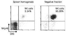
- FIG. 1B Changes of NKp46 + DX5 + natural killer cells (upper right quadrant) in spleen lymphocytes by negative MACS enrichment treatment were confirmed by flow cytometry; (FIG.
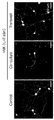
- FIG. 1C Immunostaining for embryonic DRG neurons ( ⁇ -tubulin III) after co-culture with IL-12 stimulated NK cells under direct contact conditions or conditions separated by trans-well membrane;
- FIG. 1D Granzyme B was detected by ELISA in natural killer cell-DRG neuron co-culture medium;
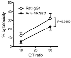
- FIG. 1E confirming the effect of NKG2D receptor on natural killer cell-mediated lysis of DRG neurons.
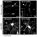
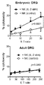
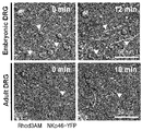
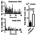
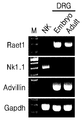
- FIG. 2 shows the results of susceptibility to NK-mediated cytotoxicity by RAE1 in cultured embryonic DRG neurons: (FIG. 2A) embryonic DRG neurons or adult DRG neurons co-cultured with IL-12 stimulated natural killer cells Immunolabeling of ⁇ -tubulin (magenta) and NKp46 (green) in the stomach; (FIG. 2B) LDH-release cytotoxicity analysis of acutely cultured embryonic DRG neurons or adult DRG neurons treated with various ratios of effector (NK): target (DRG) (E: T); (FIG.
- FIG. 2A embryonic DRG neurons or adult DRG neurons co-cultured with IL-12 stimulated natural killer cells Immunolabeling of ⁇ -tubulin (magenta) and NKp46 (green) in the stomach
- FIG. 2B LDH-release cytotoxicity analysis of acutely cultured embryonic DRG neurons or adult DRG neurons treated with various ratios of effector (NK): target (DRG) (E: T);
- FIG. 2C Time-lapse in vitro co-culture of embryonic DRG neurons or adult DRG neurons treated with rhodamine-3AM and IL-12-stimulated natural killer cells derived from NKp46-YFP male adult mice Confirming the focal Ca 2+ image;
- FIG. 2D As a result of co-culture of embryonic DRG neurons or adult DRG neurons with IL-12-stimulated natural killer cells, the frequency of neurite Ca2 + events was confirmed over time;
- FIG. 2E RT-PCR confirmed the expression of mRNA transcripts (Raet1, NK1.1, Advillin) in untreated splenic killer cells, embryonic DRG neurons, or adult DRG neurons;
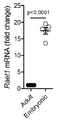
- FIG. 2F Raet1 mRNA expression in embryonic DRG neurons or adult DRG neurons was confirmed by qRT-PCR;
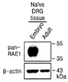
- FIG. 2G Western blot using pan-RAE1 antibody in embryonic DRG neurons or adult DRG neurons;
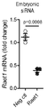
- FIG. 2H Results of knocking down Raet1 mRNA in embryonic DRG neurons using Raet1 selective siRNA;
- FIG. 2I LDH-releasing cytotoxicity assays of embryonic DRG neurons knocked down Raet1 mRNA.
- FIG. 3 shows increased expression of Raet1 and fragmentation of natural killer cell-mediated neurites in injured adult DRG neurons:
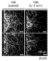
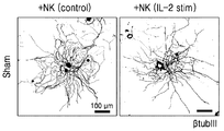
- FIG. 3A Microfluidity of adult DRG neurons with exposure to natural killer cells stimulated with IL-12 After culture, immunolabeling of ⁇ -tubulin III in neurites;
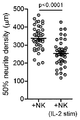
- FIG. 3B Quantification of the density of DRG neurites;
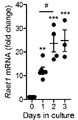
- FIG. 3C Changes in Raet1 mRNA expression over time in adult DRG neuron cultures were confirmed by qRT-PCR;
- FIG. 3D Days 1 and 2 confirmed Western blot RAE1 protein expression in adult DRG cultures in vitro;
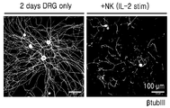
- FIG. 3E Immunolabeling of ⁇ -tubulin III in adult DRG alone culture (day 2 in vitro) or adult DRG culture co-cultured with IL-12-stimulated natural killer cells;
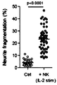
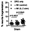
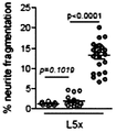
- FIG. 3F Quantification of fragmentation of DRG neurites;
- FIG. 3G Results of knocking down Raet1 mRNA in in vitro adult DRG neurons using Raet1 selective siRNA;
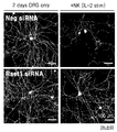
- FIG. 3H Immunolabeling of ⁇ -tubulin III in adult DRG alone cultures transfected with Raet1 siRNA (day 2 in vitro), or adult DRG cultures co-cultured with IL-12-stimulated natural killer cells ;
- FIG. 3I quantification of fragmentation of DRG neurites.
- Figure 4 shows the effect of blocking NKG2D receptors on natural killer cells on neurite degeneration of adult DRG neurons:
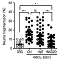
- Figure 4A Pretreatment of anti-NKG2D antibodies to adult DRG neurons and stimulated with IL-12 Co-culture with natural killer cells followed by immunolabeling of ⁇ -tubulin III;
- FIG. 4B quantification of fragmentation of DRG neurites.
- FIG. 5 shows the effects of peripheral nerve damage on Raet1 expression and the effects of sensory neuronal damage on neurite fragmentation by stimulated natural killer cells:
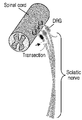
- FIG. 5A Lumbar spine (L3) , L4, and L5) diagrams showing spinal nerve cutting sites associated with DRG;
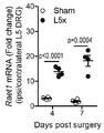
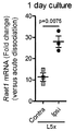
- FIG. 5B After spinal neurotomy for contralateral DRG, expression of Raet1 mRNA in ipsilateral L5 DRG over time was confirmed by qRT-PCR;
- FIG. 5C Expression of Raet1 mRNA associated with injury in adult DRG neurons (day 1 in vitro) was confirmed by qRT-PCR;
- FIG. 5A Lumbar spine (L3) , L4, and L5) diagrams showing spinal nerve cutting sites associated with DRG
- FIG. 5B After spinal neurotomy for contralateral DRG, expression of Raet1 mRNA in ipsilateral L5 DRG over time was confirmed by qRT-PCR;
- FIG. 5C Expression of Raet1 mRNA associated with injury in adult DRG neurons (day
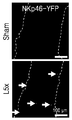
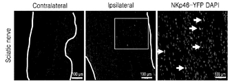
- FIG. 5H Sciatic nerve tissue sections obtained from adult male NKp46-YFP mice induced with L5 spinal cord nerve cutting injury were immunolabeled with anti-GFP;
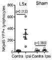
- FIG. 5I Quantification of the total number of YFP-positive events in lymphocytes FCC / SSC gated from total sciatic nerve cell grinding;
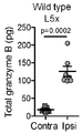
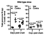
- FIG. 5J After inducing L5x damage, the content of granzyme B in the total sciatic nerve of wild-type mice was quantified by ELISA;
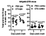
- FIG. 5K after inducing L5x damage, the results of quantification of granzyme B in total sciatic nerve of NKp46-DTR mice by ELISA.
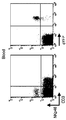
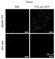
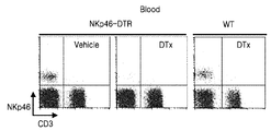
- FIG. 6 shows the results of characterizing NKp46-cre mice: (FIG. 6A) flow cytometry analysis of peripheral blood lymphocytes derived from NKp46-YFP mice labeled with anti-NKp46 and anti-CD3 antibodies; (FIG. 6B) Spleen tissue sections from wild type and NKp46-YFP mice were immunolabeled with anti-GFP antibody (green); And (FIG. 6C) flow cytometry of peripheral blood lymphocytes derived from wild-type and NKp46-DTR mice 24 hours after intravenous administration of DTx (100 ng).
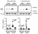
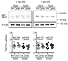
- FIG. 7 shows the effect of sciatic nerve compression on RAE1 in peripheral nerve exons:
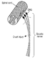
- FIG. 7A Diagram showing sciatic nerve compression injury associated with lumbar spine (L3, L4, and L5) DRG;
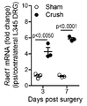
- FIG. 7B 3 and 7 days after surgery, qRT-PCR confirmed the expression of Raet1 mRNA associated with compression injury in ipsilateral L3-5 DRG;
- FIG. 7C Expression of RAE1 protein in sciatic nerve after peripheral nerve compression injury (day 3 and 7);
- FIG. 7D Expression of RAE1 protein in DRG neurons after peripheral nerve compression injury (day 3 and 7);
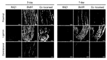
- FIG. 7E after strong ligation (day 3 and 7), co-localization of exon marker ⁇ -tubulin III (magenta) and RAE1 (green) in the sciatic nerve.
- FIG. 8A Sciatic nerve tissue sections derived from adult male NKp46-YFP mice at day 7 after sciatic nerve compression injury. Confirmed results;
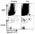
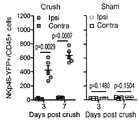
- FIG. 8B Flow cytometry was performed on sciatic nerve cell grinding fluid obtained from adult male NKp46-YFP mice on day 3 after sciatic nerve compression injury;
- FIG. 8C Quantification of the total number of CD45 + / YFP + double positive events in lymphocyte FSC / SSC gates from total sciatic nerve cell lysate;
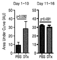
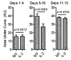
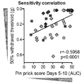
- FIG. 8D results showing area under the curve (AUC) values associated with sensory accumulation in the entire sciatic nerve injury and recovery process;
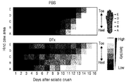
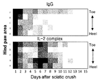
- FIG. 8F Heat map showing mean sensitivity to fin stimulation in lateral hind paws;
- FIG. 8G NKp46 + / DX5 + double positive lymphocytes in peripheral blood 16 days after sciatic nerve compression.
- Figure 9 shows the results of quantifying Granzyme B in whole sciatic nerve after crush injury or sham injury:
- Figure 9A Results of quantifying Granzyme B in whole sciatic nerve after crush injury or sham injury in wild type mice ;
- FIG. 9B Granzyme B in whole sciatic nerve after crush injury or sham injury in NKp46-DTR mice.
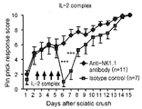
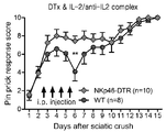
- FIG. 10 shows the effect of IL-2 / IL-2 antibody complex on spontaneous killer cell-dependent acute sensory loss due to partial sciatic nerve compression:
- FIG. 10A Treatment of IL-2 / IL-2 antibody complex Decrease in transient sensitivity by daily pin stimulus response scores;
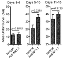
- FIG. 10B Results showing AUC values associated with loss of sensory cumulative in mice treated with IL-2 / IL-2 antibody complex;
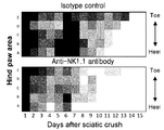
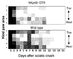
- FIG. 10C Heat map showing mean sensitivity to fin stimulation in lateral hind paws;
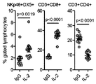
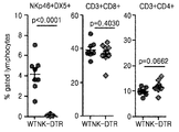
- FIG. 10D Confirmation of changes in NKp46 + DX5 + natural killer cells, CD3 + CD8 + T cells, or CD3 + CD4 + T cells in peripheral blood by treatment of the IL-2 / IL-2 antibody complex;
- FIG. 10E In mice treated with anti-NK1.1 to deplete natural killer cells, transient decrease in sensitivity by treatment of IL-2 / IL-2 antibody complex was confirmed by daily pin stimulation response scores;
- FIG. 10F Results showing AUC values associated with loss of sensory cumulative in mice treated with IL-2 / IL-2 antibody complex;
- FIG. 10G Heat map showing mean sensitivity to fin stimulation in lateral hind paws;
- FIG. 10H the change of NKp46 + DX5 + natural killer cells, CD3 + CD8 + T cells, or CD3 + CD4 + T cells in peripheral blood by treatment of the IL-2 / IL-2 antibody complex.
- FIG. 11 shows the effect of depletion of natural killer cells on acute sensory loss by treatment of IL-2 complex after partial sciatic nerve compression: (FIG. 11A) with partial sciatic nerve compression in NKp46-DTR mice. In the case of DTx treatment, the transient decrease in sensitivity due to the treatment of the IL-2 / IL-2 antibody complex was confirmed by daily pin stimulus response scores; (FIG. 11B) Heat map showing mean sensitivity to fin stimulation in lateral hind paws; And (FIG. 11C) changes of NKp46 + DX5 + natural killer cells, CD3 + CD8 + T cells, or CD3 + CD4 + T cells in peripheral blood by treatment of the IL-2 / IL-2 antibody complex.
- FIG. 12 shows the effect of IL-2 / IL-2 antibody complex on the long-term mechanical threshold by axon injury in the sciatic nerve:
- FIG. 12A Partial in mice treated with IL-2 / IL-2 antibody complex On day 6 after induction of injury, immunolabeling and quantification of ⁇ -tubulin III in full length anisotropic sciatic nerve sections (a-proximal, b-damaged site, c-distal site);
- FIG. 12B 6 days after inducing partial damage in mice treated with the IL-2 / IL-2 antibody complex, immunolabeling and quantifying Stathmin 2 in full length anisotropic sciatic nerve sections;
- FIGS. 12A and 12B Immunofluorescence of ⁇ -tubulin III and Stathmin 2 was observed at high magnification in FIGS. 12A and 12B;
- FIG. 12D 16 days after the partial injury was induced in mice treated with the IL-2 / IL-2 antibody complex, the ipsilateral mechanical sensitivity threshold of the hind paw was confirmed;
- FIG. 12E Induced partial injury in mice treated with anti-NK1.1 antibody and confirmed ipsilateral mechanical sensitivity threshold of hind paw 15 days after treatment with IL-2 / IL-2 antibody complex;
- FIG. 12F confirming the correlation between the mechanical sensitivity of the damaged limb and the accumulated pin stimulus sensitivity.
- FIG. 13 shows the results of simultaneous immunolabeling of RAE1 (green) and STMN2 (magenta) in chronically ligation of the sciatic nerve.
- 14A to 14C are photographs confirming that natural killer cells selectively infiltrate brain tissues induced by excitatory toxicity of the central nervous system.
- Ncr1icre mice (RRID: MGI: 5308422) containing cre-recombinases inserted by homologous recombination at the 3 'end of the Ncr1 (Nkp46) gene (Narni-Mancinelli et al., 2011) are provided by Dr. Vivi Vivier's laboratory received. All mice remained homozygous in the SPF facility.
- Nrc1icre / wt; rosa26eyfp / wt (abbreviated NKp46-YFP) and Nrc1icre / wt; rosa26dtr / wt (abbreviated NKp46-DTR) mice were crossbred in an SPF facility and transferred to a conventional room at least one week before the experiment. Mice were maintained at a 12 hour: 12 hour contrast cycle (turned on at 8:00 a.m.), 4-6 mice per cage were placed in wood chip bedding and randomly provided with standard laboratory feed and water.
- DRG neurons and killer cells were prepared from male C57BL / 6 mice (6-8 dwells); Embryonic DRG neurons were prepared from embryonic day 15 (E15) of euthanized female C57BL / 6 mouse uterus. Nerve damage experiments were performed in male mice with the indicated genotypes at weeks 7-9. Animals were treated by inhaling lethal doses of isoflurane in accordance with Annex 1 of the British Animals (Scientific Procedures) Act of 1986 and then inhaling lethal doses of carbon dioxide for tissue culture.
- NKp46-DTR mice were injected with diphtheria toxin (DTx; 100 ng) or sterile PBS solution (100 ⁇ l) intravenously by retro-orbital injection (Yardeni et al., 2011). Under anesthesia, ophthalmic reagents (0.5% proparacaine, Alcon, Belgium) were applied as local anesthetics under anesthesia (3% induction, 1-2% maintained at 100% O2). Insulin (0.3 ml, BD Biosciences) was injected slowly into the nasal cavity of the eye and injected both eyes alternately. Injections of blinding diphtheria toxin or sterile PBS solution that started one day before surgery continued at 4-5 day intervals during the experiment.
- mice Male mice were placed under isoflurane inhalation, shaved back lumbar region and treated with iodine solution (Potadine) to make unilateral incisions parallel to the L6 vertebrae.
- Muscle tissue was dissociated with a blunt separation to reveal the L6 transverse process in a x20 dissecting microscope illuminated with a cold light source, which was then cut and removed.
- the wound was washed with sterile saline and closed in two layers with 6-0 silk suture (Ailee, Korea) and 9 mm skin clip (MikRon Precision, CA, USA). Mice were placed in warm dark cages to recover from surgery.
- a single unilateral compression injury was performed on the ridge nerve (Bridge et al., 1994) in adult male mice (7-9 d.). Specifically, the right thigh was shaved under isofluraine anesthesia and treated with iodine to make an incision. The umbilical nerves were exposed out of the bulge by separating the muscles with a blunt forceps rupture. The nerve was completely compressed for 15 seconds using carefully removed forceps from the connective tissue and finely mirrored forceps (No 5, Dumont, Fine Science Tools, Germany). The wound site was closed with two layers of overlapping fascia using two sutures and one skin clip.
- the hemostat was then closed in the first locking position and held for 15 seconds before the nerve was carefully released.
- the wound was closed with two layers of overlapping fascia using two sutures and one skin clip. Mice recovered in a warm dark cage. All instruments were autoclaved prior to surgery and stringent sterile conditions were maintained.
- Recombinant mouse IL-2 (Cat no. 212-12, lot no. 0608108, Peprotech, Rocky Hill, NJ, USA) was prepared as a raw material of 0.1 mg / ml in PBS (without carrier protein) and according to the manufacturer's instructions Stored at 4 ° C. for 1 week. On the day of treatment, pre-mix IL-2 (1.5 ⁇ g per mouse) with anti-mouse IL-2 monoclonal antibody (50 ⁇ g per mouse) (S4B6-1 clone) (BioXCell; RRID: AB_1107705) and 15 minutes at room temperature.
- the 50% withdrawal threshold was determined using the up-down method as previously described (Chaplan et al., 1994). Active hind foot lift or withdrawal in response to von Frey filament stimulation was considered a withdrawal response. The 0.4g filament was the first stimulus used and the weaker filament was used after the withdrawal response was obtained. This process was repeated until there was no reaction, followed by the strongest filaments. Interpolation of 50% threshold was performed using the method of Dixon (Dixon, 1980). All behavioral experiments were performed by the experimenter who could not see the treatment of the mouse. Pin-prick sensory tests were performed with some modifications as previously described (Ma et al., 2011). Mice became accustomed to elevated mesh within detached compartments.
- the affected hind paws were divided into five areas from the toe to the heel and stimulated with a stainless steel Austerlitz insect pin (Size 000, FST, Germany). Sensory responses were confirmed by rapid lifting or withering of the feet. The number of responses to two consecutive pin applications on the skin was recorded as 10 out of 10 per area. Responses from direct movement of the foot or hind paw (representing extraterritorial proprioception) were excluded. Tests were performed daily until all mice had a full sensory recovery (10 points).
- HBSS Hank's buffered saline
- Welgene including 20 mM HEPES
- Additional digestion was performed for 5-7 minutes in trypsin (0.25%) and stopped with trypsin inhibitor (2.5 mg / ml) (Sigma, T9003) in PBS, followed by Dubellco containing 10% serum (Gibco, Life Technologies). Washed in Dubellco's Modified Eagle Medium (Gibco, Life Technologies).
- DRG was dissociated by trituration with a flame polished glass pipette in DMEM containing DNase I (125 U / ml), B27 supplement, L-glutamine (1 mM), penicillin (100 U / ml) and 50 ng / Bovine serum albumin layer (15% BSA solution) before resuspending in neurobasal medium (Gibco, Life Technologies) with streptomycin (100 U / ml) supplemented with ml of nerve growth factor (NGF 2.5S) Sigma) was centrifuged at 200 g.
- DNase I 125 U / ml
- B27 L-glutamine
- penicillin 100 U / ml
- 50 ng / Bovine serum albumin layer (15% BSA solution
- DRG was seeded in poly-D-lysine and laminin-coated glass bottom dishes (103 cells per dish) and incubated overnight in nerve fiber medium containing NGF (50 ng / ml).
- NGF 50 ng / ml
- DRG neurons isolated from adult mice are suspended in nerve fiber medium and poly-D-lysine (10 ⁇ g / ml) and laminin (10 ⁇ g / ml) (sigma).
- NGF 100 ng / ml
- Neurons were incubated for 5 days, where neurites grew along a 3 ⁇ m ⁇ 500 ⁇ m channel connected to neurites reservoirs.
- Natural killer cells were prepared from the spleen of adult male C57BL / 5 mice (6-8 weeks old). Spleen cells were homogenized by sequentially passing 70 ⁇ m and 40 ⁇ m cell filters (Falcon, BD Biosciences). Erythrocytes were lysed by incubating for 2 minutes in ACK lysis buffer (mM: 150 NH 4 Cl, 10 KHCO 3 , 0.1 Na 2 EDTA, pH 7.3). Single cell suspensions were then passed through nylon wool columns (Polysciences, Warrington, PA) for depletion of the adherent population consisting of B cells and macrophages.
- ACK lysis buffer mM: 150 NH 4 Cl, 10 KHCO 3 , 0.1 Na 2 EDTA, pH 7.3
- Eluted cells were resuspended in 0.01 M phosphate buffered saline (PBS) + 2 mM EDTA and 2% FBS.
- Natural killer cells were prepared using magnetic associated cell sorting (in combination with a negative selection protocol (mouse natural killer cell separation kit II, cat no. 130-096-892, Miltenyi Biotech GmbH, Germany) according to the manufacturer's instructions). Concentrated using the MACS) method. Specifically, the cell suspension was incubated sequentially at 4 ° C. with a cocktail of biotin-conjugated monoclonal antibodies against non natural killer cells, followed by anti-biotin microbeads.
- the cell suspension was then passed through an LS column placed in a magnetic field (MidiMACS Separator, Miltenyi Biotech GmbH). Bead-conjugated non-natural killer cells remained in the column while untargeted natural killer cells passed through the eluent. Enriched natural killer cells were used directly at 1000 U / ml (control) or recombinant mouse interleukin (IL) -2 (cat no. 212-12, lot number 0608108, Peprotech, Rocky Hill, 2 days prior to use in experiments). NJ, USA).
- IL mouse interleukin
- Natural killer cells were plated with RPMI 1640 medium (Gibco) supplemented with fetal bovine serum (FBS) (10%) and penicillin / streptomycin (100 U / ml) for 48 hours in 96-well U-bottomed (Falcon, BD Biosciences) plates. , Life Technologies) at 2 ⁇ 10 6 cells / ml. Purity of natural killer cells (NKp46 + DX5 +) in the eluate was consistently tested to more than 90% by flow cytometry. The cells were then harvested and used as effector cells in co-culture and cytotoxicity experiments.
- RPMI 1640 medium Gibco
- FBS fetal bovine serum
- penicillin / streptomycin 100 U / ml
- Purity of natural killer cells (NKp46 + DX5 +) in the eluate was consistently tested to more than 90% by flow cytometry. The cells were then harvested and used as effector cells in co-culture and cytotoxicity experiments.
- Control or IL-2 stimulated natural killer cells were harvested, washed with RPMI and resuspended in nerve fiber media.
- DRG are washed once in neuronal medium (NGF- non-existence) is added to the NK neurite compartment (5 ⁇ 10 5 cells) for microfluidic or DRG cultures were immediately seeded directly on to the glass cover slip cultures (2.5x10 5 Cells), co-cultured at 37 ° C. and 5% CO 2 for 4 hours.
- the coculture was carefully washed once in warm HBSS, fixed with 2-4% PFA in 0.01 M PBS (pH 7.4) for 30 minutes at room temperature, and then for ⁇ -tubulin III and NKp46 (see immunofluorescence). Store at 4 ° C.
- Natural killer cells (2.5 ⁇ 10 5 per well) were seeded in a 6.5 mm diameter polycarbonate trans-well membrane insert with 0.4 ⁇ m pore size (Corning) and incubated at 37 ° C. and 5% CO 2 for 4 hours as described above. The membrane and fixative were removed.
- natural killer cells were LEAF purified anti-mouse CD314 (NKG2D) (CX5 clone) for 15 minutes at room temperature prior to addition to target DRG neurons (Biolegend, cat no.
- RRID AB_1227715
- LEAF purified rat IgG1 isotype control clone RTK2071 (Biolegend, cat no. 400414.RRID: AB_326520) at 30 ⁇ g / ml (2.5 ⁇ 10 6 natural killer cells / ml).
- Effector natural killer cells were analyzed for cytotoxicity against DRG neuron targets by measuring the release of lactate dehydrogenase (LDH) into the culture medium using the LDH Cytotoxicity Assay Kit (Thermo Scientific Pierce, IL, US).
- Control or IL-2 stimulated natural killer cells were harvested, washed with RPMI, added to DRG cultures in 96 well plates (Nunclon, Thermo Scientific) in various ratios of neuronal serum medium, followed by incubation for 4 hours, Media supernatants were analyzed for LDH activity according to the manufacturer's instructions. Absorbance values were obtained on a microplate spectrophotometer (BioTek Instruments, VT, US). Each ratio was determined to be triple.
- Cytotoxicity% [(Experimental Release-Natural Release) / (Maximum Release-Natural Release)] ⁇ 100.
- the optimal target cell count (DRG neurons) for detecting maximum LDH release was Prior to the experiment (8x10 embryonic DRG neurons, 10 3 adult DRG neurons) were determined.
- the dish containing the DRG-NK co-culture was immediately transferred to a confocal microscope (LSM 700, Zeiss) and kept in a humidified atmosphere at 37 ° C. and 5% CO 2 (Live Cell Instruments, Seoul Korea).
- LSM 700, Zeiss confocal microscope
- the time series of a single z-section image (512 x 512) uses a multi-track setup (488 nm and 555 nm fluorescence emission and differential interference contrast (DIC) bright-field) at 30-60 second intervals, and under the control of Definite Focus Acquired using location. Images were collected for up to 3 hours and exported at continuous low speed in AVI format.
- mice Male NKp46-YFP mice (park 8-9) underwent sagittal nerve compression or sham surgery on one side. On day 3, mice were anesthetized with pentobarbital (80 mg / kg, i.p.) supplemented with 20 mg / kg immediately before recording. Some mice were administered Dextran-Texas red (neutral 40,000 m.w.) (Molecular Probes) via retro-orbital injection (100 ⁇ l, 10 mg / ml) to confirm blood flow retention during visualization and recording of blood vessels. The mandibular nerves were again exposed and washed in sterile saline. Mice were placed on warm pads maintained at 35 ° C. during imaging.
- the arches were carefully lifted with two glass rods through micromanipulators at both ends of the exposed nerve.
- the W plan-Apochromat 20X immersion lens was lowered to the nerve surface 1-2 mm from the compression site.
- Rapidly isolated adult or E15 embryo DRGs were transfected with siRNA by electroporation (Neon, Invitrogen) according to the manufacturer's instructions. Specifically, a single cell suspension of DRG was suspended in electroporation medium containing siRNA oligonucleotides and drawn into the tip containing gold-plated electrodes (5 ⁇ 10 4 cells per 10 ⁇ l). The cell tip was placed in a tube containing electrolyte buffer (1500V, 20ms), electroporated (1500V, 20ms), immediately ejected into penicillin / streptomycin-free nerve fiber medium containing 50ng / ml NGF and 37 Knockdown or functional experiments were evaluated by incubation for 48 hours at 5 ° C., 5% CO 2 .
- siRNA oligonucleotide Prior to the experiment, transfection efficiency was optimized by electroporation of DRG with cDNA plasmid encoding green fluorescent protein and GFP fluorescence 2 days after culture.
- Two siRNA oligonucleotides per target were tested for knockdown efficiency by real time PCR; SiRNA oligos with at least 70% reduction in mRNA expression compared to the negative control siRNA oligonucleotide were used in functional experiments.
- Gapdh siRNA oligonucleotide (10 nM) (Silencer Select, Ambion, Life Technologies, cat no..4390849) was used as a positive control.
- Peripheral blood samples obtained from retro-orbital bleeding confirmed systemic depletion of natural killer cells at the end of the experiment. Specifically, a 15 ml glass Pasteur pipette was inserted into the retro-orbital sinus under isoflurane anesthesia and twisted slowly to rupture the orbital venous plexus. 50-100 ⁇ l of blood was removed by capillary action and ejected into heparin-coated tubes (Idexx Laboratories, USA). Blood samples were diluted with equal volume of serum free RPMI, suspended on lymphocyte separation media (Lympholyte Mammal, Cedarlane Labs, Canada) and centrifuged at 1000 g for 20 minutes with a slow acceleration gradient. Peripheral blood mononuclear cells were harvested from the monolayer and washed with RPMI and suspended in FACS buffer (5% FBS, 0.002% NaN 3 in 0.01M PBS) for flow cytometry analysis.
- FACS buffer 5% FBS, 0.002% NaN 3 in 0.
- Lymphocytes were initially gated according to the FSC-SSC scatter profile; Cell populations were identified by fluorescent gating compared to unlabeled or IgG controls.
- Antibodies used were PE rat anti-mouse NKp46 (clone 29A1.4) monoclonal antibody (1: 200, eBioscience, cat no. 12-3351.RRID: AB_1210743), APC rat anti-mouse CD49b (clone DX5) monoclonal Antibody (1: 500; eBioscience, cat no. 17-5971. RRID: AB_469484), FITC Armenian Hamster anti-mouse CD3e (clone 145-2C11) monoclonal antibody (1: 1000, eBioscience, cat no.
- RRID AB_464881
- APC rat anti-mouse CD45 clone 30-F11 monoclonal antibody (1: 200, eBioscience, cat no 17-0451, RRID: AB_469393)
- PE rat anti-mouse CD4 clone GK1.5
- Monoclonal antibody (1: 1000, eBioscience, cat no. 12-0041, RRID: AB_465507), APC rat anti-mouse CD8a (clone 53-6.7) monoclonal antibody (1: 1000; eBioscience, cat no. 17-0081 , RRID: AB_469335).
- the titres of flow cytometry antibodies were determined before the experiment compared to equivalent concentrations of the fluorescence-conjugated IgG isotype control.
- the percentage of peripheral blood cell population was calculated from 20,000 gated lymphocyte events.
- Granzyme B cell suspensions were washed with RPMI to label into cells, resuspended in FACS buffer, and the Fc receptor was blocked at 4 ° C. for 15 minutes. Cell fixation and permeablisation were then performed in BD Cytofix / Cytoperm buffer (BD Biosciences, cat no.554554) at 4 ° C. for 20 minutes, followed by PE rat anti-mouse granzyme B (clone NGZB) monoclonal antibody (1 : 100; eBioscience, cat no. 12-8898. RRID: AB_10853811) or PE rat IgG2a isotype control (1: 100; eBioscience, cat no. 12-4321, RRID: AB_470052) for 30 min at 4 ° C. Washed with BD Perm / Wash buffer.
- NKp46-YFP mice were deeply anesthetized with pentobarbital (100 mg / kg, ip), and then PBS (0.01M, pH) at various time points after peripheral nerve injury. Myocardial perfusion was performed in 7.4) to remove peripheral blood from the circulation. Bilateral buttocks are quickly removed with ice-cold Ca2 + and Mg2 + free HBSS (Welgene) (including 20 mM HEPES) and cut into 1-2 mm pieces. The tissues were transferred to 15 ml tubes and centrifuged at 500 g for 5 minutes.
- HBSS was replaced with collagenase A (1 mg / ml) and dispase II (2.4 U / ml) (Roche, Switzerland) and incubated at 37 ° C. for 90 minutes with frequent stirring. Further digestion was performed for 5 minutes in trypsin (0.25%), stopped with trypsin inhibitor (2.5 mg / ml) (Sigma, T9003) in PBS and washed in RPMI (Gibco, Life Technologies) containing 10% serum.
- Natural killer cells were identified by lymphocyte FSC-SSC scattering profile and YFP fluorescence; In some experiments total lymphocytes were additionally labeled with APC-conjugated anti-mouse CD45 antibody (1: 400, eBioscience, cat no. 17-0451, RRID: AB_469393).
- mice are deeply anesthetized with pentobarbital (100 mg / kg, ip) and then subjected to myocardial perfusion with PBS (0.01 M, pH 7.4) containing heparin (500 U / L). Bleeding was performed. The entire DRG (Lumbar L3-L5) and full length umbilical nerve (spinal nerve nerve for peripheral triangulation) tissues were dissected and immediately frozen in liquid nitrogen and collected in sample tubes stored at -70 ° C for molecular analysis. After PBS perfusion for immunohistochemical staining, paraformaldehyde fixative fixative (0.1M PBS, 4% PFA at pH 7.4, 0.2% picric acid) was administered.
- Primary antibodies used were: rabbit anti- ⁇ -tubulin III (1: 400-500, Sigma, cat no T2200.RRID: AB_262133), goat anti-NKp46 (1: 200, R & D Systems, cat no.AF2225, RRID: AB_355192), rabbit anti-STMN2 (1: 500, Novus Biologicals, cat no.NBP1-49461 RRID: AB_10011569), goat anti mouse pan-RAE1 antibody (1:40, R & D systems, cat no.AF1136.RRID: AB_2238016.
- the fluorescent conjugated secondary antibody was incubated for 1 hour at room temperature in 1% NDS, 0.1-0.3% triton-X 100 in PBS for 1 hour.
- Tissues and cells were homogenized in RIPA buffer (Millipore, Cat # 20-188) containing protease inhibitor cocktail (Sigma, P8340) and phosphatase inhibitor cocktail (Gendepot, Cat # P3200). Cultured cells were washed with warmed HBSS and collected in protein lysis buffer by scraping; Frozen tissue was crushed in a Minilys bead homogenizer (Precellys, Bertin, France) or glass mill. The homogenized sample was sonicated on ice (3 ⁇ 10 s, 25% amplitude) and then incubated for 40 minutes on ice, then spun at 4 ° C. for 10 minutes at high speed (10,000 g) and discarded pellets.
- Equal amounts of 5X SDS sample buffer was added to the sample lysate boiled for 5 minutes on a 9 ° C. heat block. Protein content was determined by colorimetric analysis (Lowry, BioRad). Equal amounts of protein (25-40 ⁇ g) and protein size markers were separated by SDS-polyacrylamide gel electrophoresis (5% stacking gel, 10% resolving gel) and transferred to PVDF membrane. Membranes were blocked for 1 hour in a 5% skim milk solution containing Tris buffered saline and 0.1% Tween-20 (TBS-T) at room temperature, followed by goat anti-mouse Pan-RAE1 antibody (1: 500, R & D systems, cat no.AF1136.RRID: AB_2238016).
- the blot was washed with TBS-T (3 ⁇ 10 min) and then incubated with anti-chlorine HRP-conjugated secondary antibody (1: 10,000; Santa Cruz, cat no. 2020, RRID: AB_631728) for 1 hour at room temperature. .
- color development was achieved by applying Western ECL substrate (BioRad, cat no. 1705061) according to the manufacturer's instructions, and images of sequential exposure time were obtained digitally (ChemiDoc, BioRad).
- the blot was then stripped with stripping buffer at 50 ° C. for 30 minutes followed by TBS-T wash followed by mouse anti-beta-actin (1: 10,000; Sigma, cat no.
- Granzyme B content of the umbilical nerve was determined by ELISA according to the manufacturer's instructions (DuoSet, R & D Systems, cat no DY1865). Specifically, 96 well-plates were coated with capture antibody overnight at room temperature, washed and blocked for 1 hour. Frozen tissue was homogenized in a shaker with 1.4 mm zirconium beads (Precellys, Bertin) in RIPA lysis buffer (Millipore) and the lysates were centrifuged at 13,000 rpm for 5 minutes. The supernatant was diluted 1: 1 with reagent dilution, added twice to wells coated with Granzyme B standard series and incubated for 2 hours at room temperature.
- RNA total DRG 150-250 ng, cell culture
- M-MLV 200 U / rxn
- dNTP dNTP
- oligo oligo 12-18 primers
- Invitrogen 500 ng of water
- PCR reaction 25 ⁇ l
- Go Taq Flexi DNA polymerase Promega
- PCR conditions were repeated 35 times from 95 degreeC (2 minutes), 95 degreeC (30 second) to 60 degreeC (30 second), 72 degreeC (30 second), 72 degreeC (5 minutes), and 4 degreeC.
- Reactions performed without cDNA were used as negative controls.
- PCR products were run on agarose (1.5%) gels containing DNA staining reagents (SafePinky, GenDepot, USA) and visualized with a UV transmission illuminator.
- Gene expression was determined using DRG using a pair of target specific primers (500 nm) in a microamp light tube (20 ⁇ l reaction volume) of the Power SYBR Green PCR Master Mix (applied biosystem) and 7500 Real Time PCR system (applied biosystem). Was performed on cDNA from. PCR conditions were repeated 40 times at 50 ° C. (2 min), 95 ° C. (10 min), and 95 ° C. (15 sec) at 60 ° C. (1 min). Samples were performed in triplicate. Data were analyzed using the built-in 7500 software (v2.0.4, Life Technologies), and expression was determined using the comparative Ct method (Schmittgen and Livak, 2008) for the reference gene (Gapdh) or neuronal damage experiment of adult DRG for culture.
- Primer was designed using Primer-BLAST software (NIH) (Ye et al., 2012). Primers are characterized by BLAST search, overlap of exon-exon boundaries, potential hairpin formation or lack of self priming regions, single peak of dissociation curve (single band PCR product) and equivalent amplification efficiency (linear shift of Ct value upon serial dilution of cDNA). Selection was made based on the specificity for the desired target gene. Reverse transcription products without RNA or M-MLV (-RT) were used as negative controls.
- a single z-section low magnification (x10) confocal image at 0.5-fold zoom (LSM700, Zeiss) has ⁇ -tubulin III immunofluorescence along the entire length of the neurites compartment ( 647 nm emission).
- Gamma high channel images were exported to unchanged TIFF using Zeiss imaging software (v8.1, ZEN 2012 SP1, Zeiss). The image was converted to black and white by adjusting to the set threshold, and the scale correction and horizontal pixel density were measured using the plot profile function of image J (v1.46r, NIH). The distance to 50% neurites density was calculated from the normalized cumulative pixel density.
- the neurite fragments were selected using the particle analysis function (size 0.5-25 ⁇ m 2, roundness 0-1) and saved as drawings. Total area and specific area value ( ⁇ m2) were obtained using the measurement function and used to calculate the percentage of neurites fragments for each visual field.
- the complex of full-length nerves was made of individual z-section images acquired at x10 magnification and 0.5 times magnified images along the nerve length (LSM700, Zeiss). Images containing the compression site as well as the proximal and distal portions ( ⁇ 1 mm from the compression site) were scaled with ImageJ (0.8 pixels / ⁇ m) and the brightness threshold was set in black and white mode.
- Example 1 Identified activated killer cells induce cytotoxicity by RAE1-mediated mechanism in embryonic sensory neurons
- Natural killer cells are activated by the cytokine IL-2, which causes a cytotoxic attack by increasing the intracellular content of granzyme B (FIG. 1A).
- isolated, unstimulated splenic natural killer cells were used as controls (FIG. 1B), with IL-2 against DRG neurons acutely isolated from embryonic (E15) and adult mice ( ⁇ 24 hours in vitro). The effects of stimulated natural killer cells were investigated. As in previous reports, embryonic DRG neurons were very sensitive to natural killer cell mediated cytotoxicity (FIGS. 2A and 2B). Natural killer cells and embryonic DRG with trans-well membranes despite the presence of nanogram levels of granzyme B in culture medium (FIG.
- the protein Retinoic Acid Early 1 (RAE1), encoded by the Raet1 gene family ( ⁇ , ⁇ , ⁇ , ⁇ , ⁇ ), acts as a membrane-binding ligand for the mouse activated receptor NKG2D (Cerwenka et al., 2000), which is an embryonic DRG Has been associated with NK cell mediated lysis (Backstrom et al., 2003); However, the function of Raet1 in NK cytotoxicity against DRG neurons has not yet been identified. In this example, it was confirmed that spontaneous killer cytotoxicity against embryonic DRG neurons can be attenuated by NKG2D receptor blocking antibodies (FIG. 1D).
- Raet1 transcripts were observed in acute dissociated embryos and adult DRG neurons using RT-PCR as a universal primer designed to detect all five Raet1 isoforms ( ⁇ , ⁇ , ⁇ , ⁇ , ⁇ ).
- RT-PCR Quantitative RT-PCR (qRT-PCR) showed that Raet1 transcript was 17 times more concentrated in embryonic DRG compared to adult DRG (FIG. 2F). This difference was also seen at the protein level: a single large band of about 40-50 kDa was detected by pan-RAE1 antibody in the western blot of the embryo, but not in adult DRG tissue (FIG. 2G).
- Raet1 siRNA was used to selectively knock out all Raet1 isoproteins (FIG. 2H). Compared to the negative control siRNA, embryonic DRG neurons were transfected with Raet1-selected siRNA, resulting in a 20% reduction in spontaneous killer cell mediated cell lysis (FIG. 2I). These results show that Raet1 expression of embryonic DRG neurons is involved in cytotoxicity induced by engagement with stimulated killer cells.
- Example 2 Confirmation that neuronal damage induces RAE1 expression allowing cytotoxic attack by activated natural killer cells in adult sensory neurons
- DRG neurons were cultured in a microfluidic chamber (5 days in vitro) to selectively axon projections to stimulated natural killer cells in neurites compartments. Exposed. After exposure to stimulated natural killer cells, there was a 25% loss of neurites range compared to isolated control killer cells (FIGS. 3A and 3B).
- RAE1 plays a role in spontaneous killer cell mediated neurite fragmentation in long-term adult DRG cultures.
- Raet1 mRNA was upregulated time-dependently in adult DRG cultures with the corresponding de novo expression of RAE1 protein (FIG. 3D) after 2 days in vitro (FIG. 3C).
- Example 3 Stimulated natural killer cells degenerate partially damaged sensory axons
- Ncr1 encodes tissue-resident ILC, which includes a subset of group 1 ILC (ILC1s) and group 3 ILC (NCR1 + ILC3s) as well as NKp46 receptors expressed in all natural killer cells.
- This genetic approach enabled the identification (via YFP expression) or systemic destruction (via DTR) of NKp46 + cells in vivo (FIGS. 6A, 6B and 6C).
- YFP-positive killer cells were not observed in the nasal nerves of naive mice or sham surgical mice (FIGS. 5H and 5I).
- spinal cord nerve incision L5x
- induced significant recruitment of YFP-positive killer cells to the injured sagittal nerves FIGGS. 5H and 5I).
- natural killer cells were highly motile and exhibited a multipolar morphology reminiscent of stimulated natural killer cells in vitro (not shown 6). These cells were placed to functionally ideally interact with damaged peripheral nerve axons that selectively express natural killer cell target protein RAE1. YFP + cells were not observed by two-photon imaging in the nerves of sham damaged mice (data not shown).
- Mandibular nerve squeezing injury leads to rapid loss of sensory and wallerian degeneration of the distal axonous axon at the site of injury and causes sensory recovery within about two weeks (Ma et al., 2011). After compression using micro forceps, a complete loss of response to pin-frick stimulation of the lateral hind paw was confirmed in all mice (FIGS. 8D and 8E). Control (PBS-treated) NKp46-DTR mice showed a typical pattern of sensory recovery (Ma et al., 2011; Painter et al., 2014), until relatively fast recovery began about 10-11 days after injury. Almost completely insensitive (FIG. 8D, PBS).
- mice chronically depleted of natural killer cells with diphtheria toxin (DTx) treatment to investigate the role of natural killer cells on axonal integrity after nerve compression injury in vivo.
- Figure 8G mice showed initial and sustained sensitivity to pin-frick stimulation (FIG. 8D, DTx).
- NK cell depleted mice showed an anatomically broad response pattern to stimulation throughout the hind paw prior to full recovery ( 8F, DTx), which shows sporadic innernervations remaining throughout the tissue.
- mice were treated systemically with the IL-2 / anti-IL-2 monoclonal (S4B6) antibody complex.
- Treatment of IL-2 / anti-IL-2 antibody complexes is known to enhance NK and CD8 + T cell populations (Boyman et al., 2006). Indeed, at the end of the experiment (16 days) compared to the control group, both NKp46 + DX5 + and CD3 + CD8 + (FIG. 10D) cell populations were enriched in the blood of IL-2 complex-treated mice, but CD3 + CD4 + T The proportion of cells decreased (FIG. 10D).
- FIGS. 10E, 10F and 10G To maintain a level of 'maintained' sensitivity throughout the foot until regeneration continues (FIGS. 10E, 10F and 10G).
- FIGS. 11A and 11B Peripheral blood flow analysis after recovery (day 16) confirmed that NKp46 + DX5 + natural killer cells were depleted by anti-NK1.1 (FIG. 10H) and DTx in NKp46-DTR in wild-type mice (FIG. 11C).
- the proportion of CD3 + CD4 + T cells was about 20% higher in the antibody depletion group than in the isoform control (FIG.
- stathmin 2 (STMN2), a microtubule-associated protein specifically expressed by damaged DRG neurons (Cho et al., 2013). Similar to the distribution of ⁇ -tubulin III, STMN2 labeling was observed through the compressed nerves and was reduced by IL-2 complex treatment compared to the IgG control at the compression and peripheral regions (FIGS. 12B and 12C, insert b and c). STMN2 was also localized with RAE1 immune label at the site of injury (FIG. 13). These results indicate that the transient sensory loss observed in the IL-2 complex treatment after partial compression injury results from the removal of damaged axon fibers in the arch nerve.
- the mechanical threshold was assessed after recovery from partial compression injury (15-16 days) by measuring the response of the lateral paw to von Frey hair stimulation.
- IL-2 complex treatment resulted in significantly higher mechanical thresholds in the hind paws of previously compressed (ipsilateral) limbs compared to control (IgG) animals showing relatively hypersensitivity (FIG. 12D).
- normalization of mechanical threshold by treatment of IL-2 complex after partial compression was blocked by pre-depletion of natural killer cells (FIG. 12E).
- Comprehensive data from all animals show that the cumulative pin-frick sensitivity (area under curve) during the peak effect of treatment (up to 5-10 days) is related to the mechanical sensitivity of damaged limbs after recovering from partial compression. (FIG. 12F).
- Kininic acid is a potent neuroexciting amino acid agonist that acts by activating the glutamate receptor, a major excitatory neurotransmitter in the central nervous system, and treatment of high concentrations of chiinic acid in mice leads to excitatory toxicity and neurodegeneration of the central nervous system ( Levesque et al., 2013).
- 14A to 14C are photographs confirming that natural killer cells selectively infiltrate brain tissues induced by excitatory toxicity of the central nervous system.
- NKp46 expression natural killer cells only penetrated into the brain tissue in the kininic acid treatment group. This suggests that natural killer cells can penetrate into the site of central nervous excitatory toxicity and selectively remove damaged or abnormal nerve areas.
- the natural killer cells are neurological diseases caused by nerve damage or abnormal nerve of central nervous system. It can be useful for the treatment of symptoms caused by seizures or neurodegeneration caused by.
Landscapes
- Health & Medical Sciences (AREA)
- Life Sciences & Earth Sciences (AREA)
- Chemical & Material Sciences (AREA)
- General Health & Medical Sciences (AREA)
- Medicinal Chemistry (AREA)
- Engineering & Computer Science (AREA)
- Immunology (AREA)
- Veterinary Medicine (AREA)
- Public Health (AREA)
- Animal Behavior & Ethology (AREA)
- Organic Chemistry (AREA)
- Bioinformatics & Cheminformatics (AREA)
- Pharmacology & Pharmacy (AREA)
- Epidemiology (AREA)
- Zoology (AREA)
- Proteomics, Peptides & Aminoacids (AREA)
- Gastroenterology & Hepatology (AREA)
- Biomedical Technology (AREA)
- Genetics & Genomics (AREA)
- Biochemistry (AREA)
- Molecular Biology (AREA)
- Cell Biology (AREA)
- Biophysics (AREA)
- Neurosurgery (AREA)
- Neurology (AREA)
- Chemical Kinetics & Catalysis (AREA)
- Nuclear Medicine, Radiotherapy & Molecular Imaging (AREA)
- General Chemical & Material Sciences (AREA)
- Toxicology (AREA)
- Biotechnology (AREA)
- Hematology (AREA)
- Wood Science & Technology (AREA)
- Microbiology (AREA)
- Hospice & Palliative Care (AREA)
- Psychiatry (AREA)
- Virology (AREA)
- Developmental Biology & Embryology (AREA)
- General Engineering & Computer Science (AREA)
- Mycology (AREA)
- Medicines Containing Material From Animals Or Micro-Organisms (AREA)
Abstract
La présente invention concerne une composition pour traiter une lésion nerveuse, comprenant des cellules tueuses naturelles, des cellules immunitaires, ou une substance visant à augmenter l'activité de ces cellules, et les cellules tueuses naturelles, les cellules immunitaires ou la substance pour augmenter l'activité de ces cellules, selon un aspect, peuvent être utilement utilisées dans un traitement fondamental d'un trouble neurologique dû à une lésion nerveuse ou à un nerf anormal, en permettant aux cellules tueuses naturelles de pénétrer la partie nerveuse endommagée et d'éliminer directement des cellules nerveuses endommagées.
Priority Applications (1)
| Application Number | Priority Date | Filing Date | Title |
|---|---|---|---|
| US17/139,618 US20210121501A1 (en) | 2018-07-04 | 2020-12-31 | Immune cell treatment of nerve damage |
Applications Claiming Priority (2)
| Application Number | Priority Date | Filing Date | Title |
|---|---|---|---|
| KR10-2018-0077922 | 2018-07-04 | ||
| KR20180077922 | 2018-07-04 |
Related Child Applications (1)
| Application Number | Title | Priority Date | Filing Date |
|---|---|---|---|
| US17/139,618 Continuation-In-Part US20210121501A1 (en) | 2018-07-04 | 2020-12-31 | Immune cell treatment of nerve damage |
Publications (1)
| Publication Number | Publication Date |
|---|---|
| WO2020009437A1 true WO2020009437A1 (fr) | 2020-01-09 |
Family
ID=69060151
Family Applications (1)
| Application Number | Title | Priority Date | Filing Date |
|---|---|---|---|
| PCT/KR2019/008066 Ceased WO2020009437A1 (fr) | 2018-07-04 | 2019-07-02 | Traitement de lésions nerveuses par des cellules immunitaires |
Country Status (3)
| Country | Link |
|---|---|
| US (1) | US20210121501A1 (fr) |
| KR (3) | KR102454376B1 (fr) |
| WO (1) | WO2020009437A1 (fr) |
Families Citing this family (2)
| Publication number | Priority date | Publication date | Assignee | Title |
|---|---|---|---|---|
| US20250205281A1 (en) * | 2022-03-24 | 2025-06-26 | Therabest Co., Ltd. | Pharmaceutical composition for preventing or treating degenerative brain disease comprising natural killer cell |
| KR20240147956A (ko) * | 2023-03-31 | 2024-10-10 | (주) 테라베스트 | 활성화된 자연살해세포 및 이의 용도 |
Citations (1)
| Publication number | Priority date | Publication date | Assignee | Title |
|---|---|---|---|---|
| US20070244066A1 (en) * | 2006-03-28 | 2007-10-18 | Deleo Joyce A | Method for preventing or treating neuropathic pain |
Family Cites Families (13)
| Publication number | Priority date | Publication date | Assignee | Title |
|---|---|---|---|---|
| BR0314548A (pt) * | 2002-09-27 | 2005-08-09 | Biogen Idec Inc | Terapias para polineuropatia de desmielinação inflamatória crÈnica empregando-se interferon-beta |
| AU2004224277A1 (en) * | 2003-03-21 | 2004-10-07 | Wyeth | Treating immunological disorder using agonists of interleukin-21/ interleukin-21 receptor |
| WO2008071959A1 (fr) * | 2006-12-12 | 2008-06-19 | Oxford Biomedica (Uk) Limited | Vecteurs lentiviraux comprenant des micro-arn |
| KR101035556B1 (ko) * | 2008-05-22 | 2011-05-23 | 재단법인 목암생명공학연구소 | 자연살해세포의 제조방법, 이러한 방법에 의해 제조된자연살해세포, 및 이를 유효성분으로 포함하는 고형 종양치료용 조성물 |
| CA2817495A1 (fr) * | 2009-11-10 | 2011-05-19 | National Jewish Health | Composition, methodes et applications de la stimulation de reponses immunitaires |
| WO2014120764A1 (fr) * | 2013-01-29 | 2014-08-07 | Biogen Idec Ma Inc. | Agents modulateurs de s1p |
| US20160010059A1 (en) * | 2013-03-06 | 2016-01-14 | The Regents Of The University Of California | EXPANSION OF HEMATOPOIETIC PROGENITOR CELLS BY HISTONE METHYLTRANSFERASE G9a INHIBITION |
| US11173179B2 (en) * | 2015-06-25 | 2021-11-16 | Icell Gene Therapeutics Llc | Chimeric antigen receptor (CAR) targeting multiple antigens, compositions and methods of use thereof |
| KR20170034701A (ko) * | 2015-09-21 | 2017-03-29 | 코오롱생명과학 주식회사 | 통증 치료용 조성물 |
| CA3026757A1 (fr) * | 2016-07-15 | 2018-01-18 | Poseida Therapeutics, Inc. | Recepteurs antigeniques chimeriques et leurs procedes d'utilisation |
| CA3032146A1 (fr) * | 2016-08-03 | 2018-02-08 | Bio-Techne Corporation | Identification de vsig3/vista comme nouveau point de controle immunitaire et son utilisation en immunotherapie |
| GB201709808D0 (en) * | 2017-06-20 | 2017-08-02 | Kymab Ltd | Antibodies |
| KR102292935B1 (ko) | 2018-11-06 | 2021-08-24 | 재단법인 경기도경제과학진흥원 | 신경병증 통증 치료, 완화 또는 예방용 조성물 |
-
2019
- 2019-07-02 WO PCT/KR2019/008066 patent/WO2020009437A1/fr not_active Ceased
- 2019-07-02 KR KR1020190079701A patent/KR102454376B1/ko active Active
-
2020
- 2020-12-31 US US17/139,618 patent/US20210121501A1/en active Pending
-
2022
- 2022-10-06 KR KR1020220128078A patent/KR20220142976A/ko not_active Ceased
-
2024
- 2024-10-14 KR KR1020240139722A patent/KR20240155148A/ko active Pending
Patent Citations (1)
| Publication number | Priority date | Publication date | Assignee | Title |
|---|---|---|---|---|
| US20070244066A1 (en) * | 2006-03-28 | 2007-10-18 | Deleo Joyce A | Method for preventing or treating neuropathic pain |
Non-Patent Citations (5)
| Title |
|---|
| "The Response and Function of Natural Killer Cells After Peripheral Nerve Injury and Their Contribution to Neuropathic Pain", SCIENCE AND ENGINEERING PERSONAL FUNDAMENTAL RESEARCH SUPPORT BUSINESS FINAL REPORT, pages 1 - 17 * |
| BOLLINO, D. ET AL.: "Chimeric antigen receptor-engineered natural killer and natural killer T cells for cancer immunotherapy", TRANSLATIONAL RESEARCH, vol. 187, 2017, pages 32 - 43, XP085184174, DOI: 10.1016/j.trsl.2017.06.003 * |
| CUI, J.--G. ET AL.: "Possible role of inflammatory mediators in tactile hypersensitivity in rat models of mononeuropathy", PAIN, vol. 88, 2000, pages 239 - 248, XP027336213 * |
| DAVIES, A. J. ET AL.: "Natural Killer Cells Degenerate Intact Sensory Afferents following Nerve Injury", CELL, vol. 176, no. 4, 7 February 2019 (2019-02-07), pages 716 - 728, XP055673230 * |
| POLI, A. ET AL.: "NK cells in central nervous system disorders", THE JOURNAL OF IMMUNOLOGY, vol. 190, no. 11, 17 May 2013 (2013-05-17), pages 5355 - 5362, XP055673211 * |
Also Published As
| Publication number | Publication date |
|---|---|
| KR102454376B1 (ko) | 2022-10-17 |
| US20210121501A1 (en) | 2021-04-29 |
| KR20200004758A (ko) | 2020-01-14 |
| KR20240155148A (ko) | 2024-10-28 |
| KR20220142976A (ko) | 2022-10-24 |
Similar Documents
| Publication | Publication Date | Title |
|---|---|---|
| Haghayegh Jahromi et al. | Intercellular adhesion molecule-1 (ICAM-1) and ICAM-2 differentially contribute to peripheral activation and CNS entry of autoaggressive Th1 and Th17 cells in experimental autoimmune encephalomyelitis | |
| JP5829373B2 (ja) | Nogoレセプターアンタゴニスト | |
| KR20240155148A (ko) | 신경손상의 면역세포치료 | |
| DE102017114737A1 (de) | Neue T-Zellrezeptoren und deren Verwendung in Immuntherapie | |
| CN109576217A (zh) | 调节干细胞的免疫调节作用的方法 | |
| JP2010509235A (ja) | 多発性硬化症の治療 | |
| Gonzalez-Fierro et al. | Effects of a small-molecule perforin inhibitor in a mouse model of CD8 T cell–mediated neuroinflammation | |
| AU2026200686A1 (en) | Interleukin-27 producing B-cells and uses thereof | |
| EP2898896A1 (fr) | Agents pour utilisation dans le traitement de l'inflammation de la rétine | |
| US10874712B2 (en) | Methods, compounds and compositions for modulating blood brain barrier integrity and re-myelination | |
| US20230242666A1 (en) | Methods and Compositions for the Reduction of Chimeric Antigen Receptor Tonic Signaling | |
| WO2006115026A1 (fr) | Recepteur de l'humanine ou recepteur du polypeptide de type humanine | |
| Kreymborg | Delineating the cytokine profile of encephalitogenic T cells | |
| US20180371076A1 (en) | Maternal th17 cells and psychiatric disorders | |
| JPWO2008093827A1 (ja) | 中枢神経障害治療剤及び中枢神経障害の治療方法 | |
| JP2017057173A (ja) | Tim−3をターゲットとする脳損傷疾患治療用組成物及びこのスクリーニング方法 | |
| Mitsdörffer et al. | Archive ouverte UNIGE | |
| 清水謙次 | The role of Interleukin-1 receptor type 2 in the development of collagen-induced arthritis and helper T cell differentiation | |
| WO2023234330A1 (fr) | Cellule exprimant un récepteur antigénique chimérique ciblant egfr | |
| NELISSEN | The role of mast cells and their secreted factors in central nervous system trauma | |
| Menzel | Role of the cell adhesion molecule L1 during neuroinflammation in the central nervous system | |
| Mohammadi Mahvelati | The Nod-like Receptor, Nlrp12, plays an anti-inflammatory role in Experimental Autoimmune Encephalomyelitis | |
| Cupovic | Low avidity CD8+ T cells in viral infection: From neuroinflammation to adoptive T cell therapy | |
| KR20130091044A (ko) | JunB의 발현 또는 활성 억제제를 유효성분으로 포함하는 면역질환의 예방 또는 치료용 조성물 | |
| Schalks | Chronic meningeal inflammation as a cause of cortical grey matter pathology in multiple sclerosis |
Legal Events
| Date | Code | Title | Description |
|---|---|---|---|
| 121 | Ep: the epo has been informed by wipo that ep was designated in this application |
Ref document number: 19830343 Country of ref document: EP Kind code of ref document: A1 |
|
| NENP | Non-entry into the national phase |
Ref country code: DE |
|
| 122 | Ep: pct application non-entry in european phase |
Ref document number: 19830343 Country of ref document: EP Kind code of ref document: A1 |