WO2020009444A1 - 세포융합 기술을 이용한 유전자 및 세포 치료제 및 이의 용도 - Google Patents
세포융합 기술을 이용한 유전자 및 세포 치료제 및 이의 용도 Download PDFInfo
- Publication number
- WO2020009444A1 WO2020009444A1 PCT/KR2019/008081 KR2019008081W WO2020009444A1 WO 2020009444 A1 WO2020009444 A1 WO 2020009444A1 KR 2019008081 W KR2019008081 W KR 2019008081W WO 2020009444 A1 WO2020009444 A1 WO 2020009444A1
- Authority
- WO
- WIPO (PCT)
- Prior art keywords
- disease
- cells
- cell
- diseases
- muscle
- Prior art date
- Legal status (The legal status is an assumption and is not a legal conclusion. Google has not performed a legal analysis and makes no representation as to the accuracy of the status listed.)
- Ceased
Links
Images











Classifications
-
- A—HUMAN NECESSITIES
- A61—MEDICAL OR VETERINARY SCIENCE; HYGIENE
- A61K—PREPARATIONS FOR MEDICAL, DENTAL OR TOILETRY PURPOSES
- A61K35/00—Medicinal preparations containing materials or reaction products thereof with undetermined constitution
- A61K35/12—Materials from mammals; Compositions comprising non-specified tissues or cells; Compositions comprising non-embryonic stem cells; Genetically modified cells
- A61K35/28—Bone marrow; Haematopoietic stem cells; Mesenchymal stem cells of any origin, e.g. adipose-derived stem cells
-
- A—HUMAN NECESSITIES
- A61—MEDICAL OR VETERINARY SCIENCE; HYGIENE
- A61K—PREPARATIONS FOR MEDICAL, DENTAL OR TOILETRY PURPOSES
- A61K35/00—Medicinal preparations containing materials or reaction products thereof with undetermined constitution
- A61K35/12—Materials from mammals; Compositions comprising non-specified tissues or cells; Compositions comprising non-embryonic stem cells; Genetically modified cells
- A61K35/48—Reproductive organs
- A61K35/54—Ovaries; Ova; Ovules; Embryos; Foetal cells; Germ cells
- A61K35/545—Embryonic stem cells; Pluripotent stem cells; Induced pluripotent stem cells; Uncharacterised stem cells
-
- A—HUMAN NECESSITIES
- A61—MEDICAL OR VETERINARY SCIENCE; HYGIENE
- A61K—PREPARATIONS FOR MEDICAL, DENTAL OR TOILETRY PURPOSES
- A61K48/00—Medicinal preparations containing genetic material which is inserted into cells of the living body to treat genetic diseases; Gene therapy
-
- A—HUMAN NECESSITIES
- A61—MEDICAL OR VETERINARY SCIENCE; HYGIENE
- A61P—SPECIFIC THERAPEUTIC ACTIVITY OF CHEMICAL COMPOUNDS OR MEDICINAL PREPARATIONS
- A61P21/00—Drugs for disorders of the muscular or neuromuscular system
-
- A—HUMAN NECESSITIES
- A61—MEDICAL OR VETERINARY SCIENCE; HYGIENE
- A61P—SPECIFIC THERAPEUTIC ACTIVITY OF CHEMICAL COMPOUNDS OR MEDICINAL PREPARATIONS
- A61P25/00—Drugs for disorders of the nervous system
- A61P25/28—Drugs for disorders of the nervous system for treating neurodegenerative disorders of the central nervous system, e.g. nootropic agents, cognition enhancers, drugs for treating Alzheimer's disease or other forms of dementia
Definitions
- the present invention relates to genes and cell therapies using cell fusion technology, and the use thereof. Specifically, hemagglutinin neuraminidase (HN) and F (fusion) genes are transduced into cells and overexpressed. Gene and cell therapeutics using cell fusion technology, thereby enhancing cell fusion with other cells and repairing cell damage and delivering normal genes through cell fusion with damaged or dying cells or cells with gene abnormalities. , And use in diseases associated with cell damage.
- HN hemagglutinin neuraminidase
- F fusion
- Cell damage and cell death are generally progressed by cell damage and cell death.
- Representative diseases in which cell damage and death are induced include neurodegenerative disease and muscular dystrophy.
- neurodegenerative diseases including damage to the brain, spine and peripheral nerves, continue to increase.
- the cause of neurodegenerative diseases is not yet clear.
- the pathogenesis of each neurodegenerative disease is known to work slightly differently.
- amyotrophic lateral sclerosis ALS
- spinal muscular atrophy and Kennedy disease (spinal bulbar muscular atrophy) selectively kill only motor neurons so that only motor function is degenerated.
- the result can be symptomatic of quadriplegia, also called motor neuropathy disease.
- Alzheimer's disease unlike the motor neuron disease, Alzheimer's disease, Parkinson's disease, etc. are known as a typical neurodegenerative disease that the nerve cells of the brain are lost.
- neuronal loss results in general brain atrophy.
- These pathological findings are limited to the hippocampus and the olfactory cerebral cortex, which are the main brain areas that are primarily responsible for memory, but gradually spread throughout the brain through the parietal lobe and frontal lobe.
- memory loss initially occurs mainly, and as the progress progresses, clinical symptoms become diverse and become more severe.
- Parkinson's disease is caused by the loss of dopaminergic neurons in the substantia nigra (SN) located in the midbrain. Loss of dopamine nerves causes severe dopamine reduction in the striatum, resulting in impaired motor control by the extrapyramidal system, including the basal ganglia, thalamus, and motor cortex. .
- Alzheimer's disease is a US FDA-approved drug that includes Aricept, Exelon, Namenda, and Razadyne, which activate the neurotransmitter acetylcholine, which helps the activity of brain nerve cells. In addition, they also reduce brain cell damage and alleviate the symptoms or slow the progression.
- Levodopa is used as a dopamine agonist, but the long-term effect is limited.
- Muscle is an essential tissue that supports our body and maintains life. Myoblasts differentiate into myoblasts to form multinucleated myotubes, form myocytes, and the death of myoblasts and muscle cells is the cause of various diseases. In addition, there are reports of diseases in which muscle cells are lost through genetic mutations and various causes, but there is no fundamental treatment to date. These muscle diseases vary in intensity depending on the type of symptom and disease, and may also show a complex form of neurological problems such as neurons and spinal cord.
- muscle dystrophy is a disease in which muscle cells are destroyed, caused by genetic abnormalities, symptoms begin to weaken and difficult to occur, gradually worsening, dying of respiratory muscles may die.
- the DMD and BMD are caused by abnormalities of the Dystrophin gene present on the X chromosome, and about one third of them are due to natural mutations, and the rest are due to reflection genetics.
- DMD and BMD are both caused by the same gene abnormality, but a severe phenotype due to frame-shift mutations is called DMD.
- DMD the disease progresses poorly, and it is impossible to walk in most patients between the ages of 9 and 13, and not only muscle weakness but also cognitive depression may occur, and cardiomyopathy and respiratory muscle dysfunction may lead to death.
- Exon skipping targets the splicing enhancer sequence of exon 51 of the Dystrophin gene, which recovers only the reading frame and converts it into less severe genetic mutations, and thus is not a fundamental therapeutic alternative and is not a treatment for all DMDs. .
- HN and F proteins hemagglutinin neuraminidase (HN) and F (fusion) proteins. It has been confirmed that HN and F proteins enhance cell fusion with other cells, and have a high ability to restore cell damage and deliver normal genes in damaged or dying cells or cells with gene abnormalities. Accordingly, the present invention has been completed by revealing that the cell fusion technology using HN and F proteins can be usefully used to treat neurodegenerative diseases or neurological diseases or muscle degenerative diseases or muscle diseases causing cell damage.
- HN and F proteins hemagglutinin neuraminidase
- the present invention is a neurodegenerative disease containing a vector containing a gene encoding hemagglutinin neuraminidase (HN) and F (fusion) protein as an active ingredient, or Provided is a pharmaceutical composition for preventing or treating neurological diseases, or muscle degenerative diseases or muscle diseases.
- HN hemagglutinin neuraminidase
- F fusion protein
- the present invention is a pharmaceutical composition for the prevention or treatment of neurodegenerative diseases or neurological diseases, or muscle degenerative diseases or muscle diseases, containing the cells transformed with a vector containing genes encoding the HN and F proteins as an active ingredient. To provide.
- the present invention includes administering to a subject a therapeutically effective amount of a vector comprising genes encoding HN and F proteins to a subject, preventing or treating a neurodegenerative or neurodegenerative or muscular disease or muscle disease.
- a method for treating a neurodegenerative or neurodegenerative or muscular disease or muscle disease.
- the present invention comprises administering to a subject a therapeutically effective amount of a cell transformed with a vector comprising genes encoding HN and F proteins to a neurodegenerative or neurological disease, or a muscle degenerative disease or Provided are methods for preventing or treating muscle diseases.
- the present invention also provides the use of a vector comprising genes encoding HN and F proteins for use as a pharmaceutical composition for the prevention or treatment of neurodegenerative or neurological diseases, or muscle degenerative diseases or muscle diseases.
- the present invention is the use of a cell transformed with a vector comprising a gene encoding the HN and F protein for use as a pharmaceutical composition for the prevention or treatment of neurodegenerative diseases or neurological diseases, or muscle degenerative diseases or muscle diseases To provide.
- HN and F protein overexpressing cells through the normal gene and transcription regulators in the nucleus of the damaged cells, by regulating the expression of genes involved in cellular repair of the damaged cells or by repairing normal proteins through normal gene transfer, It was confirmed to recover the cells.
- HN and F protein overexpressing cells were fused with damaged cells or cells in which gene abnormalities were induced to recover normal cells.
- cell fusion techniques using HN and F proteins can be used to repair cell damage and introduce normal genes, thereby allowing the vector or cells transformed with the genes encoding the HN and F proteins to be replaced with cell damage. It can be usefully used to treat related diseases, in particular neurodegenerative diseases or neurological diseases, or muscle degenerative diseases or muscle diseases.
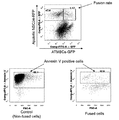
- Figure 1 shows the effect of inhibiting apoptosis of NSC34 motor neuron cell line in death by cell fusion with hATMSCs according to an embodiment of the present invention.
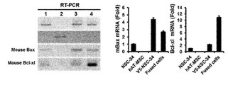
- Figure 2 shows the expression analysis of Bax and Bcl-xL in the NSC34 cell group in which cell fusion with hATMSCs according to an embodiment of the present invention.
- Figure 3 is a schematic diagram for preparing a pcDNA3.1 expression vector, each inserted with HN and F according to an embodiment of the present invention.
- Figure 4 is the analysis of HN and F mRNA expression in HN / F-hATMSCs according to an embodiment of the present invention.
- FIG. 5 is an imaging analysis of HN and F protein expression in HN / F-hATMSCs according to one embodiment of the present invention.
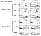
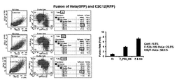
- Figure 6 analyzes the immunophenotyping (immunophenotyping) of HN / F-hATMSCs according to an embodiment of the present invention.
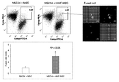
- Figure 7 analyzes the cell fusion capacity with NSC34 motor neuron line in HN / F-hATMSCs according to an embodiment of the present invention.
- FIG. 8 shows the expression of ChAT and CD105 in cells fused with HN / F-hATMSCs and NSC34 motor neuron cell lines according to an embodiment of the present invention.
- Figure 9 shows the results of proteomics analysis between the NSC34 motor neuron cell line, the killing NSC34 motor neuron cell line and HN / F-hATMSCs according to an embodiment of the present invention.
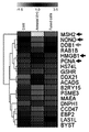
- Figure 10 shows the results of the heat map (heatmap) between the NSC34 motor neuron cell line, the killing NSC34 motor neuron cell line and HN / F-hATMSCs according to an embodiment of the present invention.
- Figure 11 analyzes the expression of genes DDB1, HMGB1 and MSH2 mRNA associated with cell recovery in NSC34 motor neurons, dying NSC34 motor neurons and HN / F-hATMSCs according to an embodiment of the present invention.
- FIG. 12 shows the effect of inhibiting apoptosis of C2C12 myoblast cells in death by HN / F-NSCs and cell fusion according to an embodiment of the present invention.
- Figure 13 analyzes the HN and F mRNA expression in HN / F-HeLa according to an embodiment of the present invention.
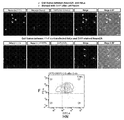
- Figure 14 analyzes the cell fusion ability with N2A neuroblastoma cell line in HN / F-HeLa according to an embodiment of the present invention.
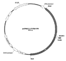
- 15 is a schematic diagram for preparing a pcDNA3.1-P2A expression vector inserted with HN and F according to an embodiment of the present invention.
- Figure 16 analyzes the cell fusion ability with C2C12 myoblasts in HN / F-HeLa or F-P2A-HN-HeLa according to an embodiment of the present invention.
- FIG. 17 illustrates a TDP-43 binding motif, a transcriptional factor (TF), in a promoter region of a gene DDB1 related to cell recovery selected according to an embodiment of the present invention.
- TF transcriptional factor
- FIG. 18 is a schematic diagram for preparing a promoter specifically binding to mouse DBB1 (mDDB1) according to an embodiment of the present invention.
- 19 is an analysis of mDDB1 expression by TDP-43 in a cell fused with an N2A neuroblastoma cell line and a HeLa cell line transduced with GFP-TDP-43 according to an embodiment of the present invention.
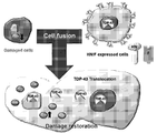
- Figure 21 illustrates the mechanism of recovery of cell damage by HN / F protein overexpressing cells in damaged cells.
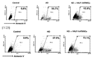
- FIG. 22 illustrates apoptosis inhibitory effect of HZ / F-hATMSCs and killed Alzheimer's disease (AD) cell model by cell fusion according to an embodiment of the present invention.
- FIG. 23 illustrates apoptosis inhibitory effect of Huntington's disease (HD) disease cell model, which is being killed by cell fusion with HN / F-hATMSCs according to an embodiment of the present invention.
- HD Huntington's disease
- FIG. 24 shows apoptosis inhibitory effect of HF / F-hATMSCs and dying heart failure disease cell models by cell fusion according to an embodiment of the present invention.
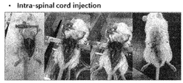
- FIG. 25 illustrates intravenous injection of HN / F-hATMSCs into a mouse model of amyotrophic lateral sclerosis (ALS) or Duchenne muscular dystrophy (DMD) disease according to one embodiment of the present invention. -Shows the cord cord injection method.
- ALS amyotrophic lateral sclerosis
- DMD Duchenne muscular dystrophy
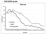
- FIG. 26 analyzes the motor performance in G93A SOD Tg mice injected with HN / F-hATMSCs according to one embodiment of the present invention.
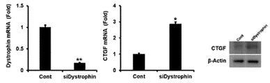
- Figure 27 analyzes the expression of Dystrophin and CTGF of the DMD disease cell model prepared according to the embodiment of the present invention.
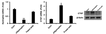
- FIG. 28 analyzes the expression of Dystrophin and CTGF in a DMD disease cell model treated with HN / F-hATMSCs according to an embodiment of the present invention.
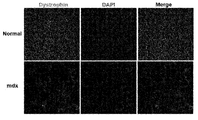
- FIG. 29 analyzes Dystrophin expression in skeletal muscle of mdx mouse, a DMD disease animal model.
- FIG. 30 analyzes the engraftment ability of HN / F-hATMSCs in skeletal muscle tissue of mdx mice injected with HN / F-hATMSCs according to one embodiment of the present invention.
- FIG. 31 analyzes dystrophin expression in skeletal muscle tissue of mdx mice injected with HN / F-hATMSCs 3 weeks after injection and 15 weeks after injection according to an embodiment of the present invention.
- FIG. 32 shows CTGF expression in skeletal muscle tissue of mdx mice injected with HN / F-hATMSCs according to one embodiment of the present invention.
- the present invention provides a vector comprising genes encoding hemagglutinin neuraminidase (HN) and F (fusion) proteins.
- HN hemagglutinin neuraminidase
- F fusion proteins
- the hemagglutinin-neuraminidase and F protein are sentai virus (sendai virus), human immunodeficiency virus 1 (human immunodeficiency virus 1), human parainfluenza virus (Human parainfluenza virus), human respiratory cells Human respiratory syncytial virus (HRSV), influenza virus or vesicular stomatitis virus may be derived from, but are not limited to, through the same or similar mechanisms of cells Various forms of hemagglutinin neuraminidase and F proteins of various origins and / or full length and / or fragments can be used so long as they can promote fusion, inhibit death, and repair damage to damaged cells. (Eckert DM, Kim PS.
- Hemagglutinin neuraminidase is a glycoprotein expressed in virus, a single viral protein having both the activity of hemagglutinin and neuraminidase, or This may be present as a separate protein, but is not limited thereto, and may be included in the present invention as long as the effect according to the present invention is achieved.
- the former include mump virus, sendai virus, human parainfluenza virus, such as human parainfluenza virus type 1 (HPIV-1), type 2 (HPIV-2), or 3 Or a paramyxovirus such as avian newcastle disease virus. Examples of the latter include those derived from influenza virus.
- hemagglutinin-neuraminidase may refer to information from Enzyme entry (http://enzyme.expasy.org) EC: 3.2.1.18, protein sequence Is derived from, for example, Sandivirus, GenBank accession no. Published as AAB06288.1.
- a Sandyvirus derived HN monoprotein which can be prepared according to the method of Example 2 according to the present invention is used.
- a fusion protein refers to a glycoprotein used for intercellular fusion or fusion / entry of a virus to a cell, endocytosis, and membrane trafficking.
- a F protein from virus is used, which is divided into classes I, II and III according to structural features.
- classes I include GP2 of Ebola virus, Mo-55 of Moloney murine leukemia virus (MoMuLv), gp41 of immunodeficiency virus HIV, Simian Virus (SIV) gp41, and the like.
- class II include SFV E1, TBEV E, and the like.
- class III examples include glycoprotein B (gB) of Herpes Simplex virus (HSV), gB of Epstein-Barr virus (EBV), protein G of Vesicular Stomatitis virus (VSV), and gp64, a glycoprotein of baculovirus, and the like.
- GB Herpes Simplex virus
- EBV Epstein-Barr virus
- VSV Vesicular Stomatitis virus
- gp64 a glycoprotein of baculovirus, and the like.
- Such fusion sugar protein and nucleic acid sequences are disclosed, for example GenBank Accession no. Published as AAC82300.1.
- genes encoding the hemagglutinin neuraminidase (HN) and F (fusion) proteins may be used in the form of full length and / or fragments.
- HN hemagglutinin neuraminidase
- F fusion proteins
- the wild type gene sequence encoding the protein disclosed in the present invention and fragments thereof, as well as genes in which a part of the nucleotide sequence is artificially modified to be advantageous, such as characteristics such as expression in cells or stability of the protein, and naturally found A portion of the nucleotide sequence includes a fragment of the modified gene or both.
- the vector means a means for expressing a gene of interest in a host cell.
- the vector includes elements for gene expression of interest, and may include a replication origin, a promoter, an operator, a transcription terminator, and the like, into the genome of the host cell.
- Appropriate enzyme sites eg, restriction enzyme sites
- RBS ribosomal binding sites
- IRES Internal Ribosome Entry Site
- the vector may further comprise transcriptional regulatory sequences (eg, enhancers, etc.) other than the promoter.
- the vector may be a plasmid DNA, recombinant vector or other mediator known in the art, specifically a linear DNA plasmid DNA, recombinant non-viral vector, recombinant viral vector or inducible gene expression vector system (inducible gene expression vector)
- the recombinant viral vector may be a retrovirus, adenovirus, adeno associated virus, helper-dependent adenovirus, and herpes simplex virus. herpes simplex virus), lentivirus, or vaccinia virus vector, but is not limited thereto.
- the vector means an expression vector for gene therapy.
- gene therapy refers to a method of normalizing its function by inserting a normal gene or providing a new function to a cell with a genetic abnormality in order to treat various genetic diseases caused by the genetic abnormality. Therefore, the expression vector for gene therapy in the present invention transfers and expresses hemagglutinin neuraminidase (HN) and F (fusion) protein genes into normal cells in the body, thereby expressing the cells with abnormal genes and normal cells.
- HN hemagglutinin neuraminidase
- F fusion
- hemagglutinin neuraminidase (HN) and F (fusion) protein genes are prepared from primers that can specifically recognize them from known sequences as described above. And, using this, the amplification of the gene through a polymerase chain amplification (Polymerase Chain Reaction) and introduced it into the expression vector as described above and can be introduced into the cell according to the invention as described below.
- Methods of introduction include, for example, liposome mediated transduction, calcium phosphate transfection, DEAE-dextran mediated transfusion, positively charged lipid mediated transduction, electroporation, transduction using a phage system or infection with a virus. Methods, and the like, but not limited thereto.
- the present invention also provides a cell transformed with a vector comprising a gene encoding hemagglutinin neuraminidase (HN) and F (fusion) protein.
- HN hemagglutinin neuraminidase
- F fusion
- the term "transformation" is that the genetic properties of the organism is changed by the DNA given from the outside, that is, DNA, which is a kind of nucleic acid extracted from cells of certain strains of the organism, may be introduced into living cells of other strains. When DNA enters the cell, the genetic traits change.
- the transformed cells may be stem cells, progenitor cells or animal cells, but is not limited thereto.
- the stem cells may be specifically embryonic stem cells, adult stem cells or induced pluripotent stem cells (iPS), but is not limited thereto, stem cells differentiated from the stem cells, such as embryonic stem Mesenchymal stem cells derived from cells, mesenchymal stem cells derived from induced pluripotent stem cells, neural stem cells derived from induced pluripotent stem cells, and the like.
- the cells may be autologous or other, or allogenic or Xenogenic cells. Most preferably, since it is autologous and derived from the recipient, there is an advantage that the administration of the pharmaceutical composition is safe and safe from the problem of the immune response.
- the embryonic stem cells are formed from the inner cell mass of the blastocytes, which are the early stages of embryonic development, and have the advantage of being able to differentiate into any tissue cell because they have the potential to differentiate into all cells. It is used in cell therapy research.
- the adult stem cells refer to undifferentiated stem cells found throughout the adult even after the embryonic development stage. These adult stem cells have a site-specific differentiation ability to differentiate themselves according to the characteristics of the surrounding tissues.
- Adult stem cells may be derived from various adult cells such as bone marrow, blood, brain, skin, fat, skeletal muscle, umbilical cord, umbilical cord blood and the like.
- mesenchymal stem cells (MSC) skeletal muscle stem cells, hematopoietic stem cells, neural stem cells (NSC), adipose derived stem cells (MSC) adipose-derived stem cells, adipose-derived progenitor cells, vascular endothelial progenitor cells, and the like, but are not limited thereto.
- the mesenchymal stem cells are stem cells derived from adult tissues obtained from the respective parts of the body already adult, umbilical cord, cord blood, bone marrow, blood, brain, skin, fat, skeletal, muscle, nerve, periosteum, amnion Or pluripotent or multipotent cells that can be separated from the placenta and can differentiate into various cells such as adipocytes and motor neurons.
- mesenchymal stem cells are characterized by being able to effectively engraft in allogeneic or heterologous recipients without the use of immunosuppressive agents.
- the mesenchymal stem cells may be mesenchymal stem cells of animals, specifically mammals, and more particularly humans.
- the mesenchymal stem cells are mesenchymal stem cells derived from adipose tissue.
- Adipose tissue-derived mesenchymal stem cells unlike bone marrow, amniotic fluid and umbilical cord blood stem cells, have a practical advantage of being provided with a large amount, and about 1% of fat cells are estimated to be stem cells.
- the plastic surgery being performed is liposuction, the adipose derived stem cells are particularly useful due to their infinite supply.
- the procedure for obtaining the mesenchymal stem cells is as follows: Isolating mesenchymal stem cells from a mammal, including human or mouse, preferably a human mesenchymal stem cell source, such as adipose tissue, blood or bone marrow . The cells are then cultured in a suitable medium. Suspension cells are removed during the culturing and passaged cells attached to the culture plate are obtained to finally obtain mesenchymal stem cells.
- any medium commonly used for culturing stem cells can be used. More specifically, a medium containing serum (eg fetal calf serum, horse serum and human serum) or serum replacement may be used.
- Medium that can be used in the present invention is, for example, RPMI series, Eagle's MEM (Eagle's minimum essential medium, Eagle, H. Science 130: 432 (1959)), ⁇ -MEM (Stanner, CP et al., Nat. New Biol. 230: 52 (1971)), Iscove's MEM (Iscove, N. et al., J. Exp. Med. 147: 923 (1978)), 199 medium (Morgan et al., Proc.
- the medium may include other components such as antibiotics or antifungal agents (eg, penicillin, streptomycin), glutamine, and the like. General descriptions of media and cultures are described in R. Ian Freshney, Culture of Animal Cells, Alan R. Liss, Inc., New York (1984), which is incorporated herein by reference.
- Identification of mesenchymal stem cells can be carried out, for example, via flow cytometry. Such flow cytometry is performed using specific surface markers of mesenchymal stem cells.
- the mesenchymal stem cells according to the present invention have a phenotype of CD29, CD44 and CD90 as markers and CD34, CD45 and HLA-DR which are negative markers.
- the induced pluripotent stem cells are introduced into four specific genes causing reverse differentiation into somatic cells, such as skin cells of adults without pluripotency, and then expressed or differentiated from cells into which four genes introducing reverse differentiation are introduced.
- somatic cells such as skin cells of adults without pluripotency
- stem cells having the same pluripotency as embryonic stem cells can be made. These are called induced pluripotent stem cells or dedifferentiated stem cells.
- Stem cell therapy using induced pluripotent stem cells somatic cells obtained from patients, induced pluripotent stem cells are made and differentiated in vitro to study the progress of various diseases, and used in disease models and cell-based research in drug development.
- the induced pluripotent stem cells are known methods, for example, Yu J et al. (2007) Induced pluripotent stem cell lines derived from human somatic cells. Science. 318, 1917-1920 or Takahashi K et al. (2007). Induction of pluripotent stem cells from adult human fibroblasts by defined factors. It can be obtained according to the method disclosed in Cell 131, 861-872 and the like.
- stem cells derived from induced pluripotent stem cells such as induced Pluripotent Stem Cells-derived mesemchymal stem cells (iPSCs-MSCs) or induced pluripotent stem cells-derived neural stem cells (induced Pluripotent Stem) Cells-derived neural stem cells, iPSCs-NSCs).
- iPSCs-MSCs induced Pluripotent Stem Cells-derived mesemchymal stem cells
- induced Pluripotent Stem induced Pluripotent Stem Cells-derived neural stem cells
- Mesenchymal stem cells derived from the induced pluripotent stem cells or neural stem cells derived from induced pluripotent stem cells are known methods, for example, L.G. VILLA-DIAZ et al. (2012) Derivation of Mesenchymal Stem Cells from Human Induced Pluripotent Stem Cells Cultured on Synthetic Substrates. STEM CELLS. 30, 1174-1181 or Hyun Soo Choi et al. (2014) Neural Stem Cells Differentiated From iPS Cells Spontaneously Regain Pluripotency. STEMCELLS. 32, 2596-2604, etc. can be obtained according to the method disclosed.
- progenitor cells are cells prior to the morphology and function of specific cells, and may be differentiated into cells of specific cell lines or formed into specific types of tissue, and have self-renewal. It means cells with extremely limited differentiation capacity. Endoderm progenitor cells, mesoderm progenitor cells, and ectoderm progenitor cells are all included here.
- the animal cell is a functional and structural basic unit derived from an animal including a human, and if the cell is derived from an animal including a human (for example, a mammal such as a monkey, dog, goat, pig or state) is within the scope of the present invention. May be included.
- the animal cells of the present invention include, but are not limited to, epithelial cells, endothelial cells, muscle cells, germ cells, skin cells (eg, fibroblasts, keratinocytes), immune cells, cancer cells and the like.
- CHO Chinese hamster ovary
- mouse myeloma (NS0) cells mouse myeloma (NS0) cells
- baby hamster kidney (BHK) cells mouse myeloma (Sp2 / 0) cells
- human retinal cells HUVEC cells, HMVEC cells
- COS-1 cells COS-7 cells
- HeLa cells HeLa cells
- HEK-293 cells HepG-2 cells
- HL-60 cells IM-9 cells
- Jurkat cells MCF-7 cells or T98G cells, etc.
- T98G cells human retinal cells
- HUVEC cells human retinal cells
- HMVEC cells human retinal cells
- COS-1 cells COS-7 cells
- HeLa cells HeLa cells
- HEK-293 cells HepG-2 cells
- HL-60 cells HepG-2 cells
- IM-9 cells Jurkat cells
- MCF-7 cells T98G cells
- a neurodegenerative disease or neurological disease, or muscle containing a vector containing a gene encoding a hemagglutinin neuraminidase (HN) and the gene encoding the F (fusion) protein as an active ingredient It provides a pharmaceutical composition for preventing or treating degenerative diseases or muscle diseases.
- HN hemagglutinin neuraminidase
- the vector is the same as the description of the vector comprising the gene encoding the hemagglutinin neuraminidase (HN) and F (fusion) protein, the specific description is the above In the following, only the specific configuration of the pharmaceutical composition for preventing or treating neurodegenerative diseases or neurological diseases, or muscle degenerative diseases or muscle diseases will be described.
- HN hemagglutinin neuraminidase
- F fusion protein
- the present invention is a cell overexpressing the HN and F protein is enhanced cell fusion capacity with other cells by the HN and F protein, the normal gene is delivered and expressed through the fusion with damaged cells, the cell damage is repaired It was confirmed that the expression of the gene is controlled. In addition, in various disease models related to cell damage, it was confirmed that HN and F protein overexpressing cells were fused with damaged cells or cells in which gene abnormalities were induced to recover normal cells.
- the vector containing the gene encoding the HN and F protein can be usefully used as an active ingredient for the prevention or treatment of neurodegenerative diseases or neurological diseases, or muscle degenerative diseases or muscle diseases.
- the neurodegenerative disease or neurological disease, or muscle degenerative disease or muscle disease is a disease associated with cell damage.
- the neurodegenerative disease or neurological disease may cause nerve cell damage, spinal nerve damage, peripheral nerve damage or neuronal cell death.
- the neurodegenerative disease or neurological disease is a neurodegenerative disease or neurological disease causing motor neuron damage or death and a neurodegenerative disease or neurological disease causing neuronal cell damage or death, spinal nerve injury or peripheral nerve damage. Can be divided into
- neurodegenerative diseases or neurological diseases causing motor neuron damage or death In order to improve or treat neurodegenerative diseases or neurological diseases causing motor neuron damage or death, preservation and recovery of motor neuron cells must be made.
- neurodegenerative or neurological diseases include spinal muscular atrophy, kennedy (spinal bulbar muscular atrophy), amyotrophic lateral sclerosis (ALS), multiple sclerosis, primary sclerosis ( primary lateral sclerosis or progressive bulbar palsy. It is not limited to this.
- Neurodegenerative diseases or neurological diseases causing nerve cell damage or death of the brain, spinal nerve damage or peripheral nerve damage may be prevented or treated through the recovery of nerve cells, peripheral nerves of the damaged brain or spinal cord.
- Examples of such neurodegenerative or neurological diseases include Alzheimer's disease (AD), dementia, multi-infact dementia (mid), frontotemporal dementia, and Lewy body dementia (AD).
- dementia with Lewy bodies mild cognitive impairment, corticobasal degeneration, Parkinson's disease (PD), depression, metabolic brain disease, multiple system atrophy Multiple system atrophy (MSA), Huntington's disease, progressive supranuclear palsy; PSP, epilepsy, dentatorubropallidoluysian atrophy (DRPLA), spinocerebellar ataxia, glaucoma, stroke, brain ischemia, encephalitis Post-encephalitic parkinsonism, Tourette's syndrome, restless legs syndrome, or attention deficit disorders with hyperactivity.
- the muscle degenerative disease or muscle disease may cause muscle cell damage or muscle cell death.
- muscle degenerative diseases or muscle diseases include myopathy, congenital myopathy, congenital muscular dystrophy, Duchenne muscular dystrophy, Becker muscular dystrophy muscular dystrophy, Limb Girdle muscular dystrophy, Facioscapulohumeral muscular dystrophy, oculopharyngeal muscular atrophy, distal muscular dystrophy, adriatic muscular dystrophy (Emery-Dreifuss muscular dystrophy), Myotonic dystrophy, Barth syndrome, heart failure, sarcopenia or X-linked dilated cardiomyopathy It may be, but is not limited thereto.
- compositions according to the invention may be formulated in a suitable form with the pharmaceutically acceptable carriers generally used.
- Pharmaceutically acceptable carriers include, for example, water, suitable oils, saline, carriers for parenteral administration such as aqueous glucose and glycols, and the like, and may further include stabilizers and preservatives.
- Suitable stabilizers include antioxidants such as sodium hydrogen sulfite, sodium sulfite or ascorbic acid.
- Suitable preservatives include benzalkonium chloride, methyl- or propyl-paraben and chlorobutanol.
- composition for cell therapy according to the present invention if necessary according to the administration method or dosage form, suspensions, dissolution aids, stabilizers, tonicity agents, preservatives, adsorption inhibitors, surfactants, diluents, excipients, pH adjusters, analgesics , Buffers, antioxidants, and the like may be appropriately included.
- Pharmaceutically acceptable carriers and formulations suitable for the present invention including those exemplified above, are described in detail in Remington's Pharmaceutical Sciences, latest edition.
- compositions of the present invention may be prepared in unit dosage form by formulating with a pharmaceutically acceptable carrier and / or excipient according to methods which can be easily carried out by those skilled in the art. Or may be prepared by incorporation into a multi-dose container.
- the pharmaceutical composition may be administered by any device that can move the active ingredient to the target cell.
- the pharmaceutical composition of the present invention may comprise a therapeutically effective amount for the treatment of a disease.
- treatment means to reverse, alleviate, inhibit, or prevent the progression of one or more symptoms of a disease or condition to which the term applies.
- therapeutically effective amount also refers to an amount of an active ingredient or pharmaceutical composition that induces a biological or medical response in a tissue system, animal or human being contemplated by a researcher, veterinarian, physician or other clinical person, Amounts that induce alleviation of the symptoms of the disease or disorder being treated. It is apparent to those skilled in the art that the active ingredient included in the pharmaceutical composition of the present invention will vary depending on the effect.
- the optimal pharmaceutical composition content can be readily determined by one skilled in the art and includes the type of disease, the severity of the disease, the amount of other ingredients contained in the composition, the type of formulation, and the patient's age, weight, general health, sex and It can be adjusted according to various factors including the diet, the time of administration, the route of administration and the rate of secretion of the composition, the duration of treatment, and the drugs used concurrently.
- the pharmaceutical composition is administered intravenously or intraspinally.
- the dosage of the composition of the present invention is 0.1 ⁇ 10 5 to 1.0 ⁇ 10 8 cells / cells transformed with a vector comprising the gene encoding the hemagglutinin-neuramidase and F protein as active ingredients. kg (body weight), more preferably 0.5 ⁇ 10 6 to 1.0 ⁇ 10 7 cells / kg body weight.
- the dosage may be variously prescribed by such factors as the formulation method, the mode of administration, the age, weight, sex, morbidity, food, time of administration, route of administration, rate of excretion, and reaction sensitivity of the patient. These factors can be taken into account to properly adjust the dosage.
- the number of administrations may be one or two or more times within the range of clinically acceptable side effects, and may be administered to one site or two or more sites for the administration site.
- the same dosage as that of humans per kg or the volume ratio (for example, average value) of the ischemic organ (heart, etc.) between the animal of interest and humans can be adjusted.
- Equivalent amounts may be administered.
- the animal to be treated according to the present invention include humans and mammals for other purposes, and specifically, humans, monkeys, mice, rats, rabbits, sheep, cattle, dogs, goats, horses, pigs, and the like. This includes.
- the present invention is a neurodegenerative disease containing a cell transformed with a vector comprising a gene encoding a hemagglutinin neuraminidase (HN) and a gene encoding F (fusion), or neurodegenerative disease
- HN hemagglutinin neuraminidase
- F fusion
- the cell is the same as the description of the cell transformed with a vector comprising a gene encoding the hemagglutinin neuraminidase (HN) and F (fusion) protein,
- HN hemagglutinin neuraminidase
- F fusion protein
- the present invention is a cell overexpressing the HN and F protein is enhanced cell fusion capacity with other cells by the HN and F protein, the normal gene is delivered and expressed through the fusion with damaged cells, the cell damage is repaired It was confirmed that the expression of the gene is controlled. In addition, in various disease models related to cell damage, it was confirmed that HN and F protein overexpressing cells were fused with damaged cells or cells in which gene abnormalities were induced to recover normal cells.
- the cells transformed with the vector containing the genes encoding the HN and F proteins can be usefully used as an active ingredient of a composition for preventing or treating neurodegenerative diseases or neurological diseases, or muscle degenerative diseases or muscle diseases. .
- the present invention also includes administering to a subject a therapeutically effective amount of a vector comprising a gene encoding hemagglutinin neuraminidase (HN) and a gene encoding F (fusion) protein,
- HN hemagglutinin neuraminidase
- F F
- the present invention provides a method for administering to a subject a therapeutically effective amount of cells transformed with a vector comprising a gene encoding hemagglutinin neuraminidase (HN) and a gene encoding F (fusion) protein.
- HN hemagglutinin neuraminidase
- F F
- a gene encoding the hemagglutinin neuraminidase (HN) and the F (fusion) protein may be used for the vector or cells transformed with the vector and a method of administration, dosage, etc. thereof.
- HN hemagglutinin neuraminidase
- F fusion protein
- the subject is a subject suffering from a neurodegenerative disease, a neurological disease, a muscle degenerative disease or a muscle disease, and more specifically, includes the gene encoding the above-described hemagglutinin-neuramidase and F protein.
- Mammals such as humans, monkeys, mice, rats, which can be alleviated and / or repaired by administering a vector or a cell transformed with said vector, such as neuronal damage or myocyte damage caused by said disease, Rabbits, sheep, cattle, dogs, goats, horses, pigs, and the like.
- the neurodegenerative disease or neurological disease may cause nerve cell damage, spinal cord injury, peripheral nerve damage or neuronal cell death.
- neurodegenerative diseases or neurological diseases are neurodegenerative diseases or neurological diseases causing motor neuron damage or death and neurodegenerative diseases or neurological diseases causing neuronal cell damage or death, spinal cord injury or peripheral nerve damage. Can be divided.
- neurodegenerative diseases or neurological diseases causing motor neuron damage or death In order to improve or treat neurodegenerative diseases or neurological diseases causing motor neuron damage or death, preservation and recovery of motor neuron cells must be made.
- neurodegenerative or neurological diseases include spinal muscular atrophy, kennedy (spinal bulbar muscular atrophy), amyotrophic lateral sclerosis (ALS), multiple sclerosis, primary sclerosis ( primary lateral sclerosis or progressive bulbar palsy. It is not limited to this.
- Neurodegenerative diseases or neurological diseases that cause nerve cell damage or death of the brain, spinal cord injury, or peripheral nerve damage may be prevented or treated through recovery of nerve cells or peripheral nerves of the damaged brain or spinal cord.
- Examples of such neurodegenerative or neurological diseases include Alzheimer's disease (AD), dementia, multi-infact dementia (mid), frontotemporal dementia, and Lewy body dementia (AD).
- dementia with Lewy bodies mild cognitive impairment, corticobasal degeneration, Parkinson's disease (PD), depression, metabolic brain disease, multiple system atrophy Multiple system atrophy (MSA), Huntington's disease, progressive supranuclear palsy (PSP), epilepsy, dentatorubropallidoluysian atrophy (DRPLA), spinal cord ventricle (spinocerebellar ataxia), glaucoma, stroke, brain ischemia, post-encephalitic parkinsonism, Tourette's syndrome, restless legs syndrome, or Uiryeok be deficient hyperactivity disorder (attention deficit disorders with hyperactivity), but is not limited thereto.
- the muscle degenerative disease or muscle disease may cause muscle cell damage or muscle cell death.
- muscle degenerative diseases or muscle diseases include myopathy, congenital myopathy, congenital muscular dystrophy, Duchenne muscular dystrophy, Becker muscular dystrophy muscular dystrophy, Limb Girdle muscular dystrophy, Facioscapulohumeral muscular dystrophy, oculopharyngeal muscular atrophy, distal muscular dystrophy, adriatic muscular dystrophy (Emery-Dreifuss muscular dystrophy), Myotonic dystrophy, Barth syndrome, heart failure, sarcopenia or X-linked dilated cardiomyopathy It may be, but is not limited thereto.
- a cell overexpressing HN and F proteins enhances cell fusion ability with other cells by HN and F proteins, expresses normal genes through fusion with damaged cells, and restores cell damage. It was confirmed that the expression of. In addition, in various disease models related to cell damage, it was confirmed that HN and F protein overexpressing cells were fused with damaged cells or cells in which gene abnormalities were induced to recover normal cells.
- the vector containing the gene encoding the HN and F protein or cells transformed therewith can be usefully used for preventing or treating neurodegenerative diseases or neurological diseases, or muscle degenerative diseases or muscle diseases.
- the present invention also provides hemagglutinin neuraminidase (HN) and F (fusion) proteins for use as a composition for preventing or treating neurodegenerative or neurological or muscular degenerative or muscular diseases. It provides a use of a vector comprising a gene encoding the.
- HN hemagglutinin neuraminidase
- F fusion proteins
- hemagglutinin neuraminidase (HN) and F (fusion) protein for use as a composition for the prevention or treatment of neurodegenerative diseases or neurological diseases, or muscle degenerative diseases or muscle diseases
- HN hemagglutinin neuraminidase
- F fusion protein
- a gene encoding the hemagglutinin neuraminidase (HN) and the F (fusion) protein may be used for the vector or cells transformed with the vector and a method of administration, dosage, etc. thereof.
- HN hemagglutinin neuraminidase
- the specific description is used above.
- the disease is the same as the description of the pharmaceutical composition for preventing or treating the neurodegenerative or neurological disease, or the muscle degenerative disease or the muscle disease, the specific description is used for the above description.
- a cell overexpressing HN and F proteins enhances cell fusion ability with other cells by HN and F proteins, expresses normal genes through fusion with damaged cells, and restores cell damage. It was confirmed that the expression of. In addition, in various disease models related to cell damage, it was confirmed that HN and F protein overexpressing cells were fused with damaged cells or cells in which gene abnormalities were induced to recover normal cells.
- the vector containing the gene encoding the HN and F protein or cells transformed therewith can be usefully used for preventing or treating neurodegenerative diseases or neurological diseases, or muscle degenerative diseases or muscle diseases.
- hATMSCs Human adipose tissue-derived mesenchymal stem cells obtained with the consent and understanding of K-STMECELL Institutional Review Board (IRB) were treated with RKCM culture medium (10% FBS, K- Incubated in STEMCELL).
- the mouse motor neuron line (NSC34 Motor Neuron-Like Hybrid Cell line, CEDARLANE, USA) was cultured in DMEM (Dulbecco's Modified Eagle's Medium) culture medium with 10% FBS.
- DMEM Dulbecco's Modified Eagle's Medium
- Mouse neuroblastoma cell line N2A cell line, ATCC was cultured in an antibiotic-added Eagle's Minimum Essential Medium (EMEM) culture medium (10% FBS addition).
- HeLa cell line Human cervical cancer cell lines (HeLa cell line, ATCC) were cultured in an antibiotic-added Eagle's Minimum Essential Medium (EMEM) culture medium (with 10% FBS).
- EMEM Eagle's Minimum Essential Medium
- Mouse C2C12 muscle myoblast cell line was incubated in DMEM culture medium (10% FBS) added with antibiotics.
- Rat heart myoblast cell line (ATC) was incubated in DMEM culture medium (10% FBS) with antibiotics.
- iPSCs human induced pluripotent stem cells prepared by receiving blood with consent from a normal person were cultured in a floating plate containing hES cell culture medium (ThermoFisher) containing no bFGF for 4 days. It was. Next, the cells were cultured in hES cell culture medium containing Retinoic acid (RA, 5 x 10 -7 M) for 4 days, and then neurobasal medium [EGF (20 ng / ml), bFGF (10 ng / ml), B27 (2) for 7 days. %), Serum-free DMEM / F12 (1: 1) culture medium containing LIF (10 ng / ml) and heparin sodium (2 ⁇ g / ml).
- RA Retinoic acid
- bFGF 10 ng / ml
- B27 (2) for 7 days.
- Serum-free DMEM / F12 (1: 1) culture medium containing LIF (10 ng / ml) and he
- iPSCs-NSCs induced Pluripotent Stem Cells-derived neural stem cells
- Example 1 Motor neuron and Local origin Mesenchymal stem cells Inhibitory Effect of Apoptosis on Motor Neuronal Cell Line by Cell Fusion
- the cell tracker (CM-DiI (Thermofisher) -labeled motor neuron cell line NSC34 cells were incubated for 24 hours after treatment with 2.5 ⁇ M of thapsigargin, apoptosis inducing factor. After Annexin V staining, Annexin V-positive NSC34 cells were isolated by flow cytometry using Annexin V antibody.
- CM-DiI Thermofisher
- HATMSCs and Annexin V-positive NSC34 cells labeled with green dyes were transferred to Sandai virus (HVJ) Envelope Cell fusion Kit (GenomONETM-CF EX Sendai virus (HVJ) Envelope Cell Fusion Kit, COSMO BIO, Japan) was used as the manufacturer's method for cell fusion and then V-positve cells were analyzed by flow cytometry.
- HVJ Sandai virus
- hSCMS and NSC34 cells with cell fusion were isolated by flow cytometry, followed by Reverse Transcription polymerase chain reaction (RT-PCR) and quantitative RT-PCR.
- RT-PCR Reverse Transcription polymerase chain reaction
- Bcl-xL an anti-apoptotic gene.
- FIG. 2 the expression of Bax mRNA was significantly decreased in the NSC34 cell group in which hATMSCs and cell fusion occurred, and the expression of Bcl-xL mRNA was significantly increased in comparison with the NSC34 cell group in which hATMSCs and cell fusion did not occur.
- Hemagglutinin Neuraminidase hemagglutinin neuraminidase , HN
- F fusion protein overexpressing human fat Mesenchymal stem cells
- HN hemagglutinin neuraminidase
- F F protein gene
- clones obtained after cloning were amplified and cultured in LB liquid medium for 24 hours, plasmid was extracted using a plasmid extraction kit (Midiprep kit (Invitrogen, USA), and then liposomes (lipofectamine 3000, Invitrogen) was transduced into hATMSCs using the manufacturer's method to obtain hATMSCs (HN / F-hATMSCs) into which the HN / F gene was introduced.
- Total RNA was extracted from the transduced cell lines and analyzed for expression of HN and F genes via RT-PCR and real-time quantitative RT-PCR. HATMSCs were used as a control. Partial HN and F primers were used to confirm expression for HN or F mRNA using RT-PCR and real-time quantitative PCR.
- the base sequence encoding myc protein was attached to the forward primer for amplification of the HN gene, and the base sequence encoding histidine was attached to the forward primer for amplification of the F gene.
- Santcruz Biotechnology, USA was used to analyze the expression of HN and F proteins in HN / F-hATMSCs through confocal laser microscopy (Nicon, Japan).
- HN and F proteins are expressed on the cell surface of HN / F-hATMSCs as shown in FIG. 5. These results indicate that human adipose-derived mesenchymal stem cells overexpressing HN and F proteins were successfully produced.
- Fusion assay with NSC34 motor neuron cell line labeled with green dye-labeled HN / F-hATMSCs and cell tracker CM-DiI to assess cell fusion capacity of HN / F-hATMSCs prepared as in Example 2 ) was performed. Two kinds of cells were mixed in a 1.5 ml test tube in a ratio of cell number 1: 1, and reacted at 4 ° C. for 5 minutes, followed by reaction at 37 ° C. for 15 minutes. The plate was transferred to a 6-well plate containing antibiotic-containing DMEM medium (10% FBS) and incubated for 16 hours at 37 ° C. under 5% CO 2 .
- HN / F-hATMSCs were harvested and analyzed for cell fusion rate of HN / F-hATMSCs and NSC34 motor neuron cells by flow cytometry.
- HATMSCs were used as a control.
- FIG. 7 the cell fusion rate of HN / F-hATMSCs and NSC34 motor neuron cell lines was increased by more than 3.5 times compared to the control group.
- an image analysis using confocal laser microscopy confirmed an image of cells fused with HN / F-hATMSCs showing green fluorescence and NSC34 motor neuron cell lines with red fluorescence.
- Example 5 HN / F- hATMSCs NSC34 Motor neuron Expression analysis of markers of hATMSCs and motor neuron markers in fused cells
- Example 4 Confocal using the motor neuron marker marker choline acetyltransferase (ChAT) and the antibody against adipose derived mesenchymal stem cell marker marker (Santcruz Biotechnology, USA) for the fusion cells of Example 4
- ChAT motor neuron marker marker choline acetyltransferase
- CD105 adipose derived mesenchymal stem cell marker
- FIG. 8 the expression of CD105, a marker of ChAT adipose-derived mesenchymal stem cell marker, which was a motor neuron marker, was confirmed in cells fused with HN / F-hATMSCs and NSC34 motor neuron.
- the annexin V-positive NSC34 motor neuron cell line of Example 2 and the fusion cells of Example 4 were analyzed for expression of DDB1, HMGB1 and MSH2 via RT-PCR and quantitative RT-PCR.
- NSC34 motor neuron line was used as a control.
- the expression of DDB1, HMGB1, and MSH2 mRNAs was decreased in dying NSC34 motor neurons, but significantly increased in cells fused with HN / F-hATMSCs and NSC34 motor neurons.
- Example 7 Killing C2C12 Myoblasts Hemagglutinin Neuraminidase (hemagglutinin) neuraminidase , HN ) / Overexpressing F (fusion) protein Induced pluripotent stem cells origin Of neural stem cells (HN / F-NSCs) By cell fusion C2C12 Myoblasts Apoptosis inhibitory effect analysis
- HN / F-NSCs induced pluripotent stem cell-derived neural stem cells
- iPSCs-NSCs pluripotent stem cell-derived neural stem cells
- liposomes lipofectamine 3000, Invitrogen
- Transduction was performed to obtain iPSCs-NSCs (HN / F-NSCs) into which the HN / F gene was introduced.
- Total RNA was extracted from the transduced cell lines and analyzed for expression of HN and F genes via RT-PCR and real-time quantitative RT-PCR (results not shown).
- the C2C12 myoblast cell line labeled with the cell tracker CM-DiI was incubated for 24 hours after treatment with apoptosis inducing factor, tapxigin 2.5 ⁇ g / ml.
- Annexin V staining Annexin V-C2C12 myoblast line was isolated by flow cytometry using Annexin V antibody.
- the cells were recovered and the V-positive cells were collected by flow cytometry. Analyzed. As a result, as shown in FIG.
- Annexin V-positive cells were identified in the C2C12 cell group without HN / F-NSCs and cell fusion, and in the C2C12 cell group with HN / F-NSCs and cell fusion. 21.6% of Annexin V-positive cells were identified.
- Example 8 N2A Neuroblastoma cell lines and Hemagglutinin Neuraminidase (hemagglutinin) neuraminidase , HN ) / Overexpressing F (fusion) protein HeLa Evaluation of cell fusion ability of cell line (HN / F-HeLa)
- HeLa cell line (HN / F-HeLa) overexpressing HN and F proteins was constructed to investigate whether cell fusion ability was observed in normal cell lines overexpressing HN and F proteins.
- the GD-HN-inserted pcDNA3.1 expression vector was cloned using the GFP-pcDNA3.1 expression vector and the HN protein gene clone obtained in Example 2.
- the pcDNA3.1 expression vector into which the RFP-F protein was inserted was cloned using the RFP-pcDNA3.1 expression vector and the F protein gene clone obtained in Example 2.
- liposomes lipofectamine 3000, Invitrogen
- heLa HeLa
- HN / F-HeLa HeLa
- Total RNA was extracted from the transduced cell lines and analyzed for expression of HN and F genes via RT-PCR and real-time quantitative RT-PCR.
- HeLa cell line was used as a control.
- overexpression of the HN and F genes was confirmed as shown in FIG. 13.
- the cells were recovered after fusion of the N2A neuroblastoma cell line labeled with HN / F-HeLa and DAPI by the same method as described in Example 4.
- Cell fusion rates of HN / F-HeLa and N2A neuroblastoma cell lines were analyzed by flow cytometry and confocal laser microscopy.
- GFP-transduced N2A neuroblastoma cell line and mCherry-transduced HeLa cell line were fused in the same manner as described in Example 4, and then cells were recovered and analyzed for cell fusion rate by confocal laser microscopy. It was. As a result, as shown in FIG.
- (fusion) protein gene was introduced into the HeLa cell line.
- the pcDNA3.1-P2A expression vector into which HN and F proteins were inserted was cloned using the pcDNA3.1-P2A expression vector and the HN protein gene and the F protein gene clone obtained in Example 2.
- liposomes liposomes (lipofectamine 3000, Invitrogen) were transduced into HeLa cell lines using the manufacturer's method to obtain HeLa cell lines (F-P2A-HN-HeLa) into which the HN / F gene was introduced.
- the pcDNA3.1 expression vector cloned with the HN and F proteins cloned in Example 2 was transduced into the HeLa cell line as described above to obtain a HeLa cell line (HN / F-HeLa) into which the HN / F gene was introduced.
- Total RNA was extracted from the transduced cell lines and analyzed for expression of HN and F genes via RT-PCR and real-time quantitative RT-PCR (results not shown).
- each of the HeLa cell lines obtained above was labeled with GFP, and after cell fusion with a C2C12 myoblast cell line labeled with RFP in the same manner as described in Example 4, cells were recovered.
- Cell flow rate of HeLa cell line and C2C12 myoblast cell line were analyzed by flow cytometry.
- GFP-labeled HeLa cell line and RFP-labeled C2C12 myoblast cell line were fused in the same manner as described in Example 4, and then cells were recovered and analyzed for cell fusion rate by flow cytometry.
- the cell fusion rate of F-P2A-HN-HeLa was 26.9%, and the cell fusion rate of HN / F-HeLa was 50.1%, as shown in FIG. It was.
- HN and F proteins are overexpressed in cells through various vectors, and cells expressing HN and F proteins are enhanced in cell fusion with other cells.
- the promoter region of the gene DDB1 related to cell repair selected in Example 6 was screened and a transcriptional factor as shown in FIG. 17. It was confirmed that a binding motif of TDP-43 (TAR DNA-binding protein 43), RF), was present at the promoter region of DDB1.
- a primer specifically binding to mouse DDB1 (mDDB1) is prepared as shown in FIG.
- Total DNA was extracted from blastoma cell line and HeLa cell line to identify mouse specific DDB1 gene by PCR.
- TDP-43 was overexpressed in N2A neuroblastoma cell line and RNA was extracted and analyzed using quantitative RT-PCR.
- the N2A neuroblastoma cell line, the HeLa cell line transduced with GFP, and the HeLa cell line transduced with the N2A neuroblastoma cell line and GFP-TDP-43 were respectively introduced into the Sandai virus (HVJ) Envelope Cell fusion Kit (GenomONETM-CF EX Sendai).
- HVJ Sandai virus
- Cell fusion was performed using virus (HVJ) Envelope Cell Fusion Kit, COSMO BIO, Japan) according to the manufacturer's method, followed by ChIP (Chromatin immunoprecipitation) assay (Millipore) using a fusion GFP antibody (Rockland).
- mDDB1 increased with fusion time in the N2A neuroblastoma cell line and HeLa cell line transduced with GFP-TDP-43, and the occupancy of TDP-43 at the mDDB1 promoter region was four times or more. Increasing TDP-43 was found to regulate the expression of DDB1.
- the cell lines were subjected to cell fusion in the same manner as described above, and analyzed for translocation of TDP-43 through confocal laser microscopy after DAPI staining, and stained with NcA neuroblastoma cell line using NucBlue (ThermoFisher) and GFP-TDP.
- Example 11 Alzheimer's disease Alzheimer's disease; In AD) cell models HN Analysis of Therapeutic Effects Using Cell Fusion with / F-hATMSCs
- AD Alzheimer's disease
- TG2576 mice 80 days old
- stem cells were isolated and filtered using trypsin from the isolated tissues.
- EGF and FGF were added to DMEM culture medium containing B27 supplement, respectively, and cultured in sphere form for 4 days. After 4 days, the cells were transferred to well-plates and cultured for 7 days in DMEM medium containing 5% FBS and B27 supplement to induce differentiation of AD mouse-derived neural stem cells.
- Example 2 HN / F-hATMSCs obtained in Example 2 and the AD mouse-derived neural stem cells obtained above were fused in the same manner as in Example 4.
- 5 ⁇ l of Annexin V antibody was added and reacted at room temperature for 15 minutes, and then V-positive cells were analyzed by flow cytometry.
- FIG. 22 50.7% of Annexin V-positive cells were identified in AD mouse-derived neural stem cells, and 15.9% of Annexin V- was found in AD mouse-derived neural stem cells in which cell fusion with HN / F-hATMSCs occurred. Positive cells were identified.
- Huntington's disease cell model was constructed to determine the therapeutic effect using cell fusion by cells overexpressing HN / F protein in Huntington's disease.
- a Huntington's disease animal model R6 / 2 Tg mice (80 days old), was provided from the Jackson laboratory. Then, subventricular zones of R6 / 2 mice were separated, and stem cells were isolated and filtered using trypsin in the separated tissues. Next, EGF and FGF were added to DMEM culture medium containing 20 ng / ml, respectively, incubated for 4 days in the form of a sphere, and then transferred to a well-plate and DMEM culture medium containing 5% FBS and B27 supplement. Incubation for 7 days at induced the differentiation of Huntington's disease-derived neural stem cells.
- Example 2 HN / F-hATMSCs obtained in Example 2 and the Huntington disease mouse-derived neural stem cells obtained above were fused in the same manner as in Example 4.
- 5 ⁇ l of Annexin V antibody was added and reacted at room temperature for 15 minutes, and then V-positive cells were analyzed by flow cytometry.
- 37.5% of Annexin V-positive cells were identified in Huntington's disease-derived neural stem cells as shown in FIG. Positive cells were identified.
- Example 13 Heart failure disease cell model HN / F- hATMSCs Treatment effect analysis using cell fusion
- a cell model of heart failure disease was prepared.
- a heart failure disease cell model was obtained by inducing oxidative stress by incubating for 200 hours with hydrogen peroxide (H 2 O 2 ) in a H9c2 cardiomyocyte line labeled with a cell tracker CMDFA (Invitrogen).
- Example 2 the HN / F-hATMSCs obtained in Example 2 were labeled with CM-DiI, and the cells were recovered after cell fusion in the same manner as described in Example 4 for the heart failure disease cell model obtained above. It was.
- the recovered cells were stained with Annexin V and isolated from Annexin V-positive cells by flow cytometry, and analyzed for green fluorescent positive cells.
- FIG. 24 29% of annexin V-positive cells were identified in a heart failure disease cell model, and 13.4% of annexin V-positive cells in a heart failure disease cell model in which cell fusion with HN / F-hATMSCs occurred. It was confirmed.
- Example 14 Amyotrophic Lateral Sclerosis (amyotrophic lateral sclerosis, ALS ) Analysis of therapeutic effect using cell fusion of HN / F-hATMSCs in disease animal models
- G93A SOD1 Tg mouse (ALS disease animal model) 80 days of age) were provided by the Jackson laboratory. Then, G93A SOD1 Tg mice were divided into a control group (Tg-saline), Tg-MSC and Tg-fusogenic MSC group, and using the intra-spinal cord injection method as shown in FIG. HATMSCs were injected into the Tg-MSC group and HN / F-hATMSCs prepared in Example 2 were injected into the Tg-fusogenic MSC group in 0.5-2 ⁇ 10 6 cells and rota rod test was performed. As a result, in the Tg-fusogenic MSC group injected with HN / F-hATMSCs as shown in FIG. It was.
- Example 15 Duchesne Muscle retraction axis ( Duchenne muscular dystrophy, DMD Analysis of therapeutic effect using cell fusion of HN / F-hATMSCs in disease cell models
- DMD disease is a muscle disease caused by Dystrophin deficiency of muscle fibers, so that siRNA (siDystrophin, GenePharma) specific to Dystrophin is obtained and introduced into C2C12 myoblasts using the manufacturer's method to suppress Dystophin expression. C2C12 myoblasts were prepared.
- HN / F-hATMSCs prepared as in Example 2 and DMD disease prepared above
- cells were recovered and analyzed for expression of Dystrophin and CTGF using quantitative RT-PCR and Western blot.
- Dystophin expression was decreased and CTGF expression was increased in DMD disease cell model, while Dystrophin expression was significantly increased and CTGF expression was significantly increased in cells fused with HN / F-hATMSCs and DMD disease cell model. It was confirmed to decrease.
- Example 16 DMD Disease in animal models HN / F- hATMSCs Treatment effect analysis using cell fusion
- mdx mouse (2-4 weeks after birth), a DMD disease animal model, was provided from the Jackson laboratory. Skeletal muscle was isolated from mdx mice, and immunohistochemistry was performed using Dystrophin antibody (abcam) to confirm that Dystrophin was decreased as shown in FIG. 29. Then, mdx mice were divided into saline, MSC and fMSC groups, and hATMSCs labeled with saline in the control group and green dye in the MSC group using the intra-spinal cord injection method as shown in FIG. 25.
- HN / F-hATMSCs of Example 2 labeled with green dye, were injected with 0.5-2 ⁇ 10 6 cell numbers, and skeletal muscle was isolated after 1 week. Then, the cells were stained with hNuclei antibody (abcam) and analyzed for engraftment ability in the extracted skeletal muscle through confocal laser microscopy. In addition, after 3 weeks or 15 weeks after injection, skeletal muscle was isolated, and immunohistochemistry was performed using Dystrophin antibody (abcam). One week after injection, skeletal muscle was isolated, total RNA was extracted from the isolated skeletal muscle, and CTGF gene expression was analyzed using quantitative RT-PCR.
- HN and F protein overexpressing cells are fused with cells damaged by HN and F proteins, and through fusion with the damaged cells, transfer of transcriptional regulators in HN and F protein overexpressing cells into the nucleus of the damaged cells, By repairing damaged cells by regulating the expression of genes involved in intracellular repair, cell fusion techniques using HN and F proteins can be used to treat neurodegenerative or neurological, muscular degenerative or muscular diseases associated with cell damage. it means.
- cell fusion technology using HN and F proteins is used for gene therapy. It can be used as a delivery tool for gene therapy.
- the present invention relates to genes and cell therapies using cell fusion technology and the use thereof, and specifically, to repair cell damage and introduce normal genes by using cell fusion technology by HN and F proteins, whereby HN and F A vector comprising a gene encoding a protein or a cell transformed therefrom can be usefully used for treating diseases related to cell damage, more specifically neurodegenerative or neurological diseases, or muscle degenerative diseases or muscle diseases.
Landscapes
- Health & Medical Sciences (AREA)
- Life Sciences & Earth Sciences (AREA)
- Engineering & Computer Science (AREA)
- Veterinary Medicine (AREA)
- Developmental Biology & Embryology (AREA)
- Medicinal Chemistry (AREA)
- Cell Biology (AREA)
- Pharmacology & Pharmacy (AREA)
- Chemical & Material Sciences (AREA)
- Animal Behavior & Ethology (AREA)
- General Health & Medical Sciences (AREA)
- Public Health (AREA)
- Biomedical Technology (AREA)
- Epidemiology (AREA)
- Biotechnology (AREA)
- Neurology (AREA)
- Bioinformatics & Cheminformatics (AREA)
- Immunology (AREA)
- General Chemical & Material Sciences (AREA)
- Nuclear Medicine, Radiotherapy & Molecular Imaging (AREA)
- Zoology (AREA)
- Reproductive Health (AREA)
- Neurosurgery (AREA)
- Virology (AREA)
- Organic Chemistry (AREA)
- Chemical Kinetics & Catalysis (AREA)
- Hospice & Palliative Care (AREA)
- Molecular Biology (AREA)
- Physical Education & Sports Medicine (AREA)
- Orthopedic Medicine & Surgery (AREA)
- Genetics & Genomics (AREA)
- Psychiatry (AREA)
- Gynecology & Obstetrics (AREA)
- Hematology (AREA)
- Medicines That Contain Protein Lipid Enzymes And Other Medicines (AREA)
- Medicines Containing Material From Animals Or Micro-Organisms (AREA)
- Enzymes And Modification Thereof (AREA)
- Micro-Organisms Or Cultivation Processes Thereof (AREA)
- Peptides Or Proteins (AREA)
Abstract
본 발명은 세포융합 기술을 이용한 유전자 및 세포 치료제 및 이의 용도에 관한 것으로, 구체적으로 헤마글루티닌-뉴라미니다아제(hemagglutinin neuraminidase, HN) 및 F(fusion) 단백질을 과발현하는 세포가 HN 및 F 단백질에 의해 다른 세포와의 세포융합력이 증진되고, 손상 또는 사멸 중인 세포, 또는 유전자 이상이 있는 세포와의 세포융합을 통해 정상 세포로의 회복이 이루어지는 것을 확인하였으므로, 상기 HN/F 단백질을 코딩하는 유전자를 포함하는 벡터 또는 이로 형질전환된 세포를 세포 손상과 관련된 질환, 예컨대 신경퇴행성 질환 또는 신경 질환, 또는 근육퇴행성 질환 또는 근육 질환 예방 또는 치료용 조성물의 유효성분으로 유용하게 이용할 수 있다.
Description
본 발명은 세포융합 기술을 이용한 유전자 및 세포 치료제, 및 이의 용도에 관한 것으로, 구체적으로 헤마글루티닌-뉴라미니다아제(hemagglutinin neuraminidase, HN) 및 F(fusion) 유전자를 세포에 형질도입시켜 과발현함으로써 다른 세포와의 세포융합력을 증진시키고, 손상 또는 사멸 중인 세포, 또는 유전자 이상이 있는 세포와의 세포융합을 통해 세포 손상을 회복하고 정상 유전자를 전달할 수 있는, 세포융합 기술을 이용한 유전자 및 세포 치료제, 및 세포 손상과 관련된 질환에서의 용도에 관한 것이다.
일반적으로 질병 및 노화는 세포 손상 및 세포 사멸에 의해 진행된다. 세포 손상 및 사멸이 유발되는 대표적인 질환으로는 신경퇴행성 질환(neurodegenerative disease), 근육퇴행위축(muscular dystrophy) 등이 있다.
급속한 노령 인구의 증가에 따라, 뇌, 척추, 말초신경의 손상을 포함한 신경퇴행성 질환이 계속해서 증가하고 있는 추세이다. 신경퇴행성질환의 원인은 아직 명확하지 않다. 그리고 각각의 신경퇴행성질환의 병리기전은 조금씩 다른 기전이 작용하는 것으로 알려져 있다.
예컨대 근위축성측삭경화증(amyotrophic lateral sclerosis, ALS), 척수성 근위축(Spinal muscular atrophy), 케네디병(spinal bulbar muscular atrophy) 등은 운동신경세포만 선택적으로 사멸하여 운동기능만 퇴행되는 것이다. 그 결과 사지마비의 증상을 나타낼 수 있으며, 운동신경원 질환으로도 불린다.
한편, 상기 운동신경원 질환과 달리 알츠하이머병, 파킨슨병 등은 뇌의 신경세포가 소실되는 대표적인 신경퇴행성 질환으로 알려져 있다. 알츠하이머병의 경우 신경세포 소실로 인해 전반적 뇌 위축 소견이 보이게 된다. 이러한 뇌 병리 소견은 질병 초기에는 주로 기억력을 담당하는 주요 뇌 부위인 해마와 내후각뇌피질 부위에 국한되어 나타나지만 점차 두정엽, 전두엽 등을 거쳐 뇌 전체로 퍼져 나간다. 이러한 뇌 병리 침범 부위의 진행에 따라 초기에는 기억력 저하가 주로 나타나다가 진행됨에 따라 점진적인 경과를 보이면서 임상 증상이 다양해지고 점점 더 심해지게 된다. 파킨슨병의 경우 중뇌에 위치한 흑질(substantia nigra, SN)의 도파민 신경(dopaminergic neuron)의 소실로 인해 유발된다. 도파민 신경의 소실은 선조체(striatum) 내의 심각한 도파민 감소를 유발하여 기저핵(basal ganglia), 시장(thalamus) 및 운동 피질(motor cortex)을 포함하는 추체외로(extrapyramidal system)에 의한 운동 조절기능의 장애가 나타난다.
이렇게 여러 가지 병리 기전이 작용하다 보니 어느 한 기전에 작용하는 치료제로는 병을 치료하는 것이 어려우며, 이러한 이유로 지금까지 거의 모든 임상시험이 실패하고 있고, 근본적 치료제가 전무한 상태이다. 구체적으로 ALS와 관련한 치료제로 미국 FDA 승인을 받은 치료제로는 운동신경세포를 파괴하는 과도한 글루타민산을 억제하는 리루졸(riruzole), 자유라디칼에 의한 산화 스트레스 손상을 억제하는 에다라본(edaravone) 외에는 거의 전무한 실정이며 그나마 약 3개월간의 생존연장 또는 신체기능의 악화를 미미하게 둔화시키는 효과밖에 없다. 알츠하이머병의 경우 미국 FDA 승인을 받은 치료제로 뇌신경세포의 활동을 돕는 신경전달물질 아세틸콜린을 활성화키는 아리셉트(Aricept), 엑셀론(Exelon), 나멘다(Namenda), 라자딘(Razadyne) 등이 있으나, 이들 역시 뇌세포 손상을 최소화시켜 증세를 완화하거나 진행 속도를 늦출 뿐 치료제로서는 역부족이다. 파킨슨병의 경우에도 도파민 작용제로 레보도파(levodopa)가 치료의 근간으로 사용되고 있으나 장기적 효과가 제한적이다. 다른 신경퇴행성 질환의 경우에도 완치를 위한 치료제가 없는 실정이다. 그렇기 때문에 좀 더 광범위한 기전에 작용하며 강력한 효과를 가지는 치료제가 시급하며, 이에 줄기세포치료를 포함한 새로운 치료방법이 절실히 요구되는 질환이라 할 수 있다.
현재, 신경퇴행성 질환을 치료하기 위해서 세포 이식, 증상을 개선하는 약물의 투여 등 다양한 치료법이 제시되고 특히 최근에는 세포치료에 관심을 두고 있다. 하지만 기존의 세포치료기술은 건강한 세포(또는 줄기세포)를 질병부위에 삽입해 사멸된 세포를 대신하거나 사멸 중인 세포의 주변 환경을 개선시켜 재생시키는 것을 목표로 하나, 이러한 시도는 많은 전임상 또는 임상시험에서 효과가 없거나 효과가 미미한 정도이다. 또한, 신경세포의 경우 다른 기관과 다르게 신경회로를 형성하는 것이 기능에서 매우 중요하기에 외부에서 공급된 세포가 신경세포로 분화되어 기존 신경회로를 그대로 복원시킬 것을 기대하는 것은 매우 어려운 현실이다. 따라서 기존의 방법 이외에 새로운 신경세포 손상 완화 또는 보호를 위한 치료 방법 개발이 절실하다.
한편, 근육은 우리 몸을 지탱하고 생명현상을 유지하는 필수 조직이다. 근육은 근아세포(myoblast)가 분화하여 다핵의 근관세포(multinucleated myotube)를 형성하고 근육세포(myocyte)를 만들며 근아세포와 근육세포의 사멸은 여러 가지 질병의 원인이 되고 있다. 또한, 유전자 돌연변이나 여러 원인을 통한 근육세포가 소실되는 질환들이 보고되고 있으나, 현재까지 근본적인 치료제가 없는 상황이다. 이러한 근육 질환은 종류에 따라 증상 및 질환의 강도가 다양하고 뉴런이나 척수 등 신경 계통의 문제로 발생하는 복합적인 형태를 보이기도 한다.
일예로 근육퇴행위축(muscular dystrophy)은 근육세포가 파괴되는 질환으로 유전자 이상에 의해 발생하며, 증상은 힘이 약해지고 일어나기가 힘든 것으로 시작되어 점차 심해져 호흡근육이 마비되어 사망하기도 한다. 뒤시엔느 근육퇴행위축(Duchenne muscular dystrophy; DMD), 베커 근육퇴행위축(Becker muscular dystrophy; BMD), 사지연결근육퇴행위축(Limb Girdle muscular dystrophy) 등 여러 가지 종류가 있으며 증상도 종류에 따라 다르게 나타난다. 상기 DMD 및 BMD는 X 염색체에 존재하는 Dystrophin 유전자의 이상에 의해 발생하며 1/3 정도는 자연 돌연변이, 나머지는 반성유전에 기인한다. DMD와 BMD 모두 같은 유전자의 이상이 원인이 되나, frame-shift mutation 등으로 인하여 심한 표현형을 보이는 경우를 DMD라 말한다. DMD의 경우 질환의 경과가 불량하여, 9-13세에는 거의 대부분의 환자에서 보행이 불가하며, 근력 약화뿐만 아니라 인지저하도 동반될 수 있으며, 심근병증, 호흡근 장애가 동반되어 사망에 이르게 된다. DMD의 경우 최근 exon skipping으로 치료를 시도하는 방법이 대두되고 있다. Exon skipping은 Dystrophin gene의 exon 51의 splicing enhancer 서열을 표적으로 하는데, reading frame만을 회복시켜 덜 위중한 유전자 변이로 전환시켜 주는 원리이므로, 근본적 치료 대안이 될 수 없고 모든 DMD를 대상으로 하는 치료방법이 아니다.
이처럼 근육 질환 환자들은 병이 진행됨에 따라 혼자서는 일상적인 생활을 할 수 없고 근본적인 치료제들 또한 전무한 상황이어서, 근육세포 손상 또는 사멸에 기인한 근육 질환을 효과적으로 치료할 수 있는 새로운 치료법에 관한 필요성이 절실하다.
이에 본 발명자들은 신경퇴행성 질환, 근육퇴행성 질환 등 세포 손상과 관련된 질환 치료제를 개발하기 위해 노력한 결과, 헤마글루티닌-뉴라미니다아제(hemagglutinin neuraminidase, HN) 및 F(fusion) 단백질을 과발현하는 세포가 HN 및 F 단백질에 의해 다른 세포와의 세포융합력이 증진되고, 손상 또는 사멸중인 세포, 또는 유전자 이상이 있는 세포에서 세포 손상을 회복하고 정상 유전자를 전달하는 능력이 높음을 확인하였다. 이에 HN 및 F 단백질에 의한 세포융합 기술을 세포 손상을 유발하는 신경퇴행성 질환 또는 신경 질환, 또는 근육퇴행성 질환 또는 근육 질환 치료에 유용하게 사용할 수 있음을 밝힘으로써, 본 발명을 완성하였다.
[선행기술문헌]
[특허문헌]
대한민국 공개특허 제10-2008-0026786호
대한민국 공개특허 제10-2006-0119064호
[비특허문헌]
Annemieke Aartsma-Rus et al., Development of Exon Skipping Therapies for Duchenne Muscular Dystrophy: A Critical Review and a Perspective on the Outstanding Issues, Nucleic Acid Ther. 2017 Oct 1; 27(5): 251-259
본 발명의 목적은 세포융합 기술을 이용한 유전자 및 세포 치료제 및 이의 세포 손상과 관련된 질환, 보다 구체적으로 신경퇴행성 질환 또는 신경 질환, 또는 근육퇴행성 질환 또는 근육 질환에서의 용도를 제공하기 위한 것이다.
상기 목적을 달성하기 위하여, 본 발명은 헤마글루티닌-뉴라미니다아제(hemagglutinin neuraminidase, HN) 및 F(fusion) 단백질을 코딩하는 유전자를 포함하는 벡터를 유효성분으로 함유하는, 신경퇴행성 질환 또는 신경 질환, 또는 근육퇴행성 질환 또는 근육 질환 예방 또는 치료용 약학적 조성물을 제공한다.
또한, 본 발명은 HN 및 F 단백질을 코딩하는 유전자를 포함하는 벡터로 형질전환된 세포를 유효성분으로 함유하는, 신경퇴행성 질환 또는 신경 질환, 또는 근육퇴행성 질환 또는 근육 질환 예방 또는 치료용 약학적 조성물을 제공한다.
또한, 본 발명은 HN 및 F 단백질을 코딩하는 유전자를 포함하는 벡터를 치료학적으로 유효한 양으로 개체에 투여하는 단계를 포함하는, 신경퇴행성 질환 또는 신경 질환, 또는 근육퇴행성 질환 또는 근육 질환 예방 또는 치료 방법을 제공한다.
또한, 본 발명은 HN 및 F 단백질을 코딩하는 유전자를 포함하는 벡터로 형질전환된 세포를 치료학적으로 유효한 양으로 개체에 투여하는 단계를 포함하는, 신경퇴행성 질환 또는 신경 질환, 또는 근육퇴행성 질환 또는 근육 질환 예방 또는 치료 방법을 제공한다.
또한, 본 발명은 신경퇴행성 질환 또는 신경 질환, 또는 근육퇴행성 질환 또는 근육 질환 예방 또는 치료용 약학적 조성물로 사용하기 위한, HN 및 F 단백질을 코딩하는 유전자를 포함하는 벡터의 용도를 제공한다.
아울러, 본 발명은 신경퇴행성 질환 또는 신경 질환, 또는 근육퇴행성 질환 또는 근육 질환 예방 또는 치료용 약학적 조성물로 사용하기 위한, HN 및 F 단백질을 코딩하는 유전자를 포함하는 벡터로 형질전환된 세포의 용도를 제공한다.
본 발명은 헤마글루티닌-뉴라미니다아제(hemagglutinin neuraminidase, HN) 및 F(fusion) 단백질을 과발현하는 세포가 HN 및 F 단백질에 의해 다른 세포와의 세포융합력이 증진되고, 손상된 세포와의 융합을 통해 HN 및 F 단백질 과발현 세포 내 정상 유전자 및 전사조절인자를 손상된 세포의 핵 내로 전달하여, 손상된 세포 내 세포 회복에 관여하는 유전자의 발현을 조절하거나 정상 유전자 전달을 통한 정상 단백질을 회복함으로써, 손상된 세포를 회복하는 것을 확인하였다. 또한, 세포 손상과 관련된 다양한 질환 모델에서 HN 및 F 단백질 과발현 세포가 손상된 세포 또는 유전자 이상이 유발된 세포와 세포융합되어 정상 세포로의 회복이 이루어짐을 확인하였다.
따라서, HN 및 F 단백질에 의한 세포융합 기술을 이용하여 세포 손상을 회복시키고 정상 유전자를 도입할 수 있으며, 이에 HN 및 F 단백질을 코딩하는 유전자를 포함하는 벡터 또는 이로 형질전환된 세포를 세포 손상과 관련된 질환, 구체적으로 신경퇴행성 질환 또는 신경 질환, 또는 근육퇴행성 질환 또는 근육 질환을 치료하는데 유용하게 사용할 수 있다.
도 1은 본 발명의 일 실시예에 따른 hATMSCs와 세포융합에 의한 사멸중인 NSC34 운동신경세포주의 세포사멸 억제효과를 나타낸 것이다.
도 2는 본 발명의 일 실시예에 따른 hATMSCs와 세포융합이 일어난 NSC34 cell group에서 Bax 및 Bcl-xL의 발현분석을 한 것이다.
도 3은 본 발명의 일 실시예에 따라 HN 및 F가 각각 삽입된 pcDNA3.1 발현벡터를 제작하기 위한 도식도이다.
도 4는 본 발명의 일 실시예에 따른 HN/F-hATMSCs에서 HN 및 F mRNA 발현을 분석한 것이다.
도 5는 본 발명의 일 실시예에 따른 HN/F-hATMSCs에서 HN 및 F 단백질 발현의 이미징 분석이다.
도 6은 본 발명의 일 실시예에 따른 HN/F-hATMSCs의 면역표현형(immunophenotyping)을 분석한 것이다.
도 7은 본 발명의 일 실시예에 따른 HN/F-hATMSCs에서 NSC34 운동신경세포주와의 세포융합 능력을 분석한 것이다.
도 8은 본 발명의 일 실시예에 따른 HN/F-hATMSCs와 NSC34 운동신경세포주가 융합된 세포에서 ChAT 및 CD105 발현을 분석한 것이다.
도 9는 본 발명의 일 실시예에 따른 NSC34 운동신경세포주, 사멸중인 NSC34 운동신경세포주 및 HN/F-hATMSCs 간 프로테오믹스(proteomics) 분석 결과를 나타낸 것이다.
도 10은 본 발명의 일 실시예에 따른 NSC34 운동신경세포주, 사멸중인 NSC34 운동신경세포주 및 HN/F-hATMSCs 간 히트맵(heatmap) 분석 결과를 나타낸 것이다.
도 11은 본 발명의 일 실시예에 따른 NSC34 운동신경세포주, 사멸중인 NSC34 운동신경세포주 및 HN/F-hATMSCs에서 세포 회복과 관련된 유전자 DDB1, HMGB1 및 MSH2 mRNA 발현을 분석한 것이다.
도 12는 본 발명의 일 실시예에 따른 HN/F-NSCs와 세포융합에 의한 사멸중인 C2C12 근육근아세포주의 세포사멸 억제효과를 나타낸 것이다.
도 13은 본 발명의 일 실시예에 따른 HN/F-HeLa에서 HN 및 F mRNA 발현을 분석한 것이다.
도 14는 본 발명의 일 실시예에 따른 HN/F-HeLa에서 N2A 신경아세포종세포주와의 세포융합 능력을 분석한 것이다.
도 15는 본 발명의 일 실시예에 따라 HN 및 F가 삽입된 pcDNA3.1-P2A 발현벡터를 제작하기 위한 도식도이다.
도 16은 본 발명의 일 실시예에 따른 HN/F-HeLa 또는 F-P2A-HN-HeLa에서 C2C12 근육근아세포주와의 세포융합 능력을 분석한 것이다.
도 17은 본 발명의 일 실시예에 따라 선별한 세포 회복과 관련된 유전자 DDB1의 프로모터 부위(promoter region)에서 전사조절인자(transcriptional factor, TF)인 TDP-43 결합모티프(binding motif)를 확인한 것이다.
도 18은 본 발명의 일 실시예에 따라 마우스 DBB1(mDDB1)에 특이적으로 결합하는 프로모터를 제작하기 위한 도식도이다.
도 19는 본 발명의 일 실시예에 따른 N2A 신경아세포종세포주와 GFP-TDP-43이 형질도입된 HeLa 세포주가 융합된 세포에서 TDP-43에 의한 mDDB1 발현을 분석한 것이다.
도 20은 본 발명의 일 실시예에 따른 N2A 신경아세포종세포주와 GFP-TDP-43이 형질도입된 HeLa 세포주가 융합된 세포에서 N2A 신경아세포종세포주의 핵 내로 TDP-43이 translocation됨을 이미징 분석한 것이다.
도 21은 손상된 세포에서 HN/ F 단백질 과발현 세포에 의한 세포 손상 회복 기전을 모식화한 것이다.
도 22는 본 발명의 일 실시예에 따른 HN/F-hATMSCs와 세포융합에 의한 사멸중인 알츠하이머병(Alzheimer's disease; AD) 세포모델의 세포사멸 억제효과를 나타낸 것이다.
도 23은 본 발명의 일 실시예에 따른 HN/F-hATMSCs와 세포융합에 의한 사멸중인 헌팅턴병(Huntington's disease; HD) 질환 세포모델의 세포사멸 억제효과를 나타낸 것이다.
도 24는 본 발명의 일 실시예에 따른 HN/F-hATMSCs와 세포융합에 의한 사멸중인 심부전 질환 세포모델의 세포사멸 억제효과를 나타낸 것이다.
도 25는 본 발명의 일 실시예에 따라 근위축성측삭경화증(amyotrophic lateral sclerosis, ALS) 또는 뒤시엔느 근육퇴행위축(Duchenne muscular dystrophy, DMD) 질환 마우스 모델에 HN/F-hATMSCs를 주입하기 위한 intra-spinal cord injection 방법을 나타낸 것이다.
도 26은 본 발명의 일 실시예에 따라 HN/F-hATMSCs를 주입한 G93A SOD Tg 마우스에서 운동성능을 분석한 것이다.
도 27은 본 발명의 실시예에 따라 제작한 DMD 질환 세포모델의 Dystrophin 및 CTGF 발현을 분석한 것이다.
도 28은 본 발명의 실시예에 따라 HN/F-hATMSCs 처리한 DMD 질환 세포모델의 Dystrophin 및 CTGF 발현을 분석한 것이다.
도 29는 DMD 질환 동물모델인 mdx 마우스의 골격근에서 Dystrophin 발현을 분석한 것이다.
도 30은 본 발명의 일 실시예에 따라 HN/F-hATMSCs를 주입한 mdx 마우스의 골격근 조직에서 HN/F-hATMSCs의 생착능을 분석한 것이다.
도 31은 본 발명의 일 실시예에 따라 HN/F-hATMSCs를 주입한 mdx 마우스의 골격근 조직에서 주입 3주 후 및 주입 15주 후 dystrophin 발현을 분석한 것이다.
도 32는 본 발명의 일 실시예에 따라 HN/F-hATMSCs를 주입한 mdx 마우스의 골격근 조직에서 CTGF 발현을 분석한 것이다.
이하, 본 발명을 하기의 실시예에 의해 상세히 설명한다.
본 발명은 헤마글루티닌-뉴라미니다아제(hemagglutinin neuraminidase, HN) 및 F(fusion) 단백질을 코딩하는 유전자를 포함하는 벡터를 제공한다.
본 발명에서, 상기 헤마글루티닌-뉴라미니다아제 및 F 단백질은 산다이바이러스(sendai virus), 인간면역결핍바이러스 1(human immunodeficiency virus 1), 인간파라인플루엔자 바이러스(Human parainfluenza virus), 인간호흡기세포융합바이러스(Human respiratory syncytial virus, HRSV), 인플루엔자 바이러스(influenza virus) 또는 베시큘라 스토마티티스 바이러스(vesicular stomatitis virus) 유래일 수 있으나, 이에 제한되지 않으며, 이들 단백질과 동일 또는 유사한 기전을 통해 세포의 융합을 촉진시키고, 사멸을 억제하며, 손상된 세포의 손상을 회복할 수 있는 한, 다양한 유래 및/또는 전장 및/또는 단편의 다양한 형태의 헤마글루티닌 뉴라미니다아제 및 F 단백질이 사용될 수 있다(Eckert DM, Kim PS. Annu Rev Biochem. 2001;70:777-810.; Sapir A, Avinoam O, Podbilewicz B, Chernomordik LV. Dev Cell. 2008 Jan;14(1):11-21.; Kouris NA, Schaefer JA, Hatta M, Freeman BT, Kamp TJ, Kawaoka Y, Ogle BM. Stem Cells Int. 2012;2012:414038).
본 발명에 따른 헤마글루티닌-뉴라미니다아제(hemagglutinin neuraminidase, HN)는 바이러스에서 발현되는 당 단백질로, 헤마글루티닌 및 뉴라미니다아제의 활성을 모두 갖고 있는 단일 바이러스 단백질이거나, 또는 각각이 별개의 단백질로 존재하는 것일 수 있으나, 이에 제한되는 것은 아니고 본 발명에 따른 효과를 달성하는 한 모두 본 발명에 포함될 수 있다. 전자의 예로는 볼거리 바이러스(mump virus), 산다이바이러스(sendai virus), 인간 파라인플루엔자 바이러스(human parainfluenza virus), 예컨대 인간 파라인플루엔자 바이러스 1형(HPIV-1), 2형(HPIV-2) 또는 3형(HPIV-3), 또는 조류 뉴캐슬병 바이러스(avian newcastle disease virus) 등과 같은 파라믹소 바이러스(paramyxovirus) 유래의 것을 들 수 있으며, 후자의 예로는 인플루엔자 바이러스 유래의 것을 들 수 있다. 이러한 단백질 및 핵산 서열은 공지되어 있으며 예를 들면 헤마글루티닌-뉴라미니다아제는 Enzyme entry(http://enzyme.expasy.org) EC:3.2.1.18의 정보를 참조할 수 있으며, 단백질 서열은 예를 들면 산다이바이러스 유래의 것으로 GenBank accession no. AAB06288.1로 공개되어 있다. 본 발명에 따른 일 구현예에서는 본 발명에 따른 실시예 2의 방법에 따라 제조될 수 있는 산다이바이러스 유래 HN 단일단백질이 사용된다.
또한, 융합 단백질(Fusion protein, F protein)은 세포간 융합 또는 바이러스의 세포에의 융합/엔트리, 엔도사이토시스(endocytosis), 멤브레인 트래피킹(membrane trafficking)에 사용되는 당 단백질을 일컫는 것이다. 본 발명에 따른 일 구현예에서는 바이러스 유래의 F 단백질이 사용되며, 이는 구조적 특징에 따라 클래스 I, II 및 III로 나뉜다. 클래스 I의 예로는 에볼라 바이러스의 GP2, MoMuLv(Moloney murine leukemia virus)의 Mo-55, 면역결핍바이러스 HIV의 gp41, Simian Virus(SIV) gp41 등을 포함한다. 클래스 II의 예로는 SFV E1, TBEV E 등을 포함한다. 클래스 III의 예로는 HSV(Herpes Simplex virus)의 gB(glycoprotein B), EBV(Epstein-Barr virus)의 gB, VSV(Vesicular Stomatitis virus)의 단백질 G 및 베큘로바이러스의 당단백질인 gp64 등을 포함한다. 이러한 융합 당 단백질 및 핵산 서열은 공개되어 있으며, 예를 들면 GenBank Accession no. AAC82300.1로 공개되어 있다.
본 발명에서, 상기 헤마글루티닌 뉴라미니다아제(hemagglutinin neuraminidase, HN) 및 F(fusion) 단백질을 코딩하는 유전자는 전장 및/또는 단편의 형태로 사용될 수 있다. 구체적으로, 본 발명에 개시된 단백질을 코딩하는 야생형 유전자 서열 및 그 단편은 물론 세포 내에서의 발현 또는 단백질의 안정성 등과 같은 특징이 유리하도록 염기서열의 일부가 인위적으로 변형된 유전자, 및 자연적으로 발견되는 염기서열 일부가 변형된 유전자 또는 이들의 모두의 단편을 포함하는 것이다.
본 발명에서, 상기 벡터는 숙주 세포에서 목적 유전자를 발현시키기 위한 수단을 의미한다. 상기 벡터는 목적 유전자 발현을 위한 요소(elements)를 포함하는 것으로, 복제원점(replication origin), 프로모터, 작동 유전자(operator), 전사 종결 서열(terminator) 등을 포함할 수 있고, 숙주 세포의 게놈 내로의 도입을 위한 적절한 효소 부위(예컨대, 제한 효소 부위) 및/또는 숙주 세포 내로의 성공적인 도입을 확인하기 위한 선별 마커 및/또는 단백질로의 번역을 위한 리보좀 결합 부위(ribosome binding site; RBS), IRES(Internal Ribosome Entry Site) 등을 추가로 포함할 수 있다. 상기 벡터는 상기 프로모터 이외의 전사 조절 서열(예를 들어, 인핸서 등)을 추가로 포함할 수 있다.
또한, 상기 벡터는 당업계에 공지된 플라스미드 DNA, 재조합 벡터 또는 기타 매개체일 수 있고, 구체적으로 선형 DNA 플라스미드 DNA, 재조합 비바이러스성 벡터, 재조합 바이러스성 벡터 또는 유전자 발현 유도 벡터 시스템(inducible gene expression vector system)일 수 있으며, 상기 재조합 바이러스성 벡터는 레트로바이러스(retrovirus), 아데노바이러스(adenovirus), 아데노 부속 바이러스(adeno associated virus), 헬퍼-의존형 아데노바이러스(helper-dependent adenovirus), 헤르페스 심플렉스 바이러스(herpes simplex virus), 렌티바이러스(lentivirus) 또는 백시니아바이러스(vaccinia virus) 벡터일 수 있으나, 이에 제한하는 것은 아니다.
또한, 상기 벡터는 유전자 치료용 발현벡터를 의미한다. 상기 용어 "유전자 치료(gene therapy)"는 유전자 이상에 의하여 유발되는 각종 유전병을 치료하기 위하여, 유전자 이상이 있는 세포에 정상 유전자를 삽입하거나 새로운 기능을 제공하여 그의 기능을 정상화시키는 방법을 일컫는다. 따라서 본 발명에서 유전자 치료용 발현벡터는 헤마글루티닌 뉴라미니다아제(hemagglutinin neuraminidase, HN) 및 F(fusion) 단백질 유전자를 체내 정상세포로 전달하여 발현시킴으로써 유전자 이상이 있는 세포와 정상세포와의 세포융합을 통해 새로운 기능을 제공하여 그의 기능을 정상화할 수 있는 발현벡터를 의미한다.
본 발명에 따른 일 구현예에서, 헤마글루티닌 뉴라미니다아제(hemagglutinin neuraminidase, HN) 및 F(fusion) 단백질 유전자는 전술한 바와 같이 공지된 서열로부터 이를 특이적으로 인식할 수 있는 프라이머를 제작하고, 이를 이용하여 중합효소연쇄증폭반응(Polymerase Chain Reaction)을 통해 상기 유전자를 증폭하고 이를 상술한 바와 같은 발현 벡터에 도입한 후 이를 후술하는 바와 같이 본 발명에 따른 세포에 도입할 수 있다. 도입 방법은 공지되어 있으며 예를 들면 리포좀 매개 전달이입, 칼슘포스페이트법, DEAE-덱스트란 매개 전달이입, 양하전 지질 매개 전달이입법, 전기천공, 파아지 시스템을 이용한 형질도입(transduction) 또는 바이러스를 이용한 감염 방법 등을 포함하나 이로 제한하는 것은 아니다.
또한, 본 발명은 헤마글루티닌-뉴라미니다아제(hemagglutinin neuraminidase, HN) 및 F(fusion) 단백질을 코딩하는 유전자를 포함하는 벡터로 형질전환된 세포를 제공한다.
본 발명에서, 상기 용어 "형질전환"은 외부로부터 주어진 DNA에 의하여 생물의 유전적인 성질이 변하는 것으로, 즉 생물의 어떤 계통의 세포에서 추출된 핵산의 일종인 DNA를 다른 계통의 살아있는 세포에 도입했을 때 DNA가 그 세포에 들어가서 유전형질이 변화하는 현상을 의미한다.
본 발명에서, 상기 형질전환된 세포는 줄기세포, 전구세포 또는 동물세포일 수 있으나, 이에 제한하는 것은 아니다. 또한, 상기 줄기세포는 구체적으로 배아줄기세포, 성체줄기세포 또는 유도만능줄기세포(induced pluripotent stem cell, iPS)일 수 있으나, 이에 제한하는 것은 아니며, 상기 줄기세포에서 분화된 줄기세포, 예컨대 배아줄기세포 유래의 중간엽줄기세포, 유도만능줄기세포 유래의 중간엽줄기세포, 유도만능줄기세포 유래의 신경줄기세포 등을 모두 포함한다. 또한, 상기 세포는 자가(autologous) 또는 타가, 또는 동종이계(allogenic) 또는 이종(Xenogenic) 세포일 수 있다. 가장 바람직하게는 자가유래의 것으로 수용자로부터 유래한 것이기 때문에 약제학적 조성물의 투여 시 면역 반응의 문제가 없고 안전하다는 이점이 있다.
상기 배아줄기세포는 배아 발생 초기인 포배기(blastocyte)의 세포내괴(inner cell mass)로부터 형성되고, 모든 세포로 분화가능한 잠재력(totipotent)을 가지고 있어 어떠한 조직 세포로도 분화될 수 있다는 장점이 있어, 세포 치료 연구에 이용되고 있다.
상기 성체줄기세포는 배아 발달 단계 이후에도 성체 전반에서 발견되는 미분화된 줄기세포를 의미한다. 이러한 성체줄기세포는 주변조직의 특성에 세포 자신을 맞추어 분화하는 조직특이적 분화능력(site-specific differentiation)을 가지고 있다. 성체줄기세포는, 골수, 혈액, 뇌, 피부, 지방, 골격근, 탯줄, 제대혈 등의 다양한 성체 세포로부터 유래할 수 있다. 구체예로, 중간엽줄기세포(Mesenchymal Stem Cell; MSC), 골격근줄기세포(skeletal muscle stem cell), 조혈줄기세포(hematopoietic stem cell), 신경줄기세포(neural stem cell; NSC), 지방유래줄기세포(adipose-derived stem cell), 지방유래전구세포(adipose-derived progenitor cell), 혈관내피전구세포(vascular endothelial progenitor cell) 등을 들 수 있으나, 이에 제한되는 것은 아니다.
또한, 상기 중간엽줄기세포는 이미 성인이 된 몸의 각 부위에서 얻어지는 성체조직으로부터 유래된 줄기세포로, 제대, 제대혈, 골수, 혈액, 뇌, 피부, 지방, 골격, 근육, 신경, 골막, 양막 또는 태반에서 분리될 수 있고, 다양한 세포 예컨대 지방세포 및 운동신경세포 등으로 분화할 수 있는 전능성(pluripotent) 또는 다능성(multipotent) 세포를 의미한다. 또한, 중간엽줄기세포는 면역억제제의 사용 없이도 동종 또는 이종 수혜자에서 효과적으로 생착될 수 있는 특징이 있다. 상기 중간엽줄기세포는 동물, 구체적으로 포유동물, 보다 구체적으로 인간의 중간엽줄기세포일 수 있다.
본 발명에 따른 일 구현예에서 중간엽줄기세포는 지방조직 유래의 중간엽줄기세포이다. 지방조직 유래의 중간엽줄기세포는 골수, 양수, 제대혈 줄기세포와 달리 다량을 제공받을 수 있는 실용적인 장점을 가지고 있고, 지방 세포의 1% 정도가 줄기 세포로 추정되고 있으며, 최근 선진국에서 광범위하게 이루어지고 있는 성형수술이 지방흡입술(liposuction)임을 고려하다면 지방유래 줄기세포는 무한적인 공급성으로 인해 유용성이 특히 높다.
상기 중간엽줄기세포를 얻는 과정을 설명하면 다음과 같다: 인간 또는 마우스를 포함하는 포유동물, 바람직하게는 인간의 중간엽줄기세포 소스, 예컨대 지방조직, 혈액 또는 골수로부터 중간엽줄기세포를 분리한다. 이어 상기 세포를 적합한 배지에서 배양한다. 배양과정에서 부유 세포를 제거하고 배양 플레이트에 부착된 세포들을 계대 배양하여, 최종적으로 구축된(established) 중간엽줄기세포를 수득한다.
또한, 골수 등에 매우 적은 양으로 존재하는 중간엽줄기세포는 이를 분리 및 배양하는 과정은 당업계에 잘 알려져 있으며, 예컨대, 미국특허 제5,486,359호에 개시되어 있다.
상기 과정에서 이용되는 배지로는, 줄기세포의 배양에 이용되는 일반적인 어떠한 배지도 이용할 수 있다. 보다 구체적으로 혈청(예컨대 우태아 혈청, 말 혈청 및 인간 혈청) 또는 혈청 대체제(serum replacement)가 함유된 배지를 이용할 수 있다. 본 발명에서 이용될 수 있는 배지는, 예를 들어 RPMI 시리즈, Eagles's MEM(Eagle's minimum essential medium, Eagle, H. Science 130:432(1959)), α-MEM(Stanner, C.P. et al., Nat. New Biol. 230:52(1971)), Iscove's MEM(Iscove, N. et al., J. Exp. Med. 147:923(1978)), 199 배지(Morgan et al., Proc. Soc. Exp. Bio. Med., 73:1(1950)), CMRL 1066, RPMI 1640(Moore et al., J. Amer. Med. Assoc. 199:519(1967)), F12(Ham, Proc. Natl. Acad. Sci. USA 53:288(1965)), F10(Ham, R.G. Exp. Cell Res. 29:515(1963)), DMEM(Dulbecco's Modified Eagle's medium, Dulbecco, R. et al., VirProcy 8:396(1959)), DMEM과 F12의 혼합물(Barnes, D. et al., Anal. Biochem. 102:255(1980)), Way-mo, h's MB752/1(Waymo, h, C. J. Natl. Cancer Inst. 22:1003(1959)), McCoy's 5A(McCoy, T.A., et al., Proc. Soc. Exp. Biol. Med. 100:115(1959)) 및 MCDB 시리즈(Ham, R.G. et al., In Vitro 14:11(1978))를 포함하나, 이에 제한되는 것은 아니다. 상기 배지에는, 다른 성분, 예를 들어, 항생제 또는 항진균제(예컨대, 페니실린, 스트렙토마이신) 및 글루타민 등이 포함될 수 있다. 배지 및 배양에 대한 일반적인 설명은 R. Ian Freshney, Culture of Animal Cells, Alan R. Liss, Inc., New York (1984)에 기재되어 있으며, 이 문헌은 본 명세서에 참조로서 삽입된다.
중간엽줄기세포의 확인은, 예컨대, 유세포 분석을 통하여 할 수 있다. 이러한 유세포 분석은, 중간엽줄기세포의 특이한 표면 마커를 이용하여 실시된다. 본 발명에 따른 일 구현예에서 본 발명에 따른 중간엽줄기세포는 마커로서 CD29, CD44 및 CD90와 음성 표지마커인 CD34, CD45 및 HLA-DR의 표현형을 가진다.
또한, 상기 유도만능줄기세포는 다능성이 없는 성인의 피부세포와 같은 체세포에 역분화를 일으키는 4가지 특정 유전자를 도입한 후 발현시키거나 역분화를 일으키는 4가지 유전자가 도입된 세포에서 만들어진 역분화 유도 단백질을 추출하여 이를 다시 체세포에 주입함으로써 배아 줄기세포와 같은 다능성을 갖는 줄기세포를 만들 수 있는데 이를 유도 다능성 줄기세포 또는 역분화 줄기세포라고 한다. 유도만능줄기세포를 이용한 줄기세포치료, 환자들에게서 체세포를 얻어 유도만능줄기세포를 만들어서 생체외에서 분화시킴으로써 여러 가지 질병의 경과를 연구하여 질병 모델, 신약 개발에서의 세포 기반 연구 등에 이용된다. 상기 유도만능줄기세포는 공지의 방법, 예를 들어 Yu J et al. (2007) Induced pluripotent stem cell lines derived from human somatic cells. Science. 318, 1917-1920 또는 Takahashi K et al. (2007). Induction of pluripotent stem cells from adult human fibroblasts by defined factors. Cell 131, 861-872 등에 개시된 방법에 따라 얻을 수 있다. 또한, 유도만능줄기세포 유래의 줄기세포, 예컨대 유도만능줄기세포 유래의 중간엽줄기세포(induced Pluripotent Stem Cells-derived mesemchymal stem cells, iPSCs-MSCs) 또는 유도만능줄기세포 유래의 신경줄기세포(induced Pluripotent Stem Cells-derived neural stem cells, iPSCs-NSCs) 등이 모두 포함된다. 본 발명의 일 구현예에서 상기 유도만능줄기세포 유래의 줄기세포는 유도만능줄기세포 유래의 신경줄기세포이다.
상기 유도만능줄기세포 유래의 중간엽줄기세포 또는 유도만능줄기세포 유래의 신경줄기세포는 공지의 방법, 예를 들어 L.G. VILLA-DIAZ et al. (2012) Derivation of Mesenchymal Stem Cells from Human Induced Pluripotent Stem Cells Cultured on Synthetic Substrates. STEM CELLS. 30, 1174-1181 또는 Hyun Soo Choi et al. (2014) Neural Stem Cells Differentiated From iPS Cells Spontaneously Regain Pluripotency. STEMCELLS. 32, 2596-2604 등에 개시된 방법에 따라 얻을 수 있다.
또한, 상기 전구세포는 특정 세포의 형태 및 기능을 갖추기 전 단계의 세포이며, 특정 세포 계통의 세포로 분화되거나 특정 유형의 조직으로 형성될 수 있는 세포로서, 자가재생산능(self-renewal)은 가지나 극히 제한된 분화능을 갖는 세포를 의미한다. 내배엽 전구세포, 중배엽 전구세포, 및 외배엽 전구세포가 모두 이에 포함된다.
또한, 상기 동물세포는 인간을 포함한 동물로부터 기원한 기능적 및 구조적 기본 단위이며, 인간을 포함한 동물(예컨대 원숭이, 개, 염소, 돼지 또는 주 등의 포유동물)로부터 기원한 세포이면 본 발명의 범위에 포함될 수 있다. 따라서 본 발명의 동물세포는 이로 제한되는 것은 아니나, 상피세포, 내피세포, 근육세포, 생식세포, 피부 세포(예로, 섬유아세포, 각질세포), 면역세포, 암세포 등을 포함한다. 구체예로, CHO(Chinese hamster ovary) 세포, NS0(mouse myeloma) 세포, BHK(baby hamster kidney) 세포, Sp2/0(mouse myeloma) 세포, 인간 망막세포(human retinal cell), HUVEC 세포, HMVEC 세포, COS-1 세포, COS-7 세포, HeLa 세포, HEK-293 세포, HepG-2 세포, HL-60 세포, IM-9 세포, Jurkat 세포, MCF-7 세포 또는 T98G 세포 등을 예시할 수 있으나, 이에 제한하는 것은 아니다.
또한, 본 발명은 헤마글루티닌-뉴라미니다아제(hemagglutinin neuraminidase, HN) 및 F(fusion) 단백질을 코딩하는 유전자를 포함하는 벡터를 유효성분으로 함유하는, 신경퇴행성 질환 또는 신경 질환, 또는 근육퇴행성 질환 또는 근육 질환 예방 또는 치료용 약학적 조성물을 제공한다.
본 발명에서, 상기 벡터에 대해서는 상기 헤마글루티닌-뉴라미니다아제(hemagglutinin neuraminidase, HN) 및 F(fusion) 단백질을 코딩하는 유전자를 포함하는 벡터에 대한 설명과 동일한 바, 구체적인 설명은 상기 내용을 원용하고, 이하에서는 신경퇴행성 질환 또는 신경 질환, 또는 근육퇴행성 질환 또는 근육 질환 예방 또는 치료용 약학적 조성물의 특유한 구성에 대해서만 설명하도록 한다.
한편, 본 발명은 HN 및 F 단백질을 과발현하는 세포가 HN 및 F 단백질에 의해 다른 세포와의 세포융합능력이 증진되고, 손상된 세포와의 융합을 통해 정상 유전자가 전달되어 발현되며, 세포 손상을 회복하는 유전자의 발현이 조절됨을 확인하였다. 또한, 세포 손상과 관련된 다양한 질환 모델에서 HN 및 F 단백질 과발현 세포가 손상된 세포 또는 유전자 이상이 유발된 세포와 세포융합되어 정상 세포로의 회복이 이루어짐을 확인하였다. 이에, 상기 HN 및 F 단백질을 코딩하는 유전자를 포함하는 벡터를 신경퇴행성 질환 또는 신경 질환, 또는 근육퇴행성 질환 또는 근육 질환의 예방 또는 치료용 조성물의 유효성분으로 유용하게 이용할 수 있다.
본 발명에서, 상기 신경퇴행성 질환 또는 신경 질환, 또는 근육퇴행성 질환 또는 근육 질환은 세포 손상과 관련된 질환이다.
상기 신경퇴행성 질환 또는 신경 질환은 신경세포 손상, 척수신경 손상, 말초신경 손상 또는 신경세포 사멸을 유발할 수 있다.
또한, 상기 신경퇴행성 질환 또는 신경 질환은 운동신경세포 손상 또는 사멸을 유발하는 신경퇴행성 질환 또는 신경 질환과 뇌의 신경세포 손상 또는 사멸, 척수신경 손상 또는 말초신경 손상을 유발하는 신경퇴행성 질환 또는 신경 질환으로 나눌 수 있다.
상기 운동신경세포 손상 또는 사멸을 유발하는 신경퇴행성 질환 또는 신경 질환의 개선 또는 치료를 위해서는 운동신경세포의 보존 및 회복이 필수적으로 이루어져야 한다. 이러한 신경퇴행성 질환 또는 신경 질환의 예로는 척수근위축증(spinal muscular atrophy), 케네디병(spinal bulbar muscular atrophy), 근위축성측삭경화증(amyotrophic lateral sclerosis; ALS), 다발성 경화증(multiple sclerosis), 원발성측삭경화증(primary lateral sclerosis) 또는 진행성 구음장애(progressive bulbar palsy)를 들 수 있으나. 이에 제한되는 것은 아니다.
상기 뇌의 신경세포 손상 또는 사멸, 척수신경 손상 또는 말초신경 손상을 유발하는 신경퇴행성 질환 또는 신경 질환은 손상된 뇌 또는 척수의 신경세포, 말초신경의 회복을 통해 질환을 예방 또는 치료할 수 있다. 이러한 신경퇴행성 질환 또는 신경 질환의 예로는 알츠하이머병(Alzheimer's disease; AD), 치매(dementia), 다발-경색성 치매(multi-infact dementia; mid), 전두측두치매(frontotemporal dementia), 루이소체 치매(dementia with Lewy bodies), 경도인지장애(mild cognitive impairment), 피질기저퇴행증(corticobasal degeneration), 파킨슨병(Parkinson's disease; PD), 우울증(depression), 대사성 뇌질환(metabolic brain disease), 다계통위축병(multiple system atrophy; MSA), 헌팅턴병(Huntington's disease), 진행성핵상마비(progressive supranuclear palsy; PSP), 간질(epilepsy), 치상핵적핵담창구시상하핵 위축증(dentatorubropallidoluysian atrophy; DRPLA), 척수소뇌실조병(spinocerebellar ataxia), 녹내장(glaucoma), 뇌졸증(stroke), 뇌허혈(brain ischemia), 뇌염후 파킨슨증(post-encephalitic parkinsonism), 투렛 증후군(Tourette's syndrome), 하지불안증후군(restless legs syndrome) 또는 주의력결핍 과잉행동장애(attention deficit disorders with hyperactivity)를 들 수 있으나, 이에 제한되는 것은 아니다.
상기 근육퇴행성 질환 또는 근육 질환은 근육세포 손상 또는 근육세포 사멸을 유발할 수 있다. 이러한 근육퇴행성 질환 또는 근육 질환의 예로는 근육병(myopathy), 선천성 근육병(congenital myopathy), 선천성 근육퇴행위축(congenital muscular dystrophy), 뒤시엔느 근육퇴행위축(Duchenne muscular dystrophy), 베커 근육퇴행위축(Becker muscular dystrophy), 사지연결근육퇴행위축(Limb Girdle muscular dystrophy), 얼굴어깨위팔근육퇴행위축(Facioscapulohumeral muscular dystrophy), 안구인두근위축증(oculopharyngeal muscular atrophy), 말단근위축증(distal muscular dystrophy), 애머리드레이푸스 근위축증(Emery-Dreifuss muscular dystrophy), 근육긴장퇴행위축(Myotonic dystrophy), 바쓰증후군(Barth syndrome), 심부전(heart failure), 근감소증(sarcopenia) 또는 X-연관 확장성 심근병증(X-linked dilated cardiomyopathy)을 들 수 있으나, 이에 제한되는 것은 아니다.
본 발명에 따른 약학적 조성물은 일반적으로 사용되는 약학적으로 허용가능한 담체와 함께 적합한 형태로 제형화될 수 있다. 약학적으로 허용되는 담체로는 예를 들면, 물, 적합한 오일, 식염수, 수성 글루코스 및 글리콜 등과 같은 비경구 투여용 담체 등이 있으며 안정화제 및 보존제를 추가로 포함할 수 있다. 적합한 안정화제로는 아황산수소나트륨, 아황산나트륨 또는 아스코르브산과 같은 항산화제가 있다. 적합한 보존제로는 벤즈알코늄 클로라이드, 메틸- 또는 프로필-파라벤 및 클로로부탄올이 있다. 또한 본 발명에 따른 세포치료용 조성물은 그 투여방법이나 제형에 따라 필요한 경우, 현탁제, 용해보조제, 안정화제, 등장화제, 보존제, 흡착방지제, 계면활성화제, 희석제, 부형제, pH 조정제, 무통화제, 완충제, 산화방지제 등을 적절히 포함할 수 있다. 상기에 예시된 것들을 비롯하여 본 발명에 적합한 약학적으로 허용되는 담체 및 제제는 문헌[Remington's Pharmaceutical Sciences, 최신판]에 상세히 기재되어 있다.
본 발명의 약학적 조성물은 당해 발명이 속하는 기술 분야에서 통상의 지식을 가진 자가 용이하게 실시할 수 있는 방법에 따라, 약학적으로 허용되는 담체 및/또는 부형제를 이용하여 제제화함으로써 단위 용량 형태로 제조되거나 또는 다용량 용기 내에 내입시켜 제조될 수 있다.
또한 상기 약학적 조성물은 유효성분이 표적 세포로 이동할 수 있는 임의의 장치에 의해 투여될 수도 있다. 본 발명의 약학적 조성물은 질환의 치료를 위하여 치료학적으로 유효한 양으로 포함할 수 있다. 본 발명에서 용어 "치료"란, 달리 언급되지 않는 한, 상기 본 용어가 적용되는 질환 또는 질병의 하나 이상의 증상을 역전시키거나, 완화시키거나, 그 진행을 억제하거나, 또는 예방하는 것을 의미한다. 또한 상기 용어 "치료학적으로 유효한 양"은 연구자, 수의사, 의사 또는 기타임상에 의해 생각되는 조직계, 동물 또는 인간에서 생물학적 또는 의학적 반응을 유도하는 유효 성분 또는 약학적 조성물의 양을 의미하는 것으로, 이는 치료되는 질환 또는 장애의 증상의 완화를 유도하는 양을 포함한다. 본 발명의 약학적 조성물에 포함되는 유효 성분은 효과에 따라 변화될 것임은 당업자에게 자명하다.
그러므로 최적의 약학적 조성물 함량은 당업자에 의해 쉽게 결정될 수 있으며, 질환의 종류, 질환의 중증도, 조성물에 함유된 다른 성분의 함량, 제형의 종류, 및 환자의 연령, 체중, 일반 건강 상태, 성별 및 식이, 투여 시간, 투여 경로 및 조성물의 분비율, 치료기간, 동시 사용되는 약물을 비롯한 다양한 인자에 따라 조절될 수 있다.
본 발명에 따른 일 구현예에서 약학적 조성물은 정맥 투여 또는 척수내 투여된다.
상기 요소를 모두 고려하여 부작용 없이 최소한의 양으로 최대 효과를 얻을 수 있는 양을 포함하는 것이 중요하다. 예컨대, 본 발명의 조성물의 투여량은 유효성분인 헤마글루티닌-뉴라미니다아제 및 F 단백질을 코딩하는 유전자를 포함하는 벡터로 형질전환된 세포 0.1 × 10⁵ 내지 1.0× 10⁸ 세포수/kg(체중), 보다 바람직하게는 0.5 × 10⁶ 내지 1.0× 10⁷ 세포수/kg(체중)일 수 있다. 다만, 투여량은 제제화 방법, 투여 방식, 환자의 연령, 체중, 성별, 병적 상태, 음식, 투여 시간, 투여 경로, 배설 속도 및 반응 감응성과 같은 요인들에 의해 다양하게 처방될 수 있고, 당업자라면 이러한 요인들을 고려하여 투여량을 적절히 조절할 수 있다. 투여 횟수는 1회 또는 임상적으로 용인가능한 부작용의 범위 내에서 2회 이상이 가능하고, 투여 부위에 대해서도 1개 부위 또는 2개 부위 이상에 투여할 수 있다. 인간 이외의 동물에 대해서도, kg당 인간과 동일한 투여량으로 하거나, 또는 예를 들면 목적의 동물과 인간과의 허혈기관(심장 등)의 용적비(예를 들면, 평균값) 등으로 상기의 투여량을 환산한 양을 투여할 수 있다. 본 발명에 따른 치료의 대상동물로서는, 인간 및 그 밖의 목적으로 하는 포유동물을 예로 들 수 있고, 구체적으로는 인간, 원숭이, 마우스, 래트, 토끼, 양, 소, 개, 염소, 말, 돼지 등이 포함된다.
또한, 본 발명은 헤마글루티닌-뉴라미니다아제(hemagglutinin neuraminidase, HN) 및 F(fusion) 단백질을 코딩하는 유전자를 포함하는 벡터로 형질전환된 세포를 유효성분으로 함유하는, 신경퇴행성 질환 또는 신경 질환, 또는 근육퇴행성 질환 또는 근육 질환 예방 또는 치료용 약학적 조성물을 제공한다.
본 발명에서, 상기 세포에 대해서는 상기 헤마글루티닌-뉴라미니다아제(hemagglutinin neuraminidase, HN) 및 F(fusion) 단백질을 코딩하는 유전자를 포함하는 벡터로 형질전환된 세포에 대한 설명과 동일한 바, 구체적인 설명은 상기 내용을 원용한다. 또한, 상기 질환, 세포의 투여방법, 투여량 등에 대해서는 상기 벡터를 포함하는 약학적 조성물의 투여 방법, 투여량 등에 대한 설명과 동일한 바, 구체적인 설명은 상기 내용을 원용한다.
한편, 본 발명은 HN 및 F 단백질을 과발현하는 세포가 HN 및 F 단백질에 의해 다른 세포와의 세포융합능력이 증진되고, 손상된 세포와의 융합을 통해 정상 유전자가 전달되어 발현되며, 세포 손상을 회복하는 유전자의 발현이 조절됨을 확인하였다. 또한, 세포 손상과 관련된 다양한 질환 모델에서 HN 및 F 단백질 과발현 세포가 손상된 세포 또는 유전자 이상이 유발된 세포와 세포융합되어 정상 세포로의 회복이 이루어짐을 확인하였다. 이에, 상기 HN 및 F 단백질을 코딩하는 유전자를 포함하는 벡터로 형질전환된 세포를 신경퇴행성 질환 또는 신경 질환, 또는 근육퇴행성 질환 또는 근육 질환의 예방 또는 치료용 조성물의 유효성분으로 유용하게 이용할 수 있다.
또한, 본 발명은 헤마글루티닌-뉴라미니다아제(hemagglutinin neuraminidase, HN) 및 F(fusion) 단백질을 코딩하는 유전자를 포함하는 벡터를 치료학적으로 유효한 양으로 개체에 투여하는 단계를 포함하는, 신경퇴행성 질환 또는 신경 질환, 또는 근육퇴행성 질환 또는 근육 질환 예방 또는 치료 방법을 제공한다.
또한, 본 발명은 헤마글루티닌-뉴라미니다아제(hemagglutinin neuraminidase, HN) 및 F(fusion) 단백질을 코딩하는 유전자를 포함하는 벡터로 형질전환된 세포를 치료학적으로 유효한 양으로 개체에 투여하는 단계를 포함하는, 신경퇴행성 질환 또는 신경 질환, 또는 근육퇴행성 질환 또는 근육 질환 예방 또는 치료 방법을 제공한다.
본 발명에서, 상기 벡터 또는 상기 벡터로 형질전환된 세포 및 이의 투여 방법, 투여량 등에 대해서는 상기 헤마글루티닌-뉴라미니다아제(hemagglutinin neuraminidase, HN) 및 F(fusion) 단백질을 코딩하는 유전자를 포함하는 벡터 및 상기 벡터로 형질전환된 세포 및 이를 포함하는 약학적 조성물의 투여 방법, 투여량 등에 대한 설명과 동일한 바, 구체적인 설명은 상기 내용을 원용한다.
본 발명에서, 상기 개체는 신경퇴행성 질환, 신경 질환, 근육퇴행성 질환 또는 근육 질환으로 고통받는 개체로서, 보다 구체적으로 상술한 헤마글루티닌-뉴라미니다아제 및 F 단백질을 코딩하는 유전자를 포함하는 벡터 또는 상기 벡터로 형질전환된 세포를 투여하여 상기 질환에 의한 세포 손상, 예컨대 신경세포 손상 또는 근육세포 손상이 완화 및/또는 회복될 수 있는 포유동물, 예를 들어 인간, 원숭이, 마우스, 래트, 토끼, 양, 소, 개, 염소, 말, 돼지 등이 포함된다.
상기 신경퇴행성 질환 또는 신경 질환은 신경세포 손상, 척수 손상, 말초신경 손상 또는 신경세포 사멸을 유발할 수 있다.
또한, 상기 신경퇴행성 질환 또는 신경 질환은 운동신경세포 손상 또는 사멸을 유발하는 신경퇴행성 질환 또는 신경 질환과 뇌의 신경세포 손상 또는 사멸, 척수 손상 또는 말초신경 손상을 유발하는 신경퇴행성 질환 또는 신경 질환으로 나눌 수 있다.
상기 운동신경세포 손상 또는 사멸을 유발하는 신경퇴행성 질환 또는 신경 질환의 개선 또는 치료를 위해서는 운동신경세포의 보존 및 회복이 필수적으로 이루어져야 한다. 이러한 신경퇴행성 질환 또는 신경 질환의 예로는 척수근위축증(spinal muscular atrophy), 케네디병(spinal bulbar muscular atrophy), 근위축성측삭경화증(amyotrophic lateral sclerosis; ALS), 다발성 경화증(multiple sclerosis), 원발성측삭경화증(primary lateral sclerosis) 또는 진행성 구음장애(progressive bulbar palsy)를 들 수 있으나. 이에 제한되는 것은 아니다.
상기 뇌의 신경세포 손상 또는 사멸, 척수 손상 또는 말초신경 손상을 유발하는 신경퇴행성 질환 또는 신경 질환은 손상된 뇌 또는 척수의 신경세포, 말초신경의 회복을 통해 질환을 예방 또는 치료할 수 있다. 이러한 신경퇴행성 질환 또는 신경 질환의 예로는 알츠하이머병(Alzheimer's disease; AD), 치매(dementia), 다발-경색성 치매(multi-infact dementia; mid), 전두측두치매(frontotemporal dementia), 루이소체 치매(dementia with Lewy bodies), 경도인지장애(mild cognitive impairment), 피질기저퇴행증(corticobasal degeneration), 파킨슨병(Parkinson's disease; PD), 우울증(depression), 대사성 뇌질환(metabolic brain disease), 다계통위축병(multiple system atrophy; MSA), 헌팅턴병(Huntington's disease), 진행성핵상마비(progressive supranuclear palsy; PSP), 간질(epilepsy), 치상핵적핵담창구시상하핵 위축증(dentatorubropallidoluysian atrophy; DRPLA), 척수소뇌실조병(spinocerebellar ataxia), 녹내장(glaucoma), 뇌졸증(stroke), 뇌허혈(brain ischemia), 뇌염후 파킨슨증(post-encephalitic parkinsonism), 투렛 증후군(Tourette's syndrome), 하지불안증후군(restless legs syndrome) 또는 주의력결핍 과잉행동장애(attention deficit disorders with hyperactivity)를 들 수 있으나 이에 제한되는 것은 아니다.
상기 근육퇴행성 질환 또는 근육 질환은 근육세포 손상 또는 근육세포 사멸을 유발할 수 있다. 이러한 근육퇴행성 질환 또는 근육 질환의 예로는 근육병(myopathy), 선천성 근육병(congenital myopathy), 선천성 근육퇴행위축(congenital muscular dystrophy), 뒤시엔느 근육퇴행위축(Duchenne muscular dystrophy), 베커 근육퇴행위축(Becker muscular dystrophy), 사지연결근육퇴행위축(Limb Girdle muscular dystrophy), 얼굴어깨위팔근육퇴행위축(Facioscapulohumeral muscular dystrophy), 안구인두근위축증(oculopharyngeal muscular atrophy), 말단근위축증(distal muscular dystrophy), 애머리드레이푸스 근위축증(Emery-Dreifuss muscular dystrophy), 근육긴장퇴행위축(Myotonic dystrophy), 바쓰증후군(Barth syndrome), 심부전(heart failure), 근감소증(sarcopenia) 또는 X-연관 확장성 심근병증(X-linked dilated cardiomyopathy)을 들 수 있으나, 이에 제한되는 것은 아니다.
본 발명은 HN 및 F 단백질을 과발현하는 세포가 HN 및 F 단백질에 의해 다른 세포와의 세포융합능력이 증진되고, 손상된 세포와의 융합을 통해 정상 유전자가 전달되어 발현되며, 세포 손상을 회복하는 유전자의 발현이 조절됨을 확인하였다. 또한, 세포 손상과 관련된 다양한 질환 모델에서 HN 및 F 단백질 과발현 세포가 손상된 세포 또는 유전자 이상이 유발된 세포와 세포융합되어 정상 세포로의 회복이 이루어짐을 확인하였다. 이에, 상기 HN 및 F 단백질을 코딩하는 유전자를 포함하는 벡터 또는 이로 형질전환된 세포를 신경퇴행성 질환 또는 신경 질환, 또는 근육퇴행성 질환 또는 근육 질환을 예방 또는 치료하는데 유용하게 이용할 수 있다.
또한, 본 발명은 신경퇴행성 질환 또는 신경 질환, 또는 근육퇴행성 질환 또는 근육 질환 예방 또는 치료용 조성물로 사용하기 위한, 헤마글루티닌-뉴라미니다아제(hemagglutinin neuraminidase, HN) 및 F(fusion) 단백질을 코딩하는 유전자를 포함하는 벡터의 용도를 제공한다.
아울러, 본 발명은 신경퇴행성 질환 또는 신경 질환, 또는 근육퇴행성 질환 또는 근육 질환 예방 또는 치료용 조성물로 사용하기 위한, 헤마글루티닌-뉴라미니다아제(hemagglutinin neuraminidase, HN) 및 F(fusion) 단백질을 코딩하는 유전자를 포함하는 벡터로 형질전환된 세포의 용도를 제공한다.
본 발명에서, 상기 벡터 또는 상기 벡터로 형질전환된 세포 및 이의 투여 방법, 투여량 등에 대해서는 상기 헤마글루티닌-뉴라미니다아제(hemagglutinin neuraminidase, HN) 및 F(fusion) 단백질을 코딩하는 유전자를 포함하는 벡터 및 상기 벡터로 형질전환된 세포 및 이를 포함하는 약학적 조성물의 투여 방법, 투여량 등에 대한 설명과 동일한 바, 구체적인 설명은 상기 내용을 원용한다. 또한, 상기 질환에 대해서는 상기 신경퇴행성 질환 또는 신경 질환, 또는 근육퇴행성 질환 또는 근육 질환 예방 또는 치료용 약학적 조성물에 대한 설명과 동일한 바, 구체적인 설명은 상기 내용을 원용한다.
본 발명은 HN 및 F 단백질을 과발현하는 세포가 HN 및 F 단백질에 의해 다른 세포와의 세포융합능력이 증진되고, 손상된 세포와의 융합을 통해 정상 유전자가 전달되어 발현되며, 세포 손상을 회복하는 유전자의 발현이 조절됨을 확인하였다. 또한, 세포 손상과 관련된 다양한 질환 모델에서 HN 및 F 단백질 과발현 세포가 손상된 세포 또는 유전자 이상이 유발된 세포와 세포융합되어 정상 세포로의 회복이 이루어짐을 확인하였다. 이에, 상기 HN 및 F 단백질을 코딩하는 유전자를 포함하는 벡터 또는 이로 형질전환된 세포를 신경퇴행성 질환 또는 신경 질환, 또는 근육퇴행성 질환 또는 근육 질환을 예방 또는 치료하는데 유용하게 이용할 수 있다.
이하, 본 발명을 하기 실시예에 의해 상세히 설명한다. 단 하기 실시예는 본 발명을 예시하는 것일 뿐, 본 발명의 내용이 하기 실시예에 의해 한정되는 것은 아니다.
실시예
세포배양
K-STMECELL IRB(Institutional review board)의 동의 및 양해하에 얻은 사람 지방유래 중간엽줄기세포(human adipose tissue-derived mesenchymal stem cells, hATMSCs)는 항생제가 첨가된 RKCM 배양배지(10% FBS 첨가, K-STEMCELL에서 제공)에 배양되었다. 생쥐 운동신경세포주 (NSC34 Motor Neuron-Like Hybrid Cell line, CEDARLANE, USA)는 항생제가 첨가된 DMEM(Dulbecco's Modified Eagle's Medium ) 배양배지(10% FBS 첨가)에 배양되었다. 마우스 신경아세포종세포주 (N2A cell line, ATCC)는 항생제가 첨가된 EMEM(Eagle's Minimum Essential Medium) 배양배지(10% FBS 첨가)에 배양되었다. 사람 자궁경부암세포주 (HeLa cell line, ATCC)는 항생제가 첨가된 EMEM(Eagle's Minimum Essential Medium) 배양배지(10% FBS 첨가)에 배양되었다. 마우스 근육근아세포주 (C2C12 muscle myoblast cell line, ATCC)는 항생제가 첨가된 DMEM 배양배지(10% FBS 첨가)에 배양되었다. 래트 심장근아세포주(H9c2 heart myoblast cell line, ATCC)는 항생제가 첨가된 DMEM 배양배지(10% FBS 첨가)에 배양되었다.
또한, 정상인으로부터 동의하에 혈액을 제공 받아 제작한 사람 유도만능줄기세포(induced Pluripotent Stem Cells, iPSCs)를 bFGF가 포함되지 않은 hES cell 배양배지(ThermoFisher)가 들어있는 배양플레이트에 4일간 부유상태로 배양하였다. 그 다음, Retinoic acid(RA, 5 x 10⁻⁷ M)가 들어있는 hES cell 배양배지에 4일간 배양하고, 7일간 neurobasal medium [EGF(20ng/ml), bFGF(10ng/ml), B27(2%), LIF(10ng/ml)와 heparin sodium(2μg/ml)이 포함된 무혈청 DMEM/F12(1:1) 배양배지]에 배양하였다. 응집상태의 colonies로부터 떨어져 나간 부유상태의 세포를 neurobasal medium이 들어있는, poly-1-ornithine(10 mg/ml)/laminin(5 μg/ml) 코팅 배양 플레이트에서 7일간 배양하고, 매 5-7일 간격으로 accutase를 처리하여 세포를 계대배양하여 사람 유도만능줄기세포 유래 신경줄기세포(induced Pluripotent Stem Cells-derived neural stem cells, iPSCs-NSCs)를 획득하였다.
상기 세포들 모두 5% CO2 공급하에 37℃ 조건하에서 배양되었다.
실시예
1. 사멸중인
운동신경세포주와
지방유래
중간엽줄기세포의
세포융합에 의한 운동신경세포주 세포사멸 억제효과 분석
세포 트랙커(tracker) CM-DiI(Thermofisher)으로 표지한 운동신경세포주 NSC34 세포에 세포사멸 유도인자인 탭시가긴(thapsigargin) 2.5 μM을 처리 후 24시간 배양하였다. 아넥신(Annexin) V 염색 후 아넥신 V 항체를 이용하여 유세포 분석기(flow cytometry)를 통해 아넥신 V-positive NSC34 세포를 분리하였다. 녹색 염료(cell-stalkerTM, Biterials)로 표지한 hATMSCs와 아넥신 V-positive NSC34 세포를 Sandai virus (HVJ) Envelope Cell fusion Kit (GenomONETM-CF EX Sendai virus (HVJ) Envelope Cell Fusion Kit, COSMO BIO, Japan)을 제조자의 방법대로 이용하여 세포융합을 실시한 후 유세포 분석기를 통해 V-positve 세포를 분석하였다. 도 1에서와 같이 hATMSCs와 세포융합이 일어나지 않은 NSC34 세포 그룹에서는 99.5%의 아넥신 V-positive 세포를 확인하였고, hATMSCs와 세포융합이 일어난 NSC34 세포 그룹에서는 32.9%의 아넥신 V-positive 세포를 확인하였다. 이러한 결과는 hATMSCs와의 세포융합에 의해 NSC34 운동신경세포주 사멸을 억제할 수 있음을 나타내는 것이다.
또한 사멸억제 효과를 분자수준에서 확인하였다. 요약하면, hATMSCs와 세포융합이 일어난 NSC34 세포 그룹을 유세포 분석기를 통해 분리한 후 역전사-중합효소 연쇄반응(Reverse Transcription polymerase chain reaction, RT-PCR) 및 정량적 역전사-중합효소 연쇄반응(quantitative RT-PCR)을 통해 pro-apoptotic 유전자인 Bax와 anti-apoptotic 유전자인 Bcl-xL의 발현을 분석하였다. 도 2에서와 같이 hATMSCs와 세포융합이 일어나지 않은 NSC34 세포 그룹에 비해 hATMSCs와 세포융합이 일어난 NSC34 세포 그룹에서 Bax mRNA의 발현이 현저하게 감소하였고, Bcl-xL mRNA의 발현은 현저하게 증가하였다.
이러한 결과는 사멸중인 NSC34 운동신경세포주가 hATMSCs와 세포융합을 하였을 때 pro-apoptotic 유전자인 Bax의 발현 감소 및 anti-apoptotic 유전자인 Bcl-xL의 발현 증가를 통해 세포사멸이 억제되는 것을 의미한다.
실시예
2.
헤마글루티닌
뉴라미니다아제(
hemagglutinin
neuraminidase
, HN) / F(fusion) 단백질을 과발현하는 사람 지방유래
중간엽줄기세포
(
HN
/F-hATMSCs) 제작
센다이 바이러스 게놈을 주형으로 표 1과 같은 프라이머를 이용하여 헤마글루티닌 뉴라미니다아제 (HN)와 F 단백질 유전자 (F) 유전자를 PCR로 증폭하였다. 증폭된 DNA는 pcDNA3.1 발현벡터에 각각 삽입시켜 E. coli strain DH5α에 클로닝하였으며, 서열을 확인하였다 (결과는 나타내지 않음). 클로닝된 HN 및 F 단백질은 각각 공개된 서열 GenBnak Accesssion No.AAB06288.1 및 AAC82300.1과 동일하다.
[표 1] 본 발명에 사용된 HN 및 F 유전자의 클로닝에 사용된 프라이머 서열

클로닝 후 획득된 클론(clone)은 LB 액체배지에서 24시간 증폭, 배양하여, 플라스미드(plasmid) 추출 키트 (Midiprep kit) (Invitrogen, USA)를 이용하여 플라스미드를 추출한 다음 리포좀 (liposome) (lipofectamine 3000, Invitrogen)을 제조사의 방법대로 이용하여 hATMSCs에 형질도입시켜 HN/F 유전자가 도입된 hATMSCs(HN/F-hATMSCs)를 획득하였다. 형질도입된 세포주로부터 총 RNA를 추출하여 RT-PCR 및 실시간 정량 RT-PCR을 통해 HN 및 F 유전자의 발현을 분석하였다. 대조군으로 hATMSCs를 이용하였다. 부분 (partial) HN 및 F 프라이머는 RT-PCR 및 실시간 정량 PCR을 이용한 HN 또는 F mRNA에 대한 발현 확인용으로 사용하였다.
발현분석 결과 도 4에서와 같이 HN 및 F 유전자의 과발현을 확인하였다. 이러한 결과는 리포좀을 이용한 형질주입(liposomal transfection)을 통하여 성공적으로 HN 및 F 유전자를 동시에 과발현시켰음을 의미한다.
또한 표 1에 제시된 바와 같이 HN 유전자 증폭을 위한 정방향 프라이머에 myc 단백질을 코딩하는 염기서열을 부착하였고 F 유전자 증폭을 위한 정방향 프라이머에 히스티딘을 코딩하는 염기서열을 부착하였기 때문에 myc 단백질과 히스티딘에 대한 항체 (Santcruz Biotechnology, USA)를 이용하여 HN/F-hATMSCs에서 공초점레이저 현미경(Nicon, Japan)을 통해 HN과 F 단백질의 발현을 분석하였다.
이미지 분석결과, 도 5에서와 같이 HN/F-hATMSCs의 세포표면에 HN과 F 단백질이 발현되는 것을 확인하였다. 이러한 결과들은 HN과 F 단백질을 과발현하는 사람 지방유래 중간엽줄기세포가 성공적으로 제작되었음을 의미한다.
실시예 3. HN/F-hATMSCs의 줄기세포 마커에 대한 발현 평가
지방유래 중간엽줄기세포의 양성 표지마커 CD29, CD44 및 CD90와 음성 표지마커인 CD34, CD45 및 HLA-DR에 대한 항체(Santcruz Biotechnology, USA)를 이용하여 유세포 분석기를 통해 HN/F-hATMSCs에서 지방유래 중간엽줄기세포 표지마커들에 대한 발현을 분석하였다. 대조군으로 hATMSCs를 이용하였다. 발현분석 결과, 도 6에서와 같이 대조군과 비교해서 양성 표지마커인 CD29, CD44 및 CD90와 음성 표지마커인 CD34, CD45 및 HLA-DR에 대한 발현차이가 없음을 확인하였다. 이러한 면역표현형분석법(Immunophenotyping)을 실시한 결과는 HN과 F 유전가 도입된 hATMSCs가 줄기세포로의 특성을 유지하고 있음을 의미하는 것이다.
실시예
4.
HN
/F-
hATMSCs와
NSC34
운동신경세포주의
세포융합
능력에 대한 평가
실시예 2에서와 같이 제조된 HN/F-hATMSCs의 세포융합 능력을 평가하기 위해서 녹색 염료로 표지한 HN/F-hATMSCs와 세포 트랙커 CM-DiI으로 표지한 NSC34 운동신경세포주와 융합분석법(fusion assay)을 실시하였다. 두 종류의 세포를 세포수 1:1의 비로 1.5 ml 시험관에 혼합하고 5분간 4℃ 조건하에서 반응시킨 후 37℃ 세포배양기에 15분간 반응시켰다. 항생제가 첨가된 DMEM 배양배지(10% FBS 첨가)가 들어있는 6 웰 플레이트에 옮긴 후 5% CO2 공급하에 37℃ 조건하에서 16시간 배양하였다. 세포를 회수하여 유세포 분석기를 통해 HN/F-hATMSCs와 NSC34 운동신경세포주의 세포융합율을 분석하였다. 대조군으로는 hATMSCs를 이용하였다. 분석결과, 도 7에서와 같이 대조군에 비해 HN/F-hATMSCs와 NSC34 운동신경세포주의 세포융합율이 3.5배 이상 증가한 것을 확인하였다. 이러한 결과는 HN/F-hATMSCs가 NSC34 운동신경세포주에 대한 세포융합율이 증진되었음을 의미한다.
또한 도 7에 제시된 바가 같이 공초점레이저 현미경을 통한 이미지 분석을 통해 녹색 형광을 나타내는 HN/F-hATMSCs와 적색 형광을 띠는 NSC34 운동신경세포주가 융합된 세포의 이미지를 확인할 수 있었다.
실시예
5.
HN
/F-
hATMSCs와
NSC34
운동신경세포주가
융합된 세포에서 hATMSCs의 표지마커 및 운동신경세포 표지마커의 발현 분석
실시예 4의 융합세포에 대하여 운동신경세포 표지마커인 콜린 아세틸트랜스퍼라아제(choline acetyltransferase, ChAT)와 지방유래 중간엽줄기세포 표지마커인 CD105에 대한 항체 (Santcruz Biotechnology, USA)를 이용하여 공초점레이저 현미경을 통해 지방유래 중간엽줄기세포 표지마커인 ChAT와 CD105의 발현을 분석하였다. 분석 결과, 도 8에서와 같이 HN/F-hATMSCs와 NSC34 운동신경세포주가 융합된 세포에서 운동신경세포 표지마커인 ChAT 지방유래 중간엽줄기세포 표지마커인 CD105의 발현을 확인하였다.
이러한 결과는 HN/F-hATMSCs와 NSC34 운동신경세포주가 융합되었음을 확인시켜주며, 융합된 세포는 지방유래 중간엽줄기세포와 운동신경세포주에서 발현되는 표지마커 단백질을 모두 발현하고 있다는 것을 의미한다.
실시예
6. 사멸중인
NSC34
운동신경세포주와
HN
/F-
hATMSCs와
NSC34
운동신경세포주가
융합된 세포의 프로테오믹스 분석
사멸중인 운동신경세포와 HN/F-hATMSCs의 세포융합을 통한 세포 손상 복구 기전을 알아보기 위해, 실시예 2의 annexin V-positive NSC34 운동신경세포주 및 실시예 4의 융합세포에 대한 프로테오믹스 분석(proteomics analysis)을 ESI-LTQ-Orbitrap(Termo Fiher) 및 nanoHPLC(RSLC, Dionex)를 이용하여 실시하고, 히트맵 분석(heatmap analysis)을 MeV software를 이용하여 실시하였다. 대조군으로 NSC34 운동신경세포주를 이용하였다. 분석 결과 도 9 및 도 10에서와 같이 세포 회복(cell repair)과 관련된 5 유전자(DDB1, HMGB1, MSH2, NONO 및 PCNA)가 사멸중인 NSC34 운동신경세포에서 감소하는 반면, HN/F-hATMSCs와 NSC34 운동신경세포주가 융합된 세포에서 증가하는 것을 확인하여, 상기 5 유전자를 세포 손상 복구 표적 유전자로 선별하였다.
또한, 실시예 2의 annexin V-positive NSC34 운동신경세포주 및 실시예 4의 융합세포에서 상기 세포 회복과 관련된 5 유전자(DDB1, HMGB1, MSH2, NONO 및 PCNA) 중 DDB1, HMGB1 및 MSH2의 발현변화를 확인하였다. 요약하면, 실시예 2의 annexin V-positive NSC34 운동신경세포주 및 실시예 4의 융합세포를 RT-PCR 및 정량 RT-PCR을 통해 DDB1, HMGB1 및 MSH2의 발현을 분석하였다. 대조군으로 NSC34 운동신경세포주를 이용하였다. 도 11에서와 같이 사멸중인 NSC34 운동신경세포에서는 DDB1, HMGB1 및 MSH2 mRNA의 발현이 감소하는 반면, HN/F-hATMSCs와 NSC34 운동신경세포주가 융합된 세포에서 현저히 증가하였다.
이러한 결과는 HN과 F 단백질을 과발현하는 hATMSCs가 사멸중인 NSC34 운동신경세포주와 융합하여 세포 손상을 회복하는 과정에서 DDB1, HMGB1 및 MSH2가 관여함을 의미한다.
실시예
7. 사멸중인
C2C12
근육근아세포주와
헤마글루티닌
뉴라미니다아제(hemagglutinin
neuraminidase
,
HN
) / F(fusion) 단백질을 과발현하는
유도만능줄기세포
유래
신경줄기세포(HN/F-NSCs)의
세포융합에 의한
C2C12
근육근아세포주
세포사멸 억제효과 분석
HN과 F 단백질을 과발현하는 세포에서 세포융합 능력이 나타나는지 알아보기 위해서 HN과 F 단백질을 과발현하는 유도만능줄기세포 유래 신경줄기세포(HN/F-NSCs)를 제작하였다. 요약하면, 실시예 2에서 클로닝한 HN 및 F 단백질이 각각 삽입된 pcDNA3.1 발현벡터를 리포좀(lipofectamine 3000, Invitrogen)을 제조사의 방법대로 이용하여 유도만능줄기세포 유래 신경줄기세포(iPSCs-NSCs)에 형질도입시켜 HN/F 유전자가 도입된 iPSCs-NSCs(HN/F-NSCs)를 획득하였다. 형질도입된 세포주로부터 총 RNA를 추출하여 RT-PCR 및 실시간 정량 RT-PCR을 통해 HN 및 F 유전자의 발현을 분석하였다(결과는 나타내지 않음).
그 다음, 세포 트랙커 CM-DiI으로 표지한 C2C12 근육근아세포주에 세포사멸 유도인자인 탭시가긴 2.5 ㎍/㎖를 처리 후 24시간 배양하였다. 아넥신 V 염색 후 아넥신 V 항체를 이용하여 유세포 분석기를 통해 아넥신 V-C2C12 근육근아세포주를 분리하였다. 상기 HN/F-NSCs를 녹색 염료로 표지하고, 실시예 1에 기재된 방법과 동일한 방법으로 아넥신 V-C2C12 근육근아세포주와 세포융합한 후, 세포를 회수하고 유세포 분석을 통해 V-positive 세포를 분석하였다. 분석결과, 도 12에서와 같이 HN/F-NSCs와 세포융합이 일어나지 않은 C2C12 세포 그룹에서는 72.3%의 아넥신 V-positive 세포를 확인하였고, HN/F-NSCs와 세포융합이 일어난 C2C12 세포 그룹에서는 21.6%의 아넥신 V-positive 세포를 확인하였다.
이러한 결과는 HN과 F 단백질을 과발현하는 iPSCs-NSCs가 사멸중인 C2C12 근육근아세포주와 융합하여 세포 손상을 회복함을 의미한다.
실시예
8.
N2A
신경아세포종세포주와
헤마글루티닌
뉴라미니다아제(hemagglutinin
neuraminidase
,
HN
) / F(fusion) 단백질을 과발현하는
HeLa
세포주(HN/F-HeLa)의 세포융합 능력에 대한 평가
HN과 F 단백질을 과발현하는 일반세포주에서도 세포융합 능력이 나타나는지 알아보기 위해서 HN과 F 단백질을 과발현하는 HeLa 세포주(HN/F-HeLa)를 제작하였다. 요약하면, GFP-pcDNA3.1 발현벡터 및 실시예 2에서 획득한 HN 단백질 유전자 클론을 이용하여 GFP-HN이 삽입된 pcDNA3.1 발현 벡터를 클로닝하였다. 또한, RFP-pcDNA3.1 발현벡터 및 실시예 2에서 획득한 F 단백질 유전자 클론을 이용하여 RFP-F 단백질이 삽입된 pcDNA3.1 발현 벡터를 클로닝하였다. 그 다음, 리포좀 (lipofectamine 3000, Invitrogen)을 제조사의 방법대로 이용하여 HeLa 세포에 형질도입시켜 HN/F 유전자가 도입된 heLa(HN/F-HeLa)를 획득하였다. 형질도입된 세포주로부터 총 RNA를 추출하여 RT-PCR 및 실시간 정량 RT-PCR을 통해 HN 및 F 유전자의 발현을 분석하였다. 대조군으로 heLa 세포주를 이용하였다. 발현분석 결과 도 13에서와 같이 HN 및 F 유전자의 과발현을 확인하였다.
상기에서 제조된 HN/F-HeLa의 세포융합 능력을 평가하기 위해서 HN/F-HeLa와 DAPI로 표지한 N2A 신경아세포종세포주를 실시예 4에 기재된 방법과 동일한 방법으로 세포융합한 후, 세포를 회수하고 유세포 분석기 및 공초점레이저 현미경을 통해 HN/F-HeLa와 N2A 신경아세포종세포주의 세포융합율을 분석하였다. 대조군으로 GFP가 형질도입된 N2A 신경아세포종세포주와 mCherry가 형질도입된 HeLa 세포주를 실시예 4에 기재된 방법과 동일한 방법으로 세포융합한 후, 세포를 회수하고 공초점레이저 현미경을 통해 세포융합율을 비교분석하였다. 분석결과, 도 14에서와 같이 HN/F-HeLa와 N2A 신경아세포종세포주에서 세포융합이 일어나고, 대조군에 비해 HN/F-HeLa와 N2A 신경아세포종세포주의 세포융합율이 7배 이상 증가한 것을 확인하였다.
이러한 결과는 세포에 상관없이 HN과 F 단백질에 의해 세포융합이 일어남을 의미한다.
실시예
9.
C2C12
근육근아세포주와
헤마글루티닌
뉴라미니다아
제(hemagglutinin
neuraminidase
,
HN
) / F(fusion) 단백질을 과발현하는
HeLa
세포주의 세포융합 능력에 대한 평가
다양한 벡터를 통해 HN과 F 단백질을 과발현하는 HeLa 세포주의 세포융합 능력을 평가하기 위해서 pcDNA3.1 발현벡터 또는 pcDNA3.1-P2A 발현벡터를 이용하여 헤마글루티닌 뉴라미니다아제(HN)과 F(fusion) 단백질 유전자를 HeLa 세포주에 도입하였다. 요약하면, 도 15와 같이 pcDNA3.1-P2A 발현벡터와 실시예 2에서 획득한 HN 단백질 유전자 및 F 단백질 유전자 클론을 이용하여 HN 및 F 단백질이 삽입된 pcDNA3.1-P2A 발현벡터를 클로닝하였다. 그 다음, 리포좀(lipofectamine 3000, Invitrogen)을 제조사의 방법대로 이용하여 HeLa 세포주에 형질도입시켜 HN/F 유전자가 도입된 HeLa 세포주(F-P2A-HN-HeLa)를 획득하였다. 또한, 실시예 2에서 클로닝한 HN 및 F 단백질이 각각 삽입된 pcDNA3.1 발현벡터를 상기와 같이 HeLa 세포주에 형질도입시켜 HN/F 유전자가 도입된 HeLa 세포주(HN/F-HeLa)를 획득하였다. 형질도입된 세포주로부터 총 RNA를 추출하여 RT-PCR 및 실시간 정량 RT-PCR을 통해 HN 및 F 유전자의 발현을 분석하였다 (결과는 나타내지 않음).
그 다음 세포융합 능력을 평가하기 위해서 상기에서 획득한 HeLa 세포주 각각을 GFP로 표지하고, 실시예 4에 기재된 방법과 동일한 방법으로 RFP로 표지한 C2C12 근육근아세포주와 세포융합한 후, 세포를 회수하고 유세포 분석기를 통해 HeLa 세포주와 C2C12 근육근아세포주의 세포융합율을 분석하였다. 대조군으로 GFP로 표지한 HeLa 세포주와 RFP로 표지한 C2C12 근육근아세포주를 실시예 4에 기재된 방법과 동일한 방법으로 세포융합한 후, 세포를 회수하고 유세포 분석기를 통해 세포융합율을 비교분석하였다. 분석결과, 도 16에서와 같이 F-P2A-HN-HeLa의 세포융합율은 26.9%, HN/F-HeLa의 세포융합율은 50.1%로 대조군에 비해 각각 약 4배, 7배씩 세포융합이 증가한 것을 확인하였다.
이러한 결과는 다양한 벡터를 통해 세포에 HN과 F 단백질이 과발현되고, HN 및 F 단백질을 과발현하는 세포가 다른 세포와의 세포융합력이 증진됨을 의미한다.
실시예
10.
N2A
신경아세포종세포주와
HeLa
세포주가 융합된 세포에서 세포 회복과 관련된 유전자 발현 분석
HN과 F 단백질에 의한 세포융합 시 세포 손상 복구 기전을 확인하기 위해서 실시예 6에서 선별한 세포 회복과 관련된 유전자 DDB1의 프로모터 부위(promoter region)를 스크리닝하여 도 17에서와 같이 전사조절인자(transcriptional factor, RF)인 TDP-43(TAR DNA-binding protein 43)의 결합모티프(binding motif)가 DDB1의 프로모터 부위에 존재함을 확인하였다.
이에 HN과 F 단백질에 의한 세포융합 시 TDP-43 증가에 따라 DDB1의 발현이 증가하는지 확인하기 위해서 도 18에서와 같이 마우스 DDB1(mDDB1)에 특이적으로 결합하는 프라이머를 제작하고 이를 이용하여 N2A 신경아세포종세포주와 HeLa 세포주로부터 총 DNA를 추출하여 PCR을 통해 마우스 특이적 DDB1 유전자를 확인하였다. DDB1의 발현이 TDP-43에 의해 조절됨을 확인하기 위해 N2A 신경아세포종세포주에 TDP-43을 과발현시킨 후 RNA를 추출하여 정량 RT-PCR을 이용해 분석하였다. 그 다음, N2A 신경아세포종세포주와 GFP가 형질도입된 HeLa 세포주, 그리고 N2A 신경아세포종세포주와 GFP-TDP-43이 형질도입된 HeLa 세포주 각각을 Sandai virus (HVJ) Envelope Cell fusion Kit (GenomONETM-CF EX Sendai virus (HVJ) Envelope Cell Fusion Kit, COSMO BIO, Japan)을 제조자의 방법대로 이용하여 세포융합을 실시한 후 fusion GFP 항체(Rockland)를 이용하여 ChIP(Chromatin immunoprecipitation) assay (Millipore)를 수행하였다. 그 결과 도 19에서와 같이 N2A 신경아세포종세포주와 GFP-TDP-43이 형질도입된 HeLa 세포주가 융합된 세포주에서 융합 시간에 따라 mDDB1이 증가하고, mDDB1 프로모터 부위에서 TDP-43의 occupancy가 4배 이상 증가하는 것으로 나타나, TDP-43의 증가에 따라서 DDB1의 발현이 조절됨을 확인하였다.
또한, HN과 F 단백질에 의한 세포융합 시 TDP-43이 수용체 세포(recipient cell)의 핵 내로 translocation 되는지 확인하기 위해서 CM-DiI으로 표지한 N2A 신경아세포종세포주와 GFP-TDP-43이 형질도입된 HeLa 세포주를 상기에 기재된 방법과 동일한 방법으로 세포융합을 실시하고 DAPI 염색 후 공초점레이저 현미경을 통해 TDP-43의 translocation을 분석하였고, N2A 신경아세포종세포주에 NucBlue (ThermoFisher)를 이용하여 염색하고 GFP-TDP-43이 형질도입된 HeLa 세포주와 상기에 기재된 방법과 동일한 방법으로 세포융합을 실시후 human nuclei 항체(abcam)를 이용하여 면역염색실시 후 공초점레이져 현미경을 통해 분석하였다. 분석결과, 도 20에서와 같이 GFP-TDP-43이 형질도입된 HeLa 세포주의 TDP-43이 N2A 신경아세포종세포주의 핵으로 translocation됨을 확인하였다.
이러한 결과는 HN과 F 단백질에 의한 세포융합 시 세포 회복과 관련된 유전자 DDB1의 발현 증가는 공여체 세포(donor cell)의 TDP-43이 수용체 세포(recipient cell)의 핵 내로 translocation 후 mDDB1의 프로모터 부위에 결합하여 발현을 조절함에 의한 것임을 의미한다. 또한, 도 21에서와 같이 세포 손상과 관련된 질환에서 HN/ F 단백질 과발현 세포가 손상된 세포와의 융합을 통해 HN/ F 단백질 과발현 세포 내 전사조절인자를 손상된 세포의 핵 내로 전달하고, 전달된 전사조절인자에 의해 손상된 세포 내 세포 회복에 관여하는 유전자의 발현을 조절함으로써, 손상된 세포를 회복하고, 이를 통해 세포 손상과 관련된 질환을 치료할 수 있음을 의미한다.
실시예
11. 알츠하이머병(
Alzheimer's
disease; AD) 세포모델에서
HN
/F-hATMSCs의 세포융합을 이용한 치료효과 분석
세포 손상과 관련된 질환으로 알츠하이머병(Alzheimer's disease; AD)에서 HN/F 단백질을 과발현하는 세포에 의한 세포융합을 이용한 치료 효과를 확인하기 위하여, AD 세포모델을 제작하였다. 요약하면, AD 동물모델인 TG2576 마우스(생후 80일)를 Jackson laboratory로부터 제공받았다. 그 다음, TG2576 마우스의 뇌실하영역(subventricular zone)을 분리하고, 분리한 조직에서 트립신을 이용해 줄기세포를 분리 및 여과하였다. 그 다음, B27 supplement가 함유된 DMEM 배양배지에 EGF 및 FGF를 각각 20 ng/ml 첨가하여 구 형태로 4일 동안 배양하였다. 4일 후 웰-플레이트로 옮기고 5% FBS 및 B27 supplement가 함유된 DMEM 배양배지에서 7일 동안 배양하여 AD 마우스 유래 신경줄기세포의 분화를 유도하였다.
그 다음, 상기 실시예 2에서 획득한 HN/F-hATMSCs와 상기에서 획득한 AD 마우스 유래 신경줄기세포를 실시예 4에 기재된 방법과 동일한 방법으로 세포융합하였다. 세포융합 24시간 후 아넥신 V 항체를 5 ㎕ 첨가하고 15분 동안 상온에서 반응한 후 유세포 분석기를 통해 V-positive 세포를 분석하였다. 분석 결과, 도 22에서와 같이 AD 마우스 유래 신경줄기세포에서는 50.7%의 아넥신 V-positive 세포를 확인하였고, HN/F-hATMSCs와와 세포융합이 일어난 AD 마우스 유래 신경줄기세포에서는 15.9%의 아넥신 V-positive 세포를 확인하였다.
이러한 결과는 HN과 F 단백질을 과발현하는 hATMSCs가 HN 및 F 단백질에 의해 AD 신경세포와 융합하여 헌팅턴병 신경세포에서 나타나는 세포 손상을 회복함을 의미한다.
실시예
12.
헌팅턴병
(
Huntington's
disease) 질환 세포모델에서
HN
/F-hATMSCs의 세포융합을 이용한 치료효과 분석
세포 손상과 관련된 질환으로 헌팅턴병(Huntington's disease)에서 HN/F 단백질을 과발현하는 세포에 의한 세포융합을 이용한 치료 효과를 확인하기 위하여, 헌팅턴병 세포모델을 제작하였다. 요약하면, 헌팅턴병 동물모델인 R6/2 Tg 마우스(생후 80일)를 Jackson laboratory로부터 제공받았다. 그 다음, R6/2 마우스의 뇌실하영역(subventricular zone)을 분리하고, 분리한 조직에서 트립신을 이용해 줄기세포를 분리 및 여과하였다. 그 다음, B27 supplement가 함유된 DMEM 배양배지에 EGF 및 FGF를 각각 20 ng/ml 첨가하여 구 형태로 4일 동안 배양한 뒤, 웰-플레이트로 옮기고 5% FBS 및 B27 supplement가 함유된 DMEM 배양배지에서 7일 동안 배양하여 헌팅턴병 마우스 유래 신경줄기세포의 분화를 유도하였다.
그 다음, 상기 실시예 2에서 획득한 HN/F-hATMSCs와 상기에서 획득한 헌팅턴병 마우스 유래 신경줄기세포를 실시예 4에 기재된 방법과 동일한 방법으로 세포융합하였다. 세포융합 24시간 후 아넥신 V 항체를 5 ㎕ 첨가하고 15분 동안 상온에서 반응한 후 유세포 분석기를 통해 V-positive 세포를 분석하였다. 분석 결과, 도 23에서와 같이 헌팅턴병 마우스 유래 신경줄기세포에서는 37.5%의 아넥신 V-positive 세포를 확인하였고, HN/F-hATMSCs와와 세포융합이 일어난 헌팅턴병 마우스 유래 신경줄기세포에서는 11.1%의 아넥신 V-positive 세포를 확인하였다.
이러한 결과는 HN과 F 단백질을 과발현하는 hATMSCs가 HN 및 F 단백질에 의해 헌팅턴병 신경세포와 융합하여 헌팅턴병 신경세포에서 나타나는 세포 손상을 회복함을 의미한다.
실시예
13. 심부전(heart failure) 질환 세포모델에서
HN
/F-
hATMSCs의
세포융합을 이용한 치료효과 분석
세포 손상과 관련된 질환으로 심부전(heart failure) 질환에서 HN/F 단백질을 과발현하는 세포에 의한 세포융합을 이용한 치료 효과를 확인하기 위하여, 심부전 질환 세포모델을 제작하였다. 요약하면, 세포 트랙커 CMDFA(Invitrogen)로 표지한 H9c2 심장근아세포주에 과산화수소(H2O2) 200 μM을 처리한 후 3시간 동안 배양하여 산화 스트레스를 유도함으로써, 심부전 질환 세포모델을 획득하였다.
그 다음, 상기 실시예 2에서 획득한 HN/F-hATMSCs를 CM-DiI으로 표지하고, 상기에서 획득한 심부전 질환 세포모델을 실시예 4에 기재된 방법과 동일한 방법으로 세포융합한 후, 세포를 회수하였다. 회수한 세포는 아넥신 V로 염색하고 유세포 분석기를 통해 아넥신 V-positive 세포를 분리한 후 분리된 세포 중에서 녹색 형광 positive 세포를 분석하였다. 분석 결과, 도 24에서와 같이 심부전 질환 세포모델에서는 29%의 아넥신 V-positive 세포를 확인하였고, HN/F-hATMSCs와와 세포융합이 일어난 심부전 질환 세포모델에서는 13.4%의 아넥신 V-positive 세포를 확인하였다.
이러한 결과는 HN과 F 단백질을 과발현하는 hATMSCs가 HN 및 F 단백질에 의해 심부전이 유도된 세포와 융합하여 세포 손상을 회복함을 의미한다.
실시예
14.
근위축성측삭경화증
(amyotrophic lateral sclerosis,
ALS
) 질환 동물모델에서 HN/F-hATMSCs의 세포융합을 이용한 치료효과 분석
세포 손상과 관련된 질환으로 근위축성측삭경화증(amyotrophic lateral sclerosis, ALS) 질환에서 HN/F 단백질을 과발현하는 세포에 의한 세포융합을 이용한 치료 효과를 확인하기 위하여, ALS 질환 동물모델인 G93A SOD1 Tg 마우스(생후 80일)를 Jackson laboratory로부터 제공받았다. 그 다음, G93A SOD1 Tg 마우스를 대조군(Tg-saline), Tg-MSC 및 Tg-fusogenic MSC 그룹으로 나누고, 도 25에서와 같이 intra-spinal cord injection 방법을 이용하여 대조군에는 염류용액(saline)을, Tg-MSC 그룹에는 hATMSCs를, Tg-fusogenic MSC 그룹에는 실시예 2에서 제조한 HN/F-hATMSCs를 0.5-2×10⁶ 세포수로 주입하고 rota rod test를 수행하였다. 그 결과, 도 26에서와 같이 HN/F-hATMSCs를 주입한 Tg-fusogenic MSC 그룹에서 HN/F 단백질에 의한 HN/F-hATMSCs과 신경세포의 융합 및 신경세포 손상 회복으로 인해 운동성이 향상됨을 확인하였다.
실시예
15.
뒤시엔느
근육퇴행위축(
Duchenne
muscular dystrophy,
DMD
) 질환 세포모델에서 HN/F-hATMSCs의 세포융합을 이용한 치료효과 분석
세포 손상과 관련된 질환으로 뒤시엔느 근육퇴행위축(DMD) 질환에서 HN/F 단백질을 과발현하는 세포에 의한 세포융합을 이용한 치료 효과를 확인하기 위하여, DMD 질환 세포모델을 제작하였다. 요약하면, DMD 질환은 근육섬유의 Dystrophin 결핍으로 인해 발병하는 근육질환인바, Dystrophin에 특이적인 siRNA(siDystrophin, GenePharma)를 입수하고, 이를 제조사의 방법대로 이용해 C2C12 근육근아세포에 도입하여 Dystophin 발현이 억제된 C2C12 근육근아세포주를 제작하였다. DMD 질환의 병리 증상으로 Dystrophin 및 CTGF 발현을 확인하기 위하여, Dystrophin 및 CTGF에 특이적으로 결합하는 프라이머를 제작하고 상기 siDystrophin을 도입한 C2C12 근육근아세포주로부터 총 RNA를 추출하여 정량 RT-PCR을 이용해 Dystrophin 및 CTGF 유전자 발현을 분석하였다. 또한, CTGF 항체(Abcam)를 이용하여 웨스턴 블럿(western blot)을 이용해 CTGF 단백질 발현을 분석하였다. 대조군으로 C2C12 세포주를 이용하였다. 발현분석 결과 도 27에서와 같이 siDystrophin을 도입한 C2C12 근육근아세포주가 DMD 질환 세포모델로서 Dystrophin의 유전자 발현이 감소하고, CTGF의 유전자 및 단백질 발현이 증가하는 것을 확인하였다.
상기에서 제작한 DMD 질환 세포모델에서 HN/F 단백질을 과발현하는 세포에 의한 세포융합을 이용한 치료 효과를 확인하기 위하여, 실시예 2에서와 같이 제조된 HN/F-hATMSCs와 상기에서 제작한 DMD 질환 세포모델을 실시예 4에 기재된 방법과 동일한 방법으로 세포융합한 후, 세포를 회수하고 정량 RT-PCR 및 웨스턴 블럿을 이용해 Dystrophin 및 CTGF의 발현을 분석하였다. 분석결과, 도 28에서와 같이 DMD 질환 세포모델에서 Dystophin 발현이 감소하고 CTGF 발현이 증가하는 반면, HN/F-hATMSCs와 DMD 질환 세포모델이 융합된 세포에서 Dystrophin 발현이 현저히 증가하고 CTGF 발현이 현저히 감소하는 것을 확인하였다.
이러한 결과는 HN과 F 단백질을 과발현하는 세포가 DMD 질환 세포모델과 세포융합하고, 세포융합된 DMD 질환 세포모델에서 정상 세포로의 회복이 이루어짐을 의미한다. 또한, 세포융합된 DMD 질환 세포모델로 정상 Dystrophin 유전자가 전달되어 Dystrophin의 발현이 회복됨을 의미한다.
실시예
16.
DMD
질환 동물모델에서
HN
/F-
hATMSCs의
세포융합을 이용한 치료효과 분석
DMD 질환에서 HN/F 단백질을 과발현하는 세포에 의한 세포융합을 이용한 치료 효과를 확인하기 위하여, DMD 질환 동물모델인 mdx 마우스(생후 2~4주)를 Jackson laboratory로부터 제공받았다. mdx 마우스에서 골격근을 분리하고, Dystrophin 항체(abcam)을 이용하여 면역조직화학을 실시하여, 도 29에서와 같이 Dystrophin이 감소해 있음을 확인하였다. 그 다음, mdx 마우스를 대조군(saline), MSC 및 fMSC 그룹으로 나누고 도 25에서와 같이 intra-spinal cord injection 방법을 이용하여 대조군에는 염류용액(saline)을, MSC 그룹에는 녹색 염료로 표지한 hATMSCs를, fMSC 그룹에는 녹색 염료로 표지한 실시예 2의 HN/F-hATMSCs를 0.5-2×10⁶ 세포수로 주입하고 1 주일 후 골격근(skeletal muscle)을 분리하였다. 그 다음, hNuclei 항체(abcam)로 염색하고, 적출한 골격근에서 공초점레이저 현미경을 통해 생착능을 분석하였다. 또한, 주입하고 3주 후 또는 15주 후 골격근을 분리하고, Dystrophin 항체(abcam)을 이용하여 면역조직화학법을 실시하였다. 또한, 주입하고 1주 후 골격근을 분리하고, 분리한 골격근로부터 총 RNA를 추출하여 정량 RT-PCR을 이용해 CTGF 유전자 발현을 분석하였다.
그 결과, 도 30에서와 같이 MSC 그룹 및 fMSC 그룹 각각에서 hATMSCs 및 HN/F-hATMSCs의 생착능을 녹색 염료로 확인하였다. 또한, 도 31에서와 같이 fMSC 그룹에서 주입 3주 후 및 주입 15주 후 사람 정상 Dystrophin 단백질 발현이 현저히 증가하여, Dystrophin 단백질 발현이 지속되는 것을 확인하였다. 또한, 도 32에서와 같이 fMSC 그룹에서 주입 1주 후 CTGF 유전자 발현이 현저히 감소하는 것을 확인하였다.
이러한 결과는 mdx 마우스의 근육에서 Dystrophin을 정상적으로 발현하는 세포가 HN 및 F 단백질에 의해 Dystrophin이 결핍된 근육 세포와의 세포융합이 증진되고, 세포융합된 근육 세포에서 정상 근육 세포로의 회복이 이루어짐을 의미한다.
상기 결과들은 HN 및 F 단백질 과발현 세포가 HN 및 F 단백질에 의해 손상된 세포와 융합되고, 손상된 세포와의 융합을 통해 HN 및 F 단백질 과발현 세포 내 전사조절인자를 손상된 세포의 핵 내로 전달하여, 손상된 세포 내 세포 회복에 관여하는 유전자의 발현을 조절함으로써 손상된 세포를 회복하므로, HN 및 F 단백질을 이용한 세포융합 기술을 세포 손상과 관련된 신경퇴행성 질환 또는 신경 질환, 근육퇴행성 질환 또는 근육 질환을 치료할 수 있음을 의미한다. 또한, HN/F 단백질 과발현 세포가 HN/F 단백질에 의해 유전자 이상이 나타나는 세포와 융합되고, 세포융합에 의해 정상 유전자가 전달되어 발현될 수 있으므로, HN 및 F 단백질을 이용한 세포융합 기술을 유전자치료(gene therapy)의 delivery tool로 사용될 수 있음을 의미한다.
본 발명은 세포융합 기술을 이용한 유전자 및 세포 치료제 및 이의 용도에 관한 것으로, 구체적으로 HN 및 F 단백질에 의한 세포융합 기술을 이용하여 세포 손상을 회복시키고 정상 유전자를 도입할 수 있으며, 이에 HN 및 F 단백질을 코딩하는 유전자를 포함하는 벡터 또는 이로 형질전환된 세포를 세포 손상과 관련된 질환, 보다 구체적으로 신경퇴행성 질환 또는 신경 질환, 또는 근육퇴행성 질환 또는 근육 질환 치료에 유용하게 사용할 수 있다.
Claims (18)
- 헤마글루티닌-뉴라미니다아제(hemagglutinin neuraminidase, HN) 및 F(fusion) 단백질을 코딩하는 유전자를 포함하는 벡터를 유효성분으로 함유하는, 신경퇴행성 질환 또는 신경 질환, 또는 근육퇴행성 질환 또는 근육 질환 예방 또는 치료용 약학적 조성물.
- 제 1항에 있어서, 상기 헤마글루티닌-뉴라미니다아제 및 F 단백질은 산다이바이러스(sendai virus), 인간면역결핍바이러스 1(human immunodeficiency virus 1), 인간파라인플루엔자 바이러스(Human parainfluenza virus), 인간호흡기세포융합바이러스(Human respiratory syncytial virus, HRSV), 인플루엔자 바이러스(influenza virus) 또는 베시큘라 스토마티티스 바이러스(vesicular stomatitis virus) 유래인 것을 특징으로 하는, 신경퇴행성 질환 또는 신경 질환, 또는 근육퇴행성 질환 또는 근육 질환 예방 또는 치료용 약학적 조성물.
- 제 1항에 있어서, 상기 벡터는 선형 DNA 플라스미드 DNA, 재조합 비바이러스성 벡터, 재조합 바이러스성 벡터 및 유전자 발현 유도 벡터 시스템(inducible gene expression vector system)으로 이루어진 군으로부터 선택되는 것을 특징으로 하는, 신경퇴행성 질환 또는 신경 질환, 또는 근육퇴행성 질환 또는 근육 질환 예방 또는 치료용 약학적 조성물.
- 제 3항에 있어서, 상기 재조합 바이러스성 벡터는 레트로바이러스(retrovirus), 아데노바이러스(adenovirus), 아데노 부속 바이러스(adeno associated virus), 헬퍼-의존형 아데노바이러스(helper-dependent adenovirus), 헤르페스 심플렉스 바이러스(herpes simplex virus), 렌티바이러스(lentivirus) 및 백시니아바이러스(vaccinia virus) 벡터로 이루어진 군으로부터 선택되는 것을 특징으로 하는, 신경퇴행성 질환 또는 신경 질환, 또는 근육퇴행성 질환 또는 근육 질환 예방 또는 치료용 약학적 조성물.
- 제 1항에 있어서, 상기 신경퇴행성 질환 또는 신경 질환은 운동신경세포 손상 또는 사멸, 뇌의 신경세포 손상 또는 사멸, 척수신경 손상 또는 말초신경 손상을 유발하는 것을 특징으로 하는, 신경퇴행성 질환 또는 신경 질환, 또는 근육퇴행성 질환 또는 근육 질환 예방 또는 치료용 약학적 조성물.
- 제 5항에 있어서, 상기 운동신경세포 손상 또는 사멸을 유발하는 신경퇴행성 질환 또는 신경 질환은 척수근위축증(spinal muscular atrophy), 케네디병(spinal bulbar muscular atrophy), 근위축성측삭경화증(amyotrophic lateral sclerosis; ALS), 다발성 경화증(multiple sclerosis), 원발성측삭경화증(primary lateral sclerosis) 및 진행성 구음장애(progressive bulbar palsy)로 이루어진 군으로부터 선택되는 것을 특징으로 하는, 신경퇴행성 질환 또는 신경 질환, 또는 근육퇴행성 질환 또는 근육 질환 예방 또는 치료용 약학적 조성물.
- 제 5항에 있어서, 상기 뇌의 신경세포 손상 또는 사멸, 척수신경 손상 또는 말초신경 손상을 유발하는 신경퇴행성 질환 또는 신경 질환은 알츠하이머병(Alzheimer's disease; AD), 치매(dementia), 다발-경색성 치매(multi-infact dementia; mid), 전두측두치매(frontotemporal dementia), 루이소체 치매(dementia with Lewy bodies), 경도인지장애(mild cognitive impairment), 피질기저퇴행증(corticobasal degeneration), 파킨슨병(Parkinson's disease; PD), 우울증(depression), 대사성 뇌질환(metabolic brain disease), 다계통위축병(multiple system atrophy; MSA), 헌팅턴병(Huntington's disease), 진행성핵상마비(progressive supranuclear palsy; PSP), 간질(epilepsy), 치상핵적핵담창구시상하핵 위축증(dentatorubropallidoluysian atrophy; DRPLA), 척수소뇌실조병(spinocerebellar ataxia), 녹내장(glaucoma), 뇌졸증(stroke), 뇌허혈(brain ischemia), 뇌염후 파킨슨증(post-encephalitic parkinsonism), 투렛 증후군(Tourette's syndrome), 하지불안증후군(restless legs syndrome) 및 주의력결핍 과잉행동장애(attention deficit disorders with hyperactivity)로 이루어진 군으로부터 선택되는 것을 특징으로 하는, 신경퇴행성 질환 또는 신경 질환, 또는 근육퇴행성 질환 또는 근육 질환 예방 또는 치료용 약학적 조성물.
- 제 1항에 있어서, 상기 근육퇴행성 질환 또는 근육 질환은 근육병(myopathy), 선천성 근육병(congenital myopathy), 선천성 근육퇴행위축(congenital muscular dystrophy), 뒤시엔느 근육퇴행위축(Duchenne muscular dystrophy), 베커 근육퇴행위축(Becker muscular dystrophy), 사지연결근육퇴행위축(Limb Girdle muscular dystrophy), 얼굴어깨위팔근육퇴행위축(Facioscapulohumeral muscular dystrophy), 안구인두근위축증(oculopharyngeal muscular atrophy), 말단근위축증(distal muscular dystrophy), 애머리드레이푸스 근위축증(Emery-Dreifuss muscular dystrophy), 근육긴장퇴행위축(Myotonic dystrophy), 바쓰증후군(Barth syndrome), 심부전(heart failure), 근감소증(sarcopenia) 및 X-연관 확장성 심근병증(X-linked dilated cardiomyopathy)으로 이루어진 군으로부터 선택되는 것을 특징으로 하는, 신경퇴행성 질환 또는 신경 질환, 또는 근육퇴행성 질환 또는 근육 질환 예방 또는 치료용 약학적 조성물.
- 헤마글루티닌-뉴라미니다아제(hemagglutinin neuraminidase, HN) 및 F(fusion) 단백질을 코딩하는 유전자를 포함하는 벡터로 형질전환된 세포를 유효성분으로 함유하는, 신경퇴행성 질환 또는 신경 질환, 또는 근육퇴행성 질환 또는 근육 질환 예방 또는 치료용 약학적 조성물.
- 제 9항에 있어서, 상기 세포는 줄기세포, 전구세포 및 동물세포로 이루어진 군으로부터 선택되는 것을 특징으로 하는, 신경퇴행성 질환 또는 신경 질환, 또는 근육퇴행성 질환 또는 근육 질환 예방 또는 치료용 약학적 조성물.
- 제 10항에 있어서, 상기 세포는 자가(autologous) 세포, 동종(allogenic) 세포 및 이종(xenogenic) 세포로 이루어진 군으로부터 선택되는 것을 특징으로 하는, 신경퇴행성 질환 또는 신경 질환, 또는 근육퇴행성 질환 또는 근육 질환 예방 또는 치료용 약학적 조성물.
- 제 10항에 있어서, 상기 줄기세포는 배아줄기세포, 성체줄기세포 및 유도만능줄기세포로 이루어진 군으로부터 선택되는 것을 특징으로 하는, 신경퇴행성 질환 또는 신경 질환, 또는 근육퇴행성 질환 또는 근육 질환 예방 또는 치료용 약학적 조성물.
- 제 12항에 있어서, 상기 성체줄기세포는 조혈줄기세포(hematopoietic stem cell), 신경줄기세포(neural stem cell) 및 중간엽줄기세포(mesenchymal stem cell)로 이루어진 군으로부터 선택되는 것을 특징으로 하는, 신경퇴행성 질환 또는 신경 질환, 또는 근육퇴행성 질환 또는 근육 질환 예방 또는 치료용 약학적 조성물.
- 제 13항에 있어서, 상기 중간엽 줄기세포는 제대, 제대혈, 골수, 혈액, 뇌, 피부, 지방, 골격, 근육, 신경, 골막, 양막 및 태반으로 이루어진 군으로부터 선택되는 1종 이상의 조직으로부터 유래된 중간엽 줄기세포인 것을 특징으로 하는, 신경퇴행성 질환 또는 신경 질환, 또는 근육퇴행성 질환 또는 근육 질환 예방 또는 치료용 약학적 조성물.
- 헤마글루티닌-뉴라미니다아제(hemagglutinin neuraminidase, HN) 및 F(fusion) 단백질을 코딩하는 유전자를 포함하는 벡터를 치료학적으로 유효한 양으로 개체에 투여하는 단계를 포함하는, 신경퇴행성 질환 또는 신경 질환, 또는 근육퇴행성 질환 또는 근육 질환 예방 또는 치료 방법.
- 헤마글루티닌-뉴라미니다아제(hemagglutinin neuraminidase, HN) 및 F(fusion) 단백질을 코딩하는 유전자를 포함하는 벡터로 형질전환된 세포를 치료학적으로 유효한 양으로 개체에 투여하는 단계를 포함하는, 신경퇴행성 질환 또는 신경 질환, 또는 근육퇴행성 질환 또는 근육 질환 예방 또는 치료 방법.
- 신경퇴행성 질환 또는 신경 질환, 또는 근육퇴행성 질환 또는 근육 질환 예방 또는 치료용 약학적 조성물로 사용하기 위한, 헤마글루티닌-뉴라미니다아제(hemagglutinin neuraminidase, HN) 및 F(fusion) 단백질을 코딩하는 유전자를 포함하는 벡터의 용도.
- 신경퇴행성 질환 또는 신경 질환, 또는 근육퇴행성 질환 또는 근육 질환 예방 또는 치료용 약학적 조성물로 사용하기 위한, 헤마글루티닌-뉴라미니다아제(hemagglutinin neuraminidase, HN) 및 F(fusion) 단백질을 코딩하는 유전자를 포함하는 벡터로 형질전환된 세포의 용도.
Priority Applications (5)
| Application Number | Priority Date | Filing Date | Title |
|---|---|---|---|
| EP19831381.9A EP3824894A4 (en) | 2018-07-02 | 2019-07-02 | GENE AND CELL THERAPY PRODUCT USING AND USING CELL FUSION TECHNOLOGY |
| CN201980044907.9A CN112384247B (zh) | 2018-07-02 | 2019-07-02 | 利用细胞融合技术的基因及细胞治疗剂以及其的用途 |
| CN202310900115.5A CN116870196A (zh) | 2018-07-02 | 2019-07-02 | 利用细胞融合技术的基因及细胞治疗剂以及其的用途 |
| JP2021521927A JP7300764B2 (ja) | 2018-07-02 | 2019-07-02 | 細胞融合技術を用いた遺伝子及び細胞治療剤、並びにその用途 |
| JP2022062691A JP2022104986A (ja) | 2018-07-02 | 2022-04-04 | 細胞融合技術を用いた遺伝子及び細胞治療剤、並びにその用途 |
Applications Claiming Priority (6)
| Application Number | Priority Date | Filing Date | Title |
|---|---|---|---|
| KR20180076719 | 2018-07-02 | ||
| KR10-2018-0076719 | 2018-07-02 | ||
| KR20180089538 | 2018-07-31 | ||
| KR10-2018-0089538 | 2018-07-31 | ||
| KR10-2019-0079666 | 2019-07-02 | ||
| KR1020190079666A KR102100490B1 (ko) | 2018-07-02 | 2019-07-02 | 세포융합 기술을 이용한 유전자 및 세포 치료제 및 이의 용도 |
Publications (1)
| Publication Number | Publication Date |
|---|---|
| WO2020009444A1 true WO2020009444A1 (ko) | 2020-01-09 |
Family
ID=69060114
Family Applications (1)
| Application Number | Title | Priority Date | Filing Date |
|---|---|---|---|
| PCT/KR2019/008081 Ceased WO2020009444A1 (ko) | 2018-07-02 | 2019-07-02 | 세포융합 기술을 이용한 유전자 및 세포 치료제 및 이의 용도 |
Country Status (2)
| Country | Link |
|---|---|
| JP (1) | JP2022104986A (ko) |
| WO (1) | WO2020009444A1 (ko) |
Citations (4)
| Publication number | Priority date | Publication date | Assignee | Title |
|---|---|---|---|---|
| KR20060119064A (ko) | 2005-05-18 | 2006-11-24 | 한훈 | 제대혈 유래 줄기세포와 만니톨을 이용한근위축성측삭경화증 환자의 세포치료용 조성물 |
| KR20080026786A (ko) | 2006-09-21 | 2008-03-26 | 부산대학교 산학협력단 | 피부 유래 줄기세포를 함유하는 신경질환 치료용세포치료제 |
| WO2015177501A1 (en) * | 2014-05-21 | 2015-11-26 | Imperial Innovations Limited | Lentiviral vectors |
| KR20170009464A (ko) * | 2015-07-17 | 2017-01-25 | 서울대학교병원 | 헤마글루티닌-뉴라미니다아제와 f 단백질을 과발현하는 중간엽줄기세포 및 그 용도 |
-
2019
- 2019-07-02 WO PCT/KR2019/008081 patent/WO2020009444A1/ko not_active Ceased
-
2022
- 2022-04-04 JP JP2022062691A patent/JP2022104986A/ja active Pending
Patent Citations (4)
| Publication number | Priority date | Publication date | Assignee | Title |
|---|---|---|---|---|
| KR20060119064A (ko) | 2005-05-18 | 2006-11-24 | 한훈 | 제대혈 유래 줄기세포와 만니톨을 이용한근위축성측삭경화증 환자의 세포치료용 조성물 |
| KR20080026786A (ko) | 2006-09-21 | 2008-03-26 | 부산대학교 산학협력단 | 피부 유래 줄기세포를 함유하는 신경질환 치료용세포치료제 |
| WO2015177501A1 (en) * | 2014-05-21 | 2015-11-26 | Imperial Innovations Limited | Lentiviral vectors |
| KR20170009464A (ko) * | 2015-07-17 | 2017-01-25 | 서울대학교병원 | 헤마글루티닌-뉴라미니다아제와 f 단백질을 과발현하는 중간엽줄기세포 및 그 용도 |
Non-Patent Citations (25)
| Title |
|---|
| "GenBnak", Database accession no. AAB06288.1 |
| "GenBnak", Database accession no. AAC82300.1 |
| ANNEMIEKE AARTSMA-RUS ET AL.: "Development of Exon Skipping Therapies for Duchenne Muscular Dystrophy: A Critical Review and a Perspective on the Outstanding Issues", NUCLEIC ACID THER, vol. 27, no. 5, 1 October 2017 (2017-10-01), pages 251 - 259, XP055699369, DOI: 10.1089/nat.2017.0682 |
| BARNES, D. ET AL., ANAL. BIOCHEM., vol. 102, 1980, pages 255 |
| DULBECCO, R. ET AL., VIRPROCY, vol. 8, 1959, pages 396 |
| EAGLE, H., SCIENCE, vol. 130, 1959, pages 432 |
| ECKERT DMKIM PS, ANNU REV BIOCHEM, vol. 70, 2001, pages 777 - 810 |
| HAM, PROC. NATL. ACAD. SCI. USA, vol. 53, 1965, pages 288 |
| HAM, R.G. ET AL., IN VITRO, vol. 14, 1978, pages 11 |
| HAM, R.G., EXP. CELL RES., vol. 29, 1963, pages 515 |
| HEE-WOO LEE, KI-YOON KIM, SUNG-WOOG LEE, HYE-MIN KIM, JUNG-A CHO, DOO- JAE LEE, YOUNG-JOO KIM, JONG-MIN LEE, SEUNG-YONG SEONG, JUN: "G-75: Enhanced fusion ability of hemagglutinin neuraminidase/fusionexpressing human adipose tissue-derived mesenchymal stem cells with motor neurons", KSBMB INTERNATIONAL CONFERENCE 2015; 2015-05-12 ~ 2015-05-14; COEX, KOREA, 12 May 2015 (2015-05-12), pages 96, XP009525666 * |
| HYUN SOO CHOI ET AL.: "Neural Stem Cells Differentiated From iPS Cells Spontaneously Regain Pluripotency", STEM CELLS, vol. 32, 2014, pages 2596 - 2604 |
| ISCOVE, N. ET AL., J. EXP. MED., vol. 147, 1978, pages 923 |
| JEONGJUN SUNG: "Novel cell based therapy and pathophysiology research for motor neuron disease using cell fusion method", FINAL REPORT. HEALTH TECHNOLOGY R&D PROJECT, 20 September 2017 (2017-09-20), pages 1 - 165, XP009525648 * |
| KOURIS NASCHAEFER JAHATTA MFREEMAN BTKAMP TJKAWAOKA YOGLE BM, STEM CELLS INT, vol. 2012, 2012, pages 414038 |
| L.G. VILLA-DIAZ ET AL.: "Derivation of Mesenchymal Stem Cells from Human Induced Pluripotent Stem Cells Cultured on Synthetic Substrates", STEM CELLS, vol. 30, 2012, pages 1174 - 1181 |
| MCCOY, T.A. ET AL., PROC. SOC. EXP. BIOL. MED., vol. 100, 1959, pages 115 |
| MOORE ET AL., J. AMER. MED. ASSOC., vol. 199, 1967, pages 519 |
| MORGAN ET AL., PROC. SOC. EXP. BIO. MED., vol. 73, 1950, pages 1 |
| SAPIR AAVINOAM 0PODBILEWICZ BCHERNOMORDIK LV, DEV CELL, vol. 14, no. 1, January 2008 (2008-01-01), pages 11 - 21 |
| STANNER, C.P. ET AL., NAT. NEW BIOL., vol. 230, 1971, pages 52 |
| TAKAHASHI K ET AL.: "Induction of pluripotent stem cells from adult human fibroblasts by defined factors", CELL, vol. 131, 2007, pages 861 - 872, XP055547222, DOI: 10.1016/j.cell.2007.11.019 |
| TSURUDOME, M. ET AL.: "The hemagglutinin-neuraminidase (HN) head domain and the fusion (f) protein stalk domain of the parainfluenza viruses affect the specificity of the HN -F interaction", FRONTIERS IN MICROBIOLOGY, vol. 9, 391, 13 March 2018 (2018-03-13), pages 1 - 9, XP055774868, DOI: 10.3389/fmicb.2018.00391 * |
| WAYMO, H, C., J. NATL. CANCER INST., vol. 22, 1959, pages 1003 |
| YU J ET AL.: "Induced pluripotent stem cell lines derived from human somatic cells", SCIENCE, vol. 318, 2007, pages 1917 - 1920, XP055435356, DOI: 10.1126/science.1151526 |
Also Published As
| Publication number | Publication date |
|---|---|
| JP2022104986A (ja) | 2022-07-12 |
Similar Documents
| Publication | Publication Date | Title |
|---|---|---|
| US11850288B2 (en) | Gene and cell therapy using cell fusion technology | |
| Stern-Straeter et al. | Evaluation of the effects of different culture media on the myogenic differentiation potential of adipose tissue-or bone marrow-derived human mesenchymal stem cells | |
| WO2011096728A2 (en) | Method for proliferating stem cells by activating c-met/hgf signaling and notch signaling | |
| WO2018048220A1 (ko) | 염증 자극된 중간엽 줄기세포를 포함하는 면역질환 또는 염증 질환의 예방 또는 치료용 약학적 조성물 | |
| WO2019147036A1 (ko) | 뇌유래-신경영양인자를 발현하는 중간엽줄기세포 및 이의 용도 | |
| CA3216719A1 (en) | Methods of generating mature corneal endothelial cells | |
| WO2020117031A9 (en) | Composition and method for inhibiting amyloid beta accumulation and/or aggregation | |
| WO2017135800A1 (ko) | Trail 및 cd를 발현하는 중간엽줄기세포 및 이의 용도 | |
| WO2017135795A1 (ko) | 간세포성장인자를 발현하는 중간엽줄기세포 및 이의 용도 | |
| Inada et al. | Alkaline phosphatase and OCT‐3/4 as useful markers for predicting susceptibility of human deciduous teeth‐derived dental pulp cells to reprogramming factor‐induced iPS cells | |
| WO2011102680A2 (ko) | Pi3k/akt/gsk3 경로를 통해 성체줄기세포의 증식, 다분화능 및 재프로그래밍을 촉진하는 cd49f | |
| WO2019035690A1 (ko) | 근육 질환 및 신경근육 질환 예방, 치료 또는 진단을 위한 miR-18b의 용도 | |
| WO2012047037A2 (ko) | 배아줄기세포 유래 심근세포 및 이를 유효성분으로 포함하는 세포치료제 | |
| WO2016178519A1 (ko) | 근육 노화 억제용 또는 근육 노화 관련 질환 치료용 조성물 | |
| An et al. | Neuroprotective effect of combined hypoxia-induced VEGF and bone marrow-derived mesenchymal stem cell treatment | |
| WO2023282423A1 (ko) | 중간엽 줄기세포, 이로부터 분리된 세포외 소포체, 및 이들의 용도 | |
| WO2019216667A1 (ko) | Sox2, c-Myc를 이용하여 비신경 세포로부터 직접 리프로그래밍된 유도신경줄기세포를 제조하는 방법 | |
| Ahmad et al. | Generation of two induced pluripotent stem cell (iPSC) lines from patients with Duchenne muscular dystrophy (IGIBi006-A and IGIBi008-A) carrying exonic deletions in the dystrophin gene | |
| KR102285613B1 (ko) | 세포융합 기술을 이용한 유전자 및 세포 치료제 및 이의 용도 | |
| WO2020106097A1 (ko) | 중간엽 줄기세포를 포함하는 지방 생성을 억제하기 위한 조성물 | |
| CN112384247B (zh) | 利用细胞融合技术的基因及细胞治疗剂以及其的用途 | |
| WO2022010220A1 (ko) | 면역원성이 감소된 신규한 이식용 세포 | |
| JP2022104986A (ja) | 細胞融合技術を用いた遺伝子及び細胞治療剤、並びにその用途 | |
| WO2021015584A1 (ko) | 불사화된 줄기세포주의 제조 방법 및 이의 용도 | |
| WO2026071648A1 (ko) | 신경능선세포 유래 신경성장인자 함유 엑소좀을 포함하는 신경질환 개선용 조성물 |
Legal Events
| Date | Code | Title | Description |
|---|---|---|---|
| 121 | Ep: the epo has been informed by wipo that ep was designated in this application |
Ref document number: 19831381 Country of ref document: EP Kind code of ref document: A1 |
|
| ENP | Entry into the national phase |
Ref document number: 2021521927 Country of ref document: JP Kind code of ref document: A |
|
| NENP | Non-entry into the national phase |
Ref country code: DE |
|
| WWW | Wipo information: withdrawn in national office |
Ref document number: 2019831381 Country of ref document: EP |