WO2020017451A1 - Échantillon cellulaire isolé, procédé de production d'un échantillon cellulaire isolé et procédé de détection d'une cellule d'intérêt - Google Patents
Échantillon cellulaire isolé, procédé de production d'un échantillon cellulaire isolé et procédé de détection d'une cellule d'intérêt Download PDFInfo
- Publication number
- WO2020017451A1 WO2020017451A1 PCT/JP2019/027690 JP2019027690W WO2020017451A1 WO 2020017451 A1 WO2020017451 A1 WO 2020017451A1 JP 2019027690 W JP2019027690 W JP 2019027690W WO 2020017451 A1 WO2020017451 A1 WO 2020017451A1
- Authority
- WO
- WIPO (PCT)
- Prior art keywords
- cells
- flat substrate
- cell
- isolated cell
- average thickness
- Prior art date
- Legal status (The legal status is an assumption and is not a legal conclusion. Google has not performed a legal analysis and makes no representation as to the accuracy of the status listed.)
- Ceased
Links
Images


Classifications
-
- C—CHEMISTRY; METALLURGY
- C12—BIOCHEMISTRY; BEER; SPIRITS; WINE; VINEGAR; MICROBIOLOGY; ENZYMOLOGY; MUTATION OR GENETIC ENGINEERING
- C12N—MICROORGANISMS OR ENZYMES; COMPOSITIONS THEREOF; PROPAGATING, PRESERVING, OR MAINTAINING MICROORGANISMS; MUTATION OR GENETIC ENGINEERING; CULTURE MEDIA
- C12N1/00—Microorganisms; Compositions thereof; Processes of propagating, maintaining or preserving microorganisms or compositions thereof; Processes of preparing or isolating a composition containing a microorganism; Culture media therefor
-
- G—PHYSICS
- G01—MEASURING; TESTING
- G01N—INVESTIGATING OR ANALYSING MATERIALS BY DETERMINING THEIR CHEMICAL OR PHYSICAL PROPERTIES
- G01N1/00—Sampling; Preparing specimens for investigation
- G01N1/28—Preparing specimens for investigation including physical details of (bio-)chemical methods covered elsewhere, e.g. G01N33/50, C12Q
-
- G—PHYSICS
- G01—MEASURING; TESTING
- G01N—INVESTIGATING OR ANALYSING MATERIALS BY DETERMINING THEIR CHEMICAL OR PHYSICAL PROPERTIES
- G01N33/00—Investigating or analysing materials by specific methods not covered by groups G01N1/00 - G01N31/00
- G01N33/48—Biological material, e.g. blood, urine; Haemocytometers
Definitions
- the present invention relates to an isolated cell specimen, a method for producing an isolated cell specimen, and a method for detecting a target cell.
- target cells to be detected for example, cells having a specific substance, cells producing a specific substance, cells infected with a pathogenic bacterium, etc.
- detection techniques are very important for experiments and diagnostics in order to confirm the presence or absence.
- Such cells include circulating cancer cells (Circulating Tumor Cells, also referred to as "CTC").
- CTC circulating Tumor Cells
- the ratio is 10 ml of blood. The number is several to several thousand for about 50 million leukocytes contained.
- Non-Patent Document 1 proposes a technique in which a cell sample is put into a microarray chip and circulating cancer cells are detected by immunostaining.
- the immunostaining method is a technique for detecting cells by detecting an antigen of the cell to be detected by using an antibody corresponding to the antigen to bind the antigen by an antigen-antibody reaction.
- Non-Patent Document 1 requires a special microarray chip and lacks simplicity. Therefore, there is a need for a technique that can more easily detect target cells.
- Patent Document 1 As a simpler method, there is a method of immobilizing a cell sample on a slide glass or the like and subjecting the cell sample to immunostaining (Patent Document 1 etc.).
- the object of the present invention is to provide an isolated cell specimen which is suitable for immunostaining and the like and can be easily stored and transported before being subjected to the immunostaining and the like.
- the present inventors have found that the above problem can be solved by an isolated cell specimen in which the average thickness of the observation target cell is adjusted to satisfy a predetermined condition, and have completed the present invention.
- the present invention includes the following embodiments.
- ⁇ 2> The isolated cell specimen according to ⁇ 1>, wherein the observation target cells include peripheral blood mononuclear cells.
- ⁇ 3> The isolated cell specimen according to ⁇ 1> or ⁇ 2>, wherein the contact angle of water on the surface of the flat substrate on which the observation target cells are arranged is 10 ° or less at 25 ° C.
- ⁇ 4> a developing step of developing the observation target cell-containing liquid on a flat substrate; After the developing step, a cell removing step of removing cells not attached to the surface of the flat substrate, After the cell removing step, the flat substrate, a drying step of naturally drying until liquid substantially does not exist on the surface of the flat substrate on which the cells are arranged, A method for producing an isolated cell specimen, which is used for immunostaining and / or nuclear staining.
- ⁇ 6> The production method according to ⁇ 4> or ⁇ 5>, wherein in the cell removing step, cells are removed using water or an aqueous solution, and the specific resistance of the water or the aqueous solution is 10 ⁇ ⁇ cm or more. .
- a method for detecting a target cell comprising a step of subjecting the isolated cell specimen according to any one of ⁇ 1> to ⁇ 3> to immunostaining and / or nuclear staining.
- observation target cells include peripheral blood mononuclear cells, and the target cells are circulating cancer cells.
- an isolated cell specimen which is suitable for immunostaining or the like and which can be easily stored or transferred before being subjected to immunostaining or the like.
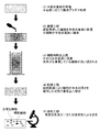
- FIG. 1 is a diagram schematically illustrating a method for producing an isolated cell specimen of the present invention.
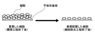
- FIG. 4 is a diagram schematically illustrating an arrangement of cells in a process of manufacturing an isolated cell specimen.
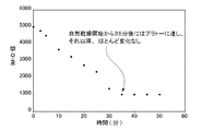
- FIG. 4 is a view showing the measurement results of the amount of water on the surface of the isolated cell specimen of Example 1.
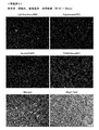
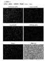
- FIG. 3 is a diagram showing the results of immunostaining using the isolated cell specimen of Example 1.
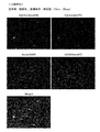
- FIG. 4 is a diagram showing the results of immunostaining using the isolated cell specimen of Comparative Example 1.
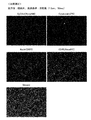
- FIG. 9 is a diagram showing the results of immunostaining using the isolated cell specimen of Comparative Example 2.
- FIG. 9 is a diagram showing the results of immunostaining using the isolated cell specimen of Comparative Example 3.
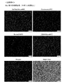
- FIG. 14 is a view showing a result of immunostaining using the isolated cell specimen of Comparative Example 4.
- FIG. 14 is a view showing a result of immunostaining using the isolated cell specimen of Comparative Example 5.
- the isolated cell specimen of the present invention is an isolated cell specimen in which cells to be observed are attached to the surface of a flat substrate, and substantially no liquid is present on the surface of the flat substrate on which the cells are arranged.
- This is an isolated cell specimen in which the average thickness of target cells satisfies the following formula (1) and is used for immunostaining and / or nuclear staining.
- T / tw 0.95 Equation (1)
- t represents the average thickness of the observation target cells attached to the surface of the flat substrate
- tw represents the average thickness of cells of the same type as the cells to be observed which have not been subjected to the drying step and which have adhered to the surface of the flat substrate.
- observation target cells In the isolated cell specimen of the present invention, cells adhere to the surface of the flat substrate. These cells are targets to be prepared and observed as a specimen, and are referred to as “observation target cells” in the present invention.
- the “observation target cell” refers to a cell group including a plurality of cells of the same type or different types.
- the isolated cell specimen of the present invention is characterized in that the average thickness of the observation target cell satisfies the formula (1).
- the expression (1) is obtained by calculating (t / tw), that is, the average thickness (t) of the observation target cells attached to the surface of the flat substrate, to the observation target that has not been subjected to the drying step and has been attached to the surface of the flat substrate.
- the value obtained by dividing by the average thickness (tw) of cells of the same kind as the cells is 0.95 or more. This equation indicates that the average thickness of the observation target cell is slightly smaller than the original average thickness of the same type of cells held under wet conditions, that is, the original average thickness of the observation target cell, Mean equivalent to the average thickness.
- T (t / tw) decreases when cells are excessively dried.
- the small (t / tw) means that the cells do not have the original thickness, size, shape, etc. (for example, the cells are flat) and cannot be used for various stainings and various detections. Means However, as a result of the investigations by the present inventors, when the surface of the flat substrate is dried so that the average thickness of the observation target cells satisfies the formula (1), the substrate is not a wet system. It has been surprisingly found that cells maintain their original thickness and the like, and can realize immunostaining properties equal to or higher than that of a wet system.
- Such an isolated cell specimen can have good staining efficiency without performing a membrane permeation treatment (treatment with saponin or the like) which is usually required for detection of intracellular antigens.
- a membrane permeation treatment treatment with saponin or the like
- Such an isolated cell specimen has a feature that conventional immunostaining samples do not have, which is extremely easy to store and transfer before being subjected to immunostaining.
- substantially no liquid is present on the surface of the flat substrate on which the cells are arranged means that there is no liquid that can be visually observed on the surface of the flat substrate on which the cells are arranged. I do.
- the isolated cell specimen of the present invention it is not excluded that a small amount of liquid exists on the surface of the flat substrate on which the cells are arranged.
- the liquid include any liquid that can retain the structure of cells and the like, such as a buffer solution, water, and a medium. Note that the observation target cells attached to the surface of the flat substrate contain sufficient moisture to satisfy the expression (1).
- Whether or not liquid is substantially not present on the surface of the flat substrate on which the cells are arranged can be determined by the following method.
- (1) The moisture content on the surface of the flat substrate at an arbitrary time point was measured at 25 ° C. and 50% relative humidity using an infrared moisture meter (for example, “Fiber Infrared Moisture Analyzer IM-3SCV MODEL-2000”, manufactured by Fuji Work Co., Ltd.). It measures under the conditions of.
- the moisture content is output as an IM-D value (unit: mV) which is an output value of the moisture meter corresponding to the moisture content.
- Flat substrate examples include a substrate having an arbitrary material and an arbitrary shape, which is usually used in cell observation.
- “flat” means that the substrate has a surface that is smooth enough to allow cells to adhere to the substrate, and an aspect in which a part of the surface of the substrate has unevenness is not excluded.
- the material of the flat substrate is not particularly limited.
- the flat substrate is preferably formed from a transparent material.
- the transparent material include various glasses and various resins (a thermoplastic resin, a thermosetting resin, and the like).
- thermoplastic resin examples include polyethylene, polypropylene, polystyrene, polyvinyl chloride, acrylate polymer, methacrylate polymer, cyclic olefin copolymer (such as norbornene), and aromatic polyesters (such as polyethylene terephthalate and polybutylene terephthalate). , Polycarbonate, polyamide and the like.
- thermosetting resin examples include a silicone resin (such as one having a polyorganosiloxane skeleton), a phenol resin, a melamine resin, and an epoxy resin.
- polystyrene, methacrylate polymer, cyclic olefin copolymer, and silicone resin are preferable.
- the size of the flat substrate is not particularly limited, and can be appropriately selected depending on the use of the isolated cell specimen, an apparatus for observing the observation target cell, and the like.
- the surface on which the observation target cells are arranged is preferably hydrophilic from the viewpoint that the observation target cell-containing liquid can be easily spread on the flat substrate.
- the contact angle of water on the surface of the flat substrate on which the cells to be observed are arranged is preferably 10 ° or less at 25 ° C., more preferably 5 ° or less.
- the surface of the flat substrate is not hydrophilic
- the surface can be subjected to a surface treatment (hydrophilization treatment) as necessary.
- a surface treatment hydrophilization treatment
- Any method can be adopted as the surface treatment method, and examples include plasma treatment, ultraviolet treatment, electron beam treatment, corona discharge treatment, and ozone treatment. Of these, plasma treatment is preferred, and oxygen plasma treatment is particularly preferred.
- the contact angle of water on the surface of the flat substrate is measured using the ⁇ / 2 method.
- the ⁇ / 2 method is to determine the angle of the straight line connecting the left and right end points and the vertices of the droplet dropped on the surface of the flat substrate to the flat substrate surface, and specify the value obtained by doubling the angle as the contact angle.
- distilled water for example, 1 to 2 ⁇ l
- a plurality of different points for example, 5 or more
- each of the dropped droplets is dropped. May be determined, and the average of the obtained values may be treated as the contact angle of water.
- the contact angle may be specified using a contact angle meter (manufactured by Kyowa Interface Science Co., Ltd.).
- the observation target cell is a cell that adheres to the surface of the flat substrate and is a target of cell observation.
- the type of the observation target cell is not particularly limited.
- preferred cells include peripheral blood mononuclear cells (also called PBMC).
- the cells whose average thickness is specified may be of the same type as each other and may be derived from the same individual, but may be derived from the same individual. It is not necessary. However, it is preferably derived from the same species of organism. For example, when t is the average thickness of peripheral blood mononuclear cells, tw also means the average thickness of peripheral blood mononuclear cells.
- Tw may be a predetermined numerical value according to the type of cell.
- the tw of a peripheral blood mononuclear cell can be about 15 ⁇ m.
- the lower limit of t of peripheral blood mononuclear cells is at least 14.2 ⁇ m (ie, (t / tw) ⁇ 0.95), preferably at least 14.4 ⁇ m (ie, (t / tw) ⁇ 0.96). ), More preferably 14.5 ⁇ m or more (that is, (t / tw) ⁇ 0.97).
- the upper limit of t of the peripheral blood mononuclear cell sphere is about 15.7 ⁇ m in consideration of the dispersion of measurement results and individual differences.
- the observation target cell may be in any mode usually used for cell observation.
- the observation target cell may be one isolated from a tissue (epithelium or the like) or a body fluid (blood or the like) by a known method.
- the method for isolating the cells to be observed is not particularly limited as long as the original size and shape of the cells are not impaired, and examples thereof include centrifugation (density gradient centrifugation, etc.) and methods using reagents.
- the observation target cell in the present invention is an isolated cell, and thus does not include aspects such as a tissue section.
- Separation solution can be used in density gradient centrifugation.
- the separation solution include a lymphocyte separation solution.
- the separation solution is usually used by being layered on a blood sample. In such a case, a reagent or the like having a density adjusted to 1.077 g / ml can be used as the separation solution.
- a separation solution For example, by using a separation solution, most of red blood cells, granulocytes (neutrophils, eosinophils, basophils) and platelets contained in whole blood can be removed. As a result, it is possible to obtain a mononuclear cell fraction mainly composed of lymphocytes (T cells, B cells, NK cells) and monocytes, which are a part of leukocytes.
- T cells lymphocytes
- B cells NK cells
- monocytes which are a part of leukocytes.
- commercially available separation solutions include, for example, “LymphoPrep” (manufactured by Alere Technologies Inc.), “NycoPrep@1.077” (manufactured by Alere Technologies Inc.), and “OptiPrep” (manufactured by AlereTechnologies Inc.) “Histopaque @ 1077” (Sigma-Aldrich), trade name “Ficoll-Paque” (GE Healthcare) and the like.
- OptiPrep it is necessary to adjust the solution density to 1.077 g / ml by mixing an appropriate amount of a reagent and a medium in advance.
- the observation target cells preferably include peripheral blood mononuclear cells.
- the “average cell thickness” refers to immunostaining (fluorescence staining) an isolated cell specimen in which cells are attached (usually in a single layer) to a smooth surface of a flat substrate using a fluorescently labeled antibody. ) Is a value obtained by analyzing the fluorescence image under the fluorescence microscope observation using image analysis software.
- FIG. 1 shows an outline of the method for measuring the average thickness of cells.
- an isolated cell specimen to which cells whose average thickness is to be specified is set horizontally on an observation table of a fluorescence microscope.
- the visual field is fixed while keeping the focus on the substrate surface to which the cells are attached, and the objective lens is moved upward (in the Z-axis direction) every 1.05 ⁇ m to acquire a fluorescent image.
- the acquired images are analyzed using image analysis software (Image @ J, Ver1.52a, NIH, https://imagej.nih.gov/ij/), and the average fluorescence signal intensity of each image is calculated.
- the distance from the lower end of the cell (substrate surface) to the upper end of the cell is obtained by plotting the intensity of the fluorescent signal with respect to the distance in the Z-axis direction (the method of specifying the upper end of the cell from the plot will be described later with reference to FIG. 2). Do).
- This distance corresponds to the average thickness of the cells.
- the average thickness of the cells measured by this method is calculated based on the sum of the average fluorescence signal intensities of the cells contained in the isolated cell specimen. Therefore, this method has an advantage that the obtained result is hardly affected by variations in the size of individual cells, individual differences among cell sample providers, and the like.
- FIG. 2 is an example of a plot of the fluorescence signal intensity versus distance in the Z-axis direction used to determine the average thickness of the cells.
- the lower end of the cell (substrate surface) is specified as a point where the value of the Z axis is “0 ⁇ m”.
- the upper end of the cell is specified as the point having the minimum value that satisfies all of the following relationships among the points counted from the lower end of the cell (substrate surface) in the plot.
- the average thickness of the cells arranged in a single layer on the flat substrate of the present invention satisfies the following expression (1).
- T / tw 0.95 Equation (1)
- t represents the average thickness of the observation target cells attached to the surface of the flat substrate
- tw represents the average thickness of cells of the same type as the cells to be observed which have not been subjected to the drying step and which have adhered to the surface of the flat substrate.
- ⁇ t is specified by obtaining the average thickness of the cells by the above-described method for the observation target cells attached to the surface of the flat substrate of the isolated cell specimen.
- Tw is specified by calculating the average thickness of the cells attached to the surface of the flat substrate and not subjected to the drying step and of the same kind as the cells to be observed, by the above method.
- the flat substrate for specifying tw is not particularly limited as long as cells can be attached to the smooth surface thereof, but is preferably the same material as the flat substrate for specifying the corresponding t.
- "the cell has not undergone the drying step” means that the cell is held only in water or an aqueous buffer solution (phosphate buffered saline (PBS) or the like), and the cell has its original size and structure. Means to keep When specifying tw, the measurement is performed while the cells are maintained in a wet system.
- PBS phosphate buffered saline
- a method for maintaining the cells in a wet system for example, a method in which a frame is provided on a substrate, and a liquid (water or an aqueous buffer (PBS or the like)) which does not affect the structure of the cells is placed in the frame.
- a liquid water or an aqueous buffer (PBS or the like)
- tw can be specified by maintaining a part of the cells to be observed in a wet system and calculating the average thickness of the cells using this.
- Tw in any isolated cell preparation can be specified as follows. (1) The type and origin of the cells attached to the isolated cell specimen are identified by microscopic observation or the like. (2) Obtain cells of the same type as the cells specified in (1) above. (3) The cells obtained in the above (2) are attached to the surface of a flat substrate, and tw is specified while maintaining the cells in a wet system.
- the lower limit of (t / tw) is preferably (t / tw) ⁇ 0.96, more preferably (t / tw) ⁇ 0.97.
- (t / tw) is less than 0.95, it usually indicates a state in which the water in the cells is lost due to drying or the like in the process of preparing the specimen, and the cells to be observed are atrophied.
- the upper limit of (t / tw) is usually 1 as understood from the definition of (t / tw). However, the value may be about 1.05.
- Expression (1) is an index of whether the observation target cell maintains the original structure. For example, when cells are dried, for example, when immunostaining is performed using an antibody, the membrane structure of the cells is destroyed by the drying, and the cells may not be able to maintain the original structure. As a result, the localization of the protein to be detected changes, and an accurate result may not be obtained. Usually, the thickness and the size of a dried cell become smaller due to the loss of water. Therefore, the equation (1) based on the original thickness (tw) of the cell is used to specify a change in the size of the observation target cell. It is effective for
- the observation target cells be arranged as densely as possible on a flat substrate.
- the density of cells arranged in a single layer on a flat substrate is preferably 4000 to 12000 cells / mm 2 , more preferably 6000 to 10000 cells / mm 2 , and still more preferably 7000 to 9000 cells / mm 2 .
- the isolated cell specimen of the present invention is used for immunostaining and / or nuclear staining.
- the isolated cell specimen of the present invention can be used for any other purpose for observing cells.
- the following method is known as a conventional immunostaining method (for example, see Non-Patent Document 1).
- the cells diluted with the liquid medium are dropped on the surface of the substrate, and then the substrate is washed with the liquid medium, so that the cells are arranged in a single layer on the surface of the substrate.
- the cells are subjected to immunostaining using an antibody in the presence of a liquid medium, and microscopic observation is performed. And all of these steps need to be performed in a wet system to avoid drying of the cells. This is because when the cells are dried, the structure of the cells may change, and accurate results may not be obtained. Therefore, in the conventional immunostaining method, it was difficult to stably store and transfer the observation cell sample before being subjected to immunostaining.
- the cells attached to the substrate retain the water and maintain the original structure of the cells, and the liquid (such as a medium or a medium) is placed on the surface of the flat substrate where the cells are arranged. Buffer).
- the isolated cell specimen is stably stored for a desired period of time (for example, one day or more) or the isolated cell specimen is transferred to a remote place until immunostaining is performed. be able to.
- the isolated cell specimen of the present invention is subjected to immunostaining, a known method can be appropriately selected, and an antibody and conditions corresponding to the marker or the like to be detected can be used.
- any fluorescent dye can be used, and for example, DAPI (4 ', 6-diamidino-2-phenylindole) and the like can be mentioned.
- the method for producing an isolated cell specimen according to the present invention includes a developing step of developing the observation target cell-containing liquid on the flat substrate, and a cell removing step of removing cells that do not adhere to the surface of the flat substrate after the developing step. After the step of removing the cells, a drying step of naturally drying the flat substrate is performed, and the obtained isolated cell specimen is used for immunostaining and / or nuclear staining.
- FIG. 3 shows an example of the method for preparing an isolated cell specimen of the present invention. Hereinafter, this will be described in detail with reference to FIG.
- a flat substrate is prepared, and a hydrophilic treatment such as an oxygen plasma treatment is performed as necessary.
- the liquid containing the cells to be observed is developed on a flat substrate.
- a method of developing the observation target cell-containing liquid any method that can be arranged on the flat substrate without destroying the cells can be adopted.
- a method of dropping the liquid on the surface of a flat substrate using a micropipette or the like may be used.
- the observation target cell-containing liquid include a cell-containing fraction such as a mononuclear cell fraction, and a dilution thereof.
- the developed liquid containing the cells to be observed spreads on the surface of the flat substrate, and observation over a wide range is possible.
- various aqueous buffers such as PBS
- pure water can be used as a liquid medium for diluting the cells.
- the cell concentration is not particularly limited, but is preferably a concentration at which the cells on the substrate are not excessively layered.
- the cell concentration is preferably 1 ⁇ 10 6 to 1 ⁇ 10 7 cells / ml, more preferably 2 ⁇ 10 6 to 5 ⁇ 10 6 cells / ml.
- Cells arranged on a flat substrate are not usually arranged in a single layer, but are arranged in a partially laminated state. In this state, the cells that are in direct contact with the surface of the flat substrate are bonded to the surface of the flat substrate by physical adsorption. That is, in the developing step, at least a part of the observation target cells arranged on the substrate surface adheres to the surface of the flat substrate.
- FIG. 4 is a schematic diagram showing the state of the stacked cells after the developing step and the state of the cells arranged in a single layer after the cell removing step.
- the method for removing cells that have not adhered to the surface of the flat substrate is not particularly limited, but from the viewpoint of preventing cell damage, a method of bringing a liquid into contact with the substrate is preferable.
- a method of immersing the plate-like substrate in a container containing a cell-removing liquid, a method of pouring the removing liquid over the surface of the plate-like substrate, and the like can be mentioned.
- the liquid for removing cells is preferably water or an aqueous solution.
- the lower limit of the specific resistance value of water or the aqueous solution is preferably 10 ⁇ ⁇ cm or more, more preferably 10 4 ⁇ ⁇ cm or more, and further preferably 10 6 ⁇ ⁇ cm or more. If the specific resistance value of water or the aqueous solution is less than 10 ⁇ ⁇ cm, the content of the ionic substance in the cell removing liquid increases, solutes precipitate in the drying step described below, or the solutes are observed with a microscope. May interfere.
- the upper limit of the specific resistance value of water or the aqueous solution is preferably 100 ⁇ ⁇ cm or less.
- aqueous solution having a specific resistance value within the above range examples include a liquid medium, tap water (15 to 16 k ⁇ ⁇ m), and various buffers (phosphate buffered saline (62 to 63 ⁇ ⁇ m), etc.).
- water having a specific resistance value of 10 4 ⁇ ⁇ cm or more and containing almost no ionic substance is preferable since it is hardly affected by precipitation in a drying step to be described later or microscopic observation. It is more preferable to use pure water having a resistance value of 10 6 ⁇ ⁇ cm or more.
- the flat substrate is dried until substantially no liquid is present on the surface of the flat substrate on which the cells are arranged.
- the amount of liquid on the surface of the flat substrate can be reduced by the drying process. Thereby, the observation target cell can be arranged on the substrate in a state where the liquid is not substantially present on the surface of the flat substrate on which the cells are arranged.
- Natural drying means drying at a temperature around room temperature (eg, 1 to 40 ° C.).
- the method of drying is not particularly limited as long as the sample can be exposed to a temperature around room temperature.
- Temperature conditions for natural drying can be adjusted according to the length of drying time and the like.
- the temperature condition of the natural drying may be in the range of room temperature, for example, preferably 1 to 40 ° C, more preferably 10 to 30 ° C, still more preferably 15 to 29 ° C, and still more preferably 20 to 29 ° C.
- the temperature is 28 ° C., most preferably 22 to 24 ° C. (around 23 ° C.). If the temperature is too high, drying of the cells proceeds, and the membrane structure of the cells is destroyed, the original structure cannot be maintained, and the subsequent observation may be affected. If the temperature is too low, the drying time may be too long.
- the relative humidity is preferably 10 to 70%, more preferably 20 to 60%, and further preferably 20 to 50%.
- the pressure condition for natural drying is preferably 0.5 to 2.0 atm, more preferably 0.8 to 1.2 atm, and still more preferably 1 atm.
- the time for natural drying is preferably 30 minutes or more, more preferably 35 minutes or more, further preferably 50 minutes or more, and still more preferably 1 hour or more. Although there is no problem if the time for natural drying is longer, it is usually preferably 100 hours or less, more preferably 90 hours or less, and still more preferably 80 hours or less from the viewpoint of work efficiency.
- the wind speed condition for natural drying may be 0.0 to 1.0 m / sec. The higher the wind speed, the shorter the drying time may be.
- the average thickness of the observation target cells satisfies the following expression (2).
- T ′ / tw ′ 0.95 Equation (2)
- t ′ represents the average thickness of the observation target cells after the drying step, which adhered to the surface of the flat substrate
- tw ′ represents the average thickness of the observation target cells before the drying step, which adhered to the surface of the flat substrate.
- the isolated cell specimen of the present invention is obtained. This isolated cell specimen is used for immunostaining and / or nuclear staining.
- the isolated cell specimen obtained as described above is subjected to immunostaining and / or nuclear staining. If necessary, the isolated cell specimen of the present invention can be subjected to various analyzes by methods other than immunostaining and / or nuclear staining.
- the isolated cell specimen obtained as described above has good immunostaining and / or nuclear staining.
- "good immunostaining and / or nuclear staining" means that the stained cells can maintain their original structure and have sufficient signal intensity by staining for detection. .
- the isolated cell specimen of the present invention is obtained.
- the obtained isolated cell specimen is subjected to immunostaining and / or nuclear staining.
- any conditions can be adopted according to the type of the target cell, and examples thereof include a method using a fluorescently labeled antibody and the like.
- the method of nuclear staining any conditions can be adopted according to the type of the target cell, and examples thereof include a method using DAPI or the like.
- the target cells include circulating cancer cells. When the target cells are circulating cancer cells, the observation target cells preferably include peripheral blood mononuclear cells.
- EpCAM a surface marker of epithelial cells
- Cytokeratin an intracellular protein marker of epithelial cells
- EpCAM-positive, Cytokeratin-positive, and CD45-negative cells can be narrowed down by simultaneously performing staining using an antibody against CD45 (a surface marker of leukocytes) in order to avoid false positives due to nonspecific adsorption of antibody molecules.
- DAPI nuclear staining is performed on the cells selected as candidates for circulating cancer cells by these antibody stainings to evaluate the normality of the nuclear structure, which may lead to cancer metastasis or recurrence. Can identify the presence of high cancer cells.
- Detection and analysis of target cells using a fluorescence microscope etc. are performed on the isolated cell specimens subjected to the staining described above, and the results of these stainings / observations are comprehensively evaluated. It is determined whether or not contains circulating cancer cells.
- ⁇ When immunostaining is performed on the isolated cell specimen obtained by the production method of the present invention, it is not necessary to perform the membrane permeation treatment. However, in the present invention, an embodiment in which a membrane permeation treatment is performed at the time of immunostaining is not excluded. In normal immunostaining, when the target to be stained is not the cell surface (for example, when cytokeratin antibody targeting intracellular proteins or DAPI targeting cell nuclei is used), a membrane using saponin or the like is used. It is necessary to perform transmission processing. That is, in order to obtain intracellular information, a membrane permeation treatment is usually required as a pretreatment. On the other hand, the isolated cell specimen obtained by the production method of the present invention can be stained without performing such a membrane permeation treatment, and can greatly contribute to simplification and efficiency of the inspection process.
- ⁇ Test 1 Examination of various isolated cell specimens> Based on the following method, isolated cell specimens were prepared under various conditions, and examination efficiency was examined.
- Example 1 An isolated cell specimen was prepared according to the following procedure and stained.
- lung cancer cells were used as a circulating cancer cell model, and a cell group containing a small number of these cells was prepared as cells to be observed.
- human lung cancer cells H1650
- human leukemia cells CEM
- the cell concentration was 3.33 ⁇ 10 3
- a cell-containing solution to be observed at 6 cells / ml was prepared.
- Development Step 3 ml of the cell-containing solution to be observed was dropped on the flat substrate using a micropipette. After the dropping, the observation target cell-containing liquid spread over almost the entire surface of the substrate.
- the water content on the surface of the flat substrate was measured at 25 ° C. and 50% relative humidity. Was measured over time, and it was confirmed that there was no liquid on the surface of the flat substrate on which the cells were arranged by natural drying for 50 minutes (FIG. 5).
- Z-stack observation (using a 20 ⁇ objective lens, using Interval: 1.05 ⁇ m) was performed using a confocal laser scanning microscope (trade name “FV-3000”, manufactured by Olympus Corporation). Based on the strength, the average thickness of the observation target cells on the flat substrate was measured.
- the obtained isolated cell specimen was subjected to immunostaining and nuclear staining by the following method.
- EpCAM a surface marker for epithelial cells
- Cytokeratin an intracellular protein marker for epithelial cells
- CD45 a surface marker for leukocytes
- Anti-EpCAM antibody trade name “Alexa488-labeled anti-EpCAM antibody”, Thermo Fisher Scientific Inc., 50-fold diluted anti-cytokeratin antibody: PE-labeled anti-cytokeratin antibody, 50-fold diluted anti-CD45 antibody: trade name “Alexa 647-labeled anti-CD45 antibody” ”, 30-fold dilution DAPI: 2 ⁇ g / ml
- Staining method 3 ml of the staining solution was developed so as to be in contact with the cells on the surface of each specimen, and staining was performed at room temperature under light shielding for 1 hour. Next, after removing excess staining solution by decantation, each specimen was immersed in PBS and washed.
- Comparative Example 1 In the drying step, the same operation as in Example 1 was performed except that hot air was blown from a distance of 1.5 cm for 10 seconds using a 1200 W dryer instead of natural drying, and the isolated cell specimen of Comparative Example 1 was dried. Was prepared. As a result of visual observation and observation with a stereoscopic microscope, no liquid was observed on the surface of the flat substrate on which the cells were arranged.
- Comparative Example 2 In the drying step, an isolated cell specimen of Comparative Example 2 was prepared in the same manner as in Example 1 except that hot air was blown from a distance of 10 cm for 30 seconds using a 1200 W dryer instead of natural drying. did. As a result of visual observation and observation with a stereoscopic microscope, no liquid was observed on the surface of the flat substrate on which the cells were arranged.
- Comparative Example 3 In the drying step, the same operation as in Example 1 was performed except that cold air was blown from a distance of 1.5 cm for 50 seconds using a 1200 W dryer instead of natural drying, and the isolated cell specimen of Comparative Example 3 was dried. Was prepared. As a result of visual observation and observation with a stereoscopic microscope, no liquid was observed on the surface of the flat substrate on which the cells were arranged.
- Example 4 (Comparative Example 4) Using the same plate-like substrate as in Example 1, an acrylic frame (2 mm in width and 2 mm in height) for holding the liquid was fixed to the peripheral portion on the plate-like substrate by an adhesive. Observation target cells were prepared in the same manner as in Example 1. After disposing the observation target cells on the substrate, the staining and observation described below were performed while the observation target cells were always kept in the buffer.
- Comparative Example 5 The same operation as in Comparative Example 4 was performed, except that a membrane permeation treatment using a conventional saponin was performed before the staining step described below.
- Table 1 shows the evaluation results. 6 to 11 show the results of immunostaining and nuclear staining.
- (t / tw) was specified as follows. That is, the thickness of the cell of Comparative Example 4 was treated as (tw).
- (T / tw) “Average thickness of observation target cell adhered to surface of flat substrate (t)” / “average thickness of observation cell adhered to surface of flat substrate of Comparative Example 4 (tw) "
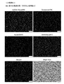
- Example 1 (FIG. 6) is a specimen in which the average thickness of the cells to be observed is in an appropriate range and the liquid is not substantially present on the surface of the flat substrate on which the cells are arranged. In this specimen, four types of markers for identifying circulating cancer cells could be clearly detected, and the cell density on the specimen was dense.
- Comparative Example 1 (FIG. 7) and Comparative Example 2 (FIG. 8) are samples prepared in the same manner as in Example 1 except that hot air drying was performed.
- the average thickness of the cells to be observed attached to the surface of the flat substrate becomes flat within a specified range, the cells do not maintain their original structure, and the staining property with the CD45 antibody is remarkable. Dropped.
- the cell density on the specimen was also low (bad).
- the average thickness of the observation target cells attached to the surface of the flat substrate was equal to or less than the specified range. It became flat and the staining with the CD45 antibody was significantly reduced.
- Comparative Example 4 (FIG. 10) and Comparative Example 5 (FIG. 11) show the results of staining the specimen in the same manner as commonly performed immunostaining.
- all operations were performed in a buffer solution throughout, so the average thickness of the cells maintained the original thickness and was within an appropriate range, but the surface of the flat substrate was This is a specimen in which a liquid having a liquid level significantly exceeding the average thickness of the attached observation target cells coexists.
- Comparative Example 4 was a sample that was not subjected to membrane permeation treatment with saponin, and could not be stained with Cytokeratin antibody and DAPI.
- the membrane permeation treatment with saponin was performed by a conventional method, and four types of markers for identifying circulating cancer cells could be detected, and the cell density on the sample was high. there were.
- Example 1 is a comparative example which is a general test method in that it has excellent staining properties with the CD45 antibody, and that nuclear staining of DAPI or the like can be performed without membrane permeation treatment and inspection efficiency is high. Excellent for 4.
- the method of the present invention is also excellent in inspection efficiency in that cells can be stably arranged in a single layer on a flat substrate.
- ⁇ Test 2 Examination of natural drying conditions> Four isolated cell specimens were prepared in the same manner as in Experimental Example 1 of Test 1 except that the time for air drying was changed within the range of 1 to 72 hours.
Landscapes
- Health & Medical Sciences (AREA)
- Life Sciences & Earth Sciences (AREA)
- Chemical & Material Sciences (AREA)
- Engineering & Computer Science (AREA)
- General Health & Medical Sciences (AREA)
- Biochemistry (AREA)
- Bioinformatics & Cheminformatics (AREA)
- Pathology (AREA)
- Physics & Mathematics (AREA)
- Biomedical Technology (AREA)
- Genetics & Genomics (AREA)
- Immunology (AREA)
- Wood Science & Technology (AREA)
- Zoology (AREA)
- Medicinal Chemistry (AREA)
- General Physics & Mathematics (AREA)
- Biotechnology (AREA)
- Organic Chemistry (AREA)
- Analytical Chemistry (AREA)
- Virology (AREA)
- Urology & Nephrology (AREA)
- Food Science & Technology (AREA)
- Molecular Biology (AREA)
- Hematology (AREA)
- General Engineering & Computer Science (AREA)
- Microbiology (AREA)
- Tropical Medicine & Parasitology (AREA)
- Investigating Or Analysing Biological Materials (AREA)
- Measuring Or Testing Involving Enzymes Or Micro-Organisms (AREA)
Abstract
La présente invention vise à fournir un échantillon cellulaire isolé qui est approprié pour une immunocoloration, etc., et qui est facilement stocké et transporté avant d'être soumis à une immunocoloration, etc. La présente invention concerne un échantillon cellulaire isolé dans lequel des cellules à observer adhèrent à la surface d'une plaque de base plane. Il n'y a pratiquement pas de liquide sur la surface de la plaque de base plane sur laquelle les cellules sont disposées. L'épaisseur moyenne des cellules à observer satisfait la formule (1) et l'échantillon cellulaire isolé est utilisé dans l'immunocoloration et/ou la coloration nucléaire. (1) : (T/tw) ≥ 0,95 (dans la formule (1), t représente l'épaisseur moyenne des cellules à observer qui adhèrent à la surface de la plaque de base plane, et tw représente l'épaisseur moyenne de cellules qui sont du même type que les cellules à observer, qui adhèrent à la surface de la plaque de base plane et qui n'ont pas été soumises à une étape de séchage).
Priority Applications (1)
| Application Number | Priority Date | Filing Date | Title |
|---|---|---|---|
| JP2020531289A JP7153365B2 (ja) | 2018-07-18 | 2019-07-12 | 単離細胞標本、単離細胞標本の製造方法、及び目的細胞の検出方法 |
Applications Claiming Priority (2)
| Application Number | Priority Date | Filing Date | Title |
|---|---|---|---|
| JP2018-135111 | 2018-07-18 | ||
| JP2018135111 | 2018-07-18 |
Publications (1)
| Publication Number | Publication Date |
|---|---|
| WO2020017451A1 true WO2020017451A1 (fr) | 2020-01-23 |
Family
ID=69163699
Family Applications (1)
| Application Number | Title | Priority Date | Filing Date |
|---|---|---|---|
| PCT/JP2019/027690 Ceased WO2020017451A1 (fr) | 2018-07-18 | 2019-07-12 | Échantillon cellulaire isolé, procédé de production d'un échantillon cellulaire isolé et procédé de détection d'une cellule d'intérêt |
Country Status (2)
| Country | Link |
|---|---|
| JP (1) | JP7153365B2 (fr) |
| WO (1) | WO2020017451A1 (fr) |
Cited By (1)
| Publication number | Priority date | Publication date | Assignee | Title |
|---|---|---|---|---|
| JP2021135141A (ja) * | 2020-02-26 | 2021-09-13 | 国立研究開発法人産業技術総合研究所 | 粒子評価用標本の作製装置及び作製方法 |
Citations (5)
| Publication number | Priority date | Publication date | Assignee | Title |
|---|---|---|---|---|
| JPH11237323A (ja) * | 1998-02-18 | 1999-08-31 | Sysmex Corp | 塗抹標本作製方法および装置 |
| JP2014533509A (ja) * | 2011-11-17 | 2014-12-15 | セルスケープ・コーポレーション | 細胞を取得し、解析するための方法、装置及びキット |
| WO2016152672A1 (fr) * | 2015-03-25 | 2016-09-29 | 富士フイルム株式会社 | Procédé pour isoler des cellules candidates de globules rouges nucléés |
| JP2018508760A (ja) * | 2015-01-14 | 2018-03-29 | バイオ−ラッド ラボラトリーズ,インコーポレイティド | 血液分析の系および方法 |
| JP2018085954A (ja) * | 2016-11-29 | 2018-06-07 | 国立研究開発法人産業技術総合研究所 | プラスチック基材及び血液標本の作製方法 |
-
2019
- 2019-07-12 WO PCT/JP2019/027690 patent/WO2020017451A1/fr not_active Ceased
- 2019-07-12 JP JP2020531289A patent/JP7153365B2/ja active Active
Patent Citations (5)
| Publication number | Priority date | Publication date | Assignee | Title |
|---|---|---|---|---|
| JPH11237323A (ja) * | 1998-02-18 | 1999-08-31 | Sysmex Corp | 塗抹標本作製方法および装置 |
| JP2014533509A (ja) * | 2011-11-17 | 2014-12-15 | セルスケープ・コーポレーション | 細胞を取得し、解析するための方法、装置及びキット |
| JP2018508760A (ja) * | 2015-01-14 | 2018-03-29 | バイオ−ラッド ラボラトリーズ,インコーポレイティド | 血液分析の系および方法 |
| WO2016152672A1 (fr) * | 2015-03-25 | 2016-09-29 | 富士フイルム株式会社 | Procédé pour isoler des cellules candidates de globules rouges nucléés |
| JP2018085954A (ja) * | 2016-11-29 | 2018-06-07 | 国立研究開発法人産業技術総合研究所 | プラスチック基材及び血液標本の作製方法 |
Non-Patent Citations (2)
| Title |
|---|
| HASHIMOTO, M. ET AL.: "Hydrophilic-treated plastic plates for wide-range analysis of Giemsa-stained red blood cells and automated Plasmodium infection rate counting", MALARIA JOURNAL, vol. 16, no. 321, 8 August 2017 (2017-08-08), pages 1 - 10, XP055678398 * |
| YAMAMURA, S. ET AL.: "Accurate Detection of Carcinoma Cells by Use of a Cell Microarray Chip", PLOS ONE, vol. 7, 1 March 2012 (2012-03-01), pages 1 - 9, XP055416178 * |
Cited By (1)
| Publication number | Priority date | Publication date | Assignee | Title |
|---|---|---|---|---|
| JP2021135141A (ja) * | 2020-02-26 | 2021-09-13 | 国立研究開発法人産業技術総合研究所 | 粒子評価用標本の作製装置及び作製方法 |
Also Published As
| Publication number | Publication date |
|---|---|
| JPWO2020017451A1 (ja) | 2021-07-01 |
| JP7153365B2 (ja) | 2022-10-14 |
Similar Documents
| Publication | Publication Date | Title |
|---|---|---|
| JP2022078344A (ja) | 生物学的サンプルの画像分析および測定 | |
| JP7079203B2 (ja) | デジタルホログラフィ顕微鏡検査および無傷の(untouched)末梢血白血球を用いる高精度の5部鑑別(5-part Differential) | |
| JP6273276B2 (ja) | 生物学的サンプルの画像分析および測定 | |
| CN105051538B (zh) | 用于检测生物状况的系统和方法 | |
| CN102036753B (zh) | 免疫磁性富集稀少细胞的改进的成像 | |
| CN105181649B (zh) | 一种新型免标记模式识别细胞仪方法 | |
| JP2021512299A5 (fr) | ||
| US5496704A (en) | Method for in vitro detection of formed elements in biological samples | |
| CN109266717A (zh) | 一种通过单细胞分析检测细菌耐药性的方法和装置 | |
| Shaw Bagnall et al. | Deformability-based cell selection with downstream immunofluorescence analysis | |
| Xie et al. | Differentiation of normal and leukemic cells by 2D light scattering label-free static cytometry | |
| CN111812071A (zh) | 一种新型循环肿瘤细胞的鉴定技术 | |
| JP7153365B2 (ja) | 単離細胞標本、単離細胞標本の製造方法、及び目的細胞の検出方法 | |
| Yapp et al. | Multiplexed 3D analysis of immune states and niches in human tissue | |
| US20110300574A1 (en) | Cell-counting method for blood hemolysis analysis in fragility measurement | |
| JP5348357B1 (ja) | 血中の目的細胞の定量方法および該細胞を定量するシステムの評価方法 | |
| Peters et al. | Classification of single extracellular vesicles in a double nanohole optical tweezer for cancer detection | |
| Boselli et al. | Unravelling plasma extracellular vesicle diversity with optimised spectral flow cytometry | |
| Hashimoto et al. | Hydrophilic-treated plastic plates for wide-range analysis of Giemsa-stained red blood cells and automated Plasmodium infection rate counting | |
| Hwang et al. | Microparticle-tagged image-based cell counting (ImmunoSpin) for CD4+ T cells | |
| US20230221319A1 (en) | A Method, A System, An Article, A Kit And Use Thereof For Biomolecule, Bioorganelle, Bioparticle, Cell And Microorganism Detection | |
| CN107328941A (zh) | 一种可同时检测多种细胞粘附因子的抗体芯片 | |
| EP4249889B1 (fr) | Procédés d'analyse de biopsies et d'échantillons biologiques | |
| RU2612007C2 (ru) | Способ автоматизированного анализа клеток крови посредством описания лейкоцитов на основе оптических особенностей структуры ядер | |
| RU2433175C2 (ru) | Способ исследования клеток с помощью биочипа |
Legal Events
| Date | Code | Title | Description |
|---|---|---|---|
| 121 | Ep: the epo has been informed by wipo that ep was designated in this application |
Ref document number: 19837563 Country of ref document: EP Kind code of ref document: A1 |
|
| ENP | Entry into the national phase |
Ref document number: 2020531289 Country of ref document: JP Kind code of ref document: A |
|
| NENP | Non-entry into the national phase |
Ref country code: DE |
|
| 122 | Ep: pct application non-entry in european phase |
Ref document number: 19837563 Country of ref document: EP Kind code of ref document: A1 |