WO2020054877A1 - 循環器障害または血栓形成の進行またはそのリスクの予測 - Google Patents
循環器障害または血栓形成の進行またはそのリスクの予測 Download PDFInfo
- Publication number
- WO2020054877A1 WO2020054877A1 PCT/JP2019/036399 JP2019036399W WO2020054877A1 WO 2020054877 A1 WO2020054877 A1 WO 2020054877A1 JP 2019036399 W JP2019036399 W JP 2019036399W WO 2020054877 A1 WO2020054877 A1 WO 2020054877A1
- Authority
- WO
- WIPO (PCT)
- Prior art keywords
- adam28
- vwf
- cells
- risk
- concentration
- Prior art date
- Legal status (The legal status is an assumption and is not a legal conclusion. Google has not performed a legal analysis and makes no representation as to the accuracy of the status listed.)
- Ceased
Links
Images

Classifications
-
- C—CHEMISTRY; METALLURGY
- C12—BIOCHEMISTRY; BEER; SPIRITS; WINE; VINEGAR; MICROBIOLOGY; ENZYMOLOGY; MUTATION OR GENETIC ENGINEERING
- C12Q—MEASURING OR TESTING PROCESSES INVOLVING ENZYMES, NUCLEIC ACIDS OR MICROORGANISMS; COMPOSITIONS OR TEST PAPERS THEREFOR; PROCESSES OF PREPARING SUCH COMPOSITIONS; CONDITION-RESPONSIVE CONTROL IN MICROBIOLOGICAL OR ENZYMOLOGICAL PROCESSES
- C12Q1/00—Measuring or testing processes involving enzymes, nucleic acids or microorganisms; Compositions therefor; Processes of preparing such compositions
- C12Q1/02—Measuring or testing processes involving enzymes, nucleic acids or microorganisms; Compositions therefor; Processes of preparing such compositions involving viable microorganisms
- C12Q1/04—Determining presence or kind of microorganism; Use of selective media for testing antibiotics or bacteriocides; Compositions containing a chemical indicator therefor
-
- C—CHEMISTRY; METALLURGY
- C12—BIOCHEMISTRY; BEER; SPIRITS; WINE; VINEGAR; MICROBIOLOGY; ENZYMOLOGY; MUTATION OR GENETIC ENGINEERING
- C12Q—MEASURING OR TESTING PROCESSES INVOLVING ENZYMES, NUCLEIC ACIDS OR MICROORGANISMS; COMPOSITIONS OR TEST PAPERS THEREFOR; PROCESSES OF PREPARING SUCH COMPOSITIONS; CONDITION-RESPONSIVE CONTROL IN MICROBIOLOGICAL OR ENZYMOLOGICAL PROCESSES
- C12Q1/00—Measuring or testing processes involving enzymes, nucleic acids or microorganisms; Compositions therefor; Processes of preparing such compositions
- C12Q1/68—Measuring or testing processes involving enzymes, nucleic acids or microorganisms; Compositions therefor; Processes of preparing such compositions involving nucleic acids
- C12Q1/6813—Hybridisation assays
-
- G—PHYSICS
- G01—MEASURING; TESTING
- G01N—INVESTIGATING OR ANALYSING MATERIALS BY DETERMINING THEIR CHEMICAL OR PHYSICAL PROPERTIES
- G01N33/00—Investigating or analysing materials by specific methods not covered by groups G01N1/00 - G01N31/00
- G01N33/48—Biological material, e.g. blood, urine; Haemocytometers
- G01N33/50—Chemical analysis of biological material, e.g. blood, urine; Testing involving biospecific ligand binding methods; Immunological testing
- G01N33/53—Immunoassay; Biospecific binding assay; Materials therefor
-
- A—HUMAN NECESSITIES
- A61—MEDICAL OR VETERINARY SCIENCE; HYGIENE
- A61B—DIAGNOSIS; SURGERY; IDENTIFICATION
- A61B5/00—Measuring for diagnostic purposes; Identification of persons
- A61B5/02—Detecting, measuring or recording for evaluating the cardiovascular system, e.g. pulse, heart rate, blood pressure or blood flow
Definitions
- the present invention relates to a method for providing data for predicting the progress of circulatory disorders or thrombus formation or the risk thereof.
- the present invention also relates to a method for assessing the risk of a drug or medical device causing circulatory disorders or thrombus formation in a living body.
- vascular endothelial cells due to these medical activities usually heal spontaneously, but vascular endothelial cells remain damaged due to the effects of biocompatibility of the medical device and the sustained release period of the drug, causing delayed onset. Deterioration of long-term clinical results such as sexual thrombosis has been an issue.
- Atherosclerotic lesions which are the main cause of vaso-occlusive disease causing circulatory disorders, have two aspects: vascular dysfunction and plaque formation (Guidelines for non-invasive assessment of vascular function, JCS # 2013).
- vascular dysfunction As for the detection of plaque, accurate evaluation can be performed with the progress of diagnostic imaging.
- plaques do not always break down and cause cardiovascular damage, and most plaque ruptures occur at lesions without severe stenosis. For this reason, it is difficult to predict a circulatory disorder due to plaque rupture by general angiography. Therefore, a method for detecting plaque that is easily broken (that is, unstable plaque) has been developed. Methods for detecting vulnerable plaque using intravascular ultrasound (IVUS), optical coherence tomography (OCT), angioscopy, etc.
- IVUS intravascular ultrasound
- OCT optical coherence tomography
- angioscopy etc.
- vascular function tests for evaluating vascular dysfunction include vascular endothelial function tests (plethysmography, flow-mediated dilatation: FMD, reactive hyperpermia peripheral arterial tonometry: RH-PAT), pulse wave velocity (PWV), cardiac ankle Vascular index (CAVI), stiffness parameter, central blood pressure, augmentation index (AI), second derivative of photoplethysmogram (SDPTG), ankle-brachial blood pressure ratio (ankle-brachial-index: ABI), toe-brachial blood pressure ratio (toe-brachial index: TBI), some of which are also used in clinical practice, but these have not yet been predicted to predict cardiovascular disorders.
- vascular endothelial function tests plethysmography, flow-mediated dilatation: FMD, reactive hyperpermia peripheral arterial tonometry: RH-PAT), pulse wave velocity (PWV), cardiac ankle Vascular index (CAVI), stiffness parameter, central blood pressure, augmentation index (AI), second derivative of photoplethys
- the primary thrombus is triggered by the binding of vWF to von Willebrand factor (vWF) on endothelial cells or collagen exposed at the site of vascular injury, and platelets bind to this vWF via GP1b / V / IX. Furthermore, fibrin is formed by binding to platelets via GPIIb / IIIa, and this primary thrombus grows, leading to vascular occlusion and stenosis. That is, it is important that vWF exists in the blood vessel wall for the formation of a thrombus that causes vascular stenosis / occlusion.
- vWF von Willebrand factor
- the thrombus (ultra-high molecular weight vWF multimer) is high in order to prevent blood vessel stenosis and occlusion due to the thrombus. It is known that shear stress is applied, the three-dimensional structure of vWF changes, and ADAMTS13 degrades vWF to prevent occlusion due to thrombus.
- ADAMTS13 cannot degrade vWF having a native tertiary structure under normal blood flow, and apparently nothing occurs in a healthy state in a mouse in which the ADAMTS13 gene is knocked out (Motto DG et al., J). Clin Invest. 2005; 115 (10): 2752-61 and Banno Fet et al., Blood. 2009; 113 (21): 5323-9). From these facts, it is considered that ADAMTS13 decomposes excessively increased thrombus to prevent stenosis and occlusion of blood vessels. However, it is physiologically decomposed vWF to control primary thrombus.
- ADAM28 degrades vWF in cancer cells (see Mochizuki Set et al., J Natl Cancer Inst. 2012; 104 (12): 906-22), but ADAM28 forms thrombus formation. There are no reports that they are involved in
- the vascular function test method In order for the vascular function test method to become a biomarker in cardiovascular disease management, (1) the extent of progression of vascular dysfunction can be determined, (2) the risk of developing cardiovascular disease or the prognosis can be estimated, (3) intervention (4) It is necessary to satisfy conditions such as (4) improvement of the result leads to improvement of prognosis (the above guidelines). In order for such a vascular function test method to be clinically applied, (1) non-invasive and simple measurement can be performed, (2) low-cost universalization is possible, (3) accuracy and reproducibility Are important, and (4) standardization of the measurement method is possible. On the other hand, the vascular function test method has a problem that certain views have not yet been determined on measurement methods, interpretation of results, clinical significance, clinical application, and the like.
- Vascular function tests allow non-invasive assessment of arterial dysfunction at the asymptomatic stage, more accurately assess the risk of developing future cardiovascular disorders, and proactively intervene to prevent events May be associated with
- no consensus has been reached on its usefulness at this time, and it is necessary to confirm that therapeutic intervention will improve vascular function tests and that the improvement will function as a surrogate marker for the reduction of cardiovascular risk. is there.
- a dedicated test device is required for a conventional vascular function test.
- biomarkers in blood are also being searched for the purpose of predicting stenosis / occlusion of blood vessels without using a dedicated test device, and high-sensitivity CRP, Lp-PLA2, pentraxin 3 (PTX3) It has been reported that these may function as markers.
- an object of the present invention is to provide means capable of predicting the progress of thrombus formation, stenosis / occlusion of blood vessels, and circulatory disorders with high accuracy by a simple method.
- the present inventors have conducted intensive studies to solve the above-mentioned problems. As a result, it was surprisingly revealed that, when endothelial function was reduced, the expression level of vWF factor in endothelial cells increased and the expression level of ADAM28 decreased for the first time. Then, based on this finding, the present invention has been completed.
- a circulatory organ is expressed by using the expression level or concentration of ADAM28 in a blood sample collected from a subject or the ratio of the expression level or concentration of ADAM28 to vWF (ADAM28 / vWF ratio) as a marker.
- a method for providing data for predicting the progress of a disorder or thrombus formation or the risk thereof, wherein the progression of a circulatory disorder or thrombus formation or the progress thereof when the value of the marker is lower than a reference value A method is provided that is predicted to be at risk.
- a method for evaluating a risk that a drug or a medical device causes a circulatory disorder or thrombus formation in a living body in the presence and absence of the drug or the medical device.
- the ratio of the expression level or concentration of ADAM28 to vWF (ADAM28 / vWF ratio), or the ratio of the expression level or concentration of ADAMTS13 to vWF (ADAMTS13 / vWF ratio) is determined in the absence of the drug or medical device. Lower than the value in the culture medium or cells after culturing. If you are, wherein the drug or medical device is evaluated to be at risk of eliciting cardiovascular disorders or thrombosis in vivo are also provided.
- One embodiment of the present invention relates to a method for predicting the presence or risk of progression or risk of circulatory disorder or thrombus formation, or providing data for predicting the presence or absence. More specifically, the method comprises using, as a marker, the expression level or concentration of ADAM28 in a blood sample collected from a subject, or the ratio of the expression level or concentration of ADAM28 to vWF (ADAM28 / vWF ratio) as a marker, and This is a method of predicting the progress of formation or the presence or absence of a risk thereof, or providing data for predicting the presence or absence of the risk.
- the method when the value of the marker is lower than a reference value, it is predicted that there is a cardiovascular disorder or thrombus formation or a risk thereof. According to the method according to the present embodiment, it is possible to predict the progress of thrombus formation, stenosis / occlusion of blood vessels, and circulatory disorders with high accuracy by a simple method.
- the present inventors aim at developing means capable of predicting the progress of thrombus formation and stenosis / occlusion of blood vessels, and thus circulatory disorders with high accuracy, by a simple method. , And studied diligently.
- the present inventors first, under the condition that the mechanical stress is not applied to the vascular endothelial cells (corresponding to the state where blood flow does not flow in the blood vessel), the condition under which the mechanical stress is applied (Equivalent to a state in which blood flow is flowing in the blood vessel), it was confirmed that the expression level of eNOS (NO synthase in vascular endothelial cells), which is a marker of endothelial function, decreased, and endothelial function decreased. did.
- eNOS NO synthase in vascular endothelial cells
- ADAMTS13 which has been conventionally considered to be a physiological degrading enzyme of vWF, has only a soluble form and, as described above, degrades vWF having a native three-dimensional structure under normal blood flow. I can't.
- ADAM28 has a membrane-bound type in addition to a soluble type, and can also degrade vWF having a native steric structure.
- ADAM28 the enzyme that physiologically controls the degradation of vWF is ADAM28 instead of ADAMTS13, which was conventionally considered.
- membrane-bound ADAM28 efficiently degrades vWF present on the same cell membrane. This is presumed to prevent the formation of primary thrombus.
- ADAM28 plays an important role as a gatekeeper for thrombus formation, and by suppressing thrombus formation in a healthy state, ADAM28 can inhibit blood vessel formation. It is considered that occlusion / stenosis is prevented.
- vWF which is a trigger of primary thrombus formation
- ADAM28 which degrades it, decreases, and thrombus formation is promoted. It is considered that occlusion / stenosis of blood vessels occurs.
- the expression amount (or concentration) of ADAM28 in a blood sample collected from a subject or the ratio of the expression amount (or concentration) of ADAM28 to vWF (ADAM28 / vWF ratio) is used as a marker to obtain a cardiovascular system. It is believed that it is possible to predict the progression or risk of a disorder or thrombus formation. According to the above-described embodiment of the present invention, the above parameters are used as markers, and when the value of the marker is lower than a reference value, it is predicted that there is progression of circulatory disorders or thrombus formation or a risk thereof. Is done.
- vWF which is a trigger of primary thrombus formation
- ADAM28 the vWF on the luminal surface of the blood vessel
- ADAM proteins are multifunctional proteins involved in transmembrane protein ectodomain shedding, cell adhesion and invasion (Edwards DR et al., Mol Aspects Med. 2008; 29 (5): 258-89; Murphy G, Semin Cell Dev Biol. 2009; 20 (2): 138-45).
- the human genome contains 25 ADAMs including 4 pseudogenes, and 21 ADAMs consist of 13 proteolytic ADAMs exhibiting proteolytic activity and 8 non-proteolytic ADAMs. (Edwards DR et al., Mol Aspects Med.
- Proteolytic ADAMs share the metalloproteinase domain of matrix metalloproteinases (MMPs), and typical proteolytic ADAMs include propeptides, metalloproteinases, disintegrin-like, cysteine-rich, epidermal growth factor-like, transmembrane and Many proteolytic ADAMs, including intracellular domains and including ADAM8, ADAM9, ADAM12, ADAM15, ADAM17, ADAM19 and ADAM28, are overexpressed in human cancers and have been implicated in cancer growth and progression. Are known.
- MMPs matrix metalloproteinases
- ADAM28 is a known protein, and its amino acid sequence and cDNA sequence are also known.
- ADAM28 has two types, a membrane type (ADAM28m) and a secretory type (ADAM28s).
- ADAM28m membrane type
- ADAM28s secretory type
- Representative amino acid sequences and cDNA sequences of human membrane type ADAM28 (and its mature form) and human secretory ADAM28 (and its mature form) are described in WO 2016/143702. Is disclosed.
- "human membrane type ADAM28” means that the amino acid sequence or nucleotide sequence of membrane type ADAM28 is the same or substantially the same as the amino acid sequence or nucleotide sequence of membrane type ADAM28 which is naturally expressed in humans. Means having a nucleotide sequence.
- Substantially identical means that the amino acid sequence or nucleotide sequence of interest is at least 70% (preferably at least 80%, more preferably at least 90%) the amino acid sequence or nucleotide sequence of membrane-type ADAM28 naturally expressed in humans. %, More preferably 95% or more, and most preferably 99% or more), and has the function of human membrane ADAM28. The same applies to non-human species, proteins and genes other than membrane-type ADAM28, and fragments thereof.
- vWF is a plasma protein that plays an important role in blood coagulation.
- vWF is mainly synthesized in the vascular endothelium and is released into the blood in the form of high molecular multimers.
- Wild-type human vWF is a polypeptide consisting of a total of 2813 amino acids including its signal peptide and proregion. The amino acid sequence of wild type human vWF and the amino acid sequence of mature wild type human vWF subunit are disclosed in WO 2004/035778.
- the expression level or concentration of ADAM28 in a blood sample collected from a subject or the ratio of the expression level or concentration of ADAM28 to vWF (ADAM28 / vWF ratio) is measured for use as a marker. I do.
- the term “subject” refers to a subject that may cause a circulatory disorder or progression of thrombus formation, and is preferably a human subject.
- the subject is a patient who has been diagnosed as having a cardiovascular disorder or a disease involving thrombus formation, more preferably a patient who has been treated after receiving the diagnosis. is there.
- coronary artery events such as coronary artery stenosis, sudden cardiac death, fatal and non-fatal myocardial infarction, coronary artery stenosis and obstruction such as unstable angina, Cerebral infarction, cancer, diabetes, traveler thrombosis (economy class syndrome), peripheral arterial embolism disease, thrombotic microangiopathy (thrombotic thrombocytopenic purpura, hemolytic uremic syndrome, etc.), venous thrombosis (lung Thromboembolism, deep vein thrombosis, portal vein thrombosis, renal vein thrombosis, hepatic vein thrombosis, jugular vein thrombosis, axillary-subclavian vein thrombosis, cerebral sinus thrombosis, etc.).
- coronary artery events such as coronary artery stenosis, sudden cardiac death, fatal and non-fatal myocardial infarction, coronary artery stenosis
- the “blood sample” may be any blood-derived sample capable of performing the above measurement. Therefore, the specific form is not particularly limited, and a whole blood sample is usually used. As the whole blood sample, blood (whole blood) collected from a subject may be used as it is, or an additive such as potassium salt of EDTA or heparin may be added for the purpose of preventing coagulation or the like. The timing for collecting blood to collect a blood sample from a subject is not particularly limited.
- a blood sample used in the method according to the present embodiment it is preferable to use a blood sample immediately after collection from a subject for measurement, but a preserved blood sample may be used.
- the method of storing the blood sample is not particularly limited as long as the value of the marker in the sample does not change. For example, a low temperature condition of 0 to 10 ° C. which does not freeze, a dark place condition, and a non-vibration condition are preferable. .
- the measurement of the marker value can be performed using a conventionally known method.
- the “expression amount” of a certain protein includes both the expression of RNA (transcript) complementary to the gene encoding the protein and the expression of the protein (translation product) itself. . Therefore, in the present specification, the expression level of a certain protein refers to the expression level or expression intensity of a transcription product or a translation product. The expression level can usually be analyzed based on the production level of a transcript or the production level, activity, and the like of a translation product thereof. Further, the “concentration” of a certain protein means the abundance (expression amount) of the target protein per unit volume of the blood sample, and the “concentration” may be used as a marker in the method according to the present embodiment.
- the expression level of the protein may be measured by measuring the transcription product of the gene encoding the protein, that is, mRNA, or may be measured by measuring the translation product of the gene, that is, the protein itself. Preferably, the measurement is performed by measuring the transcript of the gene. Note that the gene transcript also includes cDNA obtained by reverse transcription from mRNA.
- the expression level of the transcript may be measured, for example, by measuring the degree of gene expression in the sample using nucleotides containing all or a part of the mRNA base sequence as a probe or primer, for example, using a microarray (microchip). It can be measured by a conventional method, Northern blotting, quantitative PCR, or the like.
- Examples of the quantitative PCR method include agarose gel electrophoresis, polyacrylamide gel electrophoresis, fluorescent probe, RT-PCR, real-time PCR, ATAC-PCR, Taqman @ PCR (SYBR (registered trademark) green method) , The Body Map method, the Serial analysis of gene expression (SAGE) method, the Micro-analysis of Gene expression (MAGE) method, and the like are known, and these can be used as appropriate.
- the evaluation may be performed using a next-generation sequencer. Using these methods, the amount of mRNA can be measured by using a nucleotide probe or primer that hybridizes to the mRNA.
- the base length of the probe or primer used for the measurement is preferably 10 to 50 bp, more preferably 15 to 25 bp.
- a DNA microarray can be prepared by immobilizing nucleotides consisting of a nucleotide sequence of a gene or nucleotides containing a partial sequence thereof on an appropriate substrate.
- the fixed substrate include a glass plate, a quartz plate, and a silicon wafer.
- the size of the substrate may be, for example, 3.5 mm ⁇ 5.5 mm, 18 mm ⁇ 18 mm, 22 mm ⁇ 75 mm, etc., which varies depending on the number of probe spots on the substrate and the size of the spots. Can be set.
- a method for immobilizing a polynucleotide or a fragment thereof utilizing the charge of nucleotides, polylysine, polyethyleneimine, or electrostatically bonded to a solid support surface-treated with a polycation such as polyalkylamine, an amino group,
- a nucleotide having a functional group such as an amino group, an aldehyde group, a thiol group, or biotin can be covalently bonded to a solid phase surface having a functional group such as an aldehyde group or an epoxy group.
- Immobilization may be performed using an array machine. At least one gene or a fragment thereof is immobilized on a substrate to prepare a DNA microarray.
- the DNA microarray is contacted with mRNA or cDNA derived from a subject labeled with a fluorescent substance, and allowed to hybridize. By measuring the intensity, the type and amount of mRNA can be determined.
- the expression level of the translation product can be measured by quantifying the translated protein or measuring the activity of the protein.
- protein quantification can be performed using an antibody specific for the protein.
- Antibody can be prepared by a known method. Alternatively, antibodies are provided, for example, from Invitrogen, Santa Cruz Biotechnology, Sigma-Aldrich, etc., and these can be appropriately obtained and used.
- the antibody for detection may be a polyclonal antibody or a monoclonal antibody.
- the detection of a protein using an antibody is not limited, but can be performed by, for example, immunochromatography, Western blotting, EIA, ELISA, RIA, flow cytometry, and the like.
- the antibody can be labeled with a fluorescent label, a radioactive label, an enzyme, biotin, or the like, and a secondary antibody labeled for such detection can also be used.
- the presence of myocardial injury is evaluated by detecting cardiac troponin T from a blood sample by immunochromatography (Trop @ T Sensitive, Roche Diagnostics).
- the method according to the present embodiment similarly, by measuring a marker such as ADAM28 or vWF by immunochromatography, it can be easily measured in a clinically minimally invasive manner, and can be universalized at low cost. It is expected to construct an evaluation method that predicts occlusion / stenosis of a blood vessel lumen, which has high accuracy and reproducibility and enables standardization of measurement methods. As a result, occlusion / stenosis of blood vessels can be prevented beforehand, and appropriate treatment can be performed. When the above measurement is performed by the immunochromatography method, it is preferable to use immunochromatographic test paper.
- the immunochromatographic test strip comprises a sample pad area for receiving a blood sample, a conjugate pad area containing a labeled antibody capable of binding to an indicator substance such as ADAM28 or vWF, a membrane body for developing a specimen, and the indicator on the membrane body.
- an antibody capable of binding to a substance is immobilized and has a test line capable of binding to the indicator substance bound to the labeled antibody in the conjugate pad region.
- an immunochromatographic test paper having a control line capable of determining the amount of the label bound in the test line is more preferable.
- the label has a color that becomes stronger in a concentration-dependent manner.
- the present specification discloses a method for predicting the progress of circulatory disorders or thrombus formation or the presence or absence of the risk thereof.
- the present specification also discloses a method of providing data for predicting the progress of circulatory disorders or thrombus formation or the risk thereof. At this time, as described above, when the value of the marker is lower than the reference value, it is predicted that there is a cardiovascular disorder or thrombus formation or a risk thereof.
- the “reference value” is a numerical value that can be a reference for determining an increase or decrease in the expression level or concentration in the method according to the present embodiment.
- the reference value for example, the expression amount (or concentration) of ADAM28 in a blood sample collected from a person who has not been diagnosed as having the above-mentioned disease (eg, a healthy person), or the amount of ADAM28 relative to vWF A measured value of the ratio of the expression amount (or concentration) (ADAM28 / vWF ratio) is exemplified. Further, the value of the marker previously measured in the same subject may be used as the reference value.
- a detection reagent for use in the method according to the above-described one aspect of the present invention and the presence or absence of progression of a circulatory disorder or thrombus formation or a risk thereof including the detection reagent
- kits used to predict The detection reagent enables detection of the expression level or concentration of ADAM28 or ADAM28 and vWF in the above-described blood sample.
- detection reagents include, for example, DNA or RNA for hybridization with mRNA in a sample, or antibodies for specific binding to a protein in a sample.
- DNA or RNA may be a probe capable of detecting hybridization with a fluorescent label or the like.
- the DNA or RNA may be a primer that can be used for amplifying mRNA.
- the kit can include, in addition to the detection reagents described above, other reagents necessary for detection, such as buffers, various nucleotides, other reagents necessary for hybridization or antibody binding.
- Another aspect of the present invention relates to a method for evaluating a risk of a drug or a medical device causing a circulatory disorder or thrombus formation in a living body.
- the method involves culturing the cells while applying mechanical stress to the cells in the presence and absence of the drug or medical device to be evaluated.
- the expression amount or concentration of ADAM28 or ADAMTS13 the ratio of the expression amount or concentration of ADAM28 to vWF (ADAM28 / vWF ratio) in the culture solution or cells after culturing the cells in the presence of the drug or the medical device, Or when the ratio of the expression amount or concentration of ADAMTS13 to vWF (ADAMTS13 / vWF ratio) is lower than the value in a culture solution or cells after culturing the cells in the absence of the drug or the medical device.
- the above-mentioned drug or medical device is evaluated as having a risk of causing a circulatory disorder or thrombus formation in a living body.
- the drug or the medical device can be assessed as being at risk of causing circulatory disorders or thrombus formation in the body. That is, by incorporating the evaluation method according to the present embodiment in the development stage of a drug or a medical device, it becomes possible to develop a drug or a medical device that hardly causes stenosis or occlusion of a blood vessel.
- vWF which is a trigger of primary thrombus formation
- ADAM28 or ADAMTS13 ADAM28 or ADAMTS13
- ADAMTS13 is also a known protein, and its amino acid sequence and cDNA sequence are also known.
- ADAMTS13 is a zinc-based metalloprotease belonging to the ADAMTS family, and is thought to reduce the multimer size of vWF and control the function of vWF by specifically cleaving the bond between Tyr1605 and Met1606 of vWF.
- Detailed structural and sequence information on human ADAMTS13 is disclosed in Zheng Xet et al., J Biol Chem. 2001; 276 (44): 41059-63.
- the method according to this embodiment includes culturing the cells.
- Cells used in the method are not particularly limited, but are preferably endothelial cells, and more preferably vascular endothelial cells.
- the species of the cell is not particularly limited.
- the culture solution (medium) is not particularly limited as long as all the cultured cells do not die during the evaluation period.
- endothelial cells for example, Porcine Endothelial Cell Growth Medium (Applications Inc.), Endothelial Cell Growth Medium 2 Kit (Takara Bio Inc.), Endothelial Cell Growth Medium MV 2 Kit (Takara Bio Inc.) ) Is suitable.
- the method according to the present embodiment is a method for determining whether or not a certain drug or medical device has a risk of causing circulatory disorders or thrombus formation in a living body. For this reason, cells are similarly cultured in the presence and absence of the drug or the medical device, and the expression level of a specific gene or protein or the expression level of a specific gene or protein in the presence and absence thereof, respectively. The above determination is performed using the change in the value of the ratio as an index.
- drugs and medical devices there is no particular limitation on the specific types of drugs and medical devices to be evaluated, and any type can be adopted as an evaluation target.
- the drug include a nucleic acid, a peptide, a protein, an organic compound, an inorganic compound, and the like.
- the medical device include a device selected from the group consisting of an intravascular indwelling device, a PTCA balloon, an artificial blood vessel, an extracorporeal membrane oxygenator (Extracorporeal membrane oxygenator; ECMO), and a centrifugal pump artificial heart.
- ECMO extracorporeal membrane oxygenator
- the medical device is preferably an intravascular indwelling device, and an example thereof is a device selected from the group consisting of a stent, a covered stent, a stent graft, an artificial valve, a valve clip, and a pump catheter for assisting circulation. .
- the specific form of the device for performing the evaluation method according to the present embodiment is not particularly limited, and may be any device capable of holding and culturing cells to be cultured.
- the material of the site for holding and culturing the cells has an expandable and elastic property such as silicone rubber, is bendable, can hold the cells, and can have a considerable amount of cell activity during the evaluation period. It is suitable. However, it is not limited to those capable of maintaining cell activity.
- the surface of the culture site may be coated with collagen, gelatin, fibronectin, or the like.
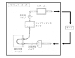
- a mechanical stress load culture device as shown in FIG. 1 is exemplified.
- mechanical stress applied to cells during culture means that a medium used for culture moves relative to cells to be cultured, or at an interface between the medium and the cells. It means a force (stress) applied to the cell by a change in pressure or the like.
- Specific examples of the mechanical stress include shear stress (shear stress) applied to cells due to a flow phenomenon (flow) of a medium simulating blood flow.
- the pressure applied to the cells that increases due to the increase in the flow rate of the medium is also included in the mechanical stress. Note that the latter increase in the flow rate of the medium simulates the pulsation of blood vessels (increase in blood pressure), and in a living body, corresponds to stretching stimulation that occurs on endothelial cells as the blood vessel diameter increases.
- the mechanical stress load culturing apparatus shown in FIG. 1 includes, as main components, a pump, a reservoir, a cell culturing unit for holding or culturing cells, a compliance tank, a valve (a resistance valve and a flow rate adjusting valve), and a tube for connecting them. It is composed of According to this mechanical stress load culturing apparatus, it is possible to flow (circulate) the medium in the cell culture unit by driving the pump, thereby applying a shear stress to the cells. At this time, by setting the flow conditions corresponding to the pulse rate, blood viscosity, pulse rate, etc. in the living body, it is possible to reproduce the shear stress due to the blood flow in the living body (load on the endothelial cells in the living body).
- the evaluation method according to the present embodiment is performed using the mechanical stress load culturing apparatus (FIG. 1) prepared above, the drug to be evaluated is added to the culture medium, and the culture is performed. In the presence of ". Similarly, by arranging and culturing the medical device to be evaluated in contact with a medium (preferably, a medium and cells), culturing in the "presence of a medical device" can be performed. In this manner, the culture in the presence of the evaluation target is performed, and the drug or medical device to be evaluated is used as an index based on the change in the marker between when the evaluation target does not exist (in the case where the evaluation target does not exist). It can be determined whether there is a risk of causing a circulatory disorder or thrombus formation in the living body.
- a medium preferably, a medium and cells
- the evaluation method according to the present embodiment is characterized in that any one of the following is used as an index (marker): Expression or concentration of ADAM28 Expression or concentration of ADAMTS13 Ratio of expression or concentration of ADAM28 to vWF (ADAM28 / vWF ratio) The ratio of the expression amount or concentration of ADAMTS13 to vWF (ADAMTS13 / vWF ratio).
- the amount and concentration or activity of the protein contained in the medium after completion of the culture may be employed, or after the completion of the culture.
- the amount, concentration or activity of the protein contained in the cells in or the amount of the gene (mRNA or cDNA prepared therefrom) encoding the same may be adopted.
- the evaluation method according to the present embodiment is characterized in that cells are cultured while applying mechanical stress to the cells.
- a means for culturing cells while applying mechanical stress to the cells for example, a mechanical stress load culturing apparatus as shown in FIG. 1 is used.
- the cell culturing unit is bent as shown in FIG. A method of culturing while circulating the medium in a connected state and further pulsating the medium.
- expression of eNOS, ADAM28 and ADAMTS13 was induced in a system in which cells were cultured while applying mechanical stress (circulating culture).
- the expression was reduced based on the observation that the expression was reduced in a system to which no mechanical stress was added (static culture). That is, while the expression of ADAM28 and ADAMTS13 is enhanced only when mechanical stress is applied to cells like blood flow in healthy blood vessels, blood flow decreases or stays like a lesion site.
- the expression of ADAM28 or ADAMTS13 is reduced in a state where no mechanical stress is applied to the cells, and therefore, it is considered that these expressions change to reflect the living body.
- a silicone rubber tube was prepared as a culture vessel for imitating a blood vessel. Then, collagen coating was performed on the inside of the silicone rubber tube by the following method. That is, first, collagen (5 mg / mL; 1 mM HCl solution, AteloCell Native collagen Bovine Dermis, Koken) was diluted 10-fold with dilute hydrochloric acid (pH 3.0, 10 ⁇ 3 M) and filled in a tube. Subsequently, coating was performed by incubating at 4 ° C. for 3 days. Then, after discarding the diluted collagen solution, the tube was washed with Hank's buffer and used for the subsequent test.
- collagen 5 mg / mL; 1 mM HCl solution, AteloCell Native collagen Bovine Dermis, Koken
- dilute hydrochloric acid pH 3.0, 10 ⁇ 3 M
- porcine coronary artery endothelial cells were cultured in a culture flask containing an endothelial cell culture medium (Porcine Endothelial Cell Growth Medium, APPLICATIONS INC) under conditions of 37 ° C. and 5% CO 2 to sufficiently increase the number of cells.
- the porcine coronary artery endothelial cells were seeded in the lumen of a silicone rubber tube coated with collagen as described above, and the cells were uniformly adhered to the inner surface of the tube while rotating the tube along the central axis. Then, the cells were cultured overnight at 37 ° C. and 5% CO 2 until they became confluent. In this way, a silicone rubber tube having a lumen covered with vascular endothelial cells was produced.
- the mechanical stress load culturing apparatus shown in FIG. 1 includes, as main components, a pump, a reservoir, a cell culturing unit for holding or culturing cells, a compliance tank, a valve (a resistance valve and a flow rate adjusting valve), and a tube for connecting them. It is composed of According to this mechanical stress load culturing apparatus, the medium in the cell culture unit can be caused to flow (circulate) or pulsate by driving the pump.
- the vascular endothelial cell-coated silicone rubber tube prepared as described above was connected to the cell culture unit portion of the mechanical stress load culture device (FIG. 1) prepared above as shown in FIG. Thereafter, the medium was circulated under a condition of 37 ° C. and 5% CO 2 at a pressure of 15 dyn / cm 2 (1.5 ⁇ 10 ⁇ 4 N / cm 2 ) and cultured for about one day.
- the medium since the silicone rubber tube is connected to the cell culture unit in a curved state, the culture is performed by circulating the culture medium so that vascular endothelial cells adhered to the inner surface of the silicone rubber tube can be removed. In this case, mechanical stress (shear stress) is applied. Under the same culture conditions, stationary culture without circulating the medium was also performed.
- Glyceraldehyde-3-phosphate dehydrogenase (GAPDH) gene was used as an internal standard.
- PCR primers were designed using the Primer3 (v.4.0.0) Pick primers from DNA sequence program, and the risk of self-annealing and mis-annealing was reduced in silico by the DNASIS Mac program (Hitachi Software Engineering). I confirmed that it was low.
- the nucleotide sequences of various primers used for the analysis of the expression level of the gene are shown below.
- Tables 1 to 5 The results of gene expression analysis performed in this way are shown in Tables 1 to 5 below. Note that the results shown in Tables 1 to 5 are all relative values when the amount of gene expression (or the ratio thereof) in static culture is set to 1.
- the expression of the eNOS gene analyzed above is generally used as an index of the function of vascular endothelial cells.
- the expression amount of the eNOS gene was higher in the static culture in which the mechanical stress was not applied than in the circulating culture in which the mechanical stress was applied to the vascular endothelial cells. Decreased. This confirms that in the static culture system, the function of vascular endothelial cells decreases due to a decrease in mechanical stress, such as a lesion in which blood flow is reduced due to the progression of blood vessel stenosis in vivo. Was done.
- ADAM28 gene (Evaluation of thrombus formation control) Further, as is clear from the results shown in Table 2, compared to the system in which circulating culture in which mechanical stress was applied to vascular endothelial cells, in static culture in which mechanical stress was not applied, ADAM28 gene The expression level also decreased significantly.
- the mechanical stress decreases in the downstream of a stenosis or occlusion site of a blood vessel in a living body, but as described above, in the stationary culture, the function of vascular endothelial cells is reduced. It is considered that it can be an index (biomarker) of decrease and stenosis / occlusion of blood vessels.
- the expression level of the vWF gene which is a trigger of primary thrombus formation, was higher in the static culture system than in the circulation culture system.
- ADAM28 showed a higher value in the circulating culture system, calculating the ratio of the expression level of ADAM28 to the expression level of vWF (ADAM28 / vWF ratio), It is believed that occlusion can be more accurately predicted. That is, when the expression level of ADAM28 (or its concentration) or the ADAM28 / vWF ratio decreases, vWF, which triggers the formation of a primary thrombus, accumulates without being sufficiently decomposed in the lumen of the blood vessel.
Landscapes
- Health & Medical Sciences (AREA)
- Life Sciences & Earth Sciences (AREA)
- Chemical & Material Sciences (AREA)
- Engineering & Computer Science (AREA)
- Organic Chemistry (AREA)
- Immunology (AREA)
- Wood Science & Technology (AREA)
- Molecular Biology (AREA)
- Proteomics, Peptides & Aminoacids (AREA)
- Zoology (AREA)
- Biochemistry (AREA)
- General Health & Medical Sciences (AREA)
- Biotechnology (AREA)
- Analytical Chemistry (AREA)
- Microbiology (AREA)
- Physics & Mathematics (AREA)
- Bioinformatics & Cheminformatics (AREA)
- Hematology (AREA)
- General Engineering & Computer Science (AREA)
- Biophysics (AREA)
- Genetics & Genomics (AREA)
- Biomedical Technology (AREA)
- Urology & Nephrology (AREA)
- Toxicology (AREA)
- Cell Biology (AREA)
- Food Science & Technology (AREA)
- Medicinal Chemistry (AREA)
- General Physics & Mathematics (AREA)
- Pathology (AREA)
- Measuring Or Testing Involving Enzymes Or Micro-Organisms (AREA)
Abstract
【課題】簡便な手法により、血栓形成の進行や血管の狭窄・閉塞、ひいては循環器障害を高精度に予測することを可能としうる手段を提供する。 【解決手段】被験体から採取した血液サンプルにおけるADAM28の発現量もしくは濃度、または、フォン・ヴィレブランド因子(vWF)に対するADAM28の発現量もしくは濃度の比率(ADAM28/vWF比)を、循環器障害または血栓形成の進行またはそのリスクの有無を予測するための指標として用いる。
Description
本発明は、循環器障害または血栓形成の進行またはそのリスクの有無を予測するためのデータを提供する方法に関する。また、本発明は、薬剤または医療用具が生体において循環器障害または血栓形成を惹起するリスクを評価する方法にも関する。
血管内皮機能の低下や、動脈硬化の進展によりプラークが破綻すると、血栓が形成され、血管内腔の狭窄や閉塞が起こる。その結果として、心筋梗塞や脳梗塞、終動脈閉塞などの循環器障害による臓器不全などが発症し、生命予後に大きな影響を与えることが問題となっている。さらに物質や薬剤の投与、血管内への治療や診断のための医療機器の挿入または留置などの医療介入により血管内皮が障害されると、血栓形成のリスクが高まり、血管内腔が狭窄したり、さらには閉塞をきたして循環器障害を引き起こしたりすることが知られている。治療や診断の目的で医療機器を挿入または留置する医療介入の一例として、バルーンなどの血管内治療、ステントやカバードステント・ステントグラフトなどの血管への留置、人工血管の移植などが挙げられる。これらの医療行為による血管内皮細胞の傷害は、通常は自然に治癒していくものの、医療機器の生体適合性や薬剤徐放期間の影響によって、血管内皮細胞が傷害されたままになり、遅発性血栓症などの長期臨床成績の悪化をもたらすことが課題となっている。
循環器障害を引き起こす血管閉塞性の疾患の主原因である動脈硬化病変には、血管機能不全およびプラーク形成の2つの側面がある(血管機能の非侵襲的評価法に関するガイドライン、JCS 2013)。プラークの検出に関しては、画像診断の進歩によって正確な評価ができるようになりつつある。しかしながら、プラークは必ずしも破綻して循環器障害を引き起こすわけではなく、プラーク破綻の多くは高度狭窄を有さない病変部位で生じる。このため、一般的に行われている血管造影ではプラークの破綻による循環器障害を予測することは困難である。そこで、破綻しやすいプラーク(すなわち、不安定プラーク)を検出する方法の開発が行われてきた。血管内超音波法(IVUS)や光干渉断層撮影(OCT)、血管内視鏡などを用いて不安定プラークを検出する方法が検討されているが、現在ではその破綻や血栓形成、およびそれらによって生じる循環器障害を予測するまでには至っていない。一方、血管機能不全を評価する血管機能検査には、血管内皮機能検査(プレチスモグラフィ、flow-mediated dilatation:FMD、reactive hyperemia peripheral arterial tonometry:RH-PAT)、脈波伝播速度(PWV)、心臓足首血管指数(CAVI)、stiffness parameter、中心血圧、増大係数(augmentation index:AI)、second derivative of photoplethysmogram(SDPTG)、足関節上腕血圧比(ankle brachial index:ABI)、足趾上腕血圧比(toe brachial index:TBI)などがあり、その一部は臨床でも使用されているが、これについても循環器障害を予測するまでには至っていない。
ところで、一次血栓は、内皮細胞上のフォン・ヴィレブランド因子(vWF)または血管障害部位に露出したコラーゲンにvWFが結合することがトリガーとなり、このvWFにGP1b/V/IXを介して血小板が結合し、さらにGPIIb/IIIaを介して血小板にフィブリンが結合することにより形成され、この一次血栓が成長して血管閉塞・狭窄に至る。すなわち、血管狭窄・閉塞をもたらす血栓の形成には、vWFが血管壁に存在することが重要である。一方、生体内においては、血栓が血管内腔に対して著しく増長して血管内腔を閉塞するような状況では、血栓による血管狭窄や閉塞を防ぐため、血栓(超高分子量vWFマルチマー)に高いシェアストレスがかかり、vWFの立体構造が変化して、ADAMTS13がvWFを分解することで血栓による閉塞を防止していることが知られている。しかしながら、ADAMTS13は通常の血流下でネイティブな立体構造を有するvWFを分解できず、かつADAMTS13の遺伝子をノックアウトしたマウスでは、健常状態では見かけ上は何も生じない(Motto DG et al., J Clin Invest. 2005; 115 (10): 2752-61およびBanno F et al., Blood. 2009; 113 (21): 5323-9を参照)。これらのことから、ADAMTS13は過剰に増大した血栓を分解して血管の狭窄・閉塞を防止しているものと考えられるが、生理的にvWFを分解して一次血栓を制御しているのは別の因子であると考えられる。なお、近年、ADAM28ががん細胞においてvWFを分解することが報告された(Mochizuki S et al., J Natl Cancer Inst. 2012; 104 (12): 906-22を参照)が、ADAM28が血栓形成に関与しているとの報告はない。
血管機能検査法が心血管疾患管理におけるバイオマーカーとなるためには、(1)血管機能不全の進展程度がわかる、(2)心血管疾患の発病リスクまたは予後の推定ができる、(3)介入による効果が評価できる、(4)結果が改善すれば予後の改善につながる、などの条件を満たすことが必要とされる(上記ガイドライン)。そして、このような血管機能検査法が臨床応用されるためには、(1)非侵襲的で簡便に計測できる、(2)低コストで普遍化が可能である、(3)精度および再現性が高い、(4)計測法の標準化が可能である、などの指標が重要である。一方、血管機能検査法に関しては、測定方法、結果の解釈、臨床的意義、臨床応用などについて、依然として一定の見解が定まっていないという問題がある。
血管機能検査によれば、無症候の段階で非侵襲的に動脈機能障害を把握することができ、将来の循環器障害の発症リスクをより的確に評価し、積極的な介入によりイベントの予防へと結びつけられる可能性がある。しかしながら、現時点での有用性に関してはコンセンサスが得られておらず、今後、治療介入により血管機能検査が改善し、その改善が循環器障害リスクの低下のサロゲートマーカーとして機能することを確認する必要がある。また、従来の血管機能検査には専用の検査機器が必要である。このため、専用の検査機器を用いることなく血管の狭窄・閉塞を予測することを目的として、血中のバイオマーカーの探索も行われており、高感度CRP、Lp-PLA2、ペントラキシン3(PTX3)などがマーカーとして機能しうる可能性が報告されている。しかしながら、これらのマーカーは必ずしも内皮傷害を特異的に反映しているわけではない。このように、血管の閉塞または狭窄などによる循環器障害を高精度に予測することは難しいのが現状であり、このために治療介入が遅れて予後不良になることがしばしば起きている。したがって、簡便な手法により、血栓形成の進行や血管の狭窄・閉塞、ひいては循環器障害を高精度に予測する方法が求められている。
そこで本発明は、簡便な手法により、血栓形成の進行や血管の狭窄・閉塞、ひいては循環器障害を高精度に予測することを可能としうる手段を提供することを目的とする。
本発明者らは、上記課題を解決すべく鋭意検討を行った。その結果、驚くべきことに、内皮機能が低下した状態では、内皮細胞におけるvWF因子の発現量が上昇するとともに、ADAM28の発現量が低下することを初めて明らかにした。そしてこの知見に基づき、本発明を完成させるに至った。
すなわち、本発明の一形態によれば、被験体から採取した血液サンプルにおけるADAM28の発現量もしくは濃度、または、vWFに対するADAM28の発現量もしくは濃度の比率(ADAM28/vWF比)をマーカーとして、循環器障害または血栓形成の進行またはそのリスクの有無を予測するためのデータを提供する方法であって、前記マーカーの値が基準値よりも低下している場合に循環器障害または血栓形成の進行またはそのリスクがあると予測される方法が提供される。
また、本発明の他の形態によれば、薬剤または医療用具が生体において循環器障害または血栓形成を惹起するリスクを評価する方法であって、前記薬剤または医療用具の存在下および非存在下において、細胞に対してメカニカルストレスを負荷しながら前記細胞を培養することを含み、前記薬剤または医療用具の存在下で前記細胞を培養した後の培養液または細胞における、ADAM28もしくはADAMTS13の発現量もしくは濃度、vWFに対するADAM28の発現量もしくは濃度の比率(ADAM28/vWF比)、またはvWFに対するADAMTS13の発現量もしくは濃度の比率(ADAMTS13/vWF比)が、前記薬剤または医療用具の非存在下で前記細胞を培養した後の培養液または細胞における値よりも低下している場合に、前記薬剤または医療用具は生体において循環器障害または血栓形成を惹起するリスクがあると評価する方法もまた、提供される。
以下、添付した図面を参照しながら、本発明の実施形態を説明する。
≪循環器障害または血栓形成の進行またはそのリスクの有無の予測≫
本発明の一形態は、循環器障害または血栓形成の進行またはそのリスクの有無を予測するか、または、その有無を予測するためのデータを提供する方法に関する。より詳細に、当該方法は、被験体から採取した血液サンプルにおけるADAM28の発現量もしくは濃度、または、vWFに対するADAM28の発現量もしくは濃度の比率(ADAM28/vWF比)をマーカーとして、循環器障害または血栓形成の進行またはそのリスクの有無を予測するか、または、その有無を予測するためのデータを提供する方法である。ここで、当該方法においては、前記マーカーの値が基準値よりも低下している場合に、循環器障害または血栓形成の進行またはそのリスクがあると予測される。本形態に係る方法によれば、簡便な手法により、血栓形成の進行や血管の狭窄・閉塞、ひいては循環器障害を高精度に予測することが可能となる。
本発明の一形態は、循環器障害または血栓形成の進行またはそのリスクの有無を予測するか、または、その有無を予測するためのデータを提供する方法に関する。より詳細に、当該方法は、被験体から採取した血液サンプルにおけるADAM28の発現量もしくは濃度、または、vWFに対するADAM28の発現量もしくは濃度の比率(ADAM28/vWF比)をマーカーとして、循環器障害または血栓形成の進行またはそのリスクの有無を予測するか、または、その有無を予測するためのデータを提供する方法である。ここで、当該方法においては、前記マーカーの値が基準値よりも低下している場合に、循環器障害または血栓形成の進行またはそのリスクがあると予測される。本形態に係る方法によれば、簡便な手法により、血栓形成の進行や血管の狭窄・閉塞、ひいては循環器障害を高精度に予測することが可能となる。
上述したように、本発明者らは、簡便な手法により、血栓形成の進行や血管の狭窄・閉塞、ひいては循環器障害を高精度に予測することを可能としうる手段を開発することを目的として、鋭意検討を行った。その過程で、本発明者らは、まず、血管内皮細胞に対してメカニカルストレスが負荷されない条件下(血管内を血流が流れていない状態に相当)では、メカニカルストレスが負荷されている条件下(血管内を血流が流れている状態に相当)と比較して、内皮機能のマーカーであるeNOS(血管内皮細胞におけるNO合成酵素)の発現量が低下し、内皮機能が低下することを確認した。そして、eNOSの発現量の低下を伴うほど内皮機能が低下した状態では、内皮細胞におけるvWFの発現量が上昇する一方で、ADAM28の発現量が低下するという事実を初めて明らかにした。ここで、従来、vWFの生理的な分解酵素であると考えられてきたADAMTS13には可溶型のみが存在し、しかも上述したように通常の血流下でネイティブな立体構造を有するvWFを分解することはできない。これに対し、ADAM28には可溶型に加えて膜結合型も存在し、しかもネイティブな立体構造を有するvWFをも分解可能である。これらのことから、vWFの分解を生理的に制御している酵素は、従来考えられていたADAMTS13ではなくADAM28であり、特に膜結合型のADAM28が同じ細胞膜上に存在するvWFを効率よく分解することで一次血栓の形成を防止しているものと推察される。また、vWFの血管壁への結合は血栓形成の第一ステップであることから、ADAM28は血栓形成のゲートキーパーとして重要な役割を担っており、健常状態において血栓形成を抑制することで、血管の閉塞・狭窄を防止していると考えられる。さらに、メカニカルストレスの減少などに起因してひとたび内皮機能が低下すると、一次血栓形成のトリガーであるvWFの発現が上昇するとともに、それを分解するADAM28の発現が低下し、血栓形成が促進されて血管の閉塞・狭窄が生じるものと考えられる。
以上のことから、被験体から採取した血液サンプルにおけるADAM28の発現量(もしくは濃度)、またはvWFに対するADAM28の発現量(もしくは濃度)の比率(ADAM28/vWF比)をマーカーとして用いることで、循環器障害または血栓形成の進行またはそのリスクがあると予測することが可能であると考えられる。本発明の上述した一形態によれば、上記のパラメータをマーカーとして用い、当該マーカーの値が基準値よりも低下している場合に、循環器障害または血栓形成の進行またはそのリスクがあると予測される。このように、上記マーカーの値が基準値よりも低下している場合には、患者の血管内腔面において一次血栓形成のトリガーであるvWFが蓄積し、その結果として、血管内で血栓が発達し、ひいては血管が閉塞または狭窄して循環器障害が引き起こされる可能性が高まる。一方、上記マーカーの値が基準値と比較して変化せず、または上昇している場合には、血管内腔面のvWFがADAM28により分解されるため、血栓が形成されずに血管の閉塞や狭窄が生じる可能性は低いものと考えられる。
以下では、上述した本発明の一形態に係る方法について、詳細に説明する。
ADAMタンパク質(ADAMs:a disintegrin and metalloproteinases)は、膜貫通タンパク質の外部ドメインシェディング、細胞接着および浸潤に関わる多機能タンパク質である(Edwards DR et al., Mol Aspects Med. 2008; 29 (5): 258-89;Murphy G, Semin Cell Dev Biol. 2009; 20 (2): 138-45)。ヒトゲノムには、4種の偽遺伝子を含む25種のADAMsが含まれており、21種のADAMsは、タンパク質分解活性を示す13種のタンパク質分解性ADAMsと8種の非タンパク質分解性ADAMsからなる(Edwards DR et al., Mol Aspects Med. 2008; 29 (5): 258-89;Shiomi T et al., Pathol Int. 2010; 60 (7): 477-96)。タンパク質分解性ADAMsは、マトリックスメタロプロテイナーゼ(MMPs)のメタロプロテイナーゼドメインを共有し、典型的なタンパク質分解性ADAMsは、プロペプチド、メタロプロテイナーゼ、ディスインテグリン様、システインリッチ、上皮増殖因子様、膜貫通および細胞内ドメインを含み、ADAM8、ADAM9、ADAM12、ADAM15、ADAM17、ADAM19およびADAM28を含む多くのタンパク質分解性ADAMsは、ヒト癌において過剰発現しており、癌の増殖および進行と関係していることが知られている。
このように、ADAM28は公知のタンパク質であり、そのアミノ酸配列やcDNA配列も公知である。ADAM28には、膜型(ADAM28m)と、分泌型(ADAM28s)の2種類が存在する。ヒト膜型ADAM28(およびその成熟型)の代表的なアミノ酸配列およびcDNA配列、並びにヒト分泌型ADAM28(およびその成熟型)の代表的なアミノ酸配列およびcDNA配列は、国際公開第2016/143702号パンフレットに開示されている。なお、「ヒト膜型ADAM28」とは、膜型ADAM28のアミノ酸配列またはヌクレオチド配列が、ヒトにおいて天然に発現している膜型ADAM28のアミノ酸配列またはヌクレオチド配列と同一または実質的に同一のアミノ酸配列またはヌクレオチド配列を有することを意味する。「実質的に同一」とは、着目したアミノ酸配列またはヌクレオチド配列が、ヒトにおいて天然に発現している膜型ADAM28のアミノ酸配列またはヌクレオチド配列と70%以上(好ましくは80%以上、より好ましくは90%以上、さらに好ましくは95%以上、最も好ましくは99%以上)の同一性を有しており、かつ、ヒト膜型ADAM28の機能を有することを意味する。ヒト以外の生物種や、膜型ADAM28以外のタンパク質、遺伝子、それらの断片についても同様に解釈する。
一方、vWFは、血液凝固において重要な役割を果たす血漿タンパク質である。vWFは主に血管内皮で合成され、高分子多量体の形態で血中に放出される。野生型ヒトvWFは、そのシグナルペプチドおよびプロ領域を含めて全部で2813個のアミノ酸からなるポリペプチドである。野生型ヒトvWFのアミノ酸配列と、野生型ヒト成熟vWFサブユニットのアミノ酸配列は、国際公開第2004/035778号パンフレットに開示されている。
本形態に係る方法においては、被験体から採取した血液サンプルにおけるADAM28の発現量もしくは濃度、または、vWFに対するADAM28の発現量もしくは濃度の比率(ADAM28/vWF比)を、マーカーとして使用するために測定する。
本明細書において、「被験体」とは、循環器障害または血栓形成の進行を生じる可能性のある被験体であり、好ましくはヒト被験体である。他の好ましい実施形態において、被験体は、循環器障害または血栓形成が関与する疾患に罹患していると診断された患者であり、より好ましくは当該診断を受けた後にその治療を受けた患者である。ここで、循環器障害または血栓形成が関与する疾患としては、冠動脈の狭窄、心臓性突然死、致死性および非致死性心筋梗塞、不安定狭心症等の冠動脈の狭窄および閉塞といった冠動脈イベント、脳梗塞、癌、糖尿病、旅行者血栓症(エコノミークラス症候群)、末梢動脈塞栓疾患、血栓性微小血管障害症(血栓性血小板減少性紫斑病、溶血性尿毒症症候群など)、静脈血栓症(肺血栓塞栓症、深部静脈血栓症、門脈血栓症、腎静脈血栓症、肝静脈血栓症、頚静脈血栓症、腋窩-鎖骨下静脈血栓症、脳静脈洞血栓症など)等が挙げられる。
本明細書において、「血液サンプル」とは、上記の測定が実施可能な血液由来のサンプルであればよい。したがって、その具体的な形態について特に制限はなく、通常は全血サンプルが用いられる。この全血サンプルとしては、被検体から採取された血液(全血)をそのまま用いてもよいし、凝固防止等を目的としてEDTAカリウム塩やヘパリン等の添加剤が添加されてもよい。被検体から血液サンプルを採取するために採血するタイミングは、特に制限されない。
本形態に係る方法で使用する血液サンプルは、被験体から採取直後のものを測定に用いることが好ましいが、保存したものを用いてもよい。血液サンプルの保存方法としては、サンプル中の上記マーカーの値が変化しない条件であれば特に制限はなく、例えば0~10℃の凍結しない程度の低温条件、暗所条件および無振動条件下が好ましい。
本形態に係る方法において、上記マーカーの値の測定は、従来公知の手法を用いて行うことができる。
本明細書において、あるタンパク質の「発現量」とは、当該タンパク質をコードする遺伝子と相補的なRNA(転写産物)の発現、および当該タンパク質(翻訳産物)自体の発現の双方を含めるものとする。したがって、本明細書において、あるタンパク質の発現量とは、転写産物または翻訳産物の発現量または発現強度をいう。発現量は通常、転写産物の産生量、またはその翻訳産物の産生量、活性等により解析することができる。また、あるタンパク質の「濃度」とは、血液サンプルの単位体積あたりにおける対象タンパク質の存在量(発現量)を意味し、本形態に係る方法においては、この「濃度」をマーカーとしてもよい。
タンパク質の発現量の測定は、当該タンパク質をコードする遺伝子の転写産物、すなわちmRNAの測定により行ってもよいし、当該遺伝子の翻訳産物、すなわちタンパク質自体の測定により行ってもよい。好ましくは、当該遺伝子の転写産物の測定により行う。なお、遺伝子の転写産物には、mRNAから逆転写されて得られたcDNAも含まれるものとする。
転写産物の発現量の測定は、例えばmRNAの塩基配列の全部または一部を含むヌクレオチドをプローブまたはプライマーとして用いてサンプル中の遺伝子発現の程度を測定すればよく、例えばマイクロアレイ(マイクロチップ)を用いた方法、ノーザンブロット法、定量的PCR法等で測定することが可能である。定量的PCR法としては、アガロースゲル電気泳動法、ポリアクリルアミドゲル電気泳動法、蛍光プローブ法、RT-PCR法、リアルタイムPCR法、ATAC-PCR法、Taqman PCR法(SYBR(登録商標)グリーン法)、Body Map法、Serial analysis of gene expression(SAGE)法、Micro-analysis of Gene Expression(MAGE)法等が知られており、これらを適宜使用することができる。また次世代シークエンサーを用いて評価してもよい。これらの方法を用いて、mRNAの量を、当該mRNAにハイブリダイズするヌクレオチドプローブまたはプライマーの使用により測定することができる。測定に用いるプローブまたはプライマーの塩基長は、好ましくは10~50bpであり、より好ましくは15~25bpである。
複数の遺伝子の発現、特に多種類の遺伝子の発現を同時に測定する場合には、DNAマイクロアレイを用いることが好適である。DNAマイクロアレイは、遺伝子の塩基配列からなるヌクレオチドまたはその一部配列を含むヌクレオチドを適当な基板上に固定化することにより作製することができる。固定基板としては、ガラス板、石英板、シリコンウェハーなどが挙げられる。基板の大きさとしては、例えば3.5mm×5.5mm、18mm×18mm、22mm×75mmなどが挙げられるが、これは基板上のプローブのスポット数やそのスポットの大きさなどに応じて様々に設定することができる。ポリヌクレオチドまたはその断片の固定化方法としては、ヌクレオチドの荷電を利用して、ポリリジン、ポリエチレンイミン、ポリアルキルアミンなどのポリ陽イオンで表面処理した固相担体に静電結合させたり、アミノ基、アルデヒド基、エポキシ基などの官能基を導入した固相表面に、アミノ基、アルデヒド基、チオール基、ビオチンなどの官能基を導入したヌクレオチドを共有結合により結合させることもできる。固定化は、アレイ機を用いて行えばよい。少なくとも1個の遺伝子またはその断片を基板に固相化してDNAマイクロアレイを作製し、当該DNAマイクロアレイと蛍光物質で標識した被験体由来のmRNAまたはcDNAを接触させてハイブリダイズさせ、DNAマイクロアレイ上の蛍光強度を測定することにより、mRNAの種類と量を決定することができる。
翻訳産物の発現量の測定は、翻訳されたタンパク質を定量するか、またはタンパク質の活性を測定することによって行うことができる。あるいはまた、タンパク質の定量は、タンパク質に対して特異的な抗体を用いて行うことができる。
抗体は公知の方法により、作製することができる。あるいはまた、抗体は、例えばInvitrogen社、Santa Cruz Biotechnology社、Sigma-Aldrich社等から提供されており、これらを適宜入手して使用することができる。検出のための抗体は、ポリクローナル抗体であってもモノクローナル抗体であってもよい。
抗体を用いたタンパク質の検出は、限定するものではないが、例えばイムノクロマトグラフィー法、ウエスタンブロッティング法、EIA法、ELISA法、RIA法、フローサイトメトリー法等によって行うことができる。抗体は、蛍光標識、放射性標識、酵素、ビオチン等による標識をすることができ、また、そのように標識された検出のための二次抗体を使用することもできる。なお、現在臨床では、血液サンプルからイムノクロマトグラフィー法により心筋トロポニンTを検出することで心筋傷害の存在を評価している(トロップ T センシティブ、ロシュ・ダイアグノスティックス社)。したがって、本形態に係る方法においてもこれと同様に、ADAM28やvWFといったマーカーをイムノクロマトグラフィー法により測定することで、臨床において低侵襲で簡便に計測でき、低コストで普遍化が可能であって、精度および再現性が高く、計測法の標準化などが可能な血管内腔の閉塞・狭窄を予測する評価法を構築することが期待される。そしてその結果、血管の閉塞・狭窄を未然に防止することができ、適切な治療を施すことが可能になる。なお、イムノクロマトグラフィー法により上記測定を行う場合には、イムノクロマト試験紙を用いることが好ましい。イムノクロマト試験紙は、血液サンプルを受容するサンプルパッド領域、ADAM28やvWFといった指標物質と結合可能な標識化抗体を含有するコンジュゲートパッド領域、検体を展開するメンブレン本体、および当該メンブレン本体上で前記指標物質と結合可能な抗体が固定化され前記コンジュゲートパッド領域において前記標識化抗体と結合した前記指標物質と結合可能なテストラインを有するものであることが好ましい。さらに、上記テストラインにおいて結合された標識の量を判別可能とするコントロールラインとを有するイムノクロマト試験紙がより好ましい。なお、前記標識は濃度依存的に発色が強くなるものであることがさらに好ましい。
本形態に係る方法においては、上述した手法によって測定された値をマーカーとして、循環器障害または血栓形成の進行またはそのリスクの有無を予測することができる。すなわち、本明細書では、循環器障害または血栓形成の進行またはそのリスクの有無を予測する方法を開示する。また、本明細書では、循環器障害または血栓形成の進行またはそのリスクの有無を予測するためのデータを提供する方法をも開示する。この際、上述したように、上記マーカーの値が基準値よりも低下している場合には、循環器障害または血栓形成の進行またはそのリスクがあると予測される。なお、本明細書において、「基準値」とは、本形態に係る方法において発現量または濃度の上昇または低下を判定するための基準となり得る数値である。基準値の一例として、例えば、上述したような疾患に罹患していると診断されていない者(例えば、健常人)から採取した血液サンプルにおけるADAM28の発現量(もしくは濃度)、またはvWFに対するADAM28の発現量(もしくは濃度)の比率(ADAM28/vWF比)の測定値が挙げられる。また、同じ被験体において過去に測定された上記マーカーの値を基準値としてもよい。
本発明のさらに他の形態によれば、上述した本発明の一形態に係る方法に使用するための検出試薬、および、当該検出試薬を含む、循環器障害または血栓形成の進行またはそのリスクの有無を予測するために使用されるキットもまた、提供される。当該検出試薬は、上述した血液サンプルにおける、ADAM28、または、ADAM28およびvWFの発現量または濃度の検出を可能とするものである。このような検出試薬としては、例えば、サンプル中のmRNAとのハイブリダイゼーションのためのDNAもしくはRNA、または、サンプル中のタンパク質との特異的結合のための抗体が挙げられる。このようなDNAまたはRNAは、ハイブリダイゼーションを蛍光標識等で検出しうるプローブであってもよい。また、当該DNAまたはRNAは、mRNAの増幅に使用可能なプライマーであってもよい。キットは、上述した検出試薬に加えて、検出のために必要な他の試薬、例えば緩衝剤、各種ヌクレオチド、ハイブリダイゼーションまたは抗体結合のために必要な他の試薬を含むことができる。
≪薬剤または医療用具が生体において循環器障害または血栓形成を惹起するリスクの評価≫
また、本発明の他の形態は、薬剤または医療用具が生体において循環器障害または血栓形成を惹起するリスクを評価する方法に関する。この方法は、評価対象である薬剤または医療用具の存在下および非存在下において、細胞に対してメカニカルストレスを負荷しながら細胞を培養することを含む。そして、当該薬剤または医療用具の存在下で上記細胞を培養した後の培養液または細胞における、ADAM28もしくはADAMTS13の発現量もしくは濃度、vWFに対するADAM28の発現量もしくは濃度の比率(ADAM28/vWF比)、またはvWFに対するADAMTS13の発現量もしくは濃度の比率(ADAMTS13/vWF比)が、上記薬剤または医療用具の非存在下で上記細胞を培養した後の培養液または細胞における値よりも低下している場合に、上記薬剤または医療用具は生体において循環器障害または血栓形成を惹起するリスクがあると評価するものである。このように、ADAM28もしくはADAMTS13の発現量(もしくは濃度)またはそのvWFに対する比の値を指標としてモニタリングすることで、薬剤または医療用具の存在下においてこれらの値が低下した場合、当該薬剤または医療用具は生体において循環器障害または血栓形成を惹起するリスクがあると評価することが可能である。すなわち、薬剤や医療用具の開発段階において本形態に係る評価方法を組み込むことで、血管の狭窄や閉塞を生じにくい薬剤の開発や医療用具の設計が可能となる。このように、薬剤または医療用具の存在下において、非存在下の場合と比較して上記マーカーの値が低下する場合には、患者の血管内腔面において一次血栓形成のトリガーであるvWFが蓄積し、その結果として、血管内で血栓が発達し、ひいては血管が閉塞または狭窄する可能性が高いと判定することができる。一方、薬剤または医療用具の存在下においても、非存在下の場合と比較して上記マーカーの値が変化せず、または上昇している場合には、血管内腔面のvWFがADAM28またはADAMTS13により分解されるため、血栓が形成されずに血管の閉塞や狭窄が生じる可能性は低いと判定することができる。
また、本発明の他の形態は、薬剤または医療用具が生体において循環器障害または血栓形成を惹起するリスクを評価する方法に関する。この方法は、評価対象である薬剤または医療用具の存在下および非存在下において、細胞に対してメカニカルストレスを負荷しながら細胞を培養することを含む。そして、当該薬剤または医療用具の存在下で上記細胞を培養した後の培養液または細胞における、ADAM28もしくはADAMTS13の発現量もしくは濃度、vWFに対するADAM28の発現量もしくは濃度の比率(ADAM28/vWF比)、またはvWFに対するADAMTS13の発現量もしくは濃度の比率(ADAMTS13/vWF比)が、上記薬剤または医療用具の非存在下で上記細胞を培養した後の培養液または細胞における値よりも低下している場合に、上記薬剤または医療用具は生体において循環器障害または血栓形成を惹起するリスクがあると評価するものである。このように、ADAM28もしくはADAMTS13の発現量(もしくは濃度)またはそのvWFに対する比の値を指標としてモニタリングすることで、薬剤または医療用具の存在下においてこれらの値が低下した場合、当該薬剤または医療用具は生体において循環器障害または血栓形成を惹起するリスクがあると評価することが可能である。すなわち、薬剤や医療用具の開発段階において本形態に係る評価方法を組み込むことで、血管の狭窄や閉塞を生じにくい薬剤の開発や医療用具の設計が可能となる。このように、薬剤または医療用具の存在下において、非存在下の場合と比較して上記マーカーの値が低下する場合には、患者の血管内腔面において一次血栓形成のトリガーであるvWFが蓄積し、その結果として、血管内で血栓が発達し、ひいては血管が閉塞または狭窄する可能性が高いと判定することができる。一方、薬剤または医療用具の存在下においても、非存在下の場合と比較して上記マーカーの値が変化せず、または上昇している場合には、血管内腔面のvWFがADAM28またはADAMTS13により分解されるため、血栓が形成されずに血管の閉塞や狭窄が生じる可能性は低いと判定することができる。
なお、ADAMTS13も公知のタンパク質であり、そのアミノ酸配列やcDNA配列も公知である。ADAMTS13は、ADAMTSファミリーに属する亜鉛系メタロプロテアーゼであり、vWFのTyr1605とMet1606との結合を特異的に切断することによって、vWFのマルチマーサイズを減少させ、vWFの機能を制御すると考えられている。ヒトADAMTS13に関する構造の詳細および配列情報は、Zheng X et al., J Biol Chem. 2001; 276 (44): 41059-63に開示されている。
本形態に係る方法は、細胞を培養することを含む。当該方法において用いられる細胞は特に制限されないが、内皮細胞であることが好ましく、血管内皮細胞であることがより好ましい。また、細胞の生物種は特に限定されない。培養液(培地)についても、評価期間中にすべての培養細胞が死滅しないものであれば特に限定されない。一例として、評価に内皮細胞を使用する場合には、例えばPorcine Endothelial Cell Growth Medium(アプリケーションズ インコーポレイテッド社)、Endothelial Cell Growth Medium 2 Kit(タカラバイオ社)、Endothelial Cell Growth Medium MV 2 Kit(タカラバイオ社)などが適する。
本形態に係る方法においては、ある薬剤または医療用具が生体において循環器障害または血栓形成を惹起するリスクがあるか否かを判定する方法である。このため、当該薬剤または医療用具の存在下および非存在下において同様に細胞の培養を行い、存在下および非存在下のそれぞれにおける特定の遺伝子またはタンパク質の発現量または特定の遺伝子またはタンパク質の発現量の比の値の変化を指標として、上記判定を行うものである。
この際、評価の対象となる薬剤や医療用具の具体的な種類について特に制限はなく、任意のものが評価の対象として採用されうる。薬剤としては、例えば、核酸、ペプチド、タンパク質、有機化合物、無機化合物などが挙げられる。また、医療用具としては、例えば、血管内留置用具、PTCAバルーン、人工血管、体外式膜型人工肺(Extracorporeal membrane oxygenation;ECMO)、および遠心ポンプ型人工心臓からなる群から選択されるものが挙げられる。なかでも、医療用具は血管内留置用具であることが好ましく、その一例としては、ステント、カバードステント、ステントグラフト、人工弁、弁クリップおよび補助循環用ポンプカテーテルからなる群から選択されるものが挙げられる。
本形態に係る評価方法を実施するための装置の具体的な形態について特に制限はなく、培養すべき細胞を保持および培養することができる装置であればよい。細胞を保持および培養する部位の材質は、シリコーンゴムなどの拡張・伸縮性を有し、屈曲可能で、細胞を保持することができ、評価期間中細胞活性を少なからず有することができるものであると好適である。ただし、細胞活性を維持可能なものに限定されるわけではない。また、細胞の種類によっては、培養部位の表面をコラーゲンやゼラチン、フィブロネクチンなどでコートしてもよい。
培養装置の一例として、図1に示すようなメカニカルストレス負荷培養装置が例示される。本明細書において、培養時に細胞に対して負荷される「メカニカルストレス」とは、培養に用いられる培地が培養される細胞に対して相対的に移動したり、当該培地と当該細胞との界面における圧力が変化したりすることによって当該細胞に対して負荷される力(応力)を意味する。このメカニカルストレスとして具体的には、血流を模した培地の流れ現象(流動)に起因して細胞に負荷されるずり応力(シェアストレス)が挙げられる。また、細胞がチューブ内腔において培養されている場合などにおいて、培地の流量の増加に起因して増大する細胞に負荷される圧力もまた、メカニカルストレスに含まれるものとする。なお、後者の培地の流量の増加は血管の拍動(血圧の上昇)を模したものであり、生体においては血管径の増大に伴って内皮細胞に対して生じる伸展刺激に相当する。
図1に示すメカニカルストレス負荷培養装置は、主な構成要素として、ポンプ、リザーバー、細胞を保持または培養する細胞培養ユニット、コンプライアンスタンク、バルブ(抵抗用バルブ、流量調整バルブ)およびそれらを接続するチューブから構成されている。このメカニカルストレス負荷培養装置によれば、ポンプを駆動させることにより、細胞培養ユニット内の培地を流動(循環)させて、細胞にシェアストレスを負荷させることも可能である。この際、生体における脈拍数や血液粘性、脈拍数などに相当する流動条件に設定することで、生体での血液の流れによるシェアストレスを再現することができる(生体内において内皮細胞に対して負荷されるシェアストレスの再現)。また、細胞培養ユニットをシリコーンゴムチューブから構成するとともにコンプライアンスを血管と合わせることで、生体における拍動に応じた血管径変化を再現することができる(生体内において内皮細胞に対して生じる伸展刺激の再現)。
例えば、上記で準備したメカニカルストレス負荷培養装置(図1)を用いて本形態に係る評価方法を実施する場合には、評価対象である薬剤を培地に添加して培養を行うことで、「薬剤の存在下」での培養を行うことができる。同様に、評価対象である医療用具を培地(好ましくは、培地および細胞)と接触するように設置して培養を行うことで、「医療用具の存在下」での培養を行うことができる。このようにして評価対象の存在下における培養を行い、当該評価対象が存在しない場合(非存在下の場合)との間での上記マーカーの変動を指標として、評価対象である薬剤または医療用具が生体において循環器障害または血栓形成を惹起するリスクがあるか否かを判定することができる。
本形態に係る評価方法においては、指標(マーカー)として、以下のいずれかのものを採用する点に特徴がある:
ADAM28の発現量もしくは濃度
ADAMTS13の発現量もしくは濃度
vWFに対するADAM28の発現量もしくは濃度の比率(ADAM28/vWF比)
vWFに対するADAMTS13の発現量もしくは濃度の比率(ADAMTS13/vWF比)。
ADAM28の発現量もしくは濃度
ADAMTS13の発現量もしくは濃度
vWFに対するADAM28の発現量もしくは濃度の比率(ADAM28/vWF比)
vWFに対するADAMTS13の発現量もしくは濃度の比率(ADAMTS13/vWF比)。
ここで、本形態に係る評価方法における「発現量」および「濃度」の値としては、培養終了後における培地中に含まれるタンパク質の量および濃度または活性を採用してもよいし、培養終了後における細胞中に含まれるタンパク質の量、濃度もしくは活性、またはこれをコードする遺伝子(mRNAまたはこれから調製したcDNA)の量を採用してもよい。
本形態に係る評価方法においては、細胞に対してメカニカルストレスを負荷しながら培養を行う点に特徴がある。ここで、細胞に対してメカニカルストレスを負荷しながら培養を行う手段としては、例えば図1に示すようなメカニカルストレス負荷培養装置を用い、この際、図2に示すように細胞培養ユニットを湾曲させて接続した状態で培地を循環させながら、さらには拍動させながら培養を実施する手法が挙げられる。このように細胞に対してメカニカルストレスを負荷しながら培養を行うことで、生体内の血液循環や血流の拍動を模擬できるため、生体の循環環境に近い状態で評価できるという利点が得られる。本形態に係る発明は、後述する実施例において示されているように、メカニカルストレスを負荷しながら細胞を培養した系(循環培養)においてはeNOS、ADAM28およびADAMTS13の発現が誘導されていたのに対し、メカニカルストレスが付加されない系(静置培養)においてはこれらの発現が低下することが観察されたことに基づきなされたものである。すなわち、ADAM28やADAMTS13は、健常な血管中の血流のように細胞に対してメカニカルストレスが負荷された状態でのみ発現が亢進される一方で、病変部位のように血流が低下したり滞留したりすることで細胞に対してメカニカルストレスが付加されない状態ではADAM28やADAMTS13の発現が低下することから、これらの発現は生体を反映して変化すると考えられる。このことから、静置培養において評価対象である薬剤や医療器具の存在下・非存在下でのこれらの発現を比較した場合には、いずれの系においてもこれらの発現は低下したままであると考えられるため、評価を行うことはできないものと考えられる。これに対し、本形態に係る評価方法のように細胞に対してメカニカルストレスを負荷しながら培養を行うことで、ADAM28やADAMTS13の発現が亢進されることから、評価対象である薬剤や医療器具の存在下・非存在下でのこれらの発現を比較することにより、評価対象物が生体において循環器障害または血栓形成を惹起するリスクがあるか否かを評価することが可能になるのである。
本発明の効果を、以下の実施例および比較例を用いて説明する。ただし、本発明の技術的範囲が以下の実施例のみに制限されるわけではない。
[血管内皮細胞の培養]
まず、血管を模すための培養容器として、シリコーンゴムチューブを準備した。そして、以下の手法によりシリコーンゴムチューブ内腔にコラーゲンコートを行った。すなわち、まず、コラーゲン(5mg/mL;1mM HCl溶液、AteloCell Native collagen Bovine Dermis、高研)を希塩酸(pH3.0、10-3M)で10倍希釈し、チューブに充填した。次いで、4℃にて3日間インキュベートしてコーティングを実施した。そして、希釈コラーゲン溶液を廃棄した後、チューブをハンクス緩衝液で洗浄して、その後の試験に用いた。
まず、血管を模すための培養容器として、シリコーンゴムチューブを準備した。そして、以下の手法によりシリコーンゴムチューブ内腔にコラーゲンコートを行った。すなわち、まず、コラーゲン(5mg/mL;1mM HCl溶液、AteloCell Native collagen Bovine Dermis、高研)を希塩酸(pH3.0、10-3M)で10倍希釈し、チューブに充填した。次いで、4℃にて3日間インキュベートしてコーティングを実施した。そして、希釈コラーゲン溶液を廃棄した後、チューブをハンクス緩衝液で洗浄して、その後の試験に用いた。
一方、ブタ冠状動脈内皮細胞を、内皮細胞培養培地(Porcine Endothelial Cell Growth Medium、APPLICATIONS INC)を含む培養フラスコ内で、37℃、5%CO2の条件下で培養し、細胞数を十分に増やした。このブタ冠状動脈内皮細胞を、上記でコラーゲンコートしたシリコーンゴムチューブの内腔に播種し、チューブを中心軸に沿って回転させながら、チューブ内面に細胞を均一に接着させた。そして、37℃、5%CO2の条件下で、コンフルエントになるまで一晩程度培養した。このようにして、内腔が血管内皮細胞で被覆されたシリコーンゴムチューブを作製した。
続いて、図1に示すメカニカルストレス負荷培養装置を準備した。図1に示すメカニカルストレス負荷培養装置は、主な構成要素として、ポンプ、リザーバー、細胞を保持または培養する細胞培養ユニット、コンプライアンスタンク、バルブ(抵抗用バルブ、流量調整バルブ)およびそれらを接続するチューブから構成されている。このメカニカルストレス負荷培養装置によれば、ポンプを駆動させることにより、細胞培養ユニット内の培地を流動(循環)させたり拍動させたりすることができる。
上記で準備したメカニカルストレス負荷培養装置(図1)の細胞培養ユニット部分に、上記で作製した血管内皮細胞被覆シリコーンゴムチューブを、図2に示すように湾曲させた状態で接続した。その後、37℃、5%CO2の条件下で、培地を15dyn/cm2(1.5×10-4N/cm2)の圧力で循環させて、1日程度培養した。この培養では、シリコーンゴムチューブが湾曲した状態で細胞培養ユニットに接続されていることから、培地を循環させて培養を実施することで、シリコーンゴムチューブの内面に接着している血管内皮細胞に対してはメカニカルストレス(せん断応力)が負荷されることになる。なお、同様の培養条件下で、培地を循環させない静置培養も行った。
[血管内皮細胞における遺伝子発現の解析]
上記の培養が終了した後、シリコーンゴムチューブから血管内皮細胞を回収し、RNeasy miniキット(キアゲン社)を用いて常法によりTotal RNAを調製および精製した。このようにして得られたTotal RNAを鋳型として、逆転写酵素(SuperScript IV VILO、サーモフィッシャー・サイエンティフィック社)を用いて常法によりcDNAを合成した。得られたcDNAを用いたリアルタイムPCR法により、eNOS遺伝子、ADAM28遺伝子、ADAMTS13遺伝子およびvWF遺伝子の発現量を解析した。なお、内部標準としてはGlyceraldehyde-3-phosphate dehydrogenase(GAPDH)遺伝子を用いた。この際、PCR装置としては、7500 First Real-Time PCR System(アプライド・バイオシステムズ社)を用いた。なお、PCRプライマーは、Primer3(v.0.4.0) Pick primers from a DNA sequenceプログラムを用いて設計し、DNASIS Mac プログラム(日立ソフトウェアエンジニアリング社)によりin silicoでセルフアニーリングやミスアニーリングのリスクが低いことを確認した。遺伝子の発現量の解析に用いた各種プライマーの塩基配列を以下に示す。
上記の培養が終了した後、シリコーンゴムチューブから血管内皮細胞を回収し、RNeasy miniキット(キアゲン社)を用いて常法によりTotal RNAを調製および精製した。このようにして得られたTotal RNAを鋳型として、逆転写酵素(SuperScript IV VILO、サーモフィッシャー・サイエンティフィック社)を用いて常法によりcDNAを合成した。得られたcDNAを用いたリアルタイムPCR法により、eNOS遺伝子、ADAM28遺伝子、ADAMTS13遺伝子およびvWF遺伝子の発現量を解析した。なお、内部標準としてはGlyceraldehyde-3-phosphate dehydrogenase(GAPDH)遺伝子を用いた。この際、PCR装置としては、7500 First Real-Time PCR System(アプライド・バイオシステムズ社)を用いた。なお、PCRプライマーは、Primer3(v.0.4.0) Pick primers from a DNA sequenceプログラムを用いて設計し、DNASIS Mac プログラム(日立ソフトウェアエンジニアリング社)によりin silicoでセルフアニーリングやミスアニーリングのリスクが低いことを確認した。遺伝子の発現量の解析に用いた各種プライマーの塩基配列を以下に示す。

このようにして行った遺伝子発現の解析の結果を下記の表1~5に示す。なお、表1~5に示す結果はいずれも、静置培養を行った場合における遺伝子発現量(またはその比)を1とした場合の相対値である。





(血管内皮細胞の機能評価)
上記で解析を行ったeNOS遺伝子の発現は、血管内皮細胞の機能の指標として一般的に用いられているものである。表1に示す結果から明らかなように、血管内皮細胞に対してメカニカルストレスが負荷される循環培養を行った系と比較して、メカニカルストレスが負荷されない静置培養においては、eNOS遺伝子の発現量が低下した。このことから、静置培養の系においては、生体内で血管の狭窄が進展して血流が低下した病変部のように、メカニカルストレスの低下によって、血管内皮細胞の機能が低下することが確認された。
上記で解析を行ったeNOS遺伝子の発現は、血管内皮細胞の機能の指標として一般的に用いられているものである。表1に示す結果から明らかなように、血管内皮細胞に対してメカニカルストレスが負荷される循環培養を行った系と比較して、メカニカルストレスが負荷されない静置培養においては、eNOS遺伝子の発現量が低下した。このことから、静置培養の系においては、生体内で血管の狭窄が進展して血流が低下した病変部のように、メカニカルストレスの低下によって、血管内皮細胞の機能が低下することが確認された。
(血栓形成制御の評価)
また、表2に示す結果から明らかなように、血管内皮細胞に対してメカニカルストレスが負荷される循環培養を行った系と比較して、メカニカルストレスが負荷されない静置培養においては、ADAM28遺伝子の発現量も顕著に低下した。ここで、生体における血管の狭窄・閉塞部位の下流などではメカニカルストレスが低下するが、上述したように静置培養では血管内皮細胞の機能低下が生じていることから、ADAM28は血管内皮細胞の機能低下および血管の狭窄・閉塞の指標(バイオマーカー)となりうるものと考えられる。
また、表2に示す結果から明らかなように、血管内皮細胞に対してメカニカルストレスが負荷される循環培養を行った系と比較して、メカニカルストレスが負荷されない静置培養においては、ADAM28遺伝子の発現量も顕著に低下した。ここで、生体における血管の狭窄・閉塞部位の下流などではメカニカルストレスが低下するが、上述したように静置培養では血管内皮細胞の機能低下が生じていることから、ADAM28は血管内皮細胞の機能低下および血管の狭窄・閉塞の指標(バイオマーカー)となりうるものと考えられる。
一方、一次血栓形成のトリガーであるvWF遺伝子の発現量は、循環培養の系と比較して、静置培養の系においてより高い値を示した。上述したように、ADAM28は循環培養の系においてより高い値を示したことから、vWFの発現量に対するADAM28の発現量の比率(ADAM28/vWF比)を算出することで、生体での血管狭窄・閉塞をより正確に予測できるものと考えられる。すなわち、ADAM28の発現量(もしくはその濃度)またはADAM28/vWF比が低下した場合には、一次血栓形成のトリガーであるvWFが血管内腔において十分に分解されずに蓄積し、その結果、血管内で血栓が発達して、血管が閉塞もしくは狭窄する可能性が高まっていると判定することができる。一方、ADAM28の発現量(もしくは濃度)またはADAM28/vWF比が変化しなかったり上昇したりした場合には、血管内腔面のvWFがADAM28により分解される結果、血栓が形成されずに血管の閉塞や狭窄は生じないと予測することができる。
本出願は、2018年9月14日に出願された日本特許出願第2018-172733号に基づいており、その開示内容は、参照により全体として組み入れられている。
Claims (9)
- 被験体から採取した血液サンプルにおけるADAM28の発現量もしくは濃度、または、フォン・ヴィレブランド因子(vWF)に対するADAM28の発現量もしくは濃度の比率(ADAM28/vWF比)をマーカーとして、循環器障害または血栓形成の進行またはそのリスクの有無を予測するためのデータを提供する方法であって、
前記マーカーの値が基準値よりも低下している場合に循環器障害または血栓形成の進行またはそのリスクがあると予測される、方法。 - 前記被験体は、循環器障害または血栓形成が関与する疾患に罹患していると診断された患者である、請求項1に記載の方法。
- 請求項1または2に記載の方法に使用するための検出試薬であって、前記血液サンプルにおける、ADAM28、または、ADAM28およびvWFの発現量または濃度の検出を可能とする、検出試薬。
- サンプル中のmRNAとのハイブリダイゼーションのためのDNAもしくはRNA、または、サンプル中のタンパク質との特異的結合のための抗体である、請求項3に記載の検出試薬。
- 請求項4に記載の検出試薬を含む、循環器障害または血栓形成の進行またはそのリスクの有無を予測するために使用されるキット。
- 薬剤または医療用具が生体において循環器障害または血栓形成を惹起するリスクを評価する方法であって、
前記薬剤または医療用具の存在下および非存在下において、細胞に対してメカニカルストレスを負荷しながら前記細胞を培養することを含み、前記薬剤または医療用具の存在下で前記細胞を培養した後の培養液または細胞における、ADAM28もしくはADAMTS13の発現量もしくは濃度、vWFに対するADAM28の発現量もしくは濃度の比率(ADAM28/vWF比)、またはvWFに対するADAMTS13の発現量もしくは濃度の比率(ADAMTS13/vWF比)が、前記薬剤または医療用具の非存在下で前記細胞を培養した後の培養液または細胞における値よりも低下している場合に、前記薬剤または医療用具は生体において循環器障害または血栓形成を惹起するリスクがあると評価する、方法。 - 前記細胞が血管内皮細胞である、請求項6に記載の方法。
- 前記薬剤または医療用具が、血管内留置用具、PTCAバルーン、人工血管、体外式膜型人工肺(Extracorporeal membrane oxygenation;ECMO)、および遠心ポンプ型人工心臓からなる群から選択される医療用具である、請求項6または7に記載の方法。
- 前記血管内留置用具が、ステント、カバードステント、ステントグラフト、人工弁、弁クリップおよび補助循環用ポンプカテーテルからなる群から選択される、請求項8に記載の方法。
Priority Applications (1)
| Application Number | Priority Date | Filing Date | Title |
|---|---|---|---|
| JP2020546244A JP7359775B2 (ja) | 2018-09-14 | 2019-09-17 | 循環器障害または血栓形成の進行またはそのリスクの予測 |
Applications Claiming Priority (2)
| Application Number | Priority Date | Filing Date | Title |
|---|---|---|---|
| JP2018-172733 | 2018-09-14 | ||
| JP2018172733 | 2018-09-14 |
Publications (1)
| Publication Number | Publication Date |
|---|---|
| WO2020054877A1 true WO2020054877A1 (ja) | 2020-03-19 |
Family
ID=69776554
Family Applications (1)
| Application Number | Title | Priority Date | Filing Date |
|---|---|---|---|
| PCT/JP2019/036399 Ceased WO2020054877A1 (ja) | 2018-09-14 | 2019-09-17 | 循環器障害または血栓形成の進行またはそのリスクの予測 |
Country Status (2)
| Country | Link |
|---|---|
| JP (1) | JP7359775B2 (ja) |
| WO (1) | WO2020054877A1 (ja) |
Cited By (2)
| Publication number | Priority date | Publication date | Assignee | Title |
|---|---|---|---|---|
| KR20240119668A (ko) * | 2023-01-30 | 2024-08-06 | 가톨릭대학교 산학협력단 | 혈전 모니터링 장치 및 방법 |
| US12590962B2 (en) | 2020-03-13 | 2026-03-31 | Terumo Kabushiki Kaisha | Method for predicting formation of thrombus or risk of thrombus formation in medical device performing blood circulation by pump |
Citations (3)
| Publication number | Priority date | Publication date | Assignee | Title |
|---|---|---|---|---|
| WO2005062054A1 (ja) * | 2003-12-22 | 2005-07-07 | Mitsubishi Kagaku Iatron, Inc. | フォンヴィルブランド因子分解酵素の測定による血栓症の検出方法 |
| JP2012510817A (ja) * | 2008-12-05 | 2012-05-17 | バクスター・インターナショナル・インコーポレイテッド | フォンビルブランド因子のadamts13媒介インビボ切断を測定する方法およびその使用 |
| WO2017119498A1 (ja) * | 2016-01-08 | 2017-07-13 | 国立大学法人京都大学 | Adamts13を主成分とする診断薬および医薬品 |
-
2019
- 2019-09-17 WO PCT/JP2019/036399 patent/WO2020054877A1/ja not_active Ceased
- 2019-09-17 JP JP2020546244A patent/JP7359775B2/ja active Active
Patent Citations (3)
| Publication number | Priority date | Publication date | Assignee | Title |
|---|---|---|---|---|
| WO2005062054A1 (ja) * | 2003-12-22 | 2005-07-07 | Mitsubishi Kagaku Iatron, Inc. | フォンヴィルブランド因子分解酵素の測定による血栓症の検出方法 |
| JP2012510817A (ja) * | 2008-12-05 | 2012-05-17 | バクスター・インターナショナル・インコーポレイテッド | フォンビルブランド因子のadamts13媒介インビボ切断を測定する方法およびその使用 |
| WO2017119498A1 (ja) * | 2016-01-08 | 2017-07-13 | 国立大学法人京都大学 | Adamts13を主成分とする診断薬および医薬品 |
Non-Patent Citations (6)
| Title |
|---|
| AKIYAMA, MASASHI: "Shear-induced unfolding and cleavage of VWF by ADAMTS13:pathophysiology of von Willebrand disease and thrombotic thrombocytopenic purpura", SEIKAGAKU, vol. 89, no. 3, 2017, pages 343 - 350 * |
| AKYOL OMER ET AL.: "Update on ADAMTS13 and VWF in cardiovascular and hematological disorders", CLINICA CHIMICA ACTA, vol. 463, 2016, pages 109 - 118, XP029830737, DOI: 10.1016/j.cca.2016.10.017 * |
| MEIRING MM ET AL.: "Tissue factor, a potent stimulator of cultured endothelial cells", INTERNATIONAL SOCIETY ON THROMBOSIS AND HAEMOSTASIS, vol. 11, no. 3, 2013, pages 31 * |
| MOCHIZUKI SATSUKI ET AL.: "Effect of ADAM28 on Carcinoma Cell Metastasis by Cleavage of von Willebrand Factor", JOURNAL OF THE NATIONAL CANCER INSTITUTE, vol. 104, no. 12, 2012, pages 906 - 922, XP055255303, DOI: 10.1093/jnci/djs232 * |
| SUGIMOTO, MITSUHIKO: "Analysis of mechanisms for thrombus formation with flow chamber system: A mechanism that regulates platelet thrombus formation by ADAMTS1", JAPANESE JOURNAL OF THROMBOSIS AND HEMOSTASIS, vol. 19, no. 6, 2008, pages 814 - 821 * |
| ZUCKER STANLEY ET AL.: "New Wrinkle Between Cancer and Blood Coagulation: Metastasis and Cleavage of von Willebrand Factor by ADAM28", JOURNAL OF THE NATIONAL CANCER INSTITUTE, vol. 104, no. 12, 2012, pages 887 - 888, XP055696036 * |
Cited By (3)
| Publication number | Priority date | Publication date | Assignee | Title |
|---|---|---|---|---|
| US12590962B2 (en) | 2020-03-13 | 2026-03-31 | Terumo Kabushiki Kaisha | Method for predicting formation of thrombus or risk of thrombus formation in medical device performing blood circulation by pump |
| KR20240119668A (ko) * | 2023-01-30 | 2024-08-06 | 가톨릭대학교 산학협력단 | 혈전 모니터링 장치 및 방법 |
| KR102856319B1 (ko) | 2023-01-30 | 2025-09-05 | 가톨릭대학교 산학협력단 | 혈전 모니터링 장치 및 방법 |
Also Published As
| Publication number | Publication date |
|---|---|
| JPWO2020054877A1 (ja) | 2021-08-30 |
| JP7359775B2 (ja) | 2023-10-11 |
Similar Documents
| Publication | Publication Date | Title |
|---|---|---|
| Subarsky et al. | The hypoxic tumour microenvironment and metastatic progression | |
| Oksala et al. | ADAM-9, ADAM-15, and ADAM-17 are upregulated in macrophages in advanced human atherosclerotic plaques in aorta and carotid and femoral arteries—Tampere vascular study | |
| ATE253647T1 (de) | Verfahren und zusammensetzung zur detektion pathogener zustände | |
| JP7359775B2 (ja) | 循環器障害または血栓形成の進行またはそのリスクの予測 | |
| JP5748652B2 (ja) | カテプシンcの使用 | |
| JP2020513574A (ja) | 疾患の診断用組成物 | |
| D’Agostino et al. | ANMCO Position Paper: long-term follow-up of patients with pulmonary thromboembolism | |
| CN105349641A (zh) | 急性心肌梗死相关基因serpinb13及其应用 | |
| JP7649286B2 (ja) | ポンプによる血液循環を行う医療機器における血栓の形成またはそのリスクを予測するための方法 | |
| JP2008509678A (ja) | 内皮前駆細胞サブセットの単離とそれらの使用方法 | |
| RU2650964C1 (ru) | Способ персонифицированного назначения агентов таргетной терапии у больных метастатическим раком почки в предоперационном режиме | |
| CN114705859A (zh) | 一种用于肝癌骨转移诊断、治疗和预后的生物标志物及其应用 | |
| RU2081416C1 (ru) | Способ диагностики диссеминированного внутрисосудистого свертывания крови | |
| CN105296657A (zh) | 颅内动脉瘤诊治标志物 | |
| CN105755146A (zh) | Serpinb3基因在制备骨关节炎诊断制剂中的应用 | |
| TW200817678A (en) | Use of ADAMTS4 gene and protein polymorphisms | |
| CN120719014B (zh) | 脑出血相关标志物Hmmr基因的应用 | |
| RU2855794C1 (ru) | Способ диагностики уролитиаза | |
| US20110086363A1 (en) | Method and apparatus to conduct kinetic analysis of platelet function in whole blood samples | |
| JP7629000B2 (ja) | 心筋炎の発症またはそのリスクを判定する方法 | |
| WO2009113495A1 (ja) | 肝癌特異的発現遺伝子による肝癌の検査方法並びに肝癌の治療及び予防剤 | |
| JP2007185127A (ja) | 中枢神経系原発悪性リンパ腫マーカーおよびその用途 | |
| EP1227146A1 (en) | A method for creating endothelial cell dysfunction in cell culture | |
| Narasimhulu et al. | Circulating platelet aggregates damage endothelial cells in culture | |
| Hu et al. | Detection of ST-2 and TGF-β2 levels in patients with atrial fibrillation and their correlation with atrial fibrillation remodeling. |
Legal Events
| Date | Code | Title | Description |
|---|---|---|---|
| 121 | Ep: the epo has been informed by wipo that ep was designated in this application |
Ref document number: 19859657 Country of ref document: EP Kind code of ref document: A1 |
|
| ENP | Entry into the national phase |
Ref document number: 2020546244 Country of ref document: JP Kind code of ref document: A |
|
| NENP | Non-entry into the national phase |
Ref country code: DE |
|
| 122 | Ep: pct application non-entry in european phase |
Ref document number: 19859657 Country of ref document: EP Kind code of ref document: A1 |