WO2020079778A1 - Appareil de simulation de corps humain - Google Patents
Appareil de simulation de corps humain Download PDFInfo
- Publication number
- WO2020079778A1 WO2020079778A1 PCT/JP2018/038674 JP2018038674W WO2020079778A1 WO 2020079778 A1 WO2020079778 A1 WO 2020079778A1 JP 2018038674 W JP2018038674 W JP 2018038674W WO 2020079778 A1 WO2020079778 A1 WO 2020079778A1
- Authority
- WO
- WIPO (PCT)
- Prior art keywords
- model
- human body
- blood vessel
- simulation device
- heart
- Prior art date
- Legal status (The legal status is an assumption and is not a legal conclusion. Google has not performed a legal analysis and makes no representation as to the accuracy of the status listed.)
- Ceased
Links
Images






Classifications
-
- G—PHYSICS
- G09—EDUCATION; CRYPTOGRAPHY; DISPLAY; ADVERTISING; SEALS
- G09B—EDUCATIONAL OR DEMONSTRATION APPLIANCES; APPLIANCES FOR TEACHING, OR COMMUNICATING WITH, THE BLIND, DEAF OR MUTE; MODELS; PLANETARIA; GLOBES; MAPS; DIAGRAMS
- G09B23/00—Models for scientific, medical, or mathematical purposes, e.g. full-sized devices for demonstration purposes
- G09B23/28—Models for scientific, medical, or mathematical purposes, e.g. full-sized devices for demonstration purposes for medicine
- G09B23/30—Anatomical models
- G09B23/303—Anatomical models specially adapted to simulate circulation of bodily fluids
-
- G—PHYSICS
- G09—EDUCATION; CRYPTOGRAPHY; DISPLAY; ADVERTISING; SEALS
- G09B—EDUCATIONAL OR DEMONSTRATION APPLIANCES; APPLIANCES FOR TEACHING, OR COMMUNICATING WITH, THE BLIND, DEAF OR MUTE; MODELS; PLANETARIA; GLOBES; MAPS; DIAGRAMS
- G09B23/00—Models for scientific, medical, or mathematical purposes, e.g. full-sized devices for demonstration purposes
- G09B23/28—Models for scientific, medical, or mathematical purposes, e.g. full-sized devices for demonstration purposes for medicine
- G09B23/285—Models for scientific, medical, or mathematical purposes, e.g. full-sized devices for demonstration purposes for medicine for injections, endoscopy, bronchoscopy, sigmoidscopy, insertion of contraceptive devices or enemas
-
- G—PHYSICS
- G09—EDUCATION; CRYPTOGRAPHY; DISPLAY; ADVERTISING; SEALS
- G09B—EDUCATIONAL OR DEMONSTRATION APPLIANCES; APPLIANCES FOR TEACHING, OR COMMUNICATING WITH, THE BLIND, DEAF OR MUTE; MODELS; PLANETARIA; GLOBES; MAPS; DIAGRAMS
- G09B23/00—Models for scientific, medical, or mathematical purposes, e.g. full-sized devices for demonstration purposes
- G09B23/28—Models for scientific, medical, or mathematical purposes, e.g. full-sized devices for demonstration purposes for medicine
- G09B23/30—Anatomical models
-
- G—PHYSICS
- G09—EDUCATION; CRYPTOGRAPHY; DISPLAY; ADVERTISING; SEALS
- G09B—EDUCATIONAL OR DEMONSTRATION APPLIANCES; APPLIANCES FOR TEACHING, OR COMMUNICATING WITH, THE BLIND, DEAF OR MUTE; MODELS; PLANETARIA; GLOBES; MAPS; DIAGRAMS
- G09B23/00—Models for scientific, medical, or mathematical purposes, e.g. full-sized devices for demonstration purposes
- G09B23/28—Models for scientific, medical, or mathematical purposes, e.g. full-sized devices for demonstration purposes for medicine
- G09B23/30—Anatomical models
- G09B23/32—Anatomical models with moving parts
-
- G—PHYSICS
- G09—EDUCATION; CRYPTOGRAPHY; DISPLAY; ADVERTISING; SEALS
- G09B—EDUCATIONAL OR DEMONSTRATION APPLIANCES; APPLIANCES FOR TEACHING, OR COMMUNICATING WITH, THE BLIND, DEAF OR MUTE; MODELS; PLANETARIA; GLOBES; MAPS; DIAGRAMS
- G09B23/00—Models for scientific, medical, or mathematical purposes, e.g. full-sized devices for demonstration purposes
- G09B23/28—Models for scientific, medical, or mathematical purposes, e.g. full-sized devices for demonstration purposes for medicine
- G09B23/30—Anatomical models
- G09B23/34—Anatomical models with removable parts
Definitions
- the present invention relates to a human body simulation device.
- the present invention has been made to solve the above problems, and an object thereof is to provide a human body simulation apparatus capable of simulating various procedures using a medical device for minimally invasive treatment or inspection.
- the first connecting portion is arranged at or near the aortic arch
- the second connecting portion is arranged at or near the ascending aorta.
- the 3rd connection part may be arranged in the abdominal aorta or its vicinity
- the 4th connection part may be arranged in the common iliac aorta or its vicinity.
- the first connecting portion is arranged at or near the aortic arch
- the second connecting portion is arranged at or near the ascending aorta
- the third connecting portion is arranged at or near the abdominal aorta.
- the aortic model and the biological model connected to the aortic model may be housed in a water tank capable of filling a liquid as a fluid. According to this configuration, since the aortic model and the biological model are housed in the water tank filled with the liquid, the aortic model and the biological model can be maintained in a wet state like each part of the actual human body.
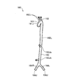
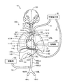
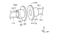
- FIG. 1 It is a figure which shows schematic structure of the human body simulation apparatus of 1st Embodiment. It is a figure which shows schematic structure of the human body simulation apparatus of 1st Embodiment. It is a figure which shows an example of a structure of an aortic model. It is a figure which shows an example of a structure of a model. It is a figure which shows an example of a structure of a model. It is a figure which shows an example of a heart model. It is explanatory drawing which shows an example of a structure of a model. It is explanatory drawing which shows the other example of a structure of a model. It is a figure explaining an example of a living body model connection part.
- a tubular body 72 connected to the breathing motion unit 70 is connected in a state where the lumen 170L of the diaphragm model 170 and the lumen of the tubular body 72 are in communication with each other.
- the brain model 130 may be a complex that further includes a bone model simulating a human skull and cervical spine.
- the skull has a hard resin case simulating the parietal bone, temporal bone, occipital bone, and sphenoid bone, and a lid simulating the frontal bone
- the cervical spine has a through hole through which a blood vessel model can pass.
- You may have multiple rectangular resin bodies which have.
- the bone model is made of a resin having a hardness different from that of an organ model such as a blood vessel model or a brain model.
- the skull can be made of acrylic resin and the vertebra can be made of PVA.
- the distal end 141D of the liver blood vessel model 141 is connected to the liver model 140, and the proximal end 141P is connected to the third connecting portion 163Ja of the aortic model 160.
- the tip 141D of the liver blood vessel model 141 imitates another blood vessel (for example, a hepatic artery) arranged on the surface and / or inside of the liver model 140, like the heart blood vessel model 111.
- the proximal end 141P of the liver blood vessel model 141 is connected to the third connecting portion 163Ja in a state where the inner lumen of the liver blood vessel model 141 and the inner lumen 160L of the aortic model 160 are in communication with each other.
- the end surface of the second connection portion 161J and the end surface of the cardiovascular model 111 are butted against each other, and the clip is performed with the lumen 161L and the lumen 111L communicating with each other.
- the second connecting portion 161J and the cardiovascular model 111 are fixed using an indwelling device (not shown) such as the above.
- the heart model 110 is connected to the aortic model 160, and the fluid flowing in the lumen 160L of the aortic model 160 can be supplied to the lumen 111L of the cardiovascular model 111.
- step S46 the control unit 40 determines whether or not the processing termination condition is satisfied. Similar to step S22, various arbitrary conditions can be adopted as the end condition. The end condition of step S46 and the end condition of step S22 may be the same or different. When the end condition is satisfied (step S46: YES), the control unit 40 ends the process. When the ending condition is not satisfied (step S46: NO), the control unit 40 causes the process to transition to step S40 and continues the first mode process.
- steps S60 to S64 after initialization of the variables n, o, and p step S54
- the variable n and the variable o are equal to the heart rate i input by the user
- the variable p is the respiratory rate j input by the user. equal. Therefore, in steps S60 to S64 after initialization of the variables n, o, p (step S54), the heart rate i kept constant (variables n, o) and the respiratory rate j kept constant (variable p). ) Is used to operate the pulsation unit 50, the pulsation unit 60, and the breathing motion unit 70.
- step S76 NO, FIG. 17: area A2 or area A3
- the control unit 40 causes the process to transition to step S80.
- step S80 the control unit 40 determines that the sum of the start time t0 acquired in step S52 and the elapsed time ⁇ t from the start time t0 (that is, the current time) is the abnormal heartbeat occurrence time m1 and the abnormal heartbeat determined in step S74. It is determined whether it is smaller than the sum of the duration m2. When it is equal to or more than the sum of the times m1 and m2 (step S80: NO, FIG. 17: area A3), the control unit 40 shifts the processing to step S52 (acquisition of the start time t0) and repeats the processing.
- the human body simulation device 1 includes a heart model 110 that imitates a heart and a blood vessel model that imitates a blood vessel (aortic model 160, heart blood vessel model 111, cerebral blood vessel model 131, liver blood vessel model 141, lower limbs). Since the blood vessel model 151) is provided, a medical device for minimally invasive treatment or examination of the heart, such as percutaneous coronary intervention (PCI) for ischemic heart disease, is used. The treatment or examination procedure that was performed can be simulated. Further, the control unit 40 of the human body simulation device 1 sends out to the respective blood vessel models such as the aortic model 160 and the pulse rate in the pulse unit 60 that causes the heart model 110 to pulse according to the set heart rate i. It is possible to change the pulsation number of the fluid pulsation unit 50 (FIG. 15: steps S12, S20, S40, S42). Therefore, it is possible to simulate the treatment or examination procedure under various pulse rate and pulse rate conditions.
- the control unit 40 sets the heart rate i and the respiratory rate j (that is, variables n, o, p) in the second mode (FIG. 16) to the patient's seriousness
- the determination is made according to one pattern randomly selected from the first to third patterns simulating the degree using the randomly determined pulsation coefficient k (step S82). Therefore, by simulating an abrupt change in the physical condition of the actual patient, the pulsation rate in the pulsation section 60, the pulsation rate in the pulsation section 50, and the breathing motion section 70 in a manner that cannot be predicted by the user of the human body simulation device 1. Breathing rate can be changed. Therefore, according to the human body simulation apparatus 1, it is possible to simulate a procedure for treatment or inspection in a more realistic manner.
- an aorta model 160a including a single biological model connecting portion (the second connecting portion 161J in the illustrated example) is provided instead of the aortic model 160 including a plurality of biological model connecting portions. May be. Even in this case, the same effect as that of the above-described first embodiment can be obtained.
- FIG. 19 is a diagram showing an example of the heart model 110b of the third embodiment.
- the tip 111D of the cardiovascular model 111 is branched into the left and right coronary artery models 112L and R that are integrally formed.
- the left and right coronary artery models 112L and R may include a plurality of blood vessel constituting sections 1121, 1122, 1125, 1126 and 1127 which respectively configure respective sections of the blood vessel.
- a lesion part simulating a lesion may be formed inside and outside the blood vessel in at least a part of each of the blood vessel constituent parts 1121, 1122, 1125, 1126, 1127.
- FIG. 6 the tip 111D of the cardiovascular model 111 is branched into the left and right coronary artery models 112L and R that are integrally formed.
- the left and right coronary artery models 112L and R may include a plurality of blood vessel constituting sections 1121, 1122, 1125, 1126 and 1127 which respectively configure respective sections of the blood vessel
- a hump-shaped lesion LP is formed outside the blood vessel of the blood vessel forming portion 1122 of the left coronary artery model 112L. Further, a stenotic lesion LP is formed inside the blood vessel of the blood vessel forming portion 1126 of the right coronary artery model 112R.
- both the blood vessel component having no lesion LP and the blood vessel component having the lesion LP are prepared for the same blood vessel component. May be replaceable.
- heart model 110b is illustrated in FIG.
- the same configuration as that of the third embodiment can be adopted for other biological models having a partial blood vessel model (FIG. 4: brain model 130, liver model 140, lower limb model 150).
- the aortic model 160 (FIG. 3) may have the same configuration as that of the third embodiment.
- FIG. 20 is a diagram illustrating a biological model connecting unit according to the fourth embodiment.
- the first flange portion 91 and the second flange portion 92 are fixed by using the fixing member 95 having the configuration shown in FIGS. 12 and 13.
- the first flange portion 91 and the second flange portion 92 may be fixed by using a fixing member having a configuration different from the fixing member 95.
- a screw 97 may be used to fix the first flange portion 91 and the second flange portion 92. Even in this case, the same effect as that of the above-described first embodiment can be obtained.
- the control unit 40e sets the heart rate i and the respiratory rate j (that is, variables n, o, p) in the second mode (FIG. 23) to the patient's weight. From the first to third patterns simulating the severity, the fluid pressure (that is, the sensor) in the peripheral portion of the blood vessel model (cardiovascular model 111, cerebral blood vessel model 131, liver blood vessel model 141, lower limb blood vessel model 151) is selected. (Measured values of 119, 139, 149, and 159) according to one pattern selected (step S82).
- control unit 40e can change the pulsation rate in the pulsation section 60, the pulsation rate in the pulsation section 50, and the respiration rate in the respiration operation section 70 in response to changes in blood pressure in the peripheral blood vessels. For this reason, according to the human body simulation apparatus 1e of the sixth embodiment, it is possible to simulate a treatment or examination procedure in a more realistic manner.
- FIG. 24 is a diagram showing a schematic configuration of a human body simulation device 1f in the seventh embodiment.
- the human body simulation device 1 may include various devices used in actual treatment or inspection.
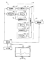
- the human body simulation device 1f of the seventh embodiment includes a monitor (display unit) 200 connected to the control unit 40f.
- the controller 40f controls the set heart rate i, the respiration rate j, and the heart rate i and the respiration rate j changed in the second mode processing (that is, variables) during execution of the processing described in FIGS.
- a graph showing n, o, p) is displayed on the monitor 200.
- the control unit 40f causes the display unit 200 to display a graph showing changes in the heart rate i and the respiratory rate j, so that the treatment is performed in a more realistic manner.
- the inspection scene can be reproduced and the convenience for the user can be improved.
- the model does not have to include at least a part of a heart model, a lung model, a brain model, a liver model, a lower limb model, and a diaphragm model.
- the pulsation part can also be omitted.
- the respiratory motion unit can also be omitted.
- the model may be configured as a composite that further comprises a bone model that mimics at least a portion of a human skeleton, such as ribs, sternum, thoracic vertebra, lumbar spine, femur, tibia and the like.
- control unit does not have to control the respiratory action unit based on the respiratory rate j and the respiratory rate j (or the variable p) (FIG. 15: steps S20 and S44, FIG. 16: step S64).
- the control unit may not control the pulsation unit based on the heart rate i and the heart rate i (or the variable n) (FIG. 15: Steps S20 and S40, FIG. 16: Step S60).
- the control unit does not have to control the pulsation unit based on the heart rate i and the heart rate i (or the variable o) (FIG. 15: steps S20 and S42, FIG. 16: step S62).
- Coronary artery model 112L Left coronary artery model 112R ... Right coronary artery model 115 ... Tubular body 119 ... Sensor 120 ... Lung model 121 ... Tracheal model 130 ... Brain model 131 ... Cerebral blood vessel model 139 ... Sensor 140 ... Liver model 141 ... Liver blood vessel model 149 ... Sensor 150 ... Lower extremity model 151 ... Lower extremity blood vessel model 159 ... Sensor 160, 160a ... Aortic model 161 ... Ascending aorta 161J ... Second connection 162 ... Aortic arch 162J ... First connection 163 ... Abdomen Aortic part 163Ja ... Third connecting part 163Jb ...
Landscapes
- Engineering & Computer Science (AREA)
- Physics & Mathematics (AREA)
- General Physics & Mathematics (AREA)
- Health & Medical Sciences (AREA)
- Mathematical Analysis (AREA)
- Pure & Applied Mathematics (AREA)
- Medical Informatics (AREA)
- Algebra (AREA)
- Computational Mathematics (AREA)
- General Health & Medical Sciences (AREA)
- Chemical & Material Sciences (AREA)
- Mathematical Optimization (AREA)
- Mathematical Physics (AREA)
- Medicinal Chemistry (AREA)
- Business, Economics & Management (AREA)
- Educational Administration (AREA)
- Educational Technology (AREA)
- Theoretical Computer Science (AREA)
- Pulmonology (AREA)
- Radiology & Medical Imaging (AREA)
- Instructional Devices (AREA)
- Prostheses (AREA)
Abstract
L'invention concerne un appareil de simulation de corps humain comprenant : un modèle d'aorte qui simule une aorte ; et un modèle biologique qui simule une partie de l'intérieur d'un corps humain et comprend un modèle de vaisseau sanguin partiel simulant certains vaisseaux sanguins. Le modèle d'aorte comprend : une partie de liaison de modèle biologique qui relie de manière amovible le modèle biologique dans un état où une lumière du modèle d'aorte et une lumière du modèle de vaisseau sanguin partiel communiquent l'une avec l'autre ; et une partie de liaison de partie d'alimentation en fluide qui relie une partie d'alimentation en fluide permettant de fournir un fluide aux lumières.
Priority Applications (6)
| Application Number | Priority Date | Filing Date | Title |
|---|---|---|---|
| CN201880097889.6A CN112868055B (zh) | 2018-10-17 | 2018-10-17 | 人体模拟装置 |
| PCT/JP2018/038674 WO2020079778A1 (fr) | 2018-10-17 | 2018-10-17 | Appareil de simulation de corps humain |
| JP2020551652A JP7200262B2 (ja) | 2018-10-17 | 2018-10-17 | 人体シミュレーション装置 |
| EP18936920.0A EP3869490B1 (fr) | 2018-10-17 | 2018-10-17 | Appareil de simulation de corps humain |
| US17/211,181 US12148319B2 (en) | 2018-10-17 | 2021-03-24 | Human body simulation device |
| US18/916,801 US20250037613A1 (en) | 2018-10-17 | 2024-10-16 | Human body simulation device |
Applications Claiming Priority (1)
| Application Number | Priority Date | Filing Date | Title |
|---|---|---|---|
| PCT/JP2018/038674 WO2020079778A1 (fr) | 2018-10-17 | 2018-10-17 | Appareil de simulation de corps humain |
Related Child Applications (1)
| Application Number | Title | Priority Date | Filing Date |
|---|---|---|---|
| US17/211,181 Continuation US12148319B2 (en) | 2018-10-17 | 2021-03-24 | Human body simulation device |
Publications (1)
| Publication Number | Publication Date |
|---|---|
| WO2020079778A1 true WO2020079778A1 (fr) | 2020-04-23 |
Family
ID=70283911
Family Applications (1)
| Application Number | Title | Priority Date | Filing Date |
|---|---|---|---|
| PCT/JP2018/038674 Ceased WO2020079778A1 (fr) | 2018-10-17 | 2018-10-17 | Appareil de simulation de corps humain |
Country Status (5)
| Country | Link |
|---|---|
| US (2) | US12148319B2 (fr) |
| EP (1) | EP3869490B1 (fr) |
| JP (1) | JP7200262B2 (fr) |
| CN (1) | CN112868055B (fr) |
| WO (1) | WO2020079778A1 (fr) |
Cited By (2)
| Publication number | Priority date | Publication date | Assignee | Title |
|---|---|---|---|---|
| WO2021214323A1 (fr) * | 2020-04-24 | 2021-10-28 | Universität Bern | Système d'imitation médicale |
| EP4318438A4 (fr) * | 2021-04-01 | 2025-03-19 | Osaka University | Simulateur de cathéter et modèle cérébrovasculaire |
Families Citing this family (3)
| Publication number | Priority date | Publication date | Assignee | Title |
|---|---|---|---|---|
| US12462709B2 (en) * | 2018-12-01 | 2025-11-04 | Syndaver Labs, Inc. | Artificial canine model |
| CN116982097A (zh) * | 2021-04-01 | 2023-10-31 | 国立大学法人大阪大学 | 导管-模拟器及脑血管模型 |
| CN114913749A (zh) * | 2022-04-18 | 2022-08-16 | 中国人民解放军西部战区总医院 | 食道胃底静脉出血及止血教学模型 |
Citations (17)
| Publication number | Priority date | Publication date | Assignee | Title |
|---|---|---|---|---|
| JPS51149251U (fr) * | 1975-05-23 | 1976-11-30 | ||
| JPH04129175U (ja) * | 1991-05-14 | 1992-11-25 | 株式会社京都科学 | 人体解剖模型 |
| JPH0550477U (ja) * | 1991-12-04 | 1993-07-02 | 株式会社高研 | 心臓血管模型 |
| JP2005241883A (ja) * | 2004-02-25 | 2005-09-08 | Hirona Gi | カテーテル検査シミュレーションシステム |
| JP2008237304A (ja) * | 2007-03-26 | 2008-10-09 | Univ Kansai Medical | 心血管系シミュレーションモデル |
| JP2011064918A (ja) * | 2009-09-16 | 2011-03-31 | Terumo Corp | 訓練用生体モデル |
| JP2013029820A (ja) * | 2011-06-20 | 2013-02-07 | Arai Medphoton Research Laboratoried Corp | 心臓モデル |
| JP2013213986A (ja) * | 2012-04-03 | 2013-10-17 | Toshio Nakanishi | 先天性心疾患治療のためのトレーニング装置およびそれを用いたトレーニング方法 |
| JP2014170075A (ja) * | 2013-03-01 | 2014-09-18 | Terumo Corp | トレーニング装置 |
| JP2015064487A (ja) | 2013-09-25 | 2015-04-09 | 国立大学法人広島大学 | シミュレーションシステム、及びステントグラフトの設置シミュレーション方法 |
| JP2015105958A (ja) * | 2013-11-28 | 2015-06-08 | 国立大学法人 筑波大学 | 医療用超音波診断訓練システム及び医療用超音波診断訓練方法 |
| WO2016075732A1 (fr) * | 2014-11-10 | 2016-05-19 | 国立大学法人大阪大学 | Simulateur de cathéter et procédé d'imagerie pour un simulateur de cathéter |
| JP2017040812A (ja) | 2015-08-20 | 2017-02-23 | テルモ株式会社 | 心臓シミュレーション装置、心臓シミュレーション装置の制御方法及び心臓シミュレーション装置の制御プログラム |
| WO2017127724A1 (fr) * | 2016-01-21 | 2017-07-27 | The University Of North Carolina At Chapel Hill | Systèmes et procédés de simulation |
| US20180108275A1 (en) * | 2016-10-17 | 2018-04-19 | Sanmina Corporation | System and method for a blood flow simulator |
| WO2018079711A1 (fr) * | 2016-10-28 | 2018-05-03 | 国立大学法人大阪大学 | Modèle d'organe pour cathéter et/ou simulateur |
| JP2018072387A (ja) * | 2016-10-24 | 2018-05-10 | 株式会社アールテック | 心臓模型、心臓局部模型、心臓弁模型、冠動脈模型及び大動脈模型 |
Family Cites Families (19)
| Publication number | Priority date | Publication date | Assignee | Title |
|---|---|---|---|---|
| US5634797A (en) * | 1995-05-05 | 1997-06-03 | Montgomery; Mary B. | Heart defect teaching aid |
| US6062866A (en) * | 1998-03-27 | 2000-05-16 | Prom; James M. | Medical angioplasty model |
| US6790043B2 (en) * | 2002-03-28 | 2004-09-14 | Board Of Trustees Of The University Of Arkansas | Method and apparatus for surgical training |
| AU2002302094B2 (en) * | 2002-11-21 | 2008-06-05 | Northern Sydney Area Health Service | Patient Simulator Manikin and System |
| US7021940B2 (en) * | 2002-11-21 | 2006-04-04 | Northern Sydney Area Health Service | Patient simulator manikin and system |
| US7427199B2 (en) * | 2005-02-03 | 2008-09-23 | Christopher Sakezles | Models and methods of using same for testing medical devices |
| US20070218437A1 (en) * | 2006-03-03 | 2007-09-20 | Lotano Vincent E | Heart model for training or demonstrating heart valve replacement or repair |
| US20090246747A1 (en) * | 2008-03-25 | 2009-10-01 | Operative Experience, Inc. | Simulator for major surgical operations |
| AU2011210745B2 (en) * | 2010-01-29 | 2013-08-29 | Jonathan M. Gurdin | Circulatory heart model |
| US20140370490A1 (en) * | 2013-06-12 | 2014-12-18 | Medtronic, Inc. | Heart-lung preparation and method of use |
| CN203786996U (zh) * | 2014-03-13 | 2014-08-20 | 西安交通大学 | 一种高度仿真介入手术训练器 |
| ES2843324T3 (es) * | 2014-11-18 | 2021-07-16 | Maximum Fidelity Surgical Simulations Llc | Reconstitución de la circulación post mortem |
| WO2016158222A1 (fr) * | 2015-03-30 | 2016-10-06 | 国立大学法人大阪大学 | Récipient pour simulateur de cathéter et modèle de cœur contenu dans ledit récipient |
| KR102419617B1 (ko) * | 2015-08-03 | 2022-07-11 | 테루모 가부시키가이샤 | 수기 시뮬레이터 |
| JP6667503B2 (ja) * | 2015-11-09 | 2020-03-18 | 国立大学法人大阪大学 | カテーテル・シミュレーター用容器、及びこの容器内に収容される心臓モデル |
| IT201600114762A1 (it) * | 2016-11-14 | 2018-05-14 | Milano Politecnico | Assieme di banco prova per la simulazione di interventi di cardiochirurgia e/o procedure di cardiologia interventistica |
| US10127838B2 (en) * | 2016-11-22 | 2018-11-13 | PraxiCut, LLC | Surgical simulation systems, methods, and compositions |
| CN106935093A (zh) * | 2017-05-08 | 2017-07-07 | 喻诗哲 | 一种人体心脏外科手术操作训练系统 |
| CN107798980A (zh) * | 2017-10-24 | 2018-03-13 | 西安交通大学医学院第附属医院 | 一种手术用仿真胸腔镜模拟训练装置 |
-
2018
- 2018-10-17 JP JP2020551652A patent/JP7200262B2/ja active Active
- 2018-10-17 EP EP18936920.0A patent/EP3869490B1/fr active Active
- 2018-10-17 WO PCT/JP2018/038674 patent/WO2020079778A1/fr not_active Ceased
- 2018-10-17 CN CN201880097889.6A patent/CN112868055B/zh active Active
-
2021
- 2021-03-24 US US17/211,181 patent/US12148319B2/en active Active
-
2024
- 2024-10-16 US US18/916,801 patent/US20250037613A1/en active Pending
Patent Citations (17)
| Publication number | Priority date | Publication date | Assignee | Title |
|---|---|---|---|---|
| JPS51149251U (fr) * | 1975-05-23 | 1976-11-30 | ||
| JPH04129175U (ja) * | 1991-05-14 | 1992-11-25 | 株式会社京都科学 | 人体解剖模型 |
| JPH0550477U (ja) * | 1991-12-04 | 1993-07-02 | 株式会社高研 | 心臓血管模型 |
| JP2005241883A (ja) * | 2004-02-25 | 2005-09-08 | Hirona Gi | カテーテル検査シミュレーションシステム |
| JP2008237304A (ja) * | 2007-03-26 | 2008-10-09 | Univ Kansai Medical | 心血管系シミュレーションモデル |
| JP2011064918A (ja) * | 2009-09-16 | 2011-03-31 | Terumo Corp | 訓練用生体モデル |
| JP2013029820A (ja) * | 2011-06-20 | 2013-02-07 | Arai Medphoton Research Laboratoried Corp | 心臓モデル |
| JP2013213986A (ja) * | 2012-04-03 | 2013-10-17 | Toshio Nakanishi | 先天性心疾患治療のためのトレーニング装置およびそれを用いたトレーニング方法 |
| JP2014170075A (ja) * | 2013-03-01 | 2014-09-18 | Terumo Corp | トレーニング装置 |
| JP2015064487A (ja) | 2013-09-25 | 2015-04-09 | 国立大学法人広島大学 | シミュレーションシステム、及びステントグラフトの設置シミュレーション方法 |
| JP2015105958A (ja) * | 2013-11-28 | 2015-06-08 | 国立大学法人 筑波大学 | 医療用超音波診断訓練システム及び医療用超音波診断訓練方法 |
| WO2016075732A1 (fr) * | 2014-11-10 | 2016-05-19 | 国立大学法人大阪大学 | Simulateur de cathéter et procédé d'imagerie pour un simulateur de cathéter |
| JP2017040812A (ja) | 2015-08-20 | 2017-02-23 | テルモ株式会社 | 心臓シミュレーション装置、心臓シミュレーション装置の制御方法及び心臓シミュレーション装置の制御プログラム |
| WO2017127724A1 (fr) * | 2016-01-21 | 2017-07-27 | The University Of North Carolina At Chapel Hill | Systèmes et procédés de simulation |
| US20180108275A1 (en) * | 2016-10-17 | 2018-04-19 | Sanmina Corporation | System and method for a blood flow simulator |
| JP2018072387A (ja) * | 2016-10-24 | 2018-05-10 | 株式会社アールテック | 心臓模型、心臓局部模型、心臓弁模型、冠動脈模型及び大動脈模型 |
| WO2018079711A1 (fr) * | 2016-10-28 | 2018-05-03 | 国立大学法人大阪大学 | Modèle d'organe pour cathéter et/ou simulateur |
Non-Patent Citations (1)
| Title |
|---|
| IKEDA SEIICHI , FUKUDA TOSHI , IRIE KEIKO , ARIA FUMIHITO , NEGORO MAKATO , TAKAHASI IKUO: "In Vitro In vitro patient-tailored cerebral arterial model for surgical simulation", THE 22ND ANNUAL CONFERENCE OF THE ROBOTICS SOCIETY OF JAPAN, 15 September 2004 (2004-09-15), pages 1 - 4, XP009526664 * |
Cited By (2)
| Publication number | Priority date | Publication date | Assignee | Title |
|---|---|---|---|---|
| WO2021214323A1 (fr) * | 2020-04-24 | 2021-10-28 | Universität Bern | Système d'imitation médicale |
| EP4318438A4 (fr) * | 2021-04-01 | 2025-03-19 | Osaka University | Simulateur de cathéter et modèle cérébrovasculaire |
Also Published As
| Publication number | Publication date |
|---|---|
| JP7200262B2 (ja) | 2023-01-06 |
| EP3869490A1 (fr) | 2021-08-25 |
| US20250037613A1 (en) | 2025-01-30 |
| EP3869490A4 (fr) | 2022-05-18 |
| CN112868055B (zh) | 2023-10-17 |
| JPWO2020079778A1 (ja) | 2021-09-02 |
| US20210209968A1 (en) | 2021-07-08 |
| US12148319B2 (en) | 2024-11-19 |
| CN112868055A (zh) | 2021-05-28 |
| EP3869490B1 (fr) | 2024-07-10 |
Similar Documents
| Publication | Publication Date | Title |
|---|---|---|
| US12148319B2 (en) | Human body simulation device | |
| US11908342B2 (en) | Human body simulation device, method for controlling human body simulation device, and computer program | |
| JP7464997B2 (ja) | 心臓シミュレーション装置 | |
| CN103260519B (zh) | 用于重建图像投影的方法和设备 | |
| CN106570313B (zh) | 获取四维血管变形行为与管壁在体应力的方法及系统 | |
| CN105943160B (zh) | 主动脉覆膜支架介入手术模拟装置 | |
| CN113168781B (zh) | 血管模型以及脏器模拟器 | |
| JP2022501643A (ja) | 患者固有の心血管シミュレーション装置 | |
| US20240420596A1 (en) | Heart model | |
| JP7270733B2 (ja) | 血管モデル | |
| US20210272481A1 (en) | Organ simulator | |
| US20210327305A1 (en) | System for validating and training invasive interventions | |
| WO2021005938A1 (fr) | Simulateur cardiaque | |
| US20240071253A1 (en) | System for advanced physician training and patient specific rehearsals |
Legal Events
| Date | Code | Title | Description |
|---|---|---|---|
| 121 | Ep: the epo has been informed by wipo that ep was designated in this application |
Ref document number: 18936920 Country of ref document: EP Kind code of ref document: A1 |
|
| ENP | Entry into the national phase |
Ref document number: 2020551652 Country of ref document: JP Kind code of ref document: A |
|
| NENP | Non-entry into the national phase |
Ref country code: DE |
|
| ENP | Entry into the national phase |
Ref document number: 2018936920 Country of ref document: EP Effective date: 20210517 |