WO2020095974A1 - Greffon et utilisation correspondante - Google Patents
Greffon et utilisation correspondante Download PDFInfo
- Publication number
- WO2020095974A1 WO2020095974A1 PCT/JP2019/043582 JP2019043582W WO2020095974A1 WO 2020095974 A1 WO2020095974 A1 WO 2020095974A1 JP 2019043582 W JP2019043582 W JP 2019043582W WO 2020095974 A1 WO2020095974 A1 WO 2020095974A1
- Authority
- WO
- WIPO (PCT)
- Prior art keywords
- cells
- hydrogel
- implant
- graft
- sol
- Prior art date
- Legal status (The legal status is an assumption and is not a legal conclusion. Google has not performed a legal analysis and makes no representation as to the accuracy of the status listed.)
- Ceased
Links
Images






Classifications
-
- A—HUMAN NECESSITIES
- A61—MEDICAL OR VETERINARY SCIENCE; HYGIENE
- A61P—SPECIFIC THERAPEUTIC ACTIVITY OF CHEMICAL COMPOUNDS OR MEDICINAL PREPARATIONS
- A61P43/00—Drugs for specific purposes, not provided for in groups A61P1/00-A61P41/00
-
- A—HUMAN NECESSITIES
- A61—MEDICAL OR VETERINARY SCIENCE; HYGIENE
- A61L—METHODS OR APPARATUS FOR STERILISING MATERIALS OR OBJECTS IN GENERAL; DISINFECTION, STERILISATION OR DEODORISATION OF AIR; CHEMICAL ASPECTS OF BANDAGES, DRESSINGS, ABSORBENT PADS OR SURGICAL ARTICLES; MATERIALS FOR BANDAGES, DRESSINGS, ABSORBENT PADS OR SURGICAL ARTICLES
- A61L27/00—Materials for grafts or prostheses or for coating grafts or prostheses
- A61L27/36—Materials for grafts or prostheses or for coating grafts or prostheses containing ingredients of undetermined constitution or reaction products thereof, e.g. transplant tissue, natural bone, extracellular matrix
- A61L27/38—Materials for grafts or prostheses or for coating grafts or prostheses containing ingredients of undetermined constitution or reaction products thereof, e.g. transplant tissue, natural bone, extracellular matrix containing added animal cells
-
- A—HUMAN NECESSITIES
- A61—MEDICAL OR VETERINARY SCIENCE; HYGIENE
- A61K—PREPARATIONS FOR MEDICAL, DENTAL OR TOILETRY PURPOSES
- A61K35/00—Medicinal preparations containing materials or reaction products thereof with undetermined constitution
- A61K35/12—Materials from mammals; Compositions comprising non-specified tissues or cells; Compositions comprising non-embryonic stem cells; Genetically modified cells
-
- A—HUMAN NECESSITIES
- A61—MEDICAL OR VETERINARY SCIENCE; HYGIENE
- A61K—PREPARATIONS FOR MEDICAL, DENTAL OR TOILETRY PURPOSES
- A61K35/00—Medicinal preparations containing materials or reaction products thereof with undetermined constitution
- A61K35/12—Materials from mammals; Compositions comprising non-specified tissues or cells; Compositions comprising non-embryonic stem cells; Genetically modified cells
- A61K35/14—Blood; Artificial blood
-
- A—HUMAN NECESSITIES
- A61—MEDICAL OR VETERINARY SCIENCE; HYGIENE
- A61K—PREPARATIONS FOR MEDICAL, DENTAL OR TOILETRY PURPOSES
- A61K35/00—Medicinal preparations containing materials or reaction products thereof with undetermined constitution
- A61K35/12—Materials from mammals; Compositions comprising non-specified tissues or cells; Compositions comprising non-embryonic stem cells; Genetically modified cells
- A61K35/14—Blood; Artificial blood
- A61K35/17—Lymphocytes; B-cells; T-cells; Natural killer cells; Interferon-activated or cytokine-activated lymphocytes
-
- A—HUMAN NECESSITIES
- A61—MEDICAL OR VETERINARY SCIENCE; HYGIENE
- A61K—PREPARATIONS FOR MEDICAL, DENTAL OR TOILETRY PURPOSES
- A61K35/00—Medicinal preparations containing materials or reaction products thereof with undetermined constitution
- A61K35/12—Materials from mammals; Compositions comprising non-specified tissues or cells; Compositions comprising non-embryonic stem cells; Genetically modified cells
- A61K35/30—Nerves; Brain; Eyes; Corneal cells; Cerebrospinal fluid; Neuronal stem cells; Neuronal precursor cells; Glial cells; Oligodendrocytes; Schwann cells; Astroglia; Astrocytes; Choroid plexus; Spinal cord tissue
-
- A—HUMAN NECESSITIES
- A61—MEDICAL OR VETERINARY SCIENCE; HYGIENE
- A61K—PREPARATIONS FOR MEDICAL, DENTAL OR TOILETRY PURPOSES
- A61K35/00—Medicinal preparations containing materials or reaction products thereof with undetermined constitution
- A61K35/12—Materials from mammals; Compositions comprising non-specified tissues or cells; Compositions comprising non-embryonic stem cells; Genetically modified cells
- A61K35/34—Muscles; Smooth muscle cells; Heart; Cardiac stem cells; Myoblasts; Myocytes; Cardiomyocytes
-
- A—HUMAN NECESSITIES
- A61—MEDICAL OR VETERINARY SCIENCE; HYGIENE
- A61K—PREPARATIONS FOR MEDICAL, DENTAL OR TOILETRY PURPOSES
- A61K35/00—Medicinal preparations containing materials or reaction products thereof with undetermined constitution
- A61K35/12—Materials from mammals; Compositions comprising non-specified tissues or cells; Compositions comprising non-embryonic stem cells; Genetically modified cells
- A61K35/37—Digestive system
- A61K35/39—Pancreas; Islets of Langerhans
-
- A—HUMAN NECESSITIES
- A61—MEDICAL OR VETERINARY SCIENCE; HYGIENE
- A61M—DEVICES FOR INTRODUCING MEDIA INTO, OR ONTO, THE BODY; DEVICES FOR TRANSDUCING BODY MEDIA OR FOR TAKING MEDIA FROM THE BODY; DEVICES FOR PRODUCING OR ENDING SLEEP OR STUPOR
- A61M37/00—Other apparatus for introducing media into the body; Percutany, i.e. introducing medicines into the body by diffusion through the skin
-
- A—HUMAN NECESSITIES
- A61—MEDICAL OR VETERINARY SCIENCE; HYGIENE
- A61P—SPECIFIC THERAPEUTIC ACTIVITY OF CHEMICAL COMPOUNDS OR MEDICINAL PREPARATIONS
- A61P3/00—Drugs for disorders of the metabolism
- A61P3/08—Drugs for disorders of the metabolism for glucose homeostasis
- A61P3/10—Drugs for disorders of the metabolism for glucose homeostasis for hyperglycaemia, e.g. antidiabetics
-
- A—HUMAN NECESSITIES
- A61—MEDICAL OR VETERINARY SCIENCE; HYGIENE
- A61L—METHODS OR APPARATUS FOR STERILISING MATERIALS OR OBJECTS IN GENERAL; DISINFECTION, STERILISATION OR DEODORISATION OF AIR; CHEMICAL ASPECTS OF BANDAGES, DRESSINGS, ABSORBENT PADS OR SURGICAL ARTICLES; MATERIALS FOR BANDAGES, DRESSINGS, ABSORBENT PADS OR SURGICAL ARTICLES
- A61L2400/00—Materials characterised by their function or physical properties
- A61L2400/06—Flowable or injectable implant compositions
Definitions
- the present invention relates to implants and their uses. More particularly, the present invention relates to implants, implant manufacturing devices, and implant manufacturing methods.
- the present application claims priority based on Japanese Patent Application No. 2018-212188 filed in Japan on Nov. 9, 2018, and the content thereof is incorporated herein.
- pluripotent stem cells such as induced pluripotent stem cells (iPS cells) and embryonic stem cells (ES cells), cells differentiated from tissue stem cells, cells collected from humans or pigs, etc.
- iPS cells induced pluripotent stem cells
- ES cells embryonic stem cells
- Patent Document 1 it may be difficult to remove the graft in an emergency, and there is still a problem in that the function of the graft is deteriorated due to a foreign body reaction.
- the graft tends to be large, and the functional transplanted cells may decrease due to central cell necrosis or the like.
- an object of the present invention is to provide a graft that allows cells to be transplanted into a living body without being subjected to immune attack and can be taken out of a living body.
- the present invention includes the following aspects.
- An implant wherein the implant has a shell part that comes into contact with body fluid when implanted in a living body, and a core part that is located inside the shell part and does not substantially come into contact with the body fluid.
- the core portion includes a plurality of first hydrogel fibers containing cells
- the shell portion includes a second hydrogel functioning as a semipermeable membrane
- the implant has an outer diameter of 1 mm or more.
- An implant having a size.
- the implant according to claim 1, wherein the semipermeable membrane has a molecular weight cutoff of 70,000 to 500,000.
- the distance between the surface of the graft and the cells contained in the first hydrogel fiber is a distance at which the cells can survive, according to any one of [1] to [5].
- Graft. [7]
- the cells contained in the first hydrogel fiber are at least one cell selected from the group consisting of endocrine cells, exocrine cells, nervous cells, skeletal muscle cells, blood cells and stromal cells.
- the implant according to any one of [1] to [6].
- a graft manufacturing apparatus including a container part and a plurality of columnar jigs detachably arranged inside the container part.
- a graft in which cells can be transplanted into a living body without being subjected to immune attack and can be taken out of the living body.
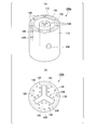
- (A) is a schematic diagram which shows an example of a graft.
- (B) is a photograph of an implant having the structure shown in (a).
- (A) is a perspective view which shows the structure of an example of a modification of a graft.
- (B) is sectional drawing which shows the structure of an example of the modification of an implant.
- It is a schematic diagram which shows the structure of an example of a graft manufacturing apparatus.
- (A) is a schematic diagram which shows the structure of an example of a graft manufacturing apparatus.
- FIG. 6 is a photograph of a state in which a graft is transplanted to a mouse in Experimental Example 4.
- 7 is a graph showing the results of measuring changes in blood glucose concentration in mice in Experimental Example 4.
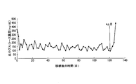
- 9 is a graph showing the results of measuring the concentration of human C-peptide in mouse blood in Experimental Example 4.
- 7 is a graph showing the results of measuring changes in blood glucose concentration in mice in Experimental Example 5.
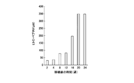
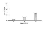
- 7 is a graph showing the results of measuring the concentration of human C-peptide in mouse blood in Experimental Example 5.
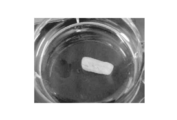
- 9 is a photograph of a graft extracted in Experimental Example 6.
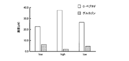
- 9 is a graph showing the measurement results of the concentrations of C-peptide and glucagon in Experimental Example 6.
- (A) and (b) are micrographs showing the results of hematoxylin-eosin staining of a thin section of a graft in Experimental Example 6.
- the present invention is a graft, wherein the graft is a shell portion that comes into contact with body fluid when implanted in a living body, and is located inside the shell portion, and the body fluid is substantially the same. And a core portion that does not come into contact with each other, the core portion includes a plurality of first hydrogel fibers containing cells, the shell portion includes a second hydrogel that functions as a semipermeable membrane, and the implant Provides an implant having an outer diameter of 1 mm or greater.
- FIG. 1A is a schematic diagram showing an example of the implant of the present embodiment.
- FIG. 1 (b) is a photograph of an implant having the structure shown in FIG. 1 (a).
- the implant 100 has a shell portion 110 and a core portion 120.
- the shell part 110 is located on the surface of the implant, and comes into contact with body fluid when implanted in the living body.
- the core part 120 is located inside the shell part 110 and does not substantially come into contact with body fluid even when it is implanted in a living body.
- “not substantially contacting body fluid” means that the core portion may be exposed on the surface of the graft at the end portion of the implant, for example, and the core portion may contact body fluid at this portion. It means to allow.
- the core part 120 includes a plurality of first hydrogel fibers 130.
- a hydrogel is a gel containing a large amount of water and having a three-dimensional network structure.
- the hydrogel can be formed by gelling a sol such as an aqueous solution of a water-soluble polymer, which will be described in detail later.
- the hydrogel fiber means a fibrous hydrogel.
- the core portion 120 includes the plurality of first hydrogel fibers 130, the number of cells 140 included in the graft can be increased. Moreover, it becomes easy to adjust the outer diameter D described later to an appropriate size and to adjust the distance between the surface of the implant 100 and the cells 140 to a distance in which the cells 140 can survive.
- the hydrogel fiber 130 includes cells 140.
- the shell part 110 is composed of a second hydrogel that functions as a semipermeable membrane.
- the shell part 110 preferably functions as a semipermeable membrane having a molecular weight cutoff of 70,000 to 500,000.
- the cells 140 are not subject to host immune attack.
- the secreted substance of the cells 140 penetrates the shell 110 and is secreted into the living body.
- the molecular weight cutoff of the shell part 110 can be arbitrarily controlled according to the purpose by adjusting the density of the second hydrogel or the like.
- the cross-sectional shape of the surface of the implant 100 orthogonal to the axial direction is not particularly limited, and may be a circle, an ellipse, a polygon such as a quadrangle or a pentagon.
- the axial direction of the graft may be the length direction of the graft or the long axis direction of the first hydrogel fiber 130.
- the implant 100 has an outer diameter D.
- the outer diameter D may be the diameter of a circle having the same area as the cross-sectional area of the surface of the implant perpendicular to the axial direction.
- the outer diameter D may be 1 to 100 mm.
- the lower limit of the outer diameter D may be 1.5 mm, 2 mm, 3 mm, 4 mm, 5 mm, or 6 mm. ..
- the upper limit value of the outer diameter D may be 100 mm, 80 mm, 60 mm, 40 mm, 30 mm, or 20 mm. It may be 10 mm, 9 mm, 8 mm, or 7 mm.
- the lower limit and the upper limit of the outer diameter D can be arbitrarily combined.
- the outer diameter D may have a size greater than 1.5 mm.
- the outer diameter D of the graft when the outer diameter D of the graft is in the above range, the number of cells derived from the living body attached to the surface of the graft is 5 ⁇ even after 2 weeks after transplanting in the living body. It tends to be kept at 10 5 / cm 2 or less. Therefore, adhesion between the tissue in the living body and the graft is suppressed, and the graft can be easily taken out of the living body as needed even after a long time has elapsed after the transplantation.
- the length of the graft can be appropriately adjusted depending on the symptoms of the transplant target and the purpose of the transplant, and is preferably in the range of 1 to 100 cm, for example.
- the distance between the surface of the implant 100 and the cells 140 contained in the first hydrogel fiber 130 is preferably a distance at which the cells 140 can survive.
- the surface of the graft 100 is a surface with which body fluid comes into contact when the graft 100 is transplanted in a living body.
- the distance is preferably 2 mm or less, more preferably 1 mm or less.
- the cells 140 contained in the first hydrogel fiber 130 may be allogeneic or heterologous with the living body into which the graft 110 is transplanted.
- transplanting allogeneic or xenogeneic cells is often more realistic than transplanting autologous cells.
- allogeneic or xenogeneic cells are transplanted into a living body, the transplanted cells are rejected by the host's immune system, and the function of the transplanted cells is reduced.
- the graft of the present embodiment even if the cells 140 are allogeneic or allogeneic, they are not immune to the host.
- the cells 140 included in the first hydrogel fiber 130 include endocrine cells, exocrine cells, nervous system cells, skeletal muscle system cells, blood cells, stromal cells, and the like.
- endocrine cells for example, neurosecretory cells, pituitary cells, thyroid cells, parathyroid cells, pancreatic islet cells, gastrointestinal endocrine cells, cardiomyocytes, hepatocytes, kidney cells, adipocytes, adrenal cells, gonadal cells, etc. Is mentioned.
- exocrine cells include digestive tract epithelial cells and the like.
- neural cells include neural stem cells, central nerve cells, peripheral nerve cells, glial cells and the like.
- skeletal muscle cells include bone cells and chondrocytes.
- the blood cells include hematopoietic stem cells, white blood cells, red blood cells, platelets and the like.
- examples of the stromal cells include fibroblasts and vascular cells.
- These cells may be cells differentiated from iPS cells or ES cells. As will be described later in Examples, for example, when the cells 140 are pancreatic islet cells, diabetes can be treated by transplanting the graft of the present embodiment into a living body.
- the cells 140 included in the first hydrogel fiber 130 may include multiple types of cells. This allows cells that interact with each other to be placed inside the implant and allowed to interact within the implant.
- the plurality of types of cells may be a mixture of any of the cells described above.
- the plurality of types of cells may be a mixture of endocrine cells, exocrine cells, neural cells, skeletal muscle cells or blood cells and stromal cells. Examples of cells that interact with each other include a combination of pancreatic islet cells and vascular endothelial cells, a combination of hepatocytes and non-parenchymal cells, and the like.
- one hydrogel fiber 130 may include a mixture of a plurality of types of cells, and one hydrogel fiber 130 includes one type of cells.
- the first hydrogel fibers 130 include cells, and a part of the plurality of first hydrogel fibers 130 may include cells of a different type from the other first hydrogel fibers 130.
- the arrangement of the first hydrogel fiber 130 inside the core portion 120 is not limited to that shown in FIG. In FIG. 1A, the first hydrogel fibers 130 are arranged at intervals along the peripheral portion of the core portion 120.
- the number of the first hydrogel fibers 130 may be increased and a large number of the first hydrogel fibers 130 may be arranged in close contact with the peripheral portion of the core portion 120.
- the hydrogel fiber 130 will be annularly arranged. Thereby, the number of cells 140 included in the graft 100 can be increased.
- FIG. 2A is a perspective view showing the structure of a graft 100 a, which is one of the modified examples of the graft 100.
- the implant 100a has a shell 110 and a core 120.
- the shell 110 of the implant 100a has a double structure of an outer side and an inner side.
- the core portion 120 is located inside the outer shell portion 110. Further, the core portion 120 is also located inside the inner shell portion 110.
- the core part 120 includes a plurality of first hydrogel fibers 130.
- the hydrogel fiber 130 contains cells 140.
- the space between the outer shell portion 110 and the inner shell portion 110 forms a recess into which body fluid enters when the implant 100a is implanted in a living body.
- the surface of the recess is also the surface of the implant 100a.
- the distance between the surface of the implant 100a and the cells 140 included in the first hydrogel fiber 130 can expand the region where the cells 140 can survive. it can. This can increase the number of viable cells 140 in the graft 100a.
- a through hole 150 may be provided on the outer surface of the implant 100a.
- the through hole 150 communicates the outer surface of the implant 100a with the recess of the implant 100a.
- body fluid will also enter through the through holes 150.
- FIG. 2B is a cross-sectional view showing the structure of the implant 100 b, which is one of the modified examples of the implant 100. As shown in FIG. 2B, the implant 100b has a shell 110 and a core 120.
- the implant 100b has a recess inside.
- the cross-sectional shape of the recess is such that three rectangular parallelepipeds are arranged radially from the center of the recess.
- the region in contact with the recess is also the shell 110.
- the core portion 120 is located inside the shell portion 110.
- the core part 120 includes a plurality of first hydrogel fibers 130.
- the hydrogel fiber 130 contains cells 140.
- the implant manufacturing apparatus of the first embodiment includes a container part and a plurality of columnar jigs detachably arranged inside the container part.
- FIG. 3 is a schematic view showing the structure of the implant manufacturing apparatus of this embodiment. As shown in FIG. 3, the implant manufacturing apparatus 300 includes a container part 310 and a plurality of columnar jigs 320 detachably arranged inside the container part 310.
- the graft manufacturing apparatus of the first embodiment can manufacture the above-described graft.
- the implant manufacturing apparatus of the second embodiment includes a first cylindrical portion and a plurality of second cylindrical portions arranged inside the first cylindrical portion.
- FIG. 4A is a schematic diagram showing the structure of the implant manufacturing apparatus of this embodiment. As shown in FIG. 4A, the implant manufacturing apparatus 400 includes a first cylindrical portion 410 and a plurality of second cylindrical portions 420 arranged inside the first cylindrical portion 410. A tube 411 that supplies the second sol to the first cylindrical portion 410 is preferably connected to the first cylindrical portion 410. Further, it is preferable that a tube 421 that supplies the first sol to the second cylindrical portion 420 is connected to the second cylindrical portion 420.
- FIG. 4B is a photograph showing a specific example of the graft manufacturing apparatus of the second embodiment. The graft manufacturing apparatus of the second embodiment can manufacture the above-described graft.
- the graft manufacturing method according to the first embodiment is a manufacturing method using the graft manufacturing apparatus according to the first embodiment described above.
- the second sol is injected into the container part 310 of the implant manufacturing apparatus 300 to form the second hydrogel.
- the plurality of columnar jigs 320 are removed after the formation of the second hydrogel.
- a second hydrogel is obtained in which the portion where the columnar jig 320 was present has a recess.
- the first sol containing cells is injected into the concave portion of the second hydrogel to form the first hydrogel.
- the cells described above can be used as the cells.
- the implant described above can be manufactured.
- the method for producing an implant according to the first embodiment preferably further includes a step of forming an exposed portion of the first hydrogel with the second hydrogel after forming the first hydrogel.
- the first hydrogel containing cells can be completely covered with the second hydrogel.
- the cells are completely encapsulated by the second hydrogel, and the possibility of immune attack of the cells when transplanted in vivo can be further reduced.
- a sol that gels to form a hydrogel in the presence of metal ions a sol that gelates to form a hydrogel in response to temperature, a gel that forms a hydrogel in response to pH A sol that forms a gel, a sol that forms a hydrogel in response to light, a sol that forms a hydrogel in response to a magnetic field, and the like can be used.
- the sol and the gel may be referred to without distinction.
- metal ions are contacted, temperature is adjusted to gelling conditions, pH is adjusted to gelling conditions, and light under gelling conditions is irradiated, depending on the characteristics of the gel used.
- An operation such as applying a magnetic field under gelation conditions may be performed.
- Hydrogels that gel in the presence of metal ions include alginate gels that gel in the presence of divalent or trivalent metal ions, carrageenan gels that gel in the presence of calcium and potassium ions, and sodium ions. Examples thereof include acrylic acid-based synthetic gels that gel in the presence.
- temperature-responsive hydrogel examples include poly (N-isopropylacrylamide) cross-linked with polyethylene glycol, a temperature-responsive hydrogel (commercial name: mebiol gel), methyl cellulose, hydroxypropyl cellulose, a copolymer of polylactic acid and polyethylene glycol, polyethylene.
- examples thereof include triblock copolymers of glycol and polypropylene oxide (commercial names: Pluronic, poloxamer), agarose, polyvinyl alcohol and the like.
- pH-responsive hydrogels examples include alginic acid gel, chitosan gel, carboxymethylcellulose gel, acrylic acid-based synthetic gel and the like.
- photoresponsive hydrogel examples include a synthetic gel having a combination of azobenzene and cyclodextrin in the skeleton, a gel composed of supramolecules having fumaric acid amide as a spacer, a gel crosslinked through a nitrobenzyl group, or a gel bonded to the gel. Can be mentioned.
- magnétique field responsive hydrogels examples include gels made of crosslinked poly (N-isopropylacrylamide) containing magnetic particles.
- the graft manufacturing method according to the second embodiment is a manufacturing method using the graft manufacturing apparatus of the second embodiment described above.
- an implant is manufactured by forming a laminar flow of a first sol and a laminar flow of a second sol and gelling the same. It is preferable that either the first sol or the second sol gels first.
- the second sol is supplied to the first cylindrical portion 410 from one side (the upper side in FIG. 4A) of the graft manufacturing apparatus 400, and the other side of the graft manufacturing apparatus 400 (FIG. ), The step of forming a laminar flow of the second sol and gelling the second sol to form the second hydrogel, and one of the implant manufacturing apparatus 400 (
- the first sol containing cells is supplied from the upper side in FIG. 4A to the second cylindrical portion 420 and discharged from the other side of the graft manufacturing apparatus 400 (the lower side in FIG. 4A).
- the step of forming a laminar flow of No. 1 sol and gelling the first sol to form the first hydrogel is simultaneously performed.
- the cells described above can be used as the cells. As a result, the implant described above can be manufactured.
- the method for manufacturing an implant according to the second embodiment forms only the second hydrogel before and after the step of simultaneously forming the step of forming the second hydrogel and the step of forming the first hydrogel. It is preferable to further include the step of
- the implant produced can completely cover the first hydrogel containing cells with the second hydrogel.
- the cells are completely encapsulated by the second hydrogel, and the possibility of immune attack of the cells when transplanted in vivo can be further reduced.
- the supply of the second sol to the first cylindrical portion 410 can be performed through the tube 411. Further, the supply of the first sol to the second cylindrical portion 420 can be performed through the tube 421.
- the same sol and hydrogel as those in the manufacturing method of the first embodiment can be used.
- the invention is a method of treating a disease, comprising implanting a graft comprising an effective amount of cells into a patient in need thereof, said graft being implanted in vivo.
- it has a shell part that comes into contact with body fluid, and a core part that is located inside the shell part and does not substantially come into contact with the body fluid, and the core part has a plurality of first hydrogels containing the cells
- a method of treatment is provided that includes a fiber, the shell portion includes a second hydrogel that functions as a semipermeable membrane, and the implant has a size greater than an outer diameter of 1.5 mm.
- the cell type is selected according to the disease.
- transplantation of pancreatic islet cells is effective in treating diabetes.
- transplantation of hepatocytes is effective for the treatment of liver diseases.
- transplantation of nerve cells is effective for treating Parkinson's disease.
- Example 1 (Production of hydrogel fiber) First, a method for producing a hydrogel fiber will be described.
- the method for producing the hydrogel fiber is not particularly limited, but it can be easily produced by using, for example, a dual coaxial microfluidic device as shown in FIG.
- a microfluidic device capable of ejecting two fluids by dividing them into a core part and a shell part so as to be coaxial is, for example, Wonje Jeong, et al., Hydrodynamic microfabrication via "on the fly” photopolymerization of microscale fibers and tubes.
- Lab Chip, 2004, 4, 576-580 Fig. 1 is also described in detail.
- FIG. 5 is a schematic diagram illustrating an example of a manufacturing process of the hydrogel fiber 600.
- a collagen solution is used as the core material and a sodium alginate solution before crosslinking is used as the shell material will be described.
- the collagen solution is introduced and injected from the inlet 510 of the microfluidic device 500.
- the sodium alginate solution before crosslinking is introduced and injected from the inlet 520 of the microfluidic device 500.
- a calcium chloride solution is introduced and injected from the introduction port 530 of the microfluidic device 500.
- the sodium alginate solution in the shell portion gels, and a hydrogel fiber in which the core portion 610 is a collagen gel and the shell portion 620 is an alginate gel can be manufactured.
- the injection speed of the solution at the inlets 510 and 520 is not particularly limited, but may be about 10 to 500 ⁇ L / min when the diameter of the microfluidic device 500 is about 50 ⁇ m to 2 mm. By adjusting the injection speed of the solution at the inlets 510 and 520, the diameter of the core and the coating thickness of the shell can be adjusted appropriately.
- the injection speed of the solution at the inlet 530 is not particularly limited, but may be, for example, about 1 to 10 mL / min.
- hydrogel fibers having different outer diameter sizes were manufactured using the microfluidic device shown in FIG.
- the outer diameter size was 0.35 mm, 0.5 mm, 1 mm, 1.5 mm, 2 mm, 3 mm, 6 mm.
- the length of the hydrogel fiber was 3 cm.
- a collagen solution or a laminin solution containing alginic acid was supplied as a core fluid to the coaxial microchannel of the microfluidic device. Further, an aqueous solution of sodium alginate was supplied as a shell fluid. Further, an aqueous barium chloride solution was supplied as a sheath fluid. As a result, gelation was induced at the interface between the shell and the sheath, and a hydrogel fiber was obtained. The shell of the hydrogel fiber was a barium alginate gel.
- each hydrogel fiber was taken out from the abdominal cavity of the mouse. The period of 14 days is suitable for observing a foreign body reaction (FBR) on the transplanted tissue.
- FBR foreign body reaction
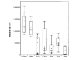
- FIG. 6 is a graph showing the results of measuring the number of cells attached to the surface of each hydrogel fiber.
- the vertical axis of the graph shows the cell density (cells / cm 2 ).
- Example 3 (Preparation of graft) A graft was produced using the graft manufacturing apparatus shown in FIGS. 4 (a) and 4 (b). A collagen solution or a laminin solution was used as the first sol. The first sol gelled under a temperature condition of a certain temperature or higher. In addition, human islet cells differentiated from human iPS were mixed in the first sol. An alginic acid solution was used as the second sol. The second sol gelled when reacted with a liquid containing a polyvalent cation.
- the second sol is supplied from one side of the graft manufacturing apparatus to the first cylindrical portion, and a laminar flow of the second sol discharged from the other side of the graft manufacturing apparatus is formed, and the second sol is gelled.
- the step of forming a laminar flow of No. 1 sol and gelling the first sol to form the first hydrogel was simultaneously performed. As a result, a graft was formed on the other side of the graft manufacturing apparatus.
- the implant had a cylindrical shape with a length of 20 to 30 mm and had an outer diameter of 6 mm.
- two types of grafts containing 6 ⁇ 10 6 or 1.2 ⁇ 10 7 islet cells per graft were prepared.
- Example 4 Transplantation of graft into immunodeficient diabetes model mouse
- the graft prepared in Experimental Example 3 was transplanted intraperitoneally or subcutaneously into an immunodeficient diabetic model mouse, and the transition of blood glucose concentration was measured.
- As the graft one containing 1.2 ⁇ 10 7 islet cells per graft was used.
- NOD-Scid mice were used as immunodeficient diabetes model mice.
- FIG. 7 is a photograph of a state in which the graft is transplanted into a mouse.
- Non-fasting blood glucose concentration in mice after transplantation of grafts was measured at least once a day.
- a blood glucose concentration of 200 mg / dL or less was determined to be euglycemia.
- FIG. 8 is a graph showing the results of measuring changes in blood glucose concentration in mice.
- the blood glucose concentration which was 200 mg / dL or more before the transplantation of the graft, was maintained at 200 mg / dL or less after the transplantation of the graft.
- the blood glucose concentration again increased to 200 mg / dL or more. This result indicates that the blood glucose concentration was maintained at 200 mg / dL or less due to the presence of the graft.
- FIG. 9 is a graph showing the results of measuring the concentration of human C-peptide.
- the C-peptide is a constituent of proinsulin, which is a precursor of insulin. In the islet cells, it is separated into an insulin part and a 31-amino acid C-peptide part and released into the blood. Therefore, it can be said that the concentration of human C-peptide in blood corresponds to the amount of human insulin secreted by human islet cells contained in the graft. Human C-peptide can also be measured in the presence of anti-human insulin antibody and mouse insulin.
- Example 5 Transplantation of graft into diabetic model mouse having immune function
- the graft prepared in Experimental Example 3 was transplanted intraperitoneally or subcutaneously into a diabetic model mouse having an immune function, and the transition of blood glucose concentration was measured.
- As the graft one containing 6 ⁇ 10 6 islet cells per graft was used.
- As a diabetic mouse having an immune function C57BL / 6 mouse in which diabetes was induced by administration of streptozocin (CAS number: 18883-66-4) was used.
- Non-fasting blood glucose concentration in mice after transplantation of grafts was measured at least once a day.
- a blood glucose concentration of 200 mg / dL or less was determined to be euglycemia.
- FIG. 10 is a graph showing the results of measuring changes in blood glucose concentration in mice.
- the blood glucose concentration which was 200 mg / dL or more before the transplantation of the graft, tended to be maintained at 200 mg / dL or less after the transplantation of the graft. This effect was maintained for at least 180 days after transplantation.
- FIG. 11 is a graph showing the results of measuring the concentration of human C-peptide in mouse blood. From the results of FIG. 11, it was confirmed that the function of human pancreatic islet cells contained in the graft was maintained even 10 weeks after transplantation of the graft.
- Example 6 (Examination of graft) From the C57BL / 6 mouse transplanted with the graft in Experimental Example 5, the graft was extracted 4 months later and analyzed.
- FIG. 12 is a photograph of the removed graft.
- the graft was incubated in a medium, the glucose concentration in the medium was adjusted to 3 mg / mL, and the concentrations of human C-peptide and glucagon secreted in the medium when the blood glucose level was lowered were measured. Subsequently, the glucose concentration in the medium was adjusted to 22 mg / mL, and the concentrations of human C-peptide and glucagon secreted into the medium when the hyperglycemic state was reached were measured. Subsequently, the glucose concentration in the medium was adjusted to 3 mg / mL, and the concentrations of human C-peptide and glucagon secreted in the medium when the hypoglycemia was again made were measured.
- Glucagon is a peptide hormone that is secreted from pancreatic islet cells when blood glucose level decreases and insulin becomes necessary, as opposed to insulin.
- FIG. 13 is a graph showing the measurement results of the concentrations of C-peptide and glucagon.
- “low” indicates the measurement result in the hypoglycemic state
- “high” indicates the measurement result in the hyperglycemic state.
- FIG. 14 (a) and 14 (b) are micrographs showing the results of hematoxylin / eosin staining.
- FIG. 14B is a micrograph of a part of FIG. 14A enlarged and photographed.
- the graft was extracted 4 months later and the same analysis was performed. As a result, the same result as this experimental example was obtained. That is, even in the NOD-Scid mouse, it was confirmed that the human pancreatic islet cells contained in the graft survived, although the graft was transplanted into the body of the mouse for 4 months. Furthermore, it was confirmed that the human pancreatic islet cells contained in the excised graft changed the concentrations of C-peptide and glucagon depending on the glucose concentration in the medium.
- a graft in which cells can be transplanted into a living body without being subjected to immune attack and can be taken out of the living body.
Landscapes
- Health & Medical Sciences (AREA)
- Life Sciences & Earth Sciences (AREA)
- Engineering & Computer Science (AREA)
- Cell Biology (AREA)
- Biomedical Technology (AREA)
- Chemical & Material Sciences (AREA)
- Public Health (AREA)
- General Health & Medical Sciences (AREA)
- Animal Behavior & Ethology (AREA)
- Veterinary Medicine (AREA)
- Medicinal Chemistry (AREA)
- Developmental Biology & Embryology (AREA)
- Pharmacology & Pharmacy (AREA)
- Zoology (AREA)
- Epidemiology (AREA)
- Immunology (AREA)
- Virology (AREA)
- Biotechnology (AREA)
- Diabetes (AREA)
- Hematology (AREA)
- Chemical Kinetics & Catalysis (AREA)
- Dermatology (AREA)
- Bioinformatics & Cheminformatics (AREA)
- Organic Chemistry (AREA)
- Nuclear Medicine, Radiotherapy & Molecular Imaging (AREA)
- General Chemical & Material Sciences (AREA)
- Nutrition Science (AREA)
- Ophthalmology & Optometry (AREA)
- Obesity (AREA)
- Emergency Medicine (AREA)
- Transplantation (AREA)
- Oral & Maxillofacial Surgery (AREA)
- Gastroenterology & Hepatology (AREA)
- Botany (AREA)
- Physiology (AREA)
- Neurology (AREA)
- Neurosurgery (AREA)
- Endocrinology (AREA)
- Cardiology (AREA)
- Vascular Medicine (AREA)
Abstract
L'invention concerne un greffon comprenant : une partie coque qui vient en contact avec un fluide corporel lorsque le greffon est transplanté dans un corps vivant ; et une partie noyau qui est située à l'intérieur de la partie coque et ne vient pratiquement pas en contact avec le fluide corporel. La partie noyau comprend une pluralité de premières fibres d'hydrogel comprenant des cellules et la partie coque comprend un deuxième hydrogel servant de film semi-perméable. Le greffon présente un diamètre externe supérieur à 1,5 mm.
Priority Applications (3)
| Application Number | Priority Date | Filing Date | Title |
|---|---|---|---|
| CN201980087975.3A CN113271892A (zh) | 2018-11-09 | 2019-11-07 | 移植物及其应用 |
| US17/290,123 US20220088272A1 (en) | 2018-11-09 | 2019-11-07 | Graft and use thereof |
| EP19881001.2A EP3878512A4 (fr) | 2018-11-09 | 2019-11-07 | Greffon et utilisation correspondante |
Applications Claiming Priority (2)
| Application Number | Priority Date | Filing Date | Title |
|---|---|---|---|
| JP2018-211288 | 2018-11-09 | ||
| JP2018211288A JP7306647B2 (ja) | 2018-11-09 | 2018-11-09 | 移植片及びその使用 |
Publications (1)
| Publication Number | Publication Date |
|---|---|
| WO2020095974A1 true WO2020095974A1 (fr) | 2020-05-14 |
Family
ID=70610732
Family Applications (1)
| Application Number | Title | Priority Date | Filing Date |
|---|---|---|---|
| PCT/JP2019/043582 Ceased WO2020095974A1 (fr) | 2018-11-09 | 2019-11-07 | Greffon et utilisation correspondante |
Country Status (5)
| Country | Link |
|---|---|
| US (1) | US20220088272A1 (fr) |
| EP (1) | EP3878512A4 (fr) |
| JP (1) | JP7306647B2 (fr) |
| CN (1) | CN113271892A (fr) |
| WO (1) | WO2020095974A1 (fr) |
Cited By (1)
| Publication number | Priority date | Publication date | Assignee | Title |
|---|---|---|---|---|
| JPWO2022092307A1 (fr) * | 2020-10-30 | 2022-05-05 |
Citations (2)
| Publication number | Priority date | Publication date | Assignee | Title |
|---|---|---|---|---|
| JP2014136128A (ja) * | 2013-01-18 | 2014-07-28 | Univ Of Tokyo | 移植用神経束及びその製造方法 |
| JP2018078917A (ja) * | 2016-11-14 | 2018-05-24 | 一般財団法人生産技術研究奨励会 | 体内移植用のハイドロゲルファイバ及びハイドロゲルファイバを用いた体内移植方法 |
Family Cites Families (7)
| Publication number | Priority date | Publication date | Assignee | Title |
|---|---|---|---|---|
| US20050222688A1 (en) * | 2002-05-10 | 2005-10-06 | Medtronic, Inc. | Transmural concentric multilayer ingrowth matrix within well-defined porosity |
| JP6215541B2 (ja) | 2013-02-28 | 2017-10-18 | 国立大学法人 東京大学 | 束状構造を有するゲルファイバー集合体の製造方法 |
| US20160355951A1 (en) | 2013-03-15 | 2016-12-08 | Arsenal Medical, Inc. | Core-sheath fibers and methods of making and using same |
| JP2017525438A (ja) * | 2014-07-29 | 2017-09-07 | ユニヴァーシテト チューリッヒ | 組織移植片 |
| JP6439918B2 (ja) * | 2014-10-17 | 2018-12-19 | 国立大学法人 東京大学 | 3次元細胞構造体の製造方法 |
| US11602588B2 (en) | 2017-06-07 | 2023-03-14 | Forcyte Medical, Llc | Connective tissue progenitor cell aspiration and processing system |
| CN107376033B (zh) * | 2017-07-26 | 2019-03-26 | 湖南赛诺生物科技股份有限公司 | 一种细胞移植增效保护装置及其制备方法 |
-
2018
- 2018-11-09 JP JP2018211288A patent/JP7306647B2/ja active Active
-
2019
- 2019-11-07 WO PCT/JP2019/043582 patent/WO2020095974A1/fr not_active Ceased
- 2019-11-07 CN CN201980087975.3A patent/CN113271892A/zh active Pending
- 2019-11-07 EP EP19881001.2A patent/EP3878512A4/fr not_active Withdrawn
- 2019-11-07 US US17/290,123 patent/US20220088272A1/en not_active Abandoned
Patent Citations (2)
| Publication number | Priority date | Publication date | Assignee | Title |
|---|---|---|---|---|
| JP2014136128A (ja) * | 2013-01-18 | 2014-07-28 | Univ Of Tokyo | 移植用神経束及びその製造方法 |
| JP2018078917A (ja) * | 2016-11-14 | 2018-05-24 | 一般財団法人生産技術研究奨励会 | 体内移植用のハイドロゲルファイバ及びハイドロゲルファイバを用いた体内移植方法 |
Non-Patent Citations (7)
| Title |
|---|
| CAS , no. 18883-66-4 |
| FUJIMOTO, K. ET AL.: "Micro-tube mass production device for microbial culture", CONF. PROC. IEEE ENG. MED. BIOL. SOC., vol. 2016, 16 August 2016 (2016-08-16), pages 481 - 484, XP032979196, DOI: 10.1109/EMBC.2016.7590744 * |
| OZAWA FUMISATO : "P-02-095: Manufacturing of multi-core shell cell for rapid formation of three dimensional hetero-structure", REGENERATIVE MEDICINE; PROGRAMS OF THE 17TH CONGRESS OF THE JAPANESE SOCIETY FOR REGENERATIVE MEDICINE, vol. 17, March 2018 (2018-03-01), pages 567, XP009528270, ISSN: 1347-7919 * |
| OZAWA FUMISATO : "P-02-107 Development of islet transplantation graft using cell fiber", REGENERATIVE MEDICINE, JOURNAL OF THE JAPANESE SOCIETY FOR REGENERATIVE MEDICINE, vol. 16, 2017, pages 420, XP009528269, ISSN: 1347-7919 * |
| See also references of EP3878512A4 |
| WONJE JEONG ET AL.: "Hydrodynamic microfabrication via ''on the fly'' photopolymerization of microscale fibers and tubes", LAB CHIP, vol. 4, 2004, pages 576 - 580 |
| YAO CHENG, ZHENG FUYIN, LU JIE, SHANG LUORAN, XIE ZHUOYING, ZHAO YUANJIN, CHEN YONGPING, GU ZHONGZE: "Bioinspired Multicompartmental Microfibers from Microfluidics", ADVANCED MATERIALS, vol. 26, no. 30, 2014, pages 5184 - 5190, XP055134083, ISSN: 0935-9648, DOI: 10.1002/adma.201400798 * |
Cited By (2)
| Publication number | Priority date | Publication date | Assignee | Title |
|---|---|---|---|---|
| JPWO2022092307A1 (fr) * | 2020-10-30 | 2022-05-05 | ||
| WO2022092307A1 (fr) * | 2020-10-30 | 2022-05-05 | 株式会社セルファイバ | Structure d'hydrogel, procédé de production d'une structure d'hydrogel, agent et procédé de transplantation |
Also Published As
| Publication number | Publication date |
|---|---|
| JP2020075895A (ja) | 2020-05-21 |
| JP7306647B2 (ja) | 2023-07-11 |
| EP3878512A4 (fr) | 2022-11-30 |
| US20220088272A1 (en) | 2022-03-24 |
| EP3878512A1 (fr) | 2021-09-15 |
| CN113271892A (zh) | 2021-08-17 |
Similar Documents
| Publication | Publication Date | Title |
|---|---|---|
| US20240398872A1 (en) | Implantable therapeutic delivery system and methods thereof | |
| EP3924008B1 (fr) | Dispositifs et méthodes de vascularisation pour agents diagnostiques et thérapeutiques implantés | |
| US6023009A (en) | Artificial pancreas | |
| US20140017304A1 (en) | Method for encapsulated therapeutic products and uses thereof | |
| CN110129253A (zh) | 人工组织前体及制备其的方法 | |
| US20190167572A1 (en) | Subcutaneous implant-type device for cell transplantation therapy | |
| WO2014197798A2 (fr) | Dispositif de greffe et procédé d'utilisation | |
| JP7306647B2 (ja) | 移植片及びその使用 | |
| CN112587730B (zh) | 复合细胞支架及其制备方法 | |
| JP7260563B2 (ja) | 細胞移植キット、袋状構造物の製造方法、および糖尿病治療剤 | |
| US20260076997A1 (en) | Cell implant including biodegradable porous microwell with stem cell-derived insulin-secreting cell aggregate supported therein, and use thereof | |
| KR100740169B1 (ko) | 세포를 포함하는 알긴산 마이크로 섬유 지지체 및 그제작방법 | |
| US20020151055A1 (en) | Novel artificial pancreas | |
| US20240181137A1 (en) | Superporous Gel Matrix for Encapsulation of Cells | |
| Dharmaraj | A perfusable encapsulation device housing stem cell derived pancreatic islets for diabetes cell therapy | |
| Jiang et al. | Investigating design principles of micropatterned encapsulation systems containing high-density microtissue arrays | |
| Baffoni | diffusive and convective transfer of O, and nutrients for cells from the bloodstream, and secretions from the cultured cells, while | |
| Broadhead | Elucidating the structural and functional properties of poly (acrylonitrile-vinylchloride) phase-inversion membranes and their influence in cell encapsulation |
Legal Events
| Date | Code | Title | Description |
|---|---|---|---|
| 121 | Ep: the epo has been informed by wipo that ep was designated in this application |
Ref document number: 19881001 Country of ref document: EP Kind code of ref document: A1 |
|
| NENP | Non-entry into the national phase |
Ref country code: DE |
|
| ENP | Entry into the national phase |
Ref document number: 2019881001 Country of ref document: EP Effective date: 20210609 |