WO2020138487A1 - TfRに結合するバイスペシフィック抗体 - Google Patents
TfRに結合するバイスペシフィック抗体 Download PDFInfo
- Publication number
- WO2020138487A1 WO2020138487A1 PCT/JP2019/051594 JP2019051594W WO2020138487A1 WO 2020138487 A1 WO2020138487 A1 WO 2020138487A1 JP 2019051594 W JP2019051594 W JP 2019051594W WO 2020138487 A1 WO2020138487 A1 WO 2020138487A1
- Authority
- WO
- WIPO (PCT)
- Prior art keywords
- amino acid
- bispecific antibody
- seq
- antibody
- acid sequence
- Prior art date
- Legal status (The legal status is an assumption and is not a legal conclusion. Google has not performed a legal analysis and makes no representation as to the accuracy of the status listed.)
- Ceased
Links
Images













Classifications
-
- C—CHEMISTRY; METALLURGY
- C07—ORGANIC CHEMISTRY
- C07K—PEPTIDES
- C07K16/00—Immunoglobulins [IG], e.g. monoclonal or polyclonal antibodies
- C07K16/18—Immunoglobulins [IG], e.g. monoclonal or polyclonal antibodies against material from animals or humans
- C07K16/28—Immunoglobulins [IG], e.g. monoclonal or polyclonal antibodies against material from animals or humans against receptors, cell surface antigens or cell surface determinants
- C07K16/2881—Immunoglobulins [IG], e.g. monoclonal or polyclonal antibodies against material from animals or humans against receptors, cell surface antigens or cell surface determinants against CD71
-
- A—HUMAN NECESSITIES
- A61—MEDICAL OR VETERINARY SCIENCE; HYGIENE
- A61P—SPECIFIC THERAPEUTIC ACTIVITY OF CHEMICAL COMPOUNDS OR MEDICINAL PREPARATIONS
- A61P35/00—Antineoplastic agents
-
- C—CHEMISTRY; METALLURGY
- C07—ORGANIC CHEMISTRY
- C07K—PEPTIDES
- C07K16/00—Immunoglobulins [IG], e.g. monoclonal or polyclonal antibodies
- C07K16/18—Immunoglobulins [IG], e.g. monoclonal or polyclonal antibodies against material from animals or humans
- C07K16/28—Immunoglobulins [IG], e.g. monoclonal or polyclonal antibodies against material from animals or humans against receptors, cell surface antigens or cell surface determinants
- C07K16/2863—Immunoglobulins [IG], e.g. monoclonal or polyclonal antibodies against material from animals or humans against receptors, cell surface antigens or cell surface determinants against receptors for growth factors, growth regulators
-
- C—CHEMISTRY; METALLURGY
- C07—ORGANIC CHEMISTRY
- C07K—PEPTIDES
- C07K16/00—Immunoglobulins [IG], e.g. monoclonal or polyclonal antibodies
- C07K16/18—Immunoglobulins [IG], e.g. monoclonal or polyclonal antibodies against material from animals or humans
- C07K16/28—Immunoglobulins [IG], e.g. monoclonal or polyclonal antibodies against material from animals or humans against receptors, cell surface antigens or cell surface determinants
- C07K16/30—Immunoglobulins [IG], e.g. monoclonal or polyclonal antibodies against material from animals or humans against receptors, cell surface antigens or cell surface determinants from tumour cells
- C07K16/303—Liver or Pancreas
-
- G—PHYSICS
- G01—MEASURING; TESTING
- G01N—INVESTIGATING OR ANALYSING MATERIALS BY DETERMINING THEIR CHEMICAL OR PHYSICAL PROPERTIES
- G01N33/00—Investigating or analysing materials by specific methods not covered by groups G01N1/00 - G01N31/00
- G01N33/48—Biological material, e.g. blood, urine; Haemocytometers
- G01N33/50—Chemical analysis of biological material, e.g. blood, urine; Testing involving biospecific ligand binding methods; Immunological testing
- G01N33/53—Immunoassay; Biospecific binding assay; Materials therefor
- G01N33/575—Immunoassay; Biospecific binding assay; Materials therefor for cancer
-
- A—HUMAN NECESSITIES
- A61—MEDICAL OR VETERINARY SCIENCE; HYGIENE
- A61K—PREPARATIONS FOR MEDICAL, DENTAL OR TOILETRY PURPOSES
- A61K39/00—Medicinal preparations containing antigens or antibodies
- A61K2039/505—Medicinal preparations containing antigens or antibodies comprising antibodies
-
- C—CHEMISTRY; METALLURGY
- C07—ORGANIC CHEMISTRY
- C07K—PEPTIDES
- C07K2317/00—Immunoglobulins specific features
- C07K2317/30—Immunoglobulins specific features characterized by aspects of specificity or valency
- C07K2317/31—Immunoglobulins specific features characterized by aspects of specificity or valency multispecific
-
- C—CHEMISTRY; METALLURGY
- C07—ORGANIC CHEMISTRY
- C07K—PEPTIDES
- C07K2317/00—Immunoglobulins specific features
- C07K2317/30—Immunoglobulins specific features characterized by aspects of specificity or valency
- C07K2317/34—Identification of a linear epitope shorter than 20 amino acid residues or of a conformational epitope defined by amino acid residues
-
- C—CHEMISTRY; METALLURGY
- C07—ORGANIC CHEMISTRY
- C07K—PEPTIDES
- C07K2317/00—Immunoglobulins specific features
- C07K2317/50—Immunoglobulins specific features characterized by immunoglobulin fragments
- C07K2317/515—Complete light chain, i.e. VL + CL
-
- C—CHEMISTRY; METALLURGY
- C07—ORGANIC CHEMISTRY
- C07K—PEPTIDES
- C07K2317/00—Immunoglobulins specific features
- C07K2317/50—Immunoglobulins specific features characterized by immunoglobulin fragments
- C07K2317/55—Fab or Fab'
-
- C—CHEMISTRY; METALLURGY
- C07—ORGANIC CHEMISTRY
- C07K—PEPTIDES
- C07K2317/00—Immunoglobulins specific features
- C07K2317/50—Immunoglobulins specific features characterized by immunoglobulin fragments
- C07K2317/56—Immunoglobulins specific features characterized by immunoglobulin fragments variable (Fv) region, i.e. VH and/or VL
-
- C—CHEMISTRY; METALLURGY
- C07—ORGANIC CHEMISTRY
- C07K—PEPTIDES
- C07K2317/00—Immunoglobulins specific features
- C07K2317/50—Immunoglobulins specific features characterized by immunoglobulin fragments
- C07K2317/56—Immunoglobulins specific features characterized by immunoglobulin fragments variable (Fv) region, i.e. VH and/or VL
- C07K2317/565—Complementarity determining region [CDR]
-
- C—CHEMISTRY; METALLURGY
- C07—ORGANIC CHEMISTRY
- C07K—PEPTIDES
- C07K2317/00—Immunoglobulins specific features
- C07K2317/60—Immunoglobulins specific features characterized by non-natural combinations of immunoglobulin fragments
- C07K2317/64—Immunoglobulins specific features characterized by non-natural combinations of immunoglobulin fragments comprising a combination of variable region and constant region components
-
- C—CHEMISTRY; METALLURGY
- C07—ORGANIC CHEMISTRY
- C07K—PEPTIDES
- C07K2317/00—Immunoglobulins specific features
- C07K2317/70—Immunoglobulins specific features characterized by effect upon binding to a cell or to an antigen
- C07K2317/73—Inducing cell death, e.g. apoptosis, necrosis or inhibition of cell proliferation
-
- C—CHEMISTRY; METALLURGY
- C07—ORGANIC CHEMISTRY
- C07K—PEPTIDES
- C07K2317/00—Immunoglobulins specific features
- C07K2317/70—Immunoglobulins specific features characterized by effect upon binding to a cell or to an antigen
- C07K2317/76—Antagonist effect on antigen, e.g. neutralization or inhibition of binding
-
- C—CHEMISTRY; METALLURGY
- C07—ORGANIC CHEMISTRY
- C07K—PEPTIDES
- C07K2317/00—Immunoglobulins specific features
- C07K2317/90—Immunoglobulins specific features characterized by (pharmaco)kinetic aspects or by stability of the immunoglobulin
- C07K2317/92—Affinity (KD), association rate (Ka), dissociation rate (Kd) or EC50 value
Definitions
- the present invention encodes a bispecific antibody, a bispecific antibody fragment, a bispecific antibody or a bispecific antibody fragment containing an antigen-binding site that binds to transferrin receptor (TfR) and an antigen-binding site that binds to a cell surface antigen.
- DNA a vector containing the DNA, a hybridoma and a transformant producing the bispecific antibody or the bispecific antibody fragment, a method for producing the bispecific antibody or the bispecific antibody fragment, the bispecific antibody or the bispecific antibody
- the present invention relates to a therapeutic and diagnostic agent containing a fragment, a therapeutic and diagnostic method using the bispecific antibody or the bispecific antibody fragment, and a detection or measurement reagent containing the bispecific antibody or the bispecific antibody fragment.
- Antibodies are glycoproteins present in serum and tissue fluids of all mammals, and recognize foreign antigens in vivo. Antibodies such as phagocytic ability of FcR-expressing cells, antibody-dependent cytotoxicity, mediator releasing ability, and antigen presenting ability through activation of the complement system and binding to a receptor (FcR) existing on the cell surface. Activates effector functions and participates in biological defense.
- FcR receptor
- 1 molecule of antibody consists of two homologous light chains (L chains) and two homologous heavy chains (H chains), and has two antigen binding sites.
- the class and subclass of an antibody is determined by the heavy chain, and each class and subclass has a different unique function.
- IgG is further classified into subclasses of IgG1, IgG2, IgG3 and IgG4, and IgA is classified into subclasses of IgA1 and IgA2 (Charles A. J. et al., Immunobiology, 1997, Current Biology Ltd/Garland Publishing Inc.) ..
- Multivalent antibody is an antibody with multiple antigen-binding sites in one molecule.
- a hybrid hybridoma is used to express H chains and L chains derived from two different types of antibodies in one cell, and thereby monovalently bind to two different types of antigens. It was reported that a bivalent antibody was produced (Non-Patent Document 1). However, in this method, about 10 combinations of antibody H and L chains occur. Therefore, the amount of a polyvalent antibody having a desired combination of H chain and L chain produced is low, and it is difficult to selectively isolate and purify such a multivalent antibody, so that the yield of the desired antibody decreases. To do.
- an antibody containing a single chain Fv (scFv) in which the antigen binding sites of H chain and L chain are linked by one polypeptide is known. Furthermore, the CH1 domain of the H1 constant region of IgG1 or a partial fragment of the domain and the L chain constant region, or a flexible linker (Gly-Gly-Gly-Gly-Ser) was used to link the two antigen-binding sites. Antibodies and the like have been reported (Patent Documents 1 and 2).
- the conventional multivalent antibodies had the drawbacks that they were easily aggregated and had low stability and productivity.
- a multivalent antibody having a plurality of antigen-binding sites in a single H-chain polypeptide and the antibody heavy chain variable region bound via a linker having the amino acid sequence of an immunoglobulin domain or a fragment thereof. Has been found to have high stability and good productivity (Patent Document 3).
- Transferrin receptor (hereinafter also referred to as TfR) was identified as a cell membrane structure expressed on the surface of reticulocytes.
- TfR has a function of taking up iron bound to transferrin (hereinafter also referred to as Tf) into cells. Since iron is an essential metal for maintaining cell homeostasis and cell proliferation, TfR for uptake of it is trophoblast cells of placenta, reticulocytes, activated lymphocytes and normal tissues with high iron uptake. Is often expressed in. It is also known that it is highly expressed in various tumor cells that actively grow (Non-patent Documents 3 and 4). It is known that when TfR is crosslinked on the cell membrane, internalization by clathrin-dependent endocytosis is promoted and degraded (Non-Patent Documents 5 and 6).
- TfR Since the expression level of TfR in normal cells other than bone marrow cells is low and the expression level in active cancer cells is high, TfR has long been recognized as a molecular target for cancer therapy.
- a TfR neutralizing antibody showing a strong drug effect in a mouse tumor-bearing model is known (Patent Document 4).
- Non-patent Document 7 an antibody drug targeting an epidermal growth factor receptor (hereinafter referred to as EGFR) for colon cancer and head and neck cancer has been clinically confirmed to be effective.
- EGFR epidermal growth factor receptor
- EGFR is a tyrosine kinase-type receptor discovered as a receptor for Epidermal growth factor (hereinafter referred to as EGF).
- EGF Epidermal growth factor
- EGFR is a transmembrane membrane glycoprotein with a molecular weight of 170 kDa, which forms a dimer when bound with a ligand and activates a downstream intracellular signal transduction pathway, thereby promoting cell proliferation, differentiation, invasion, metastasis, etc. prompt.
- EGFR is expressed in various cells of epithelial system, mesenchymal system, and nervous system, it is highly expressed in many cancer cells such as brain tumor and squamous cell carcinoma (Non-patent document 8, Non-patent document 9). Therefore, it is known as a cancer antigen.
- one antigen molecule can be used as a scaffold to internalize and decompose the other antigen molecule by using a molecule that can bind to two types of membrane proteins (Patent Documents 5 and 6). Further, when a peptide that recognizes a cancer cell-specific membrane protein and a peptide that recognizes TfR are chemically bound or linked by a hybridoma fusion method, both antigens are expressed under the condition of adding an iron chelating agent. It has been reported to exhibit a growth inhibitory activity on cells (Patent Document 7).
- the present invention provides a novel bispecific antibody that binds to TfR and a cell surface antigen, the bispecific antibody fragment, a DNA encoding the bispecific antibody or the bispecific antibody fragment, a vector containing the DNA, the bispecific antibody or the bispecific antibody.
- Hybridomas and transformants that produce specific antibody fragments, methods for producing the bispecific antibody or bispecific antibody fragment, therapeutic and diagnostic agents containing the bispecific antibody or bispecific antibody fragment, the bispecific antibody or bispecific antibody fragment. It is an object of the present invention to provide a therapeutic and diagnostic method using, and a detection or measurement reagent containing the bispecific antibody or bispecific antibody fragment.
- the present invention provides a polypeptide having an antigen-binding site for a cell surface antigen (also referred to as N-terminal polypeptide) on the N-terminal side of an IgG portion that binds to TfR, directly or by a linker.
- a cell surface antigen also referred to as N-terminal polypeptide
- a linker Provided is a bispecific antibody or a bispecific antibody fragment that binds to TfR and a cell surface antigen, which binds via TGFR.
- the present invention relates to the following.
- a bispecific antibody or a bispecific antibody fragment comprising an IgG portion that binds to a Transferrin Receptor (TfR) and an N-terminal side polypeptide that binds to a cell surface antigen, wherein the polypeptide is N of the heavy chain of the IgG portion.
- a bispecific antibody or a bispecific antibody fragment thereof which binds to a TfR and a cell surface antigen, which is bound to the terminus directly or via a linker.
- the IgG portion is From the amino acid sequence represented by SEQ ID NO: 32 or the amino acid sequence represented by SEQ ID NO: 32, at least one selected from the substitution of the second tyrosine with alanine or phenylalanine and the substitution of the third threonine with alanine or glycine A complementarity determining region (CDR) containing an amino acid sequence modified by substitution 1, From the amino acid sequence represented by SEQ ID NO:33 or the amino acid sequence represented by SEQ ID NO:33, substitution of the first valine with alanine, substitution of the second isoleucine with alanine or leucine, and glutamic acid of the seventh valine Amino acid sequence represented by SEQ ID NO: 34 and a CDR2 containing an amino acid sequence modified by at least one substitution selected from the following substitutions: 10th substitution of aspartic acid with alanine and 13th substitution of aspartic acid with proline.
- CDR complementarity determining region
- VH Heavy chain variable region containing a CDR3 containing an amino acid sequence modified by at least one substitution selected from the following substitutions: a substitution of 7th tyrosine with alanine or phenylalanine, and a 13th substitution of valine with leucine.
- VL light chain variable region
- the IgG portion comprises VH comprising the amino acid sequence represented by any one of SEQ ID NOs: 26, 31, 36, 41 and 46, and VL comprising the amino acid sequence represented by SEQ ID NO: 16 Or the bispecific antibody or the bispecific antibody fragment according to 2 above. 5.
- the IgG portion contains a VL containing CDR1 to 3 containing the amino acid sequences represented by SEQ ID NOs: 17 to 19 and one VH selected from the following (a) to (m): Or a bispecific antibody fragment thereof.
- A CDR2 and 3 containing the amino acid sequences represented by SEQ ID NOS: 33 and 34, respectively, and the amino acid modified from the amino acid sequence represented by SEQ ID NO: 32 by substitution of the second tyrosine with alanine or phenylalanine VH containing CDR1 containing sequence
- B CDR2 and 3 containing the amino acid sequences represented by SEQ ID NOS: 33 and 34, respectively, and an amino acid modified from the amino acid sequence represented by SEQ ID NO: 32 by substitution of threonine at position 3 with alanine or glycine VH containing CDR1 containing sequence
- C CDR1 and 3 containing the amino acid sequences represented by SEQ ID NOS: 32 and 34, and an amino acid sequence modified by substitution of the first amino acid sequence represented by SEQ ID NO: 33 with valine at alanine.
- CDR2 including VH D
- E CDR1 and 3 containing the amino acid sequences represented by SEQ ID NOs: 32 and 34, respectively, and an amino acid sequence modified from the amino acid sequence represented by SEQ ID NO: 33 by substitution of valine at position 7 with glutamic acid.
- the CDR1 and 2 containing the amino acid sequences represented by SEQ ID NOS: 32 and 33, and the amino acid sequence represented by SEQ ID NO: 34 were modified by substitution of glutamine at position 3 with alanine or aspartic acid.
- VH containing CDR3 containing amino acid sequence (I)
- VH containing CDR3 containing amino acid sequence From CDRs 1 and 2 containing the amino acid sequences represented by SEQ ID NOS: 32 and 33, respectively, and the amino acid sequence represented by SEQ ID NO: 34, alanine, tyrosine, serine, aspartic acid, glutamine of the fourth proline, VH containing CDR3 containing an amino acid sequence modified by substitution with glutamic acid, threonine, arginine, glycine, lysine, methionine, valine, leucine, isoleucine, tryptophan, phenylalanine or histidine (K) CDR1 and 2 containing the amino acid sequences represented by SEQ ID NOS: 32 and 33, respectively, and substitution of the amino acid sequence represented by SEQ ID NO: 34 with alanine, phenylalanine, histidine or tyrosine at the 5th tryptophan.
- the IgG portion is VL containing the amino acid sequence represented by SEQ ID NO: 16, and from the amino acid sequence represented by SEQ ID NO: 31, Y32A, Y32F, T33A represented by the EU index by Kabat et al. (hereinafter, EU index), At least 1 selected from the substitution of T33G, L45A, V48A, V50A, I51A, I51L, V55E, D58A, D61P, Q97A, Q97D, W99A, W99F, W99H, W99Y, Y100AA, Y100AF, V102L, and P98 with any amino acid. 2.
- the bispecific antibody or the bispecific antibody fragment according to 1 above which comprises a VH containing an amino acid sequence modified by one substitution. 7. From the VL in which the IgG portion includes the amino acid sequence represented by SEQ ID NO: 16 and the amino acid sequence represented by SEQ ID NO: 31, Y32A, Y32F, T33A, T33G, L45A, V48A, V50A, I51A represented by EU index , I51L, V55E, D58A, D61P, Q97A, Q97D, W99A, W99F, W99H, W99Y, Y100AA, Y100AF, V102L, P98A, P98Y, P98S, P98D, P98Q, P98E, P98T, P98R, P98G, P98P, P98K, P98P, P98K, P98P, P98K, P98P, P98K, P98P, P98K, P98P, P98K, P98P, P98K,
- the bispecific antibody or the bispecific antibody fragment according to 1 above which comprises a VH containing an amino acid sequence modified by the substitution.
- the IgG portion has at least one amino acid residue selected from the 352nd Asp, the 355th Ser, the 356th Asp, and the 358th Lys of the amino acid sequence of TfR represented by SEQ ID NO: 6, and 365.
- the bispecific antibody or the bispecific antibody fragment according to 1 above which recognizes at least one amino acid residue selected from the second Met, the 366th Val, and the 369th Glu. 9.
- Antibody fragment 13.
- Said N-terminal polypeptide is Fab of an antibody against EGFR, said antibody comprising CDRs 1 to 3 containing the amino acid sequences represented by SEQ ID NOs: 60 to 62 or SEQ ID NOs: 65 to 67, respectively;
- 20. A DNA encoding the bispecific antibody or the bispecific antibody fragment according to any one of 1 to 19 above.
- 21. A recombinant vector containing the DNA according to the above 20.
- 22. A transformant obtained by introducing the recombinant vector according to 21 above into a host cell.
- the transformant described in 22 above is cultured in a medium, and the bispecific antibody or bispecific antibody fragment according to any one of 1 to 19 above is produced and accumulated in the culture, and the bispecific antibody is produced from the culture.
- a therapeutic and/or diagnostic agent for a disease involving at least one of human TfR and a cell surface antigen which comprises the bispecific antibody or the bispecific antibody fragment described in any one of 1 to 19 above as an active ingredient.
- 26. A method for treating and/or diagnosing a disease associated with at least one of human TfR and a cell surface antigen, which uses the bispecific antibody or the bispecific antibody fragment according to any one of 1 to 19 above. 27. 27.
- the cancer associated with at least one of human TfR and cell surface antigen is cancer.
- a reagent for detecting or measuring at least one of human TfR and cell surface antigen which comprises the bispecific antibody or the bispecific antibody fragment according to any one of 1 to 19 above.
- a novel bispecific antibody that binds to TfR and a cell surface antigen, the bispecific antibody fragment, the DNA encoding the bispecific antibody or the bispecific antibody fragment, the vector containing the DNA, the bispecific antibody or the bispecific antibody Hybridomas and transformants that produce specific antibody fragments, methods for producing the bispecific antibody or bispecific antibody fragment, therapeutic and diagnostic agents containing the bispecific antibody or bispecific antibody fragment, the bispecific antibody or bispecific antibody fragment It is possible to provide a therapeutic and diagnostic method using, and a detection or measurement reagent containing the bispecific antibody or the bispecific antibody fragment.
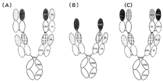
- FIG. 1 shows an example of the structure of the bispecific antibody of the present invention.
- FIG. 1(A) shows a bispecific antibody in which the N-terminal polypeptide is Fab and the polypeptide containing VH of Fab is bound to the N-terminus of the IgG portion.
- FIG. 1(B) shows a bispecific antibody in which the N-terminal polypeptide is VHH.
- FIG. 1(C) shows a bispecific antibody in which the N-terminal polypeptide is Fab, and the polypeptide containing the VL of Fab is bound to the N-terminus of the IgG portion.
- FIG. 2 shows the results of evaluation of antigen expression levels in various cancer cell lines by flow cytometry.
- 2A to 2C show OE21 cells, T.
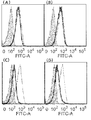
- FIG. 3 shows the results of evaluation of the antigen expression level in liver cancer cell lines by flow cytometry.
- FIGS. 3(A) to 3(C) show the results of evaluating the expression level of GPC3 in HepG2 cells, HuH-7 cells and HLE cells by a flow cytometer, respectively.
- FIGS. 3D to 3F show the results of evaluating the TfR expression level in HepG2 cells, HuH-7 cells, and HLE cells by a flow cytometer, respectively.
- FIGS. 3(G) to (I) show the results of evaluating the EGFR expression level in HepG2 cells, HuH-7 cells, and HLE cells by a flow cytometer, respectively.
- the vertical axis represents the number of cells, and the horizontal axis represents the fluorescence intensity.
- the solid line represents the binding of anti-GPC3 antibody, anti-TfR antibody or anti-EGFR antibody, and the gray-filled histogram represents the binding of negative control isotype antibody or anti-DNP antibody.
- 4(A) to (I) show the results of evaluation of the binding properties of each antibody to OE21 cells by a flow cytometer. The vertical axis represents the number of cells, and the horizontal axis represents the fluorescence intensity.
- the solid line shows the binding of the anti-EGFR antibody or the anti-TfR antibody, and the gray-filled histogram shows the binding of the secondary antibody as a negative control.
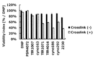
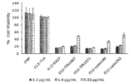
- FIG. 5 shows the results of evaluation of the growth inhibitory activity of each TfR antibody on OE21 cells.
- the vertical axis represents the cell viability, which is represented by the relative value of the amount of ATP in living cells with respect to the negative control.
- the black bar graph shows the result when only the monoclonal antibody was added, and the white bar graph shows the result when the cross-linked antibody was added to cross-link the monoclonal antibody.
- FIG. 6 shows the results of evaluation of the growth inhibitory activity of each EGFR antibody on OE21 cells.
- the vertical axis represents the cell viability, which is represented by the relative value of the amount of ATP in living cells with respect to the negative control.
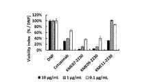
- FIG. 7A shows the results of evaluation of the growth inhibitory activity of various bispecific antibodies against OE21 cells.
- the vertical axis represents the cell viability, which is represented by the relative value of the amount of ATP in living cells with respect to the negative control.
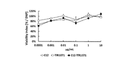
- FIG. 7B shows the results of evaluation of the growth inhibitory activity of various bispecific antibodies against OE21 cells.
- the vertical axis represents the cell viability, which is represented by the relative value of the amount of ATP in living cells with respect to the negative control.
- FIG. 7C shows the results of evaluation of the growth inhibitory activity of various bispecific antibodies against OE21 cells.
- the vertical axis represents the cell viability, which is represented by the relative value of the amount of ATP in living cells with respect to the negative control.
- FIG. 8A shows the results of evaluation of the growth inhibitory activity of the EGFR-TfR bispecific antibody against OE21, which is a cancer cell line expressing EGFR.
- the vertical axis represents the cell viability, which is shown by the relative value of the amount of ATP in living cells with respect to the negative control.
- FIG. 8B is a schematic diagram of EGFR expressing cancer cell line T.
- 5 shows the results of evaluation of the growth inhibitory activity of EGFR-TfR bispecific antibody against Tn.
- the vertical axis represents the cell viability, which is shown by the relative value of the amount of ATP in living cells with respect to the negative control.
- FIG. 8A shows the results of evaluation of the growth inhibitory activity of the EGFR-TfR bispecific antibody against OE21, which is a cancer cell line expressing EGFR.
- the vertical axis represents the cell viability, which is shown by the relative value of the amount of ATP in living cells with respect to the negative control.
- FIG. 8C shows the results of evaluation of the growth inhibitory activity of the EGFR-TfR bispecific antibody against U-937, which is a cancer cell line expressing EGFR.
- the vertical axis represents the cell viability, which is shown by the relative value of the amount of ATP in living cells with respect to the negative control.
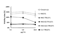
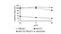
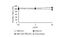
- FIG. 9 shows the results of evaluation of the growth inhibitory activity of GPC3-TfR bispecific antibody against HepG2 cells.
- the vertical axis represents the cell viability, which is represented by the relative value of the amount of ATP in living cells with respect to the negative control.
- FIG. 10A shows the results of evaluation of the growth inhibitory activity of GPC3-TfR bispecific antibody against HepG2, which is a hepatoma cell line expressing GPC3.
- the vertical axis represents the cell viability, which is shown by the relative value of the amount of ATP in living cells with respect to the negative control.
- FIG. 10B shows the results of evaluation of the growth inhibitory activity of the GPC3-TfR bispecific antibody against HuH-7, which is a hepatoma cell line expressing GPC3.
- the vertical axis represents the cell viability, which is shown by the relative value of the amount of ATP in living cells with respect to the negative control.
- FIG. 10C shows the results of evaluation of the growth inhibitory activity of the GPC3-TfR bispecific antibody against HLE, which is a hepatoma cell line expressing GPC3.
- FIGS. 11A to 11E show the results of evaluation of the inhibitory activity of each antibody or bispecific antibody against the binding of TfR and Tf on OE21 cells.
- the vertical axis represents the number of cells, and the horizontal axis represents the fluorescence intensity.
- the thin solid line is the binding activity when the fluorescently labeled transferrin is added after the addition of the negative control
- the thick solid line is the binding activity when the fluorescently labeled transferrin is added after the addition of the antibody or the bispecific antibody
- the gray filled histogram is fluorescently labeled. Represents a negative control with no transferrin added.
- FIGS. 12 shows the results of evaluation by Western blotting of the effect of adding EGFR-TfR bispecific antibody to OE21 cells on the EGFR downstream signal and TfR expression level. Each band represents the expression level of each protein shown on the right side of the figure when each bispecific antibody or antibody was added at the concentration shown at the top of the figure.
- pEGFR is phosphorylated EGFR protein
- AKT is a downstream signal protein of EGFR
- pAKT is phosphorylated AKT protein
- TFRC is TfR protein
- ACTB is ⁇ -actin.
- FIG. 13(A) to (D) show the results of evaluation of the TfR expression level on the cell surface when a EGFR-TfR bispecific antibody was added to OE21 cells using a flow cytometer.
- the vertical axis represents the number of cells, and the horizontal axis represents the fluorescence intensity.
- the thin solid line shows the binding activity when the negative control was added and the fluorescently labeled transferrin was added after the culture for 24 hours, and the thick solid line is when the antibody or the bispecific antibody was added and the fluorescently labeled transferrin was added after the culture for 24 hours. Binding activity of H., gray-filled histograms represent negative controls without the addition of fluorescently labeled transferrin.
- FIG. 14 shows the results of evaluation whether the growth inhibitory activity when the EGFR-TfR bispecific antibody was added to OE21 cells was due to iron depletion.
- the vertical axis represents the cell viability, which is shown by the relative value of the amount of ATP in living cells with respect to the negative control under the condition that PBS is added and FAS is not added.
- the black bar graph shows the survival rate when the antibody or the bispecific antibody alone was added, and the hatched bar graph shows the survival rate when the FAS was added together with the antibody or the bispecific antibody.
- FIG. 15 shows the results of evaluation of the growth inhibitory activity when the EGFR-TfR bispecific antibody was added to OE21 cells.
- the vertical axis represents the ratio (%) of cells in the well.
- FIG. 16 shows the results of evaluating the growth inhibitory activity when the EGFR-TfR bispecific antibody was added to GEMM.
- the vertical axis represents the survival rate when the amount of ATP in living cells when PBS was added was 100%.
- FIG. 17A shows the results of evaluation of the growth inhibitory activity of the EGFR-TfR bispecific antibody.
- FIG. 17B shows the results of evaluation of the growth inhibitory activity of the EGFR-TfR bispecific antibody.
- FIG. 18(A) is a schematic diagram showing the structure of the EGFR-TfR bispecific antibody prepared in Example 6.
- FIG. 18B is a schematic diagram showing the structure of the heterodimeric antibody prepared in Example 23.
- FIG. 19A shows the results of evaluation of the growth inhibitory activity of the EGFR-TfR bispecific antibody.
- FIG. 19B shows the results of evaluation of the growth inhibitory activity of the EGFR-TfR bispecific antibody.
- FIG. 20 shows the results of evaluating the growth inhibitory activity of the EGFR-TfR bispecific antibody using the growth inhibitory activity against cancer cell lines as an index.
- FIG. 21 shows the results of evaluation of the growth inhibitory activity of the EGFR-TfR bispecific antibody.
- the present invention relates to TfR and cells in which a polypeptide having an antigen-binding site for a cell surface antigen (also referred to as N-terminal side polypeptide) is bound to the N-terminal side of an IgG portion that binds to TfR directly or via a linker.
- the present invention relates to a bispecific antibody that binds to a surface antigen or the bispecific antibody fragment (hereinafter referred to as the bispecific antibody or the bispecific antibody fragment of the present invention).
- TfR in the present invention is used synonymously with CD71, TFR1, TR, T9, p90, and IMD46.
- TfR include GenBank accession No. in NCBI (http://www.ncbi.nlm.nih.gov/).
- SEQ ID NO: 6, GenBank accession No. NP — 003225 or the amino acid sequence represented by SEQ ID NO: 8 is composed of an amino acid sequence in which one or more amino acids are deleted, substituted or added, and includes a polypeptide having a TfR function.
- SEQ ID NO: 6 GenBank accession No. NP — 003225 or a polypeptide containing an amino acid sequence having 70% or more, preferably 80% or more, more preferably 90% or more homology with the amino acid sequence represented by SEQ ID NO: 8, and most preferably 95%, 96% or 97.
- Polypeptides consisting of amino acid sequences having homology of 98%, 98% and 99% or more and having the function of TfR are also included in the TfR of the present invention.
- SEQ ID NO: 6 GenBank accession No. NP_003225 or a polypeptide having an amino acid sequence in which one or more amino acid residues are deleted, substituted, or added in the amino acid sequence shown in SEQ ID NO: 8 is a site-directed mutagenesis method [Molecular Cloning, A-Laboratory Manual, Second Edition, Cold Spring Laboratory ⁇ Press (1989), Current Protocols in in Molecular Biology, John Wiley&Sons (1987-1997), Nucleic Acids Research, 10, 6487 (1982), Proc. Natl. Acad. Sci. ,USA.
- GenBank accession No. It can be obtained by introducing a site-specific mutation into DNA encoding NP — 003225 or the amino acid sequence shown by SEQ ID NO:8.
- the number of amino acids to be deleted, substituted or added is not particularly limited, but is preferably 1 to several tens, for example 1 to 20, more preferably 1 to several, for example 1 to 5 amino acids. Is.
- Examples of the gene encoding TfR include SEQ ID NO: 5 or GenBank accession No. Examples include the human TfR base sequence represented by NM_003234, and the monkey TfR base sequence represented by SEQ ID NO:7. In addition, for example, SEQ ID NO: 5, GenBank accession No. NM_003234 or a gene consisting of a nucleotide sequence having one or more bases deleted, substituted or added in the nucleotide sequence shown in SEQ ID NO: 7, and containing a DNA encoding a polypeptide having a TfR function, SEQ ID NO: 5, GenBank accession No.
- a gene comprising a DNA that hybridizes under stringent conditions with a DNA consisting of NM — 003234 or the nucleotide sequence shown in SEQ ID NO: 7 and that contains a DNA encoding a polypeptide having the function of TfR also has the TfR of the present invention. Included in the encoding gene. ..
- DNA that hybridizes under stringent conditions for example, SEQ ID NO: 5 or GenBank accession No. It means a hybridizable DNA obtained by a colony hybridization method, a plaque hybridization method, a Southern blot hybridization method, a DNA microarray method, or the like, which uses a DNA having the nucleotide sequence represented by NM_003234 as a probe. .. Specifically, the presence of 0.7 to 1.0 mol/L of sodium chloride is obtained by using a filter or a slide glass on which DNA derived from hybridized colony or plaque, or PCR product or oligo DNA having the sequence is immobilized.
- hybridizable DNA for example, SEQ ID NO: 5 or GenBank accession No.
- a DNA having a homology of 60% or more with the nucleotide sequence represented by NM_003234, a DNA having a homology of 80% or more, more preferably a DNA having a homology of 95% or more can be mentioned.
- Gene polymorphisms are often found in the nucleotide sequences of genes that encode eukaryotic proteins.
- a gene having a small-scale mutation in the nucleotide sequence due to such a polymorphism is also included in the gene encoding TfR of the present invention.
- the numerical value of homology in the present invention may be a numerical value calculated using a homology search program known to those skilled in the art, unless otherwise specified.
- BLAST Japanese Mol. ., 215, 403 (1990)
- amino acid sequences such as the values calculated using the default parameters
- BLAST2 Nucleic Acids Research, 25, 3389 (1997), Genome Research, 7, 649 (1997), [http://www.ncbi.nlm.nih.gov/Education/BLASTinfo/information3.html]
- numerical values calculated using default parameters are included. ..
- the default parameters are 5 when G (Cost to open gap) is a base sequence, 11 when an amino acid sequence, 2 when -E (Cost to extend gap) is a base sequence, and 1 when an amino acid sequence.
- -Q (Penalty for nucleotides mismatch) is -3
- -r (reward for nucleotides match) is 1
- -e (expect value) is 10
- -W wordsize
- amino acid sequence In case of 3 residues, -y [Dropoff(X)for blast extensions in bits] is blastn, 20 in programs other than blastn, -X (X dropoff value for gapped alignment in bits) and 15 are -X (X dropoff value for gapped alignment in bits).
- a polypeptide consisting of a partial sequence of the amino acid sequence of TfR can be produced by a method known to those skilled in the art.
- SEQ ID NO: 6, GenBank accession No. It can be prepared by deleting a part of the DNA encoding the amino acid sequence represented by NP — 003225 or SEQ ID NO: 8 and culturing a transformant into which an expression vector containing this is introduced.
- a polypeptide having an amino acid sequence in which one or more amino acids are deleted, substituted or added in the partial sequence of NP — 003225 or the amino acid sequence shown in SEQ ID NO: 8 can be obtained.
- a polypeptide consisting of a partial sequence of the amino acid sequence of TfR, or a polypeptide having an amino acid sequence in which one or more amino acids are deleted, substituted or added in the partial sequence of the amino acid sequence of TfR is fluorenylmethyloxycarbonyl. It can also be produced by a chemical synthesis method such as the (Fmoc) method and the t-butyloxycarbonyl (tBoc) method.
- TfR extracellular region of TfR in the present invention
- GenBank accession No. The amino acid sequence of human TfR shown in NP — 003225 can be obtained by using the known transmembrane region prediction program SOSUI (http://sosui.proteome.bio.tuat.ac.jp/sosuiframe0.html), TMHMM ver. 2 (http://www.cbs.dtu.dk/services/TMHMM-2.0/) or ExPASy Proteomics Server (http://Ca.expasy.org/).
- SOSUI transmembrane region prediction program
- TMHMM ver. 2 http://www.cbs.dtu.dk/services/TMHMM-2.0/
- ExPASy Proteomics Server http://Ca.expasy.org/.
- TfR The functions of TfR include the uptake of iron, which is essential for cell survival and proliferation.
- iron When the complex of iron and transferrin binds to TfR, it is taken up into the cell by endocytosis. When the pH in the endosome is lowered, iron is released from transferrin, migrates into the cytoplasm via DMT1, and is used for cell growth and energy production. It is known that the transferrin-TfR complex in which iron is released is not normally decomposed and re-migrates to the cell surface (Reference: Yamashiro DJ et al., Cell 37 37-789-800, 1984).
- TfR-expressing cells include, for example, many normal tissue cells such as bone marrow and placenta cells, colon cancer, head and neck cancer, brain tumor, hematopoietic tumor, liver cancer, and esophageal cancer. Many types of cancer cells, HT29, HSC-2, RAMOS, K562, HepG2, OE21, T. Many cancer cell lines such as Tn, U-937, HuH-7, and HLE can be mentioned.
- the cell surface antigen of the present invention refers to an antigen other than TfR among antigens such as proteins and peptide chains expressed on the cell membrane of cancer cells.
- the cell surface antigen of the present invention may be any as long as it can bind to a protein or polypeptide such as an antibody, but is preferably a cell surface antigen that is internalized and/or degraded by the binding of a protein or polypeptide such as an antibody. ..
- a protein highly expressed in cancer cells or a cell population involved in a specific disease is preferable.
- Preferred examples of such cell surface antigens include EGFR and GPC3.
- EGFR in the present invention is used synonymously with ERBB, ERBB1, HER1, PIG61, MENA, NISBD2, SA7, and c-Erb-1.
- SEQ ID NO: 14, NP005219 or GenBank accession No. A polypeptide having an EGFR function and comprising an amino acid sequence in which one or more amino acids have been deleted, substituted or added in the amino acid sequence shown in XP — 005549616.1.
- polypeptide comprising an amino acid sequence having a homology of preferably 70% or more, more preferably 80% or more, still more preferably 90% or more with the amino acid sequence shown in XP — 005549616.1, most preferably 95%, 96%, 97.
- Polypeptides consisting of amino acid sequences having homology of at least 98%, 98% and 99% and having EGFR function are also included in the EGFR of the present invention.
- a polypeptide having an amino acid sequence in which one or more amino acid residues have been deleted, substituted, or added in the amino acid sequence shown in XP — 005549616.1 can be prepared by, for example, SEQ ID NO: 14 using the site-directed mutagenesis method described above. , NP005219 or GenBank accession No. It can be obtained by introducing a site-specific mutation into the DNA encoding the amino acid sequence shown in XP — 005549616.1.
- the number of amino acids to be deleted, substituted or added is not particularly limited, but is preferably 1 to several tens, for example 1 to 20, more preferably 1 to several, for example 1 to 5 amino acids. Is.
- the gene encoding EGFR in the present invention includes, for example, Genbank accession No.
- An example is a human EGFR gene containing NM_005228 or the base sequence shown in SEQ ID NO:13.
- SEQ ID NO: 13 Genbank accession No.
- a gene comprising DNA encoding a polypeptide having a function of EGFR which consists of a nucleotide sequence in which one or more nucleotides are deleted, substituted or added in the nucleotide sequence represented by NM_005228, SEQ ID NO: 13 or Genbank accession No.
- a gene containing a DNA encoding a polypeptide having a function and SEQ ID NO: 13 or Genbank accession No.
- a gene comprising a DNA that hybridizes under stringent conditions with a DNA containing the nucleotide sequence represented by NM_005228 and that contains a DNA encoding a polypeptide having an EGFR function is also a gene encoding EGFR of the present invention. included.
- GenBank accession No. The amino acid sequence of human EGFR shown in NP005219 can be obtained by using the known transmembrane region prediction program SOSUI (http://sosui.proteome.bio.tuat.ac.jp/sosuiframe0.html), TMHMM ver. 2 (http://www.cbs.dtu.dk/services/TMHMM-2.0/) or ExPASy Proteomics Server (http://Ca.expasy.org/). Specifically, SEQ ID NO: 10 or GenBank accession No. The amino acid sequence shown from the 25th position to the 645th position of NP005219 can be mentioned.
- EGFR functions as a receptor with EGF, forms a dimer after binding EGF, and through signals such as Ras/Raf/MAPK pathway, PI3K/Akt pathway, Jak/STAT pathway, It includes promoting cell proliferation, survival, invasion, migration and the like. It is known that EGFR on the cell membrane is internalized when EGF binds, and is transferred to lysosomes by endocytosis and degraded (Ebner R et al., Cell Regul. 2 599-612, 1991). ..
- Cells expressing EGFR include, for example, epithelial cells of normal tissues such as skin, large intestine and lung, epithelial cancer cells including large intestine cancer, head and neck cancer, lung cancer, esophageal cancer, HT29, HSC-2, NCI-H1975, OE21, T.I. Cancer cell lines such as Tn and A431 can be mentioned.
- the GPC3 in the present invention is used synonymously with DGSX, GTR2-2, MXR7, OCI-5, SDYS, SGB, SGBS, SGBS1.
- GPC3 The function of GPC3 is, for example, to be involved in the cell division and proliferation of cancer cells such as liver cancer by binding to proteins related to Wnt pathway and Frizzled pathway.
- Genbank accession No. Human GPC3 including the amino acid sequence shown in P51654 can be mentioned.
- the extracellular region of GPC3 in the present invention includes, for example, GenBank accession No.
- GenBank accession No. The amino acid sequence of human GPC3 shown in P51654 is the known transmembrane region prediction program SOSUI (http://sosui.proteome.bio.tuat.ac.jp/sosuiframe0.html), TMHMM ver. 2 (http://www.cbs.dtu.dk/services/TMHMM-2.0/) or ExPASy Proteomics Server (http://Ca.expasy.org/).
- SOSUI Transmembrane region prediction program
- TMHMM ver. 2 http://www.cbs.dtu.dk/services/TMHMM-2.0/
- ExPASy Proteomics Server http://Ca.expasy.org/.
- GenBank accession No. The amino acid sequences shown as 1st to 559th of P51654 are mentioned.
- Examples of cells expressing GPC3 include fetal liver cells, cancer cells such as liver cancer, and cancer cell lines such as HepG2 and HuH-7.
- An antibody is a gene that encodes all or part of a heavy chain variable region and a heavy chain constant region, and a light chain variable region and a light chain constant region that make up an immunoglobulin (referred to as “antibody gene”). Is a protein derived from.
- the antibody of the present invention includes an antibody or an antibody fragment having any immunoglobulin class and subclass.
- Heavy chain refers to the polypeptide with the larger molecular weight of the two types of polypeptides that make up the immunoglobulin molecule. Heavy chains determine the class and subclass of antibody. IgA, IgD, IgE, IgG and IgM have ⁇ chain, ⁇ chain, ⁇ chain, ⁇ chain and ⁇ chain as heavy chains, respectively, and the constant regions of the heavy chains are characterized by different amino acid sequences.
- the light chain (L chain) refers to the polypeptide having the smaller molecular weight of the two types of polypeptides constituting the immunoglobulin molecule. In the case of human antibodies, there are two types of light chains, the ⁇ chain and the ⁇ chain.
- variable region usually refers to a highly diverse region existing in the amino acid sequence on the N-terminal side of immunoglobulin. Since the portion other than the variable region has a structure with little diversity, it is called a constant region (C region).
- C region constant region
- variable region corresponds to the amino acid sequence from the 1st to 117th in the EU index of Kabat et al. (Kabat et al., Sequences of proteins of immunological interest, 1991 Fifth edition) and the constant region It corresponds to the 118th and subsequent amino acid sequences.
- amino acid sequence from the 1st to 107th in the numbering (Kabat numbering) by Kabat et al. corresponds to the variable region
- amino acid sequence from the 108th to the constant region corresponds to the constant region.
- the heavy chain variable region or the light chain variable region is abbreviated as VH or VL.
- An antigen-binding site is a site that recognizes and binds an antigen in an antibody and refers to a site that forms a three-dimensional structure complementary to an antigenic determinant (epitope).
- the antigen binding site produces strong intermolecular interactions with the antigenic determinant.
- the antigen binding site is composed of VH and VL containing at least three complementarity determining regions (CDRs). For human antibodies, VH and VL each have three CDRs. These CDRs are referred to as CDR1, CDR2 and CDR3 in order from the N-terminal side.
- the heavy chain constant region or the light chain constant region is expressed as CH or CL, respectively.
- CH is classified by the heavy chain subclasses ⁇ chain, ⁇ chain, ⁇ chain, ⁇ chain and ⁇ chain.
- CH is composed of a CH1 domain, a hinge domain, a CH2 domain, and a CH3 domain arranged in order from the N-terminal side, and the CH2 domain and the CH3 domain are collectively referred to as an Fc region.
- CL is classified into two subclasses called C ⁇ chain and C ⁇ chain.
- Monoclonal antibody is an antibody secreted by antibody-producing cells that retain monoclonality and recognizes a single epitope (also called an antigenic determinant). Monoclonal antibody molecules have the same amino acid sequence (primary structure) and have a single structure. Polyclonal antibody refers to a population of antibody molecules secreted by antibody-producing cells of different clones. An oligoclonal antibody refers to a population of antibody molecules in which a plurality of different monoclonal antibodies are mixed.
- An epitope is a structural site of an antigen that an antibody recognizes and binds to.
- Examples of the epitope include a single amino acid sequence recognized and bound by a monoclonal antibody, a three-dimensional structure consisting of an amino acid sequence, a three-dimensional structure consisting of an amino acid sequence bound to a sugar chain and an amino acid sequence bound to a sugar chain. ..
- Examples of the monoclonal antibody in the present invention include an antibody produced by a hybridoma and a gene recombinant antibody produced by a transformant transformed with an expression vector containing an antibody gene.
- a hybridoma can be prepared, for example, by preparing an antigen, obtaining an antibody-producing cell having antigen specificity from an animal immunized with the antigen, and further fusing the antibody-producing cell with a myeloma cell. it can.
- the desired monoclonal antibody can be obtained by culturing the hybridoma, or by administering the hybridoma to an animal to cause ascites tumor in the hybridoma and separating and purifying the culture medium or ascites.
- an animal to be immunized with the antigen any animal can be used as long as a hybridoma can be produced, and mouse, rat, hamster, rabbit and the like are preferably used. It is also possible to obtain cells having antibody-producing ability from such immunized animals, immunize the cells in vitro, and then fuse the cells with myeloma cells to prepare hybridomas.
- the gene recombinant antibody in the present invention examples include recombinant mouse antibody, recombinant rat antibody, recombinant hamster antibody, recombinant rabbit antibody, human chimeric antibody (also referred to as chimeric antibody), humanized antibody (CDR-grafted antibody). (Also referred to as) and human antibodies, and antibodies produced by gene recombination techniques.
- the variable region and constant region of the heavy and light chains derived from any animal species can be determined according to the target animal species and purpose. For example, when the target animal species is human, the variable region can be derived from a non-human animal such as human or mouse, and the constant region and the linker can be derived from human.
- Chimeric antibody refers to an antibody composed of VH and VL of antibodies of non-human animals (non-human animals) and CH and CL of human antibodies.
- non-human animal any animal such as mouse, rat, hamster and rabbit can be used as long as a hybridoma can be produced.
- a chimeric antibody is obtained by obtaining cDNAs encoding VH and VL from a hybridoma derived from a non-human animal producing a monoclonal antibody, and inserting the cDNAs into animal cell expression vectors having DNAs encoding CH and CL of human antibody, respectively. It can be produced by constructing a chimeric antibody expression vector, introducing it into an animal cell and expressing it.
- Humanized antibody refers to an antibody in which CDRs of non-human animal antibody VH and VL are transplanted to corresponding CDRs of human antibody VH and VL. Regions other than CDRs of VH and VL are called framework regions (hereinafter referred to as FR).
- the humanized antibody includes a cDNA encoding a VH amino acid sequence consisting of the VH CDR amino acid sequence of a non-human animal antibody and the VH FR amino acid sequence of any human antibody, and the VL CDR amino acid of a non-human animal antibody.
- a cDNA encoding the amino acid sequence of VL consisting of the sequence and the amino acid sequence of FR of VL of any human antibody is constructed and inserted into an expression vector for animal cells having DNAs encoding CH and CL of the human antibody, respectively It can be produced by constructing a modified antibody expression vector, introducing it into an animal cell and expressing it.
- the human antibody originally refers to an antibody that naturally exists in the human body, but a human antibody phage library and a human antibody-producing transgene produced by recent advances in genetic engineering, cell engineering, and developmental engineering techniques. Antibodies obtained from transgenic animals are also included.
- Antibodies that naturally occur in the human body are obtained by, for example, immunizing human peripheral blood lymphocytes with EB virus or the like to immortalize and cloning the lymphocytes producing the antibody, and culturing the lymphocytes producing the antibody from the culture supernatant. It can be obtained by purifying the antibody.
- the human antibody phage library is a library in which antibody fragments such as Fab and scFv are expressed on the phage surface by inserting an antibody gene prepared from human B cells into the phage gene. Phage expressing an antibody fragment having a desired antigen-binding activity on the surface can be recovered from the library by using the binding activity to the substrate on which the antigen is immobilized as an index. The antibody fragment can be further converted into a human antibody molecule consisting of two complete H chains and two complete L chains by a genetic engineering technique.
- Human antibody-producing transgenic animal means an animal in which a human antibody gene is incorporated into cells.
- a human antibody-producing transgenic mouse can be produced by introducing a human antibody gene into a mouse ES cell, transplanting the ES cell into an early embryo of a mouse, and then developing an individual.
- Human antibody-producing human antibodies are derived from transgenic animals by producing and accumulating the antibodies in the culture supernatant by obtaining and culturing the hybridomas using the hybridoma production method that is usually performed in non-human animals.
- the CH of the gene recombinant antibody may be any as long as it belongs to a human immunoglobulin, but a human immunoglobulin G (hIgG) class is preferable. Further, any of the subclasses belonging to the hIgG class, such as hIgG1, hIgG2, hIgG3 and hIgG4, can be used.
- the CL of the recombinant antibody may be any CL as long as it belongs to human immunoglobulin, and CL of the ⁇ class or ⁇ class can be used.
- a bispecific antibody refers to a polypeptide or protein that specifically binds to each of two different types of epitopes.
- Each antigen binding site of a bispecific antibody may bind to different epitopes of a single antigen or may bind to different antigens.
- binding of a polypeptide, an antibody or an antibody fragment thereof or a bispecific antibody or a bispecific antibody fragment to any one of a cell surface antigen such as EGFR or GPC3 and TfR is known, for example, in known immunology. It can be confirmed by a method for confirming the binding property between the cell surface antigen or the TfR-expressing cells to be evaluated and the antibody using a dynamic detection method, preferably a fluorescent cell staining method.
- the antigen binding site that binds to a cell surface antigen such as EGFR or GPC3 or TfR may be any as long as it specifically recognizes and binds to a cell surface antigen such as EGFR or GPC3 or TfR. ..
- polypeptides, protein molecules and fragments thereof that can be produced by gene recombination techniques such as antibodies, ligands, receptors, and naturally occurring interacting molecules, and conjugates of the protein molecules with small molecules or natural products It may be in any form.
- the antigen-binding site may be a binding protein that is recombined using a binding domain of a known binding molecule such as an antibody, a ligand, and a receptor.
- a recombinant protein containing the CDR of an antibody that binds to each antigen is used.
- examples include proteins, antibody variable regions containing CDRs, recombinant proteins containing antibody variable regions and the binding domains of ligands that bind to each antigen.
- the antigen binding site is preferably the variable region of the antibody.
- the bispecific antibody or the bispecific antibody fragment of the present invention includes, for example, a bispecific antibody having the TfR internalization and/or degrading activity or the bispecific antibody fragment.
- the bispecific antibody or the bispecific antibody fragment of the present invention is a cell such as EGFR or GPC3 that does not show TfR internalization and/or degrading activity in cells that do not express cell surface antigens such as EGFR or GPC3.
- a bispecific antibody or a bispecific antibody fragment thereof that exhibits TfR internalization and/or degradation activity only in cells that express the surface antigen is preferred.
- Such a bispecific antibody or the bispecific antibody fragment expresses a cell surface antigen such as EGFR or GPC3, and selectively exhibits TfR internalization and/or degrading activity in pathogenic cells such as cancer cells. It is preferable in that it does not cause side effects associated with nonspecific TfR internalization and/or degradation.
- the internalizing and/or degrading activity of TfR possessed by the bispecific antibody or the bispecific antibody fragment of the present invention means that the cell surface antigen such as EGFR or GPC3 on cells and TfR are both bispecific antibodies or the bispecific antibody fragments.
- the bispecific antibody or the bispecific antibody fragment of the present invention may bind to a cell surface antigen such as EGFR or GPC3 expressed on the same cell and TfR, or EGFR expressed on a different cell or although it may bind to a cell surface antigen such as GPC3 and TfR, it preferably binds to a cell surface antigen such as EGFR or GPC3 expressed on the same cell and TfR.
- the bispecific antibody or the bispecific antibody fragment of the present invention induces internalization and/or degradation of TfR to induce cell death of cells expressing a target cell surface antigen such as EGFR or GPC3. Those that induce are preferred.
- the internalization and/or degradation activity of TfR possessed by the bispecific antibody or the bispecific antibody fragment of the present invention can be determined by, for example, OE21, T.R.
- Cell surface and intracellular TfR protein amount of cells expressing TfR such as Tn, TE-8, U-937, HepG2, HuH-7 and HLE, viable cell number, cell proliferation rate, cell viability or cell It can be confirmed by evaluating the uptake of iron.
- the bispecific antibody or the bispecific antibody fragment of the present invention specifically, the bispecific antibody or the bispecific antibody that induces the degradation of TfR when bound to both TfR and a cell surface antigen such as EGFR or GPC3.
- the bispecific antibody or the bispecific antibody fragment of the present invention specifically, the bispecific antibody or the bispecific antibody that induces the degradation of TfR when bound to both TfR and a cell surface antigen such as EGFR or GPC3.
- Examples include bispecific antibody fragments.
- the number of binding domains for a given antigen possessed by one molecule of bispecific antibody is called the valency of binding.
- the bispecific antibody binds to EGFR and TfR with divalent antigens, respectively. Join with.
- the bispecific antibody of the present invention includes an antibody having a plurality of antigen-binding sites bound via an appropriate linker such as a linker containing an immunoglobulin domain or a fragment thereof.
- the bispecific antibody of the present invention can be produced by existing production techniques ([Nature Protocols, 9, 2450-2463 (2014)], International Publication No. 1998/050431, International Publication No. 2001/7734, International Publication No. 2002/002773. And International Publication No. 2009/131239).
- a polypeptide capable of binding to a cell surface antigen binds to the N-terminus of the heavy chain of the IgG portion that binds to TfR directly or via a linker. It has a structure.
- the immunoglobulin domain has, as a minimum unit, a peptide having an amino acid sequence similar to that of an immunoglobulin and consisting of about 100 amino acid residues having at least two cysteine residues.
- the immunoglobulin domain also includes a polypeptide containing a plurality of the above-mentioned minimum unit immunoglobulin domains. Examples of the immunoglobulin domain include VH, CH1, CH2 and CH3 of immunoglobulin heavy chain, and VL and CL of immunoglobulin light chain.
- the animal species of immunoglobulin is not particularly limited, but human is preferred.
- the subclass of the constant region of the immunoglobulin heavy chain may be any of IgD, IgM, IgG1, IgG2, IgG3, IgG4, IgA1, IgA2 and IgE, preferably IgG-derived and IgM-derived.
- the subclass of the constant region of immunoglobulin light chain may be either ⁇ or ⁇ .
- the immunoglobulin domain also exists in proteins other than immunoglobulins, and for example, an immunoglobulin domain included in proteins belonging to the immunoglobulin superfamily such as major histocompatibility complex (MHC), CD1, B7, and T cell receptor (TCR). Are listed.
- MHC major histocompatibility complex
- CD1, B7 CD1, B7, and T cell receptor (TCR).
- TCR T cell receptor
- CH1 refers to the region having the amino acid sequence of 118th to 215th amino acids shown by EU index.
- CH2 refers to a region having an amino acid sequence from the 231st to 340th position shown by the EU index of Kabat et al.
- CH3 refers to a region having an amino acid sequence from the 341st to 447th position shown by the EU index of Kabat et al. ..
- a hinge region hereinafter sometimes referred to as a hinge).
- the hinge region refers to a region having an amino acid sequence from the 216th position to the 230th position shown by the EU index of Kabat et al.
- CL is a region having the amino acid sequence from 108th to 214th indicated by Kabat numbering in the case of human antibody ⁇ chain, and a region having the 108th to 215th amino acid sequence in the case of ⁇ chain. Refer to each.
- the IgG type antibody of the present invention which is also referred to as an IgG portion, refers to a partial structure of IgG constituting the bispecific antibody of the present invention or an IgG having a modified Fc portion, and is composed of one light chain and one heavy chain. It has a heterotetramer structure in which two heterodimers are associated.
- the IgG portion that binds to TfR of the present invention refers to an IgG portion that recognizes TfR, that is, an IgG portion that specifically recognizes and binds to the extracellular region of TfR.
- the heavy chain constant region of IgG may be any of IgG1, IgG2, IgG3, and IgG4 subclasses.
- a part of those amino acid sequences may be deleted, added, substituted, and/or inserted.
- all or part of the amino acid sequence consisting of CH1, hinge, CH2, and CH3 of the heavy chain of IgG can be appropriately combined and used.
- those amino acid sequences can be partially deleted or the order can be changed.
- the subclass of IgG used for the IgG part is not particularly limited, but IgG4 variant in which the Ser residue at position 228 of the heavy chain constant region of IgG4, IgG4 is replaced with Pro and the Leu residue at position 235 is replaced with Asn, respectively.
- IgG4PE an IgG4 mutant
- IgG4PE R409K an IgG4 mutant
- the constant region of the heavy chain (CH1-hinge-CH2-CH3 in order from the N-terminal side) comprises an IgG portion consisting of IgG4PE containing the amino acid sequence represented by SEQ ID NO:86, or the amino acid sequence represented by SEQ ID NO:84.
- IgG part consisting of IgG4PE R409K containing is preferred.
- the two variable regions contained in the IgG portion of the present invention preferably recognize the same antigen. It is also preferable that they have the same structure and amino acid sequence.
- the N-terminal side polypeptide that binds to a cell surface antigen is a partial structure constituting the bispecific antibody of the present invention, which exists on the N-terminal side of the heavy chain or light chain of the IgG part, and is present in each cell.
- a polypeptide that specifically recognizes and binds to the extracellular region of a surface antigen is present in each cell.
- the bispecific antibody of the present invention may have one N-terminal polypeptide at each of the N-terminals of the heavy chains or light chains of both of the two heterodimers constituting the IgG portion, or one heterodimer. Although it may have one at the N-terminal of each of them, it is preferable to have one at the N-terminals of both heavy chains or light chains. When both heavy chains or light chains have one N-terminal polypeptide at each N-terminal, they may be the same or different, but they are the same N-terminal polypeptide. Is preferred.
- the N-terminal polypeptide of the present invention may be a single chain or a multimer composed of a plurality of polypeptide chains, as long as it has the ability to bind to a cell surface antigen.
- Examples of the N-terminal side polypeptide include Fab, Fab', scFv, dsFv and VHH which are antibodies against cell surface antigens, and Fab or VHH is preferable.
- ligand molecules and receptor molecules for cell surface antigens can be used in the same manner.
- Antibodies against cell surface antigens refer to monoclonal antibodies that specifically recognize and bind to the extracellular region of each cell surface antigen.
- anti-EGFR antibody and anti-GPC3 antibody refer to monoclonal antibodies that specifically recognize and bind to the extracellular regions of EGFR and GPC3, respectively.
- the linker may have any molecular structure as long as it binds the IgG part and the N-terminal side polypeptide, but a peptide chain is preferable.
- the amino acid sequence of the peptide chain includes, for example, a sequence consisting of a repeating sequence of ES, ESKYG, ESKYGPP, GGGGS, or GGGGS, or a sequence consisting of part or all of the constant region of the antibody hinge region and CH1 domain. And so on.
- the light chain contained in the Fab and the light chain of the IgG part may be the same or different. , And preferably the same.
- the light chain may be either ⁇ chain or ⁇ chain, but is preferably ⁇ chain.
- the antibody fragment thereof is also included in the bispecific antibody of the present invention or the antibody fragment thereof.
- the number of amino acids to be deleted, substituted, inserted and/or added is 1 or more, and the number is not particularly limited. Molecular Cloning, The Second Second Edition, Cold Spring Harbor Laboratory (1989), Current Protocols in in Molecular Biology , John Wiley&Sons (1987-1997), Nucleic Acids Research, 10, 6487 (1982), Proc. Natl. Acad. Sci., USA, 79, 6409 (1982), Gene, 34, 315 (1985), Nucleic Acids Research, 13, 4431 (1985), Proc. Natl. Acad. Sci USA, 82, 488 (1985) etc., deletion, substitution, insertion or addition by well-known techniques such as site-directed mutagenesis It is the number that can be done. For example, it is usually 1 to several tens, preferably 1 to 20, more preferably 1 to 10, and further preferably 1 to 5.
- deletion, substitution, insertion or addition of one or more amino acid residues in the above amino acid sequence of the bispecific antibody of the present invention means the following. It means that there is a deletion, substitution, insertion or addition of one or more amino acid residues in any one or more amino acid sequences in the same sequence. In addition, deletion, substitution, insertion or addition may occur at the same time, and the amino acid residue to be substituted, insertion or addition may be natural type or non-natural type.
- natural amino acid residues include L-alanine, L-asparagine, L-aspartic acid, L-glutamine, L-glutamic acid, glycine, L-histidine, L-isoleucine, L-leucine, L-lysine, L -Arginine, L-methionine, L-phenylalanine, L-proline, L-serine, L-threonine, L-tryptophan, L-tyrosine, L-valine and L-cysteine.
- Group A leucine, isoleucine, norleucine, valine, norvaline, alanine, 2-aminobutanoic acid, methionine, O-methylserine, t-butylglycine, t-butylalanine, cyclohexylalanine
- Group B aspartic acid, glutamic acid, isoaspartic acid, Isoglutamic acid, 2-aminoadipic acid, 2-aminosuberic acid
- Group C asparagine, glutamine
- D lysine, arginine, ornithine, 2,4-diaminobutanoic acid, 2,3-diaminopropionic acid
- Group E proline, 3 -Hydroxyproline, 4-hydroxyproline
- F group serine, threonine, homoserine
- G group phenylalanine, tyrosine
- the bispecific antibody or the antibody fragment of the present invention includes an antibody containing any post-translationally modified amino acid residue.
- Post-translational modifications include, for example, deletion of a lysine residue at the C-terminus of the H chain [lysine clipping] and substitution of a glutamine residue at the N-terminus of a polypeptide with pyroglutamine (pyroGlu). [Beck et al, Analytical Chemistry, 85, 715-736 (2013)].
- bispecific antibody of the present invention include any one bispecific antibody selected from the group consisting of the following (1) to (4).
- a bispecific antibody comprising a Fab containing a variable region of an antibody against a cell surface antigen and an IgG portion binding to TfR, wherein the Fab directly binds to the N-terminus of the heavy chain of the IgG portion
- a bispecific antibody comprising a VH containing CDR1 to 3 of VH and a VL containing CDR1 to 3 of VL of an antibody against a cell surface antigen, and a bispecific antibody containing an IgG portion binding to TfR, wherein the Fab is an IgG portion.
- Antibody that binds directly to the N-terminus of the heavy chain of (3) Bispecific antibody containing Fab containing VH and VL of antibody to cell surface antigen and IgG portion binding to TfR, wherein the Fab directly binds to N terminus of heavy chain of IgG portion And (4) a bispecific antibody comprising a VHH that binds to a cell surface antigen and an IgG portion that binds to TfR, wherein the VHH directly binds to the N-terminus of the heavy chain of the IgG portion.
- the VL of the IgG portion that binds to TfR and the VL of the Fab may be the same or different, but they are the same. preferable.
- VL including CDR1, 2 and 3 containing the amino acid sequences represented by SEQ ID NOs: 17, 18 and 19 and CDR1 to 3 shown in 1) to 3) below are included.
- Examples include IgG moieties that bind to TfR, including VH.
- CDR1 comprising an amino acid sequence modified by one substitution.
- substitution of the first valine with alanine substitution of the second isoleucine with alanine or leucine, and substitution of the seventh valine A CDR2 comprising an amino acid sequence modified by at least one substitution selected from glutamic acid substitution, 10th aspartic acid substitution with alanine and 13th aspartic acid substitution with proline.
- CDR3 including substitution of proline at position 4 with any natural amino acid, for example, alanine, tyrosine, serine, aspartic acid, glutamine, glutamic acid, threonine, arginine, glycine, lysine, methionine, valine, leucine, isoleucine, tryptophan. , Phenylalanine or histidine.
- any natural amino acid for example, alanine, tyrosine, serine, aspartic acid, glutamine, glutamic acid, threonine, arginine, glycine, lysine, methionine, valine, leucine, isoleucine, tryptophan.
- Phenylalanine or histidine substitution of proline at position 4 with any natural amino acid, for example, alanine, tyrosine, serine, aspartic acid, glutamine, glutamic acid, threonine, arginine, glycine
- the IgG portion that binds to TfR of the present invention includes the amino acid sequences of CDR1 to 3 of VH shown in SEQ ID NOs: 32, 33 and 34, and the amino acid sequences of CDR1 to 3 of VL shown in SEQ ID NOs: 17 to 19, respectively.
- the IgG part that binds to TfR of the present invention also includes the antibodies described in (ai) or (aii) below.
- An IgG moiety that binds to TfR that competes with an anti-TfR antibody that comprises TfR.
- VL containing CDR1, 2 and 3 containing the amino acid sequences shown in SEQ ID NOS: 17, 18 and 19, respectively, and VH containing the CDR1, 2 and 3 containing amino acid sequences shown in SEQ ID NOs: 32, 33 and 34, respectively.
- An IgG portion that binds to TfR that binds to the same epitope as an anti-TfR antibody that comprises.
- the TfR-binding IgG portion of the present invention also includes the antibodies described in (bi) or (bii) below.
- VL containing CDR1, 2 and 3 containing the amino acid sequences shown in SEQ ID NOS: 17, 18 and 19, respectively, and VH containing the CDR1, 2 and 3 containing amino acid sequences shown in SEQ ID NOS: 42, 43 and 44, respectively.
- the IgG moiety that binds to TfR of the present invention also includes the antibody described in (ci) or (cii) below.
- a VL containing the amino acid sequence represented by SEQ ID NO: 16 and a TfR containing VH containing the amino acid sequence represented by 26, 31, 36, 41, 46 or 106 Included are the IgG moieties that bind.
- the IgG portion that binds to TfR of the present invention binds to TfR that includes VL including CDR1 to 3 containing the amino acid sequences represented by SEQ ID NOs: 17 to 19 and 1 VH selected from the following, respectively.
- An IgG portion is included.
- A CDR2 and 3 containing the amino acid sequences represented by SEQ ID NOS: 33 and 34, respectively, and the amino acid modified from the amino acid sequence represented by SEQ ID NO: 32 by substitution of the second tyrosine with alanine or phenylalanine VH containing CDR1 containing sequence
- B CDR2 and 3 containing the amino acid sequences represented by SEQ ID NOS: 33 and 34, respectively, and an amino acid modified from the amino acid sequence represented by SEQ ID NO: 32 by substitution of threonine at position 3 with alanine or glycine VH containing CDR1 containing sequence
- C CDR1 and 3 containing the amino acid sequences represented by SEQ ID NOS: 32 and 34, and an amino acid sequence modified by substitution of the first amino acid sequence represented by SEQ ID NO: 33 with valine at alanine.
- CDR2 including VH D
- E CDR1 and 3 containing the amino acid sequences represented by SEQ ID NOs: 32 and 34, respectively, and an amino acid sequence modified from the amino acid sequence represented by SEQ ID NO: 33 by substitution of valine at position 7 with glutamic acid.
- the CDR1 and 2 containing the amino acid sequences represented by SEQ ID NOS: 32 and 33, and the amino acid sequence represented by SEQ ID NO: 34 were modified by substitution of glutamine at position 3 with alanine or aspartic acid.
- VH containing CDR3 containing amino acid sequence (I)
- VH containing CDR3 containing amino acid sequence From CDRs 1 and 2 containing the amino acid sequences represented by SEQ ID NOS: 32 and 33, respectively, and the amino acid sequence represented by SEQ ID NO: 34, alanine, tyrosine, serine, aspartic acid, glutamine of the fourth proline, VH containing CDR3 containing an amino acid sequence modified by substitution with glutamic acid, threonine, arginine, glycine, lysine, methionine, valine, leucine, isoleucine, tryptophan, phenylalanine or histidine (K) CDR1 and 2 containing the amino acid sequences represented by SEQ ID NOS: 32 and 33, respectively, and substitution of the amino acid sequence represented by SEQ ID NO: 34 with alanine, phenylalanine, histidine or tyrosine at the 5th tryptophan.
- the VL including the amino acid sequence represented by SEQ ID NO: 16 and the amino acid sequence represented by SEQ ID NO: 31 are used to determine the EU index (hereinafter, EU index) by Kabat et al. Any amino acid of Y32A, Y32F, T33A, T33G, L45A, V48A, V50A, I51A, I51L, V55E, D58A, D61P, Q97A, Q97D, W99A, W99F, W99H, W99Y, Y100AA, Y100AF, V102L, and P98 shown.
- the IgG portion that binds to TfR of the present invention at least one selected from the 352nd Asp, 355th Ser, 356th Asp, and 358th Lys of the amino acid sequence of TfR represented by SEQ ID NO: 6.
- IgG portion that binds to TfR that recognizes one amino acid residue and at least one amino acid residue selected from Met at position 365, Val at position 366, and Glu at position 369.
- at least two amino acid residues of the 352nd Asp, the 355th Ser, the 356th Asp and the 358th Lys, and the 365th Met, 366 are included.
- IgG part that binds to TfR that recognizes at least two amino acid residues of the second Val and the 369th Glu; at least 3 selected from the 352nd Asp, the 355th Ser, the 356th Asp, and the 358th Lys IgG moiety that binds to TfR that recognizes one amino acid residue and all amino acid residues of Met at position 365, Val at position 366, and Glu at position 369; Asp at position 352, Ser at position 355, Asp at position 356.
- anti-EGFR antibody of the present invention any one selected from VL containing CDR1, 2 and 3 containing the amino acid sequences shown in SEQ ID NOS: 17, 18 and 19 and (2a) to (2b) below, respectively.
- Anti-EGFR antibodies including VH are included.
- the anti-EGFR antibody of the present invention comprises the amino acid sequences of CDR1 to 3 of VH shown in any one of (2a) to (2b) above and the amino acid sequences of CDR1 to 3 of VL shown in SEQ ID NOs: 17 to 19, respectively. And at least 35%, at least 40%, at least 45%, at least 50%, at least 55%, at least 60%, at least 65%, at least 70%, at least 75%, at least 80%, at least 85%, at least 90%, respectively. , At least 95%, at least 96%, at least 97%, at least 98%, at least 99% homologous, including anti-EGFR antibodies comprising the VH and VL CDR1-3 amino acid sequences.
- the anti-EGFR antibody of the present invention also includes the antibodies described in (i) or (ii) below.
- anti-EGFR antibody of the present invention include one bispecific antibody selected from the following (a) to (e).
- (b) containing the amino acid sequence represented by SEQ ID NO: 110 Antibody containing VH and VL containing the amino acid sequence represented by SEQ ID NO:111
- VH containing the amino acid sequence represented by SEQ ID NO: 116 and sequence Antibody containing VL containing the amino acid sequence represented by No. 117
- an N-terminal polypeptide and an IgG portion that binds to TfR are included, the N-terminal polypeptide is Fab, and the C-terminal of VH-CH1 of the Fab binds to the TfR. Also included are bispecific antibodies linked to the N-terminus of the heavy chain directly or via a linker.
- the polypeptide comprises an N-terminal side polypeptide and an IgG portion that binds to TfR, the N-terminal side polypeptide is Fab, and the C-terminal of VL-CL of the Fab binds to the TfR.
- bispecific antibodies that are attached to the N-terminus of the heavy chain of the IgG moiety either directly or via a linker.
- One aspect of the present invention is a bispecific antibody containing an IgG portion that binds to TfR and an N-terminal polypeptide that binds to EGFR.
- bispecific antibody of the present invention includes bispecific antibodies represented by the following (i) to (iv).
- a specific antibody A bispecific antibody in which the Fab binds to the N-terminus of the heavy chain of the IgG portion that binds to the TfR directly or via a linker.
- A VH containing CDR1, 2 and 3 containing the amino acid sequences shown in SEQ ID NOs: 22, 23 and 24, respectively
- B VH containing CDR1, 2 and 3 containing the amino acid sequences shown in SEQ ID NOs: 27, 28 and 29, respectively
- C VH containing CDR1, 2 and 3 containing the amino acid sequences shown in SEQ ID NOs: 32, 33 and 34, respectively
- D VH containing CDR1, 2 and 3 containing the amino acid sequences shown in SEQ ID NOs: 37, 38 and 39, respectively
- E VH containing CDR1, 2 and 3 containing the amino acid sequences shown in SEQ ID NOs: 42, 43 and 44, respectively
- F VH containing CDR1, 2 and 3 containing the amino acid sequences shown in SEQ ID NOs: 47, 48 and 49, respectively
- (Iv) Fab of anti-EGFR antibody including VL containing the amino acid sequence shown by SEQ ID NO: 16 and VH containing the amino acid sequence shown by SEQ ID NO: 59, and VL containing the amino acid sequence shown by SEQ ID NO: 16, and SEQ ID NO:
- a bispecific antibody comprising an IgG portion that binds to TfR, comprising a VH comprising an amino acid sequence represented by 21, 26, 31, 36, 41 or 46, comprising:
- a bispecific antibody in which the Fab binds to the N-terminus of the heavy chain of the IgG portion that binds to the TfR directly or via a linker.
- V Fab of anti-EGFR antibody, including VL including the amino acid sequence represented by SEQ ID NO: 16 and VH including the amino acid sequence represented by SEQ ID NO: 64, and VL including the amino acid sequence represented by SEQ ID NO: 16, and SEQ ID NO:
- a bispecific antibody in which the Fab binds to the N-terminus of the heavy chain of the IgG portion that binds to the TfR directly or via a linker.
- (Vi) Fab of anti-EGFR antibody including VL containing the amino acid sequence shown by SEQ ID NO: 16 and VH containing the amino acid sequence shown by SEQ ID NO: 69, and VL containing the amino acid sequence shown by SEQ ID NO: 16, and SEQ ID NO:
- a bispecific antibody in which the Fab binds to the N-terminus of the heavy chain of the IgG portion that binds to the TfR directly or via a linker.
- VL-CL As the bispecific antibody represented by the above (i) to (vi), even if the Fab VH-CH1 is bound directly or via a linker to the heavy chain N-terminus of the IgG portion that binds to the TfR, VL-CL may be used, but VH-CH1 is preferable.
- a bispecific antibody in which a polypeptide at the N-terminal side binds to the C-terminal of the heavy chain of an IgG part that binds to TfR directly or via a linker, wherein Bispecific antibodies that are IgG1, IgG4 or variants thereof are mentioned.
- the heavy chain constant region of the IgG portion that binds to the TfR contains IgG4PE or IgG4PE R409K
- a bispecific antibody in which the N-terminal polypeptide is bound to the N-terminal of the heavy chain of the IgG portion that binds to the TfR directly or via a linker is included.
- an IgG part that binds to TfR and an N-terminal side polypeptide that binds to EGFR are included, the N-terminal side polypeptide is Fab, and Fab is a heavy chain N-terminal of the IgG part via a linker.
- Bispecific antibodies that are bound are included.
- a more preferable embodiment is a bispecific antibody in which the amino acid sequence of the linker is one selected from the repeating sequences of ES, ESKYG, ESKYGPP, GGGGS, or GGGGS.
- bispecific antibody of the present invention examples include E08-TfR1071 or E12-TfR1071.
- the bispecific antibody or the antibody fragment of the present invention also includes an antibody having the effector activity or the antibody fragment.
- the effector activity refers to antibody-dependent cytotoxic activity caused through the Fc region of the antibody, and includes, for example, antibody-dependent cytotoxic activity (Antibody-Dependent Cellular Cytotoxicity; ADCC activity), complement-dependent cytotoxicity. Activity (Complement-Dependent Cytotoxicity; CDC activity), antibody-dependent phagocytic activity such as macrophage and dendritic cells (Antibody-dependent cellular phagocytosis activity; ADCP activity, etc.) and ADCP activity.
- ADCC activity and CDC activity can be measured using a known measurement method [Cancer Immunol. Immunother., 36, 373 (1993)].
- ADCC activity means that the antibody bound to the antigen on the target cell activates immune cells (natural killer cells, etc.) by binding to the Fc receptor of immune cells through the Fc region of the antibody, and damages the target cells. Activity.
- Fc receptor is a receptor that binds to the Fc region of an antibody, and induces various effector activities upon binding of the antibody.
- Each FcR corresponds to a subclass of antibody, and IgG, IgE, IgA, and IgM specifically bind to Fc ⁇ R, Fc ⁇ R, Fc ⁇ R, and Fc ⁇ R, respectively.
- Fc ⁇ R has subtypes of Fc ⁇ RI (CD64), Fc ⁇ RII (CD32) and Fc ⁇ RIII (CD16), and each of the subtypes has Fc ⁇ RIA, Fc ⁇ RIB, Fc ⁇ RIC, Fc ⁇ RIIA, Fc ⁇ RIIB, Fc ⁇ RIIC, Fc ⁇ RIIIA and Fc ⁇ RIIIA isoforms.
- Fc ⁇ RIIIB is specifically expressed on neutrophils
- Fc ⁇ RIIIA is expressed on monocytes, natural killer cells (NK cells), macrophages and some T cells.
- NK cell-dependent ADCC activity is elicited through binding of the antibody to Fc ⁇ RIIIA.
- CDC activity refers to the activity in which an antibody bound to an antigen on a target cell activates a series of cascades (complement activation pathway) consisting of a group of complement-related proteins in blood to damage the target cell.
- cascades complement activation pathway
- migration and activation of immune cells are induced by protein fragments generated by activation of complement.
- the cascade of CDC activity is initiated by C1q binding to the Fc region and then to the two serine proteases C1r and C1s to form the C1 complex.
- the CDC activity or the ADCC activity of the bispecific antibody or the antibody fragment of the present invention against the cells expressing the antigen can be evaluated by a known measurement method [Cancer Immunol. Immunother., 36, 373 (1993)].
- the ADCC activity of the antibody can be increased or decreased.
- a bispecific antibody is expressed using a host cell deficient in ⁇ 1,6-fucosyltransferase gene. By doing so, a bispecific antibody having high ADCC can be obtained.
- the antibody was expressed using a host cell into which ⁇ 1,6-fucosyltransferase gene was introduced. By doing so, a bispecific antibody having low ADCC activity can be obtained.
- ADCC activity and CDC activity can be increased or decreased by modifying the amino acid residue of the Fc region of the bispecific antibody.
- the CDC activity of a bispecific antibody can be increased by using the amino acid sequence of the Fc region described in US Patent Application Publication No. 2007/0148165.
- ADCC activity can be obtained by making amino acid modifications described in US Pat. No. 6,737,056, US Pat. No. 7,297,775, US Pat. No. 7,317,091, and the like.
- CDC activity can be increased or decreased.
- the stability of the bispecific antibody of the present invention can be evaluated by measuring the amount of aggregates (oligomers) formed in a sample stored under a purification process or under certain conditions. That is, when the amount of aggregates is reduced under the same conditions, the stability of the antibody is evaluated as improved.
- the amount of aggregates can be measured by separating the aggregated antibody from the non-aggregated antibody using an appropriate chromatography including gel filtration chromatography.
- the productivity of the bispecific antibody of the present invention can be evaluated by measuring the amount of antibody produced in the culture medium from antibody-producing cells. More specifically, it can be evaluated by measuring the amount of antibody contained in the culture supernatant obtained by removing the producing cells from the culture broth by an appropriate method such as the HPLC method or the ELISA method.
- the antibody fragment is a protein having an antigen-binding site and having an antigen-binding activity with respect to the antigen.
- a peptide containing Fab, Fab′, F(ab′) 2 , scFv, Diabody, dsFv, VHH or CDR and the like can be mentioned.
- Fab is a fragment obtained by treating an IgG antibody with papain, which is a proteolytic enzyme (cleaved at the 224th amino acid residue of the H chain), and the N-terminal half of the H chain and the entire L chain are disulfides. It is an antibody fragment having a molecular weight of about 50,000 and having an antigen-binding activity, which is bound by a bond (SS bond).
- the Fab heavy chain containing VH and CH1 is referred to as VH-CH1.
- the L chain of Fab containing VL and CL is referred to as VL-CL.
- F(ab′) 2 is a fragment obtained by treating IgG with the proteolytic enzyme pepsin (which is cleaved at the 234th amino acid residue of the H chain), and Fab is mediated by the SS bond in the hinge region. It is an antibody fragment having a molecular weight of about 100,000 and having an antigen-binding activity, which is slightly larger than that of the antibody.
- Fab′ is an antibody fragment having a molecular weight of about 50,000 and having antigen-binding activity, which is obtained by cleaving the SS bond in the hinge region of F(ab′) 2 .
- scFv is a VH-P-VL or VL-P-VH polypeptide in which one VH and one VL are linked using an appropriate peptide linker (P) of 12 residues or more, and antigen-binding It is an antibody fragment having activity.
- Diabody is an antibody fragment in which scFv having the same or different antigen-binding specificity forms a dimer, and has divalent antigen-binding activity for the same antigen or specific antigen-binding activity for different antigens. Is.
- DsFv refers to a polypeptide obtained by substituting a cysteine residue for each one amino acid residue in VH and VL, and binding the polypeptide via an SS bond between the cysteine residues.
- VHH also called Nanobody
- VHH refers to the heavy chain variable region in VHH antibodies and is capable of binding antigen in the absence of other polypeptides.
- VHH antibody is an antibody that exists in camelid animals such as alpaca and cartilage fish such as sharks. It consists of only heavy chain without light chain and CH1.
- a peptide containing CDR is constituted by including at least one region of CDR of VH or VL.
- a peptide containing a plurality of CDRs can be produced by binding CDRs directly or via an appropriate peptide linker.
- the peptide containing CDR is constructed by constructing DNAs encoding CDRs of VH and VL of the bispecific antibody of the present invention, inserting the DNA into a prokaryotic expression vector or a eukaryotic expression vector, and It can be expressed and produced by introduction into an organism or a eukaryote.
- the peptide containing CDR can also be produced by a chemical synthesis method such as the Fmoc method or the tBoc method.
- the bispecific antibody fragment is a bispecific antibody fragment having an antigen-binding activity with respect to two types of antigens, which essentially consists of a partial structure of the bispecific antibody.
- a fusion protein in which the Fc of the bispecific antibody of the present invention is bound to an antibody fragment, an Fc fusion protein in which the Fc is bound to a naturally occurring ligand or receptor (also referred to as immunoadhesin), and a plurality of Fc regions are included.
- the fused Fc fusion protein and the like are also included in the present invention.
- the Fc region containing amino acid residue modifications for the purpose of enhancing or lacking antibody effector activity, stabilizing antibody, and controlling blood half-life can also be used in the bispecific antibody of the present invention.
- a radioisotope, a low molecular drug, a high molecular drug, a protein or an antibody drug is chemically added to the bispecific antibody or the bispecific antibody fragment of the present invention.
- it includes a derivative of an antibody that is genetically coupled.
- the derivative of the antibody in the present invention is the N-terminal side or C-terminal side of the H chain or L chain of the bispecific antibody or the bispecific antibody fragment of the present invention, an appropriate substituent or side chain in the antibody or the antibody fragment thereof.
- a chemical method such as a radioisotope, a low-molecular drug, a high-molecular drug, an immunostimulant, a protein or an antibody drug can be chemically applied to the sugar chain in the antibody or the antibody fragment thereof [Introduction to antibody engineering, native people] It can be manufactured by combining them according to Shokan (1994)].
- the derivative of the antibody in the present invention is a DNA encoding the bispecific antibody or the bispecific antibody fragment of the present invention and a DNA encoding a desired protein or an antibody drug, which is inserted into an expression vector, It can be produced by a genetic engineering technique in which the expression vector is introduced into an appropriate host cell and expressed.
- radioisotope examples include 111 In, 131 I, 125 I, 90 Y, 64 Cu, 99 Tc, 77 Lu, or 211 At.
- the radioisotope can be directly bound to the antibody by the chloramine T method or the like.
- a substance that chelate a radioisotope may be bound to the antibody.
- the chelating agent include 1-isothiocyanatebenzyl-3-methyldiethylenetriaminepentaacetic acid (MX-DTPA) and the like.
- low molecular weight agents examples include alkylating agents, nitrosourea agents, antimetabolites, antibiotics, plant alkaloids, topoisomerase inhibitors, hormone therapeutic agents, hormone antagonists, aromatase inhibitors, P-glycoprotein inhibitors, platinum.
- anti-cancer agents such as M-phase inhibitors or kinase inhibitors [Clinical Oncology, Cancer and Chemotherapy (1996)]
- steroid agents such as hydrocortisone or prednisone
- nonsteroidal agents such as aspirin or indomethacin
- gold thiomalate gold thiomalate
- Immunomodulators such as penicillamine
- immunosuppressants such as cyclophosphamide or azathioprine
- anti-inflammatory agents such as antihistamines such as chlorpheniramine maleate or clemacitin [inflammation and anti-inflammatory therapy, Ito Denshaku Publishing Co., Ltd. (1982 )] and the like.
- anticancer agent examples include amifostine (ethiol), cisplatin, dacarbazine (DTIC), dactinomycin, mechlorethamine (nitrogen mustard), streptozocin, cyclophosphamide, ifosfamide, carmustine (BCNU), lomustine (CCNU), doxorubicin.
- amycin epirubicin
- gemcitabine gemcitabine
- daunorubicin procarbazine
- mitomycin cytarabine
- etoposide 5-fluorouracil, fluorouracil, vinblastine, vincristine, bleomycin, daunomycin, peplomycin, estramustine, paclitaxel (taxataxel), estramustine, paclitaxel, taxataxel.
- nogitecan mitoxantrone, topotecan, leuprolide, megestrol, melphalan, mercaptopurine, hydroxycarbamide, plicamycin, mitotan, pegasparagase, pentostatin, pipobroman, streptozocin, tamoxifen, goserelin, leuprorenin, flutamide.
- STI571 STI571
- erlotinib FMS-like tyrosine kinase 3 (Flt3) inhibitor
- Flt3 FMS-like tyrosine kinase 3
- VFGFR vascural endothreptorghre receptor
- VGFR fibroblast ghrephthorphate receptor
- Radicicol 17-allylamino-17-demethoxygeldanamycin, rapamycin, amsacrine, all-trans retinoic acid, sa Lidomide, lenalidomide, anastrozole, fadrozole, letrozole, exemestane, gold thiomalate, D-penicillamine, bucillamine, azathioprine, mizoribine, cyclosporine, rapamycin, hydrocortisone, bexarotene (talgretin), tamoxifen, dexamethasone, progestins, estrogens, estrogens.
- Strozole (Arimidex), Leuprine, Aspirin, Indomethacin, Celecoxib, Azathioprine, Penicillamine, Gold thiomalate, Chlorpheniramine maleate, Chloropheniramine, Clemacitin, Tretinoin, Bexarotene, Arsenic, Bortezomib, Allopurinol, Calicaremycin, Ibritumomabuchi. Examples include oxetane, targretin, ozogamimine, clarithromasin, leucovorin, ketoconazole, aminoglutethimide, suramin, methotrexate or maytansinoid or derivatives thereof.
- Examples of the method for binding the low molecular weight drug to the bispecific antibody or the bispecific antibody fragment of the present invention include, for example, a method for binding the drug and the amino group of the antibody via glutaraldehyde, or a water-soluble carbodiimide. Examples include a method of linking the amino group of the drug and the carboxyl group of the antibody via
- high-molecular drugs examples include polyethylene glycol (PEG), albumin, dextran, polyoxyethylene, styrene-maleic acid copolymer, polyvinylpyrrolidone, pyran copolymer, and hydroxypropylmethacrylamide.
- a method of reacting with a PEGylation modifying reagent and the like can be mentioned [Bioconjugate drug, Hirokawa Shoten (1993)].
- the PEGylation modifying reagent include a modifier for the ⁇ -amino group of lysine (JP-A 61-178926) and a modifier for the carboxyl group of aspartic acid and glutamic acid (JP-A 56-23587). (Japanese Patent Application Laid-Open No. 2-119920) and the like, or a modifier for the guanidino group of arginine.
- an agent that enhances immunity is ⁇ (1 ⁇ 3) glucan (for example, lentinan or sizofiran), or ⁇ -galactosylceramide ( KRN7000) and the like.
- proteins examples include cytokines or growth factors or toxin proteins that activate immunocompetent cells such as NK cells, macrophages or neutrophils.
- cytokines or growth factors examples include interferon (hereinafter referred to as IFN)- ⁇ , IFN- ⁇ , IFN- ⁇ , interleukin (hereinafter referred to as IL)-2, IL-12, IL-15, IL- 18, IL-21, IL-23, granulocyte colony stimulating factor (G-CSF), granulocyte/macrophage colony stimulating factor (GM-CSF) or macrophage colony stimulating factor (M-CSF).
- IFN interferon
- IFN- ⁇ interleukin-2
- IL-12 interleukin
- IL-15 interleukin
- IL-12 interleukin
- IL-15 interleukin- 18, IL-21
- IL-23 granulocyte colony stimulating factor
- G-CSF granulocyte colony stimulating factor
- GM-CSF granulocyte/macrophage colony stimulating factor
- M-CSF macrophage colony stimulating factor
- toxin proteins examples include ricin, diphtheria toxin, and ONTAK, and also include protein toxins in which a protein has been mutated to control toxicity.
- a fusion antibody with a protein or an antibody drug is prepared by ligating a cDNA encoding a protein to a cDNA encoding the bispecific antibody or antibody fragment of the present invention to construct a DNA encoding the fusion antibody, and synthesizing the DNA in a prokaryote or an eukaryote. It can be produced by inserting it into an expression vector for nuclear organisms and introducing the expression vector into prokaryotes or eukaryotes for expression. ..
- the agent that binds to the bispecific antibody or the antibody fragment thereof of the present invention can be used for usual immunological detection.
- a label used in the measuring method may be mentioned.
- the label include enzymes such as alkaline phosphatase, peroxidase or luciferase, luminescent substances such as acridinium ester or lophine, fluorescein isothiocyanate (FITC) or tetramethylrhodamine isothiocyanate (RITC), Alexa (registered trademark).
- fluorescent substances such as Fluor 488 and R-phycoerythrin (R-PE).
- the present invention includes a bispecific antibody having a cytotoxic activity such as CDC activity or ADCC activity, and the bispecific antibody fragment.
- a cytotoxic activity such as CDC activity or ADCC activity
- the CDC activity or ADCC activity of the bispecific antibody or the bispecific antibody fragment of the present invention on the cells expressing the antigen can be evaluated by a known measurement method [Cancer Immunol. Immunother., 36, 373 (1993)].
- the present invention also provides a composition containing a bispecific antibody or a bispecific antibody fragment that specifically recognizes and binds to a cell surface antigen such as TfR and EGFR, or a composition containing the bispecific antibody or the bispecific antibody fragment as an active ingredient.
- the present invention relates to a therapeutic agent for a disease associated with at least one of cell surface antigens such as TfR and EGFR, preferably a disease involving cells expressing cell surface antigens such as TfR and EGFR.
- the disease related to at least one of cell surface antigens such as TfR and EGFR may be any disease related to at least one of cell surface antigens such as TfR and EGFR, and examples thereof include malignant tumors and And so on.
- examples of the malignant tumor and cancer include colorectal cancer, colorectal cancer, lung cancer, breast cancer, glioma, malignant melanoma, thyroid cancer, renal cell cancer, leukemia, lymphoma, T Cellular lymphoma, gastric cancer, pancreatic cancer, cervical cancer, endometrial cancer, ovarian cancer, bile duct cancer, esophageal cancer, liver cancer, head and neck cancer, skin cancer, urinary tract cancer, Bladder cancer, prostate cancer, choriocarcinoma, pharyngeal cancer, laryngeal cancer, mesothelioma, pleural tumor, male embryo, endometrial hyperplasia, endometriosis, germinoma, fibrosarcoma, Kaposi's sarcoma , Hemangiomas, cavernous hemangiomas, hemangioblastoma, retinoblastoma, astrocytoma, neurofibromas,
- the therapeutic agent containing the bispecific antibody or the bispecific antibody fragment of the present invention, or a derivative thereof contains the bispecific antibody or the bispecific antibody fragment as an active ingredient, or a derivative thereof alone, However, it is usually preferable to provide it as a pharmaceutical preparation produced by any method known in the technical field of pharmaceutics by mixing it with one or more pharmaceutically acceptable carriers.
- oral administration or parenteral administration such as buccal, respiratory tract, rectal, subcutaneous, intramuscular or intravenous administration.
- parenteral administration such as buccal, respiratory tract, rectal, subcutaneous, intramuscular or intravenous administration.
- intravenous administration is preferred.
- dosage forms include sprays, capsules, tablets, powders, granules, syrups, emulsions, suppositories, injections, ointments or tapes.
- the dose or frequency of administration varies depending on the intended therapeutic effect, administration method, treatment period, age, body weight, etc., but is usually 10 ⁇ g/kg to 10 mg/kg per day for an adult.
- the present invention provides a reagent for immunological detection or measurement of at least one of cell surface antigens such as TfR and EGFR containing the bispecific antibody or the bispecific antibody fragment of the present invention, or a reagent such as TfR and EGFR.
- the present invention relates to a diagnostic agent for diseases associated with at least one of cell surface antigens, preferably diseases involving cells expressing cell surface antigens such as TfR and EGFR.
- the present invention also provides a method for immunologically detecting or measuring at least one of cell surface antigens such as TfR and EGFR using the bispecific antibody or the bispecific antibody fragment of the present invention, and a cell such as TfR and EGFR.
- a method for treating a disease involving at least one of surface antigens preferably a disease involving cells expressing cell surface antigens such as TfR and EGFR, or a disease involving at least one of cell surface antigens such as TfR and EGFR, preferably The present invention relates to a method for diagnosing a disease involving cells expressing cell surface antigens such as TfR and EGFR.
- any known method can be used as a method for detecting or measuring the amount of at least one of cell surface antigens such as TfR and EGFR.
- immunological detection or measurement method and the like can be mentioned.
- the immunological detection or measurement method is a method of detecting or measuring the amount of antibody or the amount of antigen using a labeled antigen or antibody.
- immunological detection or measurement methods include radioimmunoassay (RIA), enzyme immunoassay (EIA or ELISA), fluorescent immunoassay (FIA), luminescent immunoassay, and western blotting.
- RIA radioimmunoassay
- EIA or ELISA enzyme immunoassay
- FIA fluorescent immunoassay
- luminescent immunoassay luminescent immunoassay
- western blotting Alternatively, a physicochemical method may be used.
- At least one of cell surface antigens such as TfR and EGFR is expressed using the bispecific antibody or the bispecific antibody fragment of the present invention.
- at least one of cell surface antigens such as TfR and EGFR is Related diseases, preferably diseases involving cells expressing cell surface antigens such as TfR and EGFR can be diagnosed.
- Known immunological detection methods can be used to detect cells expressing at least one of cell surface antigens such as TfR and EGFR.
- immunoprecipitation method immune cell staining method
- immunohistological staining method can be used.
- Method or fluorescent antibody staining method can be used.
- a fluorescent antibody staining method such as FMAT8100HTS system (manufactured by Applied Biosystems) can be used.
- cell surface antigens such as TfR and EGFR
- tissue cells for example, tissue cells, blood, plasma, serum, pancreatic juice, urine, feces, tissue fluid or culture fluid
- cell surface antigens such as TfR and EGFR
- a diagnostic agent containing the bispecific antibody or the bispecific antibody fragment of the present invention, or a derivative thereof includes a reagent for carrying out an antigen-antibody reaction and a reagent for detecting the reaction, depending on the intended diagnostic method. But it's okay.
- reagents for carrying out the antigen-antibody reaction include buffers and salts.
- the detection reagent examples include a labeled secondary antibody that binds to the bispecific antibody or the bispecific antibody fragment, or a derivative thereof, or a conventional immunological detection or measurement method for a substrate corresponding to the label.
- the method for producing a monoclonal antibody in the present invention includes the following working steps. That is, (1) at least one of purification of the antigen used as an immunogen and preparation of cells in which the antigen is overexpressed on the cell surface, (2) after immunizing an animal with the antigen, blood is collected and its antibody titer is assayed.
- the time to remove the spleen and the like to prepare antibody-producing cells (3) preparation of myeloma cells (myeloma), (4) cell fusion between antibody-producing cells and myeloma, (5) purpose Selection of antibody-producing hybridoma group, (6) isolation of monoclonal cells from the hybridoma group (cloning), (7) culture of hybridoma for producing a large amount of monoclonal antibody, or hybridoma And (8) examination of physiological activity of the monoclonal antibody thus produced and its antigen-binding specificity, or assay of characteristics as a labeling reagent.
- the method for producing a monoclonal antibody that binds to a TfR and a cell surface antigen such as EGFR used for producing a bispecific antibody that binds to a cell surface antigen such as TfR and EGFR in the present invention is described above. It will be described in detail along with the process.
- the method for producing the antibody is not limited to this, and for example, antibody-producing cells other than spleen cells and myeloma may be used.
- a cell surface antigen such as EGFR or TfR can be transformed into Escherichia coli, yeast, insect cells or animal cells by using an expression vector containing a cDNA encoding the full length or a partial length of the cell surface antigen such as EGFR or TfR. It can be obtained by introducing it.
- at least one of cell surface antigens such as TfR and EGFR is purified from various human tumor cell cultures or human tissues that express a large amount of at least one of cell surface antigens such as TfR and EGFR and used as an antigen. You can Further, the tumor-cultured cells or the tissue can be used as an antigen as it is.
- a cell surface antigen such as EGFR or a synthetic peptide having a partial sequence of TfR can be prepared by a chemical synthesis method such as Fmoc method or tBoc method and used as an antigen.
- Cell surface antigens such as EGFR or TfR used in the present invention are Molecular Cloning, A Laboratory Manual, Second Edition, Cold Spring Harbor Laboratory Press (1989) and Current Protocols In Molecular Biology, John Wiley & Sons (1987-1997). It can be produced by expressing a DNA encoding a cell surface antigen such as EGFR or TfR in a host cell using the method described in 1) or the like, for example, by the following method. ..
- a recombinant vector is prepared by inserting a full-length cDNA containing a cell surface antigen such as EGFR or a portion encoding TfR downstream of the promoter of an appropriate expression vector.
- a DNA fragment of an appropriate length containing a portion encoding a polypeptide prepared based on the full-length cDNA may be used.
- the obtained recombinant vector is introduced into a host cell suitable for the expression vector, whereby a transformant producing a cell surface antigen such as EGFR or TfR can be obtained. ..
- the expression vector is one that can autonomously replicate in the host cell to be used or can be integrated into the chromosome, and contains a suitable promoter at a position where the DNA encoding the cell surface antigen such as EGFR or TfR can be transcribed. Any of them can be used.
- a microorganism belonging to the genus Escherichia such as Escherichia coli, yeast, insect cell or animal cell can be used as long as it can express the gene of interest.
- the recombinant vector is capable of autonomous replication in a prokaryote and is a DNA containing a promoter, a ribosome binding sequence, a cell surface antigen such as EGFR, or a portion encoding TfR. , And a vector containing a transcription termination sequence.
- the transcription termination sequence is not necessarily required in the recombinant vector, but it is preferable to arrange the transcription termination sequence immediately below the structural gene.
- the recombinant vector may contain a gene that controls a promoter.
- the recombinant vector it is preferable to use a plasmid in which the Shine-Dalgarno sequence which is a ribosome binding sequence and the initiation codon are adjusted to an appropriate distance (for example, 6 to 18 bases).
- the base sequence of the cell surface antigen such as EGFR or the DNA encoding TfR can be substituted so that it becomes an optimal codon for expression in the host, and thus the target EGFR The production rate of cell surface antigen or TfR can be improved.
- Any expression vector can be used as long as it can exhibit the function in the host cell to be used.
- pBTrp2, pBTac1, pBTac2 above, manufactured by Roche Diagnostics
- pKK233-2 Pharmacia
- pSE280 Invitrogen
- pGEMEX-1 Promega
- pQE-8 Qiagen
- pKYP10 Japanese Patent Laid-Open No. 58-110600
- pKYP200 [Agricultural Biological Chemistry, 48, 669 (1984)]
- pLSA1 Agric.Biol. Chem., 53, 277 (1989)]
- pGEL1 Proc. Natl. Acad. Sci.
- the promoter may be any promoter that functions in the host cell used. Examples thereof include promoters derived from Escherichia coli or phage such as trp promoter (Ptrp), lac promoter, PL promoter, PR promoter or T7 promoter. In addition, for example, artificially designed and modified promoters such as tandem promoter, tac promoter, lacT7 promoter, or let I promoter in which two Ptrp are connected in series are also included.
- promoters derived from Escherichia coli or phage such as trp promoter (Ptrp), lac promoter, PL promoter, PR promoter or T7 promoter.
- trp promoter trp promoter
- lac promoter lac promoter
- PL promoter PL promoter
- PR promoter PR promoter
- T7 promoter trp promoter
- artificially designed and modified promoters such as tandem promoter, tac promoter, lacT7 promoter, or let I promoter in which
- Examples of host cells include E. coli XL1-Blue, E. coli XL2-Blue, E. coli DH1, E. coli MC1000, E. coli KY3276, E. coli W1485, E. coli JM109, E. coli HB101, E. coli No. 49, Escherichia coli W3110, E. coli NY49, Escherichia coli DH5 ⁇ and the like.
- any method for introducing a recombinant vector into a host cell any method can be used as long as it is a method for introducing DNA into a host cell to be used.
- a method using calcium ion Proc. Natl. Acad. Sci. USA ,, 69, 2110 (1972), Gene, 17, 107 (1982), Molecular & General Genetics, 168, 111 (1979)]. ..
- any expression vector can be used as long as it functions in the animal cell.
- pcDNAI Invitrogen
- pcDM8 Frakoshi
- pAGE107 [Japan JP-A-3-22979; Cytotechnology, 3, 133 (1990)]
- pAS3-3 JP-A-2-227075
- pCDM8 [Nature, 329, 840 (1987)]
- pcDNAI/Amp Invitrogen
- pcDNA3.1 manufactured by Invitrogen
- pREP4 manufactured by Invitrogen
- pAGE103 [J.Biochemistry, 101, 1307 (1987)]
- any promoter can be used as long as it can exhibit a function in animal cells.
- the promoter of the immediate early (IE) gene of cytomegalovirus (CMV) the early promoter of SV40, the promoter of retrovirus.
- IE immediate early
- SV40 the early promoter of SV40
- retrovirus the promoter of retrovirus.
- Metallothionein promoter heat shock promoter
- SR ⁇ promoter Moloney murine leukemia virus promoter or enhancer.
- the enhancer of the IE gene of human CMV may be used together with the promoter.
- host cells include human Burkitt's lymphoma cell Namalwa, African green monkey kidney-derived cell COS, Chinese hamster ovary-derived cell CHO, and human leukemia cell HBT5637 (Japanese Patent Laid-Open No. 63-000299).
- any method for introducing a recombinant vector into a host cell any method can be used as long as it is a method for introducing DNA into an animal cell.
- electroporation [Cytotechnology, 3, 133 (1990)]
- calcium phosphate method Japanese Patent Laid-Open No. 2-227075
- lipofection method Proc. Natl. Acad. Sci. USA, 84, 7413 (1987)] and the like. ..
- a transformant derived from a microorganism or an animal cell having a recombinant vector incorporating a cell surface antigen such as EGFR or a DNA encoding TfR obtained as described above is cultured in a medium,
- the cell surface antigen such as EGFR or TfR can be produced by producing and accumulating at least one of the TfR and the cell surface antigen such as EGFR and collecting from the culture.
- the method for culturing the transformant in a medium can be carried out according to a usual method used for culturing a host.
- a cell surface antigen such as EGFR or TfR having a sugar or sugar chain added thereto can be obtained.
- an inducer may be added to the medium if necessary.
- an inducer may be added to the medium if necessary.
- indole acrylic acid or the like may be added to each medium.
- a medium for culturing the transformant obtained by using animal cells as a host for example, a commonly used RPMI1640 medium [The Journal of the American Medical Association, 199, 519 (1967)], Eagle's MEM medium [Science , 122, 501 (1952)], Dulbecco's modified MEM medium [Virology, 8, 396 (1959)], 199 medium [Proc. Soc. Exp. Biol. Med., 73, 1 (1950)], Include's Modified Examples thereof include Dulbecco's Medium (IMDM) medium, and a medium obtained by adding fetal bovine serum (FBS) or the like to these mediums.
- IMDM Dulbecco's Medium
- FBS fetal bovine serum
- Culturing is usually carried out for 1 to 7 days under conditions such as pH 6 to 8, 30 to 40° C. and 5% CO 2 in the presence.
- antibiotics such as kanamycin and penicillin may be added to the medium during the culture, if necessary.
- a method for expressing a gene encoding a cell surface antigen such as EGFR or TfR in addition to direct expression, methods such as secretory production or fusion protein expression [Molecular Cloning, A Laboratory Manual, Second Edition, Cold Spring Harbor Laboratory Press (1989 )] can be used.
- Examples of the method for producing a cell surface antigen such as EGFR or TfR include a method for producing inside a host cell, a method for secreting outside a host cell, and a method for producing on a host cell outer membrane.
- a suitable method can be selected by changing the structure of cell surface antigen such as EGFR or TfR to be produced.
- DNA encoding the amino acid sequence of the extracellular region DNA encoding the Fc region of an antibody, DNA encoding glutathione S-transferase (GST), DNA encoding FLAG tag or DNA encoding Histidine tag, etc.
- An antigen fusion protein can be prepared by preparing a DNA ligated with, expressing it, and purifying it. Specifically, for example, an Fc fusion protein in which a cell surface antigen such as EGFR or the extracellular region of TfR is bound to the Fc region of human IgG, a cell surface antigen such as EGFR or the extracellular region of TfR, and glutathione S-transferase ( GST) and fusion proteins.
- a cell surface antigen such as EGFR or the extracellular region of TfR
- GST glutathione S-transferase
- a gene amplification system using a dihydrofolate reductase gene and the like can be used to increase the production amount of cell surface antigens such as EGFR or TfR.
- the produced cell surface antigen such as EGFR or TfR can be isolated and purified as follows, for example.
- a cell surface antigen such as EGFR or TfR When a cell surface antigen such as EGFR or TfR is expressed in a dissolved state in the cells, the cells are collected by centrifugation after the completion of the culture, suspended in an aqueous buffer solution, and then ultrasonically disrupted, French press, Mantongaulin. The cells are disrupted using a homogenizer or Dynomill to obtain a cell-free extract.
- a usual protein isolation and purification method that is, a solvent extraction method, a salting-out method using ammonium sulfate, a desalting method, a precipitation method using an organic solvent, diethylaminoethyl ( DEAE)-sepharose, DIAION HPA-75 (manufactured by Mitsubishi Kagaku) and other anion exchange chromatography methods, resin such as S-Sepharose FF (Pharmacia) and other cation exchange chromatography methods, Butyl sepharose, hydrophobic chromatography using a resin such as phenyl sepharose, gel filtration using a molecular sieve, affinity chromatography, chromatofocusing, or an electrophoretic method such as isoelectric focusing.
- a purified protein can be obtained by using it alone or in combination.
- a cell surface antigen such as EGFR or TfR When a cell surface antigen such as EGFR or TfR is expressed by forming an insoluble substance in the cells, the cells are collected, crushed and centrifuged in the same manner as described above to give a precipitate fraction to the cells such as EGFR. The surface antigen or insoluble matter of TfR is collected. The recovered cell surface antigen such as EGFR or the insoluble matter of TfR is solubilized with a protein denaturing agent. The solubilized solution is diluted or dialyzed to restore the cell surface antigen such as EGFR or TfR to a normal three-dimensional structure, and then a purified protein of the polypeptide can be obtained by the same isolation and purification method as described above. ..
- a cell surface antigen such as EGFR or a derivative such as TfR or a sugar modified product thereof is secreted extracellularly
- the cell surface antigen such as EGFR or TfR, or a derivative such as a sugar modified product thereof is added to the culture supernatant.
- a soluble fraction is obtained by treating the culture supernatant with a method such as centrifugation in the same manner as above, and a purified protein is obtained from the soluble fraction by using the same isolation and purification method as described above. be able to.
- the cell surface antigen such as EGFR or TfR used in the present invention can also be produced by a chemical synthesis method such as Fmoc method or tBoc method.
- a chemical synthesis method such as Fmoc method or tBoc method.
- a peptide synthesizer manufactured by Advanced Chemtech, Perkin Elmer, Pharmacia, Protein Technology Instrument, Synthesel-Vega, Perceptive, or Shimadzu. can be used for chemical synthesis.
- Immunization is performed by administering the antigen with Freund's complete adjuvant or with an appropriate adjuvant such as aluminum hydroxide gel and B. pertussis vaccine.
- the immunogen administration method for immunizing a mouse may be any of subcutaneous injection, intraperitoneal injection, intravenous injection, intradermal injection, intramuscular injection or foot pad injection, but intraperitoneal injection, foot pad injection or intravenous injection. Is preferred.
- a conjugate with a carrier protein such as BSA (bovine serum albumin) or KLH (Keyhole Limpet hemocyanin) is prepared and used as an immunogen.
- ⁇ Antigen is administered 5 to 10 times every 1 to 2 weeks after the first administration. Blood is collected from the fundus venous plexus 3 to 7 days after each administration, and the antibody titer of the serum is measured using an enzyme immunoassay [Antibodies-A Laboratory Manual, Cold Spring Harbor Laboratory (1988)] or the like. If an animal whose serum has a sufficient antibody titer against the antigen used for immunization is used as the source of the fusion antibody-producing cells, the effect of the subsequent operation can be enhanced.
- the tissue containing antibody-producing cells such as spleen is extracted from the immunized animal, and the antibody-producing cells are collected.
- Antibody-producing cells are lymphocytes that are plasma cells and their progenitor cells, which may be obtained from any part of an individual, and are generally spleen, lymph nodes, bone marrow, tonsils, peripheral blood, or a combination thereof. , But spleen cells are most commonly used. When spleen cells are used, the spleen is shredded and loosened, then centrifuged, and red blood cells are removed to obtain fusion antibody-producing cells.
- Myeloma preparation step As the myeloma, cells derived from mammals such as mouse, rat, guinea pig, hamster, rabbit or human, which are not capable of producing autoantibodies, can be used. Established cell lines, for example, 8-azaguanine resistant mouse (BALB/c-derived) myeloma cell line P3-X63Ag8-U1 (P3-U1) [Current Topics in Microbiology and Immunology, 18, 1 (1978)], P3- NS1/1-Ag41 (NS-1) [European J.
- BALB/c-derived myeloma cell line P3-X63Ag8-U1 (P3-U1) [Current Topics in Microbiology and Immunology, 18, 1 (1978)]
- P3- NS1/1-Ag41 (NS-1) [European J.
- the cell line may be a suitable medium such as 8-azaguanine medium [RPMI-1640 medium supplemented with glutamine, 2-mercaptoethanol, gentamicin, FCS and 8-azaguanine], Iscove's Modified Dulbecco's medium.
- IMDM IMDM
- DMEM Dulbecco's Modified Eagle Medium
- the above-mentioned cell lines are subcultured in a normal medium (for example, DMEM medium containing 10% FCS) 3 to 4 days before cell fusion, and 2 ⁇ 10 7 or more cells are secured on the day of the fusion.
- a normal medium for example, DMEM medium containing 10% FCS
- MEM Minimum Essential Medium
- MEM medium After adding 1 to 2 mL of MEM medium several times every 1 to 2 minutes, MEM medium is added so that the total amount becomes 50 mL. After centrifugation, the supernatant is removed, the precipitated cell clumps are gently loosened, and then the cells are gently suspended in HAT medium [normal medium containing hypoxanthine, thymidine and aminopterin]. This suspension is cultured at 37° C. in a 5% CO 2 incubator for 7 to 14 days.
- HAT medium normal medium containing hypoxanthine, thymidine and aminopterin
- cell fusion can be performed by the following method. Spleen cells and myeloma cells were thoroughly washed with serum-free medium (for example, DMEM) or phosphate buffered saline (hereinafter referred to as “phosphate buffer solution”), and the ratio of the number of spleen cells to myeloma cells was 5: Mix so as to be about 1 to 10:1 and centrifuge. After removing the supernatant and thoroughly loosening the precipitated cell clumps, 1 mL of a serum-free medium containing 50% (w/v) polyethylene glycol (molecular weight 1000-4000) is added dropwise with stirring. Then, 10 mL of serum-free medium is slowly added, followed by centrifugation.
- serum-free medium for example, DMEM
- phosphate buffer solution phosphate buffered saline
- HAT medium hypoxanthine/aminopterin/thymidine
- IL-2 human interleukin-2
- the myeloma cells used for fusion are 8-azaguanine-resistant strains, that is, if they are hypoxanthine/guanine/phosphoribosyl transferase (HGPRT)-deficient strains, unfused myeloma cells and myeloma cells Fused cells cannot survive in HAT medium.
- HGPRT hypoxanthine/guanine/phosphoribosyl transferase
- HT medium a medium obtained by removing aminopterin from the HAT medium
- a part of the culture supernatant is collected, and the antibody-producing hybridoma can be selected by using the antibody titer measuring method described later.
- the method for measuring the antibody titer include various known techniques such as radioisotope immunoassay (RIA method), solid-phase enzyme immunoassay (ELISA method), fluorescent antibody method and passive hemagglutination method.
- RIA method or the ELISA method is preferable from the viewpoints of detection sensitivity, rapidity, accuracy, and possibility of automation of operation.
- this cloning method include, for example, a limiting dilution method of diluting and culturing so that one cell contains one cell, a soft agar method of culturing in a soft agar medium to collect colonies, and a micromanipulator. Examples include a method for isolating individual cells, a method for isolating single cells by a cell sorter, and the like.
- a hybridoma strain producing a monoclonal antibody against a cell surface antigen such as TfR or EGFR is selected from wells in which antibody titers are recognized, for example, cloning by the limiting dilution method is repeated 2 to 4 times, and those in which stable antibody titers are recognized. To choose as. ..
- Ascites fluid was collected from this mouse, centrifuged to remove solids, salted out with 40 to 50% ammonium sulfate, and purified by a caprylic acid precipitation method, a DEAE-Sepharose column, a protein A column or a gel filtration column, The IgG or IgM fractions are collected and used as a purified monoclonal antibody.
- a caprylic acid precipitation method for example, BALB/c
- Nu/Nu mouse, rat, guinea pig, hamster or rabbit it binds to a cell surface antigen such as TfR or EGFR.
- Ascites fluid containing a large amount of monoclonal antibodies can be obtained.
- the subclass of the antibody is determined by enzyme immunoassay using a subclass typing kit.
- Binding Assay of Monoclonal Antibody to Cell Surface Antigens such as TfR or EGFR The binding activity of monoclonal antibodies to cell surface antigens such as TfR or EGFR can be determined by the Ouchterlony method, ELISA method, RIA method, flow cytometry method ( FCM) or surface plasmon resonance (SPR).
- the octerlonie method is simple, but if the antibody concentration is low, concentration is necessary.
- the culture supernatant is reacted with the antigen-adsorbing solid phase as it is, and further, by using various immunoglobulin isotypes and antibodies corresponding to subclasses as secondary antibodies, the antibody isotype, It is possible to identify the subclass and measure the binding activity of the antibody.
- purified or partially purified cell surface antigen such as recombinant EGFR or TfR is adsorbed on a solid phase surface such as a 96-well plate for ELISA, and the solid phase surface on which the antigen is not adsorbed is used as an antigen.
- Blocking is performed with an irrelevant protein such as bovine serum albumin (BSA).
- BSA bovine serum albumin
- the serially diluted primary antibody eg mouse serum, culture supernatant, etc.
- the antibody is bound to the immobilized antigen.
- biotin an enzyme (horse radish peroxidase; HRP, alkalin phosphatase; ALP, etc.) as a second antibody, an anti-immunoglobulin antibody labeled with a chemiluminescent substance, a radiation compound, or the like was dispensed, and the first antibody bound to the plate The second antibody is reacted with the antibody. After thorough washing with Tween-PBS, a reaction depending on the labeling substance of the second antibody is performed to select a monoclonal antibody that specifically reacts with the target antigen.
- HRP horseradish peroxidase
- ALP alkalin phosphatase
- the binding activity of the antibody to the antigen-expressing cells can be measured [Cancer Immunol. Immunother., 36, 373 (1993)].
- the binding of the antibody to the membrane protein antigen expressed on the cell membrane means that the antibody recognizes and binds to the naturally occurring three-dimensional structure of the antigen.
- the SPR method includes kinetics analysis by Biacore.
- Biacore T100 kinetics in the binding between the antigen and the test substance is measured, and the result is analyzed by the analysis software attached to the device.
- an anti-mouse IgG antibody is immobilized on a sensor chip CM5 by an amine coupling method, and then a test substance such as a hybridoma culture supernatant or a purified monoclonal antibody is flowed to bind an appropriate amount, and the concentration is further known. Multiple concentrations of antigen are run and binding and dissociation is measured. Next, the obtained data is subjected to kinetics analysis by a 1:1 binding model using software attached to the device, and various parameters are acquired.
- a cell surface antigen such as EGFR or TfR is immobilized on a sensor chip by, for example, an amine coupling method, and then purified monoclonal antibodies at a plurality of known concentrations are flown to measure binding and dissociation.
- the obtained data is subjected to kinetics analysis by a bivalent binding model using software attached to the device, and various parameters are acquired.
- an antibody that binds to a cell surface antigen such as TfR or EGFR by competing with an antibody against a cell surface antigen such as TfR or EGFR is prepared by reacting a test antibody in the binding assay system described above. , Can be selected. That is, it is possible to obtain an antibody that competes with the antibody obtained above for binding to a cell surface antigen such as TfR or EGFR by screening an antibody whose binding to the antigen is inhibited when a test antibody is added. it can.
- the epitope recognized and bound by the antibody can be identified as follows.
- partial deletions and variants of the antigen may be obtained as a secretory protein using an appropriate host cell such as Escherichia coli, yeast, plant cell or mammalian cell, or on the cell membrane of the host cell. It may be expressed to prepare an antigen-expressing cell.
- a membrane-type antigen it is preferable to express it on the membrane of a host cell in order to express it while maintaining the three-dimensional structure of the antigen. It is also possible to confirm the reactivity of the antibody by preparing a synthetic peptide that mimics the primary or tertiary structure of the antigen. Examples of the synthetic peptide include a method of producing various partial peptides of the molecule using a known peptide synthesis technique.
- a chimeric protein is prepared by appropriately combining the domains constituting each region, and the reactivity of the antibody against the protein is confirmed to confirm that the antibody Can be identified. After that, further finely synthesizing oligopeptides of the corresponding portions, or mutants of the peptides, etc., by variously synthesizing using oligopeptide synthesis techniques well known to those skilled in the art, and confirming the reactivity of the antibody against the peptides Can be specified.
- kits eg, SPOTs kit (manufactured by Genosis Biotechnology), a series of multipin peptide synthesis kits using multipin synthesis method (manufactured by Chiron), etc.] ] Can also be used.
- An antibody that binds to the same epitope as an epitope bound by an antibody that binds to a cell surface antigen such as TfR or EGFR identifies the epitope of the antibody obtained in the above-mentioned binding assay system, and a partial synthetic peptide of the epitope, It can be obtained by preparing and immunizing a synthetic peptide imitating the three-dimensional structure of the epitope or a recombinant of the epitope.
- the epitope when the epitope is a membrane protein, a recombinant fusion protein in which the whole extracellular region or a part of the extracellular domain is linked to an appropriate tag, for example, FLAG tag, Histidine tag, GST protein or antibody Fc region, etc.
- an appropriate tag for example, FLAG tag, Histidine tag, GST protein or antibody Fc region, etc.
- Acquisition of cDNA encoding V region of monoclonal antibody from hybridoma Acquisition of cDNA encoding VH and VL of monoclonal antibody can be performed, for example, as follows.
- mRNA is extracted from a hybridoma producing a monoclonal antibody to synthesize cDNA.
- the synthesized cDNA is cloned into a vector such as a phage or a plasmid to prepare a cDNA library.
- a recombinant phage or a recombinant plasmid having a cDNA encoding VH or VL is isolated using a DNA encoding the C region portion or V region portion of the antibody as a probe.
- the entire base sequence of VH or VL in the isolated recombinant phage or recombinant plasmid is determined, and the entire amino acid sequence of VH or VL is deduced from the base sequence.
- a non-human animal used for producing a hybridoma a mouse, a rat, a hamster, a rabbit or the like is used, but any animal can be used as long as a hybridoma can be produced.
- RNA easy kit Qiagen
- oligo(dT)-immobilized cellulose column method For preparation of mRNA from total RNA, oligo(dT)-immobilized cellulose column method [Molecular Cloning, Laboratory Manual, Second Edition, Cold Spring Harbor Laboratory Press (1989)], or Oligo-dT30 ⁇ Super>mRNA Purification Kit
- a kit such as (manufactured by Takara Bio Inc.) is used.
- mRNA can be prepared using a kit such as Fast Track mRNA Isolation Kit (manufactured by Invitrogen) or QuickPrep mRNA Purification Kit (manufactured by Pharmacia).
- Any vector can be used as a vector into which the cDNA synthesized by using the mRNA extracted from the hybridoma as a template is incorporated when the cDNA library is prepared.
- any Escherichia coli can be used as long as the cDNA library can be introduced, expressed and maintained.
- XL1-Blue MRF' [Strategies, 5, 81 (1992)], C600 [Genetics, 39, 440 (1954)], Y1088, Y1090 [Science, 222, 778 (1983)], NM522 [J. Mol. Biol., 166, 1 (1983)], K802 [J. Mol. Biol., 16, 118 (1966)], or JM105 [Gene, 38, 275 (1985)].
- PCR method Molecular Cloning, A Laboratory Manual, Second Edit , Cold Spring Harbor Laboratory Press (1989 ), Current Protocols in Molecular Biology, Supplement 1, John Wiley & Sons (1987-1997)
- PCR method Molecular Cloning, A Laboratory Manual, Second Edit , Cold Spring Harbor Laboratory Press (1989 ), Current Protocols in Molecular Biology, Supplement 1, John Wiley & Sons (1987-1997)
- a plasmid such as pBluescript SK(-) (manufactured by Stratagene) and the nucleotide sequence of the cDNA is determined by a commonly used nucleotide sequence analysis method.
- a reaction such as dideoxy method [Proc. Natl. Acad. Sci. USA, 74, 5463 (1977)]
- A. L. F. The analysis is performed using an automatic nucleotide sequence analyzer such as a DNA sequencer (Pharmacia).
- the total amino acid sequences of VH and VL are deduced from the determined total nucleotide sequences, and compared with the total amino acid sequences of VH and VL of the known antibody [Sequences of Proteins of Immunological Interest, US Dept. Health and Human Services (1991)] By doing so, it is confirmed whether or not the obtained cDNA encodes the complete amino acid sequences of VH and VL of the antibody including the secretory signal sequence.
- amino acid sequences of the CDRs of VH and VL should be estimated by comparison with the amino acid sequences of VH and VL of known antibodies [Sequences of Proteins of Immunological Interest, US Dept. Health and and Human Services (1991)]. You can estimate the amino acid sequences of VH and VL of known antibodies [Sequences of Proteins of Immunological Interest, US Dept. Health and and Human Services (1991)]. You can
- the BLAST method [J. Mol. Biol., 215, 403 (1990)] using any database such as SWISS-PROT or PIR-Protein etc. It is possible to confirm whether or not the complete amino acid sequences of the VH and VL are novel by conducting a homology search using.
- the gene recombinant antibody expression vector can be constructed by cloning a DNA encoding at least one of CH and CL of a human antibody into an animal cell expression vector. ..
- CH and CL of any human antibody can be used, for example, CH of ⁇ 1 subclass of human antibody and CL of ⁇ class can be used.
- cDNA is used as the DNA encoding the CH and CL of the human antibody, chromosomal DNA consisting of exons and introns can also be used.
- any expression vector can be used as long as it can express by incorporating a gene encoding the C region of human antibody, for example, pAGE107 [Cytotechnol., 3, 133 (1990)], pAGE103 [J.Biochem., 101, 1307 (1987)], pHSG274 [Gene, 27, 223 (1984)], pKCR [Proc. Natl. Acad. Sci.
- the promoter and enhancer of the expression vector for animal cells include SV40 early promoter [J. Biochem., 101, 1307 (1987)], Moloney murine leukemia virus LTR. [Biochem. Biophys. Res. Commun., 149, 960 (1987)], CMV promoter (US Pat. No. 5,168,062) or immunoglobulin H chain promoter [Cell, 41, 479 (1985)]. And enhancers [Cell, 33, 717 (1983)] and the like can be used.
- Expression of the gene recombinant antibody includes antibody H chain and L chain from the viewpoints of easiness of constructing a vector, easiness of introduction into an animal cell, and balance of expression levels of antibody H chain and L chain in cells.
- a vector carrying both genes of the chain (tandem type vector) [J. Immunol. Methods, 167, 2761, 271 (1994)] is used, but multiple vectors carrying separate genes for the antibody H and L chains (separate) Type vector) can also be used in combination.
- pKANTEX93 International Publication No. 97/10354
- pEE18 Hybridoma, 17, 17, 559 (1998)]
- N5KG1val US Pat. No. 6,001,358
- N5KG4PE R409K described in International Publication No. 2006/033386
- N5KG2 vector described in International Publication No. 2003/033538
- Tol2 transposon vector International Publication No. 2010/143698
- a chimeric antibody expression vector can be constructed by cloning cDNAs encoding VH or VL, respectively.
- the base sequence of the linking portion encodes an appropriate amino acid.
- VH and VL cDNAs designed to have appropriate restriction enzyme recognition sequences are prepared.
- the prepared VH and VL cDNAs are expressed in an appropriate form upstream of each gene encoding the human antibody CH or CL in the recombinant antibody expression vector obtained in (2). And clone each to construct a chimeric antibody expression vector.
- a cDNA encoding VH or VL of a non-human antibody is amplified by PCR using synthetic DNAs having appropriate restriction enzyme recognition sequences at both ends, and is used for expression of the recombinant antibody obtained in (2).
- a chimeric antibody expression vector can also be constructed by cloning into a vector.
- cDNA encoding V region of humanized antibody cDNA encoding VH or VL of humanized antibody can be prepared as follows. First, the amino acid sequences of the framework regions (hereinafter, referred to as FR) of human antibody VH or VL to which the CDR amino acid sequences of non-human antibody VH or VL obtained in (1) are transplanted are selected.
- FR framework regions
- Any amino acid sequence of FR can be used as long as it is derived from a human antibody.
- the FR amino acid sequence of human antibody registered in databases such as Protein Data Bank or the common amino acid sequence of each subgroup of FR of human antibody [Sequences of Proteins of Immunological Interest, US Dept. Health and Human Services ( 1991)] and the like.
- an amino acid sequence of human FR having a high homology (60% or more) with the amino acid sequence of FR of VH or VL of the original non-human antibody is selected.
- the amino acid sequence of the CDR of the original non-human antibody is transplanted to the amino acid sequence of the FR of VH or VL of the selected human antibody to design the amino acid sequence of VH or VL of the humanized antibody.
- the designed amino acid sequence into a DNA sequence considering the frequency of codon usage found in the nucleotide sequence of antibody genes [Sequences of Proteins of Immunological Interest, US Dept. Health and Human Services (1991)]
- the VH or VL cDNA sequences of the purified antibody are designed respectively.
- the recombinant antibody expression vector obtained in (2) can be easily transformed into VH or VL of humanized antibody.
- the cDNA coding for can be cloned.
- the amplified product was cloned into a plasmid such as pBluescript SK(-) (manufactured by Stratagene), and the nucleotide sequence was determined by the same method as described in (1) to obtain the desired humanized antibody.
- a plasmid having a DNA sequence encoding the amino acid sequence of VH or VL is obtained.
- a humanized antibody has the same antigen-binding activity as that obtained by transplanting only CDRs of VH and VL of non-human antibody to FRs of VH and VL of human antibody. Compared to non-human antibodies of [BIO/TECHNOLOGY, 9, 266 (1991)]. Therefore, of the VH and VL FR amino acid sequences of human antibodies, the amino acid residues that are directly involved in antigen binding, the amino acid residues that interact with the CDR amino acid residues, and the three-dimensional structure of the antibody are maintained. By identifying the amino acid residues indirectly involved in the binding to the antigen and replacing those amino acid residues with the amino acid residues of the original non-human antibody, the reduced antigen-binding activity of the humanized antibody Can be raised.
- VH and VL FR amino acid residues of the human antibody can be modified by performing the PCR reaction described in (4) using synthetic DNA for modification.
- the nucleotide sequence is determined by the method described in (1), and it is confirmed that the desired modification has been made.
- each gene is cloned upstream of each gene encoding CH or CL of the human antibody in the gene recombinant antibody expression vector obtained in (2) so that they can be expressed in an appropriate form.
- any cell can be used as long as it can express the recombinant antibody.
- COS-7 cell American Type Culture Collection (ATCC) number: CRL1651] can be used.
- ATCC American Type Culture Collection
- the DEAE-dextran method Method of Nucleic Acids Res., CRC press (1991)
- the lipofection method Proc. Natl. Acad. Sci. USA, 84, 7413 ( 1987)] and the like.
- the expression level and the antigen-binding activity of the recombinant antibody in the culture supernatant were analyzed by the enzyme-linked immunosorbent assay [Monoclonal Antibodies-Principles and practice, Third edition, Academic Press (1996), Antibodies-A Laboratory Laboratory Manual. , Cold Spring Harbor Laboratory (1988), monoclonal antibody experiment manual, Kodansha Scientific (1987)] and the like. ..
- Examples of the introduction of the expression vector into the host cell include electroporation method [JP-A-2-257891, Cytotechnology, 3, 133 (1990)], calcium ion method, electroporation method, spheroplast. Method, lithium acetate method, calcium phosphate method, lipofection method and the like.
- Examples of the method for introducing a gene into an animal described below include a microinjection method, a method for introducing a gene into ES cells by electroporation or lipofection, and a nuclear transfer method.
- any cell can be used as long as it can express the gene recombinant antibody.
- mouse SP2/0-Ag14 cells ATCC CRL1581
- mouse P3X63-Ag8.653 cells ATCC CRL1580
- Chinese hamster CHO-K1 cells ATCC CCL-61
- DUKXB11 ATCC CCL-9096
- Cells ATCC CCL-1781
- CHO-S cells Life Technologies, Cat No. 11619
- CHO cells deficient in the dihydrofolate reductase gene (dhfr) CHO/DG44 cells
- a protein such as an enzyme involved in the synthesis of intracellular sugar nucleotide GDP-fucose, a sugar chain modification in which the 1-position of fucose is ⁇ -linked to the 6-position of N-acetylglucosamine at the reducing end of the N-glycoside-bonded complex sugar chain
- a host cell having a decreased or deleted activity of a protein such as an enzyme involved in GTP, or a protein involved in the transport of the intracellular sugar nucleotide GDP-fucose to the Golgi apparatus
- the ⁇ 1,6-fucosyltransferase gene is deficient CHO cells (WO 2005/035586, WO 02/31140) and the like can also be used.
- a transformant that stably expresses the recombinant antibody is selected by culturing in an animal cell culture medium containing a drug such as G418 sulfate (hereinafter referred to as G418) ( Japanese Patent Laid-Open No. 2-257891).
- RPMI1640 medium (Invitrogen), GIT medium (Nippon Pharmaceutical), EX-CELL301 medium (JR), EX-CELL302 medium (JR), EX -CELL325 medium (manufactured by JR H), IMDM medium (manufactured by Invitrogen) or Hybridoma SFM medium (manufactured by Invitrogen), or a medium prepared by adding various additives such as FBS to these media is used.
- the recombinant antibody is expressed and accumulated in the culture supernatant.
- the expression level and antigen-binding activity of the recombinant antibody in the culture supernatant can be measured by the ELISA method or the like.
- the expression level of the gene recombinant antibody produced by the transformed strain can be increased by utilizing a DHFR amplification system (Japanese Patent Laid-Open No. 2-257891).
- Recombinant antibodies can be purified from the culture supernatant of transformants using a protein A column [Monoclonal Antibodies-Principles and Practice, Third edition, Academic Press (1996), Antibodies- A Laboratory Manual, Cold Spring Harbor Laboratory (1988)]. It is also possible to combine and purify the methods used for protein purification, such as gel filtration, ion exchange chromatography, and ultrafiltration.
- the molecular weight of purified H-chain, L-chain or whole antibody molecule of the recombinant antibody is determined by polyacrylamide gel electrophoresis [Nature, 227, 680 (1970)] or Western blotting [Monoclonal Antibodies - Principles and practice, Third edition, Academic Press (1996), Antibodies-A Laboratory Laboratory, Cold Spring Harbor Laboratory (1988)] and the like.
- the bispecific antibody of the present invention can be produced by designing an N-terminal polypeptide and an IgG portion, respectively, and designing a bispecific antibody in which they are linked.
- IgG part is the same as in 1.
- Monoclonal antibodies were obtained using the method described in 2. above, and the CDR and variable region cDNA sequences of each antibody were determined as described in 2. above. It can be obtained by designing the IgG portion containing the CDR or variable region of the antibody, as determined by the method described in 1.
- N-terminal side polypeptide When the CDR or variable region of the antibody is included in the N-terminal side polypeptide, the above 1. And 2.
- the antibody can be prepared by determining the DNA sequence of the CDR or variable region of the antibody and designing the N-terminal side polypeptide containing them by the method described in 1.
- Such an N-terminal polypeptide may be a single chain such as scFv in which VH and VL are linked directly or via an appropriate linker, or a double chain such as Fab and dsFv. , Those designed for S—S binding after expression, VHH and the like can also be used.
- the antigen-binding activity of the N-terminal side polypeptide can be evaluated by the above-mentioned method, and those having the antigen-binding activity can be selected.
- bispecific antibody 4-1 Preparation of light chain common type bispecific antibody A bispecific antibody in which the N-terminal side polypeptide is Fab and the light chain of the IgG part is common to the Fab can be specifically prepared as follows.
- Light chain non-common type bispecific antibody in which N-terminal side polypeptide is Fab is specifically prepared as follows. can do. A DNA encoding a polypeptide in which VL-CL of the N-terminal side polypeptide and VH of the IgG portion are linked, a DNA encoding VH-CH1 of the N-terminal side polypeptide, and a DNA encoding the VL of the IgG portion are synthesized. , These DNAs as described in 2. It can be produced by incorporating the gene recombinant antibody expression vector described in (2) and expressing the bispecific antibody. When the IgG portion and the N-terminal side polypeptide are linked via a linker, the DNA including the linker sequence is also synthesized.
- the bispecific antibody in which the N-terminal polypeptide is VHH can be specifically produced as follows. DNAs encoding a polypeptide in which VHH and VH of the IgG portion are ligated and DNAs encoding VL of the IgG portion were synthesized, and these DNAs were prepared as described in 2. above. It can be produced by incorporating the gene recombinant antibody expression vector described in (2) and expressing the bispecific antibody. When the VHH and the IgG portion are bound via a linker, the DNA encoding the linker sequence is also included in the synthesis.
- the bispecific antibody which is a polypeptide other than the above (1) and (2), in which the N-terminal polypeptide contains scFv, dsFv or CDR, can be specifically produced as follows.
- the N-terminal side polypeptide is a single chain
- DNA in which the DNA encoding the N-terminal side polypeptide and the DNA encoding VH of the IgG portion are linked is synthesized.
- the N-terminal side polypeptide is an association consisting of two single-chain polypeptides, one of the single-chain polypeptides constituting the N-terminal side polypeptide is ligated to the VH-encoding DNA of the IgG part.
- DNA encoding the other main chain polypeptide constituting the N-terminal side polypeptide is also synthesized.
- DNA encoding VL of the IgG portion is also synthesized.
- the DNA including the linker sequence is also synthesized.
- the bispecific antibody of the present invention can be specifically prepared as follows. ..
- the N-terminal side polypeptide is a single chain
- a DNA in which the N-terminal side polypeptide-encoding DNA and the IgG portion VH-encoding DNA are linked is synthesized.
- the N-terminal side polypeptide is an aggregate consisting of two or more single-chain polypeptides
- one single-chain polypeptide constituting the N-terminal side polypeptide encodes VH of the IgG portion.
- DNA encoding the remaining single-chain polypeptide constituting the N-terminal side polypeptide is also synthesized.
- DNA encoding VL of the IgG portion is also synthesized.
- the DNA including the linker sequence is also synthesized.
- DNAs are used in the above 2. It can be produced by incorporating the gene recombinant antibody expression vector described in (2) and expressing the bispecific antibody. When the IgG portion and the N-terminal side polypeptide are bound via a linker, the DNA encoding the linker sequence is also included in the synthesis.
- the antigen binding site is the same as in 1.
- it can be isolated and obtained by techniques such as the Phase Display method and the Yeast display method [Emmanuelle Laffy et al., Human Antibodies 14,33-55, (2005)].
- each antigen-binding site contained in the bispecific antibody becomes a specific antigen. Screening using a phage display method or the like is performed so as to react, and each VH most suitable for a single VL is selected.
- the animal is immunized with the first antigen to produce a hybridoma from its spleen, and the DNA sequence encoding the first antigen-binding site is cloned. Then, the animal is immunized with the second antigen, a cDNA library is prepared from the spleen, and the DNA encoding the amino acid sequence of VH is obtained from the library by PCR.
- a scFv-expressing phage library in which VH obtained by immunization with the second antigen and VL of the first antigen-binding site was linked was prepared, and the phage library was used to perform second panning using the phage library.
- the phage that displayed scFv that specifically binds to the antigen is selected. From the selected phage, the DNA sequence encoding the amino acid sequence of VH of the second antigen binding site is cloned.
- the N-terminal polypeptide when the N-terminal polypeptide is Fab, it encodes the amino acid sequence of a polypeptide in which VH of the first antigen-binding site and VH of the second antigen-binding site are CH1 or CH1 and a linker are linked.
- the expression vector for the bispecific antibody of the present invention can be constructed.
- activity evaluation of bispecific antibody or antibody fragment thereof of the present invention The activity evaluation of the purified bispecific antibody or the bispecific antibody fragment can be carried out as follows.
- the binding activity of the bispecific antibody of the present invention to a cell line expressing at least one of cell surface antigens such as TfR and EGFR is 1. It can be measured using the binding assay system described in (7).
- the CDC activity or ADCC activity on cells expressing at least one of cell surface antigens such as TfR and EGFR can be measured by a known measurement method [Cancer Immunol. Immunol., 36, 373 (1993)].
- the anti-cell activity (also called growth inhibitory activity) of the bispecific antibody of the present invention can be measured by the following method. For example, cells are seeded on a 96-well plate, an antibody is added thereto, and the cells are cultured for a certain period of time. Then, WST-8 reagent (manufactured by Dojindo) is reacted, and the absorbance at 450 nm is measured by a plate reader to determine the survival of the cells. Measure the rate.
- Signal transduction from cell surface antigens such as TfR or EGFR into cells can be evaluated by detecting phosphorylation of intracellular proteins by Western blot or the like.
- the bispecific antibody or bispecific antibody fragment of the present invention is a disease associated with at least one of cell surface antigens such as TfR and EGFR, preferably TfR. Also, it can be used for treating diseases involving cells expressing cell surface antigens such as EGFR. Examples of the disease associated with at least one of cell surface antigens such as TfR and EGFR include malignant tumors and cancers.
- malignant tumors and cancers are colon cancer, colorectal cancer, lung cancer, breast cancer, glioma, malignant melanoma (melanoma), thyroid cancer, renal cell cancer, leukemia, lymphoma, T cell lymphoma, gastric cancer.
- sarcoma sarcoma, leiomyosarcoma
- the therapeutic agent containing the bispecific antibody or the bispecific antibody fragment of the present invention, or a derivative thereof may contain only the antibody or the antibody fragment as an active ingredient, or a derivative thereof, Usually, it is mixed with one or more pharmacologically acceptable carriers, and provided as a pharmaceutical preparation produced by a method known in the technical field of pharmaceutics.
- the route of administration includes, for example, oral administration or parenteral administration such as buccal, respiratory tract, rectal, subcutaneous, intramuscular or intravenous administration.
- Dosage forms include, for example, sprays, capsules, tablets, powders, granules, syrups, emulsions, suppositories, injections, ointments or tapes.
- Various preparations include commonly used excipients, fillers, binders, wetting agents, disintegrants, surfactants, lubricants, dispersants, buffers, preservatives, solubilizing agents, preservatives, colorings. It can be produced by a conventional method using a flavor, a flavoring agent, a stabilizer and the like.
- Excipients include, for example, lactose, fructose, glucose, corn starch, sorbitol, crystalline cellulose, sterilized water, ethanol, glycerol, physiological saline and buffer solutions.
- examples of the disintegrant include starch, sodium alginate, gelatin, calcium carbonate, calcium citrate, dextrin, magnesium carbonate and synthetic magnesium silicate.
- binder examples include methyl cellulose or a salt thereof, ethyl cellulose, gum arabic, gelatin, hydroxypropyl cellulose, polyvinylpyrrolidone and the like.
- Lubricants include, for example, talc, magnesium stearate, polyethylene glycol and hydrogenated vegetable oils.
- the stabilizer examples include amino acids such as arginine, histidine, lysine and methionine, human serum albumin, gelatin, dextran 40, methyl cellulose, sodium sulfite, sodium metasulfite and the like.
- additives include, for example, syrup, petrolatum, glycerin, ethanol, propylene glycol, citric acid, sodium chloride, sodium nitrite and sodium phosphate.
- Formulations suitable for oral administration include, for example, emulsions, syrups, capsules, tablets, powders or granules.
- Liquid preparations such as emulsions or syrups can be prepared with water, sugars such as sucrose, sorbitol or fructose, glycols such as polyethylene glycol or propylene glycol, oils such as sesame oil, olive oil or soybean oil, p-hydroxybenzoic acid. It is produced using an antiseptic such as ester or a flavor such as strawberry flavor or peppermint as an additive. ..
- Capsules, tablets, powders or granules include excipients such as lactose, glucose, sucrose or mannitol, disintegrating agents such as starch or sodium alginate, lubricants such as magnesium stearate or talc, polyvinyl alcohol, hydroxy. It is produced using a binder such as propylcellulose or gelatin, a surfactant such as fatty acid ester, or a plasticizer such as glycerin as an additive.
- Formulations suitable for parenteral administration include, for example, injections, suppositories or sprays.
- the injection is manufactured by using a carrier such as a salt solution, a glucose solution, or a mixture of both.
- Suppositories are manufactured using a carrier such as cacao butter, hydrogenated fat or carboxylic acid.
- the propellant is produced by using a carrier which does not irritate the oral cavity and airway mucosa of the recipient and disperses the monoclonal antibody or the antibody fragment thereof of the present invention as fine particles to facilitate absorption.
- the carrier include lactose, glycerin and the like. It can also be manufactured as an aerosol or dry powder. Further, also in the above-mentioned parenteral preparation, the components exemplified as the additive in a formulation suitable for oral administration can be added.
- the effective amount of the bispecific antibody of the present invention is 0.0001 mg to 100 mg per 1 kg of body weight, and the effective amount is 2 days. To 8 weeks apart.
- Method for diagnosing a disease using the bispecific antibody or the bispecific antibody fragment of the present invention Cells in which at least one of cell surface antigens such as TfR and EGFR is expressed using the bispecific antibody of the present invention or the bispecific antibody fragment By detecting or measuring, it is possible to diagnose a disease associated with at least one of cell surface antigens such as TfR and EGFR, preferably a disease involving cells expressing cell surface antigens such as TfR and EGFR.
- Diagnosis of a malignant tumor or cancer which is a disease related to at least one of cell surface antigens such as TfR and EGFR is performed by detecting or measuring at least one of cell surface antigens such as TfR and EGFR as follows. be able to.
- the bispecific antibody or the bispecific antibody fragment of the present invention or a derivative thereof, using the immunological method described below, such as TfR and EGFR
- the immunological method described below such as TfR and EGFR
- At least one of the cell surface antigens is detected or measured, and the abundance of at least one of the cell surface antigens such as TfR and EGFR in the biological sample of a healthy person is examined.
- the abundance of at least one of cell surface antigens such as TfR and EGFR is similarly examined in the biological sample of the subject, and the abundance is compared with the abundance of healthy subjects.
- the abundance of at least one of cell surface antigens such as TfR and EGFR in a subject is increased as compared with a healthy subject, the subject is diagnosed with cancer.
- Other diseases associated with at least one of cell surface antigens such as TfR and EGFR can be diagnosed by the same method.
- the immunological method is a method of detecting or measuring the amount of antibody or the amount of antigen using a labeled antigen or antibody.
- a radioactive substance-labeled immunoantibody method an enzyme immunoassay method, a fluorescence immunoassay method, a luminescence immunoassay method, a Western blotting method, a physicochemical method and the like can be mentioned.
- the radioactive substance-labeled immunoantibody method includes, for example, reacting the antigen or cells expressing the antigen with the bispecific antibody of the present invention or the antibody fragment thereof, and further reacting with a radiolabeled anti-immunoglobulin antibody or binding fragment. After the treatment, a method of measuring with a scintillation counter or the like can be mentioned.
- the enzyme immunoassay for example, the antigen or cells expressing the antigen is reacted with the bispecific antibody or the bispecific antibody fragment of the present invention, and further with a labeled anti-immunoglobulin antibody or binding fragment.
- a method of measuring the coloring dye with an absorptiometer can be mentioned.
- a sandwich ELISA method and the like can be mentioned.
- a known enzyme label [enzyme immunoassay, Medical Shoin (1987)] can be used.
- an alkaline phosphatase label, a peroxidase label, a luciferase label, a biotin label, or the like is used.
- the sandwich ELISA method is a method of binding an antibody to a solid phase, trapping an antigen to be detected or measured, and reacting the trapped antigen with a second antibody.
- a second antibody two kinds of antibodies or antibody fragments that bind to an antigen to be detected or measured and have different antigen-binding sites are prepared, and the first antibody or antibody fragment is pre-plated (for example, 96 Then, the second antibody or antibody fragment is labeled with a fluorescent substance such as FITC, an enzyme such as peroxidase, or biotin.
- the antibody-adsorbed plate was reacted with cells or a disrupted solution thereof, a tissue or a disrupted solution thereof, a cell culture supernatant, serum, pleural effusion, ascites fluid, ocular fluid, or the like, which was separated from the living body, and then labeled.
- An antibody or antibody fragment is reacted and a detection reaction according to the labeling substance is performed.
- the antigen concentration in the test sample is calculated from a calibration curve prepared by serially diluting an antigen of known concentration.
- the antibody used in the sandwich ELISA method either a polyclonal antibody or a monoclonal antibody may be used, and an antibody fragment such as Fab, Fab′, or F(ab) 2 may be used.
- the combination of two types of antibodies used in the sandwich ELISA method may be a combination of monoclonal antibodies or antibody fragments that bind to different epitopes, or a combination of a polyclonal antibody and a monoclonal antibody or an antibody fragment thereof.
- fluorescence immunoassay for example, the method described in the literature [Monoclonal Antibodies-Principles and practice, Third edition, Academic Press (1996), monoclonal antibody experiment manual, Kodansha Scientific (1987)] is used. ..
- the label used in the fluorescent immunoassay a known fluorescent label [fluorescent antibody method, Soft Science Co. (1983)] can be used. For example, FITC or RITC is used.
- luminescence immunoassay for example, the method described in the literature [Bioluminescence and Chemiluminescence Clinical Laboratory 42, Hirokawa Shoten (1998)] is used.
- the label used in the luminescent immunoassay include known luminescent labels such as acridinium ester or lophine.
- the antigen or cells expressing the antigen is fractionated by SDS (sodium dodecyl sulfate)-PAGE [Antibodies-A Laboratory Laboratory Manual Cold Spring Harbor Laboratory (1988)], and the gel is then polyvinylidene fluoride (1988).
- PVDF polyvinylidene fluoride
- the membrane is reacted with an antibody or antibody fragment that binds to an antigen, and a fluorescent substance such as FITC, an enzyme label such as peroxidase, or a biotin-labeled anti-IgG antibody or It is measured by visualizing the label after reacting the antibody fragment.
- FITC fluorescent substance
- an enzyme label such as peroxidase
- biotin-labeled anti-IgG antibody a biotin-labeled anti-IgG antibody
- cells or tissues expressing a polypeptide having a desired amino acid sequence are lysed, and 0.1 to 30 ⁇ g of protein per lane is electrophoresed by the SDS-PAGE method under reducing conditions.
- the electrophoresed protein is transferred to a PVDF membrane and reacted with PBS containing 1 to 10% BSA (hereinafter referred to as BSA-PBS) for 30 minutes at room temperature to perform a blocking operation.
- BSA-PBS PBS containing 1 to 10% BSA
- the bispecific antibody of the present invention is reacted, washed with PBS containing 0.05 to 0.1% Tween-20 (Tween-PBS), and reacted with peroxidase-labeled goat anti-mouse IgG for 2 hours at room temperature. ..
- the antigen is detected by washing with Tween-PBS and detecting the band bound with the antibody using ECL Western Blotting Detection Reagents (manufactured by Amersham).
- ECL Western Blotting Detection Reagents manufactured by Amersham.
- an antibody used for detection by Western blotting an antibody that can bind to a polypeptide that does not have a native three-dimensional structure is used.
- a physicochemical method for example, by binding at least one of the cell surface antigens such as TfR and EGFR, which are antigens, and the bispecific antibody or the bispecific antibody fragment of the present invention, an aggregate is formed, The aggregate is detected.
- a physicochemical method a capillary method, a one-dimensional immunodiffusion method, an immunoturbidimetric method or a latex immunoturbidimetric method [Clinical Test Method Recommendation, Kanehara Shuppan (1998)] and the like can be used.
- a carrier such as polystyrene latex having a particle size of about 0.1 to 1 ⁇ m sensitized with an antibody or an antigen is used, and when an antigen-antibody reaction is caused by the corresponding antigen or antibody, the reaction solution in the reaction solution is The scattered light increases and the transmitted light decreases. By detecting this change as absorbance or integrated turbidity, the antigen concentration in the test sample is measured.
- a known immunological detection method can be used for the detection or measurement of cells expressing at least one of cell surface antigens such as TfR and EGFR, but immunoprecipitation method and immune cell staining method are preferable. Immunohistochemical staining method or fluorescent antibody staining method is used.
- cells expressing at least one of cell surface antigens such as TfR and EGFR are reacted with the bispecific antibody of the present invention or an antibody fragment thereof, and then specific to immunoglobulin such as protein G sepharose.
- a carrier having binding ability is added to precipitate the antigen-antibody complex.
- the bispecific antibody or the bispecific antibody fragment of the present invention is immobilized on a 96-well plate for ELISA and then blocked with BSA-PBS.
- BSA-PBS blocked with BSA-PBS.
- a lysate of cells or tissues expressing at least one of cell surface antigens such as TfR and EGFR is reacted.
- the immunoprecipitate is extracted from the well-washed plate with a sample buffer for SDS-PAGE, and then detected by Western blotting as described above.
- the immunocytostaining method or the immunohistostaining method means that cells or tissues expressing an antigen are treated with a surfactant or methanol in order to improve the permeability of the antibody, and then the bispecific method of the present invention is used. After reacting with an antibody and further reacting with an anti-immunoglobulin antibody or a binding fragment thereof which has been subjected to a fluorescent label such as FITC, an enzyme label such as peroxidase or a biotin label, the label is visualized and observed under a microscope. Is the way.
- the bispecific antibody or the bispecific antibody fragment of the present invention can detect at least one of cell surface antigens such as TfR and EGFR expressed on the cell membrane by a fluorescent antibody staining method.
- the fluorescent antibody staining method when the FMAT8100HTS system (manufactured by Applied Biosystems) or the like is used, the formed antibody-antigen complex and free radicals not involved in the formation of the antibody-antigen complex are used.
- the amount of antigen or the amount of antibody can be measured without separating the antibody or the antigen.
- Example 1 Preparation of soluble human and monkey TfR antigens and soluble human and monkey EGFR antigens Preparation of Human TfR and Monkey TfR Soluble Antigens Human and monkey TfR extracellular domain proteins with N-terminally added FLAG-Fc were prepared by the methods described below.
- the nucleotide sequence encoding the human TfR extracellular domain is SEQ ID NO: 1
- the amino acid sequence deduced from the nucleotide sequence is SEQ ID NO: 2
- the nucleotide sequence encoding the monkey TfR extracellular domain is SEQ ID NO: 3
- the nucleotide sequence is The amino acid sequence deduced from is shown in SEQ ID NO:4.
- FLAG-Fc- human TfR and FLAG-Fc-monkey TfR produce human TfR gene sequences of the vector (Genbank Accession Number: NM _ 003234.2 , SEQ ID NO: 5; amino acid sequence encoded by the gene is the SEQ ID NO: 6 Based on the above), a gene fragment of the extracellular domain of human TfR consisting of the nucleotide sequence represented by SEQ ID NO: 1 was synthesized.
- a FLAG-Fc-human TfR expression vector was prepared by digesting an INPEP4 vector (manufactured by Biogen-IDEC) containing FAG regions of FLAG-tag and human IgG with restriction enzymes BamHI and KpnI and inserting the nucleotide sequence shown in SEQ ID NO: 1 into Was produced.
- the gene of the extracellular domain of monkey TfR consisting of the nucleotide sequence shown in SEQ ID NO: 3 based on the monkey TfR gene sequence (SEQ ID NO: 7; the amino acid sequence encoded by the gene is shown in SEQ ID NO: 8)
- SEQ ID NO: 7 the amino acid sequence encoded by the gene is shown in SEQ ID NO: 8
- a FLAG-Fc-monkey TfR expression vector containing the fragment was created.
- the culture supernatant was affinity-purified using Protein A resin (MabSelect, manufactured by GE Healthcare).
- the antibody adsorbed to protein A is referred to as Dulbecco's phosphate buffered saline [D-PBS(-) without Ca and Mg, liquid; hereinafter D-PBS(-). Nacalai Tesque, Inc.] and eluted with 100 mM sodium citrate buffer (pH 3.5), and collected in a tube containing 2M Tris-HCl (pH 8.5).
- Human EGFR gene sequence Human EGFR cells comprising the nucleotide sequence of SEQ based on (Genbank Accession Number NM _ 005228.4, SEQ ID NO: 13 said gene shows the amino acid sequence encoded by SEQ ID NO: 14): 9 A gene fragment of the outer domain was synthesized. Similarly, a gene fragment of the extracellular domain of monkey EGFR consisting of the nucleotide sequence represented by SEQ ID NO: 11 was synthesized. Human and monkey EGFR-FLAG-Fc proteins and human and monkey EGFR-GST proteins were obtained in the same manner as in 1-(1) and (2).
- the concentration of the obtained protein was calculated by measuring the absorbance at a wavelength of 280 nm and using the extinction coefficient estimated from the amino acid sequence of each protein.
- Example 2 Preparation of human or monkey TfR membrane-expressing CHO cells
- the human TfR gene shown in SEQ ID NO: 5 was cloned into pKANTEX (described in US Pat. No. 6,423,511), and a human TfR expression vector pKANTEX-human for membrane expression was cloned. TfR full was obtained.
- the obtained expression vector pKANTEX-human TfR full was introduced into CHO cells and cultured to express the protein in a transient expression system. After gene introduction, single cell cloning was performed to obtain human TfFR/CHO cells.
- the monkey TfR gene shown in SEQ ID NO: 7 was cloned by the same method to obtain monkey TfR/CHO cells.
- CFSE Carblxyl fluorescein succinininidyl ester
- the cells were centrifuged and washed with 1% BSA-PBS (Nacalai Tesque, Inc.) containing 0.02% EDTA and 0.05% NaN 3 .
- BSA-PBS Nacalai Tesque, Inc.
- the obtained CFSE-stained cells were cryopreserved in Cellbanker 1 (Nippon Zenyaku Kogyo Co., Ltd.).
- Example 3 Immunization of human antibody-producing animals
- FLAG-Fc-human TfR and human EGFR-FLAG-Fc mixed with an adjuvant in human antibody-producing animals were used. Immunized 4-5 times. Serum was collected from the immunized animal, and the binding activity to human TfR and human EGFR was analyzed by ELISA to confirm that the antibody titer was increased.
- Example 4 Acquisition of anti-TfR antibody Preparation of TfR Immunized Human Antibody M13 Phage Library with A27 Light Chain RNA from spleen cells or peripheral blood mononuclear cells (PBMC) obtained from immunized animals using RNeasy Mini kit (QIAGEN) was extracted, the cDNA was amplified with the SMARTer RACE cDNA amplification kit (manufactured by Clontech), and the VH gene fragment was further amplified by PCR.
- PBMC peripheral blood mononuclear cells
- VH gene fragment and the human antibody germ-line sequence, A27 sequence consisting of the nucleotide sequence represented by SEQ ID NO: 15, were inserted into the phagemid pCANTAB 5E (manufactured by Amersham Pharmacia) to transform E. coli TG1 (manufactured by Lucigen).
- the plasmid was obtained by conversion.
- the A27 sequence encodes the human antibody light chain variable region (VL) consisting of the amino acid sequence represented by SEQ ID NO: 16, and CDRs 1, 2 and 3 of the VL (also referred to as LCDR1, 2 and 3 respectively)
- VL human antibody light chain variable region
- CDRs 1, 2 and 3 of the VL also referred to as LCDR1, 2 and 3 respectively
- the amino acid sequences of are shown in SEQ ID NOs: 17, 18 and 19, respectively.
- the phage was eluted with 0.1 M Gly-HCl (pH 2.2), and the eluate was neutralized with 2 M Tris-HCl (pH 8.5).
- AAD 7-aminoactinomycin D
- BD Biosciences 7-aminoactinomycin D
- flow cytometer The 7-AAD-negative live cell fraction and the CFSE-positive antigen-expressing cell fraction were sorted by BD Bioscience, FACS Aria III).
- APC-positive phage-binding fractions were sorted in addition to the above. Phages were eluted from the sorted cells with 0.1 M Gly-HCl (pH 2.2), and the eluate was neutralized with 2 M Tris-HCl (pH 8.5).
- TG1 was infected with the concentrated phages, and SOBAG plates (2.0% tryptone, 0.5% Yeast extract, 0.05% NaCl, 2.0% glucose, 10 mM MgCl 2 , 100 ⁇ g/mL ampicillin, 1.5) were used. % Agar) to form colonies.
- a single clone phage was obtained by infecting with VCSM13 Interference Resistant Helper Phase and culturing again.
- clones that bind to both human and monkey TfR were selected by flow cytometry (Millipore, Guava Easy Cyt HT).
- Flow cytometry was performed by adding a phage clone to the human or monkey TfR/CHO of Example 2. After reacting at 4°C for 30 minutes, each well was washed 3 times with PBS-T.
- ELISA was performed to confirm TfR reactivity of the cloned phage. Specifically, a phage solution diluted with PBS-T containing 10% Block Ace (manufactured by Dainippon Pharmaceutical Co., Ltd.) was added to a 96-well immunoplate (Nunc) on which an antigen protein was immobilized, and the mixture was allowed to stand at room temperature for 1 hour. After reacting for a time, washed with PBS-T, added anti-M13 antibody (manufactured by GE Healthcare) labeled with horseradish peroxidase diluted with 1% BSA-PBS, and reacted at room temperature for 1 hour. went.
- PBS-T 96-well immunoplate
- TMB color development substrate solution manufactured by DAKO
- 2N HCl solution was added to stop the color development reaction
- the absorbance at wavelength 450 nm (reference wavelength 570 nm) was measured with a plate reader (Emax; Molecular Devices).
- Table 1 shows the obtained full-length nucleotide sequence of each anti-TfR antibody, the amino acid sequence deduced from the nucleotide sequence, and the CDR1 to 3 of VH (hereinafter sometimes referred to as HCDR1 to 3). The sequence number of the amino acid sequence is shown. In addition, "-" in the table indicates that sequence information is not described.
- the cell suspension was centrifuged (900 rpm, 5 minutes) to gently loosen the cells in the obtained precipitate fraction, and the concentration of spleen cells was then added using Esclone cloning medium to which HAT SUPPLEMENT (Thermo Fisher Scientific) was added.
- HAT SUPPLEMENT Thermo Fisher Scientific
- Thermo Fisher Scientific was added.
- the HAT-added cloning medium was dispensed at 100 ⁇ L/well into a 96-well culture plate in advance, and the cell suspension was dispensed at 100 ⁇ L/well, and the CO 2 incubator (5% CO 2 , 37° C.) was dispensed. ) For 8 to 10 days.
- the SMARTer RACE Kit (Clontech) was used for the synthesis of the RACE cDNA.
- the amplified antibody variable region fragment was cloned using TOPO TA cloning (Invitrogen) to confirm the nucleotide sequence of the DNA fragment.
- amino acid sequences deduced from the nucleotide sequences encoding the variable regions of the heavy and light chains of clone 2230 are shown in SEQ ID NO: 53 and SEQ ID NO: 54, respectively.
- the VH gene was subcloned into N5KG4PE or N5KG4PE R409K to obtain an expression vector of an anti-TfR monoclonal antibody having a constant region of human IgG4PE or a constant region of human IgG4PE R409K.
- an expression vector was prepared in order to use the anti-TfR monoclonal antibodies TfR434 and TfR435 that are TfR neutralizing antibodies described in WO2012/153707 as positive control antibodies for the anti-TfR antibody.
- the amino acid sequences of VH of TfR434 and TfR435 are shown in SEQ ID NOs: 55 and 56, and the amino acid sequence of VL is shown in SEQ ID NO: 57, respectively.
- Example 5 Acquisition of antibody against cell surface antigen (1) Preparation of EGFR antibody 1. Preparation of EGFR-immunized human antibody M13 phage library By the same method as in 1. of Example 4, EGFR immunization having the VL gene consisting of A27 sequence or the VL gene of anti-TfR antibody 2230 and VH gene was made into a library. A human antibody M13 phage library was obtained.
- This operation was repeated 3 to 5 times to concentrate the phages displaying the scFv that specifically bind to human and monkey EGFR-Fc.
- the clones that specifically recognize human and monkey EGFR-Fc were confirmed by ELISA.
- the ELISA was carried out in the same manner as in Example 4 with the human or monkey EGFR-Fc of Example 1 immobilized.
- an anti-EGFR antibody having the same VL as the anti-TfR antibody 2230 was obtained.
- the amino acid sequences deduced from the nucleotide sequences encoding the variable regions of the heavy chains of clones KME07, KME09 and KME11 are shown in SEQ ID NO: 73, SEQ ID NO: 74 and SEQ ID NO: 75, respectively.
- a soluble IgG expression vector incorporating the obtained anti-EGFR antibody gene was prepared in the same manner as in Example 4.
- the constant region the constant region of human IgG4PE or human IgG4PE R409K was used.
- Cetuximab an antibody having a variable region of the anti-EGFR monoclonal antibody C225 described in International Publication No. 1996/040210 (hereinafter referred to as Cetuximab) was produced.
- the amino acid sequence of VH of Cetuximab is shown in SEQ ID NO:76.
- the amino acid sequence of Cetuximab VL is shown in SEQ ID NO: 77.
- VH1 and VH2 are either the VHs of the antibodies obtained in Example 4 or Example 5, and VH1 and VH2 are the VHs of antibodies against different antigens.
- the gene encoding the amino acid sequence of the linker is referred to as a linker gene.
- a linker sequence of the bispecific antibody produced in the following steps a part of the hinge region of IgG4 (ES (SEQ ID NO: 79), ESKYG (SEQ ID NO: 80), ESKYGPP (SEQ ID NO: 81), etc.) was linked. Or a GS linker (GGGGGS (SEQ ID NO: 82) or GGGGS three times repeated sequence) was used.
- IgG4PE or the heavy chain constant region of IgG4PE R409K described in WO 2006/033386 base sequence of SEQ ID NO: 83 and amino acid sequence of SEQ ID NO: 84 was used.
- EGFR-TfR bispecific antibody a bispecific antibody containing the variable region of the anti-EGFR antibody in the N-terminal side polypeptide and the variable region of the anti-TfR antibody in the IgG portion.
- EGFR-TfR bispecific antibody an EGFR-TfR bispecific antibody containing the variable region of the anti-EGFR antibody E12 and the variable region of the anti-TfR antibody TfR1071 is described as E12-TfR1071.
- TfR-EGFR bispecific antibody a bispecific antibody containing the variable region of the anti-TfR antibody in the N-terminal side polypeptide and the variable region of the anti-EGFR antibody in the IgG portion.
- TfR-EGFR bispecific antibody containing the variable region of the anti-TfR antibody TfR1071 and the variable region of the anti-EGFR antibody E12 is described as TfR1071-E12.
- An antibody having a linker between the N-terminal polypeptide and the IgG portion is described as EGFR-linker-TfR bispecific antibody.
- the EGFR-linker-TfR bispecific antibody having ES as a variable region of the anti-EGFR antibody E08, the variable region of the anti-TfR antibody TfR1071 and a linker is referred to as E08-ES-TfR1071.
- the GS linker (SEQ ID NO: 82) is described as GS, and the sequence obtained by repeating the GS linker three times is described as GS3.
- Table 3 shows the name, structure, linker, clone name of the anti-TfR antibody used for preparing the antibody and clone name of the antibody against the cell surface antigen of the bispecific antibody.
- "-" in the table indicates that a linker is not used and there is no corresponding sequence.
- the antibody shown in Fig. 1(B) consists of one type of vector.
- the A27 gene encoding VL was subcloned into the pCI-OtCAG-G4PE R409K vector containing the nucleotide sequence encoding the constant region of human IgG4PE R409K.
- the constant region sequence of the pCI-OtCAG_hG4PE R409K vector has a Ser residue at position 228 in the EU-index of the heavy chain constant region of human IgG4 as Pro, a Leu residue at position 235 as Glu, and an Arg residue at 409. It is a heavy chain constant region of an IgG4 mutant [hereinafter referred to as IgG4PE R409K (WO 2006/033386)] in which the groups are respectively replaced by Lys.
- these vectors are vectors prepared by total synthesis by introducing a restriction enzyme site necessary for expressing a human antibody gene with the pCI vector from Promega as a common main skeleton.
- pCI was obtained by fully synthesizing the VH gene of the antibody obtained in Example 5, the gene of the linker if there was a linker, and the nucleotide sequence of the polypeptide linked to the VH gene of the anti-TfR antibody obtained in Example 4. -Subcloned into A27 to obtain an expression vector.
- Example 7 Preparation of monoclonal antibody binding to TfR, monoclonal antibody binding to cell surface antigen and bispecific antibody Anti-TfR monoclonal antibody subcloned into antibody expression vector in Examples 4 to 6, monoclonal against cell surface antigen Antibodies and bispecific antibodies having binding sites for both antigens were expressed and purified by the following methods.
- the antibody expression vector was co-transfected into Expi293 cells by Expi293 (trademark) Expression System (Thermo Fisher), and 12 to 16 hours later, Transfection Enhancer was added to express the antibody or bispecific antibody in a transient expression system. It was
- the culture supernatant was collected 4 to 7 days after the introduction of the vector, filtered through a membrane filter (Millipore) having a pore size of 0.22 ⁇ m, and the antibody was affinity-purified using Protein A resin (MabSelect SuRe, GE Healthcare). .. D-PBS(-) was used as a washing solution.
- the antibody or bispecific antibody adsorbed to Protein A was eluted with 100 mM sodium citrate buffer (pH 3.5) and collected in a tube containing 2 M Tris-HCl buffer (pH 8.5).
- the solution was concentrated using VIVASPIN (manufactured by Sartrius stealin), and the buffer solution was replaced with D-PBS(-) using Nap Column (manufactured by GE Healthcare). Furthermore, when purifying the monomer, AKTA FPLC (manufactured by GE Healthcare) and Superdex High-performance Columns (manufactured by GE Healthcare) are used to prepare a monomer fraction from the antibody or bispecific antibody solution. The fraction was collected. The purified antibody or the purified bispecific antibody was obtained by filtration sterilization with a membrane filter (Millex-GV, manufactured by MILLIPORE) having a pore diameter of 0.22 ⁇ m in the AKTA system.
- VIVASPIN manufactured by Sartrius stealin
- D-PBS(-) Nap Column
- AKTA FPLC manufactured by GE Healthcare
- Superdex High-performance Columns manufactured by GE Healthcare
- the absorbance of the thus obtained antibody solution or bispecific antibody solution at a wavelength of 280 nm is measured, and the concentration of the purified antibody or the purified bispecific antibody is calculated using the extinction coefficient estimated from the amino acid sequence of each antibody or bispecific antibody. did.
- Example 8 Evaluation of Bispecific Antibody Binding to Antigen by Biacore
- SPR method surface plasmon resonance method
- the FLAG-Fc-human TfR, FLAG-Fc-monkey TfR, human EGFR-GST and monkey EGFR-GST prepared in Example 1 were used.
- a binding test was performed.
- Biacore T100 manufactured by GE Healthcare
- the Anti-human IgG antibody was immobilized on a CM5 sensor chip (manufactured by GE Healthcare) using a Human Antibody Capture Kit (manufactured by GE Healthcare) according to the package insert.
- a test bispecific antibody prepared at 1 to 3 ⁇ g/mL was added to the flow cell at a flow rate of 10 ⁇ L/min for 10 seconds.
- an antigen protein solution (diluted with HBS-EP+ containing 0.1% BSA (manufactured by GE Healthcare)) diluted 3-fold in 5 steps from 1 or 9 nM was added as an analyte at a flow rate of 30 ⁇ L/min.
- the binding reaction between each bispecific antibody and the analyte was measured for 2 minutes, and the dissociation reaction was measured for 10 minutes.
- the measurement was performed by the single cycle kinetics method.
- the obtained sensorgram was analyzed using Bia Evaluation Software (manufactured by GE Healthcare), and the kinetic constant of each antibody was calculated.
- the K D values of the EGFR-TfR bispecific antibody for human TfR and monkey TfR were in the order of 10 ⁇ 8 to 10 ⁇ 9 M.
- the EGFR-TfR bispecific antibody had a K D value for monkey TfR between 1/10 and 10 times the K D value for human TfR, indicating that there is cross-species between human TfR and monkey TfR.
- the K D values of the EGFR-TfR bispecific antibody for human EGFR and monkey EGFR were in the order of 10 ⁇ 9 M to 10 ⁇ 10 M.
- the K D value for monkey EGFR was between 1/10 and 10 times the K D value for human EGFR, indicating that there is cross-species between human EGFR and monkey EGFR.
- Example 9 Measurement of antigen expression level of cancer cells by flow cytometer and evaluation of binding property of obtained antibody Various antigen expression levels in various cancer cell lines were measured by fluoresence activated cell sorting (FACS) method according to the following procedure. evaluated. A PE-labeled anti-human CD71 antibody (BD Pharmingen) and an AlexaFluor488-labeled anti-EGFR antibody (Santa Cruz) were used for evaluation.
- FACS fluoresence activated cell sorting
- TfR and EGFR The expression levels of TfR and EGFR in various cancer cell lines were evaluated as follows.
- OE21 cells were suspended in Staining Buffer (SB) of D-PBS(-) containing 0.1% NaN 3 and 1% FBS at a concentration of 1 ⁇ 10 6 cells/mL, and a 96-well round bottom plate was prepared at 100 ⁇ L/well.
- SB Staining Buffer
- the expression level of GPC3 in HepG2 cells, HuH-7 cells and HLE cells was also evaluated. Specifically, cells were prepared in the same manner as above, HN3 antibody was used as the primary antibody, and then Alexa Fluor (registered trademark) 488 goat anti-human IgG (H+L) (Molecular Probes) was used as the secondary antibody. In addition, the cells were incubated at ice temperature for 30 minutes and the fluorescence intensity was measured by the same method.
- 2(A) to (C) show OE21 cells and T.O.
- the result of having evaluated the expression level of EGFR in Tn cell and U-937 cell by a flow cytometer is shown.
- 2(D) to (G) show OE21 cells, T.
- the result of having evaluated the expression level of TfR in Tn cell and U-937 cell by a flow cytometer is shown.
- OE21 cells showed high expression of EGFR, T. Tn cells were expressed in EGFR and U-937 cells were low in EGFR expression.
- FIGS. 3(A) to 3(C) show the results of evaluating the expression level of GPC3 in HepG2 cells, HuH-7 cells and HLE cells by a flow cytometer, respectively. Further, FIGS. 3(D) to (F) show the results of evaluation of the TfR expression level in HepG2 cells, HuH-7 cells and HLE cells by a flow cytometer, respectively. As shown in FIG. 3, HepG2 cells were high in GPC3 expression, HuH-7 cells were in GPC3 expression, and HLE cells were GPC3 negative. In addition, HepG2 cells, HuH-7 cells, and HLE cells all had low EGFR expression.
- 4A to 4I show the results of E08, E12, E17, KME07, KME09, KME11, TfR1071, 2230, and T14, respectively.
- KME11 As a result, among the anti-EGFR antibodies, only KME11 could not be confirmed to bind to OE21 cells. KME11 was selected as an antibody that binds to the soluble EGFR protein as described in Example 5, but the above results indicate that it does not bind to cell surface membrane EGFR. Further, among the anti-TfR antibodies, only T14 was not confirmed to bind to OE21 cells. T14 was selected as an antibody that binds to the soluble TfR protein by ELISA as described in Example 4, but the above results showed that it did not bind to the membrane-type TfR on the cell surface.
- Example 10 Evaluation of antiproliferative activity of anti-TfR antibody The antiproliferative activity of the TfR monoclonal antibody obtained in Example 4 was evaluated as follows using the antiproliferative activity against cancer cell lines as an index.
- test antibody was evaluated by adding 10 ⁇ g/mL of an anti-human IgG polyclonal antibody (Sigma) (hereinafter also referred to as a cross-link antibody) together with the test antibody to crosslink the test antibody.
- an antibody that did not show growth inhibitory activity with the monoclonal antibody alone was selected as a TfR non-neutralizing antibody.
- Example 11 Evaluation of growth inhibitory activity of antibody against cell surface antigen Using the growth inhibitory activity against cancer cell lines as an index, the growth inhibitory activity of the antibody against the cell surface antigen obtained in Example 5 was evaluated as follows. did.
- OE21 cells 1 ⁇ 10 3 cells of OE21 cells were seeded on a flat-bottom 96-well plate (manufactured by Falcon), and test antibodies diluted to various concentrations with RPMI1640 medium containing 10% FBS (manufactured by Sigma-Aldrich) were added, and the temperature was 37° C., 5 After culturing in 0.0% carbon dioxide gas for 4 to 6 days, CellTiter-Glo (registered trademark) Luminescent CellViality Assay (Promega) was added, and the fluorescence intensity was measured using a microplate reader (1420 ARVO multilabel counter: WALLAC). ). Under each condition, three independent wells were used for evaluation, and an average value was calculated. The obtained fluorescence intensity value reflects the amount of ATP in living cells in each well. An anti-DNP antibody was used as a negative control.
- OE21 cells were used to evaluate the growth inhibitory activity of the anti-EGFR antibody. Results for OE21 cells are shown in FIG. As shown in FIG. 6, Cetuximab and clone E08 showed strong growth inhibitory activity, E17 showed weak growth inhibitory activity, and clone E12 did not show growth inhibitory activity. This result is considered to reflect the EGF neutralizing activity of each antibody.
- Example 12 Evaluation of growth inhibitory activity of bispecific antibody The growth inhibitory activity of the bispecific antibody obtained in Example 6 was evaluated as follows using the growth inhibitory activity against cancer cell lines as an index.
- OE21 cells were used to evaluate the growth inhibitory activity of the bispecific antibody.
- 7A to C show the evaluation results of the bispecific antibody having the variable region of the anti-EGFR antibody E08, the bispecific antibody having the variable region of the anti-EGFR antibody E12, and the bispecific antibody having the variable region of the anti-TfR antibody 2230, respectively. ..
- variable region of a clone (TfR1071 or the like) that had growth inhibitory activity when crosslinked with a cross-linking antibody in Example 10 was placed on the IgG portion side, and the variable region of the anti-EGFR antibody was placed at the N-terminus.
- the variable region of the anti-EGFR antibody was placed at the N-terminus.
- ES amino acid sequence of the hinge region of IgG4
- ESKYG ESKYGPP
- Example 9 since the anti-TfR antibody T14 does not react with TfR on the cell surface, the growth inhibitory activity of E08-T14 and T14-E08 is purely E08-derived EGFR signal inhibitory activity (Example It is considered to be derived from 11).
- a bispecific antibody containing the variable region of the clones (R327, TfR1071, TfR4016, cyno186 and cyno292) that exhibited growth inhibitory activity when crosslinked with a cross-linking antibody in Example 10 in the IgG portion.
- the bispecific antibody containing the variable region in the IgG portion of the clones (PSM4072, TfR1007 and cyno163) that did not show growth inhibitory activity even when crosslinked with a cross-linking antibody.
- KME07-2230, KME09-2230, and KME11-2230 showed growth inhibitory activity, as shown in FIG. 7C.
- KME11 does not bind to EGFR on cells, it was revealed that the bispecific antibody KME11-2230 exerts cell growth inhibitory activity independently of EGFR.
- a clone having such characteristics is likely to be toxic to EGFR-negative and TfR-positive normal cells such as bone marrow cells.
- bispecific antibody having a variable region of an anti-TfR antibody which does not show growth inhibitory activity by itself such as R327, TfR1071, TfR4016, cyno186, cyno292, and which shows growth inhibitory activity when cross-linked, in an IgG portion
- the cell surface antigen selectively shows strong growth inhibitory activity.
- the growth inhibitory activity of bispecific antibodies was not affected by the presence or absence of a linker.
- E12-TfR1071 shows a strong growth inhibitory activity against OE21 cells and a moderate growth inhibitory activity against T.Tn cells, but shows a strong growth inhibitory activity against U-937 cells. Showed no growth inhibitory activity. From this, it was found that E12-TfR1071 exhibits growth inhibitory activity in an EGFR expression level-dependent manner.
- HepG2 cells were used to evaluate the growth inhibitory activity of various GPC3-TfR bispecific antibodies. As shown in FIG. 9, HN3-TfR1071, HN3-GS-TfR1071 and HN3-GS3-TfR1071 showed very strong growth inhibitory activity.
- the growth inhibitory activity of the bispecific antibody HN3-TfR1071 obtained in Example 6 was examined using cell lines with different GPC3 expression levels. Evaluation was carried out in the same manner as in Example 10.
- Example 9 the results using HepG2 cells as GPC3 high-expressing cells are shown in FIG. 10A, the results using HuH-7 cells as medium-expressing cells are shown in FIG. 10B, and the results using HLE cells as negative cells. Is shown in FIG. 10C.
- HN3-TfR1071 showed a strong growth inhibitory activity against HepG2 cells highly expressing GPC3 and a moderate growth inhibitory activity against HuH-7 cells expressing GPC3. However, it showed no growth inhibitory activity on GPC3-negative HLE cells. From this, it was found that HN3-TfR1071 exhibits a growth inhibitory activity in a GPC3 expression level-dependent manner.
- the bispecific antibody having the variable region of the anti-TfR antibody in the IgG portion shows a strong cell growth inhibitory activity in a cell surface antigen-dependent manner.
- Example 13 Binding Inhibitory Activity of Anti-TfR Antibody and Bispecific Antibody against TfR and Transferrin
- EGFR-TfR bispecific antibody Whether the binding of TfR and transferrin on cancer cells was inhibited was evaluated as follows using a flow cytometer.
- the EGFR-TfR bispecific antibody of the present invention has no TfR neutralizing activity and does not inhibit the binding between TfR and transferrin on the cell surface.
- OE21 cells 1 ⁇ 10 6 cells of OE21 cells were seeded on a flat-bottom 6- well plate (manufactured by Falcon) with RPMI1640 medium (manufactured by Sigma-Aldrich) containing 10% FBS, and a test antibody was added thereto, and the test antibody was added thereto at 37° C. and 5.0% carbonic acid. It was cultured under gas for 24 hours. Then, the mixture was left to stand on ice and washed once with PBS(-), then RIPA buffer (Thermo Fisher Co.) containing Protease/Phosphase Inhibitor Cocktail (CST Japan Co.) was added, and a lysis solution was used using a cell scraper (Co.). Was recovered.
- RPMI1640 medium manufactured by Sigma-Aldrich
- Prestain protein size marker III (Wako) was used as a standard for electrophoresis.
- the electrophoresed gel was set on a blotter (ATTO) together with a filter paper (ATTO) and a membrane (ATTO), and the gel was blotted at 125 mA for 1 hour.
- the membrane after blotting was shaken for 1 hour at room temperature or overnight at 4° C. using a blocking buffer solution (PIERCE).
- TBS-T After washing 3 times with TBS-T (Wako), a secondary antibody against each primary antibody (Streptavidin antibody-HRP or anti-rabbit IgG antibody-HRP; Cell Signaling Technology) was added and shaken at room temperature for 1 hour. .. After washing three times with TBS-T, ECL Prime Western Blotting Detection Reagent (GE Healthcare) was added and detected by Amersham Imager 600 (GE Healthcare).
- the EGFR phosphorylated by the addition of E08-TfR1071 and the phosphorylated AKT were decreased similarly to the positive control Cetuximab, but the addition was not observed by the addition of E12-TfR1071.
- the amount of TfR protein was decreased by the addition of E08-TfR1071 and E12-TfR1071, but was not decreased by Cetuximab.
- E08-TfR1071 has EGFR signal inhibitory activity and TfR degrading activity, and E12-TfR1071 does not have EGFR signal inhibitory activity but only TfR degrading activity.
- Example 15 Evaluation of the effect of EGFR-TfR bispecific antibody on TfR expression To clarify the mechanism of action of the growth inhibitory activity of bispecific antibody on cancer cells, EGFR-TfR bispecific antibody The effect on the expression level was evaluated as follows.
- FIG. 13(A) to (D) show the results of Cetuximab, TfR1071 antibody, E08-TfR1071 bispecific antibody, and E12-TfR1071 bispecific antibody, respectively.
- TfR1071 antibody when the TfR1071 antibody was added, the amount of transferrin bound to the cell surface did not decrease, whereas when the EGFR-TfR bispecific antibody was added, the transferrin bound to the cell surface. The amount of transferrin was reduced.
- TfR-mediated growth inhibitory activity of the EGFR-TfR bispecific antibody of the present invention is due to the degradation of the TfR protein of cancer cells and the reduction of the TfR on the cell surface. It was
- Example 16 Evaluation of influence of EGFR-TfR bispecific antibody on iron uptake In order to confirm whether iron is involved in the growth inhibitory activity against cancer cells of EGFR-TfR bispecific antibody, the following evaluation was performed.
- Example 17 Comparison of Efficacy of EGFR-TfR Bispecific Antibody and EGFR Signal Inhibitor IncuCyte (Sartorius) was used to evaluate the activity between EGFR signal inhibition, TfR inhibition and two growth inhibition mechanisms.
- TfR inhibition maintains the cell growth inhibitory activity even after the removal of the inhibitor, as compared with the EGFR signal inhibition.
- human bone marrow cells are induced to differentiate into a multipotential hematopoetic stem cell (hereinafter referred to as GEMM), Recombinant Human SCF (Peprotech) at a final concentration of 50 ng/mL and Human IL-3 (Humanen IL-3) are human IL-3 (Human IL).
- Human IL-6 Sigma-Aldrich
- Human GM-CSF Human GM-CSF
- G-CSF Miltenyi Biotec
- the TfR neutralizing antibody TfR435 showed growth inhibitory activity, whereas the bispecific antibodies E08-TfR1071 and E12-TfR1071 did not show growth inhibitory activity.
- the bispecific antibody of the present invention does not show a growth inhibitory activity on normal bone marrow cells expressing TfR. Therefore, the bispecific antibody of the present invention is expected to have reduced myelotoxicity as compared with the TfR neutralizing antibody.
- Example 19 Growth-inhibitory activity of amino acid-modified EGFR-TfR bispecific antibody
- the growth-inhibitory activity of E12-TfR1071 bispecific antibody in which the amino acid of VH of TfR1071 was modified was determined in the same manner as in Example 12 using OE21 cells. It was evaluated by.
- an amino acid-modified EGFR-TfR bispecific antibody was prepared as follows.
- Expression and preparation of the amino acid-modified bispecific antibody were performed according to the method described in Example 7. The following were prepared as amino acid-modified bispecific antibodies.
- Y32A means that the 32nd amino acid residue Y (tyrosine) in the EU index is replaced with A (alanine).
- 52A and 100A are branch numbers that mean the amino acid residues “between the 52nd and 53rd” and “between the 100th and 101st” in the EU index, respectively.
- N52A-D means that the 52A-th amino acid residue in the EU index is replaced by N to A
- Y100A-A means that the 100A-th amino acid residue in the EU index is replaced by Y-A.
- the N52A-A and N52A-D variants of clone TfR1071 showed no growth inhibitory activity, and the V55E, W99A, and W99H variants had slightly reduced growth inhibitory activity.
- the variant showed the same strong growth inhibitory activity as before the modification
- N52 is important for exhibiting the growth inhibitory activity. Further, it was suggested that P98 exhibited the same growth inhibitory activity even if it was substituted with various other amino acids, and that it could be substituted with any other natural amino acid.
- Example 20 Epitope identification of anti-TfR antibody
- the epitope of anti-TfR antibody 1071 was identified as follows. Based on the sequence information described in WO 2014/189973, the anti-TfR antibodies 15G11v5, 7A4v15, and 16F6v4 that bind to the human TfR Apical domain, and the anti-TfR antibody 7G7v1 that recognizes the Protease-like domain are described in Example 7. It was prepared in the same manner as the method described in 1.
- the extracellular domain protein of human TfR described in Example 1 was prepared. Further, among the extracellular domains of human TfR, a chimera in which the Apical domain or the Protease-like domain is replaced with the corresponding domain of mouse TfR (including the amino acid sequence shown in GenBank accession No. NP_001344227 or the amino acid shown in SEQ ID NO: 87) The protein was made. Specifically, the nucleotide sequences encoding these amino acid sequences were subcloned into a His tag-introduced pCI-OtCMV vector to obtain a protein expression vector.
- His-huTfR is a His-tagged human TfR extracellular domain
- huTfR with mouse apical domain is a His-tagged human TfR extracellular domain in which the Apical domain is replaced with the corresponding sequence of the mouse
- huTfR with mouse-useliteotease Shows a His-tagged human TfR extracellular domain in which the Protease-like domain has been replaced with the corresponding mouse sequence.
- chimeric TfRs A01 to A16 were prepared by substituting some amino acid residues of the human TfR Apical domain with corresponding amino acid residues of mouse TfR.
- the amino acid sequences of the chimeric TfRs produced in Table 5 are shown in SEQ ID NOs: 90 to 105, respectively.
- Table 5 shows the sites of amino acid substitution in each chimeric protein.
- Q285K-T286N-T362S of A01 is 285th amino acid from Q to K, 286th amino acid from T to N, and 362th amino acid. Means that T is replaced with S. Amino acids were deleted when the corresponding mouse amino acid was absent, such as V210 of A08.
- TfR epitope analysis of various TfR antibodies was performed.
- the reactivity of the anti-TfR antibody produced in the present invention to human TfR/CHO produced in Example 2 and mouse TfR/CHO produced in the same manner as in Example 2 was examined by the same method as in Example 9. It was analyzed by flow cytometry.
- R327, TfR1071, cyno186, cyno292 and 2230 all bound to human TfR but not mouse TfR.
- 15G11v5, 7A4v15, 16F6v4, and 7G7v1 do not cross into mice (WO2014/189973).
- the binding activity of the anti-TfR antibody to the various chimeric TfRs prepared above was analyzed by ELISA, and the binding domain of the anti-TfR antibody and the recognition epitope were identified.
- the amino acid residue substituted in the chimeric TfR was identified as an epitope.
- ELISA was performed by the following method. 1% (w/v) BSA-PBS (-) pH 7.0 without KCl (hereinafter referred to as blocking solution, Nacalai Tesque) was added to a 96-well immunoplate (Nunc) on which antigen proteins were immobilized. After blocking at room temperature, anti-TfR antibody diluted with the same solution was added and reacted at room temperature for 1 hour. After that, Anti-Human IgG (Fc) was washed with 0.05% (w/v)-tPBS (1x) without KCl (pH 7.2) (hereinafter referred to as a washing solution. Nacalai Tesque, Inc.) and diluted with a blocking solution.
- Goat IgG Fab''-HRP (IBL) was added, reacted at room temperature for 1 hour, washed with a washing solution, and added with 1-Step (registered trademark) Ultra TMB-ELISA Substrate Solution (Thermo) at room temperature.
- the reaction was performed under the following conditions to stop the color reaction by adding a 5N HCl solution, and the absorbance at a wavelength of 450 nm (reference wavelength 570 nm) was measured by a plate reader (EPOCH2: Bio Tek).
- N/A in the table indicates that data is not acquired.
- the value obtained by subtracting the absorbance at the reference wavelength of 570 nm from the absorbance at a wavelength of 450 nm is regarded as the binding activity to various antigenic proteins, and the value obtained by dividing the binding activity to various chimeric TfRs by the binding activity to His-huTfR is 0.5 or more.
- T14, R327, TfR1071, cyno186, cyno292, 15G11v5, 7A4v15, and 16F6v4 recognize the Apical domain
- 2230 and 7G7v1 recognize the Protease-like domain.
- T14 is the amino acid substitution site of A05
- R327, TfR1007, TfR1071 and cyno292 are the amino acid substitution sites of A02 and A07
- cyno186 is the amino acid substitution of A02, A07 and A14.
- 15G11v5 recognized the amino acid substitution sites of A02, A03, and A04
- 7A4v15 and 16F6v4 recognized the amino acid substitution sites of A02.
- the amino acid residue recognized by TfR1071 was shown to be the site where the amino acid substitution was made with A02 and A07. Therefore, it was suggested that the amino acid residues recognized by TfR1071 are 7 amino acid residues of D352, S355, D356, K358, M365, V366, and E369. The same applies to R327, TfR1007, and cyno292.
- Example 21 Anti-TfR antibody epitope and growth inhibitory activity of EGFR-TfR bispecific antibody To analyze the relationship between the growth inhibitory activity of the bispecific antibody of the present invention and the epitope of the variable region of the IgG portion that binds to TfR. , E12-TfR bispecific antibody was prepared with various anti-TfR antibodies having different epitopes, and the growth inhibitory activity was evaluated.
- a bispecific antibody having a structure as shown in FIG. 18(A) was produced by the method described in Example 6.
- Penta-His Antibody Qiagen
- His-huTfR was added, and the binding activity of each bispecific antibody to TfR as an analyte was measured to prepare.
- Kinetic constants of various bispecific antibodies were calculated. The results are shown in Table 7.
- the EGFR-TfR bispecific antibody using R327, TfR1007, TfR1071, cyno186 and cyno292 had stronger maximum activity than the other bispecific antibodies. From this result and the results of Example 20, by using the amino acid substitution sites of R327, TfR1007, TfR1071, and cyno292 whose epitopes are A02 and A07, and cyno186 whose epitopes are the amino acid substitution sites of A02, A07, and A14. It was found that the growth inhibitory activity of the specific antibody was high. Therefore, it was shown that including the amino acid substitution sites of A02 and A07 in the epitope is important for high growth inhibitory activity.
- the bispecific antibody using TfR1071 and R327 showed the highest activity from the lowest concentration, and these clones showed high activity. Among them, it was shown that TfR1071 and R327 are particularly excellent.
- EGFR-TfR bispecific antibody growth inhibitory activity using various anti-EGFR antibodies and TfR1071 An EGFR-TfR bispecific antibody was prepared with an anti-EGFR antibody and a TfR antibody TfR1071 and the growth inhibitory activity was evaluated.
- Bispecific antibodies of anti-EGFR antibody (Cetuximab, Panitumumab, Necitumumab, Nimotuzumab) and TfR antibody TfR1071 were prepared in the same manner as in Example 6.
- the sequences of the variable regions of Cetuximab, Panitumumab, Necitumumab, and Nimotuzumab are described in International Publication No. WO 1996/040210, International Publication No. 1998/050433, International Publication No. 2011/116387, and US Patent Application Publication No. 2012/03085576, respectively.
- the one described in 1. was used.
- a schematic diagram of the structure of the prepared bispecific antibody is shown in FIG. 1(c).
- the EGFR-TfR bispecific antibody having the CDR of TfR1071 exhibited a strong growth inhibitory activity regardless of the variable region of any anti-EGFR antibody.
- Example 23 Comparison of Growth Inhibitory Activity with Heterodimeric Bispecific Antibody
- an anti-EGFR antibody was analyzed.
- a heterodimeric bispecific antibody (hereinafter referred to as a heterodimeric antibody) having one antigen-binding domain of E12 and one antigen-binding domain of the anti-TfR antibody TfR1071 was prepared, and its growth inhibitory activity was evaluated.
- FIG. 18B shows a schematic diagram of the structure of the produced heterodimeric antibody.
- a bispecific antibody Hetero E12-TfR1071 having one E12 and one TfR1071 antigen-binding domain was prepared by the method described in the patent document (US Patent Application Publication No. 2014/0348839), and the same procedure as in Example 11 was performed. The growth inhibitory activity was evaluated by the method. The results are shown in Fig. 21.
- Example 24 TfR binding activity of E12-TfR1071YE bispecific antibody
- the E12-TfR1071YE bispecific antibody having the above was prepared by a conventional method. Similar to the method described in Example 20, the binding activity to human TfR protein was analyzed by ELISA. As a result, E12-TfR1071YE showed the same binding activity as E12-TfR1071.
- SEQ ID NO: 1 human TfR extracellular domain nucleotide sequence
- SEQ ID NO: 2 human TfR extracellular domain amino acid sequence
- SEQ ID NO: 3 monkey TfR extracellular domain nucleotide sequence
- SEQ ID NO: 4 monkey TfR extracellular domain amino acid sequence
- SEQ ID NO: 5 Human TfR nucleotide sequence
- SEQ ID NO: 6 Human TfR amino acid sequence
- SEQ ID NO: 8 Cynomolgus TfR amino acid sequence
- SEQ ID NO: 9 Human EGFR extracellular domain nucleotide sequence (signal sequence Including)
- SEQ ID NO: 10 Human EGFR extracellular domain amino acid sequence (including signal sequence)
- SEQ ID NO: 11 Base sequence of monkey EGFR extracellular domain (including signal sequence)
- SEQ ID NO: 12 Amino acid sequence of monkey EGFR extracellular domain (including signal sequence)
Landscapes
- Health & Medical Sciences (AREA)
- Chemical & Material Sciences (AREA)
- Immunology (AREA)
- Life Sciences & Earth Sciences (AREA)
- Organic Chemistry (AREA)
- General Health & Medical Sciences (AREA)
- Medicinal Chemistry (AREA)
- Molecular Biology (AREA)
- Biochemistry (AREA)
- Genetics & Genomics (AREA)
- Proteomics, Peptides & Aminoacids (AREA)
- Biophysics (AREA)
- Engineering & Computer Science (AREA)
- Urology & Nephrology (AREA)
- Hematology (AREA)
- Biomedical Technology (AREA)
- Pharmacology & Pharmacy (AREA)
- Chemical Kinetics & Catalysis (AREA)
- Veterinary Medicine (AREA)
- Public Health (AREA)
- Animal Behavior & Ethology (AREA)
- Nuclear Medicine, Radiotherapy & Molecular Imaging (AREA)
- General Chemical & Material Sciences (AREA)
- Cell Biology (AREA)
- Biotechnology (AREA)
- Microbiology (AREA)
- Food Science & Technology (AREA)
- Analytical Chemistry (AREA)
- Physics & Mathematics (AREA)
- Pathology (AREA)
- General Physics & Mathematics (AREA)
- Gastroenterology & Hepatology (AREA)
- Peptides Or Proteins (AREA)
- Micro-Organisms Or Cultivation Processes Thereof (AREA)
- Preparation Of Compounds By Using Micro-Organisms (AREA)
- Medicinal Preparation (AREA)
- Medicines Containing Antibodies Or Antigens For Use As Internal Diagnostic Agents (AREA)
- Hospice & Palliative Care (AREA)
- Oncology (AREA)
Abstract
Description
1.Transferrin Receptor(TfR)に結合するIgG部分、および細胞表面抗原に結合するN末端側ポリペプチドを含むバイスペシフィック抗体または該バイスペシフィック抗体断片であって、該ポリペプチドが該IgG部分の重鎖のN末端に直接またはリンカーを介して結合する、TfRおよび細胞表面抗原に結合するバイスペシフィック抗体または該バイスペシフィック抗体断片。
2.前記IgG部分が、
配列番号32で表されるアミノ酸配列もしくは配列番号32で表されるアミノ酸配列から、2番目のチロシンのアラニンもしくはフェニルアラニンへの置換および3番目のスレオニンのアラニンもしくはグリシンへの置換から選ばれる少なくとも1つの置換により改変されたアミノ酸配列を含む相補性決定領域(complementarity determining region;CDR)1、
配列番号33で表されるアミノ酸配列もしくは配列番号33で表されるアミノ酸配列から、1番目のバリンのアラニンへの置換、2番目のイソロイシンのアラニンもしくはロイシンへの置換、7番目のバリンのグルタミン酸への置換、10番目のアスパラギン酸のアラニンへの置換および13番目のアスパラギン酸のプロリンへの置換から選ばれる少なくとも一つの置換により改変されたアミノ酸配列を含むCDR2および
配列番号34で表されるアミノ酸配列もしくは配列番号34で表されるアミノ酸配列から、3番目のグルタミンのアラニンもしくはアスパラギン酸への置換、4番目のプロリンの任意の天然アミノ酸への置換、5番目のトリプトファンのアラニン、フェニルアラニン、ヒスチジンもしくはチロシンへの置換、7番目のチロシンのアラニンもしくはフェニルアラニンへの置換および13番目のバリンのロイシンへの置換から選ばれる少なくとも1つの置換により改変されたアミノ酸配列を含むCDR3を含む重鎖可変領域(VH)、ならびに
それぞれ配列番号17~19で表されるアミノ酸配列を含むCDR1~3を含む軽鎖可変領域(VL)を含む、前記1に記載のバイスペシフィック抗体または該バイスペシフィック抗体断片。
3.前記IgG部分が、以下の(ai)~(ci)から選ばれる1のVH、およびそれぞれ配列番号17~19で表されるアミノ酸配列を含むCDR1~3を含むVLを含む、前記1に記載のバイスペシフィック抗体または該バイスペシフィック抗体断片。
(ai)それぞれ配列番号32~34で表されるアミノ酸配列を含むCDR1~3を含むVH
(bi)それぞれ配列番号42~44で表されるアミノ酸配列を含むCDR1~3を含むVH
(ci)それぞれ配列番号47~49で表されるアミノ酸配列を含むCDR1~3を含むVH
4.前記IgG部分が、配列番号26、31、36、41、および46のいずれか1つで表されるアミノ酸配列を含むVH、および配列番号16で表されるアミノ酸配列を含むVLを含む、前記1または2に記載のバイスペシフィック抗体または該バイスペシフィック抗体断片。
5.前記IgG部分が、それぞれ配列番号17~19で表されるアミノ酸配列を含むCDR1~3を含むVL、および、以下の(a)~(m)から選ばれる1のVHを含む、前記1に記載のバイスペシフィック抗体または該バイスペシフィック抗体断片。
(a)それぞれ配列番号33および34で表されるアミノ酸配列を含むCDR2および3、ならびに、配列番号32で表されるアミノ酸配列から、2番目のチロシンのアラニンまたはフェニルアラニンへの置換により改変されたアミノ酸配列を含むCDR1を含むVH
(b)それぞれ配列番号33および34で表されるアミノ酸配列を含むCDR2および3、ならびに、配列番号32で表されるアミノ酸配列から、3番目のスレオニンのアラニンまたはグリシンへの置換により改変されたアミノ酸配列を含むCDR1を含むVH
(c)それぞれ配列番号32および34で表されるアミノ酸配列を含むCDR1および3、ならびに、配列番号33で表されるアミノ酸配列から、1番目のバリンのアラニンへの置換により改変されたアミノ酸配列を含むCDR2を含むVH
(d)それぞれ配列番号32および34で表されるアミノ酸配列を含むCDR1および3、ならびに、配列番号33で表されるアミノ酸配列から、2番目のイソロイシンのアラニンまたはロイシンへの置換により改変されたアミノ酸配列を含むCDR2を含むVH
(e)それぞれ配列番号32および34で表されるアミノ酸配列を含むCDR1および3、ならびに、配列番号33で表されるアミノ酸配列から、7番目のバリンのグルタミン酸への置換により改変されたアミノ酸配列を含むCDR2を含むVH
(f)それぞれ配列番号32および34で表されるアミノ酸配列を含むCDR1および3、ならびに、配列番号33で表されるアミノ酸配列から、10番目のアスパラギン酸のアラニンへの置換により改変されたアミノ酸配列を含むCDR2を含むVH
(g)それぞれ配列番号32および34で表されるアミノ酸配列を含むCDR1および3、ならびに、配列番号33で表されるアミノ酸配列から、13番目のアスパラギン酸のプロリンへの置換により改変されたアミノ酸配列を含むCDR2を含むVH
(h)それぞれ配列番号32および33で表されるアミノ酸配列を含むCDR1および2、ならびに、配列番号34で表されるアミノ酸配列から、3番目のグルタミンのアラニンまたはアスパラギン酸への置換により改変されたアミノ酸配列を含むCDR3を含むVH
(i)それぞれ配列番号32および33で表されるアミノ酸配列を含むCDR1および2、ならびに、配列番号34で表されるアミノ酸配列から、4番目のプロリンの任意の天然アミノ酸への置換により改変されたアミノ酸配列を含むCDR3を含むVH
(j)それぞれ配列番号32および33で表されるアミノ酸配列を含むCDR1および2、ならびに、配列番号34で表されるアミノ酸配列から、4番目のプロリンのアラニン、チロシン、セリン、アスパラギン酸、グルタミン、グルタミン酸、スレオニン、アルギニン、グリシン、リジン、メチオニン、バリン、ロイシン、イソロイシン、トリプトファン、フェニルアラニンもしくはヒスチジンへの置換により改変されたアミノ酸配列を含むCDR3を含むVH
(k)それぞれ配列番号32および33で表されるアミノ酸配列を含むCDR1および2、ならびに、配列番号34で表されるアミノ酸配列から、5番目のトリプトファンのアラニン、フェニルアラニン、ヒスチジンまたはチロシンへの置換により改変されたアミノ酸配列を含むCDR3を含むVH
(l)それぞれ配列番号32および33で表されるアミノ酸配列を含むCDR1および2、ならびに、配列番号34で表されるアミノ酸配列から、7番目のチロシンのアラニンまたはフェニルアラニンへの置換により改変されたアミノ酸配列を含むCDR3を含むVH
(m)それぞれ配列番号32および33で表されるアミノ酸配列を含むCDR1および2、ならびに、配列番号34で表されるアミノ酸配列から、バリンのロイシンへの置換により改変されたアミノ酸配列を含むCDR3を含むVH
6.前記IgG部分が、配列番号16で表されるアミノ酸配列を含むVL、ならびに配列番号31で表されるアミノ酸配列から、KabatらによるEUインデックス(以下、EUインデックス)により示されるY32A、Y32F、T33A、T33G、L45A、V48A、V50A、I51A、I51L、V55E、D58A、D61P、Q97A、Q97D、W99A、W99F、W99H、W99Y、Y100AA、Y100AF、V102L、およびP98の任意のアミノ酸への置換から選ばれる少なくとも1つの置換により改変されたアミノ酸配列を含むVHを含む、前記1に記載のバイスペシフィック抗体または該バイスペシフィック抗体断片。
7.前記IgG部分が、配列番号16で表されるアミノ酸配列を含むVL、ならびに配列番号31で表されるアミノ酸配列から、EUインデックスにより示されるY32A、Y32F、T33A、T33G、L45A、V48A、V50A、I51A、I51L、V55E、D58A、D61P、Q97A、Q97D、W99A、W99F、W99H、W99Y、Y100AA、Y100AF、V102L、P98A、P98Y、P98S、P98D、P98Q、P98E、P98T、P98R、P98G、P98K、P98M、P98V、P98L、P98I、P98W、P98F、およびP98Hから選ばれるいずれか1つの置換により改変されたアミノ酸配列を含むVHを含む、前記1に記載のバイスペシフィック抗体または該バイスペシフィック抗体断片。
8.前記IgG部分が、配列番号6で表されるTfRのアミノ酸配列の352番目のAsp、355番目のSer、356番目のAsp、および358番目のLysから選ばれる少なくとも1つのアミノ酸残基、並びに、365番目のMet、366番目のVal、および369番目のGluから選ばれる少なくとも1つのアミノ酸残基を認識する、前記1に記載のバイスペシフィック抗体または該バイスペシフィック抗体断片。
9.前記IgG部分の重鎖定常領域が配列番号84または配列番号86で表されるアミノ酸配列を含む前記1~8のいずれか1に記載のバイスペシフィック抗体または該バイスペシフィック抗体断片。
10.前記細胞表面抗原がEGFRまたはGPC3である、前記1~9のいずれか1に記載のバイスペシフィック抗体または該バイスペシフィック抗体断片。
11.前記細胞表面抗原がEGFRである、前記1~10のいずれか1に記載のバイスペシフィック抗体または該バイスペシフィック抗体断片。
12.前記N末端側ポリペプチドが前記細胞表面抗原に対する抗体のFab、Fab’、scFv、dsFvおよびVHHのいずれか1つである、前記1~11のいずれか1に記載のバイスペシフィック抗体または該バイスペシフィック抗体断片。
13.前記N末端側ポリペプチドが、前記細胞表面抗原に対する抗体のFabである、前記1~12のいずれか1に記載のバイスペシフィック抗体または該バイスペシフィック抗体断片。
14.前記N末端側ポリペプチドが、EGFRに対する抗体のFabであって、該抗体がそれぞれ配列番号60~62またはそれぞれ配列番号65~67で表されるアミノ酸配列を含むCDR1~3を含むVH、およびそれぞれ配列番号17~19で表されるアミノ酸配列を含むCDR1~3を含むVLを含む抗体である、前記1~9のいずれか1に記載のバイスペシフィック抗体または該バイスペシフィック抗体断片。
15.前記EGFRに対する抗体が、以下の(a)~(e)から選ばれる1の抗体である、前記14に記載のバイスペシフィック抗体または該バイスペシフィック抗体断片。
(a)配列番号59または配列番号64で表されるアミノ酸配列を含むVH、および配列番号16で表されるアミノ酸配列を含むVLを含む抗体
(b)配列番号110で表されるアミノ酸配列を含むVH、および配列番号111で表されるアミノ酸配列を含むVLを含む抗体
(c)配列番号112で表されるアミノ酸配列を含むVH、および配列番号113で表されるアミノ酸配列を含むVLを含む抗体
(d)配列番号114で表されるアミノ酸配列を含むVH、および配列番号115で表されるアミノ酸配列を含むVLを含む抗体
(e)配列番号116で表されるアミノ酸配列を含むVH、および配列番号117で表されるアミノ酸配列を含むVLを含む抗体
16.前記Fabの重鎖C末端が前記IgG部分の重鎖N末端に直接結合する、前記13~15のいずれか1に記載のバイスペシフィック抗体または該バイスペシフィック抗体断片。
17.前記Fabの重鎖C末端がIgG部分の重鎖N末端にリンカーを介して結合する、前記13~15のいずれか1に記載のバイスペシフィック抗体または該バイスペシフィック抗体断片。
18.上記リンカーのアミノ酸配列がIgGのヒンジ領域のアミノ酸配列の一部または全部からなる、前記17に記載のバイスペシフィック抗体または該バイスペシフィック抗体断片。
19.前記Fabおよび前記IgG部分が同一のアミノ酸配列からなる軽鎖を含む前記13~18のいずれか1に記載のバイスペシフィック抗体または該バイスペシフィック抗体断片。
20.前記1~19のいずれか1に記載のバイスペシフィック抗体または該バイスペシフィック抗体断片をコードするDNA。
21.前記20に記載のDNAを含有する組換え体ベクター。
22.前記21に記載の組換え体ベクターを宿主細胞に導入して得られる形質転換株。
23.前記22に記載の形質転換株を培地に培養し、培養物中に前記1~19のいずれか1に記載のバイスペシフィック抗体または該バイスペシフィック抗体断片を生産蓄積させ、該培養物からバイスペシフィック抗体または該バイスペシフィック抗体断片を採取することを特徴とする前記1~19のいずれか1に記載のバイスペシフィック抗体または該バイスペシフィック抗体断片の製造方法。
24.前記1~19のいずれか1に記載のバイスペシフィック抗体または該バイスペシフィック抗体断片を有効成分として含有する、ヒトTfRおよび細胞表面抗原の少なくとも一方が関係する疾患の治療および/または診断薬。
25.ヒトTfRおよび細胞表面抗原の少なくとも一方が関係する疾患ががんである前記24に記載の治療薬および/または診断薬。
26.前記1~19のいずれか1に記載のバイスペシフィック抗体または該バイスペシフィック抗体断片を用いる、ヒトTfRおよび細胞表面抗原の少なくとも一方が関係する疾患の治療および/または診断方法。
27.ヒトTfRおよび細胞表面抗原の少なくとも一方が関係する疾患ががんである前記26に記載の方法。
28.ヒトTfRおよび細胞表面抗原の少なくとも一方が関係する疾患の治療および/または診断に使用するための、前記1~19のいずれか1に記載のバイスペシフィック抗体または該バイスペシフィック抗体断片。
29.ヒトTfRおよび細胞表面抗原の少なくとも一方が関係する疾患ががんである前記28に記載のバイスペシフィック抗体または該バイスペシフィック抗体断片。
30.ヒトTfRおよび細胞表面抗原の少なくとも一方が関係する疾患の治療および/または診断剤の製造のための、前記1~19のいずれか1に記載のバイスペシフィック抗体または該バイスペシフィック抗体断片の使用。
31.ヒトTfRおよび細胞表面抗原の少なくとも一方が関係する疾患ががんである前記30に記載の使用。
32.前記1~19のいずれか1に記載のバイスペシフィック抗体または該バイスペシフィック抗体断片を含む、ヒトTfRおよび細胞表面抗原の少なくとも一方を検出または測定するための試薬。
B群:アスパラギン酸、グルタミン酸、イソアスパラギン酸、イソグルタミン酸、2-アミノアジピン酸、2-アミノスベリン酸
C群:アスパラギン、グルタミン
D群:リジン、アルギニン、オルニチン、2,4-ジアミノブタン酸、2,3-ジアミノプロピオン酸
E群:プロリン、3-ヒドロキシプロリン、4-ヒドロキシプロリン
F群:セリン、スレオニン、ホモセリン
G群:フェニルアラニン、チロシン
(1)細胞表面抗原に対する抗体の可変領域を含むFabおよびTfRに結合するIgG部分を含むバイスペシフィック抗体であって、該FabがIgG部分の重鎖のN末端に直接結合するバイスペシフィック抗体、
(2)細胞表面抗原に対する抗体のVHのCDR1~3を含むVHおよびVLのCDR1~3を含むVLを含むFabならびにTfRに結合するIgG部分を含むバイスペシフィック抗体であって、該FabがIgG部分の重鎖のN末端に直接結合するバイスペシフィック抗体、
(3)細胞表面抗原への抗体のVHおよびVLを含むFabならびにTfRに結合するIgG部分を含むバイスペシフィック抗体であって、該FabがIgG部分の重鎖のN末端に直接結合するバイスペシフィック抗体、ならびに
(4)細胞表面抗原に結合するVHHおよびTfRに結合するIgG部分を含むバイスペシフィック抗体であって、該VHHがIgG部分の重鎖のN末端に直接結合するバイスペシフィック抗体。
1)配列番号32で表されるアミノ酸配列もしくは配列番号32で表されるアミノ酸配列から、2番目のチロシンのアラニンもしくはフェニルアラニンへの置換および3番目のスレオニンのアラニンもしくはグリシンへの置換から選ばれる少なくとも1つの置換により改変されたアミノ酸配列を含むCDR1。
2)配列番号33で表されるアミノ酸配列もしくは配列番号33で表されるアミノ酸配列から、1番目のバリンのアラニンへの置換、2番目のイソロイシンのアラニンもしくはロイシンへの置換、7番目のバリンのグルタミン酸への置換、10番目のアスパラギン酸のアラニンへの置換および13番目のアスパラギン酸のプロリンへの置換から選ばれる少なくとも一つの置換により改変されたアミノ酸配列を含むCDR2。
3)配列番号34で表されるアミノ酸配列もしくは配列番号34で表されるアミノ酸配列から、3番目のグルタミンのアラニンもしくはアスパラギン酸への置換、4番目のプロリンの任意の天然アミノ酸への置換、5番目のトリプトファンのアラニン、フェニルアラニン、ヒスチジンもしくはチロシンへの置換、7番目のチロシンのアラニンもしくはフェニルアラニンへの置換および13番目のバリンのロイシンへの置換から選ばれる少なくとも1つの置換により改変されたアミノ酸配列を含むCDR3。4番目のプロリンの任意の天然アミノ酸への置換としては、例えば、アラニン、チロシン、セリン、アスパラギン酸、グルタミン、グルタミン酸、スレオニン、アルギニン、グリシン、リジン、メチオニン、バリン、ロイシン、イソロイシン、トリプトファン、フェニルアラニンもしくはヒスチジンへの置換が挙げられる。
(ai)それぞれ配列番号17、18および19で示されるアミノ酸配列を含むCDR1、2および3を含むVLならびにそれぞれ配列番号32、33および34で示されるアミノ酸配列を含むCDR1、2および3を含むVHを含む抗TfR抗体と、競合してTfRに結合するTfRに結合するIgG部分。
(aii)それぞれ配列番号17、18および19で示されるアミノ酸配列を含むCDR1、2および3を含むVLならびにそれぞれ配列番号32、33および34で示されるアミノ酸配列を含むCDR1、2および3を含むVHを含む抗TfR抗体と同じエピトープに結合するTfRに結合するIgG部分。
(bi)それぞれ配列番号17、18および19で示されるアミノ酸配列を含むCDR1、2および3を含むVLならびにそれぞれ配列番号42、43および44で示されるアミノ酸配列を含むCDR1、2および3を含むVHを含む抗TfR抗体と、競合してTfRに結合するIgG部分。
(bii)それぞれ配列番号17、18および19で示されるアミノ酸配列を含むCDR1、2および3を含むVLならびにそれぞれ配列番号42、43および44で示されるアミノ酸配列を含むCDR1、2および3を含むVHを含む抗TfR抗体と同じエピトープに結合する、TfRに結合するIgG部分。
(ci)それぞれ配列番号17、18および19で示されるアミノ酸配列を含むCDR1、2および3を含むVLならびにそれぞれ配列番号47、48および49で示されるアミノ酸配列を含むCDR1、2および3を含むVHを含む抗TfR抗体と、競合してTfRに結合するIgG部分。
(cii)それぞれ配列番号17、18および19で示されるアミノ酸配列を含むCDR1、2および3を含むVLならびにそれぞれ配列番号47、48および49で示されるアミノ酸配列を含むCDR1、2および3を含むVHを含む抗TfR抗体と同じエピトープに結合する、TfRに結合するIgG部分。
(a)それぞれ配列番号33および34で表されるアミノ酸配列を含むCDR2および3、ならびに、配列番号32で表されるアミノ酸配列から、2番目のチロシンのアラニンまたはフェニルアラニンへの置換により改変されたアミノ酸配列を含むCDR1を含むVH
(b)それぞれ配列番号33および34で表されるアミノ酸配列を含むCDR2および3、ならびに、配列番号32で表されるアミノ酸配列から、3番目のスレオニンのアラニンまたはグリシンへの置換により改変されたアミノ酸配列を含むCDR1を含むVH
(c)それぞれ配列番号32および34で表されるアミノ酸配列を含むCDR1および3、ならびに、配列番号33で表されるアミノ酸配列から、1番目のバリンのアラニンへの置換により改変されたアミノ酸配列を含むCDR2を含むVH
(d)それぞれ配列番号32および34で表されるアミノ酸配列を含むCDR1および3、ならびに、配列番号33で表されるアミノ酸配列から、2番目のイソロイシンのアラニンまたはロイシンへの置換により改変されたアミノ酸配列を含むCDR2を含むVH
(e)それぞれ配列番号32および34で表されるアミノ酸配列を含むCDR1および3、ならびに、配列番号33で表されるアミノ酸配列から、7番目のバリンのグルタミン酸への置換により改変されたアミノ酸配列を含むCDR2を含むVH
(f)それぞれ配列番号32および34で表されるアミノ酸配列を含むCDR1および3、ならびに、配列番号33で表されるアミノ酸配列から、10番目のアスパラギン酸のアラニンへの置換により改変されたアミノ酸配列を含むCDR2を含むVH
(g)それぞれ配列番号32および34で表されるアミノ酸配列を含むCDR1および3、ならびに、配列番号33で表されるアミノ酸配列から、13番目のアスパラギン酸のプロリンへの置換により改変されたアミノ酸配列を含むCDR2を含むVH
(h)それぞれ配列番号32および33で表されるアミノ酸配列を含むCDR1および2、ならびに、配列番号34で表されるアミノ酸配列から、3番目のグルタミンのアラニンまたはアスパラギン酸への置換により改変されたアミノ酸配列を含むCDR3を含むVH
(i)それぞれ配列番号32および33で表されるアミノ酸配列を含むCDR1および2、ならびに、配列番号34で表されるアミノ酸配列から、4番目のプロリンの任意の天然アミノ酸への置換により改変されたアミノ酸配列を含むCDR3を含むVH
(j)それぞれ配列番号32および33で表されるアミノ酸配列を含むCDR1および2、ならびに、配列番号34で表されるアミノ酸配列から、4番目のプロリンのアラニン、チロシン、セリン、アスパラギン酸、グルタミン、グルタミン酸、スレオニン、アルギニン、グリシン、リジン、メチオニン、バリン、ロイシン、イソロイシン、トリプトファン、フェニルアラニンもしくはヒスチジンへの置換により改変されたアミノ酸配列を含むCDR3を含むVH
(k)それぞれ配列番号32および33で表されるアミノ酸配列を含むCDR1および2、ならびに、配列番号34で表されるアミノ酸配列から、5番目のトリプトファンのアラニン、フェニルアラニン、ヒスチジンまたはチロシンへの置換により改変されたアミノ酸配列を含むCDR3を含むVH
(l)それぞれ配列番号32および33で表されるアミノ酸配列を含むCDR1および2、ならびに、配列番号34で表されるアミノ酸配列から、7番目のチロシンのアラニンまたはフェニルアラニンへの置換により改変されたアミノ酸配列を含むCDR3を含むVH
(m)それぞれ配列番号32および33で表されるアミノ酸配列を含むCDR1および2、ならびに、配列番号34で表されるアミノ酸配列から、バリンのロイシンへの置換により改変されたアミノ酸配列を含むCDR3を含むVH
(2a)それぞれ配列番号60、61および62で示されるアミノ酸配列を含むCDR1、2および3を含むVH、
(2b)それぞれ配列番号65、66および67で示されるアミノ酸配列を含むCDR1、2および3を含むVH。
(i)それぞれ配列番号17、18および19で示されるアミノ酸配列を含むCDR1、2および3を含むVLならびに上記(2a)~(2b)のいずれか1つに示されるVHを含む抗EGFR抗体と、競合してEGFRに結合する抗体。
(ii)それぞれ配列番号17、18および19で示されるアミノ酸配列を含むCDR1、2および3を含むVLならびに上記(2a)~(2b)のいずれか1つに示されるVHを含む抗EGFR抗体と同じエピトープに結合する抗体。
(a)配列番号59または配列番号64で表されるアミノ酸配列を含むVH、および配列番号16で表されるアミノ酸配列を含むVLを含む抗体
(b)配列番号110で表されるアミノ酸配列を含むVH、および配列番号111で表されるアミノ酸配列を含むVLを含む抗体
(c)配列番号112で表されるアミノ酸配列を含むVH、および配列番号113で表されるアミノ酸配列を含むVLを含む抗体
(d)配列番号114で表されるアミノ酸配列を含むVH、および配列番号115で表されるアミノ酸配列を含むVLを含む抗体
(e)配列番号116で表されるアミノ酸配列を含むVH、および配列番号117で表されるアミノ酸配列を含むVLを含む抗体
該TfRに結合するIgG部分の重鎖N末端に直接またはリンカーを介して該Fabが結合するバイスペシフィック抗体。
(a)それぞれ配列番号22、23および24で示されるアミノ酸配列を含むCDR1、2および3を含むVH
(b)それぞれ配列番号27、28および29で示されるアミノ酸配列を含むCDR1、2および3を含むVH
(c)それぞれ配列番号32、33および34で示されるアミノ酸配列を含むCDR1、2および3を含むVH
(d)それぞれ配列番号37、38および39で示されるアミノ酸配列を含むCDR1、2および3を含むVH
(e)それぞれ配列番号42、43および44で示されるアミノ酸配列を含むCDR1、2および3を含むVH
(f)それぞれ配列番号47、48および49で示されるアミノ酸配列を含むCDR1、2および3を含むVH
該TfRに結合するIgG部分の重鎖N末端に直接またはリンカーを介して該Fabが結合するバイスペシフィック抗体。
(a)それぞれ配列番号22、23および24で示されるアミノ酸配列を含むCDR1、2および3を含むVH
(b)それぞれ配列番号27、28および29で示されるアミノ酸配列を含むCDR1、2および3を含むVH
(c) それぞれ配列番号32、33および34で示されるアミノ酸配列を含むCDR1、2および3を含むVH
(d)それぞれ配列番号37、38および39で示されるアミノ酸配列を含むCDR1、2および3を含むVH
(e)それぞれ配列番号42、43および44で示されるアミノ酸配列を含むCDR1、2および3を含むVH
(f)それぞれ配列番号47、48および49で示されるアミノ酸配列を含むCDR1、2および3を含むVH
該TfRに結合するIgG部分の重鎖N末端に直接またはリンカーを介して該Fabが結合するバイスペシフィック抗体。
(a)それぞれ配列番号22、23および24で示されるアミノ酸配列を含むCDR1、2および3を含むVH
(b)それぞれ配列番号27、28および29で示されるアミノ酸配列を含むCDR1、2および3を含むVH
(c)それぞれ配列番号32、33および34で示されるアミノ酸配列を含むCDR1、2および3を含むVH
(d)それぞれ配列番号37、38および39で示されるアミノ酸配列を含むCDR1、2および3を含むVH
(e)それぞれ配列番号42、43および44で示されるアミノ酸配列を含むCDR1、2および3を含むVH
(f)それぞれ配列番号47、48および49で示されるアミノ酸配列を含むCDR1、2および3を含むVH
該TfRに結合するIgG部分の重鎖N末端に直接またはリンカーを介して該Fabが結合するバイスペシフィック抗体。
該TfRに結合するIgG部分の重鎖N末端に直接またはリンカーを介して該Fabが結合するバイスペシフィック抗体。
該TfRに結合するIgG部分の重鎖N末端に直接またはリンカーを介して該Fabが結合するバイスペシフィック抗体。
該TfRに結合するIgG部分の重鎖N末端に直接またはリンカーを介してN末端側ポリペプチドが結合するバイスペシフィック抗体が挙げられる。
本発明におけるモノクローナル抗体の製造方法は、下記の作業工程を包含する。すなわち、(1)免疫原として使用する抗原の精製および細胞表面に抗原が過剰発現した細胞の作製の少なくとも一方、(2)抗原を動物に免疫した後、血液を採取しその抗体価を検定して脾臓などを摘出する時期を決定し、抗体産生細胞を調製する工程、(3)骨髄腫細胞(ミエローマ)の調製、(4)抗体産生細胞とミエローマとの細胞融合、(5)目的とする抗体を産生するハイブリドーマ群の選別、(6)ハイブリドーマ群からの単クローン(monoclonal)細胞の分離(クローニング)、(7)場合によっては、モノクローナル抗体を大量に製造するためのハイブリドーマの培養、またはハイブリドーマを移植した動物の飼育、(8)このようにして製造されたモノクローナル抗体の生理活性およびその抗原結合特異性の検討、または標識試薬としての特性の検定、などである。
EGFR等の細胞表面抗原またはTfRを発現させた細胞は、EGFR等の細胞表面抗原またはTfRの全長またはその部分長をコードするcDNAを含む発現ベクターを、大腸菌、酵母、昆虫細胞または動物細胞などに導入することにより、得ることができる。また、TfRおよびEGFR等の細胞表面抗原の少なくとも一方を多量に発現している各種ヒト腫瘍培養細胞またはヒト組織などからTfRおよびEGFR等の細胞表面抗原の少なくとも一方を精製し、抗原として使用することができる。また、該腫瘍培養細胞または該組織などをそのまま抗原として用いることもできる。さらに、Fmoc法またはtBoc法などの化学合成法によりEGFR等の細胞表面抗原またはTfRの部分配列を有する合成ペプチドを調製し、抗原に用いることもできる。
マウス、ラット、ハムスター、ウサギ、ウシ、またはアルパカなどの動物に、(1)で得られる抗原を免疫して、その動物の脾臓、リンパ節または末梢血中の抗体産生細胞を採取する。また、動物としては、例えば、富塚らの文献[Tomizuka. et al., Proc Natl Acad Sci USA., 97、722,(2000)]に記載されているヒト由来抗体を産生するトランスジェニックマウス、免疫原性を高めるためにTfRまたはEGFR等の細胞表面抗原のコンディショナルノックアウトマウスなどが被免疫動物として挙げられる。
ミエローマとしては、マウス、ラット、モルモット、ハムスター、ウサギまたはヒト等の哺乳動物に由来する自己抗体産生能のない細胞を用いることが出来るが、一般的にはマウスから得られた株化細胞、例えば、8-アザグアニン耐性マウス(BALB/c由来)ミエローマ細胞株P3-X63Ag8-U1(P3-U1)[Current Topics in Microbiology and Immunology, 18, 1(1978)]、P3-NS1/1-Ag41(NS-1)[European J. Immunology, 6, 511 (1976)]、SP2/0-Ag14(SP-2)[Nature, 276, 269(1978)]、P3-X63-Ag8653(653)[J. Immunology, 123, 1548 (1979)]、またはP3-X63-Ag8(X63)[Nature, 256, 495(1975)]などが用いられる。該細胞株は、適当な培地、例えば8-アザグアニン培地[グルタミン、2-メルカプトエタノール、ゲンタマイシン、FCSおよび8-アザグアニンを加えたRPMI-1640培地]、イスコフ改変ダルベッコ培地(Iscove’s Modified Dulbecco’s Medium;以下「IMDM」という)、またはダルベッコ改変イーグル培地(Dulbecco’s Modified Eagle Medium;以下「DMEM」という)などの培地で継代培養する。細胞融合の3~4日前に上記の細胞株を正常培地(例えば、10% FCSを含むDMEM培地)で継代培養し、融合を行う当日に2×10⁷個以上の細胞数を確保する。
(2)で得られる融合用抗体産生細胞と(3)で得られるミエローマ細胞を、Minimum Essential Medium(MEM)培地またはPBS(リン酸二ナトリウム1.83g、リン酸一カリウム0.21g、食塩7.65g、蒸留水1リットル、pH7.2)でよく洗浄し、融合用抗体産生細胞:ミエローマ細胞=5:1~10:1になるように混合し、遠心分離した後、上清を除く。沈澱した細胞集塊をよくほぐした後、ポリエチレングリコール-1000(PEG-1000)、MEM培地およびジメチルスルホキシドの混合液を、37℃にて攪拌しながら加える。さらに1~2分間毎にMEM培地1~2mLを数回加えた後、MEM培地を加えて全量が50mLになるようにする。遠心分離後、上清を除き、沈澱した細胞集塊を緩やかにほぐした後、HAT培地[ヒポキサンチン、チミジンおよびアミノプテリンを加えた正常培地]中に緩やかに細胞を懸濁する。この懸濁液を5%CO2インキュベーター中、37℃にて7~14日間培養する。
融合に用いたミエローマ細胞が8-アザグアニン耐性株である場合、すなわちヒポキサンチン・グアニン・ホスホリボシルトランスフェラーゼ(HGPRT)欠損株である場合は、融合しなかったミエローマ細胞およびミエローマ細胞同士の融合細胞は、HAT培地中では生存することができない。一方、抗体産生細胞同士の融合細胞および抗体産生細胞とミエローマ細胞とのハイブリドーマは、HAT培地中で生存することができるが、抗体産生細胞同士の融合細胞はやがて寿命に達する。したがって、HAT培地中での培養を続けることによって、抗体産生細胞とミエローマ細胞とのハイブリドーマのみが生き残り、結果的にハイブリドーマを取得することができる。
プリスタン処理[2,6,10,14-テトラメチルペンタデカン(Pristane)0.5mLを腹腔内投与し、2週間飼育する]した8~10週令のマウスまたはヌードマウスに、(5)で得られるモノクローナル抗体産生ハイブリドーマを腹腔内に注射する。10~21日でハイブリドーマは腹水癌化する。このマウスから腹水を採取し、遠心分離して固形分を除去後、40~50%硫酸アンモニウムで塩析し、カプリル酸沈殿法、DEAE-セファロースカラム、プロテインAカラムまたはゲル濾過カラムによる精製を行い、IgGまたはIgM画分を集め、精製モノクローナル抗体とする。また、同系統のマウス(例えば、BALB/c)若しくはNu/Nuマウス、ラット、モルモット、ハムスターまたはウサギ等の腹腔内で該ハイブリドーマを増殖させることにより、TfRまたはEGFR等の細胞表面抗原に結合するモノクローナル抗体を大量に含む腹水を得ることができる。
TfRまたはEGFR等の細胞表面抗原に対するモノクローナル抗体の結合活性は、オクテルロニー(Ouchterlony)法、ELISA法、RIA法、フローサイトメトリー法(FCM)または表面プラズモン共鳴法(SPR)などのバインディングアッセイ系により測定することができる。
本発明において、抗体が認識し結合するエピトープの同定は以下のようにして行うことができる。
遺伝子組換え抗体の作製例として、P. J. Delves., ANTIBODY PRODUCTION ESSENTIAL TECHNIQUES., 1997 WILEY、P. Shepherd and C. Dean. Monoclonal Antibodies., 2000 OXFORD UNIVERSITY PRESSおよびJ. W. Goding., Monoclonal Antibodies:principles and practice., 1993 ACADEMIC PRESSなどに概説されているが、以下にキメラ抗体、ヒト化抗体およびヒト抗体の作製方法を示す。また、遺伝子組換えマウス、ラット、ハムスターおよびラビット抗体についても、同様の方法で作製することができる。
モノクローナル抗体のVHおよびVLをコードするcDNAの取得は、例えば以下のようにして行うことができる。
遺伝子組換え抗体発現用ベクターは、動物細胞用発現ベクターにヒト抗体のCHおよびCLの少なくとも一方をコードするDNAをクローニングすることにより構築することができる。
[Biochem. Biophys. Res. Commun., 149, 960 (1987)]、CMVプロモーター(米国特許第5,168,062号明細書)または免疫グロブリンH鎖のプロモーター[Cell, 41, 479 (1985)]とエンハンサー[Cell, 33, 717(1983)]などを用いることができる。
(2)で得られる遺伝子組換え抗体発現用ベクター内のヒト抗体のCHまたはCLをコードする遺伝子の各々の上流に、(1)で得られる非ヒト抗体のVHまたはVLをコードするcDNAを各々クローニングすることで、キメラ抗体発現ベクターを構築することができる。
ヒト化抗体のVHまたはVLをコードするcDNAは、以下のようにして作製することができる。まず、(1)で得られる非ヒト抗体のVHまたはVLのCDRのアミノ酸配列を移植するヒト抗体のVHまたはVLのフレームワーク領域(以下、FRと表記する)のアミノ酸配列をそれぞれ選択する。
ヒト化抗体は、非ヒト抗体のVHおよびVLのCDRのみをヒト抗体のVHおよびVLのFRに移植しただけでは、その抗原結合活性は元の非ヒト抗体に比べて低下する[BIO/TECHNOLOGY, 9, 266(1991)]。そのため、ヒト抗体のVHおよびVLのFRのアミノ酸配列のうち、直接抗原との結合に関与しているアミノ酸残基、CDRのアミノ酸残基と相互作用するアミノ酸残基、および抗体の立体構造を維持し間接的に抗原との結合に関与しているアミノ酸残基を同定し、それらのアミノ酸残基を元の非ヒト抗体のアミノ酸残基に置換することにより、低下したヒト化抗体の抗原結合活性を上昇させることができる。
(2)で得られる遺伝子組換え抗体発現用ベクターのヒト抗体のCHまたはCLをコードするそれぞれの遺伝子の上流に、構築したヒト化抗体のVHまたはVLをコードするcDNAをそれぞれクローニングし、ヒト化抗体発現ベクターを構築することができる。
ヒト抗体を産生する動物を被免疫動物として用いて、モノクローナル抗体を産生するハイブリドーマを樹立した場合には、(1)において、ヒト抗体のVHおよびVLのアミノ酸配列およびcDNA配列を得ることができる。そこで、(2)で得られる遺伝子組換え抗体発現用ベクターのヒト抗体のCHまたはCLをコードするそれぞれの遺伝子の上流に、(1)で得たヒト抗体のVHまたはVLをコードする遺伝子をそれぞれクローニングすることで、ヒト抗体発現ベクターを構築することができる。
(3)、(6)および(7)で得られる遺伝子組換え抗体発現用ベクター、またはそれらを改変した発現ベクターを用いて遺伝子組換え抗体を一過性に発現させ、得られた多種類の遺伝子組換え抗体の抗原結合活性を効率的に評価することができる。
(3)、(6)および(7)で得られた遺伝子組換え抗体発現ベクターを適当な宿主細胞に導入することにより遺伝子組換え抗体を安定的に発現する形質転換株を得ることができる。
本発明のバイスペシフィック抗体は、N末端側ポリペプチドとIgG部分とをそれぞれ設計し、それらを連結させたバイスペシフィック抗体を設計することによって作製することができる。
IgG部分は、上記1.に記載の方法を用いてモノクローナル抗体を取得し、各抗体のCDRおよび可変領域のcDNA配列を上記2.に記載の方法を用いて決定し、抗体のCDRまたは可変領域を含むIgG部分を設計することにより得ることができる。
N末端側ポリペプチドに、抗体のCDRまたは可変領域が含まれる場合、上記1.および2.に記載の方法で、抗体のCDRまたは可変領域のDNA配列を決定し、それらを含むN末端側ポリペプチドを設計することによって作製することができる。このようなN末端側ポリペプチドとしては、scFvなどのようにVHとVLを直接もしくは適当なリンカーを介して結合させた1本鎖のもの、FabおよびdsFvなどのように2本鎖で発現させ、発現後にS-S結合するよう設計したもの、またはVHHなども用いることができる。N末端側ポリペプチドの抗原結合活性は、上記方法で評価し、抗原結合活性を保持しているものを選択することができる。
4-1.軽鎖共通型バイスペシフィック抗体の作製
N末端側ポリペプチドがFabであり、該FabとIgG部分の軽鎖が共通であるバイスペシフィック抗体は、具体的に以下のように作製することができる。
(1)N末端側ポリペプチドがFabである軽鎖非共通型バイスペシフィック抗体は、具体的に以下のように作製することができる。
N末端側ポリペプチドのVL-CLとIgG部分のVHを連結したポリペプチドをコードするDNA、N末端側ポリペプチドのVH-CH1をコードするDNA、およびIgG部分のVLをコードするDNAを合成し、これらのDNAを上記2.(2)に記載の遺伝子組換え抗体発現ベクターに組み込み、当該バイスペシフィック抗体を発現させることで、作製することができる。IgG部分とN末端側ポリペプチドがリンカーを介して結合している場合、そのリンカー配列をコードするDNAも含めて合成する。
VHHとIgG部分のVHを連結したポリペプチドをコードするDNA、およびIgG部分のVLをコードするDNAを合成し、これらのDNAを上記2.(2)に記載の遺伝子組換え抗体発現ベクターに組み込み、当該バイスペシフィック抗体を発現させることで、作製することができる。該VHHとIgG部分がリンカーを介して結合している場合、そのリンカー配列をコードするDNAも含めて合成する。
N末端側ポリペプチドが1本鎖の場合は、N末端側ポリペプチドをコードするDNAとIgG部分のVHをコードするDNAとを連結したDNAを合成する。N末端側ポリペプチドが2つの一本鎖ポリペプチドからなる会合体の場合は、N末端側ポリペプチドを構成する一方の一本鎖ポリペプチドを、IgG部分のVHをコードするDNAと連結して合成するとともに、N末端側ポリペプチドを構成するもう一方の本鎖ポリペプチドをコードするDNAも合成する。また、IgG部分のVLをコードするDNAも合成する。IgG部分とN末端側ポリペプチドがリンカーを介して結合している場合、そのリンカー配列をコードするDNAも含めて合成する。これらのDNAを上記2.(2)に記載の遺伝子組換え抗体発現ベクターに組み込み、当該バイスペシフィック抗体を発現させることで、作製することができる。
N末端側ポリペプチドが上記以外のポリペプチドである場合、本発明のバイスペシフィック抗体は具体的には以下のように作製することができる。
N末端側ポリペプチドが1本鎖の場合は、N末端側ポリペプチドをコードするDNAとIgG部分のVHをコードするDNAとを連結したDNAを合成する。N末端側ポリペプチドが2本以上の一本鎖ポリペプチドからなる会合体の場合は、N末端側ポリペプチドを構成するうちの1本の一本鎖ポリペプチドを、IgG部分のVHをコードするDNAと連結して合成するとともに、N末端側ポリペプチドを構成する残りの一本鎖ポリペプチドをコードするDNAも合成する。また、IgG部分のVLをコードするDNAも合成する。IgG部分とN末端側ポリペプチドがリンカーを介して結合している場合、そのリンカー配列をコードするDNAも含めて合成する。これらのDNAを上記2.(2)に記載の遺伝子組換え抗体発現ベクターに組み込み、当該バイスペシフィック抗体を発現させることで、作製することができる。
精製したバイスペシフィック抗体または該バイスペシフィック抗体断片の活性評価は、以下のように行うことができる。
本発明のバイスペシフィック抗体または該バイスペシフィック抗体断片は、TfRおよびEGFR等の細胞表面抗原の少なくとも一方が関係する疾患、好ましくはTfRおよびEGFR等の細胞表面抗原の発現細胞が関与する疾患の治療に用いることができる。TfRおよびEGFR等の細胞表面抗原の少なくとも一方が関係する疾患としては、例えば、悪性腫瘍およびがんなどが挙げられる。
本発明のバイスペシフィック抗体または該バイスペシフィック抗体断片を用いて、TfRおよびEGFR等の細胞表面抗原の少なくとも一方が発現した細胞を検出または測定することにより、TfRおよびEGFR等の細胞表面抗原の少なくとも一方が関係する疾患、好ましくはTfRおよびEGFR等の細胞表面抗原の発現細胞が関与する疾患を診断することができる。
N末端にFLAG-Fcが付加されたヒトおよびサルTfRの細胞外ドメインタンパク質をそれぞれ以下に記載する方法で作製した。ヒトTfR細胞外ドメインをコードする塩基配列を配列番号1に、当該塩基配列から推定されるアミノ酸配列を配列番号2に、サルTfR細胞外ドメインをコードする塩基配列を配列番号3に、当該塩基配列から推定されるアミノ酸配列を配列番号4に示す。
ヒトTfR遺伝子配列(Genbank Accession Number:NM_003234.2、配列番号5;該遺伝子がコードするアミノ酸配列を配列番号6に示す)を基に配列番号1で示される塩基配列からなるヒトTfRの細胞外ドメインの遺伝子断片を合成した。
C末端にFLAG-FcまたはGSTが付加されたヒトまたはサルEGFRの細胞外ドメインタンパク質をそれぞれ以下に記載する方法で作製した。ヒトEGFRのシグナル配列および細胞外ドメインをコードする塩基配列を配列番号9に、当該塩基配列から推定されるアミノ酸配列を配列番号10に、サルEGFRのシグナル配列および細胞外ドメインをコードする塩基配列を配列番号11に、当該塩基配列から推定されるアミノ酸配列を配列番号12に示す。
pKANTEX(米国特許第6423511号明細書に記載)に配列番号5に示すヒトTfR遺伝子をクローニングし、膜発現用ヒトTfR発現ベクターpKANTEX-ヒトTfR fullを得た。
ヒトTfR、ヒトEGFRに対するモノクローナル抗体を取得するために、ヒト抗体産生動物にアジュバントで混合したFLAG-Fc-ヒトTfR、ヒトEGFR-FLAG-Fcを計4~5回免疫した。被免疫動物から血清を回収し、ヒトTfR、ヒトEGFRに対する結合活性をELISAにより解析し、抗体価が上がっていることを確認した。
1.軽鎖がA27のTfR免疫ヒト抗体M13ファージライブラリーの作製
免疫動物から得られた脾臓細胞または末梢血単核細胞(peripheral blood mononuculear cell;PBMC)からRNeasy Mini kit(QIAGEN社製)を用いてRNAを抽出し、SMARTer RACE cDNA増幅キット(Clontech社製)にてcDNAを増幅し、さらにPCRにてVH遺伝子断片を増幅させた。該VH遺伝子断片およびヒトの抗体germ-line配列であり、配列番号15で示される塩基配列からなるA27配列をファージミドpCANTAB 5E(Amersham Pharmacia社製)に挿入し、大腸菌TG1(Lucigen社製)を形質転換してプラスミドを得た。
このTfR免疫ヒト抗体M13ファージライブラリーを用いて、下記のファージディスプレイ法により、A27にコードされるVLを有する抗TfRモノクローナル抗体を取得した。
ヒトTfR(BBI Solutions社製)を固相化し、SuperBlock Blockig Buffer(Thermo Fisher社製)を用いてヒトTfRが結合していない部位をブロックしたMAXISORP STARTUBE(NUNC社製)と、ヒト抗体M13ファージライブラリーを室温下で1~2時間反応させ、D-PBS(-)および0.1% Tween20含有PBS(以下PBS-Tと記載する。和光純薬工業社製)でそれぞれ3回洗浄後に0.1MのGly-HCl(pH2.2)でファージを溶出し、溶出液を2MのTris-HCl(pH8.5)で中和した。
CHO細胞にヒト抗体M13ファージライブラリーを氷上で30分間反応させ、遠心分離により細胞を除いた上清を回収してCHO細胞と反応後のファージ溶液を調製した。このファージ液と、実施例1で取得したCFSE染色されたヒトTfR/CHOを氷上で30分間~1時間反応させた後、遠心分離により上清を除去した。5%ウシ胎仔血清、1mMのEDTAおよび0.1%NaN3を含有したD-PBS(-)(以下SMバッファーと記載する)で洗浄を複数回繰り返し行った後、1次抗体として抗M13抗体(GEヘルスケア社)を加えて氷上で30分間反応させた。遠心分離により上清を除去し、SMバッファーによる洗浄を複数回繰り返し行った後、2次抗体としてAPC標識Goat抗mouse IgG(H+L)抗体(Southern Biotech社)を加えて氷上で30分間反応させた。遠心分離により上清を除去し、SMバッファーによる洗浄を複数回繰り返し行った後、7-aminoactinomycin D(AAD)(BD Biosciences社)を添加し、氷上で10分間反応させた後、フローサイトメーター(BDバイオサイエンス社、FACS AriaIII)で7-AAD陰性の生細胞画分かつCFSE陽性の抗原発現細胞画分のソーティングをおこなった。なお、抗原発現細胞結合ファージの濃縮が確認できたラウンドにおいては上記に加えてさらにAPC陽性のファージ結合画分をソートした。ソートされた細胞から0.1MのGly-HCl(pH2.2)でファージを溶出し、溶出液を2MのTris-HCl(pH8.5)で中和した。

(1)細胞融合によるハイブリドーマ作製
マウスミエローマ細胞株 P3-U1(P3X63Ag8U.1、ATCC CRL-1597)をエスクロンクローニングメディウム(エーディア社)で培養して無血清馴化したのち、細胞融合の親株として用いた。被免疫動物の脾臓を無菌的に採取しRED BLOOD CELL LYSING BUFFER(Sigma-Aldrich社)で溶血後、PBSで2回洗浄し、脾細胞とP3-U1の細胞数が、脾臓細胞:P3-U1=8:1になるよう混合し、遠心分離(1200rpm、5分間)した。
ハイブリドーマ培養上清に含まれる抗体のTfRへの結合活性をELISAで評価した。被験サンプルにはハイブリドーマ培養上清を使用し、測定は上記2.(2)と同様の手順で実施した。
スクリーニングで陽性であったウェルの細胞についてサブクローニングを行い、クローニングメディウム中で約7~10日間培養した。
クローニングしたハイブリドーマからRneasy mini kit(Qiagen社)を用いてトータルRNAを抽出し、5’-RACE法により抗体遺伝子を増幅させた。RACE用cDNAの合成にはSMARTer RACE Kit(Clontech社)を用いた。増幅させた抗体可変領域断片を、TOPO TA cloning(Invitrogen社)を用いてクローニングして該DNA断片の塩基配列を確認した。
取得した抗TfR抗体の遺伝子が組み込まれた可溶性IgGの発現ベクターをそれぞれ作製した。まず、共通のVLをコードするA27遺伝子をN5KG4PE(IDEC Pharmaceuticals)またはN5KG4PE R409K(国際公開第2006/033386号に記載)にサブクローニングした。
(1)EGFR抗体の作製
1.EGFR免疫ヒト抗体M13ファージライブラリーの作製
実施例4の1.と同様の方法により、A27配列からなるVL遺伝子または抗TfR抗体2230のVL遺伝子を有し、VH遺伝子がライブラリー化されたEGFR免疫ヒト抗体M13ファージライブラリーを得た。
実施例1で取得したヒトEGFR-FcまたはサルEGFR-Fcを固相化し、SuperBlock Blockig Buffer(Thermo Fisher社製)を用いて抗原が結合していない部位をブロックしたMAXISORP STARTUBE(NUNC社製)と、EGFR免疫ヒト抗体M13ファージライブラリーを用い、実施例4の2と同様の方法によりヒトEGFR及びサルEGFRに特異的に結合するscFvを提示したファージを単クローン化した。

国際公開第2012/145469号に記載された抗Glypican-3モノクローナル抗体HN3の可変領域をもつ抗体を作製した。HN3のVHのアミノ酸配列を配列番号78に示す。HN3のVHをコードする遺伝子を合成し、実施例4と同様の方法で、N5KG4PE R409Kにサブクローニングし、ヒトIgG4PE R409Kの定常領域をもつ抗Glypican-3モノクローナル抗体HN3の発現ベクターを得た。
図1(A)に記載する構造を有する、ヒト及びサルTfRならびにヒト及びサルEGFRに結合するバイスペシフィック抗体を以下の方法で作製した。図1(A)~(C)において、N末端側ポリペプチドに含まれるVHをVH1、C末端側のIgG部分に含まれるVHをVH2という。バイスペシフィック抗体の形状には国際公開第2009/131239号に記載の形状を採用した。
以下の工程で作製されるバイスペシフィック抗体のリンカー配列としては、IgG4のヒンジ領域の一部(ES(配列番号79)、ESKYG(配列番号80)およびESKYGPP(配列番号81)など)を連結させたもの、あるいはGSリンカー(GGGGS(配列番号82)やGGGGSの3回繰り返し配列)を用いた。また、IgG部分の重鎖定常領域として、IgG4PEまたは国際公開第2006/033386号記載のIgG4PE R409Kの重鎖定常領域(塩基配列を配列番号83、アミノ酸配列を配列番号84で示す)を用いた。
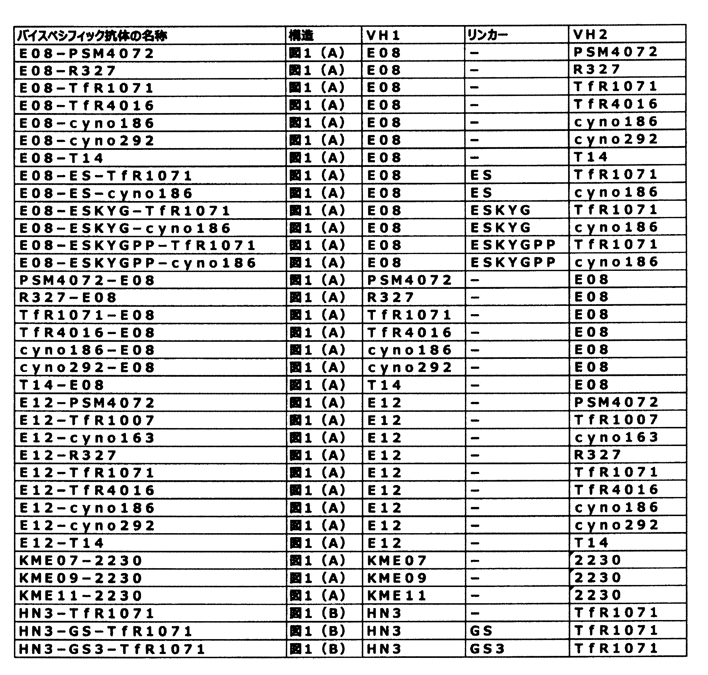
表3に記載されたバイスペシフィック抗体のうち、図1(A)に示す軽鎖共通型バイスペシフィック抗体発現ベクターを以下に記載する方法で作製した。
この二つの遺伝子断片を実施例4または実施例5で取得した抗体の発現ベクターのVH1とIgG部分のCH1の間にサブクローニングして、バイスペシフィック抗体発現ベクターを得た。
表3に記載されたバイスペシフィック抗体のうち、軽鎖非共通型バイスペシフィック抗体について、発現ベクターを以下に記載する方法で作製した。
実施例4から実施例6で抗体発現ベクターにサブクローニングした抗TfRモノクローナル抗体、細胞表面抗原に対するモノクローナル抗体、および両抗原に対する結合部位を有するバイスペシフィック抗体を下記の方法でそれぞれ発現させ、精製した。
実施例7で得られたEGFR-TfRバイスペシフィック抗体のヒトTfRおよびサルTfRならびにヒトEGFRおよびサルEGFRそれぞれに対する結合活性と種交差性を確認することを目的として、実施例1で作製したFLAG-Fc-ヒトTfR、FLAG-Fc-サルTfR、ヒトEGFR-GSTおよびサルEGFR-GSTを用いて、表面プラズモン共鳴法(SPR法)による結合性試験を実施した。測定機器として、BiacoreT100(GEヘルスケア社製)を使用した。

各種がん細胞株における各種抗原発現量を、以下の手順に従いfluoresence activated cell sorting(FACS)法により評価した。評価にはPE標識抗ヒトCD71抗体(BD Pharmingen社)および、AlexaFluor488標識抗EGFR抗体(SantaCruz社)を用いた。
がん細胞株に対する増殖阻害活性を指標として、実施例4で得られたTfRモノクローナル抗体の増殖阻害活性を以下のように評価した。
がん細胞株に対する増殖阻害活性を指標として、実施例5で得られた細胞表面抗原に対する抗体の増殖阻害活性を以下のように評価した。
がん細胞株に対する増殖阻害活性を指標として、実施例6で得られたバイスペシフィック抗体の増殖阻害活性を以下のように評価した。
抗TfR抗体の可変領域をN末端側ポリペプチドに含むバイスペシフィック抗体は、ほとんど増殖阻害活性を示さなかった。
バイスペシフィック抗体のがん細胞に対する増殖阻害活性の作用機序を明らかにするため、EGFR-TfRバイスペシフィック抗体が、がん細胞上のTfRとトランスフェリンの結合を阻害するかフローサイトメーターを用いて以下のように評価した。
バイスペシフィック抗体のがん細胞に対する増殖阻害活性の作用機序を明らかにするため、がん細胞にEGFR-TfRバイスペシフィック抗体を添加したときの、EGFRの下流シグナル、およびTfRタンパク質の量への影響をウェスタンブロッティングにより以下のように評価した。
バイスペシフィック抗体のがん細胞に対する増殖阻害活性の作用機序を明らかにするため、EGFR-TfRバイスペシフィック抗体が細胞膜上のTfR発現量に影響するか以下のように評価した。
EGFR-TfRバイスペシフィック抗体のがん細胞に対する増殖阻害活性に鉄が関係しているか確認するため、以下のように評価した。
EGFRシグナル阻害とTfR阻害、2つの増殖阻害メカニズム間の活性を比較するため、IncuCyte(Sartorius社)を用いて評価した。
バイスペシフィック抗体E08-TfR1071およびE12-TfR1071の正常ヒト骨髄細胞(EGFR陰性、TfR陽性)に対する影響を確認するため、以下のように評価した。
TfR1071のVHのアミノ酸を改変したE12-TfR1071バイスペシフィック抗体の増殖阻害活性を、OE21細胞を用いて実施例12と同様の方法で評価した。まず、アミノ酸改変したEGFR-TfRバイスペシフィック抗体を以下のように作製した。アミノ酸改変(アミノ酸置換とも言う)したバイスペシフィック抗体の発現ベクターは、E12-TfR1071の発現ベクターにおいて、置換前のアミノ酸をコードする塩基配列を、置換後のアミノ酸をコードする塩基配列に置き換えたベクターを、実施例6に記載の方法に準じて作製した。アミノ酸改変したバイスペシフィック抗体の発現および調製は、実施例7に記載の方法に準じて行った。アミノ酸改変したバイスペシフィック抗体としては、以下のものを作製した。
Y32A、Y32F、T33A、T33G、L45A、V48A、V50A、I51A、I51L、N52A-A、N52A-D、V55E、D58A、D61P、Q97A、Q97D、P98A、W99A、W99F、W99H、W99Y、Y100A-A、Y100A-F、V102L、P98Y、P98S、P98D、P98Q、P98E、P98T、P98R、P98G、P98K、P98M、P98V、P98L、P98I、P98W、P98F、P98H
抗TfR抗体1071のエピトープを以下のように同定した。
国際公開第2014/189973号に記載されている配列情報から、ヒトTfRのApicalドメインに結合する抗TfR抗体15G11v5、7A4v15、および16F6v4、ならびにProtease-likeドメインを認識する抗TfR抗体7G7v1を実施例7に記載した方法と同様にして作製した。


本発明のバイスペシフィック抗体の増殖阻害活性とTfRに結合するIgG部分の可変領域のエピトープの関連性を解析するため、エピトープの異なる各種抗TfR抗体でE12-TfRバイスペシフィック抗体を作製し、増殖阻害活性を評価した。

バイスペシフィック抗体の増殖阻害活性にTfR1071が重要であるか確認するため、上市されているEGFR阻害活性のある抗EGFR抗体とTfR抗体TfR1071でEGFR-TfRバイスペシフィック抗体を作製し、増殖阻害活性を評価した。
本発明のバイスペシフィック抗体の増殖阻害活性において、TfRとEGFRに結合する価数の影響を解析するため、抗EGFR抗体E12と抗TfR抗体TfR1071の抗原結合ドメインをそれぞれ一つずつ有するヘテロ二量体型バイスペシフィック抗体(以下ヘテロ二量体抗体と記載する。)を作製し、増殖阻害活性を評価した。図18(B)に、作製したヘテロ二量体抗体の構造の模式図を示す。
TfR1071のVH配列(配列番号31)の1番目のアミノ酸をY(チロシン)からE(グルタミン酸)に置換したVH配列(配列番号106)を有する、E12-TfR1071YEバイスペシフィック抗体を、常法により作製した。実施例20に記載の方法と同様にして、ELISAによりヒトTfRタンパク質への結合活性を解析した。その結果、E12-TfR1071YEはE12-TfR1071と同等の結合活性を示した
配列番号2:ヒトTfR細胞外ドメインのアミノ酸配列
配列番号3:サルTfR細胞外ドメインの塩基配列
配列番号4:サルTfR細胞外ドメインのアミノ酸配列
配列番号5:ヒトTfRの塩基配列
配列番号6:ヒトTfRのアミノ酸配列
配列番号7:カニクイザルTfRの塩基配列
配列番号8:カニクイザルTfRのアミノ酸配列
配列番号9:ヒトEGFR細胞外ドメインの塩基配列(シグナル配列を含む)
配列番号10:ヒトEGFR細胞外ドメインのアミノ酸配列(シグナル配列を含む)
配列番号11:サルEGFR細胞外ドメインの塩基配列(シグナル配列を含む)
配列番号12:サルEGFR細胞外ドメインのアミノ酸配列(シグナル配列を含む)
配列番号13:ヒトEGFRの塩基配列
配列番号14:ヒトEGFRのアミノ酸配列
配列番号15:A27 VLの塩基配列
配列番号16:A27 VLのアミノ酸配列
配列番号17:A27 LCDR1のアミノ酸配列
配列番号18:A27 LCDR2のアミノ酸配列
配列番号19:A27 LCDR3のアミノ酸配列
配列番号20:PSM4072 VHの塩基配列
配列番号21:PSM4072 VHのアミノ酸配列
配列番号22:PSM4072 HCDR1のアミノ酸配列
配列番号23:PSM4072 HCDR2のアミノ酸配列
配列番号24:PSM4072 HCDR3のアミノ酸配列
配列番号25:R327 VHの塩基配列
配列番号26:R327 VHのアミノ酸配列
配列番号27:R327 HCDR1のアミノ酸配列
配列番号28:R327 HCDR2のアミノ酸配列
配列番号29:R327 HCDR3のアミノ酸配列
配列番号30:TfR1071 VHの塩基配列
配列番号31:TfR1071 VHのアミノ酸配列
配列番号32:TfR1071 HCDR1のアミノ酸配列
配列番号33:TfR1071 HCDR2のアミノ酸配列
配列番号34:TfR1071 HCDR3のアミノ酸配列
配列番号35:TfR4016 VHの塩基配列
配列番号36:TfR4016 VHのアミノ酸配列
配列番号37:TfR4016 HCDR1のアミノ酸配列
配列番号38:TfR4016 HCDR2のアミノ酸配列
配列番号39:TfR4016 HCDR3のアミノ酸配列
配列番号40:cyno186 VHの塩基配列
配列番号41:cyno186 VHのアミノ酸配列
配列番号42:cyno186 HCDR1のアミノ酸配列
配列番号43:cyno186 HCDR2のアミノ酸配列
配列番号44:cyno186 HCDR3のアミノ酸配列
配列番号45:cyno292 VHの塩基配列
配列番号46:cyno292 VHのアミノ酸配列
配列番号47:cyno292 HCDR1のアミノ酸配列
配列番号48:cyno292 HCDR2のアミノ酸配列
配列番号49:cyno292 HCDR3のアミノ酸配列
配列番号50:TfR1007 VHのアミノ酸配列
配列番号51:cyno163 VHのアミノ酸配列
配列番号52:2230 VHのアミノ酸配列
配列番号53:2230 VLのアミノ酸配列
配列番号54:T14 VHのアミノ酸配列
配列番号55:TfR434 VHのアミノ酸配列
配列番号56:TfR435 VHのアミノ酸配列
配列番号57:TfR434、435 VLのアミノ酸配列
配列番号58:E08 VHの塩基配列
配列番号59:E08 VHのアミノ酸配列
配列番号60:E08 HCDR1のアミノ酸配列
配列番号61:E08 HCDR2のアミノ酸配列
配列番号62:E08 HCDR3のアミノ酸配列
配列番号63:E12 VHの塩基配列
配列番号64:E12 VHのアミノ酸配列
配列番号65:E12 HCDR1のアミノ酸配列
配列番号66:E12 HCDR2のアミノ酸配列
配列番号67:E12 HCDR3のアミノ酸配列
配列番号68:E17 VHの塩基配列
配列番号69:E17 VHのアミノ酸配列
配列番号70:E17 HCDR1のアミノ酸配列
配列番号71:E17 HCDR2のアミノ酸配列
配列番号72:E17 HCDR3のアミノ酸配列
配列番号73:KME07 VHのアミノ酸配列
配列番号74:KME09 VHのアミノ酸配列
配列番号75:KME11 VHのアミノ酸配列
配列番号76:Cetuximab VHのアミノ酸配列
配列番号77:Cetuximab VLのアミノ酸配列
配列番号78:HN3 VHのアミノ酸配列
配列番号79:リンカーのアミノ酸配列
配列番号80:リンカーのアミノ酸配列
配列番号81:リンカーのアミノ酸配列
配列番号82:リンカーのアミノ酸配列
配列番号83:IgG4PE R409K 定常領域の塩基配列
配列番号84:IgG4PE R409K 定常領域のアミノ酸配列
配列番号85:IgG4 CH1の改変コドンの塩基配列
配列番号86:IgG4PE 定常領域のアミノ酸配列
配列番号87:マウスTfRのアミノ酸配列
配列番号88:ヒトTfR細胞外ドメインのApicalドメインマウスキメラ体のアミノ酸配列
配列番号89:ヒトTfR細胞外ドメインのProtease-likeドメインマウスキメラ体のアミノ酸配列
配列番号90:A01のアミノ酸配列
配列番号91:A02のアミノ酸配列
配列番号92:A03のアミノ酸配列
配列番号93:A04のアミノ酸配列
配列番号94:A05のアミノ酸配列
配列番号95:A06のアミノ酸配列
配列番号96:A07のアミノ酸配列
配列番号97:A08のアミノ酸配列
配列番号98:A09のアミノ酸配列
配列番号99:A10のアミノ酸配列
配列番号100:A11のアミノ酸配列
配列番号101:A12のアミノ酸配列
配列番号102:A13のアミノ酸配列
配列番号103:A14のアミノ酸配列
配列番号104:A15のアミノ酸配列
配列番号105:A16のアミノ酸配列
配列番号106:TfR1071 EVQLで始まるVHのアミノ酸配列
配列番号107:TfR1007 HCDR1のアミノ酸配列
配列番号108:TfR1007 HCDR2のアミノ酸配列
配列番号109:TfR1007 HCDR3のアミノ酸配列
配列番号110:Cetuximab VHのアミノ酸配列
配列番号111:Cetuximab VLのアミノ酸配列
配列番号112:Panitumumab VHのアミノ酸配列
配列番号113:Panitumumab VLのアミノ酸配列
配列番号114:Necitumumab VHのアミノ酸配列
配列番号115:Necitumumab VLのアミノ酸配列
配列番号116:Nimotuzumab VHのアミノ酸配列
配列番号117:Nimotuzumab VLのアミノ酸配列
Claims (32)
- Transferrin Receptor(TfR)に結合するIgG部分、および細胞表面抗原に結合するN末端側ポリペプチドを含むバイスペシフィック抗体または該バイスペシフィック抗体断片であって、該N末端側ポリペプチドが該IgG部分の重鎖のN末端に直接またはリンカーを介して結合する、TfRおよび細胞表面抗原に結合するバイスペシフィック抗体または該バイスペシフィック抗体断片。
- 前記IgG部分が、
配列番号32で表されるアミノ酸配列もしくは配列番号32で表されるアミノ酸配列から、2番目のチロシンのアラニンもしくはフェニルアラニンへの置換および3番目のスレオニンのアラニンもしくはグリシンへの置換から選ばれる少なくとも1つの置換により改変されたアミノ酸配列を含む相補性決定領域(complementarity determining region;CDR)1、
配列番号33で表されるアミノ酸配列もしくは配列番号33で表されるアミノ酸配列から、1番目のバリンのアラニンへの置換、2番目のイソロイシンのアラニンもしくはロイシンへの置換、7番目のバリンのグルタミン酸への置換、10番目のアスパラギン酸のアラニンへの置換および13番目のアスパラギン酸のプロリンへの置換から選ばれる少なくとも一つの置換により改変されたアミノ酸配列を含むCDR2および
配列番号34で表されるアミノ酸配列もしくは配列番号34で表されるアミノ酸配列から、3番目のグルタミンのアラニンもしくはアスパラギン酸への置換、4番目のプロリンの任意の天然アミノ酸への置換、5番目のトリプトファンのアラニン、フェニルアラニン、ヒスチジンもしくはチロシンへの置換、7番目のチロシンのアラニンもしくはフェニルアラニンへの置換および13番目のバリンのロイシンへの置換から選ばれる少なくとも1つの置換により改変されたアミノ酸配列を含むCDR3を含む重鎖可変領域(VH)、ならびに
それぞれ配列番号17~19で表されるアミノ酸配列を含むCDR1~3を含む軽鎖可変領域(VL)を含む、請求項1に記載のバイスペシフィック抗体または該バイスペシフィック抗体断片。 - 前記IgG部分が、以下の(ai)~(ci)から選ばれる1のVH、およびそれぞれ配列番号17~19で表されるアミノ酸配列を含むCDR1~3を含むVLを含む、請求項1に記載のバイスペシフィック抗体または該バイスペシフィック抗体断片。
(ai)それぞれ配列番号32~34で表されるアミノ酸配列を含むCDR1~3を含むVH
(bi)それぞれ配列番号42~44で表されるアミノ酸配列を含むCDR1~3を含むVH
(ci)それぞれ配列番号47~49で表されるアミノ酸配列を含むCDR1~3を含むVH - 前記IgG部分が、配列番号26、31、36、41、および46のいずれか1つで表されるアミノ酸配列を含むVH、および配列番号16で表されるアミノ酸配列を含むVLを含む、請求項1または2に記載のバイスペシフィック抗体または該バイスペシフィック抗体断片。
- 前記IgG部分が、それぞれ配列番号17~19で表されるアミノ酸配列を含むCDR1~3を含むVL、および、以下の(a)~(m)から選ばれる1のVHを含む、請求項1に記載のバイスペシフィック抗体または該バイスペシフィック抗体断片。
(a)それぞれ配列番号33および34で表されるアミノ酸配列を含むCDR2および3、ならびに、配列番号32で表されるアミノ酸配列から、2番目のチロシンのアラニンまたはフェニルアラニンへの置換により改変されたアミノ酸配列を含むCDR1を含むVH
(b)それぞれ配列番号33および34で表されるアミノ酸配列を含むCDR2および3、ならびに、配列番号32で表されるアミノ酸配列から、3番目のスレオニンのアラニンまたはグリシンへの置換により改変されたアミノ酸配列を含むCDR1を含むVH
(c)それぞれ配列番号32および34で表されるアミノ酸配列を含むCDR1および3、ならびに、配列番号33で表されるアミノ酸配列から、1番目のバリンのアラニンへの置換により改変されたアミノ酸配列を含むCDR2を含むVH
(d)それぞれ配列番号32および34で表されるアミノ酸配列を含むCDR1および3、ならびに、配列番号33で表されるアミノ酸配列から、2番目のイソロイシンのアラニンまたはロイシンへの置換により改変されたアミノ酸配列を含むCDR2を含むVH
(e)それぞれ配列番号32および34で表されるアミノ酸配列を含むCDR1および3、ならびに、配列番号33で表されるアミノ酸配列から、7番目のバリンのグルタミン酸への置換により改変されたアミノ酸配列を含むCDR2を含むVH
(f)それぞれ配列番号32および34で表されるアミノ酸配列を含むCDR1および3、ならびに、配列番号33で表されるアミノ酸配列から、10番目のアスパラギン酸のアラニンへの置換により改変されたアミノ酸配列を含むCDR2を含むVH
(g)それぞれ配列番号32および34で表されるアミノ酸配列を含むCDR1および3、ならびに、配列番号33で表されるアミノ酸配列から、13番目のアスパラギン酸のプロリンへの置換により改変されたアミノ酸配列を含むCDR2を含むVH
(h)それぞれ配列番号32および33で表されるアミノ酸配列を含むCDR1および2、ならびに、配列番号34で表されるアミノ酸配列から、3番目のグルタミンのアラニンまたはアスパラギン酸への置換により改変されたアミノ酸配列を含むCDR3を含むVH
(i)それぞれ配列番号32および33で表されるアミノ酸配列を含むCDR1および2、ならびに、配列番号34で表されるアミノ酸配列から、4番目のプロリンの任意の天然アミノ酸への置換により改変されたアミノ酸配列を含むCDR3を含むVH
(j)それぞれ配列番号32および33で表されるアミノ酸配列を含むCDR1および2、ならびに、配列番号34で表されるアミノ酸配列から、4番目のプロリンのアラニン、チロシン、セリン、アスパラギン酸、グルタミン、グルタミン酸、スレオニン、アルギニン、グリシン、リジン、メチオニン、バリン、ロイシン、イソロイシン、トリプトファン、フェニルアラニンもしくはヒスチジンへの置換により改変されたアミノ酸配列を含むCDR3を含むVH
(k)それぞれ配列番号32および33で表されるアミノ酸配列を含むCDR1および2、ならびに、配列番号34で表されるアミノ酸配列から、5番目のトリプトファンのアラニン、フェニルアラニン、ヒスチジンまたはチロシンへの置換により改変されたアミノ酸配列を含むCDR3を含むVH
(l)それぞれ配列番号32および33で表されるアミノ酸配列を含むCDR1および2、ならびに、配列番号34で表されるアミノ酸配列から、7番目のチロシンのアラニンまたはフェニルアラニンへの置換により改変されたアミノ酸配列を含むCDR3を含むVH
(m)それぞれ配列番号32および33で表されるアミノ酸配列を含むCDR1および2、ならびに、配列番号34で表されるアミノ酸配列から、バリンのロイシンへの置換により改変されたアミノ酸配列を含むCDR3を含むVH - 前記IgG部分が、配列番号16で表されるアミノ酸配列を含むVL、ならびに配列番号31で表されるアミノ酸配列から、KabatらによるEUインデックス(以下、EUインデックス)により示されるY32A、Y32F、T33A、T33G、L45A、V48A、V50A、I51A、I51L、V55E、D58A、D61P、Q97A、Q97D、W99A、W99F、W99H、W99Y、Y100AA、Y100AF、V102L、およびP98の任意のアミノ酸への置換から選ばれる少なくとも1つの置換により改変されたアミノ酸配列を含むVHを含む、請求項1に記載のバイスペシフィック抗体または該バイスペシフィック抗体断片。
- 前記IgG部分が、配列番号16で表されるアミノ酸配列を含むVL、ならびに配列番号31で表されるアミノ酸配列から、EUインデックスにより示されるY32A、Y32F、T33A、T33G、L45A、V48A、V50A、I51A、I51L、V55E、D58A、D61P、Q97A、Q97D、W99A、W99F、W99H、W99Y、Y100AA、Y100AF、V102L、P98A、P98Y、P98S、P98D、P98Q、P98E、P98T、P98R、P98G、P98K、P98M、P98V、P98L、P98I、P98W、P98F、およびP98Hから選ばれるいずれか1つの置換により改変されたアミノ酸配列を含むVHを含む、請求項1に記載のバイスペシフィック抗体または該バイスペシフィック抗体断片。
- 前記IgG部分が、配列番号6で表されるTfRのアミノ酸配列の352番目のAsp、355番目のSer、356番目のAsp、および358番目のLysから選ばれる少なくとも1つのアミノ酸残基、並びに、365番目のMet、366番目のVal、および369番目のGluから選ばれる少なくとも1つのアミノ酸残基を認識する、請求項1に記載のバイスペシフィック抗体または該バイスペシフィック抗体断片。
- 前記IgG部分の重鎖定常領域が配列番号84または配列番号86で表されるアミノ酸配列を含む請求項1~8のいずれか1項に記載のバイスペシフィック抗体または該バイスペシフィック抗体断片。
- 前記細胞表面抗原がEGFRまたはGPC3である、請求項1~9のいずれか1項に記載のバイスペシフィック抗体または該バイスペシフィック抗体断片。
- 前記細胞表面抗原がEGFRである、請求項1~10のいずれか1項に記載のバイスペシフィック抗体または該バイスペシフィック抗体断片。
- 前記N末端側ポリペプチドが前記細胞表面抗原に対する抗体のFab、Fab’、scFv、dsFvおよびVHHのいずれか1つである、請求項1~11のいずれか1項に記載のバイスペシフィック抗体または該バイスペシフィック抗体断片。
- 前記N末端側ポリペプチドが、前記細胞表面抗原に対する抗体のFabである、請求項1~12のいずれか1項に記載のバイスペシフィック抗体または該バイスペシフィック抗体断片。
- 前記N末端側ポリペプチドが、EGFRに対する抗体のFabであって、該抗体がそれぞれ配列番号60~62またはそれぞれ配列番号65~67で表されるアミノ酸配列を含むCDR1~3を含むVH、およびそれぞれ配列番号17~19で表されるアミノ酸配列を含むCDR1~3を含むVLを含む抗体である、請求項1~9のいずれか1項に記載のバイスペシフィック抗体または該バイスペシフィック抗体断片。
- 前記EGFRに対する抗体が、以下の(a)~(e)から選ばれる1の抗体である、請求項14に記載のバイスペシフィック抗体または該バイスペシフィック抗体断片。
(a)配列番号59または配列番号64で表されるアミノ酸配列を含むVH、および配列番号16で表されるアミノ酸配列を含むVLを含む抗体
(b)配列番号110で表されるアミノ酸配列を含むVH、および配列番号111で表されるアミノ酸配列を含むVLを含む抗体
(c)配列番号112で表されるアミノ酸配列を含むVH、および配列番号113で表されるアミノ酸配列を含むVLを含む抗体
(d)配列番号114で表されるアミノ酸配列を含むVH、および配列番号115で表されるアミノ酸配列を含むVLを含む抗体
(e)配列番号116で表されるアミノ酸配列を含むVH、および配列番号117で表されるアミノ酸配列を含むVLを含む抗体 - 前記Fabの重鎖C末端が前記IgG部分の重鎖N末端に直接結合する、請求項13~15のいずれか1項に記載のバイスペシフィック抗体または該バイスペシフィック抗体断片。
- 前記Fabの重鎖C末端がIgG部分の重鎖N末端にリンカーを介して結合する、請求項13~15のいずれか1項に記載のバイスペシフィック抗体または該バイスペシフィック抗体断片。
- 上記リンカーのアミノ酸配列がIgGのヒンジ領域のアミノ酸配列の一部または全部からなる、請求項17に記載のバイスペシフィック抗体または該バイスペシフィック抗体断片。
- 前記Fabおよび前記IgG部分が同一のアミノ酸配列からなる軽鎖を含む請求項13~18のいずれか1項に記載のバイスペシフィック抗体または該バイスペシフィック抗体断片。
- 請求項1~19のいずれか1項に記載のバイスペシフィック抗体または該バイスペシフィック抗体断片をコードするDNA。
- 請求項20に記載のDNAを含有する組換え体ベクター。
- 請求項21に記載の組換え体ベクターを宿主細胞に導入して得られる形質転換株。
- 請求項22に記載の形質転換株を培地に培養し、培養物中に請求項1~19のいずれか1項に記載のバイスペシフィック抗体または該バイスペシフィック抗体断片を生産蓄積させ、該培養物からバイスペシフィック抗体または該バイスペシフィック抗体断片を採取することを特徴とする請求項1~19のいずれか1項に記載のバイスペシフィック抗体または該バイスペシフィック抗体断片の製造方法。
- 請求項1~19のいずれか1項に記載のバイスペシフィック抗体または該バイスペシフィック抗体断片を有効成分として含有する、ヒトTfRおよび細胞表面抗原の少なくとも一方が関係する疾患の治療および/または診断薬。
- ヒトTfRおよび細胞表面抗原の少なくとも一方が関係する疾患ががんである請求項24に記載の治療薬および/または診断薬。
- 請求項1~19のいずれか1項に記載のバイスペシフィック抗体または該バイスペシフィック抗体断片を用いる、ヒトTfRおよび細胞表面抗原の少なくとも一方が関係する疾患の治療および/または診断方法。
- ヒトTfRおよび細胞表面抗原の少なくとも一方が関係する疾患ががんである請求項26に記載の方法。
- ヒトTfRおよび細胞表面抗原の少なくとも一方が関係する疾患の治療および/または診断に使用するための、請求項1~19のいずれか1項に記載のバイスペシフィック抗体または該バイスペシフィック抗体断片。
- ヒトTfRおよび細胞表面抗原の少なくとも一方が関係する疾患ががんである請求項28に記載のバイスペシフィック抗体または該バイスペシフィック抗体断片。
- ヒトTfRおよび細胞表面抗原の少なくとも一方が関係する疾患の治療および/または診断剤の製造のための、請求項1~19のいずれか1項に記載のバイスペシフィック抗体または該バイスペシフィック抗体断片の使用。
- ヒトTfRおよび細胞表面抗原の少なくとも一方が関係する疾患ががんである請求項30に記載の使用。
- 請求項1~19のいずれか1項に記載のバイスペシフィック抗体または該バイスペシフィック抗体断片を含む、ヒトTfRおよび細胞表面抗原の少なくとも一方を検出または測定するための試薬。
Priority Applications (14)
| Application Number | Priority Date | Filing Date | Title |
|---|---|---|---|
| MX2021007797A MX2021007797A (es) | 2018-12-28 | 2019-12-27 | Anticuerpo biespecifico que se une al receptor de transferrina (tfr). |
| NZ777568A NZ777568B2 (en) | 2019-12-27 | BISPECIFIC ANTIBODY BINDING TO TfR | |
| SG11202106990PA SG11202106990PA (en) | 2018-12-28 | 2019-12-27 | BISPECIFIC ANTIBODY BINDING TO TfR |
| EP19901837.5A EP3903816A4 (en) | 2018-12-28 | 2019-12-27 | BISPECIFIC ANTIBODY BINDING TO TFR |
| AU2019413278A AU2019413278B2 (en) | 2018-12-28 | 2019-12-27 | Bispecific antibody binding to TfR |
| BR112021012569-8A BR112021012569A2 (pt) | 2018-12-28 | 2019-12-27 | Anticorpo biespecífico de ligação ao tfr |
| MYPI2021003612A MY210422A (en) | 2018-12-28 | 2019-12-27 | Bispecific antibody binding to tfr |
| CN201980086662.6A CN113226371B (zh) | 2018-12-28 | 2019-12-27 | 与TfR结合的双特异性抗体 |
| JP2020562545A JP7506607B2 (ja) | 2018-12-28 | 2019-12-27 | TfRに結合するバイスペシフィック抗体 |
| KR1020217019406A KR20210108961A (ko) | 2018-12-28 | 2019-12-27 | TfR에 결합하는 이중 특이적 항체 |
| US17/418,447 US12258413B2 (en) | 2018-12-28 | 2019-12-27 | Bispecific antibody binding to TfR |
| CA3125316A CA3125316A1 (en) | 2018-12-28 | 2019-12-27 | Bispecific antibody binding to tfr |
| JP2024096802A JP2024119984A (ja) | 2018-12-28 | 2024-06-14 | TfRに結合するバイスペシフィック抗体 |
| US19/057,265 US20250250352A1 (en) | 2018-12-28 | 2025-02-19 | BISPECIFIC ANTIBODY BINDING TO TfR |
Applications Claiming Priority (2)
| Application Number | Priority Date | Filing Date | Title |
|---|---|---|---|
| JP2018-248334 | 2018-12-28 | ||
| JP2018248334 | 2018-12-28 |
Related Child Applications (2)
| Application Number | Title | Priority Date | Filing Date |
|---|---|---|---|
| US17/418,447 A-371-Of-International US12258413B2 (en) | 2018-12-28 | 2019-12-27 | Bispecific antibody binding to TfR |
| US19/057,265 Division US20250250352A1 (en) | 2018-12-28 | 2025-02-19 | BISPECIFIC ANTIBODY BINDING TO TfR |
Publications (1)
| Publication Number | Publication Date |
|---|---|
| WO2020138487A1 true WO2020138487A1 (ja) | 2020-07-02 |
Family
ID=71125764
Family Applications (1)
| Application Number | Title | Priority Date | Filing Date |
|---|---|---|---|
| PCT/JP2019/051594 Ceased WO2020138487A1 (ja) | 2018-12-28 | 2019-12-27 | TfRに結合するバイスペシフィック抗体 |
Country Status (13)
| Country | Link |
|---|---|
| US (2) | US12258413B2 (ja) |
| EP (1) | EP3903816A4 (ja) |
| JP (2) | JP7506607B2 (ja) |
| KR (1) | KR20210108961A (ja) |
| CN (1) | CN113226371B (ja) |
| AU (1) | AU2019413278B2 (ja) |
| BR (1) | BR112021012569A2 (ja) |
| CA (1) | CA3125316A1 (ja) |
| MX (1) | MX2021007797A (ja) |
| MY (1) | MY210422A (ja) |
| SG (1) | SG11202106990PA (ja) |
| TW (2) | TWI907249B (ja) |
| WO (1) | WO2020138487A1 (ja) |
Cited By (1)
| Publication number | Priority date | Publication date | Assignee | Title |
|---|---|---|---|---|
| US20220259330A1 (en) * | 2020-04-29 | 2022-08-18 | Sunshine Guojian Pharmaceutical (Shanghai) Co., Ltd. | Tetravalent bispecific antibody, preparation method therefor, and use thereof |
Families Citing this family (4)
| Publication number | Priority date | Publication date | Assignee | Title |
|---|---|---|---|---|
| WO2023083381A1 (zh) | 2021-11-15 | 2023-05-19 | 成都百利多特生物药业有限责任公司 | 双特异性抗体-喜树碱类药物偶联物及其医药用途 |
| KR20250162888A (ko) * | 2023-03-24 | 2025-11-19 | 바이오악틱 에이비 | 인간 트랜스페린 수용체 htfr1의 프로테아제-유사 도메인에 결합하는 이중특이적 항체 |
| AU2024299328A1 (en) | 2023-07-21 | 2026-01-22 | Marrow Therapeutics, Inc. | Hematopoietic cell targeting conjugates and related methods |
| TW202517685A (zh) * | 2023-10-26 | 2025-05-01 | 美商百健Ma公司 | 抗運鐵蛋白受體抗體及其用途 |
Citations (47)
| Publication number | Priority date | Publication date | Assignee | Title |
|---|---|---|---|---|
| US570561A (en) | 1896-11-03 | S peters co | ||
| JPS5623587A (en) | 1979-08-03 | 1981-03-05 | Mitsuwa Seiki Co Ltd | Vane type compressor |
| JPS58110600A (ja) | 1981-12-25 | 1983-07-01 | Kyowa Hakko Kogyo Co Ltd | ヒトβ型インタ−フエロン遺伝子を含む組みかえ体プラスミド |
| JPS60221091A (ja) | 1983-12-21 | 1985-11-05 | Kyowa Hakko Kogyo Co Ltd | 新規プロモ−タ− |
| JPS61178926A (ja) | 1984-03-06 | 1986-08-11 | Takeda Chem Ind Ltd | 化学修飾ペプチドホルモンおよびその製造法 |
| JPS63299A (ja) | 1986-04-22 | 1988-01-05 | イミユネツクス・コ−ポレ−シヨン | ヒトg−csfタンパク質の発現 |
| JPH02117920A (ja) | 1988-05-06 | 1990-05-02 | Sumitomo Pharmaceut Co Ltd | ポリエチレングリコール誘導体、修飾ペプチドおよびその製造方法 |
| US4939094A (en) | 1985-08-28 | 1990-07-03 | Kyowa Hakko Kogyo Co., Ltd. | Fused antigen polypeptide |
| JPH02227075A (ja) | 1988-09-29 | 1990-09-10 | Kyowa Hakko Kogyo Co Ltd | 新規ポリペプチド |
| JPH02257891A (ja) | 1989-03-31 | 1990-10-18 | Kyowa Hakko Kogyo Co Ltd | 組換え動物細胞による蛋白質の製造 |
| JPH0322979A (ja) | 1989-06-19 | 1991-01-31 | Kyowa Hakko Kogyo Co Ltd | 新規プラスミノーゲン活性化因子 |
| US5168062A (en) | 1985-01-30 | 1992-12-01 | University Of Iowa Research Foundation | Transfer vectors and microorganisms containing human cytomegalovirus immediate-early promoter-regulatory DNA sequence |
| JPH05336963A (ja) | 1991-12-17 | 1993-12-21 | Kyowa Hakko Kogyo Co Ltd | 新規α2→3シアリルトランスフェラーゼ |
| WO1994023021A1 (fr) | 1993-03-29 | 1994-10-13 | Kyowa Hakko Kogyo Co., Ltd. | α-1,3-FUCOSYLTRANSFERASE |
| WO1996040210A1 (en) | 1995-06-07 | 1996-12-19 | Imclone Systems Incorporated | Antibody and antibody fragments for inhibiting the growth of tumors |
| WO1997010354A1 (fr) | 1995-09-11 | 1997-03-20 | Kyowa Hakko Kogyo Co., Ltd. | Anticorps de la chaine alpha du recepteur de l'interleukine 5 humaine |
| WO1998050431A2 (en) | 1997-05-02 | 1998-11-12 | Genentech, Inc. | A method for making multispecific antibodies having heteromultimeric and common components |
| WO1998050433A2 (en) | 1997-05-05 | 1998-11-12 | Abgenix, Inc. | Human monoclonal antibodies to epidermal growth factor receptor |
| US6001358A (en) | 1995-11-07 | 1999-12-14 | Idec Pharmaceuticals Corporation | Humanized antibodies to human gp39, compositions containing thereof |
| WO2000042072A2 (en) | 1999-01-15 | 2000-07-20 | Genentech, Inc. | Polypeptide variants with altered effector function |
| WO2000061739A1 (fr) | 1999-04-09 | 2000-10-19 | Kyowa Hakko Kogyo Co., Ltd. | Methode de regulation de l'activite d'une molecule immunologiquement fonctionnelle |
| WO2002002773A2 (en) | 2000-06-29 | 2002-01-10 | Abbott Laboratories | Dual specificity antibodies and methods of making and using |
| WO2002031140A1 (en) | 2000-10-06 | 2002-04-18 | Kyowa Hakko Kogyo Co., Ltd. | Cells producing antibody compositions |
| US6423511B1 (en) | 1992-09-07 | 2002-07-23 | Kyowa Hakko Kogyo Co., Ltd. | Humanized antibodies |
| WO2003033538A1 (en) | 2001-10-15 | 2003-04-24 | Kirin Beer Kabushiki Kaisha | Anti-hla-dr antibody |
| US6737056B1 (en) | 1999-01-15 | 2004-05-18 | Genentech, Inc. | Polypeptide variants with altered effector function |
| WO2005035586A1 (ja) | 2003-10-08 | 2005-04-21 | Kyowa Hakko Kogyo Co., Ltd. | 融合蛋白質組成物 |
| WO2006033386A1 (ja) | 2004-09-22 | 2006-03-30 | Kirin Beer Kabushiki Kaisha | 安定化されたヒトIgG4抗体 |
| US20070071675A1 (en) | 2005-08-19 | 2007-03-29 | Chengbin Wu | Dual variable domain immunoglobulin and uses thereof |
| US20070148165A1 (en) | 2005-07-22 | 2007-06-28 | Kyowa Hakko Kogyo Co., Ltd. | Recombinant antibody composition |
| US7297775B2 (en) | 1998-04-02 | 2007-11-20 | Genentech, Inc. | Polypeptide variants |
| US7317091B2 (en) | 2002-03-01 | 2008-01-08 | Xencor, Inc. | Optimized Fc variants |
| WO2009131239A1 (ja) | 2008-04-25 | 2009-10-29 | Kyowa Hakko Kirin Co Ltd | 安定な多価抗体 |
| WO2010143698A1 (ja) | 2009-06-11 | 2010-12-16 | 大学共同利用機関法人情報・システム研究機構 | タンパク質の生産方法 |
| WO2011116387A1 (en) | 2010-03-19 | 2011-09-22 | Tetragenetics, Inc. | Production of aglycosylated monoclonal antibodies in ciliates |
| WO2012145469A1 (en) | 2011-04-19 | 2012-10-26 | The United States Of America, As Represented By The Secretary, Department Of Health & Human Services | Human monoclonal antibodies specific for glypican-3 and use thereof |
| WO2012153707A1 (ja) | 2011-05-09 | 2012-11-15 | 株式会社ペルセウスプロテオミクス | トランスフェリン受容体を特異的に認識できる抗体 |
| US20120308576A1 (en) | 2011-06-03 | 2012-12-06 | Merrimack Pharmaceuticals, Inc. | Monoclonal and oligoclonal anti-egfr antibodies for use in the treatment of tumors expressing predominantly high affinity egfr ligands or tumors expressing predominantly low affinity egfr ligands |
| JP2014500886A (ja) * | 2010-11-30 | 2014-01-16 | ジェネンテック, インコーポレイテッド | 低親和性血液脳関門受容体抗体及びその使用 |
| WO2014189973A2 (en) | 2013-05-20 | 2014-11-27 | Genentech, Inc. | Anti-transferrin receptor antibodies and methods of use |
| US20140348839A1 (en) | 2011-12-20 | 2014-11-27 | Medimmune, Llc | Modified polypeptides for bispecific antibody scaffolds |
| EP2808035A1 (en) | 2013-05-29 | 2014-12-03 | Samsung Electronics Co., Ltd | Composition for target membrane protein depletion |
| JP2015523336A (ja) * | 2012-05-21 | 2015-08-13 | ジェネンテック, インコーポレイテッド | 血液脳関門輸送の安全性を改善するための方法 |
| WO2016111344A1 (ja) * | 2015-01-08 | 2016-07-14 | 協和発酵キリン株式会社 | Trailr2とpsmaに結合するバイスペシフィック抗体 |
| JP2018503357A (ja) * | 2014-11-19 | 2018-02-08 | ジェネンテック, インコーポレイテッド | 抗トランスフェリン受容体抗体及びその使用方法 |
| JP2018520143A (ja) * | 2015-06-24 | 2018-07-26 | エフ.ホフマン−ラ ロシュ アーゲーF. Hoffmann−La Roche Aktiengesellschaft | 目的に合わせた親和性を有する抗トランスフェリンレセプター抗体 |
| WO2020017734A1 (ko) | 2018-07-16 | 2020-01-23 | 주식회사 인프라웨어테크놀러지 | 운동 처방 정보를 제공하는 서버 및 컴퓨터 프로그램 |
Family Cites Families (12)
| Publication number | Priority date | Publication date | Assignee | Title |
|---|---|---|---|---|
| US2808035A (en) | 1953-04-23 | 1957-10-01 | Riley Stoker Corp | Burning wet materials |
| US5705614A (en) | 1993-04-09 | 1998-01-06 | Chiron Corporation | Methods of producing antigen forks |
| JP2003531588A (ja) | 2000-04-11 | 2003-10-28 | ジェネンテック・インコーポレーテッド | 多価抗体とその用途 |
| DK2990420T3 (en) * | 2000-05-26 | 2017-04-03 | Immunex Corp | USE OF INTERLEUKIN-4 RECEPTOR ANTIBODIES AND COMPOSITIONS THEREOF |
| CA2742969A1 (en) * | 2008-11-07 | 2010-05-14 | Fabrus Llc | Anti-dll4 antibodies and uses thereof |
| KR20120024763A (ko) * | 2009-05-15 | 2012-03-14 | 추가이 세이야쿠 가부시키가이샤 | 항axl 항체 |
| EP2825553B1 (en) | 2012-03-14 | 2018-07-25 | Regeneron Pharmaceuticals, Inc. | Multispecific antigen-binding molecules and uses thereof |
| DK3078744T3 (da) * | 2013-12-04 | 2020-09-28 | Chugai Pharmaceutical Co Ltd | Antigen-bindende molekyler, antigen-bindingsaktiviteten af hvilke varierer i henhold til koncentrationen af forbindelser, og biblioteker af molekylerne |
| SG11201608253XA (en) | 2014-04-04 | 2016-10-28 | Kyowa Hakko Kirin Co Ltd | Anti-death receptor 3 (dr3) antagonistic antibodies with reduced agonistic activity |
| WO2016081640A1 (en) * | 2014-11-19 | 2016-05-26 | Genentech, Inc. | Anti-transferrin receptor / anti-bace1 multispecific antibodies and methods of use |
| KR20180030030A (ko) * | 2015-07-15 | 2018-03-21 | 교와 핫꼬 기린 가부시키가이샤 | 인간 crth2에 특이적으로 결합하는 항체 |
| CA3023667A1 (en) * | 2016-05-09 | 2017-11-16 | Annexon, Inc. | Compositions and methods for treating spinal muscular atrophy |
-
2019
- 2019-12-27 WO PCT/JP2019/051594 patent/WO2020138487A1/ja not_active Ceased
- 2019-12-27 CA CA3125316A patent/CA3125316A1/en active Pending
- 2019-12-27 MY MYPI2021003612A patent/MY210422A/en unknown
- 2019-12-27 CN CN201980086662.6A patent/CN113226371B/zh active Active
- 2019-12-27 US US17/418,447 patent/US12258413B2/en active Active
- 2019-12-27 BR BR112021012569-8A patent/BR112021012569A2/pt unknown
- 2019-12-27 JP JP2020562545A patent/JP7506607B2/ja active Active
- 2019-12-27 SG SG11202106990PA patent/SG11202106990PA/en unknown
- 2019-12-27 MX MX2021007797A patent/MX2021007797A/es unknown
- 2019-12-27 KR KR1020217019406A patent/KR20210108961A/ko active Pending
- 2019-12-27 EP EP19901837.5A patent/EP3903816A4/en active Pending
- 2019-12-27 AU AU2019413278A patent/AU2019413278B2/en active Active
- 2019-12-27 TW TW114101537A patent/TWI907249B/zh active
- 2019-12-27 TW TW108148133A patent/TWI873112B/zh active
-
2024
- 2024-06-14 JP JP2024096802A patent/JP2024119984A/ja active Pending
-
2025
- 2025-02-19 US US19/057,265 patent/US20250250352A1/en active Pending
Patent Citations (50)
| Publication number | Priority date | Publication date | Assignee | Title |
|---|---|---|---|---|
| US570561A (en) | 1896-11-03 | S peters co | ||
| JPS5623587A (en) | 1979-08-03 | 1981-03-05 | Mitsuwa Seiki Co Ltd | Vane type compressor |
| JPS58110600A (ja) | 1981-12-25 | 1983-07-01 | Kyowa Hakko Kogyo Co Ltd | ヒトβ型インタ−フエロン遺伝子を含む組みかえ体プラスミド |
| US4686191A (en) | 1981-12-25 | 1987-08-11 | Hakko Kogyo Co., Ltd. Kyowa | Recombinant plasmid containing human interferon-beta gene |
| JPS60221091A (ja) | 1983-12-21 | 1985-11-05 | Kyowa Hakko Kogyo Co Ltd | 新規プロモ−タ− |
| JPS61178926A (ja) | 1984-03-06 | 1986-08-11 | Takeda Chem Ind Ltd | 化学修飾ペプチドホルモンおよびその製造法 |
| US5168062A (en) | 1985-01-30 | 1992-12-01 | University Of Iowa Research Foundation | Transfer vectors and microorganisms containing human cytomegalovirus immediate-early promoter-regulatory DNA sequence |
| US4939094A (en) | 1985-08-28 | 1990-07-03 | Kyowa Hakko Kogyo Co., Ltd. | Fused antigen polypeptide |
| JPS63299A (ja) | 1986-04-22 | 1988-01-05 | イミユネツクス・コ−ポレ−シヨン | ヒトg−csfタンパク質の発現 |
| JPH02117920A (ja) | 1988-05-06 | 1990-05-02 | Sumitomo Pharmaceut Co Ltd | ポリエチレングリコール誘導体、修飾ペプチドおよびその製造方法 |
| JPH02227075A (ja) | 1988-09-29 | 1990-09-10 | Kyowa Hakko Kogyo Co Ltd | 新規ポリペプチド |
| JPH02257891A (ja) | 1989-03-31 | 1990-10-18 | Kyowa Hakko Kogyo Co Ltd | 組換え動物細胞による蛋白質の製造 |
| US5160735A (en) | 1989-06-19 | 1992-11-03 | Kyowa Hakko Kogyo Co. Ltd. | Plasminogen activator |
| JPH0322979A (ja) | 1989-06-19 | 1991-01-31 | Kyowa Hakko Kogyo Co Ltd | 新規プラスミノーゲン活性化因子 |
| JPH05336963A (ja) | 1991-12-17 | 1993-12-21 | Kyowa Hakko Kogyo Co Ltd | 新規α2→3シアリルトランスフェラーゼ |
| US6423511B1 (en) | 1992-09-07 | 2002-07-23 | Kyowa Hakko Kogyo Co., Ltd. | Humanized antibodies |
| WO1994023021A1 (fr) | 1993-03-29 | 1994-10-13 | Kyowa Hakko Kogyo Co., Ltd. | α-1,3-FUCOSYLTRANSFERASE |
| WO1996040210A1 (en) | 1995-06-07 | 1996-12-19 | Imclone Systems Incorporated | Antibody and antibody fragments for inhibiting the growth of tumors |
| WO1997010354A1 (fr) | 1995-09-11 | 1997-03-20 | Kyowa Hakko Kogyo Co., Ltd. | Anticorps de la chaine alpha du recepteur de l'interleukine 5 humaine |
| US6001358A (en) | 1995-11-07 | 1999-12-14 | Idec Pharmaceuticals Corporation | Humanized antibodies to human gp39, compositions containing thereof |
| WO1998050431A2 (en) | 1997-05-02 | 1998-11-12 | Genentech, Inc. | A method for making multispecific antibodies having heteromultimeric and common components |
| WO1998050433A2 (en) | 1997-05-05 | 1998-11-12 | Abgenix, Inc. | Human monoclonal antibodies to epidermal growth factor receptor |
| US7297775B2 (en) | 1998-04-02 | 2007-11-20 | Genentech, Inc. | Polypeptide variants |
| WO2000042072A2 (en) | 1999-01-15 | 2000-07-20 | Genentech, Inc. | Polypeptide variants with altered effector function |
| US6737056B1 (en) | 1999-01-15 | 2004-05-18 | Genentech, Inc. | Polypeptide variants with altered effector function |
| WO2000061739A1 (fr) | 1999-04-09 | 2000-10-19 | Kyowa Hakko Kogyo Co., Ltd. | Methode de regulation de l'activite d'une molecule immunologiquement fonctionnelle |
| WO2002002773A2 (en) | 2000-06-29 | 2002-01-10 | Abbott Laboratories | Dual specificity antibodies and methods of making and using |
| WO2002031140A1 (en) | 2000-10-06 | 2002-04-18 | Kyowa Hakko Kogyo Co., Ltd. | Cells producing antibody compositions |
| WO2003033538A1 (en) | 2001-10-15 | 2003-04-24 | Kirin Beer Kabushiki Kaisha | Anti-hla-dr antibody |
| US7317091B2 (en) | 2002-03-01 | 2008-01-08 | Xencor, Inc. | Optimized Fc variants |
| WO2005035586A1 (ja) | 2003-10-08 | 2005-04-21 | Kyowa Hakko Kogyo Co., Ltd. | 融合蛋白質組成物 |
| WO2006033386A1 (ja) | 2004-09-22 | 2006-03-30 | Kirin Beer Kabushiki Kaisha | 安定化されたヒトIgG4抗体 |
| US20070148165A1 (en) | 2005-07-22 | 2007-06-28 | Kyowa Hakko Kogyo Co., Ltd. | Recombinant antibody composition |
| US20070071675A1 (en) | 2005-08-19 | 2007-03-29 | Chengbin Wu | Dual variable domain immunoglobulin and uses thereof |
| WO2009131239A1 (ja) | 2008-04-25 | 2009-10-29 | Kyowa Hakko Kirin Co Ltd | 安定な多価抗体 |
| WO2010143698A1 (ja) | 2009-06-11 | 2010-12-16 | 大学共同利用機関法人情報・システム研究機構 | タンパク質の生産方法 |
| WO2011116387A1 (en) | 2010-03-19 | 2011-09-22 | Tetragenetics, Inc. | Production of aglycosylated monoclonal antibodies in ciliates |
| JP2014500886A (ja) * | 2010-11-30 | 2014-01-16 | ジェネンテック, インコーポレイテッド | 低親和性血液脳関門受容体抗体及びその使用 |
| WO2012145469A1 (en) | 2011-04-19 | 2012-10-26 | The United States Of America, As Represented By The Secretary, Department Of Health & Human Services | Human monoclonal antibodies specific for glypican-3 and use thereof |
| WO2012153707A1 (ja) | 2011-05-09 | 2012-11-15 | 株式会社ペルセウスプロテオミクス | トランスフェリン受容体を特異的に認識できる抗体 |
| US20120308576A1 (en) | 2011-06-03 | 2012-12-06 | Merrimack Pharmaceuticals, Inc. | Monoclonal and oligoclonal anti-egfr antibodies for use in the treatment of tumors expressing predominantly high affinity egfr ligands or tumors expressing predominantly low affinity egfr ligands |
| US20140348839A1 (en) | 2011-12-20 | 2014-11-27 | Medimmune, Llc | Modified polypeptides for bispecific antibody scaffolds |
| JP2015523336A (ja) * | 2012-05-21 | 2015-08-13 | ジェネンテック, インコーポレイテッド | 血液脳関門輸送の安全性を改善するための方法 |
| WO2014189973A2 (en) | 2013-05-20 | 2014-11-27 | Genentech, Inc. | Anti-transferrin receptor antibodies and methods of use |
| JP2016526878A (ja) * | 2013-05-20 | 2016-09-08 | ジェネンテック, インコーポレイテッド | 抗トランスフェリン受容体抗体及び使用方法 |
| EP2808035A1 (en) | 2013-05-29 | 2014-12-03 | Samsung Electronics Co., Ltd | Composition for target membrane protein depletion |
| JP2018503357A (ja) * | 2014-11-19 | 2018-02-08 | ジェネンテック, インコーポレイテッド | 抗トランスフェリン受容体抗体及びその使用方法 |
| WO2016111344A1 (ja) * | 2015-01-08 | 2016-07-14 | 協和発酵キリン株式会社 | Trailr2とpsmaに結合するバイスペシフィック抗体 |
| JP2018520143A (ja) * | 2015-06-24 | 2018-07-26 | エフ.ホフマン−ラ ロシュ アーゲーF. Hoffmann−La Roche Aktiengesellschaft | 目的に合わせた親和性を有する抗トランスフェリンレセプター抗体 |
| WO2020017734A1 (ko) | 2018-07-16 | 2020-01-23 | 주식회사 인프라웨어테크놀러지 | 운동 처방 정보를 제공하는 서버 및 컴퓨터 프로그램 |
Non-Patent Citations (90)
| Title |
|---|
| "A Practical Approach", 1995, OXFORD UNIVERSITY, article "DNA Cloning 1: Core Techniques" |
| "Antibodies - A Laboratory Manual", 1988, COLD SPRING HARBOR LABORATORY |
| "Bioluminescence and Chemiluminescence Clinical Test 42", 1998, KANEHARA & CO., LTD. |
| "Clinical oncology", JAPANESE JOURNAL OF CANCER AND CHEMOTHERAPY, 1996 |
| "enzyme immunoassay method", 1987, IGAKU-SHOIN LTD. |
| "Fluorescent Antibody Method", 1983, SOFT SCIENCE, INC. |
| "GenBank", Database accession no. XP-005 549616. 1 |
| "Methods in Nucleic Acids Res.", 1991, CRC PRESS |
| "Molecular Cloning, A Laboratory Manual", 1989, COLD SPRING HARBOR LABORATORY PRESS, article "PCR method" |
| "Monoclonal Antibodies-Principles and practice", 1996, ACADEMIC PRESS |
| AGRIC. BIOL. CHEM., vol. 53, 1989, pages 277 |
| AGRICULTURAL BIOLOGICAL CHEMISTRY, vol. 48, 1984, pages 669 |
| ANNU. REV. IMMUNOL., vol. 9, 1991, pages 457 - 492 |
| BECK ET AL., ANALYTICAL CHEMISTRY, vol. 85, 2013, pages 715 - 736 |
| BIO/TECHNOLOGY, vol. 9, 1991, pages 266 |
| BIOCHEM. BIOPHYS. RES. COMMUN., vol. 149, 1987, pages 960 |
| CANCER IMMUNOL. IMMUNOTHER., vol. 36, 1993, pages 373 |
| CELL, vol. 33, 1983, pages 717 |
| CELL, vol. 41, 1985, pages 479 |
| CURRENT TOPICS IN MICROBIOLOGY AND IMMUNOLOGY, vol. 18, 1978, pages 1 |
| CYTOTECHNOL., vol. 13, 1993, pages 79 |
| CYTOTECHNOL., vol. 4, 1990, pages 173 |
| CYTOTECHNOLOGY, vol. 3, 1990, pages 133 |
| DNA CLONING: A PRACTICAL APPROACH, vol. 1, 1985, pages 49 |
| EBNER R ET AL., CELL REGUL., vol. 2, 1991, pages 599 - 612 |
| EMMANUELLE LAFFY, HUMAN ANTIBODIES, vol. 14, 2005, pages 33 - 55 |
| ERBITUX US PACKAGE INSERT, June 2018 (2018-06-01) |
| EUROPEAN J. IMMUNOLOGY, vol. 6, 1976, pages 511 |
| GENE, vol. 17, 1982, pages 107 |
| GENE, vol. 27, 1984, pages 223 |
| GENE, vol. 38, 1985, pages 275 |
| GENES DEVELOP., vol. 4, 1990, pages 1288 |
| GENETICS, vol. 39, 1954, pages 440 |
| GENOME RESEARCH, vol. 7, 1997, pages 649, Retrieved from the Internet <URL:http://www.ncbi.nlm.nih.gov/Education/BLASTinfo/infonnation3.html> |
| HAMILTON TA ET AL., PNAS USA, vol. 76, 1979, pages 6406 - 10 |
| HENDLER FJ ET AL., J CLIN INVEST., vol. 74, 1984, pages 647 - 51 |
| HYBRIDOMA, vol. 17, 1998, pages 559 |
| J. BACTERIOL., vol. 172, 1990, pages 2392 |
| J. BIOCHEM., vol. 101, 1987, pages 1307 |
| J. BIOCHEMISTRY, vol. 101, 1987, pages 1307 |
| J. IMMUNOL. METHODS, vol. 167, 1994, pages 271 |
| J. IMMUNOLOGY, vol. 123, 1979, pages 1548 |
| J. MOL. BIOL., vol. 112, 1977, pages 535 |
| J. MOL. BIOL., vol. 16, 1966, pages 118 |
| J. MOL. BIOL., vol. 166, 1983, pages 1 |
| J. MOL. BIOL., vol. 215, 1990, pages 403 |
| KRANZ, J. HEMATOTHER., vol. 5, 1995, pages 403 - 408 |
| LARSON SM ET AL., J NATL CANCER INST., vol. 64, 1980, pages 41 - 53 |
| LIBERMANN TA ET AL., CANCER RES., vol. 44, 1984, pages 753 - 60 |
| LIU AP ET AL., J CELL BIOL., vol. 191, 2010, pages 1381 - 93 |
| LOWE ET AL., PROC. NATL. ACAD. SCI., vol. 86, 1989, pages 8227 |
| METHODS IN ENZYMOL., vol. 154, 1987, pages 3 |
| MOL IMMUNOL., vol. 33, no. 9, June 1996 (1996-06-01), pages 759 - 68 |
| MOL. CELL. BIOL., vol. 3, 1983, pages 280 |
| MOLECULAR & GENERAL GENETICS, vol. 168, 1979, pages 111 |
| NATURE PROTOCOLS, vol. 9, 2014, pages 2450 - 2463 |
| NATURE, vol. 227, 1970, pages 680 |
| NATURE, vol. 256, 1975, pages 495 |
| NATURE, vol. 276, 1978, pages 269 |
| NATURE, vol. 329, 1987, pages 840 |
| NUCLEIC ACIDS RESEARCH, vol. 10, 1982, pages 6487 |
| NUCLEIC ACIDS RESEARCH, vol. 13, 1985, pages 4431 |
| NUCLEIC ACIDS RESEARCH, vol. 17, 1989, pages 9494 |
| NUCLEIC ACIDS RESEARCH, vol. 25, 1997, pages 3389 |
| P. J. DELVES: "ANTIBODY PRODUCTION ESSENTIAL TECHNIQUES", 1997, WILEY |
| P. SHEPHERDC. DEAN: "Monoclonal Antibodies", 2000, OXFORD UNIVERSITY PRESS |
| PAULSON ET AL., J. BIOL. CHEM., vol. 264, 1989, pages 17619 |
| PROC. NATL. ACAD. SCI USA, vol. 82, 1985, pages 488 |
| PROC. NATL. ACAD. SCI. USA, vol. 69, 1972, pages 2110 |
| PROC. NATL. ACAD. SCI. USA, vol. 74, 1977, pages 5463 |
| PROC. NATL. ACAD. SCI. USA, vol. 77, 1980, pages 4216 |
| PROC. NATL. ACAD. SCI. USA, vol. 78, 1981, pages 1527 |
| PROC. NATL. ACAD. SCI. USA, vol. 79, 1982, pages 6409 |
| PROC. NATL. ACAD. SCI. USA, vol. 82, 1985, pages 4306 |
| PROC. NATL. ACAD. SCI. USA, vol. 84, 1987, pages 7413 |
| PROC. NATL. ACAD. SCI., vol. 79, 1982, pages 6409 |
| PROC. SOC. EXP. BIOL. MED., vol. 73, 1950, pages 1 |
| PROCEEDING OF THE NATIONAL ACADEMY OF SCIENCES IN USA, vol. 82, 1985, pages 488 |
| PROTEIN ENGINEERING, vol. 7, 1994, pages 1501 |
| SCIENCE, vol. 122, 1952, pages 501 |
| SCIENCE, vol. 222, 1983, pages 778 |
| See also references of EP3903816A4 |
| SOMATIC CELL AND MOLECULAR GENETICS, vol. 12, 1986, pages 55 |
| STRATEGIES, vol. 5, 1992, pages 81 |
| SURESH, METHODS ENZYMOL., vol. 121, 1986, pages 210 - 228 |
| THE JOURNAL OF THE AMERICAN MEDICAL ASSOCIATION, vol. 199, 1967, pages 519 |
| TOMIZUKA ET AL., PROC NATL ACAD SCI USA., vol. 97, 2000, pages 722 |
| VIROLOGY, vol. 8, 1959, pages 396 |
| WEISSMAN AM, J CELL BIOL., vol. 102, 1986, pages 951 - 8 |
| YAMASHIRO DJ ET AL., CELL, vol. 37, 1984, pages 789 - 800 |
Cited By (2)
| Publication number | Priority date | Publication date | Assignee | Title |
|---|---|---|---|---|
| US20220259330A1 (en) * | 2020-04-29 | 2022-08-18 | Sunshine Guojian Pharmaceutical (Shanghai) Co., Ltd. | Tetravalent bispecific antibody, preparation method therefor, and use thereof |
| JP2022540319A (ja) * | 2020-04-29 | 2022-09-15 | 三生国健薬業(上海)股▲ふん▼有限公司 | 四価二重特異性抗体、その製造方法および用途 |
Also Published As
| Publication number | Publication date |
|---|---|
| CA3125316A1 (en) | 2020-07-02 |
| SG11202106990PA (en) | 2021-07-29 |
| KR20210108961A (ko) | 2021-09-03 |
| US20250250352A1 (en) | 2025-08-07 |
| US12258413B2 (en) | 2025-03-25 |
| EP3903816A4 (en) | 2022-09-07 |
| MX2021007797A (es) | 2021-10-26 |
| MY210422A (en) | 2025-09-22 |
| CN113226371B (zh) | 2025-05-30 |
| US20220017635A1 (en) | 2022-01-20 |
| JP7506607B2 (ja) | 2024-06-26 |
| AU2019413278A1 (en) | 2021-07-15 |
| NZ777568A (en) | 2025-05-02 |
| JPWO2020138487A1 (ja) | 2021-11-11 |
| EP3903816A1 (en) | 2021-11-03 |
| TWI907249B (zh) | 2025-12-01 |
| CN113226371A (zh) | 2021-08-06 |
| TW202039581A (zh) | 2020-11-01 |
| AU2019413278B2 (en) | 2026-04-09 |
| BR112021012569A2 (pt) | 2021-09-14 |
| TWI873112B (zh) | 2025-02-21 |
| JP2024119984A (ja) | 2024-09-03 |
| TW202517296A (zh) | 2025-05-01 |
Similar Documents
| Publication | Publication Date | Title |
|---|---|---|
| US20240059788A1 (en) | BISPECIFIC ANTIBODY WHICH BINDS TO CD40 AND EpCAM | |
| US20220259328A1 (en) | Bispecific antibody binding to cd40 and fap | |
| US20250250352A1 (en) | BISPECIFIC ANTIBODY BINDING TO TfR | |
| US20180016346A1 (en) | Bispecific antibody binding to trailr2 and psma | |
| US12466891B2 (en) | Bispecific antibody binding to CD40 and GPC3 | |
| US12187803B2 (en) | Bispecific antibody that binds to CD116 and CD131 | |
| WO2021261597A1 (ja) | GPC3とTfRに結合するバイスペシフィック抗体 | |
| HK40062464A (en) | Bispecific antibody binding to tfr | |
| HK40110685A (en) | Bispecific antibody that binds to cd116 and cd131 | |
| HK40038050A (en) | Bispecific antibody which binds to cd40 and epcam |
Legal Events
| Date | Code | Title | Description |
|---|---|---|---|
| 121 | Ep: the epo has been informed by wipo that ep was designated in this application |
Ref document number: 19901837 Country of ref document: EP Kind code of ref document: A1 |
|
| ENP | Entry into the national phase |
Ref document number: 2020562545 Country of ref document: JP Kind code of ref document: A |
|
| ENP | Entry into the national phase |
Ref document number: 3125316 Country of ref document: CA |
|
| NENP | Non-entry into the national phase |
Ref country code: DE |
|
| REG | Reference to national code |
Ref country code: BR Ref legal event code: B01A Ref document number: 112021012569 Country of ref document: BR |
|
| ENP | Entry into the national phase |
Ref document number: 2019413278 Country of ref document: AU Date of ref document: 20191227 Kind code of ref document: A |
|
| ENP | Entry into the national phase |
Ref document number: 2019901837 Country of ref document: EP Effective date: 20210728 |
|
| ENP | Entry into the national phase |
Ref document number: 112021012569 Country of ref document: BR Kind code of ref document: A2 Effective date: 20210624 |
|
| WWG | Wipo information: grant in national office |
Ref document number: 17418447 Country of ref document: US |
|
| WWG | Wipo information: grant in national office |
Ref document number: 201980086662.6 Country of ref document: CN |
|
| WWG | Wipo information: grant in national office |
Ref document number: 777568 Country of ref document: NZ |