WO2020189538A1 - 哺乳動物細胞の保存液 - Google Patents
哺乳動物細胞の保存液 Download PDFInfo
- Publication number
- WO2020189538A1 WO2020189538A1 PCT/JP2020/011002 JP2020011002W WO2020189538A1 WO 2020189538 A1 WO2020189538 A1 WO 2020189538A1 JP 2020011002 W JP2020011002 W JP 2020011002W WO 2020189538 A1 WO2020189538 A1 WO 2020189538A1
- Authority
- WO
- WIPO (PCT)
- Prior art keywords
- solution
- cells
- storage
- salt
- cell
- Prior art date
- Legal status (The legal status is an assumption and is not a legal conclusion. Google has not performed a legal analysis and makes no representation as to the accuracy of the status listed.)
- Ceased
Links
Images











Classifications
-
- C—CHEMISTRY; METALLURGY
- C12—BIOCHEMISTRY; BEER; SPIRITS; WINE; VINEGAR; MICROBIOLOGY; ENZYMOLOGY; MUTATION OR GENETIC ENGINEERING
- C12N—MICROORGANISMS OR ENZYMES; COMPOSITIONS THEREOF; PROPAGATING, PRESERVING, OR MAINTAINING MICROORGANISMS; MUTATION OR GENETIC ENGINEERING; CULTURE MEDIA
- C12N1/00—Microorganisms; Compositions thereof; Processes of propagating, maintaining or preserving microorganisms or compositions thereof; Processes of preparing or isolating a composition containing a microorganism; Culture media therefor
- C12N1/04—Preserving or maintaining viable microorganisms
-
- C—CHEMISTRY; METALLURGY
- C12—BIOCHEMISTRY; BEER; SPIRITS; WINE; VINEGAR; MICROBIOLOGY; ENZYMOLOGY; MUTATION OR GENETIC ENGINEERING
- C12N—MICROORGANISMS OR ENZYMES; COMPOSITIONS THEREOF; PROPAGATING, PRESERVING, OR MAINTAINING MICROORGANISMS; MUTATION OR GENETIC ENGINEERING; CULTURE MEDIA
- C12N5/00—Undifferentiated human, animal or plant cells, e.g. cell lines; Tissues; Cultivation or maintenance thereof; Culture media therefor
- C12N5/06—Animal cells or tissues; Human cells or tissues
- C12N5/0602—Vertebrate cells
- C12N5/0618—Cells of the nervous system
- C12N5/0622—Glial cells, e.g. astrocytes, oligodendrocytes; Schwann cells
-
- C—CHEMISTRY; METALLURGY
- C12—BIOCHEMISTRY; BEER; SPIRITS; WINE; VINEGAR; MICROBIOLOGY; ENZYMOLOGY; MUTATION OR GENETIC ENGINEERING
- C12N—MICROORGANISMS OR ENZYMES; COMPOSITIONS THEREOF; PROPAGATING, PRESERVING, OR MAINTAINING MICROORGANISMS; MUTATION OR GENETIC ENGINEERING; CULTURE MEDIA
- C12N5/00—Undifferentiated human, animal or plant cells, e.g. cell lines; Tissues; Cultivation or maintenance thereof; Culture media therefor
- C12N5/06—Animal cells or tissues; Human cells or tissues
- C12N5/0602—Vertebrate cells
- C12N5/0634—Cells from the blood or the immune system
- C12N5/0636—T lymphocytes
-
- C—CHEMISTRY; METALLURGY
- C12—BIOCHEMISTRY; BEER; SPIRITS; WINE; VINEGAR; MICROBIOLOGY; ENZYMOLOGY; MUTATION OR GENETIC ENGINEERING
- C12N—MICROORGANISMS OR ENZYMES; COMPOSITIONS THEREOF; PROPAGATING, PRESERVING, OR MAINTAINING MICROORGANISMS; MUTATION OR GENETIC ENGINEERING; CULTURE MEDIA
- C12N5/00—Undifferentiated human, animal or plant cells, e.g. cell lines; Tissues; Cultivation or maintenance thereof; Culture media therefor
- C12N5/06—Animal cells or tissues; Human cells or tissues
- C12N5/0602—Vertebrate cells
- C12N5/0634—Cells from the blood or the immune system
- C12N5/0644—Platelets; Megakaryocytes
-
- C—CHEMISTRY; METALLURGY
- C12—BIOCHEMISTRY; BEER; SPIRITS; WINE; VINEGAR; MICROBIOLOGY; ENZYMOLOGY; MUTATION OR GENETIC ENGINEERING
- C12N—MICROORGANISMS OR ENZYMES; COMPOSITIONS THEREOF; PROPAGATING, PRESERVING, OR MAINTAINING MICROORGANISMS; MUTATION OR GENETIC ENGINEERING; CULTURE MEDIA
- C12N5/00—Undifferentiated human, animal or plant cells, e.g. cell lines; Tissues; Cultivation or maintenance thereof; Culture media therefor
- C12N5/06—Animal cells or tissues; Human cells or tissues
- C12N5/0602—Vertebrate cells
- C12N5/0652—Cells of skeletal and connective tissues; Mesenchyme
- C12N5/0662—Stem cells
-
- C—CHEMISTRY; METALLURGY
- C12—BIOCHEMISTRY; BEER; SPIRITS; WINE; VINEGAR; MICROBIOLOGY; ENZYMOLOGY; MUTATION OR GENETIC ENGINEERING
- C12N—MICROORGANISMS OR ENZYMES; COMPOSITIONS THEREOF; PROPAGATING, PRESERVING, OR MAINTAINING MICROORGANISMS; MUTATION OR GENETIC ENGINEERING; CULTURE MEDIA
- C12N5/00—Undifferentiated human, animal or plant cells, e.g. cell lines; Tissues; Cultivation or maintenance thereof; Culture media therefor
- C12N5/06—Animal cells or tissues; Human cells or tissues
- C12N5/0602—Vertebrate cells
- C12N5/0652—Cells of skeletal and connective tissues; Mesenchyme
- C12N5/0662—Stem cells
- C12N5/0663—Bone marrow mesenchymal stem cells (BM-MSC)
-
- C—CHEMISTRY; METALLURGY
- C12—BIOCHEMISTRY; BEER; SPIRITS; WINE; VINEGAR; MICROBIOLOGY; ENZYMOLOGY; MUTATION OR GENETIC ENGINEERING
- C12N—MICROORGANISMS OR ENZYMES; COMPOSITIONS THEREOF; PROPAGATING, PRESERVING, OR MAINTAINING MICROORGANISMS; MUTATION OR GENETIC ENGINEERING; CULTURE MEDIA
- C12N2500/00—Specific components of cell culture medium
- C12N2500/30—Organic components
- C12N2500/34—Sugars
-
- C—CHEMISTRY; METALLURGY
- C12—BIOCHEMISTRY; BEER; SPIRITS; WINE; VINEGAR; MICROBIOLOGY; ENZYMOLOGY; MUTATION OR GENETIC ENGINEERING
- C12N—MICROORGANISMS OR ENZYMES; COMPOSITIONS THEREOF; PROPAGATING, PRESERVING, OR MAINTAINING MICROORGANISMS; MUTATION OR GENETIC ENGINEERING; CULTURE MEDIA
- C12N2500/00—Specific components of cell culture medium
- C12N2500/30—Organic components
- C12N2500/38—Vitamins
-
- C—CHEMISTRY; METALLURGY
- C12—BIOCHEMISTRY; BEER; SPIRITS; WINE; VINEGAR; MICROBIOLOGY; ENZYMOLOGY; MUTATION OR GENETIC ENGINEERING
- C12N—MICROORGANISMS OR ENZYMES; COMPOSITIONS THEREOF; PROPAGATING, PRESERVING, OR MAINTAINING MICROORGANISMS; MUTATION OR GENETIC ENGINEERING; CULTURE MEDIA
- C12N2500/00—Specific components of cell culture medium
- C12N2500/70—Undefined extracts
- C12N2500/80—Undefined extracts from animals
- C12N2500/84—Undefined extracts from animals from mammals
-
- C—CHEMISTRY; METALLURGY
- C12—BIOCHEMISTRY; BEER; SPIRITS; WINE; VINEGAR; MICROBIOLOGY; ENZYMOLOGY; MUTATION OR GENETIC ENGINEERING
- C12N—MICROORGANISMS OR ENZYMES; COMPOSITIONS THEREOF; PROPAGATING, PRESERVING, OR MAINTAINING MICROORGANISMS; MUTATION OR GENETIC ENGINEERING; CULTURE MEDIA
- C12N2501/00—Active agents used in cell culture processes, e.g. differentation
- C12N2501/60—Transcription factors
-
- C—CHEMISTRY; METALLURGY
- C12—BIOCHEMISTRY; BEER; SPIRITS; WINE; VINEGAR; MICROBIOLOGY; ENZYMOLOGY; MUTATION OR GENETIC ENGINEERING
- C12N—MICROORGANISMS OR ENZYMES; COMPOSITIONS THEREOF; PROPAGATING, PRESERVING, OR MAINTAINING MICROORGANISMS; MUTATION OR GENETIC ENGINEERING; CULTURE MEDIA
- C12N2501/00—Active agents used in cell culture processes, e.g. differentation
- C12N2501/60—Transcription factors
- C12N2501/606—Transcription factors c-Myc
-
- C—CHEMISTRY; METALLURGY
- C12—BIOCHEMISTRY; BEER; SPIRITS; WINE; VINEGAR; MICROBIOLOGY; ENZYMOLOGY; MUTATION OR GENETIC ENGINEERING
- C12N—MICROORGANISMS OR ENZYMES; COMPOSITIONS THEREOF; PROPAGATING, PRESERVING, OR MAINTAINING MICROORGANISMS; MUTATION OR GENETIC ENGINEERING; CULTURE MEDIA
- C12N2506/00—Differentiation of animal cells from one lineage to another; Differentiation of pluripotent cells
- C12N2506/45—Differentiation of animal cells from one lineage to another; Differentiation of pluripotent cells from artificially induced pluripotent stem cells
Definitions
- the present invention is a preservation solution for mammalian cells containing niacin or a derivative thereof or a salt thereof (hereinafter, may be collectively referred to as "niacins") and an antioxidant, and a powder for preparing the preservation solution.
- niacins niacins
- the present invention relates to a preparation and a method for preserving mammalian cells using the preservative solution.
- Platelet preparations are administered to patients with platelet depletion as well as during surgery and wound bleeding.
- platelet preparations are produced from blood obtained by donating blood, but there is concern that the amount of blood donated will decrease as the population composition changes, resulting in a shortage of platelet preparations.
- Non-Patent Document 1 a method for producing platelets in vitro has been developed (Non-Patent Document 1), and various techniques for stable mass production have been established (Patent Documents 1 and 2).
- Platelet preparations are manufactured by mixing platelets with an appropriate storage solution and filling them in a blood bag or the like. Since the morphology and function of platelets change rapidly with low temperature, platelet preparations are stored at room temperature (20 to 24 ° C.) with shaking. However, it is known that various problems such as a decrease in platelet function occur during storage, and long-term storage has not been possible. Therefore, there is a need for the development of a storage solution that prevents the functional deterioration of platelets and enables the extension of the storage period of platelet preparations.
- mesenchymal stem cells are somatic stem cells present in bone marrow, adipose tissue, etc., and have the ability to differentiate into bone, cartilage, fat, etc. For this reason, mesenchymal stem cells are attracting attention as a promising cell source in transplantation therapy, and development of culture and storage methods is underway (Patent Documents 3 and 4). More recently, various cancer immunotherapies using T cells have been developed and clinical trials are being conducted (Non-Patent Document 2). However, it has been reported that when T cells are cryopreserved, the expression of PD-1 (immune checkpoint molecule) on the T cell membrane is significantly reduced (Non-Patent Document 3). Development is required.
- PD-1 immune checkpoint molecule
- An object of the present invention is to provide a new storage solution which is superior in storage stability to a conventional storage solution and can store mammalian cells for a long period of time.
- vitamin C platelets in an isotonic solution supplemented with ascorbic acid
- VC ascorbic acid
- niacin hereinafter, may be referred to as “vitamin B3” or "VB3”
- VC niacin
- the present invention has been found to be effective for cryopreservation, and (4) the cell preservation effect of VC and VB3 as described above is inhibited by riboflavin (vitamin B2, hereinafter sometimes referred to as "VB2"). It came to be completed.
- a preservation solution for mammalian cells containing (1) niacin or a derivative thereof or a salt thereof, and an antioxidant, and (2) the antioxidant is ascorbic acid or a derivative thereof or a salt thereof.
- the preservative solution according to (1) or (2) above, wherein the concentration of niacin or a derivative thereof or a salt thereof is 30 to 3000 mg / L, or (5) niacin or a derivative thereof or a derivative thereof.
- the concentration of the preservative solution according to (1) or (2) above, which has a salt concentration of 120 to 1200 mg / L, or (6) ascorbic acid or a derivative thereof, or a salt thereof, is 1 to 10000 mg / L.
- the concentration of the preservative solution according to any one of (2) to (5) above, or (7) ascorbic acid or a derivative thereof or a salt thereof is 30 to 6000 mg / L, the above (2).
- Any one of (2) to (5) above, wherein the concentration of the preservative solution according to any one of (5) and (8) ascorbic acid or a derivative thereof or a salt thereof is 300 to 3000 mg / L.
- the storage solution according to item 1 or (9) the storage solution according to any one of (1) to (8) above for storing mammalian cells at 0 to 40 ° C., or (10).
- the preservation solution according to any one of (1) to (10) above, wherein the (11) mammalian cells are platelets or megakaryocytes, and (12) platelets are the following ( Concentration step of concentrating the preservation solution according to (11) above, which is a purified platelet obtained by a method containing A) and (B), and (A) a culture of megakaryocytes; (B) Concentration obtained. Centrifugation step of centrifuging platelets from a substance; (13) The preservation solution according to (11) or (12) above, which further contains albumin, and (14) the concentration of albumin is 1.25 to 10% (w).
- the preservation solution according to (13) above (15) the preservation solution according to (13) or (14) above, which further contains sugar, and (16) sugar are glucose.
- the preservation solution according to (18) above, which is a stem cell the preservation solution according to (18) above, where (20) immune cells are T cells, (21) niacin or a derivative thereof, or a salt thereof.
- the preservative solution according to any one of (18) to (20) above, which contains an antioxidant in the isotonic solution, and the isotonic solution (22) are lactated Ringer's solution, according to (21) above.
- Item 1 of any one of (21) to (23) above which comprises the above-mentioned preservation solution, (23) further containing trehalose, the above-mentioned (21) or (22), and (24) further containing dextran.
- the present invention relates to a powder preparation for preparing a preservation solution according to any one of (1) to (25) above, which comprises salts thereof and an antioxidant.
- the present invention comprises a method for preserving mammalian cells in a solution containing (27) niacin or a derivative thereof or a salt thereof, and an antioxidant, and (28) anti-preservation of mammalian cells.
- the method according to any one of (27) to (36) above, wherein the (37) mammalian cell is a platelet or a megakaryocyte, and (38) the platelet is the following (A). ) And (B), which are purified platelets obtained by the method according to (37) above, and (A) a concentration step of concentrating a culture of megakaryocytes; (B) from the obtained concentrate. Centrifugal separation step of centrifuging platelets; (39) The method according to (37) or (38) above, wherein the solution further contains albumin, and (40) the concentration of albumin is 1.25 to 10% (w).
- niacin or a derivative thereof or a salt thereof, and an antioxidant i) it is used for preparing niacin or a derivative thereof or a salt thereof, and an antioxidant, and ii) a preservation solution for mammalian cells for use in the preservation of mammalian cells.
- niacin or derivatives thereof or salts thereof, and antioxidants for the purpose can be mentioned.
- mammalian cells such as platelets, which are difficult to cryopreserve, can be stored for a long period of time, and a high-quality cell-containing solution for transplantation in medical treatment can be provided.
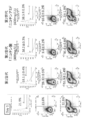
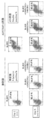
- FIG. 1 It is a schematic diagram which shows the enrichment system of the iPS cell-derived platelet used in the Example of this application. It is a figure which shows the influence of the preservation solution of this invention on the Annexin V positive rate of platelets after storage for 5 days, the P-Selectin positive rate at no stimulation, and the PAC-1 / P-Selectin positive rate at the time of ATR stimulation. ..
- “1st generation” is a conventional preservation solution
- “1st generation + VC” is a preservation solution of the present invention containing 1000 mg / L VC
- “1st generation” is a conventional preservation solution
- “1st generation + VC” is a preservation solution of the present invention containing 1000 mg / L VC
- “1st generation” is a conventional preservation solution
- "+ VC” is a preservation solution of the present invention containing 1000 mg / L VC
- “+ VC, VB3M” is 1000 mg / L VC and 400 mg / L nicotine.
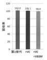
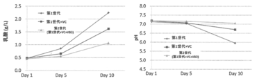
- the preservation solution of the present invention containing an acid is shown respectively. It is a figure which shows the influence of the preservation solution of this invention on the lactic acid concentration and pH of a platelet sample after storage for 5 or 10 days.
- “1st generation” is a conventional preservation solution
- “1st generation + VC” is a preservation solution of the present invention containing 1000 mg / L VC
- “2nd generation (1st generation + VC + VB3)” is 1000 mg /
- the preservation solution of the present invention containing L VC and 400 mg / L nicotinic acid is shown respectively.
- “1st generation” is a conventional preservation solution
- “1st generation + VC” is a preservation solution of the present invention containing 1000 mg / L VC
- “2nd generation (1st generation + VC + VB3)” is 1000 mg /
- the preservation solution of the present invention containing L VC and 400 mg / L nicotinic acid is shown respectively. It is a figure which shows the influence of the preservation solution of this invention on the platelet recovery rate in a platelet sample after storage for 5 or 10 days.
- “1st generation” is a conventional preservation solution
- “1st generation + VC” is a preservation solution of the present invention containing 1000 mg / L VC
- “2nd generation (1st generation + VC + VB3)” is 1000 mg /
- the preservation solution of the present invention containing L VC and 400 mg / L nicotinic acid is shown respectively.
- “plt / mL” indicates the platelet concentration in the sample
- “%” indicates the platelet recovery rate. It is a figure which shows the influence of the preservation solution of this invention on the aggregation ability of platelets after storage for 5 or 10 days. TRAP-6 was used as the stimulant in this experiment.
- new preservation solution indicates the preservation solution of the present invention containing 1000 mg / L VC and 400 mg / L nicotinic acid. It is a figure which shows the influence of the preservation solution of this invention on the aggregation ability of platelets after storage for 5 or 10 days. Collagen was used as a stimulant in this experiment.
- new preservation solution indicates the preservation solution of the present invention containing 1000 mg / L VC and 400 mg / L nicotinic acid. It is a figure which shows the influence of the preservation solution of this invention on the aggregation ability of platelets after storage for 5 or 10 days. ADP was used as the stimulant in this experiment.
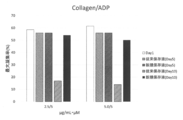
- new preservation solution indicates the preservation solution of the present invention containing 1000 mg / L VC and 400 mg / L nicotinic acid. It is a figure which shows the influence of the preservation solution of this invention on the aggregation ability of platelets after storage for 5 or 10 days. Collagen / ADP was used as a stimulant in this experiment. In the figure, “new preservation solution” indicates the preservation solution of the present invention containing 1000 mg / L VC and 400 mg / L nicotinic acid.
- “Second generation (nicotinamide (400 mg / L)” indicates a preservation solution of the present invention containing 1000 mg / L VC and 400 mg / L nicotinamide, respectively. It is a figure which shows the influence of the preservation solution of this invention on the Annexin V positive rate of platelets after storage for 5 days, the P-Selectin positive rate at no stimulation, and the PAC-1 / P-Selectin positive rate at the time of ATR stimulation. .. In this experiment, a VC reagent containing no additives was used. In the figure, “1st generation” indicates a conventional preservation solution, and “1st generation VC reagent” indicates a preservation solution of the present invention containing 1000 mg / L VC.
- the preservation solution of the present invention Effects of the preservation solution of the present invention and known preservation solutions on the Annexin V positive rate of platelets after storage for 5 days, the P-Selectin positive rate without stimulation, and the PAC-1 / P-Selectin positive rate during ATR stimulation. It is a figure which shows the result of comparison.
- the "new platelet preservation solution” is a preservation solution of the present invention containing 1000 mg / L VC and 400 mg / L nicotinic acid
- the "JRC platelet preservation solution” is about 1 pair of ACD-A solution and bicarbonate Ringer solution.
- the preservation solutions mixed in 20 are shown respectively. It is a figure which shows the hemostatic effect by the platelet sample prepared using the preservation solution of this invention.
- “Vehicle” is a mouse to which only the preservation solution of the present invention (without platelets) was administered
- Platinum is a mouse to which a platelet sample prepared using the preservation solution of the present invention was administered. Shown. It is a figure which shows the influence of the preservation solution of this invention on the survival rate and viable cell recovery rate of mesenchymal stem cells.
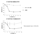
- “+ solvent” is a preservation solution obtained by adding water to a CSP-01 solution
- “+ VC + nicotinic acid” is a present invention containing 1000 mg / L VC and 400 mg / L nicotinic acid in a CSP-01 solution. The preservation solutions of are shown respectively.
- “1st generation” is a conventional preservation solution
- “1st generation + water-soluble vitamin” is a preservation solution of the present invention containing 9 kinds of water-soluble vitamins including VB2, "1st generation + water-soluble”.
- "Vitamins (excluding VB2)” indicates the preservation solution of the present invention containing eight kinds of water-soluble vitamins containing no VB2. It is a figure which shows the influence of VB2 on the P-Selectin positive rate at the time of non-stimulation of platelets, and the PAC-1 / P-Selectin positive rate at the time of ATR stimulation.
- 1st generation is a conventional preservation solution
- 1st generation + water-soluble vitamin is a preservation solution of the present invention containing 9 kinds of water-soluble vitamins including VB2, "1st generation + water-soluble”.
- Volitamins (excluding VB2) indicates the preservation solution of the present invention containing eight kinds of water-soluble vitamins containing no VB2. It is a figure which shows the influence of the preservation solution of this invention on the survival rate and viable cell recovery rate of mesenchymal stem cells.
- + solvent is a preservative solution in which water is added to a CSP-01 solution
- + VC + nicotinic acid is a book in which 1000 mg / L VC and 400 mg / L nicotinic acid are added to a CSP-01 solution.
- the preservation solutions of the invention are shown respectively. It is a figure which shows the influence of the preservation solution of this invention on the survival rate and viable cell recovery rate of mesenchymal stem cells.
- CSP-01 + VC + nicotinic acid is a preservative solution of the present invention in which 1000 mg / L VC and 400 mg / L nicotinic acid are added to a CSP-01 solution
- "Lactated Ringer's solution + VC + nicotinic acid” is The preservation solution of the present invention in which 1000 mg / L of VC and 400 mg / L of nicotinic acid are added to Lactated Ringer's solution is shown, respectively. It is a figure which shows the influence of the preservation solution of this invention on the survival rate and viable cell recovery rate of mesenchymal stem cells.
- CSP-01 + VC + nicotinic acid is a preservation solution of the present invention in which 1000 mg / L VC and 400 mg / L nicotinic acid are added to a CSP-01 solution
- CSP-01 + VC is CSP-.
- the preservation solution of the present invention in which 1000 mg / L of VC is added to the 01 solution is indicated
- the term "CSP-01 + nicotinic acid” indicates the preservation solution of the present invention in which 400 mg / L of nicotinic acid is added to the CSP-01 solution. ..
- LR indicates Lactated Ringer's solution
- “GLC” is 80 mg / dL of glucose
- “VC” is 1000 mg / L of VC
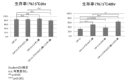
- nicotinic acid is 400 mg / L of nicotinic acid. Shown. It is a figure which shows the influence of the preservation solution of this invention on the T cell viable cell recovery rate. Further, in the figure, “LR” indicates a lactated Ringer solution. Further, “GLC” indicates 80 mg / dL of glucose, “VC” indicates 1000 mg / L of VC, and “nicotinic acid” indicates 400 mg / L of nicotinic acid.
- the mammalian cell preservation solution of the present invention (hereinafter, may be referred to as "preservation solution of the present invention") contains niacins and / or antioxidants, and is referred to as "preservation of mammalian cells".
- preservation solution of the present invention contains niacins and / or antioxidants, and is referred to as "preservation of mammalian cells”.
- the powder preparation of the present invention is not particularly limited as long as it is for preparing the preservation solution of the present invention, and further, the method for preserving mammalian cells of the present invention is in the preservation solution of the present invention.
- the invention is not particularly limited as long as it includes a step of preserving mammalian cells.
- preservation of mammalian cells means (i) maintaining the survival of the target mammalian cells, (ii) suppressing deterioration, (iii) maintaining the desired function, and /. Or, (iv) means suppressing differentiation or proliferation.
- the preservation solution of the present invention means a solution that exerts the above-mentioned preservation effect when mixed with mammalian cells
- the preservation solution of the present invention includes the embodiment of a preservation solution that does not yet contain cells to be preserved. The aspect of the preservation solution already containing the cells to be preserved is also included.
- the preservation solution of the present invention may contain niacins alone or an antioxidant as an active ingredient for preservation of mammalian cells, but niacins may be contained.
- niacins and / or antioxidants may be referred to as "essential protective components of the present invention".
- additives for preparing preservatives or powder formulations of the present invention are also included in the present invention.
- the additive of the present invention always contains the essential protective component of the present invention.
- the additive of the present invention does not have to contain the "arbitrary active ingredient" described later.
- Niacin (VB3) in the above “niacins” means nicotinic acid and / or nicotinamide. That is, the preservation solution of the present invention may be any of a preservation solution containing nicotinic acid, a preservation solution containing nicotinamide, and a preservation solution containing nicotinamide and nicotinamide, but the preservation solution containing nicotinamide. Is preferable. Further, niacin (VB3) can be produced by a known method such as chemical synthesis, but a commercially available product can also be used.
- nicotinic acid injection preparation manufactured by Toa Seiyo Ltd.
- nicotinamide manufactured by Fujifilm Wako Pure Chemical Industries, Ltd.
- niacin derivative in the above-mentioned “niacins” is not particularly limited, and for example, tocopherol nicotinic acid ester, niceritrol, nicomol, inositol hexanicotinic acid ester, 2-chloronicotinamide, 6-methylnicotinamide, 6-aminonicotinamide.
- Preferable examples include nicotinic acid, methylisonicotinic acid, thionicotinamide, nearramid, 2-mercaptonicotinic acid, nearprazine, methyl nicotinate, sodium nicotinate and the like.
- salt of niacin or a derivative thereof in the above “niacins” is not particularly limited, but is an alkali metal salt such as sodium salt and potassium salt, and an alkaline earth metal salt ammonium salt such as calcium salt and magnesium salt.
- alkali metal salt such as sodium salt and potassium salt
- alkaline earth metal salt ammonium salt such as calcium salt and magnesium salt.
- organic base salts such as trialkylamine salts, mineral salts such as hydrochlorides and sulfates, and organic acid salts such as acetates.
- antioxidant is not particularly limited, but for example, ascorbic acid (VC), superoxide dismutase 1, superoxide dismutase 2, superoxide dismutase 3, glutathione, lipoic acid, epigalocatecingalate, etc.
- Derivatives thereof such as curcumin, metranine, hydroxytyrosol, ubiquinone, catalase, vitamin E, uric acid and the like, or salts thereof can be preferably mentioned, among which ascorbic acid (VC) or its derivatives or salts thereof (hereinafter , "Ascorbic acids”) can be preferably mentioned, and ascorbic acid (VC) can be particularly preferably mentioned.
- Ascorbic acid (VC) can be produced by a known method such as chemical synthesis, but a commercially available product can also be used.
- commercially available products such as ascorbic acid injection (manufactured by Sawai Pharmaceutical Co., Ltd.) and L (+)-ascorbic acid standard product (manufactured by Fujifilm Wako Pure Chemical Industries, Ltd.) can be mentioned.
- the derivative of ascorbic acid (VC) in the above “ascorbic acids” is not particularly limited, but for example, ascorbic acid 2-phosphate, ascorbic acid 2-sulfuric acid, ascorbyl-2-glucoside, ascorbyl-6-glucoside and the like are preferable. Can be listed in.
- the salt of ascorbic acid (VC) or a derivative thereof in the above “ascorbic acids” is not particularly limited, but is an alkali metal salt such as sodium salt and potassium salt, and an alkaline earth metal salt ammonium such as calcium salt and magnesium salt.
- organic base salts such as salts and trialkylamine salts, mineral salts such as hydrochlorides and sulfates, and organic acid salts such as acetates.
- the concentration of niacins contained in the preservation solution of the present invention may be, for example, in the range of 1 to 10000 mg / L in terms of nicotinic acid, and specifically, 10 to 5000 mg / L and 20 to 8000 mg / L. , 30-6000 mg / L, 30-4000 mg / L, 30-3000 mg / L, 30-2000 mg / L, 30-1500 mg / L, 30-1200 mg / L, among others, 120-1200 mg. It is preferably within the range of / L.
- the concentration of ascorbic acids contained in the preservation solution of the present invention may be, for example, in the range of 1 to 10000 mg / L in terms of ascorbic acid, for example, 10 to 8000 mg / L, 20 to 7000 mg / L, 30. 6000 mg / L, 30-5000 mg / L, 30-4000 mg / L, 30-3000 mg / L, 50-3000 mg / L, 100-3000 mg / L, among others, 300-3000 mg / L. It is preferably within the range.
- the preservative solution of the present invention may contain the essential protective component of the present invention in an isotonic solution.
- isotonic solution is particularly limited as long as it is an isotonic solution in which the salt concentration or sugar concentration is adjusted by sodium ion, potassium ion, calcium ion or the like so as to be substantially the same as the osmotic pressure of body fluid or cell fluid.
- physiological saline physiological saline having a buffering effect
- PBS phosphate buffered saline
- Tris buffered saline Tris Buffered Saline
- HEPES buffering Phosphate saline etc.
- Ringer's solution Lactated Ringer's solution
- Acetate Ringer's solution Dicarbonate Ringer's solution and the like
- bicarbonate Ringer's solution or Lactated Ringer's solution is preferable.
- the isotonic solution can be produced based on a known composition, but commercially available products can also be used.
- Otsuka Raw Food Injection physiological saline solution, manufactured by Otsuka Pharmaceutical Factory
- Ringer's solution “Otsuka” Ringer's solution, manufactured by Otsuka Pharmaceutical Factory
- Lactec registered trademark
- injection Lactated Ringer's solution, Otsuka Pharmaceutical Factory
- examples thereof include commercially available products such as Veen (registered trademark) F infusion (Ringel acetate solution, manufactured by Fuso Yakuhin Kogyo Co., Ltd.) and Vicanate (registered trademark) infusion solution (Ringel bicarbonate solution, manufactured by Otsuka Pharmaceutical Factory).
- the term “isotonic” means that the osmotic pressure is in the range of 250 to 380 mOsm / L.
- the preservation solution of the present invention may contain albumin or sugar as an arbitrary active ingredient for preservation of mammalian cells.
- arbitrary active ingredient means an ingredient that may or may not be included.
- albumin and / or sugar may be referred to as "an optional protective ingredient of the present invention”.
- examples of the above-mentioned "albumin” include human serum albumin (HSA), bovine serum albumin (BSA), fetal bovine serum albumin (FBS), and the like, but HSA is preferable.
- the concentration of albumin may be in the range of 0.1 to 30 (w / v)%, for example, 1.0 to 20 (w / v)%.
- glucose examples include glucose, trehalose, dextran, hydroxyethyl starch and the like.
- concentration of glucose may be in the range of 1 to 10000 mg / L, for example, 10 to 8000 mg / L, 20 to 6000 mg / L, 30 to 6000 mg / L. , 40-6000 mg / L, 50-6000 mg / L, 100-6000 mg / L, 200-6000 mg / L, 500-6000 mg / L, 1000-6000 mg / L, 2000-6000 mg / L, 2000-5000 mg / L. be able to.
- the concentration of trehalose can be 0.1 to 100 g / L, 5 to 80 g / L, or 20 to 60 g / L.
- the concentration of dextran can be 0.1 to 100 g / L, 5 to 80 g / L, or 40 to 70 g / L.
- the concentration of hydroxyethyl starch can be 1 to 500 g / L or 10 to 100 g / L.
- the preservation solution of the present invention includes (a) vitamin B2 (VB2, also referred to as riboflavin) or a derivative thereof or a salt thereof (hereinafter, may be referred to as "vitamin B2"), (b) a medium or an essential component thereof. Alternatively, (c) it preferably does not contain a cell differentiation promoter.
- vitamin B2 vitamin B2
- a medium or an essential component thereof e.g., riboflavin
- a salt of vitamin B2 or a derivative thereof for example, a sodium salt and the like can be mentioned.
- the “medium” in (b) above means a cell culture solution that provides nutrients necessary for cell maintenance and proliferation in an in vitro environment, and includes, for example, Eagle's Minimal Essential (EME) Medium and Iskov Modified Dalveco Medium.
- EME Eagle's Minimal Essential
- IMDM Dalbecco Modified Eagle's Medium
- TC199 Medium Alpha-Minimum Essential Medium
- ⁇ -MEM Alpha-Minimum Essential Medium
- RPMI1640 Ham-F-12
- E199 MCDB
- Reybowitz L-15 William E Medium, etc. it can.
- essential components of the medium are aqueous nutrients and electrolytes, glycosaminoglycans, deswelling agents, energy sources, buffers, antioxidants, membrane stabilizers, antibiotics (or antispasmodics).
- Fungal agent aqueous nutrient and electrolyte
- one or more media selected from the above-mentioned media may be used.
- glycosaminoglycan includes chondroitin sulfate, dermatane sulfate, dermatine sulfate, heparin sulfate, heparan sulfate, keratin sulfate, keratin sulfate, or hyaluronic acid, and the above-mentioned “deswelling agent” includes dextran, dextran sulfate, and polyvinyl.
- Pyrrolidone polyethylene glycol, polyvinyl acetate, hydroxypropylmethyl cellulose, or carboxypropyl methyl cellulose is used as the above-mentioned "energy source”, pyruvate, shoe cloth, fructose, or dextrose, and the above-mentioned “buffer” is a hydrogen carbonate buffer solution.
- HEPES buffer 2-mercaptoethanol, glutathione, or ⁇ -tocopherol as the "antioxidant”
- vitamin A retinic acid
- ethanolamine ethanolamine
- phosphoethanolamine as the "membrane stabilizer”
- Serene or transferase is used as the above-mentioned "antibiotics and / or antifungal agents”
- amphotelicin-B gentamycin sulfate
- adenosine, inosin, or adenin as the above-mentioned "cell nutritional supplement", such as cholesterol, L-hydroxyproline, d-biotin, calciferol, niacin, p-aminobenzoic acid, pyridoxin hydrochloride, vitamin B12, Fe ( NO 3 ) 3 or non-essential amino acids can be mentioned, and phenol red can be mentioned as the above-mentioned "pH indicator”.
- the “cell differentiation promoter” in (c) above means a drug added to a medium or the like in order to obtain cells of a desired type from cells having differentiation ability, and for example, retinol, vitamin D2, vitamin D3, and the like.
- the "mammalian cell” which is the target of the preservation solution of the present invention is not particularly limited as long as it is a living cell derived from a mammal, and even a primary cell obtained from an individual organism can be a primary cell.
- the cells may be cells grown by culturing for generations or multiple generations, but specifically, cells used for treating diseases / trauma, megakaryocytes used for their production, regenerative medicine / immunity, etc. Stem cells, immune cells and the like used for therapy and the like can be preferably exemplified.
- the "mammals” include rodents such as mice, rats, hamsters and guinea pigs, lagomorphs such as rabbits, ungulates such as pigs, cows, goats, horses and sheep, and cats such as dogs and cats. Primates such as eyes, humans, monkeys, red-tailed monkeys, cynomolgus monkeys, marmosets, orangutans, and chimpanzees can be exemplified, and among them, humans can be preferably exemplified.
- the above-mentioned "platelet” is one of the cell components in blood and means a cell component in which CD41a and CD42b are positive.
- the platelets to be the subject of the preservation solution of the present invention are artificially produced from megakaryocytes cultured in vitro, even if they are concentrated platelets obtained by removing erythrocytes and leukocytes from mammalian blood (whole blood). Purified platelets may be used, but purified platelets are preferable.
- the method for producing the purified platelets is not particularly limited, but is a step of (A) a concentration step of concentrating a culture of megakaryocytes; and (B) a centrifugation step of centrifuging platelets from the obtained concentrate; A method including A) and (B) can be preferably exemplified.
- the "megakaryocytes" used in this method for producing platelets are artificial pluripotent stem cells (iPS cells), embryonic stem cells (ES cells), nuclear transplant ES cells (ntES cells), and reproductive stem cells (EG cells).
- Somatic stem cells Somatic stem cells, embryonic tumor cells, and other pluripotent cells, as well as hematopoietic stem cells, hematopoietic progenitor cells, CD34-positive cells, megakaryocytes / red isolated from bone marrow, umbilical cord blood, peripheral blood, etc. It can also be derived from embryonic stem cells, megakaryocyte progenitor cells, and the like.
- stem cell means an immature cell having self-renewal ability and differentiation / proliferation ability.
- Stem cells include subpopulations such as pluripotent stem cells (pluripotent stem ce11), multipotent stem cells (multipotent stem ce11), and monopotent stem cells (unipotent stem ce11), depending on their ability to differentiate.
- Pluripotent stem cells mean cells that cannot become individuals by themselves, but have the ability to differentiate into all the tissues and cells that make up the living body, and pluripotent stem cells are all types. Although not present, it means a cell capable of differentiating into a plurality of types of tissues and cells, and a unipotent stem cell means a cell having an ability to differentiate into a specific tissue or cell.
- the stem cell to be stored in the preservation solution of the present invention may be any of pluripotent stem cell, pluripotent stem cell, and monopoly stem cell, for example, iPS cell, ES cell, ntES cell, EG cell and the like.
- pluripotent stem cells somatic stem cells such as mesenchymal stem cells, hematopoietic stem cells, neural stem cells, bone marrow stem cells, and germline stem cells, and among them, mesenchymal stem cells are preferable.
- Mesenchymal stem cells can be collected from mammalian bone marrow, adipose tissue, peripheral blood, umbilical cord blood, etc.
- Leafy stem cells can be isolated. Among them, mesenchymal stem cells derived from human bone marrow and mesenchymal stem cells derived from immature porcine bone marrow can be preferably exemplified.
- immune cells mean cells that are present in blood or lymph and are involved in the immune system.
- immune cells to be preserved in the preservation solution of the present invention include T cells, macrophages, dendritic cells, B cells, NK cells, neutrophils, eosinophils, myeloid-derived inhibitory cells (MDSC) and the like.
- T cells a cell expressing an antigen receptor called a T cell receptor (TCR) on the surface, and is, for example, a cytotoxic T cell that is a CD8 positive cell or a CD4 positive cell.
- T cells can be collected from mammalian peripheral blood, lymph nodes, bone marrow, thymus, spleen, umbilical cord blood, etc. by a known general method, or commercially available products can also be used.
- the preservation solution of the present invention can be used to preserve mammals in a non-frozen state.
- the temperature at which mammalian cells are stored using the present invention can be arbitrarily selected depending on the type of cells to be stored, but is preferably in the range of 0 to 40 ° C.
- the temperature at which platelets and megakaryocytes are stored using the preservation solution of the present invention is preferably 15 to 30 ° C, more preferably 20 to 24 ° C, and most preferably 21 to 23 ° C. ..
- the temperature at which the mesenchymal stem cells and T cells are stored using the storage solution of the present invention is preferably 1 to 38 ° C, more preferably 1 to 30 ° C, and particularly preferably 1 to 15.
- the temperature at which mammalian cells are stored using the present invention can be varied within the range of 0-40 ° C.
- T cells when stored using the storage solution of the present invention, they are stored at a low temperature (for example, 1 to 5 ° C, preferably 5 ° C) for a certain period of time, and then stored. It can be stored at room temperature (eg, 20-26 ° C, preferably 22-25 ° C, more preferably 25 ° C) for a further few hours.
- the preservation solution of the present invention can be used for storing mammalian cells for several hours to several tens of days.
- the storage period varies depending on the type of cells to be stored, but as shown in the following examples, 1 to 15 days, preferably 1 to 10 days for platelets and megakaryocytes, and 1 for mesenchymal stem cells. ⁇ 63 days, preferably 1-35 days, more preferably 1-30 days, more preferably 1-28 days, even more preferably 1-14 days, T cells 1-30 days, preferably 1-14 It can be stored for days, more preferably 1-2 days, even more preferably 30 hours.
- the preservation solution of the present invention may be used for the purpose of administering mammalian cells to a mammal. That is, after storing the mixed solution containing the preservation solution of the present invention and the mammalian cells under predetermined conditions, the mixed solution can be directly administered (for example, intravenously) into the living body of the mammal. Therefore, it is preferable that the preservation solution of the present invention does not contain a component that may adversely affect the living body of the mammal when administered into the living body of the mammal. Examples of such "components having an adverse effect" include polyvinylpyrrolidone, 2-mercaptoethanol, okadaic acid, sodium butyrate, G418 and the like.
- a mammalian deterioration inhibitor containing the essential protective component of the present invention can be mentioned.
- Such a deterioration inhibitor may further contain the optional protective component of the present invention.
- the mammalian deterioration inhibitor of the present invention can be used to prepare the preservation solution of the present invention by adding it to the above isotonic solution, and is also used to enhance the preservation property by adding it to a known mammalian preservation solution. You can also do it.
- the present invention also relates to a mammalian cell storage container in which the preservation solution or powder preparation of the present invention is enclosed.
- the storage container of the present invention may be in any form as long as it can maintain sterility after injecting a suspension of mammalian cells, for example, a blood bag, an infusion bag, a syringe, etc. Ampoules, vials and the like can be mentioned, but blood bags are preferred.
- Platelets derived from iPS cells were prepared according to the method described in PCT / JP2018 / 034667. The specific procedure is shown in (1-1) to (1-12) below.
- human ES / iPS cell colonies are co-cultured with C3H10T1 / 2 feeder cells for 14 days in the presence of 20 ng / mL VEGF (manufactured by R & D SYSTEMS) to produce hematopoietic progenitor cells (HPC).
- HPC hematopoietic progenitor cells
- the gene transfer system used the lentiviral vector system.
- the lentiviral vector is a Tetracycline-controlled Tet-on® gene expression induction system vector.
- Tetracycline-controlled Tet-on® gene expression induction system vector By rearranging the mOKS cassette of LV-TRE-mOKS-Ubc-tTA-I2G (Kobayashi et al., Cell, 2010, vol.142, No. 5,787-799) to c-MYC, BMI1, or BCL-xL. Made.
- Vectors into which c-MYC, BMI1, or BCL-xL have been introduced are introduced into LV-TRE-c-Myc-Ubc-tTA-I2G, LVTRE-BMI1-Ubc-tTA-I2G, and LV-TRE-BCL-, respectively. It was designated as xL-Ubc-tTA-I2G.
- the c-MYC, BMI1, and BCL-xL viruses were prepared by gene transfer into 293T cells with the above lentiviral vector. By infecting the cells of interest with the resulting virus, the c-MYC, BMI1, and BCL-xL genes are introduced into the genomic sequence of the cells of interest. These genes, which have been stably introduced into the genome sequence, can be forcibly expressed by adding doxycycline (clontech # 631311) to the medium.
- the spin infection was performed twice every 12 hours.
- the medium is a basal medium (15% Fetal Bovine Serum (manufactured by GIBCO), 1% Pencillin-Streptomycin-Glutamine (manufactured by GIBCO), 1% Insulin, Transferrin, Selenium Solution (manufactured by ITS-G)).
- IMDM IMDM (Sigma-Aldrich) containing .45 mmol / L 1-Thioglycerol (Sigma-Aldrich), 50 ⁇ g / mL L-Ascorbic Acid (Sigma-Aldrich), 50 ng / mL Human Medium ) (R & D SYSTEMS), 50 ng / mL Human Stem Cell Factor (SCF) (R & D SYSTEMS) and 2 ⁇ g / mL Doxycycline (DOX, clonetech, # 631311) (hereinafter, “Differentiation medium”). ”), And further, Protamine was added and used so as to have a final concentration of 10 ⁇ g / mL.
- 2nd day of infection to 11th day of infection On the second day of infection, the virus-infected blood cells obtained by the above method were collected by pipetting, centrifuged at 1200 rpm for 5 minutes to remove the supernatant, and then suspended in a new differentiation medium to obtain new cells. Seeded on C3H10T1 / 2 feeder cells (6 well plate). Subculture was performed by performing the same operation on the 9th day of infection. At the time of re-seeding, after counting the number of cells, the cells were seeded on C3H10T1 / 2 feeder cells so as to have 1 ⁇ 10 5 cells / 2 mL / well (6 well plate).
- the anti-human CD235ab-pacific blue antibody (manufactured by BioLegend) was used to react the blood cells with the antibody. After the above reaction, analysis was performed using FACS Verse TM (manufactured by BD Biosciences). On the 14th day of infection, cells having a CD41a positive rate of 50% or more were designated as megakaryocyte self-proliferating strains.
- BCL-xL Virus Infection of Megakaryocyte Self-Proliferating Strain
- BCL-xL was introduced into the megakaryocyte self-proliferating strain on the 14th day of the above infection by the lentivirus method using BCL-xL virus.
- Virus particles were added to the medium to achieve MOI 10 and infected by spin infection (32 ° C., 900 rpm, centrifuge for 60 minutes). Forced expression of the BCL-xL gene is DO so that it is 1 ⁇ g / mL DOX in the medium. It was carried out by adding X.
- Day 18 of infection The megakaryocyte self-proliferating strain after the introduction of the BCL-xL gene was collected, the number of cells was counted, and the cells were seeded so as to have 3 ⁇ 10 5 cells / 10 mL / 100 mm dish.
- Day 24 of infection The megakaryocyte self-proliferating strain after the introduction of the BCL-xL gene was collected, the number of cells was counted, and the cells were seeded so as to have 1 ⁇ 10 5 cells / 10 mL / 100 mm dish. After that, subculture was carried out in the same manner every 4-7 days, and maintenance culture was carried out.
- the BCL-xL infection day 24 were collected megakaryocyte self-propagating strains transformed gene, cells 1.0 ⁇ 10 5 per, 2 [mu] L of anti-human CD41a-APC antibody (manufactured by BioLegend Corp.), 1 [mu] L of anti-human CD42b -PE antibody (manufactured by eBioscience) and 1 ⁇ L of anti-human CD235ab-Pacific Blue antibody (Anti-CD235ab-PB; manufactured by BioLegend) were immunostained and then analyzed using FACS Verse TM.
- a strain having a CD41a positive rate of 50% or more was designated as an immortalized megakaryocyte cell line.
- These cells which were able to proliferate for 24 days or more after infection, were designated as immortalized megakaryocyte cell lines SeV2-MKCL and NIH5-MKCL.
- the obtained SeV2-MKCL and NIH5-MKCL were statically cultured in a 10 cm dish (10 mL / dish).
- IMDM was used as the basic medium, and the following components were added (concentration is final concentration).
- the culture conditions were 27 ° C. and 5% CO 2 .
- the culture of megakaryocytes was concentrated using a hollow fiber membrane (Plasmaflow OP, manufactured by Asahi Kasei Medical Co., Ltd.), and the obtained concentrate of the culture of megakaryocytes was placed in a storage bag. Recovered.
- a cell bag containing the culture to which the ACD-A solution was added was bonded to the ACP215 disposable set using a sterile bonding device. Then, ACP215 was started up in the service mode, and the rotation speed was set to 2500 rpm (350 ⁇ g). ACP215 was started and the culture in the cell bag was introduced into a separation bowl at about 100 mL / min. The liquid component flowing out of the separation bowl was collected in a collection bag. After introducing the entire amount of the culture in the cell bag into the separation bowl, an additional 500 mL of a wash-preserving solution was introduced into the separation bowl.
- centrifugation was stopped and a collection bag containing the recovery solution (recovered liquid component containing platelets) was separated using a tube sealer.
- a recovery bag containing a recovery solution (including platelets) was joined to the new ACP215 disposable set using the above sterile joining device.
- ACP215 was launched in normal mode. WPC was selected as the program setting, and the ACP215 disposable set to which the above collection bag was joined was set according to the instructions of the device.
- the collection bag containing the collection liquid was installed on the stand.
- the centrifugal speed of ACP215 was changed to 5000 rpm (1398.8 ⁇ g), and the centrifugation was started.
- the recovery liquid began to be introduced into the separation bowl, automatic injection was changed to manual injection. Specifically, the recovered solution was introduced into the separation bowl at an introduction rate of about 100 mL / min. After adding the entire amount of the recovered liquid to the separation bowl, another 500 mL of the washing and preserving liquid was added.
- a solution (ACD-A solution; manufactured by Terumo) (sodium citrate hydrate 2.20 W / V%, citric acid hydrate, 0.80 W / V%, and glucose 2.20 W / V%) was added.
- the solution was prepared.
- the solution is also referred to as a "first generation preservation solution”.
- a solution was prepared by adding a VC preparation (injection preparation containing an additive; manufactured by Sawai Pharmaceutical Co., Ltd.) to the first-generation preservation solution.
- the solution is also referred to as "VC-added preservation solution”.
- the final concentrations of the additives in each preservation solution are shown in Table 2 below.
- all the preservation solutions were adjusted to pH 7.3 ⁇ 0.1 with 1 M NaOH and incubated for 1 hour or more until use (room temperature, 5% CO 2 under shading).
- Annexin V positive rate of platelet samples is one of the indicators of platelet deterioration (activation), and when the positive rate is high, it is evaluated that the platelets are deteriorated or abnormal. Will be done. Therefore, the Annexin V positive rate in the platelet sample obtained in (2-2) above was measured, and the effect of each preservation solution on suppressing the deterioration of platelets was investigated. Specifically, the platelet sample obtained in (2-2) above was diluted 500-fold with Annexin Buffer (manufactured by Beckton Dickinson) and dispensed into three centrifuge tubes (negative control, positive control, respectively). And non-stimulating samples).
- Annexin Buffer manufactured by Beckton Dickinson
- the Annexin V positive rate of platelets (CD41 + fraction) was 60.5 ⁇ 0.3 to 64.4 ⁇ 0.1% when the 1st generation preservation solution was used, whereas VC was added.
- the preservation solution was used, it was 53.9 ⁇ 2.1 to 56.7 ⁇ 1.8%, and it was clarified that it decreased by the addition of VC. From these results, it was clarified that the addition of 300 to 3000 mg / L of VC improves the platelet deterioration inhibitory action of the platelet preservation solution.
- PAC when each platelet sample was stimulated with adenosine diphosphate (ADP) and thrombin receptor activating peptide-6 (TRAP-6), which are platelet activators, in order to know the preservation state of platelet function.
- ADP adenosine diphosphate
- TRAP-6 thrombin receptor activating peptide-6
- ATR the combination of ADP and TRAP-6 may be referred to as "ATR").
- the platelet sample obtained in (2-2) above was diluted 500-fold with Tyroad HEPES Buffer (THB) and dispensed into three centrifuge tubes (negative control, positive control, and none, respectively). Stimulation sample).
- a mixed solution of ADP (manufactured by Sigma, final concentration 20 ⁇ M) and TRAP-6 (manufactured by BACHEEM, final concentration 30 ⁇ M) was added to the positive control sample, and the positive control sample and the non-stimulated sample after stimulation were prepared with an anti-CD41 antibody (anti-CD41 antibody). Staining with BioLegend (), anti-P-Selectin antibody (BioLegend), and anti-PAC-1 antibody (Beckton Dickinson) (light-shielded, room temperature, 30 minutes).
- Negative control samples were stained with an anti-CD41 antibody (manufactured by BioLegend), an anti-P-Selectin antibody isotype control antibody (manufactured by BioLegend), and an anti-PAC-1 antibody isotype control antibody (manufactured by BioLegend) (manufactured by BioLegend). Under shade, room temperature, 30 minutes). The stained sample was fixed by adding 1% paraformaldehyde (under shading, 4 ° C., 30 minutes or more), and then the P-Selectin and / or PAC-1 positive rate was measured by FACS within 24 hours. At the time of analysis, gating was performed so that the P-Selectin positive rate and the PAC-1 positive rate in the iPS platelets (CD41 + fraction) in the negative control sample were 1.0 ⁇ 0.1% or less. It was.
- the middle part of FIG. 2 shows the P-Selectin positive rate without stimulation, and the lower part of FIG. 2 shows the PAC-1 / P-Selectin positive rate with ATR stimulation (both are the results obtained from platelet samples after storage for 5 days). ).
- the P-Selectin positive rate in unstimulated platelets (CD41 + fraction) was 33.2 ⁇ 0.3-45.6 ⁇ 1.2% when the 1st generation preservation solution was used.
- the VC-added preservative solution was used, it was 13.0 ⁇ 0.2 to 19.7 ⁇ 1.1%, and it was clarified that it decreased by the addition of VC.
- the decrease in the P-Selectin positive rate tended to depend on the VC addition concentration.
- VC and VB3 to platelet preservation solution 3-1
- nicotinic acid nicotinic acid injection preparation; manufactured by Toa Seiyo Ltd.
- VB3 means nicotinic acid and / or nicotinamide, but in Examples 3 to 5 and 8 to 13, nicotinic acid is used as VB3, and Examples In No. 6, nicotinic acid or nicotinamide is used as VB3.
- a solution obtained by adding VB3 (nicotinic acid or nicotinamide) to a VC-added preservation solution is also referred to as a “VC / VB3 addition preservation solution” or a “second generation preservation solution”.
- the final concentrations of the additives in each preservation solution are as shown in Table 3 below.
- all the preservation solutions were adjusted to pH 7.3 ⁇ 0.1 with 1 M NaOH and incubated for 1 hour or more until use (room temperature, 5% CO 2 under shading).
- the lactic acid concentration in the supernatant was measured using an N-assay L LAC Nittobo (manufactured by Nittobo Medical Co., Ltd.) and an automatic analyzer 7180 (manufactured by Hitachi High-Technologies Corporation). The results are shown in FIG.
- the lactic acid concentration in the sample supernatant was 0.35 ⁇ 0.01 g / L when the 1st generation preservation solution was used, and 0.16 ⁇ 0.01 g / L when the VC-added preservation solution was used.
- the VC / VB3 added preservative solution was used, it was 0.13 ⁇ 0.01 g / L. From these results, it was clarified that the combined use of VC and VB3 strongly suppressed anaerobic metabolism in platelets during storage as compared with VC alone.
- the above preservation solution (1st generation preservation solution, VC-added preservation solution, or 2nd generation preservation solution) is added to the iPS cell-derived platelet preparation prepared by the method described in Example 1 so as to obtain a uniform suspension. Suspended gently (platelet concentration: about 1.0 ⁇ 10 9 plts / mL). Immediately after each suspension, or after filling a blood storage bag and storing it with horizontal shaking for 5 or 10 days (under shading, 22 ° C., 50 rpm), perform the following experiments (4-2) to (4-4). Served.
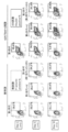
- FIG. 7 shows the P-Selectin positive rate without stimulation
- the right side of FIG. 7 shows the PAC-1 / P-Selectin positive rate with ATR stimulation.
- the P-Selectin positive rate was 18.0% before storage (Day 1) when the 1st generation storage solution was used, whereas it was 33.0% after 5 days storage (Day 5) and stored for 10 days. Later (Day 10), it increased to 31.1%.
- the P-Selectin positive rates after storage for 5 and 10 days were both lower than those when the 1st generation preservative solution was used, which were 26.2% (Day 5) and 26.2% (Day 5), respectively. It was 24.4% (Day 10).
- the P-Selectin positive rate after storage for 5 and 10 days was even lower than when the VC-added storage solution was used, which were 24.2% (Day 5) and 19. It was 3% (Day 10). From the above results, it was clarified that the use of the second generation preservation solution suppresses the deterioration of platelets even after storage for 10 days.
- the ATR-stimulated PAC-1 / P-Selectin positive rate was 43.4% before storage (Day 1) when the first-generation storage solution was used, whereas it was 43.4% after storage for 5 days (Day 5). It decreased sharply to 34.5% and 5.9% after storage for 10 days (Day 10).
- the ATR-stimulated PAC-1 / P-Selectin positive rate was the same as when the first-generation preservative solution was used after storage for 5 days (Day 5). It was about 35.9% and 35.3%, respectively, but after storage for 10 days (Day 10), it showed significantly higher values than the 1st generation storage solution (19.4% and 19 respectively). 0.9%). From the above results, it was clarified that the reactivity of platelets was maintained even after storage for 10 days by using the VC-added storage solution or the second-generation storage solution.
- [Aggregation ability of platelet samples] (5-1) Preparation of Preservative Solution and Platelet Sample A first-generation preservation solution and a second-generation preservation solution were prepared by the method described in (3-1) of Example 3. The above-mentioned preservation solution was added to the iPS cell-derived platelet preparation prepared by the method described in Example 1 and gently suspended so as to form a uniform suspension (platelet concentration: about 1.0 ⁇ 10 9 plts /). mL). Each suspension was immediately or filled in a blood storage bag and stored with horizontal shaking for 5 or 10 days (under shading, 22 degrees, 50 rpm), and then subjected to the following experiment (5-2).
- Figures 9 to 12 show the results of using TRAP-6, Collagen, ADP, and Collagen / ADP as stimulants, respectively. It was revealed that the maximum aggregation rate of platelet samples using the 1st generation preservation solution was significantly reduced during storage for 5 to 10 days regardless of which stimulant was used (FIGS. 9 to 12). "Conventional Preservation Solution (Day 10)"). On the other hand, such a sharp decrease in the aggregation rate was not observed in the platelet sample using the second generation preservation solution (“New preservation solution (Day 10)” in FIGS. 9 to 12). From the above results, it was clarified that the aggregation ability (hemostatic ability) of platelets during storage can be maintained for 10 days or more by using the second generation preservation solution.
- second generation preservation solution (nicotinic acid)
- second generation preservation solution the preservation solution obtained by adding VC and nicotinamide to the first generation preservation solution
- generational preservation solution (nicotinamide)
- the above-mentioned preservation solution was added to the iPS cell-derived platelet preparation prepared by the method described in Example 1 and gently suspended so as to form a uniform suspension (platelet concentration: about 0.3 ⁇ 10 9 plts /). mL). Each suspension was immediately or seeded on a 24-well plate and stored with horizontal shaking for 5 days (under shading, 22 degrees, 50 rpm) and then subjected to the following experiments (6-2) and (6-3). ..
- the Annexin V positive rate of platelets was 38.2 ⁇ 0.3% in the nicotinamide-added preservation solution and 38.5 ⁇ 0.3% in the nicotinamide-added preservation solution. there were. All of these values were lower than when the first generation preservation solution was used (53.1 ⁇ 0.4%). From these results, it was clarified that vitamin B3 (nicotinic acid and / or nicotinamide) also suppresses the deterioration of platelets during storage.
- the P-Selectin positive rate increased from 11.6% before storage (Day 1) to 28.3 ⁇ 0.5% after storage for 5 days (Day 5) when the 1st generation storage solution was used. ..
- the second generation storage solution to which nicotinic acid or nicotinamide was added was used, it was kept as low as 9.4 ⁇ 0.2% to 10.6 ⁇ 0.2% even after storage for 5 days. ..
- the ATR-stimulated PAC-1 / P-Selectin positive rate was 33.9 ⁇ 2.4% when the first-generation preservative solution was used, but the second-generation nicotinic acid or nicotinamide was added. When the preservative solution was used, it was 41.6 ⁇ 0.8% to 41.2 ⁇ 1.9%. From these results, it was clarified that nicotinic acid or nicotinamide maintains the reactivity of platelets during storage.
- first generation preservation solution (VC reagent)
- the above-mentioned preservation solution was added to the iPS cell-derived platelet preparation prepared by the method described in Example 1 and gently suspended so as to form a uniform suspension (platelet concentration: about 0.3 ⁇ 10 9 plts /). mL).
- Each suspension was immediately or seeded on a 24-well plate and stored with horizontal shaking for 5 days (under shading, 22 degrees, 50 rpm) and then subjected to the following experiments (7-2) and (7-3). ..
- the unstimulated P-Selectin positive rate was 9.5 ⁇ 0.4% when the 1st generation VC reagent was used than when the 1st generation preservation solution was used (28.3 ⁇ 0.5%). ) was lower. From these results, it was clarified that the addition of VC alone suppresses the deterioration of iPS-derived platelets during storage. In addition, the ATR-stimulated PAC-1 / P-Selectin positive rate was higher when the first-generation VC reagent was used (38.2) than when the first-generation preservation solution was used (33.9 ⁇ 2.4%). ⁇ 1.6%) was higher.
- Each storage solution was adjusted to pH 7.3 ⁇ 0.1 with 1 M NaOH and incubated for 1 hour or more until use (room temperature, 5% CO 2 under shading).
- the above-mentioned preservation solution was added to the iPS cell-derived platelet preparation prepared by the method described in Example 1 and gently suspended so as to form a uniform suspension (platelet concentration: about 1.0 ⁇ 10 9 plts /). mL).
- Each suspension was immediately or filled in a blood storage bag and stored with horizontal shaking for 5 days (under shading, 22 degrees, 50 rpm) and then subjected to the following experiments (8-2) and (8-3). ..
- the unstimulated P-Selectin positive rate was lower when the 2nd generation preservation solution was used (16.5%) than when the JRCS platelet lavage solution was used (30.4%). From these results, it was clarified that the 2nd generation preservative solution exerts an excellent deterioration suppressing effect by the JRCS platelet washing solution in the preservation of iPS-derived platelets.
- the ATR-stimulated PAC-1 / P-Selectin positive rate was higher when the 2nd generation preservation solution was used (24.8%) than when the JRCS platelet lavage solution was used (9.8%). It was. From these results, it was clarified that the second-generation preservative solution exerts an excellent function-maintaining effect on the preservation of iPS-derived platelets by the JRCS platelet washing solution.
- CSP-01 solution lactated Ringer's solution
- JP2012-115253 and WO2014 / 208053 A VC preparation (1000 mg / L, manufactured by Sawai Pharmaceutical Co., Ltd.) and nicotinic acid (400 mg / L, manufactured by Toa Seiyo Ltd.) were added to the above CSP-01 solution to prepare a mesenchymal stem cell preservation solution.
- a control preservation solution was prepared by adding distilled water (Otsuka Distilled Solution; manufactured by Otsuka Pharmaceutical Factory Co., Ltd.) in place of VC and nicotinic acid to the CSP-01 solution.
- (10-2) were suspended human bone marrow-derived mesenchymal stem cells (Lonza, Inc.) using a preservative solution was prepared by mesenchymal stem cells stored above (10-1) (5 ⁇ 10 5 cells / mL) .. After allowing the suspension to stand at 24, 48, 96, and 168 at 5 ° C., total cell count and dead cell count were measured using a microscope. The cell viability (%) and viable cell recovery rate (%) at each time point were calculated using the following formulas 1 and 2.
- Preservation of iPS cell-derived megakaryocytes (11-1) Preparation of iPS cell-derived megakaryocytes The concentration of platelets contained in the culture obtained in Example 1 was measured by FACS. The required amount of culture is collected, ACD-A solution (10 v / v%) and PEG1 (final concentration 2 ⁇ M; manufactured by Cayman Chemical Company) are added, and centrifugation for 12 minutes (1200 ⁇ g, 22 ° C., Brake minimum) was performed. After removing the supernatant, the first-generation or second-generation storage solution added to the pellet and gently suspended in a uniform suspension (platelet concentration: about 1.3 ⁇ 10 9 plts / mL) .. Each suspension was immediately or filled in a blood storage bag and stored with horizontal shaking for 5 or 10 days (under shading, 22 degrees, 50 rpm) before being subjected to the following experiment (11-2).
- Annexin V Negative Rate of Megakaryocyte Samples
- Annexin V is also known as a probe for detecting changes in the cell membrane (expression of phosphatidylserine to the outside of the cell membrane) in apoptotic cells. Therefore, the Annexin V negative rate in the megakaryocyte sample obtained in (11-1) above was measured, and the effect of each preservation solution on suppressing apoptosis of megakaryocytes was examined.
- the megakaryocyte sample (culture containing megakaryocytes and platelets) obtained in (11-1) above is diluted 500-fold with Annexin Buffer (manufactured by Beckton Dickinson) and divided into three centrifuge tubes.
- Annexin Buffer manufactured by Beckton Dickinson
- the Annexin V positive rate in iPS platelets (CD41 + fraction) in the negative control was set to 1.0 ⁇ 0.1%, and the Annexin V negative rate of megakaryocytes in the unstimulated sample was calculated.
- the results are shown in FIG. 18 (“Day 1” indicates a sample before storage, “Day 5” indicates a sample after storage for 5 days, and “Day 10” indicates a sample after storage for 10 days).
- the Annexin V negative rate decreased over time in both the samples using the 1st generation preservation solution and the samples using the 2nd generation preservation solution. However, it was clarified that the decrease in the Annexin V negative rate was suppressed when the second generation preservation solution was used as compared with the case where the first generation preservation solution was used.
- the sample using the second generation preservation solution contained a higher proportion of viable cells having stable cell membranes and not undergoing apoptosis. Therefore, this example shows that the second generation preservation solution is also effective for the preservation of iPS cell-derived megakaryocytes.
- T cell preservation solution 3% trehalose was added to a lactated Ringer solution (Lactec infusion; manufactured by Otsuka Pharmaceutical Factory, Ltd.) (hereinafter, such a solution may be referred to as "CSP-11 solution”).
- a VC preparation 1000 mg / L, manufactured by Sawai Pharmaceutical Co., Ltd.
- nicotinic acid 400 mg / L, manufactured by Toa Eiyo Co., Ltd.
- the cells were washed with CSP-11 solution, dispensed into a stem full tube (manufactured by Sumitomo Bakelite Co., Ltd.), and centrifuged for 10 minutes (300 ⁇ g, room temperature). After removing the supernatant, the preservation solution prepared in (12-1) above was added and suspended (T cell concentration: about 5 ⁇ 10 5 cells / 1 ml). A 20 ⁇ L cell suspension was dispensed from each stem full tube, 20 ⁇ L tripan blue (manufactured by gibco) was mixed, and the survival rate was measured using a one-cell counter (manufactured by Biomedical Science) (1 location). The total number of cells and the number of dead cells in the area of the cell counting chamber at the four corners of the cell counting section were measured). In addition, after storing the cell suspension at 5 ° C. for 48 hours, the survival rate was measured in the same manner.
- the results are shown in Fig. 19.
- the survival rate before storage did not differ between the storage solutions (Fig. 19).
- FIG. 19 After storage for 48 hours, it was revealed that the survival rate was significantly higher when VC alone or a storage solution containing VC and nicotinic acid was used (FIG. 19).
- the survival rate was significantly higher when the preservation solution containing VC and nicotinic acid was used as compared with VC alone. Therefore, it was shown that the preservation solution of the present invention is also effective for refrigerated storage of T cells.
- the cells were washed with a lactated Ringer's solution containing 3% trehalose, dispensed into a stem full tube (manufactured by Sumitomo Bakelite), and centrifuged (300 ⁇ g, room temperature) for 10 minutes. .. After removing the supernatant, the preservation solution prepared in (12-1) above was added and suspended (T cell concentration: about 5 ⁇ 10 5 cells / 1 ml). A 20 ⁇ L cell suspension was dispensed from each stem full tube, 20 ⁇ L tripan blue (manufactured by gibco) was mixed, and the survival rate was measured using a one-cell counter (manufactured by Biomedical Science) (1 location).
- the viable cell recovery rate was calculated using the following formula 3.
- Fig. 20 The results are shown in Fig. 20. There was no difference in the survival rate before storage between the storage solutions (left graph in FIG. 20). On the other hand, after storage for 24 and 48 hours, it was clarified that the survival rate and the recovery rate of living cells were higher when the preservation solution containing VC and nicotinic acid was used (center and right graph of FIG. 20). Therefore, it was shown that the preservation solution of the present invention is also effective for refrigerated storage of T cells. Moreover, it was clarified that the survival rate and the viable cell recovery rate tend to be improved by adding glucose in addition to VC and nicotinic acid.
- Annexin V positive rate was further reduced in the preservative solution to which the water-soluble vitamin group (excluding VB2) was added, and was 40.3%. From these results, it was shown that the platelet deterioration inhibitory effect of VC and VB3 may be inhibited by VB2.
- the P-Selectin positive rate increased from 27.4% before storage (Day 1) to 42.9% after storage for 5 days (Day 5) when the 1st generation storage solution was used.
- the P-Selectin positive rate after storage for 5 days was suppressed as low as 34.9% when the preservative solution containing the water-soluble vitamin group was used, and the water-soluble vitamin group (excluding VB2) was added. When the preservative solution was used, it was further suppressed to 24.0%.
- the ATR-stimulated PAC-1 / P-Selectin positive rate increased from 35.6% before storage (Day 1) to 32.4% after storage for 5 days (Day 5) when the first-generation storage solution was used. It was declining.
- the preservative solution to which the water-soluble vitamin group was added was used, it was 36.8% even after storage for 5 days.
- a preservative solution containing a water-soluble vitamin group (excluding VB2) was used, it increased to 43.0% after storage for 5 days. From these results, it was shown that the platelet function maintenance effect of VC and VB3 may be inhibited by VB2.
- a CSP-01 solution was prepared according to the description in (10-1) of Example 10.
- a VC preparation 1000 mg / L, manufactured by Sawai Pharmaceutical Co., Ltd.
- nicotinic acid 400 mg / L, manufactured by Toa Seiyo Ltd.
- a control preservative solution was prepared by adding distilled water (Otsuka Distilled Solution; manufactured by Otsuka Pharmaceutical Factory Co., Ltd.) as a solvent in place of VC and nicotinic acid to the CSP-01 solution.
- (16-2) between the mesenchymal stem cells stored above (16-1) using a preservative solution was prepared by suspending the human adipose-derived mesenchymal stem cells (Lonza, Inc.) (5x10 5 cells / mL). After allowing the suspension to stand at 5 ° C. for 7, 14, 21 and 28 days, the total number of cells and the number of dead cells were measured using a microscope. The cell viability (%) and the viable cell recovery rate (%) at each time point were calculated using the formulas 1 and 2 described in (10-2) of Example 10.
- a sodium hydrogen carbonate preparation (Meylon intravenous injection 8.4%, manufactured by Otsuka Pharmaceutical Factory, Ltd.) was added to the CSP-01 solution to adjust the pH to 7.0 to 7.3.
- a sodium hydrogen carbonate preparation (Meylon intravenous injection 8.4%, manufactured by Otsuka Pharmaceutical Factory, Ltd.) was added to the CSP-01 solution to adjust the pH to 7.0 to 7.3.
- the following four types of mesenchymal stem cell preservation solutions were prepared.
- CSP-01 CSP-01 + VC CSP-01 + nicotinic acid CSP-01 + VC + nicotinic acid
- (17-2) between leaves system using stock solutions prepared by stem cells stored above (17-1) was suspended human adipose-derived mesenchymal stem cells (Lonza, Inc.) (5x10 5 cells / mL). After allowing the suspension to stand at 5 ° C. for 7, 14, 21 and 28 days, the total number of cells and the number of dead cells were measured using a microscope. The cell viability (%) and the viable cell recovery rate (%) at each time point were calculated using the formulas 1 and 2 described in (10-2) of Example 10.
- np mesenchymal stem cells Immature pig bone marrow-derived mesenchymal stem cells (np mesenchymal stem cells) were prepared according to the method of Nishimura et al. (Xenotransplantation. 2019 May; 26 (3): e12501.). .. Such mp mesenchymal stem cells were suspended using the preservation solution prepared according to (18-1) above (5x10 5 cells / mL). After allowing the suspension to stand at 5 ° C. for 7, 14, 21 and 28 days, the total number of cells and the number of dead cells were measured using a microscope. The cell viability (%) and the viable cell recovery rate (%) at each time point were calculated using the formulas 1 and 2 described in (10-2) of Example 10.
- T cell preservation solution Lactated Ringer's solution (Lactec infusion; manufactured by Otsuka Pharmaceutical Factory), VC preparation (1000 mg / L, manufactured by Sawai Pharmaceutical Co., Ltd.), nicotinic acid (400 mg / L, manufactured by Toa Ayo), And / or glucose (80 mg / dL, manufactured by Otsuka Pharmaceutical Factory, Ltd.) was added to prepare the following eight types of T cell preservation solutions.
- the cells were washed with PBS ( ⁇ ), dispensed into a stem full tube (manufactured by Sumitomo Bakelite), and centrifuged for 10 minutes (300 ⁇ g, room temperature). After removing the supernatant, the preservation solution prepared in (19-1) above was added and suspended (T cell concentration: about 5 ⁇ 10 5 cells / 1 ml). The total number of cells and the number of dead cells were measured using a microscope at the time when the cell suspension was stored at 5 ° C. for 24 hours and then at 25 ° C. for 6 hours (after storage for a total of 30 hours). .. The cell viability (%) and the viable cell recovery rate (%) at each time point were calculated using the formulas 1 and 2 described in (10-2) of Example 10.
- FIG. 27 shows the survival rate of T cells after storage.
- the survival rate after storage at 5 ° C. for 24 hours was higher than that of the LR storage solution, that of the VC-added storage solution (LR + VC, LR + VC + nicotinic acid, LR + VC + glucose, and LR + VC + nicotinic acid + glucose. ) was used, and it was clarified that it increased significantly.
- the survival rate after storage at 5 ° C. for 24 hours + 25 ° C. for 6 hours was higher than that of the LR storage solution in terms of VC and glucose-added storage solution (LR + VC + glucose, and LR + VC + nicotine. It increased significantly when acid + glucose) was used, and further increased when VC and a nicotinic acid-added preservative solution (LR + VC + nicotinic acid) were used.
- FIG. 28 shows the recovery rate of viable T cells after storage.
- the recovery rate of living cells after storage at 5 ° C. for 24 hours was higher than that of the LR storage solution in that of the VC-added storage solution (LR + VC, LR + VC + nicotinic acid, LR + VC + glucose, and LR + VC + nicotinic acid). It was clarified that it increased significantly when + glucose) was used. Among them, in the preservation solution (LR + VC + glucose and LR + VC + nicotinic acid + glucose) added in combination of VC and glucose, a more remarkable effect of improving the recovery rate of living cells was observed. Further, as shown in the lower graph of FIG.
- the recovery rate of living cells after storage at 5 ° C. for 24 hours + 25 ° C. for 6 hours was higher than that of the LR storage solution, that of VC and glucose-added preservation solution (LR + VC + glucose, and It increased significantly when LR + VC + nicotinic acid + glucose) was used, and further increased when VC and a nicotinic acid-added preservative solution (LR + VC + nicotinic acid) were used.
- the present invention since it is possible to store platelets by shaking for at least 10 days while maintaining the function of platelets, it is useful for producing platelet preparations for treating diseases and wounds. Further, according to the present invention, long-term storage is possible even in non-freeze storage of mesenchymal stem cells, megakaryocytes, T cells, etc., which is useful in the fields of transplantation medicine and cancer treatment in regenerative medicine and the like. Is.
Landscapes
- Health & Medical Sciences (AREA)
- Engineering & Computer Science (AREA)
- Life Sciences & Earth Sciences (AREA)
- Biomedical Technology (AREA)
- Chemical & Material Sciences (AREA)
- Bioinformatics & Cheminformatics (AREA)
- Wood Science & Technology (AREA)
- Organic Chemistry (AREA)
- Zoology (AREA)
- Biotechnology (AREA)
- Genetics & Genomics (AREA)
- Microbiology (AREA)
- Biochemistry (AREA)
- General Engineering & Computer Science (AREA)
- General Health & Medical Sciences (AREA)
- Hematology (AREA)
- Immunology (AREA)
- Cell Biology (AREA)
- Developmental Biology & Embryology (AREA)
- Virology (AREA)
- Tropical Medicine & Parasitology (AREA)
- Medicinal Chemistry (AREA)
- Rheumatology (AREA)
- Neurology (AREA)
- Neurosurgery (AREA)
- Micro-Organisms Or Cultivation Processes Thereof (AREA)
- Agricultural Chemicals And Associated Chemicals (AREA)
- Medicines Containing Material From Animals Or Micro-Organisms (AREA)
- Apparatus Associated With Microorganisms And Enzymes (AREA)
- Medicinal Preparation (AREA)
Abstract
Description
PCT/JP2018/034667に記載の方法に従ってiPS細胞由来血小板を作製した。具体的な手順を以下の(1-1)~(1-12)に示す。
Takayamaら(J.Exp.Med.,2010,vol.13,2817-2830)の方法に従って、ヒトiPS細胞(TKDN SeV2及びNIH5:センダイウイルスを用いて樹立されたヒト胎児皮膚繊維芽細胞由来iPS細胞)から血球細胞への分化培養を実施した。詳細には、ヒトES/iPS細胞コロニーを、20ng/mLのVEGF(R&D SYSTEMS社製)存在下で、C3H10T1/2フィーダ細胞と共に14日間共培養して造血前駆細胞(Hematopoietic Progenitor Cells;HPC)を作製した。上記培養は、37℃、20%O2、5%CO2の条件で実施した。
遺伝子導入システムは、レンチウイルスベクターシステムを利用した。レンチウイルスベクターは、Tetracycline制御性のTet-on(登録商標)遺伝子発現誘導システムベクターである。LV-TRE-mOKS-Ubc-tTA-I2G(Kobayashiら、Cell,2010,vol.142,No.5,787-799)のmOKSカセットを、c-MYC、BMI1、又はBCL-xLに組み替えることで作製した。c-MYC、BMI1、又はBCL-xLが導入されたベクターを、それぞれ、LV-TRE-c-Myc-Ubc-tTA-I2G、LVTRE-BMI1-Ubc-tTA-I2G、及びLV-TRE-BCL-xL-Ubc-tTA-I2Gとした。c-MYC、BMI1、及びBCL-xLウイルスは、293T細胞へ上記レンチウイルスベクターで遺伝子導入することにより作製した。得られたウイルスを目的の細胞に感染させることによって、c-MYC、BMI1、及びBCL-xL遺伝子が目的の細胞のゲノム配列に導入される。安定的にゲノム配列に導入されたこれらの遺伝子は、培地にドキシサイクリン(clontech#631311)を加えることによって強制発現させることができる。
予めC3H10T1/2フィーダ細胞を播種した6well plate上に、上記(1-1)の方法で得られたHPCを5×10⁴cells/wellとなるように播種し、BMI1ウイルス及びc-MYCウイルスを用いたレンチウイルス法にてc-MYC及びBMI1を強制発現させた。このとき、細胞株1種類につき6 wellずつ使用した。具体的には、それぞれMOI(multiplicity of infection)20となるように培地中にウイルス粒子を添加し、スピンインフェクション(32℃、900rpm、60分間遠心)で感染させた。上記スピンインフェクションは、12時間おきに2回実施した。培地は、基本培地(15% Fetal Bovine Serum(GIBCO社製)、1% Penicillin-Streptomycin-Glutamine(GIBCO社製)、1% Insulin,Transferrin,Selenium Solution(ITS-G)(GIBCO社製)、0.45mmol/L 1-Thioglycerol(Sigma-Aldrich社製)、50μg/mL L-Ascorbic Acid(Sigma-Aldrich社製)を含有するIMDM(Sigma-Aldrich社製))に、50ng/mL Human thrombopoietin(TPO)(R&D SYSTEMS社製)、50ng/mL Human Stem Cell Factor(SCF)(R&D SYSTEMS社製)及び2μg/mL Doxycycline(DOX、clontech社製、#631311)を添加した培地に(以下、「分化培地」と称する)、さらに、Protamineを最終濃度が10μg/mLとなるように添加して用いた。
上記(1-3)の方法でc-MYC及びBMI1ウイルスの感染を実施した日を感染0日目として、以下の通りに、c-MYC遺伝子及びBMI1遺伝子が導入されたHPCを培養することで、巨核球自己増殖株をそれぞれ作製した。c-MYC遺伝子及びBMI1遺伝子の強制発現は、培地に1μg/mL DOXを添加することにより実施した。
感染2日目~感染11日目:
感染2日目に、ピペッティングにて上記の方法で得られたウイルス感染済み血球細胞を回収し、1200rpm、5分間遠心操作を行って上清を除去した後、新しい分化培地で懸濁して新しいC3H10T1/2フィーダ細胞上に播種した(6well plate)。感染9日目に同様の操作をすることによって継代を実施した。上記再播種時は、細胞数を計測後、1×10⁵cells/2mL/wellとなるようにC3H10T1/2フィーダ細胞上に播種した(6well plate)。
感染12日目~感染13日目:
感染2日目と同様の操作を実施した。細胞数を計測後3×10⁵cells/10mL/100mm dishとなるように、C3H10T1/2フィーダ細胞上に播種した(100mm dish)。
感染14日目:
ウイルス感染済み血球細胞を回収し、細胞1.0×10⁵個あたり、2μLの抗ヒトCD41a-APC抗体(BioLegend社製)、1μLの抗ヒトCD42b-PE抗体(eBioscience社製)、及び1μLの抗ヒトCD235ab-pacific blue抗体(BioLegend社製)をそれぞれ用いて、上記血球細胞と抗体とを反応させた。上記反応後、FACS Verse(商標)(BD Biosciences社製)を用いて解析した。感染14日目において、CD41a陽性率が50%以上である細胞を、巨核球自己増殖株とした。
上記感染14日目の巨核球自己増殖株に、BCL-xLウイルスを用いたレンチウイルス法にてBCL-xLを遺伝子導入した。MOI 10になるように培地中にウイルス粒子を添加し、スピンインフェクション(32℃、900rpm、60分間遠心)で感染させた。BCL-xL遺伝子の強制発現は、培地に1μg/mL DOXとなるようにDO
Xを添加することにより実施した。
感染14日目~感染18日目:
上記(1-5)の方法で得られたBCL-xL遺伝子を導入した巨核球自己増殖株を回収し、1200rpm、5分間遠心操作を行った。上記遠心後、沈殿した細胞を新しい分化培地で懸濁した後、新しいC3H10T1/2フィーダ細胞上に2×105cells/2mL/wellとなるように播種した(6well plate)。
感染18日目(継代):
BCL-xL遺伝子を導入後の巨核球自己増殖株を回収し、細胞数を計測後、3×10⁵cells/10mL/100mm dishとなるように播種した。
感染24日目(継代):
BCL-xL遺伝子を導入後の巨核球自己増殖株を回収し、細胞数を計測後、1×10⁵cells/10mL/100mm dishとなるように播種した。以後、4-7日毎に同様にして継代を行い、維持培養を行った。なお、継代時には、新たな分化培地に懸濁の上、播種した。
感染24日目にBCL-xLを遺伝子導入した巨核球自己増殖株を回収し、細胞1.0×10⁵個あたり、2μLの抗ヒトCD41a-APC抗体(BioLegend社製)、1μLの抗ヒトCD42b-PE抗体(eBioscience社製)、及び1μLの抗ヒトCD235ab-Pacific Blue抗体(Anti-CD235ab-PB;BioLegend社製)を用いて免疫染色した後に、FACS Verse(商標)を用いて解析した。そして、感染24日目において、CD41a陽性率が50%以上である株を不死化巨核球細胞株とした。感染後24日以上増殖することができたこれらの細胞を、不死化巨核球細胞株SeV2-MKCL及びNIH5-MKCLとした。得られたSeV2-MKCL及びNIH5-MKCLを、10cmディッシュ(10mL/ディッシュ)で静置培養した。培地は、IMDMを基本培地として、以下の成分を加えた(濃度は終濃度)。培養条件は、27℃、5%CO2とした。
FBS(シグマ社製、#172012、lot.12E261) 15%
L-Glutamin(Gibco社製、#25030-081) 2mmol/L
ITS(Gibco社製、#41400-045) 100倍希釈
MTG(monothioglycerol、sigma社製、#M6145-25ML) 450μmol/L
アスコルビン酸(sigma社製、#A4544) 50μg/mL
Puromycin(sigma社製、#P8833-100MG) 2μg/mL
SCF(和光純薬社製、#193-15513) 50ng/mL
TPO様作用物質 200ng/mL
DOXを含まない培地で培養することで強制発現を解除した。具体的には、上記(1-6)の方法で得た不死化巨核球細胞株(SeV2-MKCL及びNIH5-MKCL)を、PBS(-)で2度洗浄し、下記血小板生産培地に懸濁した。細胞の播種密度は、1.0×10⁵cells/mLとした。そして、上記血小板生産培地存在下で6日間培養して、血小板を産生させることにより、巨核球の培養物を生産させた。また、上記血小板生産培地は、IMDMを基本培地として、以下の成分を加えた(濃度は、終濃度)。
human plasma 5%
L-Glutamin(Gibco社製、#25030-081) 4mmol/L
ITS(Gibco社製、#41400-045) 100倍希釈
MTG(monothioglycerol、sigma社製、#M6145-25ML) 450μmol/L
アスコルビン酸(sigma社製、#A4544) 50μg/mL
SCF(和光純薬社製、#193-15513) 50ng/mL
TPO様作用物質 200ng/mL
ADAM阻害剤 15μmol/L
GNF351(Calbiochem社製、#182707) 500nmol/L
Y39983(Chemscene LLC社製、#CS-0096) 500nmol/L
Urokinase 5U/mL
低分子heparin(SANOFI社製、クレキサン) 1U/mL
上記(1-7)で得られた巨核球の培養物から、血小板を製造(精製)した。なお、同様の精製を2回実施した。具体的には、上記(1-7)で得られた巨核球の培養物について、培養物バッグに導入した。そして、上記培養物バッグについて、図1のように、濃縮システムに接続した。図1において、洗浄保存液バッグ1及び2は、洗浄保存液を含む。上記洗浄保存液は、ビカネイト輸液(大塚製薬社製)に20%ACD及び2.5%ヒト血清アルブミンを添加し、NaOHでpH7.2に調整したものを使用した。そして、下記表1にしたがって、中空糸膜(プラズマフローOP、旭化成メディカル社製)を用いて、上記巨核球の培養物を濃縮し、得られた巨核球の培養物の濃縮液を貯蔵バッグに回収した。

まず、無菌接合装置を用いて、ACP215ディスポーザブルセットの廃液バッグを回収用バッグに置換した。上記回収用バッグは、ハイカリックIVHバッグ(テルモ社製、HC-B3006A)を用いた。次に、上記巨核球の培養物の濃縮液に対して10%量のACD-A液(テルモ社製)を添加した。上記添加後、ACD-A液を添加した濃縮液を、細胞バッグに注入した。上記細胞バッグは、ハイカリックIVHバッグ(テルモ社製、HC-B3006A)を用いた。
さらに、無菌接合装置を用いて、ACD-A液を添加した培養物を含む細胞バッグをACP215ディスポーザブルセットに接合した。そして、ACP215をサービスモードで立ち上げ、回転数を2500rpm(350×g)にセットした。ACP215をスタートさせ、上記細胞バッグ中の培養物を約100mL/minで分離ボウルに導入した。上記分離ボウルより流出する液体成分は、回収バッグに回収した。上記細胞バッグ中の培養物の全量を分離ボウルに導入後、さらに500mLの洗浄保存液を上記分離ボウルに導入した。上記分離ボウルに上記洗浄保存液を導入後、遠心を止めてチューブシーラーを用いて回収液(血小板を含む回収された液体成分)を含む回収バッグを切り離した。
新しいACP215ディスポーザブルセットに、上記無菌接合装置を用いて回収液(血小板を含む)を含んだ回収バッグを接合した。ACP215を通常モードで立ち上げた。プログラム設定はWPCを選択し、機器の指示に従い、上記回収バッグを接合したACP215ディスポーザブルセットをセットした。なお、回収液を含んだ回収バッグはスタンドに設置した。
次に、ACP215の遠心速度を5000rpm(1398.8×g)に変更し、遠心をスタートさせた。上記分離ボウルへ上記回収液が導入され始めたとき、自動注入から手動注入に変更した。具体的には、上記回収液を約100mL/minの導入速度で上記分離ボウルに導入した。上記回収液全量を分離ボウルに添加後、さらに500mLの洗浄保存液を追加した。
洗浄は、ACP215のプログラムに従って、2000mLの上記洗浄保存液で洗浄した。
ACP215のプログラムに従って、200mLの洗浄済み培養物(血小板を含む)を血小板製剤バッグに回収した。
上記血小板製剤バッグについて、上記中空糸膜を用いて、常法により血小板を分離し、回収用バッグに回収した。
(2-1)血小板保存液の調製
重炭酸リンゲル液(ビカネイト輸液;大塚製薬工場社製)(塩化ナトリウム 5.84g/L、塩化カリウム 0.30g/L、塩化カルシウム水和物 0.22g/L、塩化マグネシウム 0.20g/L、炭酸水素ナトリウム 2.35g/L、及びクエン酸ナトリウム水和物 0.20g/L)に、ヒト血清アルブミン製剤(HSA;CSLベーリング社製)、及び、血液保存液(ACD-A液;テルモ社製)(クエン酸ナトリウム水和物 2.20W/V%、クエン酸水和物、0.80W/V%、及びブドウ糖 2.20W/V%)を加えた溶液を調製した。本明細書においては、当該溶液を「第1世代保存液」ともいう。また、第1世代保存液にVC製剤(添加剤を含む注射製剤;沢井製薬社製)を加えた溶液を調製した。本明細書においては、当該溶液を「VC添加保存液」ともいう。各保存液における上記添加剤の最終濃度を以下の表2に示す。また、いずれの保存液も1MのNaOHによりpH7.3±0.1となるように調整し、使用時まで1時間以上インキュベートした(遮光下、室温、5%CO2)。
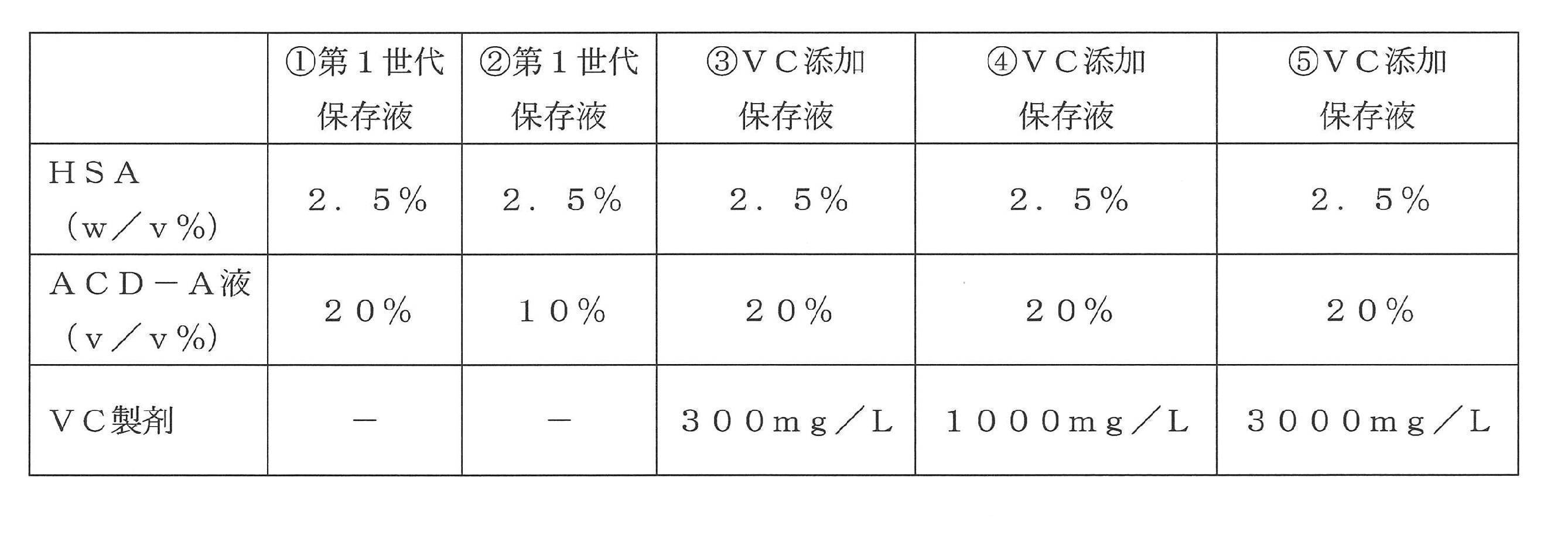
実施例1により得られた培養物に含まれる血小板の濃度を、FACSにより測定した。必要量の血小板を含む培養物を分取してACD-A液(10v/v%)及びPEG1(終濃度2μM;Cayman Chemical Company社製)を添加し、12分間の遠心分離(1200×g、22℃)を行った。上清を除去した後に、ペレットに上記(2-1)で調製した保存液を加え、均一な懸濁液となるように穏やかに懸濁した(血小板濃度:約0.3×10⁹plts/mL)。各懸濁液を24ウェルプレートに播種して最長5日間水平振盪保存(遮光下、22度、50rpm)した後に、以下の(2-3)及び(2-4)の実験に供した。
Annexin Vは血小板の劣化(活性化)の指標の一つであり、陽性率が高い場合には血小板が劣化している、又は異常であると評価される。そこで、上記(2-2)により得られた血小板サンプルにおけるAnnexin V陽性率を測定し、各保存液による血小板の劣化抑制効果を調べた。
具体的には、上記(2-2)により得られた血小板サンプルをAnnexin Buffer(Beckton Dickinson社製)で500倍希釈し、3本の遠心管に分注した(それぞれ、ネガティブコントロール、ポジティブコントロール、及び無刺激サンプル)。ネガティブコントロールサンプルにはEDTAを、ポジティブコントロールサンプルにはIonomycinをそれぞれ添加した後に、全てのサンプルを抗CD41抗体(BioLegend社製)及びAnnexin V(Beckton Dickinson社製)により染色した(遮光下、室温、20分間)。染色後に、Annexin Bufferを加えて速やかにFACSで測定した。ネガティブコントロールにおけるiPS血小板(CD41+分画)中のAnnexin V陽性率を1.0±0.1%とし、無刺激サンプルにおけるAnnexin V陽性率を算出した。
結果を図2の上段に示す(いずれも、5日間保存後血小板サンプルから得られた結果)。血小板(CD41+分画)のAnnexin V陽性率は、第1世代保存液を用いた場合には60.5±0.3~64.4±0.1%であったのに対し、VC添加保存液を用いた場合には53.9±2.1~56.7±1.8%であり、VC添加により低下することが明らかとなった。これらの結果から、300~3000mg/LのVCを添加することによって、血小板保存液の血小板劣化抑制作用が改善することが明らかとなった。
P―Selectinは血小板の劣化(活性化)の指標の一つであり、無刺激時のP―Selectin陽性率が高い場合には血小板が劣化又は異常であると評価される。そこで、上記(2-2)により得られた血小板サンプルにおけるP―Selectin陽性率を測定し、各保存液による血小板の劣化抑制効果を調べた。
また、血小板機能の保存状態を知るために、血小板活性化剤であるアデノシン二リン酸(ADP)及びトロンビン受容体活性化ペプチド-6(TRAP-6)により各血小板サンプルを刺激した時の、PAC-1及びP―Selectin陽性率を調べた(以下、ADP及びTRAP-6の組合せを「ATR」と称する場合がある)。
具体的には、上記(2-2)により得られた血小板サンプルをTyroad HEPES Buffer(THB)により500倍希釈し、3本の遠心管に分注した(それぞれ、ネガティブコントロール、ポジティブコントロール、及び無刺激サンプル)。ポジティブコントロールサンプルにADP(Sigma社製、終濃度20μM)及びTRAP-6(BACHEM社製、終濃度30μM)の混合液を添加し、刺激後のポジティブコントロールサンプル及び無刺激サンプルを、抗CD41抗体(BioLegend社製)、抗P―Selectin抗体(BioLegend社製)、及び抗PAC-1抗体(Beckton Dickinson社製)により染色した(遮光下、室温、30分間)。また、ネガティブコントロールサンプルは、抗CD41抗体(BioLegend社製)、抗P―Selectin抗体のアイソタイプコントロール抗体(BioLegend社製)、及び抗PAC-1抗体のアイソタイプコントロール抗体(BioLegend社製)により染色した(遮光下、室温、30分間)。染色後のサンプルに1%パラホルムアルデヒドを加えて固定し(遮光下、4℃、30分以上)、その後24時間以内にFACSによりP―Selectin及び/又はPAC―1陽性率を測定した。解析の際には、ネガティブコントロールサンプルにおけるiPS血小板(CD41+分画)中の、P-Selectin陽性率及びPAC-1陽性率が1.0±0.1%以下となるようにゲーティングを行った。
また、ATR刺激後の血小板(CD41+分画)におけるPAC―1/P―Selectin陽性率は、第1世代保存液を用いた場合には17.6~20.8%であったのに対し、VC添加保存液を用いた場合には23.9~26.9%であり、VC添加により上昇することが明らかとなった。これらの結果から、300~3000mg/LのVCを添加することによって、血小板保存液の血小板機能維持作用が改善することが示された。
(3-1)血小板保存液の調製
VC添加保存液にニコチン酸(ニコチン酸注射製剤;トーアエイヨー社製)を添加した溶液を調製した。なお、上述の通り、本願明細書において、「VB3」はニコチン酸及び/又はニコチンアミドを意味するが、実施例3~5、及び8~13ではVB3としてニコチン酸が使用されており、実施例6ではVB3としてニコチン酸又はニコチンアミドが使用されている。また、本明細書においては、VC添加保存液にVB3(ニコチン酸又はニコチンアミド)を添加した溶液を「VC/VB3添加保存液」又は「第2世代保存液」ともいう。各保存液における、上記添加剤の最終濃度は以下の表3に示す通りである。また、いずれの保存液も1MのNaOHによりpH7.3±0.1となるように調整し、使用時まで1時間以上インキュベートした(遮光下、室温、5%CO2)。
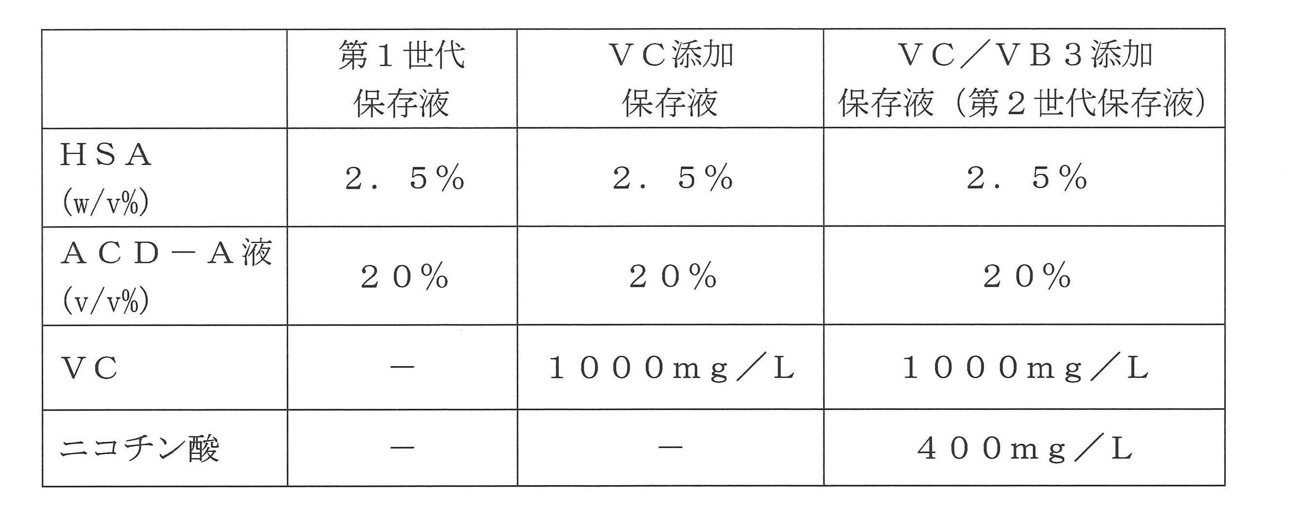
実施例1に記載の方法によって作製したiPS細胞由来血小板製剤に、上記(3-1)で調製した保存液を加え、均一な懸濁液となるように穏やかに懸濁した(血小板濃度:約0.3×10⁹plts/mL)。各懸濁液を直ちに、又は24ウェルプレートに播種して最長5日間水平振盪保存(遮光下、22度、50rpm)した後に、以下の(3-3)~(3-5)の実験に供した。
実施例2の(2-3)に記載の方法によって、上記(3-2)により得られた血小板サンプル(5日間保存後)におけるAnnexin V陽性率を測定し、各保存液による血小板の劣化抑制効果を調べた。
結果を図3に示す。血小板(CD41+分画)のAnnexin V陽性率は、第1世代保存液を用いた場合には61.4±1.9%、VC添加保存液を用いた場合には46.3±0.7%、VC/VB3添加保存液を用いた場合には44.3±0.5%であった。これらの結果から、VCとVB3を組み合わせて使用することによって、VCのみと比較して、血小板の劣化がより強く抑制されることが明らかとなった。
保存中の血小板製剤では嫌気的代謝により乳酸が産生されており、乳酸濃度の上昇に伴ってpHが低下し、結果として血小板の劣化が起こることが知られている。そこで、上記(3-2)により得られた血小板サンプルにおける乳酸濃度を測定し、各保存液による乳酸産生抑制作用を調べた。
具体的には、上記(3-2)により得られた血小板サンプル(5日間保存後)を、1.5mLチューブに入れて遠心した(1200×g、22℃、10分間)。上清を回収して新しいチューブに入れ、測定まで-80℃で凍結保存した。N-アッセイL LACニットーボー(ニットーボーメディカル株式会社製)及び自動分析装置7180(株式会社日立ハイテクノロジーズ製)を用いて、上記上清中の乳酸濃度を測定した。
結果を図4に示す。サンプル上清中の乳酸濃度は、第1世代保存液を用いた場合には0.35±0.01g/L、VC添加保存液を用いた場合には0.16±0.01g/L、VC/VB3添加保存液を用いた場合には0.13±0.01g/Lであった。これらの結果から、VCとVB3を組み合わせて使用することによって、VCのみと比較して、保存中の血小板における嫌気的代謝がより強く抑制されることが明らかとなった。
保存時における血小板の活性化は、容器への付着や、凝集塊の形成を引き起こし、血小板回収率低下の原因となると考えられている。そこで、上記(3-2)により得られた、保存前及び保存後の血小板サンプルにおける血小板濃度を測定し、各保存液による回収率の変化を調べた。
具体的には、上記(3-2)で作製した血小板サンプル(保存前、及び5日間保存後)をTHBにより500倍希釈し、TruCOUNTチューブ(Beckton Dickinson社製)に分注した。抗CD41抗体(BioLegend社製)及び抗CD42b抗体(BioLegend社製)により染色し(遮光下、室温、20分間)、再度THBを加えた後にFACSで測定した。TruCOUNTチューブのビーズカウント値に基づいて、CD41+細胞(血小板)の濃度を算出した。各血小板サンプルについて、保存前の血小板濃度を分母とし、保存後の血小板濃度を分子として、回収率を算出した。
結果を図5に示す。血小板の回収率は、第1世代保存液を用いた場合には102.0±4.2%、VC添加保存液を用いた場合には101.1±6.8%、VC/VB3添加保存液を用いた場合には99.9±3.9%であった。これらの結果から、VC及び/又はVB3を添加しても、回収率に差が認められないことが明らかとなった。
(4-1)血小板保存液の調製と、血小板サンプルの作製
第1世代保存液(20%のACD-A液を含む)に、VC製剤(1000mg/L、沢井製薬社製)を添加し、VC添加保存液を調製した。また、当該VC添加保存液にニコチン酸(400mg/L、トーアエイヨー社製)を添加し、第2世代保存液を調製した。各溶液のpHは7.3±0.1となるように調整した。以下の表4に、本実施例で使用した第2世代保存液の組成を示す。
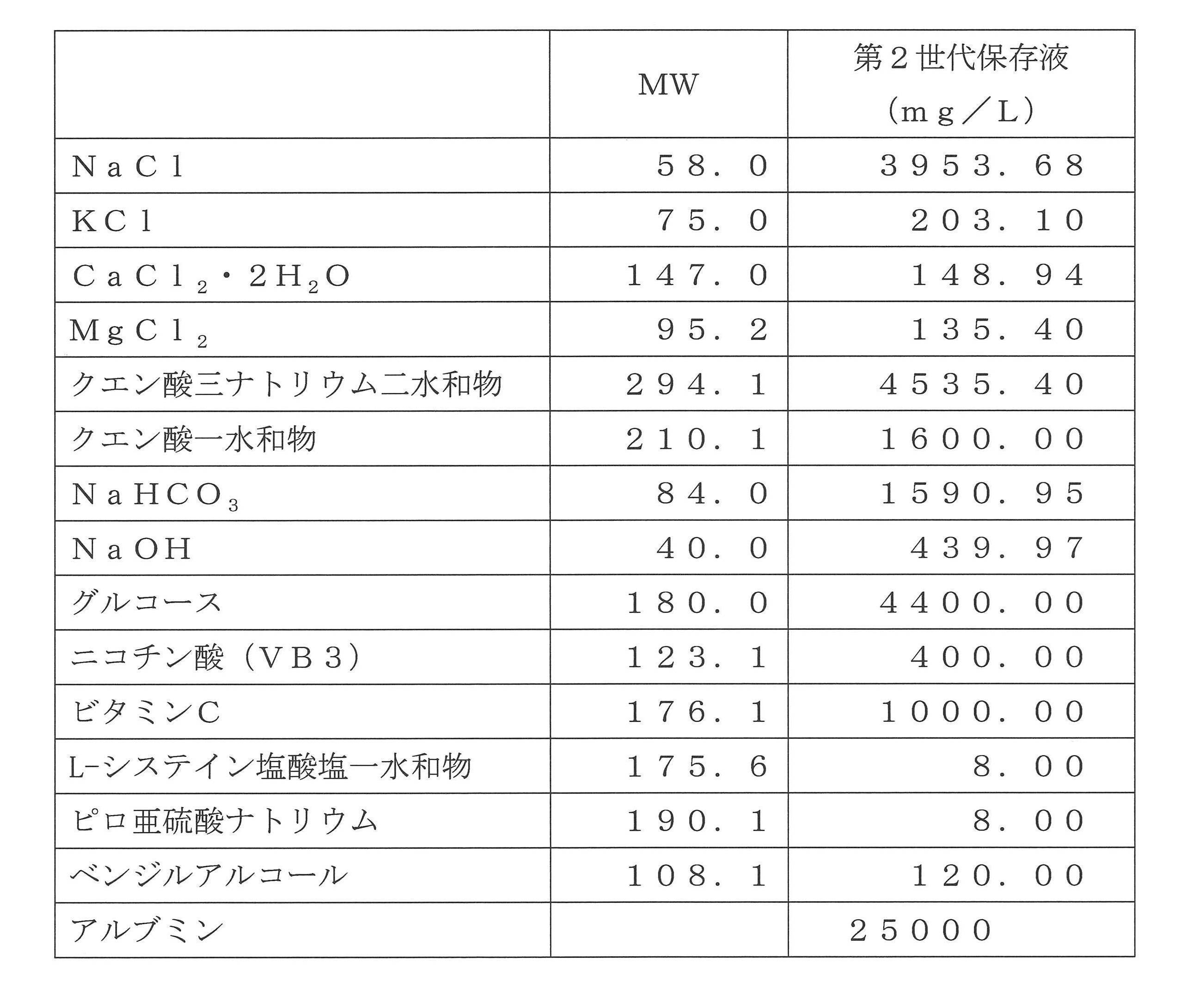
上記(2-4)でも述べたように、保存中の血小板製剤では嫌気的代謝により乳酸が産生されており、それがpHの低下の原因となることが知られている。本実験では、上記(4-1)により得られた血小板サンプルにおける乳酸濃度及びpHを測定し、各保存液による乳酸産生抑制作用を調べた。
具体的には、上記(4-1)により得られた血小板サンプル(保存前、5日間保存後、及び10日間保存後)を1.5mLチューブに入れ、細胞培養用分析装置FLEX(Nova Biomedical社製)により、乳酸濃度及びpHを測定した。
結果を図6に示す。図6の左グラフに示すように、乳酸濃度はいずれの保存液を用いた場合でも経時的に上昇していたが、第2世代保存液を用いたサンプルでは上昇の程度が最も低かった。また、図6の右グラフに示すように、pHはいずれの保存液を用いた場合でも経時的に低下していたが、第2世代保存液を用いたサンプルでは低下の程度が最も低く、10日間保存後でも中性に保たれていた。
実施例2の(2-4)でも述べたように、P―Selectinは血小板の劣化マーカーとして知られており、また、ATR(ADP/TRAP-6)刺激時のPAC―1/P―Selectinは血小板の反応性マーカーとして知られている。そこで本実験では、実施例2の(2-4)に記載の方法によって、上記(4-1)により得られた血小板サンプルにおける無刺激時のP―Selectin陽性率、及びATR刺激時のPAC―1/P―Selectin陽性率を測定し、各保存液による血小板の劣化抑制効果を調べた。
図7の左に無刺激時P―Selectin陽性率を、図7の右にATR刺激時PAC―1/P―Selectin陽性率をそれぞれ示す。P―Selectin陽性率は、第1世代保存液を用いた場合、保存前(Day1)が18.0%であったのに対し、5日間保存後(Day5)では33.0%、10日間保存後(Day10)では31.1%と上昇していた。一方、VC添加保存液を用いた場合、5及び10日間保存後のP―Selectin陽性率はいずれも第1世代保存液を用いた場合と比較して低く、それぞれ26.2%(Day5)及び24.4%(Day10)であった。また、第2世代保存液を用いた場合、5及び10日間保存後のP―Selectin陽性率は、VC添加保存液を用いた場合よりもさらに低く、それぞれ24.2%(Day5)及び19.3%(Day10)であった。以上の結果から、第2世代保存液を使用することによって、10日間保存後でも血小板の劣化が抑制されることが明らかとなった。
また、ATR刺激PAC―1/P―Selectin陽性率は、第1世代保存液を用いた場合、保存前(Day1)では43.4%であったのに対し、5日間保存後(Day5)では34.5%、10日間保存後(Day10)では5.9%と急激に低下していた。一方、VC添加保存液、又は第2世代保存液を用いた場合、ATR刺激PAC―1/P―Selectin陽性率は、5日間保存後(Day5)では第1世代保存液を用いた場合と同程度(それぞれ、35.9%及び35.3%)であったが、10日間保存後(Day10)では第1世代保存液よりも顕著に高い値を示した(それぞれ、19.4%及び19.9%)。以上の結果から、VC添加保存液、又は第2世代保存液を使用することによって、10日間保存後でも血小板の反応性が維持されることが明らかとなった。
上記(3-5)に記載の方法によって、上記(4-1)により得られた血小板サンプルにおける血小板濃度及び回収率を測定した。
結果を図8に示す。第1世代保存液を用いた場合の血小板回収率は、5日間保存後(Day5)では85.4%、10日間保存後(Day10)では86.8%であった。一方、VC添加保存液、又は第2世代保存液を用いた場合には、10日間保存後(Day10)でも血小板回収率が90%以上であった(それぞれ、90.0%及び91.4%)。これらの結果は、VC添加保存液、又は第2世代保存液を使用することによって、保存時の血小板活性化が抑制されるという上記(4-3)の結果と一致するものである。
(5-1)保存液及び血小板サンプルの調製
実施例3の(3-1)に記載の方法によって、第1世代保存液及び第2世代保存液を調製した。実施例1に記載の方法によって作製したiPS細胞由来血小板製剤に、上記保存液を加え、均一な懸濁液となるように穏やかに懸濁した(血小板濃度:約1.0×10⁹plts/mL)。各懸濁液を直ちに、又は血液保存バッグに充填して5若しくは10日間水平振盪保存(遮光下、22度、50rpm、)した後に、以下の(5-2)の実験に供した。
上記(5-1)により得られた血小板サンプルを遠心して(1200×g、室温、10分間)、上清を除去した。5%ACD-Aを含む重炭酸リンゲル液を加えて、細胞濃度が1.0×10⁹plts/mlとなるように懸濁した。得られた懸濁液をヒト血漿/CaCl2溶液(コスモ・バイオ社製)により希釈し、以下の表5に示す刺激剤を添加して刺激した。刺激後の各サンプルの凝集率を、血小板凝集能測定装置PRP313M(タイヨウ社製)により測定した。
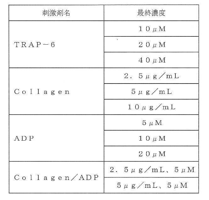
(6-1)保存液及び血小板サンプルの調製
第1世代保存液(20%のACD-A液を含む)に、VC(1000mg/L)と、ニコチン酸(400mg/L;富士フイルム和光純薬社製)又はニコチンアミド(400mg/L;富士フイルム和光純薬社製)とを添加し、1M NaOHを用いてpH7.3±0.1となるように調整した。以下、第1世代保存液にVC及びニコチン酸を添加した保存液を「第2世代保存液(ニコチン酸)」と、第1世代保存液にVC及びニコチンアミドを添加した保存液を「第2世代保存液(ニコチンアミド)」と、それぞれ称する場合がある。
実施例1に記載の方法によって作製したiPS細胞由来血小板製剤に、上記保存液を加え、均一な懸濁液となるように穏やかに懸濁した(血小板濃度:約0.3×10⁹plts/mL)。各懸濁液を直ちに、又は24ウェルプレートに播種して5日間水平振盪保存(遮光下、22度、50rpm)した後に、以下の(6-2)及び(6-3)の実験に供した。
実施例2の(2-3)に記載の方法によって、上記(6-1)により得られた血小板サンプルにおけるAnnexin V(劣化マーカー)陽性率を測定し、各保存液による血小板の劣化抑制効果を調べた。
結果を図13の上段に示す。図中、「Day1」は保存前のサンプルを、「第1世代」、「第2世代(ニコチン酸)」、「第2世代(ニコチンアミド)」は5日間保存後のサンプルをそれぞれ示す。血小板(CD41+分画)のAnnexin V陽性率は、ニコチン酸を添加した保存液では38.2±0.3%であり、ニコチンアミドを添加した保存液では38.5±0.3%であった。これらの値はいずれも、第1世代保存液を用いた場合(53.1±0.4%)よりも低かった。これらの結果から、広くビタミンB3(ニコチン酸及び/又はニコチンアミド)でも、保存中の血小板の劣化が抑制されることが明らかとなった。
実施例2の(2-4)に記載の方法によって、上記(6-1)により得られた血小板サンプルにおける無刺激時のP―Selectin(劣化マーカー)陽性率、及びATR刺激時のPAC―1/P―Selectin(反応性マーカー)陽性率を測定し、各保存液による血小板の劣化抑制効果を調べた。
図13の中段に無刺激時P―Selectin陽性率を、図13の下段にATR刺激時PAC―1/P―Selectin陽性率をそれぞれ示す。P―Selectin陽性率は、第1世代保存液を用いた場合、保存前(Day1)の11.6%から5日間保存後(Day5)の28.3±0.5%へと上昇していた。一方、ニコチン酸又はニコチンアミドを添加した第2世代保存液を用いた場合には、5日間保存後でも9.4±0.2%~10.6±0.2%と低く抑えられていた。
また、ATR刺激PAC―1/P―Selectin陽性率は、第1世代保存液を用いた場合には33.9±2.4%であったが、ニコチン酸又はニコチンアミドを添加した第2世代保存液を用いた場合には41.6±0.8%~41.2±1.9%であった。これらの結果から、ニコチン酸又はニコチンアミドにより保存中の血小板の反応性が維持されることが明らかとなった。
(7-1)保存液及び血小板サンプルの調製
上記実施例2~6では、「VC」として沢井製薬社製のVC製剤を用いた。当該VC製剤には、ピロ亜硫酸ナトリウム、L-システイン塩酸塩一水和物及びベンジルアルコールからなる添加物が含まれていることから、これら添加物が実験結果に影響していないかを、以下の実験により確認した。
第1世代保存液(20%のACD-A液を含む)に、VC試薬(添加剤を含まないVC;1000mg/L、富士フイルム和光純薬社製)を添加し、pH7.3±0.1となるように調整した。以下、かかる保存液を「第1世代保存液(VC試薬)」と称する場合がある。実施例1に記載の方法によって作製したiPS細胞由来血小板製剤に、上記保存液を加え、均一な懸濁液となるように穏やかに懸濁した(血小板濃度:約0.3×109plts/mL)。各懸濁液を直ちに、又は24ウェルプレートに播種して5日間水平振盪保存(遮光下、22度、50rpm)した後に、以下の(7-2)及び(7-3)の実験に供した。
実施例2の(2-3)に記載の方法によって、上記(7-1)により得られた血小板サンプルにおけるAnnexin V(劣化マーカー)陽性率を測定し、各保存液による血小板の劣化抑制効果を調べた。
結果を図14の上段に示す(Day1は保存前のサンプルを、第1世代及び第1世代VC試薬は5日間保存後のサンプルをそれぞれ示す)。血小板(CD41+分画)のAnnexin V陽性率は、第1世代保存液を用いた場合(53.1±0.4%)よりも、第1世代VC試薬を用いた場合(38.9±0.4%)の方が低かった。これらの結果から、VCのみの添加(VB3を含まない)により、保存中のiPS由来血小板の劣化が抑制されることが明らかとなった。
実施例2の(2-4)に記載の方法によって、上記(7-1)により得られた血小板サンプルにおける無刺激時のP―Selectin(劣化マーカー)陽性率、及びATR刺激時のPAC―1/P―Selectin(反応性マーカー)陽性率を測定し、各保存液による血小板の劣化抑制効果を調べた。
図14の中段に無刺激時P―Selectin陽性率を、図14の下段にATR刺激時PAC―1/P―Selectin陽性率をそれぞれ示す。無刺激時P―Selectin陽性率は、第1世代保存液を用いた場合(28.3±0.5%)よりも、第1世代VC試薬を用いた場合(9.5±0.4%)の方が低かった。これらの結果から、VCのみの添加により、保存中のiPS由来血小板の劣化が抑制されることが明らかとなった。
また、ATR刺激PAC―1/P―Selectin陽性率は、第1世代保存液を用いた場合(33.9±2.4%)よりも、第1世代VC試薬を用いた場合(38.2±1.6%)の方が高かった。これらの結果から、VC試薬のみの添加により、保存中のiPS由来血小板の反応性が維持されることが明らかとなった。すなわち、VCの添加効果は、沢井製薬社製のVC製剤に含まれる添加剤によるものではないことが明らかになった。
(8-1)保存液及び血小板サンプルの調製
ビカネイト輸液に、以下の表6に示す添加剤を添加して、第2世代保存液を調製した。また、既知の血小板保存液(日本赤十字社、照射洗浄血小板-LR「日赤」の添付文書、2016年3月、及び、Japanese Journal of Transfusion and Cell Therapy, Vol. 59.No. 3 59(3):492―498, 2013)と同じ組成の溶液を調製した。本明細書においては、当該既知の保存液を「日赤血小板洗浄液」ともいう。いずれの保存液も1MのNaOHによりpH7.3±0.1となるように調整し、使用時まで1時間以上インキュベートした(遮光下、室温、5%CO2)。実施例1に記載の方法によって作製したiPS細胞由来血小板製剤に、上記保存液を加え、均一な懸濁液となるように穏やかに懸濁した(血小板濃度:約1.0×10⁹plts/mL)。各懸濁液を直ちに、又は血液保存バッグに充填して5日間水平振盪保存(遮光下、22度、50rpm)した後に、以下の(8-2)及び(8-3)の実験に供した。
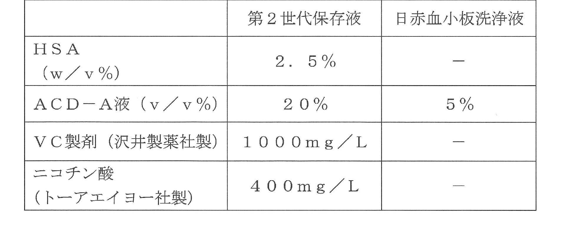
実施例2の(2-3)に記載の方法によって、上記(8-1)により得られた血小板サンプルにおけるAnnexin V(劣化マーカー)陽性率を測定し、各保存液による血小板の劣化抑制効果を調べた。
結果を図15上段の上段に示す(「Day1」は保存前のサンプルを、「Day5」は5日間保存後のサンプルをそれぞれ示す)。血小板(CD41+分画)のAnnexinV陽性率は、日赤血小板洗浄液を用いた場合(73.9%)よりも、第2世代保存液を用いた場合(49.3%)の方が低かった。これらの結果から、第2世代保存液により、保存中のiPS由来血小板の劣化が抑制されることが明らかとなった。
実施例2の(2-4)に記載の方法によって、上記(8-1)により得られた血小板サンプルにおける無刺激時のP―Selectin(劣化マーカー)陽性率、及びATR刺激時のPAC―1/P―Selectin(反応性マーカー)陽性率を測定し、各保存液による血小板の劣化抑制効果を調べた。
図15の中段に無刺激時P―Selectin陽性率を、図15の下段にATR刺激時PAC―1/P―Selectin陽性率をそれぞれ示す。無刺激時P―Selectin陽性率は、日赤血小板洗浄液を用いた場合(30.4%)よりも、第2世代保存液を用いた場合(16.5%)の方が低かった。これらの結果から、iPS由来血小板の保存において第2世代保存液は、日赤血小板洗浄液により優れた劣化抑制効果を奏することが明らかとなった。
また、ATR刺激PAC―1/P―Selectin陽性率は、日赤血小板洗浄液を用いた場合(9.8%)よりも、第2世代保存液を用いた場合(24.8%)の方が高かった。これらの結果から、iPS由来血小板の保存において第2世代保存液は、日赤血小板洗浄液により優れた機能維持効果を奏することが明らかとなった。
(9-1)血小板サンプルの調製
実施例1に記載の方法によって作製したiPS細胞由来血小板製剤に、第2世代保存液を加え、均一な懸濁液となるように穏やかに懸濁した(血小板濃度:約1.0×10⁹plts/mL)。各懸濁液を直ちに、又は血液保存バッグに充填して水平振盪保存(遮光下、22度、50rpm、10日間)した後に、以下の(9-2)の実験に供した。
血小板減少症モデルNOGマウスの尾静脈内に、上記(9-1)により得られた血小板サンプルを投与した(マウス1匹あたり200μL(2×10⁸plts))。投与10分後、注射針を用いて腹側の尾動脈に切創を作製した。切創は1個体あたり1か所とした。切創からの出血を確認した後に、切創箇所を含む尾先端部を37℃の生理食塩水に漬けて、止血までの時間を測定した。測定時間は最大600秒とした。また、対照群(Vehicle)として、第2世代保存液のみ(血小板を含まない)を用いて同様の試験を行った。
結果を図16に示す。Vehicle投与群では全てのマウスにおいて、600秒間以内での止血は認められなかった。一方、血小板投与群では、止血までの時間は平均で391秒であり、最も短い個体では130秒であった。これらの結果から、本発明の第2世代保存液により保存されたiPS細胞由来血小板が止血効果を有することが明らかとなった。
(10-1)間葉系幹細胞保存液の調製
乳酸リンゲル液(ラクテック輸液;大塚製薬工場社製)に、3%トレハロース及び5%デキストランを添加した(以下、かかる溶液を「CSP-01溶液」と称する場合がある。特開2012-115253、及びWO2014/208053参照)。上記CSP-01溶液に、VC製剤(1000mg/L、沢井製薬社製)及びニコチン酸(400mg/L、トーアエイヨー社製)を加え、間葉系幹細胞保存液を調製した。また、CSP-01溶液に、VC及びニコチン酸に代えて蒸留水(大塚蒸留液;大塚製薬工場社製)を加えたコントロール保存液を調製した。
上記(10-1)により調製した保存液を用いてヒト骨髄由来間葉系幹細胞(Lonza社製)を懸濁した(5×10⁵細胞/mL)。懸濁液を5℃で24、48、96、及び168時静置した後に、顕微鏡を用いて全細胞数及び死細胞数を計測した。各時点での細胞生存率(%)及び生細胞回収率(%)を以下の式1及び2を用いて算出した。
細胞生存率(%)=(全細胞数-死細胞数)/全細胞数×100
生細胞回収率(%)=各時点の生存細胞数/懸濁直後(保存前)の生存細胞数×100
(11-1)iPS細胞由来巨核球の調製
実施例1により得られた培養物に含まれる血小板の濃度を、FACSにより測定した。必要量の培養物を分取してACD-A液(10v/v%)及びPEG1(終濃度2μM;Cayman Chemical Company社製)を添加し、12分間の遠心分離(1200×g、22℃、ブレーキ最小)を行った。上清を除去した後に、ペレットに第1世代又は第2世代保存液を加え、均一な懸濁液となるように穏やかに懸濁した(血小板濃度:約1.3×10⁹plts/mL)。各懸濁液を直ちに、又は血液保存バッグに充填して5又は10日間水平振盪保存(遮光下、22度、50rpm)した後に、以下の(11-2)の実験に供した。
Annexin Vは、アポトーシス細胞における細胞膜の変化(フォスファチジルセリンの細胞膜外側への表出)を検出するプローブとしても知られている。そこで、上記(11-1)により得られた巨核球サンプルにおけるAnnexin V陰性率を測定し、各保存液による巨核球のアポトーシス抑制効果を調べた。
具体的には、上記(11-1)により得られた巨核球サンプル(巨核球及び血小板を含む培養物)をAnnexin Buffer(Beckton Dickinson社製)で500倍希釈し、3本の遠心管に分注した(それぞれ、ネガティブコントロール、ポジティブコントロール、及び無刺激サンプル)。ネガティブコントロールサンプルにはEDTAを、ポジティブコントロールサンプルにはIonomycinをそれぞれ添加した後に、全てのサンプルを抗CD41抗体(BioLegend社製)及びAnnexin V(Beckton Dickinson社製)により染色した(遮光下、室温、20分間)。染色後に、Annexin Bufferを加えて速やかにFACSで測定した。得られたFSC(前方散乱光)及びSSC(側方散乱光)の値に基づき血小板と巨核球とを区分した。そして、ネガティブコントロールにおけるiPS血小板(CD41+分画)中のAnnexin V陽性率を1.0±0.1%とし、無刺激サンプルにおける巨核球のAnnexin V陰性率を算出した。
結果を図18に示す(「Day1」は保存前のサンプルを、「Day5」は5日間保存後のサンプルを、「Day10」は10日間保存後のサンプルをそれぞれ示す)。Annexin V陰性率は、第1世代保存液を用いたサンプルでも、第2世代保存液を用いたサンプルでも、経時的に低下した。しかし、第2世代保存液を用いた場合には、第1世代保存液を用いた場合よりも、Annexin V陰性率の低下が抑制されることが明らかとなった。これらの結果から、第2世代保存液を用いたサンプルにおいては、アポトーシスを起こしていない、細胞膜が安定した生細胞がより高い割合で含まれることが示された。したがって、本実施例により、第2世代保存液がiPS細胞由来巨核球の保存にも有効であることが示された。
(12-1)T細胞保存液の調製
乳酸リンゲル液(ラクテック輸液;大塚製薬工場社製)に3%トレハロースを添加した(以下、かかる溶液を「CSP-11溶液」と称する場合がある)。上記CSP-11溶液に、VC製剤(1000mg/L、沢井製薬社製)及び/又はニコチン酸(400mg/L、トーアエイヨー社製)を加え、以下の4種類のT細胞保存液を調製した。
CSP-11
CSP-11+VC
CSP-11+ニコチン酸
CSP-11+VC+ニコチン酸
市販の凍結CD8陽性T細胞(ベリタス社製)を融解して、リンパ球培養培地(LGM3、Lonza社製)で洗浄した後に、約1時間インキュベートした(37℃、5%CO2)。必要量のCD8陽性T細胞を分取して、10分間の遠心分離(300×g、室温)を行った。上清を取り除いた後に、TLY CULTUREキット25(GCリンフォテック社製)を用いて細胞を浮遊させ、37℃、5%CO2条件下で培養して増殖させた(T細胞濃度:約1.1×10⁶cells/5ml)。拡大培養開始から7日後に、CSP-11溶液で細胞を洗浄して、ステムフルチューブ(住友ベークライト社製)に分注し、10分間の遠心分離(300×g、室温)を行った。上清を取り除いた後に、上記(12-1)で調製した保存液を加えて懸濁した(T細胞濃度:約5×105cells/1ml)。各ステムフルチューブから20μLの細胞懸濁液を分取し、20μLトリパンブルー(gibco社製)を混合し、ワンセルカウンター(バイオメディカルサイエンス社製)を用いて生存率を測定した(1ヵ所の細胞計数部の四隅の細胞計数室のエリアの合計細胞数及び死細胞数を計測した)。また、上記細胞懸濁液を5℃で48時間保存した後に、同様に生存率を測定した。
(13-1)T細胞保存液の調製
CSP-01溶液に、VC製剤(1000mg/L、沢井製薬社製)、ニコチン酸(400mg/L、トーアエイヨー社製)、及び/又はグルコース(80mg/dL、大塚製薬工場社製)を加え、以下の4種類のT細胞保存液を調製した。
CSP-01
CSP-01+VC+ニコチン酸
CSP-01+グルコース
CSP-01+グルコース+VC+ニコチン酸
市販の凍結CD8陽性T細胞(ベリタス社製)を融解して、リンパ球培養培地(LGM3、Lonza社製)で洗浄した後に、約6時間インキュベートした(37℃、5%CO2)。必要量のCD8陽性T細胞を分取して、10分間の遠心分離(300×g、室温)を行った。上清を取り除いた後に、TLY CULTUREキット25(GCリンフォテック社製)を用いて細胞を浮遊させ、37℃、5%CO2条件下で培養して増殖させた(T細胞濃度:約1.2×10⁶cells/5ml)。拡大培養開始から6日後に、3%トレハロース含有乳酸リンゲル液で細胞を洗浄して、ステムフルチューブ(住友ベークライト社製)に分注し、10分間の遠心分離(300×g、室温)を行った。上清を取り除いた後に、上記(12-1)で調製した保存液を加えて懸濁した(T細胞濃度:約5×105cells/1ml)。各ステムフルチューブから20μLの細胞懸濁液を分取し、20μLトリパンブルー(gibco社製)を混合し、ワンセルカウンター(バイオメディカルサイエンス社製)を用いて生存率を測定した(1ヵ所の細胞計数部の四隅の細胞計数室のエリアの合計細胞数及び死細胞数を計測した)。また、上記細胞懸濁液を5℃で24又は48時間保存した後に、同様に生存率を測定した。得られた各時点での生存率から、以下の式3を用いて生細胞回収率を算出した。
生細胞回収率(%)=(保存後の生細胞数)÷(保存前の生細胞数)×100
(14-1)水溶性ビタミン群を添加した保存液の調製
第1世代保存液(20%のACD-A液を含む)に、水溶性ビタミン群(B1、VB2、VB3、VB5、VB6、VB7、VB9、VB12、及びVC)を添加した(以下、かかる保存液を「第1世代+水溶性ビタミン」と称する場合がある)。また、第1世代保存液(20%のACD-A液を含む)に、上記水溶性ビタミン群からVB2を除いた群(B1、VB3、VB5、VB6、VB7、VB9、VB12、及びVC)を添加した(以下、かかる保存液を「第1世代+水溶性ビタミン(VB2除く)」と称する場合がある)。いずれの保存液も、1M NaOHを用いてpH7.3±0.1となるように調整した。
実施例1に記載の方法によって作製したiPS細胞由来血小板製剤に、上記保存液を加え、均一な懸濁液となるように穏やかに懸濁した(血小板濃度:約0.3×10⁹plts/mL)。各懸濁液を直ちに、又は24ウェルプレートに播種して5日間水平振盪保存(遮光下、22度、50rpm)した後に、以下の(13-2)及び(13-3)の実験に供した。
実施例2の(2-3)に記載の方法によって、上記(14-1)により得られた血小板サンプルにおけるAnnexin V(劣化マーカー)陽性率を測定し、各保存液による血小板の劣化抑制効果を調べた。
結果を図21に示す。図中、「Day1」は保存前のサンプルを、「Day5」は5日間保存後のサンプルをそれぞれ示す。5日間保存後の血小板(CD41+分画)のAnnexin V陽性率は、第1世代保存液を用いた場合には、52.0%であったのに対し、水溶性ビタミン群を添加した保存液では43.0%に低下していた。また、Annexin V陽性率は、水溶性ビタミン群(VB2除く)を添加した保存液ではさらに低下しており、40.3%であった。これらの結果から、VC及びVB3による血小板劣化抑制効果が、VB2により阻害される可能性が示された。
図22の左側に無刺激時P―Selectin陽性率を、図22の右側にATR刺激時PAC―1/P―Selectin陽性率をそれぞれ示す。P―Selectin陽性率は、第1世代保存液を用いた場合、保存前(Day1)の27.4%から5日間保存後(Day5)の42.9%へと上昇していた。一方、5日間保存後のP―Selectin陽性率は、水溶性ビタミン群を添加した保存液を用いた場合には34.9%と低く抑えられており、水溶性ビタミン群(VB2除く)を添加した保存液を用いた場合には24.0%とさらに低く抑えられていた。
また、ATR刺激PAC―1/P―Selectin陽性率は、第1世代保存液を用いた場合、保存前(Day1)の35.6%から5日間保存後(Day5)の32.4%へと低下していた。一方、水溶性ビタミン群を添加した保存液を用いた場合には、5日間保存後でも36.8%であった。さらに、水溶性ビタミン群(VB2除く)を添加した保存液を用いた場合には、5日間保存後に43.0%と上昇することが明らかとなった。これらの結果から、VC及びVB3による血小板機能維持効果が、VB2により阻害される可能性が示された。
(15-1)間葉系幹細胞保存液の調製
実施例10の(10-1)の記載に従ってCSP-01溶液を調製した。かかるCSP-01溶液に、VC製剤(1000mg/L、沢井製薬社製)及びニコチン酸(400mg/L、トーアエイヨー社製)を加え、間葉系幹細胞保存液を調製した。また、CSP-01溶液に、VC及びニコチン酸に代えて、溶媒である蒸留水(大塚蒸留液;大塚製薬工場社製)を加えたコントロール保存液を調製した。
上記(15-1)により調製した保存液を用いてヒト骨髄由来間葉系幹細胞(Lonza社製)を懸濁した(5x10⁵細胞/mL)。懸濁液を5℃で、1、2、4、7、14、21、28、35及び63日静置した後に、顕微鏡を用いて全細胞数及び死細胞数を計測した。各時点での細胞生存率(%)及び生細胞回収率(%)は、実施例10の(10-2)に記載の式1及び2を用いて算出した。
(16-1)間葉系幹細胞保存液の調製
CSP-01溶液又は乳酸リンゲル液(ラクテック輸液;大塚製薬工場社製)に、VC製剤(1000mg/L、沢井製薬社製)及びニコチン酸(400mg/L、トーアエイヨー社製)を加え、以下の4種の間葉系幹細胞保存液を調製した。
CSP-01
CSP-01+VC+ニコチン酸
乳酸リンゲル液
乳酸リンゲル液+VC+ニコチン酸
上記(16-1)により調製した保存液を用いてヒト脂肪由来間葉系幹細胞(Lonza社製)を懸濁した(5x10⁵細胞/mL)。懸濁液を5℃で7、14、21及び28日静置した後に、顕微鏡を用いて全細胞数及び死細胞数を計測した。各時点での細胞生存率(%)及び生細胞回収率(%)は、実施例10の(10-2)に記載の式1及び2を用いて算出した。
(17-1)間葉系幹細胞保存液の調製
CSP-01溶液に、VC製剤(1000mg/L、沢井製薬社製)及び/又はニコチン酸(400mg/L、トーアエイヨー社製)を加え、さらに炭酸水素ナトリウム製剤(メイロン静注8.4%、大塚製薬工場社製)を加えてpHを7.0~7.3に調整した。また、コントロール保存液として、CSP-01溶液に、炭酸水素ナトリウム製剤(メイロン静注8.4%、大塚製薬工場社製)を加えてpHを7.0~7.3に調整した。以上のようにして、以下の4種の間葉系幹細胞保存液を調製した。
CSP-01
CSP-01+VC
CSP-01+ニコチン酸
CSP-01+VC+ニコチン酸
上記(17-1)により調製した保存液を用いてヒト脂肪由来間葉系幹細胞(Lonza社製)を懸濁した(5x10⁵細胞/mL)。懸濁液を5℃で7、14、21及び28日静置した後に、顕微鏡を用いて全細胞数及び死細胞数を計測した。各時点での細胞生存率(%)及び生細胞回収率(%)は、実施例10の(10-2)に記載の式1及び2を用いて算出した。
(18-1)間葉系幹細胞保存液の調製
CSP-01溶液に、VC製剤(1000mg/L、沢井製薬社製)及びニコチン酸(400mg/L、トーアエイヨー社製)を加え、間葉系幹細胞保存液(CSP-01+VC+ニコチン酸)を調製した。また、コントロール保存液として、CSP-01溶液にのみを用いた。
Nishimuraら(Xenotransplantation. 2019 May;26(3):e12501.)の方法に従って、幼若ブタ骨髄由来間葉系幹細胞(np間葉系幹細胞)を作製した。かかるnp間葉系幹細胞を、上記(18-1)により調製した保存液を用いて懸濁した(5x10⁵細胞/mL)。懸濁液を5℃で7、14、21及び28日静置した後に、顕微鏡を用いて全細胞数及び死細胞数を計測した。各時点での細胞生存率(%)及び生細胞回収率(%)は、実施例10の(10-2)に記載の式1及び2を用いて算出した。
(19-1)T細胞保存液の調製
乳酸リンゲル液(ラクテック輸液;大塚製薬工場社製)に、VC製剤(1000mg/L、沢井製薬社製)、ニコチン酸(400mg/L、トーアエイヨー社製)、及び/又はグルコース(80mg/dL、大塚製薬工場社製)を加え、以下の8種類のT細胞保存液を調製した。
LR
LR+グルコース
LR+VC
LR+ニコチン酸
LR+VC+ニコチン酸
LR+VC+グルコース
LR+ニコチン酸+グルコース
LR+VC+ニコチン酸+グルコース
市販の凍結CD8陽性T細胞(ベリタス社製)を融解して、リンパ球培養培地(LGM3、Lonza社製)で洗浄した後に、1時間インキュベートした(37℃、5%CO2)。必要量のCD8陽性T細胞を分取して、10分間の遠心分離(300×g、室温)を行った。上清を取り除いた後に、TLY CULTUREキット25(GCリンフォテック社製)を用いて細胞を浮遊させ、37℃、5%CO2条件下で培養して増殖させた(T細胞濃度:約8×10⁵cells/5ml)。拡大培養開始から7日後に、PBS(-)で細胞を洗浄して、ステムフルチューブ(住友ベークライト社製)に分注し、10分間の遠心分離(300×g、室温)を行った。上清を取り除いた後に、上記(19-1)で調製した保存液を加えて懸濁した(T細胞濃度:約5×105cells/1ml)。上記細胞懸濁液を5℃で24時間保存した時点、及び、その後さらに25℃で6時間保存した時点(合計30時間保存後)で、顕微鏡を用いて全細胞数及び死細胞数を計測した。各時点での細胞生存率(%)及び生細胞回収率(%)は、実施例10の(10-2)に記載の式1及び2を用いて算出した。
Claims (51)
- ナイアシン若しくはその誘導体又はそれらの塩、及び抗酸化剤を含む、哺乳動物細胞の保存液。
- 抗酸化剤が、アスコルビン酸若しくはその誘導体又はそれらの塩である、請求項1に記載の保存液。
- ナイアシン若しくはその誘導体又はそれらの塩の濃度が、1~10000mg/Lである、請求項1又は2に記載の保存液。
- ナイアシン若しくはその誘導体又はそれらの塩の濃度が、30~3000mg/Lである、請求項1又は2に記載の保存液。
- ナイアシン若しくはその誘導体又はそれらの塩の濃度が、120~1200mg/Lである、請求項1又は2に記載の保存液。
- アスコルビン酸若しくはその誘導体又はそれらの塩の濃度が、1~10000mg/Lである、請求項2~5のいずれか1項に記載の保存液。
- アスコルビン酸若しくはその誘導体又はそれらの塩の濃度が、30~6000mg/Lである、請求項2~5のいずれか1項に記載の保存液。
- アスコルビン酸若しくはその誘導体又はそれらの塩の濃度が、300~3000mg/Lである、請求項2~5のいずれか1項に記載の保存液。
- 哺乳動物細胞を0~40℃で保存するための、請求項1~8のいずれか1項に記載の保存液。
- ビタミンB2若しくはその誘導体又はそれらの塩を含まない、請求項1~9のいずれか1項に記載の保存液。
- 哺乳動物細胞が、血小板又は巨核球である、請求項1~10のいずれか1項に記載の保存液。
- 血小板が、以下の(A)及び(B)を含む方法により得られた精製血小板である、請求項11に記載の保存液。
(A)巨核球の培養物を濃縮する濃縮工程;
(B)得られた濃縮物から血小板を遠心分離する遠心分離工程; - さらにアルブミンを含む、請求項11又は12に記載の保存液。
- アルブミンの濃度が、1.25~10%(w/v)である、請求項13に記載の保存液。
- さらに糖を含む、請求項13又は14に記載の保存液。
- 糖が、ブドウ糖である、請求項15に記載の保存液。
- 血小板又は巨核球を、5~10日間保存するための、請求項11~16のいずれか1項に記載の保存液。
- 哺乳動物細胞が、幹細胞又は免疫細胞である、請求項1~10のいずれか1項に記載の保存液。
- 幹細胞が、間葉系幹細胞である、請求項18に記載の保存液。
- 免疫細胞が、T細胞である、請求項18に記載の保存液。
- ナイアシン若しくはその誘導体又はそれらの塩、及び抗酸化剤を等張液中に含む、請求項18~20のいずれか1項に記載の保存液。
- 等張液が、乳酸リンゲル液である、請求項21に記載の保存液。
- さらにトレハロースを含む、請求項21又は22に記載の保存液。
- さらにデキストランを含む、請求項21~23のいずれか1項に記載の保存液。
- 幹細胞を、1~63日間保存するための、請求項18~24のいずれか1項に記載の保存液。
- ナイアシン若しくはその誘導体又はそれらの塩、及び抗酸化剤を含む、請求項1~25のいずれか1項に記載の保存液を調製するための粉末製剤。
- ナイアシン若しくはその誘導体又はそれらの塩、及び抗酸化剤を含む液中で、哺乳動物細胞を保存する工程を含む、哺乳動物細胞の保存方法。
- 抗酸化剤が、アスコルビン酸若しくはその誘導体又はそれらの塩である、請求項27に記載の方法。
- ナイアシン若しくはその誘導体又はそれらの塩の濃度が、1~10000mg/Lである、請求項27又は28に記載の方法。
- ナイアシン若しくはその誘導体又はそれらの塩の濃度が、30~3000mg/Lである、請求項27又は28に記載の方法。
- ナイアシン若しくはその誘導体又はそれらの塩の濃度が、120~1200mg/Lである、請求項27又は28に記載の方法。
- アスコルビン酸若しくはその誘導体又はそれらの塩の濃度が、1~10000mg/Lである、請求項28~31のいずれか1項に記載の方法。
- アスコルビン酸若しくはその誘導体又はそれらの塩の濃度が、30~6000mg/Lである、請求項28~31のいずれか1項に記載の方法。
- アスコルビン酸若しくはその誘導体又はそれらの塩の濃度が、300~3000mg/Lである、請求項28~31のいずれか1項に記載の方法。
- 哺乳動物細胞を0~40℃で保存することを特徴とする、請求項27~34のいずれか1項に記載の方法。
- 液が、ビタミンB2若しくはその誘導体又はそれらの塩を含まないことを特徴とする、請求項27~35のいずれか1項に記載の方法。
- 哺乳動物細胞が、血小板又は巨核球である、請求項27~36のいずれか1項に記載の方法。
- 血小板が、以下の(A)及び(B)を含む方法により得られた精製血小板である、請求項37に記載の方法。
(A)巨核球の培養物を濃縮する濃縮工程;
(B)得られた濃縮物から血小板を遠心分離する遠心分離工程; - 液中にさらにアルブミンを含む、請求項37又は38に記載の方法。
- アルブミンの濃度が、1.25~10%(w/v)である、請求項39に記載の方法。
- 液中にさらに糖を含む、請求項39又は40に記載の方法。
- 糖が、ブドウ糖である、請求項41に記載の方法。
- 血小板又は巨核球を、5~10日間保存することを特徴とする、請求項37~42のいずれか1項に記載の方法。
- 哺乳動物細胞が、幹細胞又は免疫細胞である、請求項27~36のいずれか1項に記載の方法。
- 幹細胞が、間葉系幹細胞である、請求項44に記載の方法。
- 免疫細胞が、T細胞である、請求項44に記載の方法。
- 液が、等張液である、請求項44~46のいずれか1項に記載の方法。
- 等張液が、乳酸リンゲル液である、請求項47に記載の方法。
- 液中にさらにトレハロースを含む、請求項47又は48に記載の方法。
- 液中にさらにデキストランを含む、請求項47~49のいずれか1項に記載の方法。
- 幹細胞を、1~63日間保存することを特徴とする、請求項44~50のいずれか1項に記載の方法。
Priority Applications (11)
| Application Number | Priority Date | Filing Date | Title |
|---|---|---|---|
| KR1020257008707A KR20250040760A (ko) | 2019-03-15 | 2020-03-13 | 포유동물 세포의 보존액 |
| US17/438,025 US20220145235A1 (en) | 2019-03-15 | 2020-03-13 | Storage liquid for mammalian cells |
| CN202080020194.5A CN113557295A (zh) | 2019-03-15 | 2020-03-13 | 哺乳动物细胞的保存液 |
| KR1020217029129A KR102784950B1 (ko) | 2019-03-15 | 2020-03-13 | 포유동물 세포의 보존액 |
| EP20773511.9A EP3940061A4 (en) | 2019-03-15 | 2020-03-13 | PRESERVATION LIQUID FOR MAMMALIAN CELLS |
| SG11202109825S SG11202109825SA (en) | 2019-03-15 | 2020-03-13 | Storage liquid for mammalian cells |
| AU2020241910A AU2020241910B2 (en) | 2019-03-15 | 2020-03-13 | Storage liquid for mammalian cells |
| CA3129733A CA3129733A1 (en) | 2019-03-15 | 2020-03-13 | Storage liquid for mammalian cells |
| JP2021507298A JP7169503B2 (ja) | 2019-03-15 | 2020-03-13 | 哺乳動物細胞の保存液 |
| JP2022036343A JP7653944B2 (ja) | 2019-03-15 | 2022-03-09 | 哺乳動物細胞の保存液 |
| AU2024201062A AU2024201062B2 (en) | 2019-03-15 | 2024-02-19 | Storage liquid for mammalian cells |
Applications Claiming Priority (2)
| Application Number | Priority Date | Filing Date | Title |
|---|---|---|---|
| JP2019-047852 | 2019-03-15 | ||
| JP2019047852 | 2019-03-15 |
Publications (1)
| Publication Number | Publication Date |
|---|---|
| WO2020189538A1 true WO2020189538A1 (ja) | 2020-09-24 |
Family
ID=72519323
Family Applications (1)
| Application Number | Title | Priority Date | Filing Date |
|---|---|---|---|
| PCT/JP2020/011002 Ceased WO2020189538A1 (ja) | 2019-03-15 | 2020-03-13 | 哺乳動物細胞の保存液 |
Country Status (9)
| Country | Link |
|---|---|
| US (1) | US20220145235A1 (ja) |
| EP (1) | EP3940061A4 (ja) |
| JP (4) | JP7169503B2 (ja) |
| KR (2) | KR20250040760A (ja) |
| CN (1) | CN113557295A (ja) |
| AU (2) | AU2020241910B2 (ja) |
| CA (1) | CA3129733A1 (ja) |
| SG (1) | SG11202109825SA (ja) |
| WO (1) | WO2020189538A1 (ja) |
Cited By (1)
| Publication number | Priority date | Publication date | Assignee | Title |
|---|---|---|---|---|
| CN112998009A (zh) * | 2021-03-31 | 2021-06-22 | 北京益华生物科技有限公司 | Nk细胞冻存液及其制备方法和应用 |
Families Citing this family (8)
| Publication number | Priority date | Publication date | Assignee | Title |
|---|---|---|---|---|
| JP6830294B1 (ja) * | 2019-04-26 | 2021-02-17 | 株式会社大塚製薬工場 | トレハロースを含む哺乳動物細胞保存用液 |
| CN115039759B (zh) * | 2022-03-07 | 2023-11-03 | 细胞生态海河实验室 | 一种红细胞保存液及其应用 |
| JP2023173802A (ja) * | 2022-05-26 | 2023-12-07 | 株式会社カネカ | 細胞凝集塊組成物の製造方法 |
| CN115281180A (zh) * | 2022-07-04 | 2022-11-04 | 四川大学华西医院 | 一种mRNA-血小板复合物冻存液及其制备方法 |
| CN115363016B (zh) * | 2022-08-09 | 2023-07-18 | 广州明迅生物科技有限责任公司 | 细胞储存液及其应用 |
| CN117736985B (zh) * | 2023-12-20 | 2024-12-20 | 深圳泽医细胞治疗集团有限公司 | 适用于干细胞样记忆t细胞培养的血液保存液及应用 |
| CN118370308B (zh) * | 2024-04-18 | 2025-11-11 | 维赛(佛山)生物科技有限公司 | 一种临床用犬自然杀伤细胞冷藏保存液及其制备方法和应用 |
| WO2025257595A1 (en) * | 2024-06-12 | 2025-12-18 | Biocompatibility Innovation S.r.l. | Preserving solution |
Citations (11)
| Publication number | Priority date | Publication date | Assignee | Title |
|---|---|---|---|---|
| JP2012115253A (ja) | 2010-11-09 | 2012-06-21 | Otsuka Pharmaceut Factory Inc | 幹細胞懸濁液 |
| WO2014208053A1 (ja) | 2013-06-28 | 2014-12-31 | 株式会社大塚製薬工場 | トレハロース及びデキストラン含有哺乳動物細胞移植用溶液 |
| US20160205923A1 (en) * | 2015-01-21 | 2016-07-21 | Hong Seung Kim | Activated platelet preservation composition, method for preserving activated platelet and preserved activated platelet using the same |
| JP2016538859A (ja) * | 2013-11-19 | 2016-12-15 | プラトードPlatod | 血小板産生流体装置 |
| WO2017065280A1 (ja) * | 2015-10-14 | 2017-04-20 | 株式会社メガカリオン | 精製血小板の製造方法 |
| WO2017073656A1 (ja) | 2015-10-27 | 2017-05-04 | 株式会社カネカ | 間葉系幹細胞を含む細胞集団の製造方法、間葉系幹細胞、細胞集団、並びに医薬組成物 |
| WO2017077964A1 (ja) | 2015-11-02 | 2017-05-11 | 株式会社メガカリオン | 往復動撹拌装置を用いた血小板の製造方法 |
| WO2018097226A1 (ja) * | 2016-11-25 | 2018-05-31 | テルモ株式会社 | 生細胞または生細胞を含む組成物の保存液 |
| JP2018515099A (ja) * | 2015-05-12 | 2018-06-14 | プラトードPlatod | 改善された血小板産生のための薬理学的特徴およびマイクロ流体特徴の組み合わせ |
| JP2018516955A (ja) * | 2015-06-09 | 2018-06-28 | プレジデント・アンド・フェロウズ・オブ・ハーバード・カレッジ | 血小板の長期貯蔵および保存の方法 |
| JP2018153196A (ja) | 2016-12-14 | 2018-10-04 | 株式会社大塚製薬工場 | 哺乳動物細胞投与用液 |
Family Cites Families (16)
| Publication number | Priority date | Publication date | Assignee | Title |
|---|---|---|---|---|
| US4767704A (en) * | 1983-10-07 | 1988-08-30 | Columbia University In The City Of New York | Protein-free culture medium |
| US5104787A (en) * | 1990-03-05 | 1992-04-14 | Lindstrom Richard L | Method for apparatus for a defined serumfree medical solution useful for corneal preservation |
| JPH057619A (ja) * | 1991-06-24 | 1993-01-19 | L Lyndstrom Richard | 血清不含医療用規定溶液およびその溶液を用いた角膜の保存方法 |
| WO2004112631A1 (en) * | 2003-06-20 | 2004-12-29 | Terumo Penpol Limited | A FORMULATION AND A PROCESS FOR REDUCING THE DETERIORATION OF RBCs OF BLOOD DURING STORAGE WITH PARTICULAR REFERENCE TO DECREASE IN THE LEVEL OF 2,3 DIPHOSPHOGLYCERATE |
| FR2856891B1 (fr) * | 2003-07-04 | 2007-09-07 | Stem Alpha | Milieu de conservation d'organes, de tissus biologiques ou de cellules vivantes |
| BRPI0517333A (pt) * | 2004-11-12 | 2008-10-07 | Doorzand Airdrive B V | solução de conservação e perfusão de órgãos para manter a viabilidade do órgão doador, e, método para conservar um órgão |
| CN105123670B (zh) * | 2007-02-17 | 2018-12-14 | 哈佛学院董事会 | 用于进行组织保存的组合物以及方法 |
| US7892724B2 (en) * | 2007-07-12 | 2011-02-22 | Warsaw Orthopedic, Inc | Method for enhancing the viability of mammalian cells, tissues and organs using a solution comprising two low molecular weight PEGs |
| US7960098B2 (en) * | 2007-07-12 | 2011-06-14 | Warsaw Orthoperic, Inc. | Methods and compositions for the preservation of cells and tissues |
| JP2013532661A (ja) * | 2010-07-22 | 2013-08-19 | リベン ファーマシューティカルズ インコーポレイテッド | 磁気双極子安定化溶液の使用を含む疾患を処置または改善する方法および行動を向上させる方法 |
| CN103404509B (zh) * | 2013-08-07 | 2015-04-08 | 彭乐 | 一种细胞保存液、其制备方法和应用 |
| ES2536605B1 (es) * | 2013-10-24 | 2016-03-02 | Fundación Pública Andaluza Progreso Y Salud | Método de obtención de megacariocitos y plaquetas |
| WO2016091349A1 (en) | 2014-12-11 | 2016-06-16 | Merck Patent Gmbh | Cell culture media |
| FR3040860B1 (fr) * | 2015-09-10 | 2020-05-15 | Universite de Bordeaux | Milieu de conservation injectable pour la conservation de cellules du sang placentaire, de la moelle osseuse et du sang peripherique |
| US10886195B2 (en) | 2016-08-18 | 2021-01-05 | Intel Corporation | Systems and methods for improved through-silicon-vias |
| WO2018097225A1 (ja) | 2016-11-25 | 2018-05-31 | テルモ株式会社 | 生細胞または生細胞を含む組成物の保存液 |
-
2020
- 2020-03-13 AU AU2020241910A patent/AU2020241910B2/en active Active
- 2020-03-13 CA CA3129733A patent/CA3129733A1/en active Pending
- 2020-03-13 JP JP2021507298A patent/JP7169503B2/ja active Active
- 2020-03-13 KR KR1020257008707A patent/KR20250040760A/ko active Pending
- 2020-03-13 EP EP20773511.9A patent/EP3940061A4/en active Pending
- 2020-03-13 US US17/438,025 patent/US20220145235A1/en active Pending
- 2020-03-13 CN CN202080020194.5A patent/CN113557295A/zh active Pending
- 2020-03-13 WO PCT/JP2020/011002 patent/WO2020189538A1/ja not_active Ceased
- 2020-03-13 SG SG11202109825S patent/SG11202109825SA/en unknown
- 2020-03-13 KR KR1020217029129A patent/KR102784950B1/ko active Active
-
2021
- 2021-09-28 JP JP2021158368A patent/JP7653884B2/ja active Active
-
2022
- 2022-03-09 JP JP2022036343A patent/JP7653944B2/ja active Active
-
2024
- 2024-02-19 AU AU2024201062A patent/AU2024201062B2/en active Active
-
2025
- 2025-03-17 JP JP2025042877A patent/JP2025087922A/ja active Pending
Patent Citations (11)
| Publication number | Priority date | Publication date | Assignee | Title |
|---|---|---|---|---|
| JP2012115253A (ja) | 2010-11-09 | 2012-06-21 | Otsuka Pharmaceut Factory Inc | 幹細胞懸濁液 |
| WO2014208053A1 (ja) | 2013-06-28 | 2014-12-31 | 株式会社大塚製薬工場 | トレハロース及びデキストラン含有哺乳動物細胞移植用溶液 |
| JP2016538859A (ja) * | 2013-11-19 | 2016-12-15 | プラトードPlatod | 血小板産生流体装置 |
| US20160205923A1 (en) * | 2015-01-21 | 2016-07-21 | Hong Seung Kim | Activated platelet preservation composition, method for preserving activated platelet and preserved activated platelet using the same |
| JP2018515099A (ja) * | 2015-05-12 | 2018-06-14 | プラトードPlatod | 改善された血小板産生のための薬理学的特徴およびマイクロ流体特徴の組み合わせ |
| JP2018516955A (ja) * | 2015-06-09 | 2018-06-28 | プレジデント・アンド・フェロウズ・オブ・ハーバード・カレッジ | 血小板の長期貯蔵および保存の方法 |
| WO2017065280A1 (ja) * | 2015-10-14 | 2017-04-20 | 株式会社メガカリオン | 精製血小板の製造方法 |
| WO2017073656A1 (ja) | 2015-10-27 | 2017-05-04 | 株式会社カネカ | 間葉系幹細胞を含む細胞集団の製造方法、間葉系幹細胞、細胞集団、並びに医薬組成物 |
| WO2017077964A1 (ja) | 2015-11-02 | 2017-05-11 | 株式会社メガカリオン | 往復動撹拌装置を用いた血小板の製造方法 |
| WO2018097226A1 (ja) * | 2016-11-25 | 2018-05-31 | テルモ株式会社 | 生細胞または生細胞を含む組成物の保存液 |
| JP2018153196A (ja) | 2016-12-14 | 2018-10-04 | 株式会社大塚製薬工場 | 哺乳動物細胞投与用液 |
Non-Patent Citations (10)
| Title |
|---|
| "Japanese Red Cross Society, the package insert of Irradiated Washed Platelets-LR ''NISSEKI", JAPANESE JOURNAL OF TRANSFUSION AND CELL THERAPY, vol. 59, no. 3, 2013, pages 492 - 498 |
| CAMPBELL, CLIN VACCINE IMMUNOL, vol. 16, 2009, pages 1648 - 53 |
| GIAMMONA, L. M. ET AL.: "Nicotinamide (vitamin B3) increases the polyploidisation and proplatelet formation of cultured primary human megakaryocytes", BRITISH JOURNAL OF HAEMATOLOGY, vol. 135, 2006, pages 554 - 566, XP055135582, DOI: 10.1111/j.1365-2141.2006.06341.x * |
| HIGA, YUKIE : "No. 16 Effect of preserved PC on platelet function by addition of water-soluble vitamins, About the effect of vitamin C", JOURNAL OF THE JAPAN SOCIETY OF BLOOD TRANSFUSION, vol. 48, no. 1, 2002, pages 48, XP009530637, ISSN: 0546-1448 * |
| KOBAYASHI ET AL., CELL, vol. 142, no. 5, 2010, pages 787 - 799 |
| NISHIMURA, XENOTRANSPLANTATION, vol. 26, no. 3, May 2019 (2019-05-01), pages e12501 |
| TAKAYAMA ET AL., J. EXP. MED., vol. 13, 2010, pages 2817 - 2830 |
| TAKAYAMA, BLOOD, vol. 111, 2008, pages 5298 - 5306 |
| TYLER, BLOOD, vol. 121, 2013, pages 308 - 317 |
| XU, Y. ET AL.: "Tannic acid attenuated irradiation-induced apoptosis in megakaryocytes", EXPERIMENTAL CELL RESEARCH, vol. 370, 2018, pages 409 - 416, XP085447194, DOI: 10.1016/j.yexcr.2018.07.003 * |
Cited By (1)
| Publication number | Priority date | Publication date | Assignee | Title |
|---|---|---|---|---|
| CN112998009A (zh) * | 2021-03-31 | 2021-06-22 | 北京益华生物科技有限公司 | Nk细胞冻存液及其制备方法和应用 |
Also Published As
| Publication number | Publication date |
|---|---|
| AU2024201062B2 (en) | 2025-05-29 |
| US20220145235A1 (en) | 2022-05-12 |
| JP2021192640A (ja) | 2021-12-23 |
| CA3129733A1 (en) | 2020-09-24 |
| JPWO2020189538A1 (ja) | 2021-09-13 |
| JP7653884B2 (ja) | 2025-03-31 |
| TW202102664A (zh) | 2021-01-16 |
| JP7653944B2 (ja) | 2025-03-31 |
| EP3940061A4 (en) | 2023-01-04 |
| JP2022071183A (ja) | 2022-05-13 |
| KR102784950B1 (ko) | 2025-03-21 |
| JP7169503B2 (ja) | 2022-11-11 |
| AU2020241910A1 (en) | 2021-10-07 |
| KR20210126086A (ko) | 2021-10-19 |
| AU2024201062A1 (en) | 2024-03-07 |
| EP3940061A1 (en) | 2022-01-19 |
| KR20250040760A (ko) | 2025-03-24 |
| CN113557295A (zh) | 2021-10-26 |
| AU2020241910B2 (en) | 2023-12-21 |
| SG11202109825SA (en) | 2021-10-28 |
| JP2025087922A (ja) | 2025-06-10 |
Similar Documents
| Publication | Publication Date | Title |
|---|---|---|
| JP7653944B2 (ja) | 哺乳動物細胞の保存液 | |
| JP5998265B2 (ja) | トレハロース及びデキストラン含有哺乳動物細胞移植用溶液 | |
| Douay et al. | Ex vivo production of human red blood cells from hematopoietic stem cells: what is the future in transfusion? | |
| KR102689261B1 (ko) | 정제 혈소판의 제조 방법 | |
| CA2711549A1 (en) | Method of producing a population of cells | |
| JP7570456B2 (ja) | 精製血小板の製造方法、血小板製剤の製造方法、血液製剤の製造方法、血小板保存液、血小板保存剤および血小板の保存方法 | |
| CN110882276B (zh) | 细胞治疗组合物及治疗血管病变的方法 | |
| Glück et al. | Characterization and transfusion of in vitro cultivated hematopoietic progenitor cells | |
| WO2021193606A1 (ja) | アカルボース及びデキストランを含む哺乳動物細胞保存用液 | |
| WO2025154708A1 (ja) | 高濃度細胞保存液及び高濃度細胞保存方法 | |
| WO2021145364A1 (ja) | トレハロースを含む血球系細胞保存用液 | |
| McFarland et al. | ASH 2010 meeting report—Top 10 clinically oriented abstracts in transfusion medicine |
Legal Events
| Date | Code | Title | Description |
|---|---|---|---|
| 121 | Ep: the epo has been informed by wipo that ep was designated in this application |
Ref document number: 20773511 Country of ref document: EP Kind code of ref document: A1 |
|
| ENP | Entry into the national phase |
Ref document number: 2021507298 Country of ref document: JP Kind code of ref document: A |
|
| ENP | Entry into the national phase |
Ref document number: 3129733 Country of ref document: CA |
|
| ENP | Entry into the national phase |
Ref document number: 20217029129 Country of ref document: KR Kind code of ref document: A |
|
| NENP | Non-entry into the national phase |
Ref country code: DE |
|
| ENP | Entry into the national phase |
Ref document number: 2020241910 Country of ref document: AU Date of ref document: 20200313 Kind code of ref document: A |
|
| WWE | Wipo information: entry into national phase |
Ref document number: 2020773511 Country of ref document: EP |
|
| WWD | Wipo information: divisional of initial pct application |
Ref document number: 1020257008707 Country of ref document: KR |
|
| WWP | Wipo information: published in national office |
Ref document number: 1020257008707 Country of ref document: KR |
|
| WWG | Wipo information: grant in national office |
Ref document number: 11202109825S Country of ref document: SG |
|
| WWP | Wipo information: published in national office |
Ref document number: 11202109825S Country of ref document: SG |