WO2020204149A1 - Procédé de criblage et procédé d'évaluation de toxicité - Google Patents
Procédé de criblage et procédé d'évaluation de toxicité Download PDFInfo
- Publication number
- WO2020204149A1 WO2020204149A1 PCT/JP2020/015255 JP2020015255W WO2020204149A1 WO 2020204149 A1 WO2020204149 A1 WO 2020204149A1 JP 2020015255 W JP2020015255 W JP 2020015255W WO 2020204149 A1 WO2020204149 A1 WO 2020204149A1
- Authority
- WO
- WIPO (PCT)
- Prior art keywords
- cells
- complement
- cell
- medium
- human
- Prior art date
- Legal status (The legal status is an assumption and is not a legal conclusion. Google has not performed a legal analysis and makes no representation as to the accuracy of the status listed.)
- Ceased
Links
Images


Classifications
-
- G—PHYSICS
- G01—MEASURING; TESTING
- G01N—INVESTIGATING OR ANALYSING MATERIALS BY DETERMINING THEIR CHEMICAL OR PHYSICAL PROPERTIES
- G01N33/00—Investigating or analysing materials by specific methods not covered by groups G01N1/00 - G01N31/00
- G01N33/48—Biological material, e.g. blood, urine; Haemocytometers
- G01N33/50—Chemical analysis of biological material, e.g. blood, urine; Testing involving biospecific ligand binding methods; Immunological testing
- G01N33/5005—Chemical analysis of biological material, e.g. blood, urine; Testing involving biospecific ligand binding methods; Immunological testing involving human or animal cells
- G01N33/5008—Chemical analysis of biological material, e.g. blood, urine; Testing involving biospecific ligand binding methods; Immunological testing involving human or animal cells for testing or evaluating the effect of chemical or biological compounds, e.g. drugs, cosmetics
- G01N33/5014—Chemical analysis of biological material, e.g. blood, urine; Testing involving biospecific ligand binding methods; Immunological testing involving human or animal cells for testing or evaluating the effect of chemical or biological compounds, e.g. drugs, cosmetics for testing toxicity
-
- G—PHYSICS
- G01—MEASURING; TESTING
- G01N—INVESTIGATING OR ANALYSING MATERIALS BY DETERMINING THEIR CHEMICAL OR PHYSICAL PROPERTIES
- G01N33/00—Investigating or analysing materials by specific methods not covered by groups G01N1/00 - G01N31/00
- G01N33/48—Biological material, e.g. blood, urine; Haemocytometers
- G01N33/50—Chemical analysis of biological material, e.g. blood, urine; Testing involving biospecific ligand binding methods; Immunological testing
- G01N33/5005—Chemical analysis of biological material, e.g. blood, urine; Testing involving biospecific ligand binding methods; Immunological testing involving human or animal cells
- G01N33/5008—Chemical analysis of biological material, e.g. blood, urine; Testing involving biospecific ligand binding methods; Immunological testing involving human or animal cells for testing or evaluating the effect of chemical or biological compounds, e.g. drugs, cosmetics
- G01N33/5044—Chemical analysis of biological material, e.g. blood, urine; Testing involving biospecific ligand binding methods; Immunological testing involving human or animal cells for testing or evaluating the effect of chemical or biological compounds, e.g. drugs, cosmetics involving specific cell types
Definitions
- the present invention relates to a method for screening a therapeutic agent for a disease associated with complement hyperactivation using a cytotoxic marker associated with complement as an index.
- the present invention also relates to a method for evaluating cytotoxicity caused by overactivation of complement of a test substance using a cytotoxic marker associated with complement as an index.
- Complement is not only an important effector of innate immunity, but also involved in acquired immune response, inflammation, blood coagulation, and promotion of tumor progression.
- Examples of the mechanism for eliminating foreign substances by activating complement include opsonization, anaphylatoxin (C5a, C3a) production, and formation of the final product, complement membrane attack complex (MAC).
- MAC complement membrane attack complex
- complement activation functions as an ecological defense, it carries the risk of exerting biotoxicity in pathological conditions. For example, in the kidney, complement deposition in renal tissue causes polymembrane proliferative glomerulonephritis (MPGN), post-streptococcal acute glomerulonephritis (PSAGN), and lupus nephritis.
- MPGN polymembrane proliferative glomerulonephritis
- PSAGN post-streptococcal acute glomerulonephritis
- lupus nephritis lupus
- MAC membrane-invasive complex
- osmotic fluid movement and cation influx occur, resulting in cell death.
- abnormal complement activation causes various diseases. Therefore, using complement activation as an index, focusing on hemolytic action, treatment of diseases associated with complement overactivation Although drug thinning has been performed, these conventional methods do not consider cytotoxicity such as vascular injury that actually occurs due to complement activation.
- Non-Patent Document 1 MAC deposition also occurs in various cancer tissues, and normal human serum (NHS) can be used to activate alternative pathways of complement system in osteosarcoma cells, and human endothelium. It has been reported that the production of growth factors that promote cell angiogenesis is enhanced. However, in the document, in HUVEC (human umbilical vein endothelial cells) and the like other than human osteosarcoma epithelial cells U2-OS, the degree of MAC production is determined by "normal human serum” and "human serum inactivated by heat treatment”. It has also been reported that no difference is observed.
- HUVEC human umbilical vein endothelial cells
- U2-OS human serum inactivated by heat treatment
- An object of the present invention is to provide a method for screening a therapeutic agent for a disease associated with complement hyperactivation and a kit used for the screening, using a cytotoxic marker associated with complement as an index.
- the present inventors brought vascular endothelial cells differentiated from induced pluripotent stem cells into contact with normal human serum (NHS) containing complement or NHS heat-treated to inactivate complement.
- NHS normal human serum
- MAC complement membrane attack complex
- the amount of MAC deposited when an active complement is used is an antibody against complement component C5 at the end of the complement activation cascade, and can also be used as a therapeutic agent for atypical hemolytic urinary syndrome (aHus).
- the present inventors induce differentiation of a plurality of cells other than vascular endothelial cells from induced pluripotent stem cells in the same manner as vascular endothelial cells, and use the cells and complement to generate MAC.
- MAC deposition was caused by active complement regardless of the cell type, and that the amount of MAC deposition was decreased in a dose-dependent manner by administration of eculizumab.
- the present inventors have completed the present invention.
- the present invention provides the following [1] to [6] for solving the above problems.
- [1] A method of screening for therapeutic agents for diseases associated with complement hyperactivation using cytotoxic markers associated with complement as indicators.
- (1a) A step of adding complement to cells prepared from stem cells to form a cytotoxic marker associated with the complement, and (2a) a therapeutic drug candidate substance being added to reduce the amount of the cytotoxic marker.
- the process of selecting the substance to be used Including methods.
- [3] A method for evaluating cytotoxicity caused by complement activation of a test substance using a cytotoxic marker associated with complement as an index.
- Step of adding a test substance to cells prepared from stem cells (2b) A step of measuring the amount of cytotoxic marker produced associated with complement, and (3b) a step of evaluating cytotoxicity caused by complement overactivation from the amount of the cytotoxic marker produced. , Including methods.
- the cells prepared from the stem cells are vascular endothelial cells, liver sinusoidal endothelial cells, nerve cells, oligodendrocytes, hepatocytes or retinal pigment epithelial cells.
- (I-b) Cells made from stem cells, and (ii-b) Reagents for detecting cytotoxic markers associated with complement A kit for assessing cytotoxicity due to complement overactivation, including.
- a method for screening a therapeutic agent for a disease associated with complement hyperactivation using a cytotoxic marker associated with complement as an index Since it was confirmed that the formation of the cytotoxic marker was reduced by the known therapeutic agent for the disease, the effect of complement-induced vascular injury actually occurring in the living body can be affected by using the cytotoxic marker as an index. A more reliable screening that reflects this can be expected.
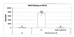
- FIG. 1 shows the formation of MAC in vascular endothelial cells prepared from human pluripotent stem cells after culturing in a medium containing a complement source (culture supernatants of human serum and organoids).
- a complement source culture supernatants of human serum and organoids.
- Heat induction shows the result of deactivating the complement supply source by allowing it to stand at 55 ° C. for 30 to 60 minutes.
- the MAC immunostaining results were quantified, and the results corrected by the same quantified nuclear staining (DAPI) were used as the results.
- FIG. 1 shows the formation of MAC in vascular endothelial cells prepared from human pluripotent stem cells after culturing in a medium containing a complement source (culture supernatants of human serum and organoids).
- 3% serum with LPS was used as the complement source (3% human serum and organoid culture supernatant treated with LPS).
- the results of the experiment are shown.
- Heat induction shows the result of de
- FIG. 2 shows the formation of MAC in vascular endothelial cells prepared from human pluripotent stem cells after culturing in a medium containing a complement source (culture supernatants of human serum and organoids).
- a complement source culture supernatants of human serum and organoids.
- FIG. 2 shows the results of experiments using no complement source, and 3% serum with LPS (3% human serum and organoid culture supernatant treated with LPS) with various concentrations of C5 antibody Eculizumab.
- the results of the experiment in which MAC was formed by adding to the above are shown.
- the MAC immunostaining results were quantified and the results corrected by the same quantified nuclear staining (DAPI) were used as the results.
- FIG. 3 shows each cell (nerve cell, hepatocyte and retinal pigment epithelial cell) prepared from human pluripotent stem cells after culturing in a medium containing a complement source (culture supernatant of human serum and organoid).
- a complement source culture supernatant of human serum and organoid.
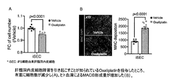
- FIG. 4 shows that administration of oxaliplatin, which is known to cause hepatic sinusoidal endothelial cell injury, significantly reduced the number of cells (A) and increased the amount of MAC formed by human serum (B). Shown.
- the present invention screens therapeutic agents for diseases associated with complement hyperactivation using a cytotoxic marker associated with complement (hereinafter, may be simply referred to as a "cytotoxic marker”) as an index.
- a method (hereinafter sometimes referred to as “screening method of the present invention”) is provided.
- the screening method of the present invention (1a) A step of adding complement to cells prepared from stem cells to form a cytotoxic marker, and (2a) a substance to which a therapeutic drug candidate substance is added to reduce the amount of the cytotoxic marker is selected. Process, including.
- atypical hemolytic uremic syndrome aHUS
- MS multiple sclerosis
- NASH non-alcoholic steatosis
- AMD age-related yellow spot degeneration
- PNH paroxysmal nocturnal hemochromatosis
- STC-HUS intestinal hemorrhagic Escherichia hemolytic uremic syndrome
- AHR acute humoral rejection
- AMR acute antibody-related rejection
- severe muscle Examples include asthenia, neuromyelitis optica, membranous proliferative glomerulonephritis (MPGN), dense deposit disease (DDD), cold agglutinosis, and fulminant antiphospholipid antibody syndrome (CAPS).
- MPGN membranous proliferative glomerulonephritis
- DDD dense deposit disease
- CAS fulminant antiphospholipid antibody syndrome
- the cytotoxic markers include, for example, MAC, lactate dehydrogenase (LDH), cell-mediated cytotoxicity (BrdU uptake), intracellular ATP, DNA fragmentation, Caspase-3 / 7, 8 , 9 reducing ability (NADH, etc.) and the like. Further, in the present specification, the formation of a cytotoxic marker is compared with a cell before contact with complement, a cell with contact with inactivated complement, or a cell without contact with complement. This means that the amount of cytotoxic markers (eg, MAC) formed (deposited) is increased.
- Non-Patent Document 1 As shown in the examples below, in multiple types of cells made from stem cells, active complement causes MAC deposition, and the amount of MAC deposition is dose-dependently reduced by administration of eculizumab. It was proven to do. This result was different from the result of human umbilical vein endothelial cells disclosed in Non-Patent Document 1. The reason for this difference is that the cell population used in Non-Patent Document 1 is a cell population separated from the living body, so that cells having a plurality of characteristics are mixed depending on the isolation method or the like. It may be due to being a group. Therefore, since the problem of the above variation can be avoided by preparing the cells to be used from stem cells, any cell type prepared from stem cells can be applied to the screening method of the present invention without any particular limitation.
- Examples of such cell types include nerve cells, oligodendrocytes, erythrocytes, mononuclear cells (eg, lymphocytes (NK cells, B cells, T cells, monospheres, dendritic cells, etc.)), granulocytes (eg, dendritic cells, etc.).
- nerve cells oligodendrocytes, erythrocytes, mononuclear cells (eg, lymphocytes (NK cells, B cells, T cells, monospheres, dendritic cells, etc.)), granulocytes (eg, dendritic cells, etc.).
- a method for producing (inducing differentiation) the cells from stem cells a method known per se can be appropriately selected according to the type of the target cell.
- vascular endothelial cells for example, Yamashita J. et al. et al. , Nature, 408 (6808): 92-96 (2000), Narazaki G. et al. et al. , Circulation. 29; 118 (5): 498-506 (2008), for example, as a method for producing a nerve cell, Yan Y. et al. , Stem Cells Trans Med, 2: 862-870 (2013), Kondo T. et al. , Et al.
- stem cells are cultured in a medium containing a serum substitute (eg B-27), BMP4 and a GSK-3 inhibitor (eg CHIR99021). After inducing the cells into mesoderm cells, the cells can be cultured in a medium containing VEGF and Folskolin to prepare vascular endothelial cells.
- a serum substitute eg B-27
- BMP4 eg BMP4
- GSK-3 inhibitor eg CHIR99021
- the cells are cultured in a medium containing Wnt protein (eg, Wnt3a) and Actibin A to induce endometrial cells, and then the cells are used as a serum substitute (B-27) and Hepatocytes can be prepared by culturing in a medium containing FGF2.
- Wnt protein eg, Wnt3a
- Actibin A to induce endometrial cells
- pluripotent stem cells examples include pluripotent stem cells.
- pluripotent stem cell can be differentiated into tissues and cells having various different morphologies and functions of the living body, and can be differentiated into three germ layers (endoderm, mesodermal, ectoderm). Refers to stem cells that have the ability to differentiate into cells of any lineage.
- the pluripotent stem cells are not particularly limited, and are, for example, artificial pluripotent stem cells (sometimes referred to as “iPS cells” in the present specification), embryonic stem cells (ES cells), and clones obtained by nuclear transplantation.
- pluripotent stem cells examples thereof include embryo-derived embryonic stem cells (nuclear transfer Embryonic stem cells: ntES cells), pluripotent embryonic stem cells, and embryonic embryonic stem cells (EG cells).
- ntES cells nuclear transfer Embryonic stem cells
- EG cells embryonic embryonic stem cells
- pluripotent stem cells examples include dental pulp stem cells, oral mucosa-derived stem cells, hair follicle stem cells, cultured fibroblasts, and somatic stem cells derived from bone marrow stem cells.
- Preferred pluripotent stem cells are ES cells and iPS cells.
- the pluripotent stem cell is an ES cell or an arbitrary cell derived from a human embryo
- the cell is a cell produced by destroying an embryo, even if the cell is produced by destroying the embryo.
- the stem cells are preferably derived from mammals (eg, mice, rats, hamsters, guinea pigs, dogs, monkeys, orangutans, chimpanzees, humans), and more preferably humans.
- “Induced pluripotent stem cell” refers to a cell obtained by introducing a specific factor (nuclear reprogramming factor) into a mammalian somatic cell or an undifferentiated stem cell and reprogramming it.
- a specific factor nuclear reprogramming factor
- iPS induced pluripotent stem cells
- Yamanaka et al. By introducing four factors, Oct3 / 4, Sox2, Klf4, and c-Myc, into mouse fibroblasts.
- human cell-derived iPS cells (Takahashi K, Yamanaka) established by introducing similar four factors into human fibroblasts.
- Nanog-iPS cells Okita, K., Ichisaka, T. et al.
- IPS cells prepared by a method that does not contain c-Myc (Nakagawa M, Yamanaka S., et al. Nature Biotechnology, (2008). , 101-106)
- iPS cells established by introducing 6 factors by a virus-free method (Okita K et al. Nat. Methods 2011 May; 8 (5): 409-12, Okita K et al.
- induced pluripotent stem cells (Yu J., Thomason JA. Et al., Science (2007) 318: established by introducing four factors of OCT3 / 4, SOX2, NANOG, and LIN28 produced by Thomason et al. 1917-1920.), Induced pluripotent stem cells produced by Dayy et al. (Park IH, Dayy GQ. Et al., Nature (2007) 451: 141-146), induced pluripotent stem cells produced by Sakurada et al. (Japanese Patent Laid-Open No. 2008-307007) and the like can also be used.
- any of the described pluripotent stem cells known in the art can be used.
- As the induced pluripotent cell line various iPS cell lines established by NIH, RIKEN, Kyoto University and the like can be used.
- RIKEN's HiPS-RIKEN-1A strain, HiPS-RIKEN-2A strain, HiPS-RIKEN-12A strain, Nippons-B2 strain, Kyoto University's 253G1 strain, 201B7 strain, 409B2 strain examples thereof include 454E2 strain, 606A1 strain, 610B1 strain, 648A1 strain and the like.
- ES cells are stem cells that are pluripotent and proliferative by self-renewal, established from the inner cell mass of early mammalian embryos (eg, blastocysts) such as humans and mice. ES cells were discovered in mice in 1981 (M.J.Evans and MH Kaufman (1981), Nature 292: 154-156), after which ES cell lines were established in primates such as humans and monkeys. (JA Thomason et al. (1998), Science 282: 1145-1147; JA Thomason et al. (1995), Proc. Nature. Acad. Sci. USA, 92: 7844-7884; JA Thomason et al. (1996), Biol.
- ES cells can be established by removing the inner cell mass from the blastocyst of the fertilized egg of the target animal and culturing the inner cell mass on a fibroblast feeder.
- a fibroblast feeder for methods of establishing and maintaining human and monkey ES cells, see, for example, USP 5,843,780; Thomson JA, et al. (1995), Proc Natl. Acad. Sci. US A. 92: 7844-7748; Thomason JA, et al. (1998), Science. 282: 1145-1147; Suemori H. et al. et al.
- ES cells can be established using only single blastomeres of embryos in the cleavage stage before the blastocyst stage (Chung Y. et al. (2008), Cell Stem Cell 2: 113-). 117), it can also be established using embryos that have stopped developing (Zhang X. et al. (2006), Stem Cells 24: 2669-2676.).
- RIKEN RIKEN
- CHB-1 to CHB-12 strains sold by ESI Bio RUES1 strains, RUES2 strains, HUES1 to HUES28 strains, etc.
- H1 strains sold by WiCell Research H9 strains, etc.
- KhES-1, KhES-2, KhES-3, KhES-4, KhES-5, SSES1, SSES2, SSES3, etc. to be sold can be used.
- nt ES cells are ES cells derived from cloned embryos produced by nuclear transfer technology and have almost the same characteristics as ES cells derived from fertilized eggs (Wakayama T. et al. (2001), Science, 292). : 740-743; S. Wakayama et al. (2005), Biol. Reprod., 72: 932-936; Byrne J. et al. (2007), Nature, 450: 497-502). That is, ES cells established from the inner cell mass of blastocysts derived from cloned embryos obtained by replacing the nuclei of unfertilized eggs with the nuclei of somatic cells are nt ES (nuclear transfer ES) cells.
- nt ES cells For the production of nt ES cells, a combination of nuclear transfer technology (Cibelli JB et al. (1998), Nature Biotechnol., 16: 642-646) and ES cell production technology (above) is used. (Seika Wakayama et al. (2008), Experimental Medicine, Vol. 26, No. 5 (Special Edition), pp. 47-52). In nuclear transfer, somatic cell nuclei can be injected into unfertilized eggs of mammals and cultured for several hours to initialize them.
- Pluripotent germ stem cells are pluripotent stem cells derived from germline stem cells (GS cells). Similar to ES cells, these cells can induce differentiation into cells of various lineages, and have properties such as being able to produce chimeric mice when transplanted into mouse blastocysts (Kanatus-Shinohara M. et al. (Kanatus-Shinohara M. et al.). 2003) Biol. Reprod., 69: 612-616; Shinohara K. et al. (2004), Cell, 119: 1001-1012). It is self-renewable in a culture medium containing a glial cell line-developed neurotrophic factor (GDNF), and is reproductive by repeating passage under the same culture conditions as ES cells. Stem cells can be obtained (Masanori Takebayashi et al. (2008), Experimental Medicine, Vol. 26, No. 5 (Special Edition), pp. 41-46, Yodosha (Tokyo, Japan)).
- GDNF
- EG cells are cells with pluripotency similar to ES cells, which are established from primordial germ cells in the embryonic period. It can be established by culturing primordial germ cells in the presence of substances such as LIF, bFGF, stem cell factor (Masui Y. et al. (1992), Cell, 70: 841-847; J. et al. L. Resnick et al. (1992), Nature, 359: 550-551).
- step (1a) -Step of forming a cytotoxic marker (step (1a))-
- the complement used in the above step (1a) is not particularly limited as long as it can form a cytotoxic marker, but for example, serum (eg, human serum, bovine serum, horse serum, sheep serum, goat serum, pig serum, llama). Serum, dog serum, chicken serum, donkey serum, cat serum, rabbit serum, guinea pig serum, hamster serum, rat serum, mouse serum, etc.)) and complements contained in the liver and vascular endothelial cells.
- serum eg, human serum, bovine serum, horse serum, sheep serum, goat serum, pig serum, llama
- Complement contained in the culture supernatant of (including), or each complement component eg, C1q, C1r, C1s, C2, C4, C3, C3a, C5s, C3b, C5a, C5b6789, and combinations thereof
- a solution containing complement such as the serum and culture supernatant may be referred to as a “complement source”.
- the present inventors have confirmed that the combination of a plurality of types of complement sources promotes the formation of cytotoxic markers as compared with the case of combining each of them alone. Therefore, the complement or complement supply
- the source is preferably a combination of multiple types (eg, a combination of serum and culture supernatant of organ organoids).
- the complement or complement source used in this step may be prepared by a protein synthesis method known per se (eg, solid phase synthesis method, liquid phase synthesis method, etc.), a method of isolating from a living body, or commercially available. May be used.
- complement can also be obtained by introducing a gene encoding complement into a host cell such as Escherichia coli to produce a protein.
- a cDNA is prepared using genomic DNA extracted from a cell having the gene or from the extracted mRNA, and the nucleic acid of a desired length is amplified by the PCR method using the DNA as a template.
- the protein thus obtained can be purified and isolated by a known purification method, for example, solvent extraction, distillation, column chromatography, liquid chromatography, recrystallization, a combination thereof, or the like.
- the culture supernatant of the organoid can also be used as a complement source.
- organoids include a three-dimensional structure prepared only from vascular endothelial cells, a three-dimensional structure prepared from vascular endothelial cells and hepatocytes (having a vascular network composed of vascular endothelial cells between hepatocytes). It is characterized by that) and so on.
- These organoids are generally produced by a culture method known per se (eg, Nature Cell Biology 18, 246-254 (2016)). Specifically, as a method for producing an organoid, for example, Nahmias Y. et al. , Tissue Eng. , 12 (6), 2006, pp. The method described in 1627-1638 and the like can be mentioned.
- the vascular endothelial cell used for producing the organoid may be a hematopoietic vascular endothelial cell (HEC) or a non-hematopoietic vascular endothelial cell (non-hemogenic endothelial cell; non-HEC).
- HEC is a vascular endothelial cell capable of producing hematopoietic stem cells (having hematopoietic ability), and is also called a blood cell-producing vascular endothelial cell.
- HEC hematopoietic vascular endothelial cell
- non-HEC non-hemogenic endothelial cell
- vascular endothelial cell progenitor cells include cells present in the differentiation process from Flk-1 (CD309, KDR) -positive vascular endothelial cell progenitor cells (eg, lateral plate mesodenal cells) to HEC cells ( See Cell Reports 2,553-567, 2012).
- the hepatocytes used for the production of such organoids are differentiated hepatocytes (differentiated hepatocytes), but cells whose differentiation fate to hepatocytes has been determined but have not yet differentiated into hepatocytes (undifferentiated hepatocytes). ), So-called hepatic progenitor cells (eg, endometrial embryo cells).
- Differentiated hepatocytes or undifferentiated hepatocytes may be cells collected from a living body (isolated from the liver in the living body), pluripotent stem cells such as ES cells and iPS cells, hepatic precursor cells, and the like. It may be a cell obtained by differentiating a cell having the ability to differentiate into other hepatocytes.
- Cells that can differentiate into hepatocytes include, for example, K. Si-Taieb, et al. Hepatology, 51 (1): 297-305 (2010), T.I. Touboul, et al. Hepatology. 51 (5): 1754-65. It can be produced according to (2010).
- mesenchymal stem cells may be used in the production of the organoid.
- mesenchymal stem cells even if they are differentiated cells (differentiated mesenchymal cells), are cells whose differentiation fate to mesenchymal cells has been determined but have not yet differentiated into mesenchymal cells (undifferentiated). It may be a mesenchymal cell), a so-called mesenchymal stem cell.
- Terms such as mesenchymal stem cells, mesenchymal progenitor cells, and mesenchymal cells (R. Peters, et al. PLoS One.30; 5 (12): e15689. (2010)) used among those skilled in the art are targeted.
- mesenchymal stem cells Corresponds to "mesenchymal cells" in the present specification.
- the ratio of the number of vascular endothelial cells and hepatocytes for producing the organoid is adjusted within an appropriate range from the viewpoint of, for example, making the components contained in the collected culture supernatant desired. can do.
- the ratio of the number of vascular endothelial cells to hepatocytes is typically 1: 0.1 to 5, preferably 1: 0.1 to 2. Is. Even when mesenchymal stem cells are used, the ratio can be appropriately adjusted, and as a preferred embodiment, the ratio of the number of cells (vascular endothelial cells: hepatocytes: stem cells) is 7:10: 1.
- a medium for organoids in which a medium for vascular endothelial cells and a medium for hepatocytes (culture solution) are mixed at an appropriate ratio (for example, 1: 1).
- the medium for vascular endothelial cells include DMEM / F-12 (Gibco), Stepro-34 SFM (Gibco), Essential 6 medium (Gibco), Essential 8 medium (Gibco), EGM (Lonza), and BulletKit (Lonza).
- EGM-2 (Lonza), BulletKit (Lonza), EGM-2 MV (Lonza), VascuLife EnGS Comp Kit (LCT), Human Endothelial-SFM Basic Growth Medium (Invitrogen) Human Endothelium (Invitrogen) Human Endothelium (Invitrogen) ) And so on.
- the medium for vascular endothelial cells is B27 Suplements (GIBCO), BMP4 (bone formation factor 4), GSK ⁇ inhibitor (eg, CHIR99021), VEGF (vascular endothelial cell growth factor), FGF2 (Fibroblast Growth Factor (bFGF (basic))).
- Examples of the medium for hepatocytes include RPMI (Fujifilm) and HCM (Lonza).
- the medium for hepatocytes is one or more selected from Wnt3a, activin A, BMP4, FGF2, FBS (fetal bovine serum), HGF (hepatocyte growth factor), oncostatin M (OSM), dexamethasone (Dex), etc. May contain additives of. Those skilled in the art can appropriately determine the amount of these additives added with reference to normal culture conditions for culturing hepatocytes.
- a medium for hepatocytes containing at least one selected from ascorbic acid, BSA-FAF, insulin, hydrocortisone and GA-1000, from HCM BulletKit (Lonza) to hEGF (recombinant human epithelial cells).
- HCM BulletKit (Lonza) and HCM BulletKit (Lonza) from RPMI1640 (Sigma-Aldrich) with 1% B27 Supplements (GIBCO) and 10 ng / mL hHGF (Sigma-Aldrich) added.
- a medium containing dexamethasone, oncostatin M, and HGF added to a 1: 1 mixture excluding (recombinant human epithelial cell growth factor) may be used.
- the culture supernatant of the organoid can be obtained by collecting the culture medium supernatant in which the organoid prepared by the above-mentioned method is cultivated.
- the collected culture supernatant may be concentrated if necessary.
- the method of adding complement to cells prepared from stem cells or contacting the cells with complement in step (1a) is not particularly limited, but for example, complement is added to the medium in which the cells are cultured. Alternatively, it can be carried out by adding a complement source, or by transferring or seeding the cells in a medium to which the complement or complement source has been added in advance.
- the formation of the cytotoxic marker is not particularly limited, but can be carried out, for example, by culturing the cells in a medium containing cells prepared from stem cells and complement.
- the medium generally used in the culture includes, for example, BME medium, BGJb medium, CMRL 1066 medium, Glasgow MEM medium, Embedded MEM (IMEM) medium, Embedded MDM (IMDM) medium, Medium 199 medium, Eagle MEM medium, ⁇ MEM medium. , DMEM medium (High glucose, Low glucose), DMEM / F12 medium, ham medium, RPMI 1640 medium, Fisher's medium, a mixed medium thereof, and the like. Further, the above-mentioned medium for vascular endothelial cells, medium for hepatocytes, medium for organoids, or a mixed medium thereof may be used.
- the medium contains amino acids, L-glutamine, GlutaMAX (product name), non-essential amino acids, vitamins, and antibiotics (for example, Antibiotic-Atimycotic (in the present specification, AA)).
- antibiotics for example, Antibiotic-Atimycotic (in the present specification, AA)
- penicillin, streptomycin, or mixtures thereof antibacterial agents (eg, amphotericin B), antioxidants, pyruvate, buffers, inorganic salts and the like may be added.
- the culture period is also not particularly limited as long as a cytotoxic marker can be formed, but is preferably 0.5 hours to 96 hours, more preferably 4 hours to 48 hours, for example.
- the culture temperature is also not particularly limited, but is preferably 30 to 40 ° C (eg, 37 ° C).
- the carbon dioxide concentration in the culture vessel is, for example, about 5%.
- Detection or measurement of a decrease in the amount of cytotoxic marker is not particularly limited, but antibody (eg, anti-MAC, C9 antibody, anti-ATP antibody, anti-Caspase 3,7 antibody, anti-CD59 antibody, anti-LDH capable of detecting MAC). It can be performed using an immunological assay using (antibody, etc.), for example, ELISA, immunostaining, western blotting, flow cytometry. In order to quantify the expression level based on the results of immunostaining, Western blotting, etc., the obtained image data can be quantified using an image processing software (eg, ImageJ, etc.).
- image processing software eg, ImageJ, etc.
- the cytotoxic marker has an enzymatic activity such as LDH
- the enzymatic activity is used as an index, and if necessary, a kit (eg, LDH Cytotoxicity Assay Kit (Funakoshi), etc.) is used. Cytotoxic markers can also be detected or measured.
- the concentration of serum in the medium is preferably 1% (v / v) to 50% (v / v), and 3% (v / v) to 20. More preferably, it is% (v / v).
- concentration of the culture supernatant in the medium is preferably 1% (v / v) to 50% (v / v). More preferably, it is 3% (v / v) to 20% (v / v).
- a person skilled in the art can appropriately determine the concentration according to the type of complement used, the form of supply, and the like.
- a complement activator may be further added to the medium to bring the cells prepared from the stem cells into contact with the complement.
- the complement activator may be added to a medium in which cells prepared from stem cells are cultured, or the cells may be transferred or seeded in a medium to which the complement activator has been added in advance. Can be done by.
- the complement activator may be added to cells prepared from stem cells before the addition of the complement or complement source, or after the addition.
- Such complement activator is not particularly limited as long as it can promote the formation of a cytotoxic marker, and examples thereof include LPS (Lipopolicaccharide) and the polysaccharide zymosan of the cell wall of yeast.
- Candidate substances for therapeutic agents used in step (2a) include, for example, cell extracts, cell culture supernatants, microbial fermentation products, extracts derived from marine organisms, plant extracts, purified proteins or crude proteins, peptides, non-peptide compounds. , Synthetic low molecular weight compounds, and natural compounds are exemplified.
- Candidate substances for therapeutic agents used in step (2a) are also (1) biological library, (2) synthetic library method using deconvolution, and (3) "1 bead, 1 compound (one-bed one-). It can be obtained using any of the many approaches in combinatorial library methods known in the art, including compound) ”library methods and (4) synthetic library methods using affinity chromatography selection. Biological library methods using affinity chromatography sorting are limited to peptide libraries, but the other four approaches are applicable to small molecule libraries of peptides, non-peptide oligomers, or compounds (Lam (1997). ) Anticancer Drug Des. 12: 145-67). Examples of methods for synthesizing molecular libraries can be found in the art (DeWitt et al. (1993) Proc. Natl.
- Bacterial libraries include solutions (see Houghten (1992) Bio / Patents 13: 421-21) or beads (Lam (1991) Nature 354: 82-4), chips (Fodor (1993) Nature 364: 555-). 6), bacteria (US Pat. No. 5,223,409), spores (US Pat. Nos. 5,571,698, 5,403,484, and 5,223,409), plasmids ( Cull et al. (1992) Proc. Nature. Acad. Sci. USA 89: 1856-9) or phage (Scott and Smith (1990) Science 249: 386-90; Devlin (1990) Science 249: 404-6; Cirla It can be prepared as et al. (1990) Proc. Nature. Acad. Sci. USA 87: 6378-82; Felici (1991) J. Mol. Biol. 222: 301-10; US Patent Application No. 20020133360).
- the method of adding the treatment candidate substance in the step (2a) or the method of contacting the cells prepared from the stem cells with the treatment candidate substance is not particularly limited, but for example, the cells after the step (1a) are subjected to. It can be carried out by adding a treatment candidate substance to the containing medium, or by transferring or seeding the cells in a medium to which the treatment candidate substance has been added in advance.
- a therapeutic candidate substance that reduces the amount of cytotoxic marker can be selected as a therapeutic agent for a disease associated with complement hyperactivation, or can be determined to be the therapeutic agent.
- the treatment candidate substance may be added in advance to the medium before the step (1a), and the step (1a) can be performed using the medium.
- the therapeutic candidate substance is used.
- a positive control such as a substance having a therapeutic effect on a disease in which complement hyperactivation is already involved (eg, eculizumab) and / or a negative control such as a placebo is used. You may use it.
- the present invention provides a method for evaluating cytotoxicity caused by overactivation of complement of a test substance (hereinafter, may be referred to as "evaluation method of the present invention") using a cytotoxicity marker as an index.
- the evaluation method of the present invention is (1b) Step of adding a test substance to cells prepared from stem cells, (2b) a step of measuring the amount of cytotoxic marker produced, and (3b) a step of evaluating cytotoxicity due to complement overactivation from the amount of the cytotoxic marker produced. including.
- Oxaliplatin which is known to cause hepatic sinusoidal endothelial cell damage
- human hepatic sinusoidal endothelial cells significantly reduced the number of cells and formed MAC by human serum. It was shown that the cytotoxicity of Oxaliplatin can be evaluated by the increase in the amount, that is, the amount of MAC formed. Therefore, according to the evaluation method of the present invention, it is possible to evaluate and predict whether or not the test substance has cytotoxicity due to the activation of complement.
- the test substance can be used for treatment of any disease. If they are drug or therapeutic drug candidate substances, do they induce complement activation using cytotoxic markers as indicators, that is, have toxicity (side effects) through complement activation?
- test substances include, for example, cell extracts, cell culture supernatants, microbial fermentation products, extracts from marine organisms, plant extracts, purified or crude proteins, peptides, non-peptide compounds, synthetic low molecular weight compounds, and Natural compounds are exemplified.
- the test substance is also (1) a biological library, (2) a synthetic library method using a deconvolution, (3) a "1 bead 1 compound (one-bed one-compound)" library method, and (4) It can be obtained using any of the many approaches in combinatorial library methods known in the art, including synthetic library methods using affinity chromatography sorting. Biological library methods using affinity chromatography sorting are limited to peptide libraries, but the other four approaches are applicable to small molecule libraries of peptides, non-peptide oligomers, or compounds (Lam (1997). ) Anticancer Drug Des. 12: 145-67). Examples of methods for synthesizing molecular libraries can be found in the art (DeWitt et al. (1993) Proc. Natl.
- Bacterial libraries include solutions (see Houghten (1992) Bio / Patents 13: 421-21) or beads (Lam (1991) Nature 354: 82-4), chips (Fodor (1993) Nature 364: 555-). 6), bacteria (US Pat. No. 5,223,409), spores (US Pat. Nos. 5,571,698, 5,403,484, and 5,223,409), plasmids ( Cull et al. (1992) Proc. Nature. Acad. Sci. USA 89: 1856-9) or phage (Scott and Smith (1990) Science 249: 386-90; Devlin (1990) Science 249: 404-6; Cirla It can be prepared as et al. (1990) Proc. Nature. Acad. Sci. USA 87: 6378-82; Felici (1991) J. Mol. Biol. 222: 301-10; US Patent Application No. 20020133360).
- cytotoxic markers see 1. As described in. Regarding cells prepared from stem cells, the above 1. The same as those described in is exemplified.
- the method of adding the treatment candidate substance to the cells prepared from the stem cells in the step (1b) or the method of contacting the cells with the treatment candidate substance is not particularly limited, but for example, the medium in which the cells are cultured is used. It can be carried out by adding a treatment candidate substance, or by transferring or seeding the cells in a medium to which the treatment candidate substance has been added in advance.
- step (2b) The measurement of the amount of cytotoxic marker produced in step (2b) is described in 1. above. It can be carried out by the same method as the method described in step (2a) of.
- step (3b) for example, cytotoxicity compared to a control to which no test substance was added, a control to which a substance having no ability to overactivate complement was added, or cells before the addition of the test substance.
- the test substance can be evaluated as having cytotoxicity due to complement overactivation, and when the amount of cytotoxic marker produced does not change, Alternatively, when the amount produced decreases, it can be evaluated that the test substance does not have cytotoxicity due to complement overactivation.
- Screening kit for therapeutic agents for diseases associated with complement hyperactivation The present invention also provides a kit for screening a therapeutic agent for a disease associated with complement hyperactivation (hereinafter, may be referred to as a "screening kit of the present invention").
- the screening method of the present invention may be carried out using the screening kit of the present invention.
- the screening kit of the present invention includes (Ia) Cells prepared from stem cells, (Iii-a) complement or complement source, and (iii-a) reagents for detecting cytotoxic markers, Is included.
- Diseases associated with complement hyperactivation include the above 1.
- the same diseases as described in are exemplified.
- the same as those described in is exemplified.
- Examples of the reagent of (iii-a) include antibodies that recognize cytotoxic markers (eg, anti-MAC, C9 antibody, anti-ATP antibody, anti-Caspase 3,7 antibody, CD59 antibody, anti-LDH that can detect MAC). Antibodies, etc.).
- the reagent can be provided as a reagent kit containing each antibody in a separate reagent.
- the antibody contained in the reagent of the present invention may be provided, for example, in the form of being bound to a fluorescent dye, a metal isotope or beads (eg, magnetic beads).
- the screening kit of the present invention may contain other substances used for screening therapeutic agents. These other substances may be provided in coexistence with the above (ia) to (iii-a), or may be provided together with a separate reagent, as long as they do not adversely affect the reaction. Examples of the other substance include the above 1.
- the screening kit of the present invention may include an instruction manual describing how to use the kit and the reagent.
- Kit for evaluating cytotoxicity caused by complement hyperactivation The present invention also provides a kit for evaluating cytotoxicity caused by overactivation of complement of a test substance (hereinafter, may be referred to as "evaluation kit of the present invention").
- the evaluation method of the present invention may be carried out using the evaluation kit of the present invention.
- the evaluation kit of the present invention includes (I-b) Cells made from stem cells, and (ii-b) Reagents for detecting cytotoxic markers associated with complement, Is included.
- the cells prepared from the stem cells of (i-b) mentioned above include the above 1. The same as those described in is exemplified.
- the reagent of the above (ii-b) As the reagent of the above (ii-b), the above 3. The same as those described in (iii-a) of the above is exemplified.
- the reagent contains two or more antibodies, the reagent can be provided as a reagent kit containing each antibody in a separate reagent.
- the antibody contained in the reagent of the present invention may be provided, for example, in the form of being bound to a fluorescent dye, a metal isotope or beads (eg, magnetic beads).
- the evaluation kit of the present invention may contain other substances used for the evaluation of cytotoxicity. These other substances may be provided in coexistence with (i-b) and (ii-b) above, or may be provided with separate reagents, as long as they do not adversely affect the reaction. Examples of the other substance include the above 2.
- the evaluation kit of the present invention may include an instruction manual that describes how to use the kit and reagents, evaluation criteria, and the like. The present invention will be described in more detail in the following examples, but the present invention is not limited thereto.
- confirmation of cell markers was performed by flow cytometry, immunostaining, and / or quantitative PCR.
- Confirmation of CD31, CD73 and CD144 markers Confirmation of the CD31, CD73 and CD144 markers showed that the cells were fluorescently labeled anti-CD31 antibody (FITC Mouse anti-Human CD31, BD Harmingen), anti-CD73 antibody (CD73-PE, human, Miltenyi Biotec or CD73-APC, human, After reacting with Miltenyi Biotec) and anti-CD144 antibody (PE Mouse anti-Human CD144, BD Harmingen), flow cytometry (FACS Fortessa (BD)) was performed.
- a marker-negative sample undifferentiated human iPS cells (1383D2; Center for iPS Cell Research and Application, Kyoto University) were used.
- a negative control was prepared by reacting an antibody having the same isotype as the labeled antibody and not recognizing the target marker.
- PCR primers forward primer; Rv: reverse primer
- probes used are as follows: HNF4 ⁇ Probe; Universal Probe Library Probe 027 (Roche) AFP Probe; Universal Probe Library Probe 061 (Roche) ALB Probe; Universal Probe Library Probe 027 (Roche) 18s rRNA (Endogenous Control) EUK 18s rRNA (20 ⁇ ) (ABI)
- Example 1 Detection of membrane-invasive complex in iPS cell-derived human vascular endothelial cells using iPS cell-derived hepatic organoid culture supernatant and human serum (1-1) of iPS human non-hemogenic vascular endothelial cells (human non-hemogenic endothelial cell) Preparation Human iPS cells (1383D2; iPS Laboratory, Kyoto University) were added to DMEM / F-12 (Gibco) (10 ml) with 1% B-27 Organoids (GIBCO), BMP4 (25 ng / ml), and CHIR99021 (8 ⁇ M). Mesodermal cells were induced by culturing at 5% CO 2 , 37 ° C.
- VEGF insulin growth factor
- Folskolin (2 ⁇ M)
- Stepro-34 SFM Stepro-34 SFM (10 ml)
- the cells were cultured at 5% CO 2 , 37 ° C. for 7 days to be CD31 positive, CD73 positive and CD144 positive.
- Human non-hematopoietic vascular endothelial cell population was obtained.
- HE Hepatic Endoderm
- Human iPS cells 1383D2; Kyoto University iPS Research Institute
- RPMI Fluji Film
- Wnt3a 50 ng / mL
- Actibin A Endoderm cells were induced by adding (100 ng / ml) and culturing at 5% CO 2 , 37 ° C. for 5 days.
- the obtained endoderm cells are cultured in the same medium at 5% CO 2 , 37 ° C. for another 5 days by adding 1% B27 Supplements (GIBCO) and FGF2 (10 ng / ml) to AFP.
- GEBCO B27 Supplements
- FGF2 10 ng / ml
- organoids three-dimensional structure
- HE human liver endoblast cells
- non-HEC human non-hematopoietic vascular endothelial cells
- MC human mesenchymal stem cells
- the culture medium was HCM (Lonza) plus FBS (5%), HGF (10 ng / ml), OSM (20 ng / ml), and Dex (100 nM) for hepatocyte medium (A).
- HCM Longza
- FBS 5%
- HGF 10 ng / ml
- OSM 20 ng / ml
- Dex 100 nM
- Stempro-34 SFM Gibco
- VEGF 50 ng / ml
- FGF2 10 ng / ml
- A vascular endothelial cells
- the present inventors have observed the formation of MAC by using either human serum or the culture supernatant as the complement source, and by using the complement source in combination, the effect of forming MAC. Has been confirmed to be improved compared to when used alone. Therefore, in the following examples, human serum and the culture supernatant were used as complement sources.
- iPS cells 1383D2; Center for iPS Cell Research and Application, Kyoto University 4000 cells were seeded on 96-well plate (corning), and the above 1-1 Human non-hematopoietic vascular endothelial cells derived from iPS were prepared on a plate according to the above method.
- the cell medium containing the CD59 antibody (1 ⁇ g / ml) was added in a volume of 100 ⁇ l / 96 well, incubated at 37 ° C.
- a primary antibody solution (1/200 Anti-terminal complement complex, HCB Hybrid Biotech, HM2167 with 1% blocking solution in PBS (-)) was added in a volume of 50 ⁇ l / 96 well, and allowed to stand at room temperature for 60 minutes or 4 ° C. overnight. Placed. Then, after washing with PBS (-) three times, a secondary antibody solution (1/1000, Alexa Flour 555, 1/1000 DAPI-HCB Hycult Biotech, HM2167 with 1% blocking solution in PBS (-)) was added to 50 ⁇ l / 96 well. Add by volume and incubate at 37 ° C. for 60 minutes.
- Example 2 Effect of complement fifth factor C5 antibody Eculizumab (AB00296, Absolute Antibody Ltd) on membrane-invasive complex formation in iPS cell-derived non-hematopoietic human vascular endothelial cells using iPS cell-derived hepatic organoid culture supernatant and human serum Tested.
- Membrane complexes were formed and detected by the method described in (1-5) above, except that Eculizumab was added at the same time as serum at a concentration of 2.028 to 676 nano mol / L. The results are shown in FIG.
- Example 3 Detection of Complement Membrane Invasion Complex in iPS Cell-Derived Human Neurons (3-1) Preparation of iPS Cell-Derived Human Neurons Human iPS cells (253G1; Center for iPS Cell Research and Application, Kyoto University) were described by Yan Y et al. , Stem Cells Trans Med, 2: 862-870. According to (2013), differentiation was induced into iPS cell-derived human neural stem cells and nerve cells using the PSC Nuclear Inducation Medium (Thermo Fisher Scientific).
- Example 4 Detection of Membrane Invasive Complex in iPS Cell-Derived Human Hepatocytes (4-1) Preparation of iPS Cell-Derived Human Hepatocytes Human iPS cells (1383D2; iPS Research Institute, Kyoto University) induce differentiation 10 according to the method of 1-2 above. After a day, a human hepatocyte population positive for AFP, ALB and HNF4 ⁇ was obtained. Further, 5% FBS, HGF (10 ng / mL), OSM (20 ng / ml), and Dex (100 nM) were added to HCM medium (LONZA), and iPS cells were cultured at 5% CO 2 , 37 ° C. for 11 days. Derived human hepatocyte progenitor cells were induced.
- Example 5 Detection of Membrane Invasive Complexes in iPS Cell-Derived Human Retinal Pigment Epithelial Cells (5-1) Preparation of iPS Cell-Derived Human Retinal Pigment Epithelial Cells Human iPS cells (253G1; Kyoto University iPS Research Institute) were prepared according to the method for producing WO 20150533375 A1. , Induced differentiation into human retinal pigment epithelial cells derived from iPS cells.
- Example 6 Detection of membrane-invasive complex during administration of oxaliplatin in iPS cell-derived human hepatic sinusoid endothelial cells (6-1) Preparation of iPS cell-derived human hepatic sinusoid endothelial cells Human iPS cells (625A4; iPS Research Institute, Kyoto University) , AK02N (Ajinomoto) (8 ml), 5% CO 2 , 37 ° C. for 6-7 days to form iPS cell colonies with a diameter of 500-700 ⁇ m.
- the obtained colonies were placed in medium 8 medium (Gibco) (8 ml) supplemented with BMP4 (80 ng / ml), VEGF (80 ng / ml) and CHIR99021 (2 ⁇ M) at 5% CO 2 , 37 ° C. for 2 days. It was cultured. Then, the medium was replaced with a medium in which VEGF (80 ng / ml), FGF2 (25 ng / ml), SCF (50 ng / ml) and SB431542 (2 ⁇ M) were added to Esential 6 medium (Gibco) (8 ml), and 5% CO 2 . Lateral plate mesodenal cells were induced by culturing at 37 ° C.
- the medium having the above composition was exchanged with a medium from which VEGF was removed, and 5% CO by culturing for 1 day at 2, 37 ° C., to induce CD34-positive, CD73-negative hematopoietic vascular endothelial cells.
- the hematopoietic vascular endothelial cells were dissociated with TrpLE Express (Gibco) and reseeded into a 96-well plate thinly coated with Matrigel (BD Harmingen) diluted 50-fold with PBS and Stepro-34 SFM (Gibco) (150 ⁇ l).
- Rock inhibitor (10 ⁇ M), VEGF (10 ng / ml), SCF (50 ng / ml), Flt-3L (10 ng / ml), IL-3 (10 ng / ml), IL-6 (10 ng / ml), TPO ( After culturing in a medium supplemented with 10 ng / ml), IL-11 (5 ng / ml), IGF-1 (25 ng / ml) and EPO (2 Unit / mL) for 24 hours, the medium having the above composition minus the Rock inhibitor was removed. By culturing at 5% CO 2 , 37 ° C. for 2 days, CD34-positive, CD32-positive, and Factor VIII-positive hepatic sinus endothelial cells were obtained.
- iPS cell-derived human hepatic sinusoid endothelial cells were prepared on 96-well plate according to the method of 6-1 above.
- the cell medium containing the CD59 antibody (1 ⁇ g / ml) was added in a volume of 100 ⁇ l / 96 well, incubated at 37 ° C. for 1 hour, and then equal amounts of the same medium (6% (v / v) humans) in the cell culture medium.
- the present invention uses a cytotoxic marker associated with complement as an index to screen for a therapeutic agent for a disease associated with the activation in consideration of vascular injury caused in vivo by activation of complement. Therefore, more reliable drug discovery screening becomes possible.
Landscapes
- Health & Medical Sciences (AREA)
- Life Sciences & Earth Sciences (AREA)
- Engineering & Computer Science (AREA)
- Biomedical Technology (AREA)
- Immunology (AREA)
- Hematology (AREA)
- Chemical & Material Sciences (AREA)
- Urology & Nephrology (AREA)
- Molecular Biology (AREA)
- Toxicology (AREA)
- Cell Biology (AREA)
- Analytical Chemistry (AREA)
- General Health & Medical Sciences (AREA)
- Bioinformatics & Cheminformatics (AREA)
- Microbiology (AREA)
- Food Science & Technology (AREA)
- Medicinal Chemistry (AREA)
- Tropical Medicine & Parasitology (AREA)
- Physics & Mathematics (AREA)
- Biochemistry (AREA)
- Biotechnology (AREA)
- General Physics & Mathematics (AREA)
- Pathology (AREA)
- Micro-Organisms Or Cultivation Processes Thereof (AREA)
- Medicines Containing Material From Animals Or Micro-Organisms (AREA)
- Investigating Or Analysing Biological Materials (AREA)
- Measuring Or Testing Involving Enzymes Or Micro-Organisms (AREA)
Abstract
Priority Applications (5)
| Application Number | Priority Date | Filing Date | Title |
|---|---|---|---|
| EP20784667.6A EP3950953A4 (fr) | 2019-03-29 | 2020-03-27 | Procédé de criblage et procédé d'évaluation de toxicité |
| US17/435,913 US20220252575A1 (en) | 2019-03-29 | 2020-03-27 | Screening method and toxicity evaluation method |
| KR1020217034246A KR20210144793A (ko) | 2019-03-29 | 2020-03-27 | 스크리닝 방법 및 독성 평가 방법 |
| CN202080017694.3A CN113748213A (zh) | 2019-03-29 | 2020-03-27 | 筛选方法及毒性评价方法 |
| JP2021512313A JP7533896B2 (ja) | 2019-03-29 | 2020-03-27 | スクリーニング方法および毒性評価法 |
Applications Claiming Priority (2)
| Application Number | Priority Date | Filing Date | Title |
|---|---|---|---|
| JP2019-066625 | 2019-03-29 | ||
| JP2019066625 | 2019-03-29 |
Publications (1)
| Publication Number | Publication Date |
|---|---|
| WO2020204149A1 true WO2020204149A1 (fr) | 2020-10-08 |
Family
ID=72668513
Family Applications (1)
| Application Number | Title | Priority Date | Filing Date |
|---|---|---|---|
| PCT/JP2020/015255 Ceased WO2020204149A1 (fr) | 2019-03-29 | 2020-03-27 | Procédé de criblage et procédé d'évaluation de toxicité |
Country Status (6)
| Country | Link |
|---|---|
| US (1) | US20220252575A1 (fr) |
| EP (1) | EP3950953A4 (fr) |
| JP (1) | JP7533896B2 (fr) |
| KR (1) | KR20210144793A (fr) |
| CN (1) | CN113748213A (fr) |
| WO (1) | WO2020204149A1 (fr) |
Cited By (2)
| Publication number | Priority date | Publication date | Assignee | Title |
|---|---|---|---|---|
| JPWO2022191171A1 (fr) * | 2021-03-09 | 2022-09-15 | ||
| WO2022230919A1 (fr) * | 2021-04-28 | 2022-11-03 | 国立大学法人 東京医科歯科大学 | Procédé de production de cellules |
Families Citing this family (1)
| Publication number | Priority date | Publication date | Assignee | Title |
|---|---|---|---|---|
| WO2025041882A1 (fr) * | 2023-08-23 | 2025-02-27 | 한국표준과학연구원 | Biomarqueur d'évaluation de toxicité médicamenteuse d'un organoïde hépatique |
Citations (19)
| Publication number | Priority date | Publication date | Assignee | Title |
|---|---|---|---|---|
| US5223409A (en) | 1988-09-02 | 1993-06-29 | Protein Engineering Corp. | Directed evolution of novel binding proteins |
| US5843780A (en) | 1995-01-20 | 1998-12-01 | Wisconsin Alumni Research Foundation | Primate embryonic stem cells |
| US20020103360A1 (en) | 1998-09-01 | 2002-08-01 | Yang Pan | Novel protein related to melanoma-inhibiting protein and uses thereof |
| WO2007069666A1 (fr) | 2005-12-13 | 2007-06-21 | Kyoto University | Facteur de reprogrammation nucleaire |
| US20080236610A1 (en) | 2004-04-10 | 2008-10-02 | Holger Bartels | Hair Rollers |
| WO2008118220A2 (fr) | 2006-11-28 | 2008-10-02 | Veritainer Corporation | Unité de détection de rayonnement pour monter un capteur de rayonnement sur une grue de contenant |
| WO2008124133A1 (fr) | 2007-04-07 | 2008-10-16 | Whitehead Institute For Biomedical Research | Reprogrammation de cellules somatiques |
| WO2008151058A2 (fr) | 2007-05-30 | 2008-12-11 | The General Hospital Corporation | Procédés de génération de cellules pluripotentes à partir de cellules somatiques |
| JP2008307007A (ja) | 2007-06-15 | 2008-12-25 | Bayer Schering Pharma Ag | 出生後のヒト組織由来未分化幹細胞から誘導したヒト多能性幹細胞 |
| US20090047263A1 (en) | 2005-12-13 | 2009-02-19 | Kyoto University | Nuclear reprogramming factor and induced pluripotent stem cells |
| JP2011514329A (ja) * | 2008-02-15 | 2011-05-06 | タフツ ユニバーシティー | マウス網膜上への細胞膜傷害複合体(mac)生成のヒト化モデル、並びに、黄斑変性の処置のための組成物とキットと方法 |
| WO2013073246A1 (fr) | 2011-11-18 | 2013-05-23 | Kyoto University | Procédé d'induction d'une différentiation de cellules souches pluripotentes en cellules du muscle squelettique |
| WO2013075222A1 (fr) | 2011-11-21 | 2013-05-30 | University Health Network | Populations de progéniteurs hématopoïétiques et procédés d'enrichissement de cellules souches associé en ceux-ci |
| JP2015503909A (ja) * | 2011-12-21 | 2015-02-05 | ノバルティス アーゲー | P因子を標的とする抗体のための組成物および方法 |
| WO2015020234A1 (fr) | 2013-08-06 | 2015-02-12 | 武田薬品工業株式会社 | Méthode de production de neurones dopaminergiques |
| WO2015053375A1 (fr) | 2013-10-09 | 2015-04-16 | 株式会社ヘリオス | Procédé de production de cellules épithéliales de pigment rétinien |
| WO2016076415A1 (fr) | 2014-11-13 | 2016-05-19 | 国立大学法人京都大学 | Procédé d'induction de lymphocytes t à partir de cellules souches pluripotentes |
| WO2017221975A1 (fr) | 2016-06-23 | 2017-12-28 | 国立大学法人京都大学 | Procédé de production de lymphocytes t doubles positifs cd4/cd8 |
| JP2019066625A (ja) | 2017-09-29 | 2019-04-25 | 日亜化学工業株式会社 | 光学エンジン及びプロジェクタ |
Family Cites Families (1)
| Publication number | Priority date | Publication date | Assignee | Title |
|---|---|---|---|---|
| WO2012016162A2 (fr) * | 2010-07-29 | 2012-02-02 | Tufts University | Compositions, procédés et kits pour modéliser, diagnostiquer et traiter des troubles du complément |
-
2020
- 2020-03-27 WO PCT/JP2020/015255 patent/WO2020204149A1/fr not_active Ceased
- 2020-03-27 EP EP20784667.6A patent/EP3950953A4/fr not_active Withdrawn
- 2020-03-27 KR KR1020217034246A patent/KR20210144793A/ko not_active Ceased
- 2020-03-27 JP JP2021512313A patent/JP7533896B2/ja active Active
- 2020-03-27 US US17/435,913 patent/US20220252575A1/en active Pending
- 2020-03-27 CN CN202080017694.3A patent/CN113748213A/zh active Pending
Patent Citations (25)
| Publication number | Priority date | Publication date | Assignee | Title |
|---|---|---|---|---|
| US5223409A (en) | 1988-09-02 | 1993-06-29 | Protein Engineering Corp. | Directed evolution of novel binding proteins |
| US5403484A (en) | 1988-09-02 | 1995-04-04 | Protein Engineering Corporation | Viruses expressing chimeric binding proteins |
| US5571698A (en) | 1988-09-02 | 1996-11-05 | Protein Engineering Corporation | Directed evolution of novel binding proteins |
| US5843780A (en) | 1995-01-20 | 1998-12-01 | Wisconsin Alumni Research Foundation | Primate embryonic stem cells |
| US20020103360A1 (en) | 1998-09-01 | 2002-08-01 | Yang Pan | Novel protein related to melanoma-inhibiting protein and uses thereof |
| US20080236610A1 (en) | 2004-04-10 | 2008-10-02 | Holger Bartels | Hair Rollers |
| WO2007069666A1 (fr) | 2005-12-13 | 2007-06-21 | Kyoto University | Facteur de reprogrammation nucleaire |
| JP2008283972A (ja) | 2005-12-13 | 2008-11-27 | Kyoto Univ | 誘導多能性幹細胞の製造方法 |
| US20090047263A1 (en) | 2005-12-13 | 2009-02-19 | Kyoto University | Nuclear reprogramming factor and induced pluripotent stem cells |
| WO2008118220A2 (fr) | 2006-11-28 | 2008-10-02 | Veritainer Corporation | Unité de détection de rayonnement pour monter un capteur de rayonnement sur une grue de contenant |
| WO2008124133A1 (fr) | 2007-04-07 | 2008-10-16 | Whitehead Institute For Biomedical Research | Reprogrammation de cellules somatiques |
| WO2008151058A2 (fr) | 2007-05-30 | 2008-12-11 | The General Hospital Corporation | Procédés de génération de cellules pluripotentes à partir de cellules somatiques |
| WO2009007852A2 (fr) | 2007-06-15 | 2009-01-15 | Izumi Bio, Inc | Cellules multipotentes/pluripotentes et procédés s'y rapportant |
| WO2009006997A1 (fr) | 2007-06-15 | 2009-01-15 | Izumi Bio, Inc. | Cellules souches pluripotentes humaines et leur utilisation à des fins médicales |
| WO2009006930A1 (fr) | 2007-06-15 | 2009-01-15 | Izumi Bio, Inc. | Cellules souches pluripotentes humaines induites à partir des cellules souches indifférenciées issues d'un tissu postnatal humain |
| JP2008307007A (ja) | 2007-06-15 | 2008-12-25 | Bayer Schering Pharma Ag | 出生後のヒト組織由来未分化幹細胞から誘導したヒト多能性幹細胞 |
| JP2011514329A (ja) * | 2008-02-15 | 2011-05-06 | タフツ ユニバーシティー | マウス網膜上への細胞膜傷害複合体(mac)生成のヒト化モデル、並びに、黄斑変性の処置のための組成物とキットと方法 |
| WO2013073246A1 (fr) | 2011-11-18 | 2013-05-23 | Kyoto University | Procédé d'induction d'une différentiation de cellules souches pluripotentes en cellules du muscle squelettique |
| WO2013075222A1 (fr) | 2011-11-21 | 2013-05-30 | University Health Network | Populations de progéniteurs hématopoïétiques et procédés d'enrichissement de cellules souches associé en ceux-ci |
| JP2015503909A (ja) * | 2011-12-21 | 2015-02-05 | ノバルティス アーゲー | P因子を標的とする抗体のための組成物および方法 |
| WO2015020234A1 (fr) | 2013-08-06 | 2015-02-12 | 武田薬品工業株式会社 | Méthode de production de neurones dopaminergiques |
| WO2015053375A1 (fr) | 2013-10-09 | 2015-04-16 | 株式会社ヘリオス | Procédé de production de cellules épithéliales de pigment rétinien |
| WO2016076415A1 (fr) | 2014-11-13 | 2016-05-19 | 国立大学法人京都大学 | Procédé d'induction de lymphocytes t à partir de cellules souches pluripotentes |
| WO2017221975A1 (fr) | 2016-06-23 | 2017-12-28 | 国立大学法人京都大学 | Procédé de production de lymphocytes t doubles positifs cd4/cd8 |
| JP2019066625A (ja) | 2017-09-29 | 2019-04-25 | 日亜化学工業株式会社 | 光学エンジン及びプロジェクタ |
Non-Patent Citations (64)
| Title |
|---|
| "experiment medicine", vol. 26, 2008, YODOSHA CO., LTD., pages: 41 - 46 |
| CAI J ET AL., HEPATOLOGY, vol. 45, 2007, pages 1229 - 1239 |
| CARELL ET AL., ANGEW. CHEM. INT. ED. ENGL., vol. 33, 1994, pages 2061 |
| CELL REPORTS, vol. 2, 2012, pages 553 - 567 |
| CHO ET AL., SCIENCE, vol. 261, 1993, pages 1303 - 5 |
| CIBELLI J.B. ET AL., NATURE BIOTECHNOL., vol. 16, 1998, pages 642 - 646 |
| CULL ET AL., PROC. NATL. ACAD. SCI. USA, vol. 89, 1992, pages 1865 - 9 |
| CWIRLA ET AL., PROC. NATL. ACAD. SCI. USA, vol. 87, 1990, pages 6378 - 82 |
| DEWITT ET AL., PROC. NATL. ACAD. SCI. USA, vol. 90, 1993, pages 6909 - 13 |
| DOI D. ET AL., STEM CELL REPORTS, vol. 2, no. 3, 2014, pages 337 - 50 |
| ERB ET AL., PROC. NATL. ACAD. SCI. USA, vol. 91, 1994, pages 11422 - 6 |
| FELICI, J. MOL. BIOL., vol. 222, 1991, pages 301 - 10 |
| FODOR, NATURE, vol. 364, 1993, pages 555 - 6 |
| HALLAM, D. ET AL.: "An Induced Pluripotent Stem Cell Patient Specific Model of Complement Factor H (Y402H) Polymorphism Displays Characteristic Features of Age-Related Macular Degeneration and Indicates a Beneficial Role for UV Light Exposure", STEM CELLS, vol. 35, 15 September 2017 (2017-09-15), pages 2305 - 2320, XP055745786 * |
| HOUGHTEN, BIO/TECHNIQUES, vol. 13, 1992, pages 412 - 21 |
| HUANGFU D.MELTON, DA. ET AL., NATURE BIOTECHNOLOGY, vol. 26, no. 7, 2008, pages 795 - 797 |
| J.A. THOMSON ET AL., BIOL. REPROD., vol. 55, 1996, pages 254 - 259 |
| J.A. THOMSON ET AL., PROC. NATL. ACAD. SCI. USA, vol. 92, 1995, pages 7844 - 7848 |
| J.A. THOMSON ET AL., SCIENCE, vol. 282, 1998, pages 1145 - 1147 |
| J.A. THOMSONV.S. MARSHALL, CURR. TOP. DEV. BIOL., vol. 38, 1998, pages 133 - 165 |
| J.L. RESNICK ET AL., NATURE, vol. 359, 1992, pages 550 - 551 |
| JEON H ET AL., SCI REP, vol. 8, no. 1, 2018, pages 5415 |
| K.SI-TAIYEB ET AL., HEPATOLOGY, vol. 51, no. 5, 2010, pages 1754 - 305 |
| KANATSU-SHINOHARA M ET AL., BIOL. REPROD., vol. 69, 2003, pages 612 - 616 |
| KAWABATA S ET AL., STEM CELL REPORTS, vol. 6, no. 1, 2016, pages 1 - 8 |
| KAWASAKI H. ET AL., PROC. NATL. ACAD. SCI. USA, vol. 99, 2002, pages 1580 - 1585 |
| KIM JB.SCHOLER HR. ET AL., NATURE, vol. 454, 2008, pages 646 - 650 |
| KIYOKA WAKAYAMA ET AL., EXPERIMENT MEDICINE, vol. 26, no. 5, 2008, pages 47 - 52 |
| KLIMANSKAYA I. ET AL., NATURE, vol. 444, 2006, pages 481 - 485 |
| KONDO T. ET AL., CELL STEM CELL, vol. 12, 2013, pages 487 - 496 |
| KURITA R. ET AL., PLOS ONE, vol. 8, 2013, pages e59890 |
| LAFLAMME M ET AL., NAT. BIOTECHNOL., vol. 25, 2007, pages 1015 - 1024 |
| LAM, ANTICANCER DRUG DES, vol. 12, 1997, pages 145 - 67 |
| LAM, NATURE, vol. 354, 1991, pages 82 - 4 |
| LIU S ET AL., CYTOTHERAPY, vol. 17, 2015, pages 344 - 358 |
| M.J. EVANSM.H. KAUFMAN, NATURE, vol. 292, 1981, pages 154 - 156 |
| MATSUI Y ET AL., CELL, vol. 70, 1992, pages 841 - 847 |
| MIHARADA K ET AL., NAT. BIOTECHNOL., vol. 24, 2006, pages 1255 - 1256 |
| NAHMIAS Y ET AL., TISSUE ENG., vol. 12, no. 6, 2006, pages 1627 - 1638 |
| NARAZAKI G ET AL., CIRCULATION, vol. 118, no. 5, 2008, pages 498 - 506 |
| NATURE CELL BIOLOGY, vol. 18, 2016, pages 246 - 254 |
| OKITA K ET AL., NAT. METHODS, vol. 8, no. 5, May 2011 (2011-05-01), pages 409 - 12 |
| OKITA, K.ICHISAKA, T.YAMANAKA, S., NATURE, vol. 450, 2007, pages 497 - 502 |
| R. PETERS ET AL., PLOS ONE, vol. 5, no. 12, 2010, pages e15689 |
| S. WAKAYAMA ET AL., BIOL. REPROD., vol. 72, 2005, pages 932 - 936 |
| SCOTTSMITH, SCIENCE, vol. 249, 1990, pages 404 - 90 |
| SHI Y.DING S. ET AL., CELL STEM CELL, vol. 2, 2008, pages 113 - 117 |
| SHIMOJI K. ET AL., CELL STEM CELL, vol. 6, 2010, pages 227 - 237 |
| SHINOHARA K ET AL., CELL, vol. 119, 2004, pages 1001 - 1012 |
| SHOJI E ET AL., SCIENCE REPORTS, vol. 5, 2015, pages 12831 |
| SUEMORI H. ET AL., DEV. DYN., vol. 222, 2001, pages 273 - 279 |
| SUEMORI H., BIOCHEM. BIOPHYS. RES. COMMUN., vol. 345, 2006, pages 926 - 932 |
| TAKAHASHI KYAMANAKA S. ET AL., CELL, vol. 131, 2007, pages 861 - 872 |
| TAKAHASHI KYAMANAKA S., CELL, vol. 126, 2006, pages 663 - 676 |
| THOMSON JA ET AL., PROC NATL. ACAD. SCI. USA., vol. 92, 1995, pages 7844 - 7848 |
| UCHIMURA T ET AL., STEM CELL RESEARCH, vol. 25, 2017, pages 98 - 106 |
| UENO M. ET AL., PROC. NATL. ACAD. SCI. USA, vol. 103, 2006, pages 9554 - 9559 |
| WAKAYAMA T ET AL., SCIENCE, vol. 292, 2001, pages 740 - 743 |
| YAMAMIZU K ET AL., J. CELL BIOL., vol. 189, 2010, pages 325 - 338 |
| YAMASHITA J ET AL., NATURE, vol. 408, no. 6808, 2000, pages 92 - 96 |
| YAN Y ET AL., STEM CELLS TRANS MED, vol. 2, 2013, pages 862 - 870 |
| YU J.THOMSON JA ET AL., SCIENCE, vol. 318, 2007, pages 1917 - 1920 |
| ZHANG X. ET AL., STEM CELLS, vol. 24, no. 3, 2006, pages 2669 - 2676 |
| ZUCKERMANN ET AL., J. MED. CHEM., vol. 37, 1994, pages 1233 - 51 |
Cited By (3)
| Publication number | Priority date | Publication date | Assignee | Title |
|---|---|---|---|---|
| JPWO2022191171A1 (fr) * | 2021-03-09 | 2022-09-15 | ||
| WO2022230919A1 (fr) * | 2021-04-28 | 2022-11-03 | 国立大学法人 東京医科歯科大学 | Procédé de production de cellules |
| EP4332215A4 (fr) * | 2021-04-28 | 2025-05-14 | National University Corporation Tokyo Medical and Dental University | Procédé de production de cellules |
Also Published As
| Publication number | Publication date |
|---|---|
| EP3950953A1 (fr) | 2022-02-09 |
| KR20210144793A (ko) | 2021-11-30 |
| CN113748213A (zh) | 2021-12-03 |
| JPWO2020204149A1 (fr) | 2020-10-08 |
| JP7533896B2 (ja) | 2024-08-14 |
| US20220252575A1 (en) | 2022-08-11 |
| EP3950953A4 (fr) | 2023-04-19 |
Similar Documents
| Publication | Publication Date | Title |
|---|---|---|
| US20260049276A1 (en) | Derivation of human microglia from pluripotent stem cells | |
| JP6823326B2 (ja) | 血管内皮細胞の誘導方法 | |
| US20250223567A1 (en) | Compositions and methods for obtaining vascularized human intestinal organoid tissue, and related uses thereof | |
| US8507275B2 (en) | Method of inducing differentiation of embryonic stem cells into hemangioblast | |
| US20130011924A1 (en) | Method for inducing differentiation of pluripotent stem cells into mesodermal cells | |
| RS63157B1 (sr) | Metode in vitro diferencijacije dopaminskih (mda) neurona srednjeg mozga | |
| WO2012011610A1 (fr) | Procédé pour induire la différenciation d'une cellule souche pluripotente humaines en cellule de mésoderme humain | |
| Gell et al. | PRDM14 is expressed in germ cell tumors with constitutive overexpression altering human germline differentiation and proliferation | |
| JP7533896B2 (ja) | スクリーニング方法および毒性評価法 | |
| WO2016108288A1 (fr) | Procédé de production de cellules progénitrices de muscle squelettique | |
| JP7541700B2 (ja) | 腎間質細胞の製造方法 | |
| JPWO2020022261A1 (ja) | 新規腎前駆細胞マーカーおよびそれを利用した腎前駆細胞の濃縮方法 | |
| JP7548494B2 (ja) | 細胞の製造方法 | |
| WO2020138510A1 (fr) | Acquisition de cellules positives à l1cam à partir de cellules de cortex cérébral et utilisation correspondante en tant que préparation cellulaire | |
| JP2023071631A (ja) | Alport症候群の予防又は治療薬のスクリーニング又は評価方法 | |
| WO2022191171A1 (fr) | Procédé de production de groupe de cellules | |
| Feige | TGFβ1 Induces Vasculogenesis and Inhibits Angiogenic Sprouting in an Embryonic Stem Cell Differentiation Model: Respective Contribution of ALK1 and ALK5 | |
| HK40007852B (zh) | 用於产生具有体内血管形成能力的中胚层和/或内皮集落形成细胞样细胞的方法 |
Legal Events
| Date | Code | Title | Description |
|---|---|---|---|
| 121 | Ep: the epo has been informed by wipo that ep was designated in this application |
Ref document number: 20784667 Country of ref document: EP Kind code of ref document: A1 |
|
| ENP | Entry into the national phase |
Ref document number: 2021512313 Country of ref document: JP Kind code of ref document: A |
|
| NENP | Non-entry into the national phase |
Ref country code: DE |
|
| ENP | Entry into the national phase |
Ref document number: 20217034246 Country of ref document: KR Kind code of ref document: A |
|
| ENP | Entry into the national phase |
Ref document number: 2020784667 Country of ref document: EP Effective date: 20211029 |
|
| WWR | Wipo information: refused in national office |
Ref document number: 1020217034246 Country of ref document: KR |
|
| WWW | Wipo information: withdrawn in national office |
Ref document number: 2020784667 Country of ref document: EP |