WO2021153604A1 - 画像処理装置および画像処理方法 - Google Patents
画像処理装置および画像処理方法 Download PDFInfo
- Publication number
- WO2021153604A1 WO2021153604A1 PCT/JP2021/002809 JP2021002809W WO2021153604A1 WO 2021153604 A1 WO2021153604 A1 WO 2021153604A1 JP 2021002809 W JP2021002809 W JP 2021002809W WO 2021153604 A1 WO2021153604 A1 WO 2021153604A1
- Authority
- WO
- WIPO (PCT)
- Prior art keywords
- image
- feature extraction
- neural network
- mth
- input
- Prior art date
- Legal status (The legal status is an assumption and is not a legal conclusion. Google has not performed a legal analysis and makes no representation as to the accuracy of the status listed.)
- Ceased
Links
Images


Classifications
-
- G—PHYSICS
- G06—COMPUTING OR CALCULATING; COUNTING
- G06T—IMAGE DATA PROCESSING OR GENERATION, IN GENERAL
- G06T12/00—Tomographic reconstruction from projections
- G06T12/10—Image preprocessing, e.g. calibration, positioning of sources or scatter correction
-
- A—HUMAN NECESSITIES
- A61—MEDICAL OR VETERINARY SCIENCE; HYGIENE
- A61B—DIAGNOSIS; SURGERY; IDENTIFICATION
- A61B6/00—Apparatus or devices for radiation diagnosis; Apparatus or devices for radiation diagnosis combined with radiation therapy equipment
- A61B6/02—Arrangements for diagnosis sequentially in different planes; Stereoscopic radiation diagnosis
- A61B6/03—Computed tomography [CT]
- A61B6/037—Emission tomography
-
- A—HUMAN NECESSITIES
- A61—MEDICAL OR VETERINARY SCIENCE; HYGIENE
- A61B—DIAGNOSIS; SURGERY; IDENTIFICATION
- A61B5/00—Measuring for diagnostic purposes; Identification of persons
- A61B5/05—Detecting, measuring or recording for diagnosis by means of electric currents or magnetic fields; Measuring using microwaves or radio waves
- A61B5/055—Detecting, measuring or recording for diagnosis by means of electric currents or magnetic fields; Measuring using microwaves or radio waves involving electronic [EMR] or nuclear [NMR] magnetic resonance, e.g. magnetic resonance imaging
-
- A—HUMAN NECESSITIES
- A61—MEDICAL OR VETERINARY SCIENCE; HYGIENE
- A61B—DIAGNOSIS; SURGERY; IDENTIFICATION
- A61B5/00—Measuring for diagnostic purposes; Identification of persons
- A61B5/72—Signal processing specially adapted for physiological signals or for diagnostic purposes
- A61B5/7203—Signal processing specially adapted for physiological signals or for diagnostic purposes for noise prevention, reduction or removal
-
- A—HUMAN NECESSITIES
- A61—MEDICAL OR VETERINARY SCIENCE; HYGIENE
- A61B—DIAGNOSIS; SURGERY; IDENTIFICATION
- A61B5/00—Measuring for diagnostic purposes; Identification of persons
- A61B5/72—Signal processing specially adapted for physiological signals or for diagnostic purposes
- A61B5/7235—Details of waveform analysis
- A61B5/7264—Classification of physiological signals or data, e.g. using neural networks, statistical classifiers, expert systems or fuzzy systems
- A61B5/7267—Classification of physiological signals or data, e.g. using neural networks, statistical classifiers, expert systems or fuzzy systems involving training the classification device
-
- A—HUMAN NECESSITIES
- A61—MEDICAL OR VETERINARY SCIENCE; HYGIENE
- A61B—DIAGNOSIS; SURGERY; IDENTIFICATION
- A61B6/00—Apparatus or devices for radiation diagnosis; Apparatus or devices for radiation diagnosis combined with radiation therapy equipment
- A61B6/52—Devices using data or image processing specially adapted for radiation diagnosis
- A61B6/5258—Devices using data or image processing specially adapted for radiation diagnosis involving detection or reduction of artifacts or noise
-
- G—PHYSICS
- G06—COMPUTING OR CALCULATING; COUNTING
- G06T—IMAGE DATA PROCESSING OR GENERATION, IN GENERAL
- G06T12/00—Tomographic reconstruction from projections
- G06T12/30—Image post-processing, e.g. metal artefact correction
-
- G—PHYSICS
- G06—COMPUTING OR CALCULATING; COUNTING
- G06T—IMAGE DATA PROCESSING OR GENERATION, IN GENERAL
- G06T5/00—Image enhancement or restoration
- G06T5/70—Denoising; Smoothing
-
- G—PHYSICS
- G06—COMPUTING OR CALCULATING; COUNTING
- G06T—IMAGE DATA PROCESSING OR GENERATION, IN GENERAL
- G06T2207/00—Indexing scheme for image analysis or image enhancement
- G06T2207/10—Image acquisition modality
- G06T2207/10072—Tomographic images
- G06T2207/10081—Computed x-ray tomography [CT]
-
- G—PHYSICS
- G06—COMPUTING OR CALCULATING; COUNTING
- G06T—IMAGE DATA PROCESSING OR GENERATION, IN GENERAL
- G06T2207/00—Indexing scheme for image analysis or image enhancement
- G06T2207/10—Image acquisition modality
- G06T2207/10072—Tomographic images
- G06T2207/10088—Magnetic resonance imaging [MRI]
-
- G—PHYSICS
- G06—COMPUTING OR CALCULATING; COUNTING
- G06T—IMAGE DATA PROCESSING OR GENERATION, IN GENERAL
- G06T2207/00—Indexing scheme for image analysis or image enhancement
- G06T2207/10—Image acquisition modality
- G06T2207/10072—Tomographic images
- G06T2207/10104—Positron emission tomography [PET]
-
- G—PHYSICS
- G06—COMPUTING OR CALCULATING; COUNTING
- G06T—IMAGE DATA PROCESSING OR GENERATION, IN GENERAL
- G06T2207/00—Indexing scheme for image analysis or image enhancement
- G06T2207/10—Image acquisition modality
- G06T2207/10072—Tomographic images
- G06T2207/10108—Single photon emission computed tomography [SPECT]
-
- G—PHYSICS
- G06—COMPUTING OR CALCULATING; COUNTING
- G06T—IMAGE DATA PROCESSING OR GENERATION, IN GENERAL
- G06T2207/00—Indexing scheme for image analysis or image enhancement
- G06T2207/20—Special algorithmic details
- G06T2207/20081—Training; Learning
-
- G—PHYSICS
- G06—COMPUTING OR CALCULATING; COUNTING
- G06T—IMAGE DATA PROCESSING OR GENERATION, IN GENERAL
- G06T2207/00—Indexing scheme for image analysis or image enhancement
- G06T2207/20—Special algorithmic details
- G06T2207/20084—Artificial neural networks [ANN]
-
- G—PHYSICS
- G06—COMPUTING OR CALCULATING; COUNTING
- G06T—IMAGE DATA PROCESSING OR GENERATION, IN GENERAL
- G06T2207/00—Indexing scheme for image analysis or image enhancement
- G06T2207/30—Subject of image; Context of image processing
- G06T2207/30004—Biomedical image processing
- G06T2207/30016—Brain
-
- G—PHYSICS
- G06—COMPUTING OR CALCULATING; COUNTING
- G06T—IMAGE DATA PROCESSING OR GENERATION, IN GENERAL
- G06T2211/00—Image generation
- G06T2211/40—Computed tomography
- G06T2211/424—Iterative
-
- G—PHYSICS
- G06—COMPUTING OR CALCULATING; COUNTING
- G06T—IMAGE DATA PROCESSING OR GENERATION, IN GENERAL
- G06T2211/00—Image generation
- G06T2211/40—Computed tomography
- G06T2211/441—AI-based methods, deep learning or artificial neural networks
Definitions
- the present disclosure relates to an apparatus and a method for creating a tomographic image after noise removal processing based on list data collected by a radiation tomography apparatus.
- Examples of the radiation tomography apparatus capable of acquiring a tomographic image of a subject (living body) include a PET (Positron Emission Tomography) apparatus and a SPECT (Single Photon Emission Computed Tomography) apparatus.
- the PET device includes a detector having a large number of small radiation detectors arranged around the measurement space in which the subject is placed.
- the PET device detects a photon pair of energy 511 keV generated by the pair annihilation of electrons and positrons in the subject to which the positron emitting isotope (RI radiation source) is injected by the coincidence counting method, and this simultaneous counting is performed. Gather information. Then, based on the large number of simultaneous counting information collected, it is possible to reconstruct a tomographic image showing the spatial distribution of the occurrence frequency of photon pairs in the measurement space (that is, the spatial distribution of the RI radiation source).
- the list data in which the coincidence counting information collected by the PET apparatus is arranged in chronological order is divided into a plurality of frames in the order of collection, and the image reconstruction process is performed using the data included in each frame of the list data.
- This makes it possible to obtain a dynamic PET image composed of tomographic images of a plurality of frames.
- This PET device plays an important role in the field of nuclear medicine and the like, and can be used to study, for example, biological functions and higher-order functions of the brain.
- the tomographic image reconstructed in this way contains a lot of noise, so noise removal processing by an image filter is required.
- Examples of the image filter used for noise removal include a Gaussian filter and a guided filter. Conventionally, a Gaussian filter has been used.
- the guided filter has been developed in recent years and has a feature that the boundary of light and shade in an image can be well preserved as compared with the Gaussian filter.
- Patent Document 1 and Non-Patent Documents 1 and 2 describe a technique for removing noise in a dynamic PET image by using a guided filter.
- the techniques described in Patent Document 1 and Non-Patent Document 1 use an image obtained by integrating dynamic PET images composed of tomographic images of a plurality of frames as a guidance image during noise removal processing by a guided filter. Further, the technique described in Non-Patent Document 2 enables more effective noise removal by using a more appropriate guidance image.
- Non-Patent Document 3 includes PET image noise by Deep Image Prior technology (Non-Patent Document 4) using a convolutional neural network (CNN), which is a type of deep neural network (DNN). The technique for removing is described.
- CNN convolutional neural network
- DNN deep neural network
- Non-Patent Document 5 describes a technique that can meet the demand for improving noise removal performance.
- the noise removing technique described in Non-Patent Document 5 requires a long time for processing.
- the present invention provides an apparatus and method capable of creating a high-performance noise-removed tomographic image based on list data collected by a radiation tomography apparatus and shortening the time required for noise removal processing.
- the purpose is to do.
- the embodiment of the present invention is an image processing device.
- the image processing device divides the list data collected by the radiation tomography device into M frames in the order of collection, and performs reconstruction processing for each m of 1 or more and M or less based on the list data included in the mth frame. It is a device that removes noise from the created m-th tomographic image, and (1) a feature extraction unit that inputs an input image to a feature extraction neural network and outputs an intermediate image from the feature extraction neural network. 2) A reconstruction unit that inputs an intermediate image to the m-th reconstruction neural network for each m of 1 or more and M or less and outputs an m-output image from the m-th reconstruction neural network, and (3) 1 or more.
- An evaluation unit that obtains an evaluation value based on the sum of the differences between the mth tomographic image and the mth output image of each m of M or less, and (4) each process of the feature extraction unit, the reconstruction unit, and the evaluation unit. ,
- the learning of each of the feature extraction neural network based on the evaluation value and the mth reconstruction neural network of each m of 1 or more and M or less is repeatedly performed, and the mth reconstruction neural network of each m of 1 or more and M or less is repeated. It includes a control unit that outputs a plurality of mth output images from the network.
- the embodiment of the present invention is a radiation tomography system.
- the radiation tomography system creates a tomography device after noise removal processing based on a radiation tomography device that collects list data for reconstructing a tomography image of a subject and a list data collected by the radiation tomography device.
- the image processing apparatus having the above configuration is provided.
- the embodiment of the present invention is an image processing method.
- the list data collected by the radiation tomography apparatus is divided into M frames in the order of collection, and reconstruction processing is performed for each m of 1 or more and M or less based on the list data included in the mth frame. It is a method of removing noise of the created mth tomographic image, and (1) a feature extraction step of inputting an input image to a feature extraction neural network and outputting an intermediate image from the feature extraction neural network, and ( 2) A reconstruction step in which an intermediate image is input to the m-th reconstruction neural network for each m of 1 or more and M or less and an m-output image is output from the m-th reconstruction neural network, and (3) 1 or more.
- a learning step for learning each of the following mth reconstructive neural networks of each m is provided, and each process of the feature extraction step, the reconstruction step, the evaluation step, and the learning step is repeatedly performed to be 1 or more and M or less.
- a plurality of mth output images are output from the mth reconstruction neural network of each m.
- the embodiment of the present invention it is possible to create a tomographic image with high performance noise removal based on the list data collected by the radiation tomography apparatus, and it is possible to shorten the time required for the noise removal processing. ..
- FIG. 1 is a diagram showing a configuration of a radiation tomography system 1.
- FIG. 2 is a diagram showing a configuration of a main part of the image processing device 3.
- FIG. 3 is a flowchart illustrating an image processing method.
- FIG. 4 is a graph showing a time-varying model of the activity of each of the white matter (WM), gray matter (GM), and tumor (tumor) used in the simulation.
- FIG. 5 is a diagram showing a numerical phantom image.
- FIG. 6 is a diagram showing a 16th tomographic image x 16 before the noise removal processing.
- FIG. 7 is a diagram showing an image obtained by performing the noise removal processing of Comparative Example 1 on the 16th tomographic image x 16 of FIG.
- FIG. 8 is a diagram showing an image obtained by performing the noise removal processing of Comparative Example 2 on the 16th tomographic image x 16 of FIG.
- FIG. 9 is a diagram showing a tomographic image (static PET image) created by performing a reconstruction process using all of the list data.
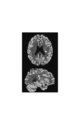
- FIG. 10 is a diagram showing an image obtained by performing the noise removal processing of Comparative Example 3 on the 16th tomographic image x 16 of FIG.
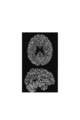
- FIG. 11 is a diagram showing an image obtained by performing the noise removal processing of Example 1 on the 16th tomographic image x 16 of FIG.
- FIG. 12 is a diagram showing an MRI image.
- FIG. 13 is a diagram showing an image obtained by performing the noise removal processing of Comparative Example 4 on the 16th tomographic image x 16 of FIG.
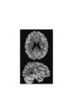
- FIG. 14 is a diagram showing an image obtained by performing the noise removal processing of Example 2 on the 16th tomographic image x 16 of FIG.
- FIG. 15 is a graph showing the time change of PSNR in each of the tomographic image before the noise removal treatment, each image after the noise removal treatment of Comparative Examples 1 to 4, and each image after the noise removal treatment of Examples 1 and 2.
- FIG. 16 is a graph showing the time change of the SSIM in each of the tomographic image before the noise removal treatment, each image after the noise removal treatment of Comparative Examples 1 to 4, and each image after the noise removal treatment of Examples 1 and 2. Is.
- FIG. 1 is a diagram showing the configuration of the radiation tomography system 1.
- the radiation tomography system 1 includes a radiation tomography apparatus 2 and an image processing apparatus 3.
- FIG. 2 is a diagram showing a configuration of a main part of the image processing device 3.
- the radiation tomography apparatus 2 is an apparatus for collecting list data for reconstructing a tomographic image of a subject.
- Examples of the radiation tomography apparatus 2 include a PET apparatus and a SPECT apparatus.
- the radiation tomography apparatus 2 will be described as a PET apparatus.
- the image processing device 3 includes an image creation unit 11, a feature extraction unit 12, a reconstruction unit 13, an evaluation unit 14, a control unit 15, an image selection unit 16, and a storage unit 17.
- a computer having a CPU, RAM, ROM, a hard disk drive, or the like is used.
- the image processing device 3 includes an input unit (for example, a keyboard and a mouse) that receives input from the operator, and includes a display unit (for example, a liquid crystal display) that displays an image or the like.
- the radiation tomography apparatus 2 includes a detection unit having a large number of small radiation detectors arranged around a measurement space in which a subject is placed.
- the radiotomography apparatus 2 detects a photon pair of energy 511 keV generated by annihilation of an electron-positron pair in a subject into which a positron emitting isotope (RI radiation source) is injected by a coincidence counting method. This coincidence counting information is accumulated. Then, the radiation tomography apparatus 2 outputs a large number of accumulated coincidence counting information arranged in chronological order to the image processing apparatus 3 as list data.
- the list data includes identification information and detection time information of a pair of radiation detectors that simultaneously count photon pairs.
- the list data may also include photon energy information detected by each radiation detector and detection time difference information of a pair of radiation detectors.
- the image processing device 3 reconstructs a tomographic image based on the list data.
- an ML-EM (maximum likelihood expectation maximization) method and a successive approximation image reconstruction technique by an improved block iterative method are known.
- OSEM ordered subset ML-EM
- RAMLA row-action maximum likelihood algorithm
- DRAMA dynamic RAMLA
- the image creation unit 11 divides the list data into a plurality of frames (first to Mth frames) in the order of collection, and reconstructs each of the plurality of frames using the data included in the mth frame of the list data.
- M number of tomographic images x 1 ⁇ x M is the dynamic PET images is used as a teacher image of the neural network.
- the image creation unit 11 may create an input image z to be input to the neural network.
- the input image z is preferably an image showing the morphological information of the subject. More specifically, the input image z is more list data from the list data may be a tomographic image created by performing reconstruction processing with that used in preparing the first m tomographic images x m.
- the input image z may be a static PET image.
- the input image z may be created for each frame, may be created in common for all frames, or may be created in common for several frames.
- the input image z may be a tomographic image created by performing a reconstruction process using all of the list data. Further, the input image z may be an MRI image of the subject or a CT image of the subject.
- the feature extraction unit 12 causes the feature extraction neural network (feature extraction NN) 18 to input the input image z, and causes the feature extraction NN 18 to output an intermediate image.
- Reconstructing unit 13 the m reconstructed neural network by inputting the intermediate image (the m reconstructed for NN) 19 m, the m output image y m from the m reconstruction NN19 m, to output the n .
- m is each integer of 1 or more and M or less.
- n is an integer of 0 or more, and represents the number of times of learning of the feature extraction NN18 and the mth reconstruction NN19 m.
- the feature extraction NN18 and the m-th reconstruction NN19 m are DNNs, preferably CNNs.
- the feature extraction NN18 may have a U-net structure.
- the M reconstruction NNs 19 1 to 19 M may have a common configuration.
- the evaluation unit 14 obtains an evaluation value based on the sum of the differences between the m-th tomographic image x m and the m-th output images ym , n. For example, the evaluation unit 14 may calculate the evaluation value by the following formula (1), and when the input image z is a static PET image, the evaluation unit 14 further evaluates by the following formula (2) using the input image z as well. The value may be calculated.
- the control unit 15 performs processing of each of the feature extraction unit 12 and the reconstruction unit 13, calculation of the evaluation value by the evaluation unit 14, and learning of each of the feature extraction NN18 and the mth reconstruction NN19 m based on the evaluation value. Have them do it repeatedly. Then, the control unit 15 outputs the mth output images ym, n from the mth reconstruction NN19 m that has been learned n times. It should be noted that each learning of the feature extraction NN18 and the mth reconstruction NN19 m is performed collectively so that the evaluation value becomes small.
- the image selection unit 16 selects one of a plurality of m output images ym , 0 to ym , N for each of the first to M frames as a tomographic image after noise removal processing. ..
- the image selection unit 16 is any one of a plurality of m output images ym , 0 to ym , N based on the comparison between the mth output images ym, n and the input image z. Select the mth output image.
- the m output image y m, to the error between the n and the input image z may be selected first m output image with the minimum, between the m-th output image y m, n and the input image z
- You may select any m output image from 1 or 2 or more m output images in which the error of is equal to or less than the threshold value.
- a doctor, a technician, or the like may select any m-output image from a plurality of m-output images ym, 0 to ym , N for each frame.
- Storage unit 17 stores list data, the input image z, the intermediate image, the m output image y m of each frame, n and the m-th tomographic image x m of each frame. Further, the storage unit 17 stores the mth output image selected from the plurality of mth output images ym, 0 to ym , N of each frame.
- FIG. 3 is a flowchart illustrating an image processing method.
- This image processing method includes an image creation step S1, a feature extraction step S2, a reconstruction step S3, an end determination step S4, an evaluation step S5, a learning step S6, and an image selection step S7.
- the image creation step S1 is a process performed by the image creation unit 11.
- the list data is divided into a plurality of frames (first to Mth frames) in the order of collection, and each of the plurality of frames is reconstructed using the data included in the mth frame of the list data.
- a tomographic image (input image z) may be created by performing a reconstruction process using all of the list data.
- the feature extraction step S2 is a process performed by the feature extraction unit 12.
- the input image z is input to the feature extraction NN18, and the intermediate image is output from the feature extraction NN18.
- the reconstruction step S3 is a process performed by the reconstruction unit 13. In reconstruction step S3, by inputting the intermediate image in the m reconstruction NN19 m, the m output image y m from the m reconstruction NN19 m, to output n.
- the end determination step S4 is a process performed by the control unit 15. In the end determination step S4, it is determined whether or not to end the repetition of each process of steps S2 to S6. This determination may be based on whether or not the number of repetitions has reached a predetermined value, or based on whether or not the error between the m output images ym, n and the input image z is equal to or less than the threshold value. May be good.
- the process proceeds to the evaluation step S5, and it is determined that the repetition of each process of steps S2 to S6 may be completed.
- the process proceeds to the image selection step S7.
- the evaluation step S5 is a process performed by the evaluation unit 14.
- an evaluation value (for example, the above equation (1) or (2)) is obtained based on the sum of the differences between the m-th tomographic image x m and the m-output images ym , n.
- the learning step S6 is a process performed by the control unit 15 on the feature extraction NN18 and the mth reconstruction NN19 m .
- batch learning is performed for each of the feature extraction NN18 and the mth reconstruction NN19 m based on the evaluation value. After this learning step S6, the process returns to the feature extraction step S2.
- the image selection step S7 is a process performed by the image selection unit 16. In the image selection step S7, for each of the first to M frames, any one of a plurality of m output images ym , 0 to ym , and N is selected as a tomographic image after noise removal processing. ..
- the numerical phantom used in this simulation simulates the human brain injected with 18 F-FDG (fluorodeoxyglucose).
- This numerical phantom includes a white matter (WM), a gray matter (GM) and a tumor (tumor).
- FIG. 4 is a graph showing a time-varying model of the activity of each of the white matter (WM), gray matter (GM), and tumor (tumor) used in the simulation.
- the time-activity change (Time-Activity Curve, TAC) generally gradually increases with the passage of time immediately after the RI radiation source is turned on, reaches a peak at a certain time, and then gradually decreases after the peak. Become.
- the total number of counts (the number of the coincidence counting information) was set to 1 ⁇ 10 9.
- the measurement time was 90 minutes, and the list data was divided into 30 frames.
- Each period of the 1st to 4th frames is 20 seconds, each period of the 5th to 8th frames is 40 seconds, each period of the 9th to 12th frames is 60 seconds, and the 13th to 16th frames.
- Each period of the frame is 180 seconds, and each period of the 17th to 30th frames is 300 seconds.
- Numerical phantoms have a three-dimensional structure of 192 x 192 x 64 voxels.
- a synogram for each frame was created based on this numerical phantom.
- the synogram is a histogram of the coincidence counting information for each pair of radiation detectors of the radiation tomography apparatus 2.
- a noise-added synogram was created by applying Poisson noise corresponding to the count number of the frame to the synogram of each frame.
- a reconstructed image (mth tomographic image x m ) was created by the OS-EM method based on this noise-added synogram. The number of iterations in the OS-EM method was set to 6, and the number of subsets was set to 16.
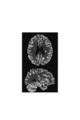
- FIG. 5 is a diagram showing a numerical phantom image.
- the arrow points to the tumor site.
- FIG. 6 is a diagram showing a 16th tomographic image x 16 before the noise removal processing. The image of FIG. 6 has a large amount of noise, and it is difficult to distinguish the tumor portion.
- FIG. 7 is a diagram showing an image obtained by performing the noise removal processing of Comparative Example 1 on the 16th tomographic image x 16 of FIG.
- the noise removal processing of Comparative Example 1 is a processing by a Gaussian filter. Although the image of FIG. 7 has a lot of noise and is not clear as a whole, the tumor portion can be discriminated.
- FIG. 8 is a diagram showing an image obtained by performing the noise removal processing of Comparative Example 2 on the 16th tomographic image x 16 of FIG.
- the noise removal process of Comparative Example 2 is the process described in Non-Patent Document 2. Although the image of FIG. 8 has a lot of noise, the boundary between the shades is clear and the tumor portion can be discriminated.
- FIG. 9 is a diagram showing a tomographic image (static PET image) created by performing a reconstruction process using all of the list data.
- this static PET image was used as an input image to the neural network.
- FIG. 10 is a diagram showing an image obtained by performing the noise removal processing of Comparative Example 3 on the 16th tomographic image x 16 of FIG.
- the noise removal process of Comparative Example 3 is the process described in Non-Patent Document 5. Compared with the images of Comparative Examples 1 and 2, the image of FIG. 10 has sufficiently removed noise, the boundary of shading becomes clear, and the tumor portion can be discriminated.
- FIG. 11 is a diagram showing an image obtained by performing the noise removal processing of Example 1 on the 16th tomographic image x 16 of FIG.
- the noise removal process of the first embodiment is based on the present embodiment. Compared with the image of FIG. 10, in the image of FIG. 11, noise is sufficiently removed, the boundary of shading becomes clear, and the tumor portion can be discriminated.
- FIG. 12 is a diagram showing an MRI image (T1-weighted image). No tumor is found on this MRI image. In Comparative Example 4 and Example 2, this MRI image was used as an input image to the neural network.
- FIG. 13 is a diagram showing an image obtained by performing the noise removal processing of Comparative Example 4 on the 16th tomographic image x 16 of FIG.
- the noise removal process of Comparative Example 4 is the process described in Non-Patent Document 5. Compared with the images of Comparative Examples 1 and 2, the image of FIG. 13 has sufficiently removed noise, the boundary of shading becomes clear, and the tumor portion can be discriminated.
- FIG. 14 is a diagram showing an image obtained by performing the noise removal processing of Example 2 on the 16th tomographic image x 16 of FIG.
- the noise removal process of the second embodiment is based on the present embodiment. Compared with the image of FIG. 13, the image of FIG. 14 further sufficiently removes noise, the boundary of shading becomes clear, and the tumor portion can be discriminated.
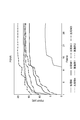
- FIG. 15 is a graph showing the time change of PSNR in each of the tomographic image before the noise removal treatment, each image after the noise removal treatment of Comparative Examples 1 to 4, and each image after the noise removal treatment of Examples 1 and 2.
- PSNR Peak Signal to Noise Ratio
- dB decibels
- FIG. 16 is a graph showing the time change of the SSIM in each of the tomographic image before the noise removal treatment, each image after the noise removal treatment of Comparative Examples 1 to 4, and each image after the noise removal treatment of Examples 1 and 2.
- SSIM Structuretural Similarity Index
- Both the PSNR and SSIM indexes show that the noise removal processing of Comparative Examples 3 and 4 is superior to the noise removal processing of Comparative Examples 1 and 2, and that the noise removal processing of Comparative Examples 3 and 4 is superior to the noise removal processing of Comparative Examples 3 and 4. It is shown that the noise removal processing of Examples 1 and 2 has better performance than the noise removal processing. The noise removal processing performance of Examples 1 and 2 is also excellent for other frames.
- the image processing apparatus 3 of this embodiment includes a processing by the feature extraction NN18 in the feature extraction unit 12, a process by the m reconstitution NN19 m of each frame in the reconstruction unit 13. Then, the image processing device 3 causes the intermediate image output from the feature extraction NN18 to be input to the mth reconstruction NN19 m of each frame, and at that time, the mth output output from the mth reconstruction NN19 m.
- An evaluation value is obtained based on the sum of the differences between the images ym and n and the mth tomographic image x m, and the feature extraction NN18 and the mth reconstruction NN19 m are trained based on this evaluation value.
- the feature extraction NN18 is extracted from the m-th tomographic image x m of each frame by repeating learning based on the above evaluation values.
- An intermediate image representing the common information can be output, and the mth reconstruction NN19 m infers the mth output images ym, n from the intermediate image. From this, it is considered that the performance of the noise removal processing is improved in the present embodiment.
- the noise removal processing described in Non-Patent Document 5 the first m tomographic images x m of each frame as a teacher image, train the neural network using a combination of the input image z this teacher image, the frame of this study Repeat for the number of.
- the learning of the feature extraction NN18 can be performed in common for a plurality of frames. For this reason, the time required for the noise removal processing of the present embodiment can be shortened as compared with the noise removal processing described in Non-Patent Document 5.
- the radiation tomography apparatus 2 is a PET apparatus in the above embodiment, but may be a SPECT apparatus.
- the image processing apparatus divides the list data collected by the radiation tomography apparatus into M frames in the order of collection, and reconstructs each m of 1 or more and M or less based on the list data included in the m-th frame. It is a device that removes noise from the m-th tomographic image created by processing. (1) Feature extraction that inputs an input image to a feature extraction neural network and outputs an intermediate image from the feature extraction neural network. And (2) a reconstruction unit that inputs an intermediate image to the m-th reconstruction neural network for each m of 1 or more and M or less and outputs the m-th output image from the m-th reconstruction neural network.
- An evaluation unit that obtains an evaluation value based on the sum of the differences between the mth tomographic image and the mth output image of each m of 1 or more and M or less, and (4) a feature extraction unit, a reconstruction unit, and an evaluation unit.
- Each process and the learning of the feature extraction neural network based on the evaluation value and the learning of each mth reconstruction neural network of 1 or more and M or less are repeatedly performed, and the mth of each m of 1 or more and M or less is performed repeatedly.
- the configuration includes a control unit that outputs a plurality of mth output images from the reconstruction neural network.
- the feature extraction unit may be configured to input an image representing the morphological information of the subject as an input image to the feature extraction neural network. Further, the feature extraction unit uses a tomographic image created by performing reconstruction processing using more list data than the list data used when creating the mth tomographic image for each m of 1 or more and M or less as an input image. It may be configured to be input to the feature extraction neural network.
- the feature extraction unit may be configured to input the MRI image of the subject as an input image to the feature extraction neural network. Further, the feature extraction unit may be configured to input the CT image of the subject as an input image to the feature extraction neural network.
- the above image processing apparatus further includes an image selection unit that selects one of the mth output images obtained for each m of 1 or more and M or less as a tomographic image after noise removal processing. It may be provided as a configuration. Further, the image selection unit may be configured to select one of the mth output images from a plurality of mth output images based on the comparison between the mth output image and the input image for each m of 1 or more and M or less. good. Further, the image selection unit may be configured to select one of the mth output images from a plurality of mth output images based on the comparison between the mth tomographic image and the input image for each m of 1 or more and M or less. good.
- the radiation tomography system is a radiation tomography apparatus that collects list data for reconstructing a tomographic image of a subject, and after noise removal processing based on the list data collected by the radiation tomography apparatus. It is configured to include an image processing device having the above configuration for creating a tomographic image.
- the list data collected by the radiation tomography apparatus is divided into M frames in the order of collection, and each m of 1 or more and M or less is reconstructed based on the list data included in the m-th frame. It is a method of removing noise of the m-th tomographic image created by processing.
- the steps and (2) a reconstruction step in which an intermediate image is input to the m-th reconstruction neural network for each m of 1 or more and M or less and the m-th output image is output from the m-th reconstruction neural network.
- An evaluation step to obtain an evaluation value based on the sum of the differences between the mth tomographic image and the mth output image of each m of 1 or more and M or less, and (4) a neural network for feature extraction based on the evaluation value.
- a learning step for learning each of the m-th reconstruction neural networks of 1 or more and M or less is provided, and each process of the feature extraction step, the reconstruction step, the evaluation step, and the learning step is repeatedly performed, and 1 A plurality of m-th output images are output from the m-th reconstruction neural network of each m of M or less.
- an image representing the morphological information of the subject may be input to the feature extraction neural network as an input image.
- a tomographic image created by performing reconstruction processing using more list data than the list data used when creating the mth tomographic image for each m of 1 or more and M or less is used as an input image. It may be configured to be input to the feature extraction neural network.
- the MRI image of the subject may be input to the feature extraction neural network as an input image.
- the CT image of the subject may be input to the feature extraction neural network as an input image.
- the above image processing method further includes an image selection step of selecting one of the mth output images obtained for each m of 1 or more and M or less as a tomographic image after noise removal processing. It may be provided as a configuration. Further, in the image selection step, one of the mth output images may be selected from a plurality of mth output images based on the comparison between the mth output image and the input image for each m of 1 or more and M or less. good. Further, in the image selection step, one of the mth output images may be selected from a plurality of mth output images based on the comparison between the mth tomographic image and the input image for each m of 1 or more and M or less. good.
- the present invention is used as an apparatus and method capable of creating a high-performance noise-removed tomographic image based on list data collected by a radiation tomography apparatus and shortening the time required for noise removal processing. It is possible.
Landscapes
- Health & Medical Sciences (AREA)
- Life Sciences & Earth Sciences (AREA)
- Engineering & Computer Science (AREA)
- Physics & Mathematics (AREA)
- Medical Informatics (AREA)
- Public Health (AREA)
- Biophysics (AREA)
- Pathology (AREA)
- Biomedical Technology (AREA)
- Heart & Thoracic Surgery (AREA)
- Nuclear Medicine, Radiotherapy & Molecular Imaging (AREA)
- Molecular Biology (AREA)
- Surgery (AREA)
- Animal Behavior & Ethology (AREA)
- General Health & Medical Sciences (AREA)
- Veterinary Medicine (AREA)
- Radiology & Medical Imaging (AREA)
- High Energy & Nuclear Physics (AREA)
- Computer Vision & Pattern Recognition (AREA)
- Artificial Intelligence (AREA)
- Optics & Photonics (AREA)
- Theoretical Computer Science (AREA)
- General Physics & Mathematics (AREA)
- Signal Processing (AREA)
- Psychiatry (AREA)
- Physiology (AREA)
- Evolutionary Computation (AREA)
- Fuzzy Systems (AREA)
- Mathematical Physics (AREA)
- Image Processing (AREA)
- Apparatus For Radiation Diagnosis (AREA)
- Image Analysis (AREA)
- Nuclear Medicine (AREA)
- Magnetic Resonance Imaging Apparatus (AREA)
Abstract
Description

Claims (15)
- 放射線断層撮影装置により収集されたリストデータを収集順にM個のフレームに分割し1以上M以下の各mについて第mフレームに含まれるリストデータに基づいて再構成処理を行って作成された第m断層画像のノイズを除去する装置であって、
特徴抽出用ニューラルネットワークに入力画像を入力させて該特徴抽出用ニューラルネットワークから中間画像を出力させる特徴抽出部と、
1以上M以下の各mについて第m再構成用ニューラルネットワークに前記中間画像を入力させて該第m再構成用ニューラルネットワークから第m出力画像を出力させる再構成部と、
1以上M以下の各mの前記第m断層画像と前記第m出力画像との間の差の総和に基づいて評価値を求める評価部と、
前記特徴抽出部、前記再構成部および前記評価部の各処理と、前記評価値に基づく前記特徴抽出用ニューラルネットワークおよび1以上M以下の各mの前記第m再構成用ニューラルネットワークそれぞれの学習とを繰り返し行わせて、1以上M以下の各mの前記第m再構成用ニューラルネットワークから複数の前記第m出力画像を出力させる制御部と、
を備える、画像処理装置。 - 前記特徴抽出部は、被検体の形態情報を表す画像を前記入力画像として前記特徴抽出用ニューラルネットワークに入力させる、請求項1に記載の画像処理装置。
- 前記特徴抽出部は、1以上M以下の各mについて前記第m断層画像を作成する際に用いたリストデータより多いリストデータを用いて再構成処理を行って作成された断層画像を前記入力画像として前記特徴抽出用ニューラルネットワークに入力させる、請求項1に記載の画像処理装置。
- 前記特徴抽出部は、被検体のMRI画像を前記入力画像として前記特徴抽出用ニューラルネットワークに入力させる、請求項1に記載の画像処理装置。
- 前記特徴抽出部は、被検体のCT画像を前記入力画像として前記特徴抽出用ニューラルネットワークに入力させる、請求項1に記載の画像処理装置。
- 1以上M以下の各mについて得られた複数の前記第m出力画像のうちから何れかの第m出力画像をノイズ除去処理後の断層画像として選択する画像選択部を更に備える、請求項1~5の何れか1項に記載の画像処理装置。
- 前記画像選択部は、1以上M以下の各mについて前記第m出力画像と前記入力画像との比較に基づいて複数の前記第m出力画像のうちから何れかの第m出力画像を選択する、請求項6に記載の画像処理装置。
- 被検体の断層画像を再構成するためのリストデータを収集する放射線断層撮影装置と、
前記放射線断層撮影装置により収集されたリストデータに基づいてノイズ除去処理後の断層画像を作成する請求項1~7の何れか1項に記載の画像処理装置と、
を備える、放射線断層撮影システム。 - 放射線断層撮影装置により収集されたリストデータを収集順にM個のフレームに分割し1以上M以下の各mについて第mフレームに含まれるリストデータに基づいて再構成処理を行って作成された第m断層画像のノイズを除去する方法であって、
特徴抽出用ニューラルネットワークに入力画像を入力させて該特徴抽出用ニューラルネットワークから中間画像を出力させる特徴抽出ステップと、
1以上M以下の各mについて第m再構成用ニューラルネットワークに前記中間画像を入力させて該第m再構成用ニューラルネットワークから第m出力画像を出力させる再構成ステップと、
1以上M以下の各mの前記第m断層画像と前記第m出力画像との間の差の総和に基づいて評価値を求める評価ステップと、
前記評価値に基づいて前記特徴抽出用ニューラルネットワークおよび1以上M以下の各mの前記第m再構成用ニューラルネットワークそれぞれを学習させる学習ステップと、
を備え、
前記特徴抽出ステップ、前記再構成ステップ、前記評価ステップおよび前記学習ステップの各処理を繰り返し行わせて、1以上M以下の各mの前記第m再構成用ニューラルネットワークから複数の前記第m出力画像を出力させる、画像処理方法。 - 前記特徴抽出ステップにおいて、被検体の形態情報を表す画像を前記入力画像として前記特徴抽出用ニューラルネットワークに入力させる、請求項9に記載の画像処理方法。
- 前記特徴抽出ステップにおいて、1以上M以下の各mについて前記第m断層画像を作成する際に用いたリストデータより多いリストデータを用いて再構成処理を行って作成された断層画像を前記入力画像として前記特徴抽出用ニューラルネットワークに入力させる、請求項9に記載の画像処理方法。
- 前記特徴抽出ステップにおいて、被検体のMRI画像を前記入力画像として前記特徴抽出用ニューラルネットワークに入力させる、請求項9に記載の画像処理方法。
- 前記特徴抽出ステップにおいて、被検体のCT画像を前記入力画像として前記特徴抽出用ニューラルネットワークに入力させる、請求項9に記載の画像処理方法。
- 1以上M以下の各mについて得られた複数の前記第m出力画像のうちから何れかの第m出力画像をノイズ除去処理後の断層画像として選択する画像選択ステップを更に備える、請求項9~13の何れか1項に記載の画像処理方法。
- 前記画像選択ステップにおいて、1以上M以下の各mについて前記第m出力画像と前記入力画像との比較に基づいて複数の前記第m出力画像のうちから何れかの第m出力画像を選択する、請求項14に記載の画像処理方法。
Priority Applications (3)
| Application Number | Priority Date | Filing Date | Title |
|---|---|---|---|
| EP21748477.3A EP4099062A4 (en) | 2020-01-29 | 2021-01-27 | IMAGE PROCESSING DEVICE AND IMAGE PROCESSING METHOD |
| CN202180009327.3A CN114981684B (zh) | 2020-01-29 | 2021-01-27 | 图像处理装置和图像处理方法 |
| US17/795,566 US12444098B2 (en) | 2020-01-29 | 2021-01-27 | Image processing device and image processing method |
Applications Claiming Priority (2)
| Application Number | Priority Date | Filing Date | Title |
|---|---|---|---|
| JP2020-012243 | 2020-01-29 | ||
| JP2020012243A JP7557944B2 (ja) | 2020-01-29 | 2020-01-29 | 画像処理装置および画像処理方法 |
Publications (1)
| Publication Number | Publication Date |
|---|---|
| WO2021153604A1 true WO2021153604A1 (ja) | 2021-08-05 |
Family
ID=77079912
Family Applications (1)
| Application Number | Title | Priority Date | Filing Date |
|---|---|---|---|
| PCT/JP2021/002809 Ceased WO2021153604A1 (ja) | 2020-01-29 | 2021-01-27 | 画像処理装置および画像処理方法 |
Country Status (5)
| Country | Link |
|---|---|
| US (1) | US12444098B2 (ja) |
| EP (1) | EP4099062A4 (ja) |
| JP (1) | JP7557944B2 (ja) |
| CN (1) | CN114981684B (ja) |
| WO (1) | WO2021153604A1 (ja) |
Families Citing this family (4)
| Publication number | Priority date | Publication date | Assignee | Title |
|---|---|---|---|---|
| JP7557944B2 (ja) * | 2020-01-29 | 2024-09-30 | 浜松ホトニクス株式会社 | 画像処理装置および画像処理方法 |
| JP2023112818A (ja) * | 2022-02-02 | 2023-08-15 | 浜松ホトニクス株式会社 | 画像処理装置および画像処理方法 |
| JP7815033B2 (ja) * | 2022-05-31 | 2026-02-17 | 浜松ホトニクス株式会社 | 画像処理装置および画像処理方法 |
| US20230401769A1 (en) * | 2022-06-14 | 2023-12-14 | Siemens Medical Solutions Usa, Inc. | Systems and methods of accelerated dynamic imaging in pet |
Citations (3)
| Publication number | Priority date | Publication date | Assignee | Title |
|---|---|---|---|---|
| CN103955899A (zh) | 2014-05-02 | 2014-07-30 | 南方医科大学 | 基于组合图像引导的动态pet图像去噪方法 |
| JP2019510969A (ja) * | 2016-02-29 | 2019-04-18 | シャンハイ・ユナイテッド・イメージング・ヘルスケア・カンパニー・リミテッド | Ect画像を再構成するシステムおよび方法 |
| JP2019211475A (ja) * | 2018-05-31 | 2019-12-12 | キヤノンメディカルシステムズ株式会社 | 医用画像処理装置及びプログラム |
Family Cites Families (15)
| Publication number | Priority date | Publication date | Assignee | Title |
|---|---|---|---|---|
| JPH0830728A (ja) * | 1994-07-12 | 1996-02-02 | Suzuki Motor Corp | 画像の二値化装置 |
| US11914674B2 (en) * | 2011-09-24 | 2024-02-27 | Z Advanced Computing, Inc. | System and method for extremely efficient image and pattern recognition and artificial intelligence platform |
| JP2018005520A (ja) * | 2016-06-30 | 2018-01-11 | クラリオン株式会社 | 物体検出装置及び物体検出方法 |
| WO2018048507A1 (en) * | 2016-09-06 | 2018-03-15 | Han Xiao | Neural network for generating synthetic medical images |
| US10096109B1 (en) | 2017-03-31 | 2018-10-09 | The Board Of Trustees Of The Leland Stanford Junior University | Quality of medical images using multi-contrast and deep learning |
| US10803984B2 (en) | 2017-10-06 | 2020-10-13 | Canon Medical Systems Corporation | Medical image processing apparatus and medical image processing system |
| JP6974159B2 (ja) | 2017-12-26 | 2021-12-01 | 浜松ホトニクス株式会社 | 画像処理装置および画像処理方法 |
| US11880962B2 (en) * | 2018-02-15 | 2024-01-23 | General Electric Company | System and method for synthesizing magnetic resonance images |
| CN108710904A (zh) * | 2018-05-10 | 2018-10-26 | 上海交通大学 | 基于递归神经网络的图像匹配方法及系统 |
| CN109003260B (zh) | 2018-06-28 | 2021-02-09 | 深圳视见医疗科技有限公司 | Ct图像肺结节检测方法、装置、设备及可读存储介质 |
| CN109598727B (zh) * | 2018-11-28 | 2021-09-14 | 北京工业大学 | 一种基于深度神经网络的ct图像肺实质三维语义分割方法 |
| CN110197516A (zh) | 2019-05-29 | 2019-09-03 | 浙江明峰智能医疗科技有限公司 | 一种基于深度学习的tof-pet散射校正方法 |
| WO2021041125A1 (en) | 2019-08-23 | 2021-03-04 | Subtle Medical, Inc. | Systems and methods for accurate and rapid positron emission tomography using deep learning |
| JP7337675B2 (ja) * | 2019-12-04 | 2023-09-04 | キヤノンメディカルシステムズ株式会社 | 医用データ処理装置 |
| JP7557944B2 (ja) * | 2020-01-29 | 2024-09-30 | 浜松ホトニクス株式会社 | 画像処理装置および画像処理方法 |
-
2020
- 2020-01-29 JP JP2020012243A patent/JP7557944B2/ja active Active
-
2021
- 2021-01-27 WO PCT/JP2021/002809 patent/WO2021153604A1/ja not_active Ceased
- 2021-01-27 EP EP21748477.3A patent/EP4099062A4/en active Pending
- 2021-01-27 US US17/795,566 patent/US12444098B2/en active Active
- 2021-01-27 CN CN202180009327.3A patent/CN114981684B/zh active Active
Patent Citations (3)
| Publication number | Priority date | Publication date | Assignee | Title |
|---|---|---|---|---|
| CN103955899A (zh) | 2014-05-02 | 2014-07-30 | 南方医科大学 | 基于组合图像引导的动态pet图像去噪方法 |
| JP2019510969A (ja) * | 2016-02-29 | 2019-04-18 | シャンハイ・ユナイテッド・イメージング・ヘルスケア・カンパニー・リミテッド | Ect画像を再構成するシステムおよび方法 |
| JP2019211475A (ja) * | 2018-05-31 | 2019-12-12 | キヤノンメディカルシステムズ株式会社 | 医用画像処理装置及びプログラム |
Non-Patent Citations (6)
| Title |
|---|
| DMITRY ULYANOV ET AL.: "Deep Image Prior", ARXIV PREPRINT ARXIV:1711.10925, 2017 |
| F. HASHIMOTO ET AL.: "Denoising of Dynamic Sinogram by Image Guided Filtering for Positron Emission Tomography", IEEE TRANSACTIONS ON RADIATION AND PLASMA MEDICAL SCIENCES, vol. 2, no. 6, 2018, pages 541 - 548, XP011698873, DOI: 10.1109/TRPMS.2018.2869936 |
| F. HASHIMOTO ET AL.: "Dynamic PET Image Denoising Using Deep Convolutional Neural Networks Without Prior Training Datasets", IEEE ACCESS, vol. 7, 2019, pages 96594 - 96603, XP011737252, DOI: 10.1109/ACCESS.2019.2929230 |
| KUANG GONG ET AL.: "PET Image Reconstruction Using Deep Image Prior", IEEE TRANSACTIONS ON MEDICAL IMAGING, 2018 |
| LIJUN LU ET AL., IEEE NUCLEAR SCIENCE SYMPOSIUM AND MEDICAL IMAGING CONFERENCE, vol. Dynamic PET Denoising Incorporating a Composite Im, 2014 |
| See also references of EP4099062A4 |
Also Published As
| Publication number | Publication date |
|---|---|
| EP4099062A4 (en) | 2024-02-28 |
| CN114981684A (zh) | 2022-08-30 |
| US20230102661A1 (en) | 2023-03-30 |
| JP7557944B2 (ja) | 2024-09-30 |
| US12444098B2 (en) | 2025-10-14 |
| CN114981684B (zh) | 2025-09-19 |
| JP2021117866A (ja) | 2021-08-10 |
| EP4099062A1 (en) | 2022-12-07 |
Similar Documents
| Publication | Publication Date | Title |
|---|---|---|
| JP7237624B2 (ja) | 画像処理装置および画像処理方法 | |
| Wang et al. | FBP-Net for direct reconstruction of dynamic PET images | |
| WO2021153604A1 (ja) | 画像処理装置および画像処理方法 | |
| CN114387359B (zh) | 一种三维x射线低剂量成像方法及装置 | |
| Kim et al. | An effective post-filtering framework for 3-D PET image denoising based on noise and sensitivity characteristics | |
| Guo et al. | Graph filtering approach to PET image denoising | |
| JP6974159B2 (ja) | 画像処理装置および画像処理方法 | |
| WO2017149399A2 (en) | Optimization-based reconstruction with an image-total-variation constraint in pet | |
| CN105488824B (zh) | 一种重建pet图像的方法和装置 | |
| WO2023228910A1 (ja) | 画像処理装置および画像処理方法 | |
| JP6495615B2 (ja) | 画像処理装置および画像処理方法 | |
| JP6986961B2 (ja) | 画像処理装置および画像処理方法 | |
| Ote et al. | List-mode PET image reconstruction using Dykstra-like splitting | |
| US20250157099A1 (en) | Image processing device and image processing method | |
| JP7815033B2 (ja) | 画像処理装置および画像処理方法 | |
| JP7018306B2 (ja) | 画像処理装置および画像処理方法 | |
| JP2024157164A (ja) | 画像処理装置および画像処理方法 | |
| CN120051712A (zh) | 图像处理装置和图像处理方法 | |
| Wang et al. | Joint Reconstruction of Dynamic PET Activity and Kinetic Parametric Images Using Learnable Descent Framework | |
| JP2016085064A (ja) | 画像処理装置および画像処理方法 |
Legal Events
| Date | Code | Title | Description |
|---|---|---|---|
| 121 | Ep: the epo has been informed by wipo that ep was designated in this application |
Ref document number: 21748477 Country of ref document: EP Kind code of ref document: A1 |
|
| NENP | Non-entry into the national phase |
Ref country code: DE |
|
| ENP | Entry into the national phase |
Ref document number: 2021748477 Country of ref document: EP Effective date: 20220829 |
|
| WWG | Wipo information: grant in national office |
Ref document number: 202180009327.3 Country of ref document: CN |
|
| WWG | Wipo information: grant in national office |
Ref document number: 17795566 Country of ref document: US |