WO2023033481A1 - 항암 활성을 가지는 adp-리보오스 결합 펩티드 및 이의 용도 - Google Patents
항암 활성을 가지는 adp-리보오스 결합 펩티드 및 이의 용도 Download PDFInfo
- Publication number
- WO2023033481A1 WO2023033481A1 PCT/KR2022/012886 KR2022012886W WO2023033481A1 WO 2023033481 A1 WO2023033481 A1 WO 2023033481A1 KR 2022012886 W KR2022012886 W KR 2022012886W WO 2023033481 A1 WO2023033481 A1 WO 2023033481A1
- Authority
- WO
- WIPO (PCT)
- Prior art keywords
- cancer
- adp
- ribose
- cells
- peptide
- Prior art date
- Legal status (The legal status is an assumption and is not a legal conclusion. Google has not performed a legal analysis and makes no representation as to the accuracy of the status listed.)
- Ceased
Links
Images




Classifications
-
- C—CHEMISTRY; METALLURGY
- C07—ORGANIC CHEMISTRY
- C07K—PEPTIDES
- C07K14/00—Peptides having more than 20 amino acids; Gastrins; Somatostatins; Melanotropins; Derivatives thereof
- C07K14/435—Peptides having more than 20 amino acids; Gastrins; Somatostatins; Melanotropins; Derivatives thereof from animals; from humans
- C07K14/46—Peptides having more than 20 amino acids; Gastrins; Somatostatins; Melanotropins; Derivatives thereof from animals; from humans from vertebrates
- C07K14/47—Peptides having more than 20 amino acids; Gastrins; Somatostatins; Melanotropins; Derivatives thereof from animals; from humans from vertebrates from mammals
-
- A—HUMAN NECESSITIES
- A61—MEDICAL OR VETERINARY SCIENCE; HYGIENE
- A61K—PREPARATIONS FOR MEDICAL, DENTAL OR TOILETRY PURPOSES
- A61K38/00—Medicinal preparations containing peptides
- A61K38/16—Peptides having more than 20 amino acids; Gastrins; Somatostatins; Melanotropins; Derivatives thereof
- A61K38/17—Peptides having more than 20 amino acids; Gastrins; Somatostatins; Melanotropins; Derivatives thereof from animals; from humans
-
- A—HUMAN NECESSITIES
- A61—MEDICAL OR VETERINARY SCIENCE; HYGIENE
- A61P—SPECIFIC THERAPEUTIC ACTIVITY OF CHEMICAL COMPOUNDS OR MEDICINAL PREPARATIONS
- A61P35/00—Antineoplastic agents
-
- C—CHEMISTRY; METALLURGY
- C07—ORGANIC CHEMISTRY
- C07H—SUGARS; DERIVATIVES THEREOF; NUCLEOSIDES; NUCLEOTIDES; NUCLEIC ACIDS
- C07H19/00—Compounds containing a hetero ring sharing one ring hetero atom with a saccharide radical; Nucleosides; Mononucleotides; Anhydro-derivatives thereof
- C07H19/02—Compounds containing a hetero ring sharing one ring hetero atom with a saccharide radical; Nucleosides; Mononucleotides; Anhydro-derivatives thereof sharing nitrogen
- C07H19/04—Heterocyclic radicals containing only nitrogen atoms as ring hetero atom
- C07H19/16—Purine radicals
- C07H19/20—Purine radicals with the saccharide radical esterified by phosphoric or polyphosphoric acids
- C07H19/207—Purine radicals with the saccharide radical esterified by phosphoric or polyphosphoric acids the phosphoric or polyphosphoric acids being esterified by a further hydroxylic compound, e.g. flavine adenine dinucleotide or nicotinamide-adenine dinucleotide
-
- C—CHEMISTRY; METALLURGY
- C07—ORGANIC CHEMISTRY
- C07K—PEPTIDES
- C07K14/00—Peptides having more than 20 amino acids; Gastrins; Somatostatins; Melanotropins; Derivatives thereof
- C07K14/435—Peptides having more than 20 amino acids; Gastrins; Somatostatins; Melanotropins; Derivatives thereof from animals; from humans
- C07K14/46—Peptides having more than 20 amino acids; Gastrins; Somatostatins; Melanotropins; Derivatives thereof from animals; from humans from vertebrates
- C07K14/47—Peptides having more than 20 amino acids; Gastrins; Somatostatins; Melanotropins; Derivatives thereof from animals; from humans from vertebrates from mammals
- C07K14/4701—Peptides having more than 20 amino acids; Gastrins; Somatostatins; Melanotropins; Derivatives thereof from animals; from humans from vertebrates from mammals not used
- C07K14/4747—Apoptosis related proteins
-
- C—CHEMISTRY; METALLURGY
- C12—BIOCHEMISTRY; BEER; SPIRITS; WINE; VINEGAR; MICROBIOLOGY; ENZYMOLOGY; MUTATION OR GENETIC ENGINEERING
- C12N—MICROORGANISMS OR ENZYMES; COMPOSITIONS THEREOF; PROPAGATING, PRESERVING, OR MAINTAINING MICROORGANISMS; MUTATION OR GENETIC ENGINEERING; CULTURE MEDIA
- C12N15/00—Mutation or genetic engineering; DNA or RNA concerning genetic engineering, vectors, e.g. plasmids, or their isolation, preparation or purification; Use of hosts therefor
- C12N15/09—Recombinant DNA-technology
- C12N15/63—Introduction of foreign genetic material using vectors; Vectors; Use of hosts therefor; Regulation of expression
-
- C—CHEMISTRY; METALLURGY
- C12—BIOCHEMISTRY; BEER; SPIRITS; WINE; VINEGAR; MICROBIOLOGY; ENZYMOLOGY; MUTATION OR GENETIC ENGINEERING
- C12N—MICROORGANISMS OR ENZYMES; COMPOSITIONS THEREOF; PROPAGATING, PRESERVING, OR MAINTAINING MICROORGANISMS; MUTATION OR GENETIC ENGINEERING; CULTURE MEDIA
- C12N9/00—Enzymes; Proenzymes; Compositions thereof; Processes for preparing, activating, inhibiting, separating or purifying enzymes
- C12N9/10—Transferases (2.)
- C12N9/1025—Acyltransferases (2.3)
- C12N9/104—Aminoacyltransferases (2.3.2)
-
- C—CHEMISTRY; METALLURGY
- C12—BIOCHEMISTRY; BEER; SPIRITS; WINE; VINEGAR; MICROBIOLOGY; ENZYMOLOGY; MUTATION OR GENETIC ENGINEERING
- C12N—MICROORGANISMS OR ENZYMES; COMPOSITIONS THEREOF; PROPAGATING, PRESERVING, OR MAINTAINING MICROORGANISMS; MUTATION OR GENETIC ENGINEERING; CULTURE MEDIA
- C12N9/00—Enzymes; Proenzymes; Compositions thereof; Processes for preparing, activating, inhibiting, separating or purifying enzymes
- C12N9/10—Transferases (2.)
- C12N9/1048—Glycosyltransferases (2.4)
- C12N9/1077—Pentosyltransferases (2.4.2)
-
- C—CHEMISTRY; METALLURGY
- C12—BIOCHEMISTRY; BEER; SPIRITS; WINE; VINEGAR; MICROBIOLOGY; ENZYMOLOGY; MUTATION OR GENETIC ENGINEERING
- C12N—MICROORGANISMS OR ENZYMES; COMPOSITIONS THEREOF; PROPAGATING, PRESERVING, OR MAINTAINING MICROORGANISMS; MUTATION OR GENETIC ENGINEERING; CULTURE MEDIA
- C12N9/00—Enzymes; Proenzymes; Compositions thereof; Processes for preparing, activating, inhibiting, separating or purifying enzymes
- C12N9/10—Transferases (2.)
- C12N9/12—Transferases (2.) transferring phosphorus containing groups, e.g. kinases (2.7)
- C12N9/1205—Phosphotransferases with an alcohol group as acceptor (2.7.1), e.g. protein kinases
-
- C—CHEMISTRY; METALLURGY
- C12—BIOCHEMISTRY; BEER; SPIRITS; WINE; VINEGAR; MICROBIOLOGY; ENZYMOLOGY; MUTATION OR GENETIC ENGINEERING
- C12N—MICROORGANISMS OR ENZYMES; COMPOSITIONS THEREOF; PROPAGATING, PRESERVING, OR MAINTAINING MICROORGANISMS; MUTATION OR GENETIC ENGINEERING; CULTURE MEDIA
- C12N9/00—Enzymes; Proenzymes; Compositions thereof; Processes for preparing, activating, inhibiting, separating or purifying enzymes
- C12N9/14—Hydrolases (3)
- C12N9/16—Hydrolases (3) acting on ester bonds (3.1)
- C12N9/18—Carboxylic ester hydrolases (3.1.1)
-
- C—CHEMISTRY; METALLURGY
- C12—BIOCHEMISTRY; BEER; SPIRITS; WINE; VINEGAR; MICROBIOLOGY; ENZYMOLOGY; MUTATION OR GENETIC ENGINEERING
- C12N—MICROORGANISMS OR ENZYMES; COMPOSITIONS THEREOF; PROPAGATING, PRESERVING, OR MAINTAINING MICROORGANISMS; MUTATION OR GENETIC ENGINEERING; CULTURE MEDIA
- C12N9/00—Enzymes; Proenzymes; Compositions thereof; Processes for preparing, activating, inhibiting, separating or purifying enzymes
- C12N9/93—Ligases (6)
-
- A—HUMAN NECESSITIES
- A61—MEDICAL OR VETERINARY SCIENCE; HYGIENE
- A61K—PREPARATIONS FOR MEDICAL, DENTAL OR TOILETRY PURPOSES
- A61K38/00—Medicinal preparations containing peptides
-
- C—CHEMISTRY; METALLURGY
- C07—ORGANIC CHEMISTRY
- C07K—PEPTIDES
- C07K2319/00—Fusion polypeptide
- C07K2319/01—Fusion polypeptide containing a localisation/targetting motif
- C07K2319/10—Fusion polypeptide containing a localisation/targetting motif containing a tag for extracellular membrane crossing, e.g. TAT or VP22
Definitions
- the present invention relates to an ADP (adenosine diphosphate)-ribose-binding peptide having anticancer activity, and specifically, an ADP-ribose-binding peptide having a specific amino acid sequence and a modification thereof, and pharmaceuticals for preventing or treating cancer containing the same as an active ingredient. It relates to an anti-cancer composition, and a pharmaceutical composition for adjuvant anti-cancer.
- ADP adenosine diphosphate
- PARylation is one of the post-translation modification processes, in which ADP-ribose polymer (poly(adenosine diphosphate-ribose)) is covalently attached to a protein by PAR polymerase. .
- a high-molecular ADP-ribose chain is produced through PARylation, which is a unique intracellular biochemical action that does not show a form such as ubiquitination or SUMOylation and is not a modification at the small molecular level such as acetylation or methylation.
- PARylation is involved in the development of systemic diseases including cancer, viral infection and neurodegeneration.
- PARylation is derived from the activation of PARP-1
- inhibition of PARP-1 activity can be used to treat ovarian cancer, prostate cancer, breast cancer, and cancer.
- Anticancer efficacy targeting other cancers is already well known (J Mateo et al., Ann Oncol., 2019, 30(9): 1437).
- NAD + can impair cellular metabolism, especially oxidative phosphorylation processes that occur in a cellular energy crisis (ATP depletion). It has been suggested that PARylation activation induced by extensive DNA damage consumes NAD + and the subsequent effects of this action may result in cell death.
- PAR polymers released from the nucleus after DNA repair can liberate apoptosis-inducing factors bound to the mitochondrial membrane in the cytoplasm and activate pathways that recruit apoptosis-inducing factors into the nucleus.
- apoptotic factors translocate to the nucleus, they induce cell death by mediating massive DNA fragmentation.
- PARylation-induced energy depletion and apoptosis-inducing factors are known to be related to PAR signaling that controls protein kinase-phosphatase pathways such as the PI3K-Akt pathway or the MAP kinase pathway, and these PAR-dependent cell death processes To a significant extent, it contributes to the complexity of death mechanisms.
- activating PARylation or inhibiting the degradation of PAR polymers can also be a strategy for discovering effective anticancer therapies.
- activation of PARylation or inhibition of PAR polymer degradation is a very promising target in a cancer cell-specific region that undergoes a metabolic process different from that of normal cells.
- the peptides having the novel amino acid sequence of the present invention bind to ADP-ribose and ultimately release ADP-ribose or PAR from the action of various degrading enzymes or signaling proteins in cells.
- the present invention was completed by confirming that PARylation can be overactivated and the decomposition process of PAR polymerase can be disturbed by preventing the polymer from being used, and as a result of this disturbance, cancer cell death can be induced to produce a breakthrough anticancer effect. .
- One object of the present invention is to provide an adenosine diphosphate (ADP)-ribose binding peptide having any one amino acid sequence selected from the group consisting of SEQ ID NO: 1 to SEQ ID NO: 14.
- ADP adenosine diphosphate
- Another object of the present invention is to provide a polynucleotide encoding the above peptide.
- Another object of the present invention is to provide vectors and transformants containing the polynucleotide.
- Another object of the present invention is a pharmaceutical composition for preventing or treating cancer, comprising the peptide or a pharmaceutically acceptable salt thereof as an active ingredient, and a pharmaceutical supplement for anticancer that enhances the reactivity to a second anticancer agent. It is to provide a pharmaceutical composition for adjuvant anti-cancer to enhance the responsiveness to the red composition and radiation anti-cancer treatment.
- Another object of the present invention is to use the peptide or a pharmaceutically acceptable salt thereof for the prevention or treatment of cancer, and to administer the peptide or a pharmaceutically acceptable salt thereof to a subject in need thereof. It is to provide a method for preventing or treating cancer, including.
- the ADP-ribose binding peptide of the present invention accumulates ADP-ribose in cancer cells, destroys the balance of cells, leads to death of cancer cells, but has excellent anti-cancer effect without being toxic to normal cells, and is not suitable for administration of other anti-cancer drugs or radiation anti-cancer. When administered in combination during treatment, it has a very good effect as an anticancer adjuvant by enhancing the responsiveness to anticancer drugs and radiation therapy.
- Figure 1 shows the level of intracellular poly ADP-ribose after treatment with the peptides of SEQ ID NOs: 1 to 4 to different concentrations of cancer cells.
- Figure 2 shows the level of intracellular poly ADP-ribose after treatment with the peptides of SEQ ID NOs: 5 to 8 to different concentrations of cancer cells.
- Figure 3 shows the level of intracellular poly ADP-ribose after treatment with the peptides of SEQ ID NOs: 9 to 12 to different concentrations of cancer cells.
- Figure 4 shows the level of intracellular poly ADP-ribose after treatment with the peptides of SEQ ID NOs: 13 and 14 to different concentrations of cancer cells.
- Figure 5 shows the results of confirming the cell viability after treating different cancer cells with the peptides of SEQ ID NO: 1, SEQ ID NO: 15, SEQ ID NO: 2, and SEQ ID NO: 16.
- the upper figure shows a cell photograph, and the lower graph shows the cell viability.
- FIG. 6 shows the result of confirming the cell viability after treating different cancer cells with the peptides of SEQ ID NO: 3, SEQ ID NO: 17, SEQ ID NO: 4, and SEQ ID NO: 18.
- the upper figure shows a cell photograph, and the lower graph shows the cell viability.
- FIG. 7 shows the result of confirming the cell viability after treating different cancer cells with the peptides of SEQ ID NO: 5, SEQ ID NO: 19, SEQ ID NO: 6, and SEQ ID NO: 20.
- the upper figure shows a cell photograph, and the lower graph shows the cell viability.
- Figure 8 shows the result of confirming the cell viability after treating different cancer cells with the peptides of SEQ ID NO: 7, SEQ ID NO: 21, SEQ ID NO: 8, and SEQ ID NO: 22.
- the upper figure shows a cell photograph, and the lower graph shows the cell viability.
- FIG. 9 shows the result of confirming cell viability after treating different cancer cells with the peptides of SEQ ID NO: 9, SEQ ID NO: 23, SEQ ID NO: 10, and SEQ ID NO: 24.
- the upper figure shows a cell photograph, and the lower graph shows the cell viability.
- FIG. 10 shows the result of confirming the cell viability after treating different cancer cells with the peptides of SEQ ID NO: 11, SEQ ID NO: 25, SEQ ID NO: 12, and SEQ ID NO: 26.
- the upper figure shows a cell photograph, and the lower graph shows the cell viability.
- FIG 11 shows the result of confirming the cell viability after treating different cancer cells with the peptides of SEQ ID NO: 13, SEQ ID NO: 27, SEQ ID NO: 14, and SEQ ID NO: 28.
- the upper figure shows a cell photograph, and the lower graph shows the cell viability.
- Figure 13 shows the cell viability after treatment with bevacizumab (left) or osimertinib (right) alone or in combination with the peptides of SEQ ID NOs: 1 to 14 to different cancer cells.
- Figure 15 shows the cell viability after treatment with 2 Gy of radiation alone or in combination with the peptides of SEQ ID NO: 1 to SEQ ID NO: 14 to different cancer cells.
- FIG. 16 shows the cell viability after treatment with 2 Gy of radiation alone or in combination with the peptides of SEQ ID NOs: 15 to 28 to different cancer cells.
- 17 shows changes in tumor volume over time after subcutaneous administration of the peptides of SEQ ID NOs: 15 to 28 in a tumor implantation animal model.
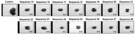
- 18 is a photograph of tumor tissue confirmed after subcutaneously administering the peptides of SEQ ID NOs: 15 to 28 in a tumor transplantation animal model.
- 20 is a photograph of tumor tissue confirmed after orally administering the peptides of SEQ ID NOs: 1 to 14 in a tumor transplantation animal model.
- 21 is a representative result of evaluating the toxicity of the peptide of SEQ ID NO: 7 to normal cells (CCD-18Co).
- ADP adenosine diphosphate
- ADP-ribose is used as a concept including both isolated ADP-ribose itself and ADP-ribose polymer (poly ADP-ribose), and "ADP-ribose binding peptide” refers to ADP-ribose or ADP-ribose. It refers to any peptide having an activity to inhibit degradation by binding to a ribose polymer.
- the peptide is not simply formed by peptide bonds between the amino acids constituting it, but also any form of peptide analog, derivative, etc. having a partially modified form to improve properties such as stability and efficacy from the viewpoint of protein drugs. Also includes variants of
- the inventors of the present invention can expect anticancer effects through activation of PARylation or inhibition of degradation of PAR polymers, in a completely different aspect from many reports of anticancer efficacy in terms of inhibition of PARylation (poly ADP-ribosylation) by conventional PARP-1 inhibitors. It was noted that there is In particular, cancer cells continue to divide at a faster rate than normal cells and actively metabolize, so by overactivating PARylation or inhibiting the degradation of PAR polymer, it is possible to specifically target cancer cells without affecting normal cells that do not overactivate PARylation. It was hoped that it could be annihilated.
- the WE domain is a globular domain conserved in many proteins, including deltex, Trip12, and poly-ADP-ribose polymerase homologs, named after the most conserved residue in the domain (L. Aravind , TRENDS in Biochemical Sciences, 2001, 26(5): 273). It is known that in some proteins having a WE domain, there is an ADP-ribose binding motif in the domain.
- the fragments were synthesized from WE domains present in various types of proteins and their anticancer activity was investigated.
- the level of ADP-ribose in cancer cells significantly increased when the ADP-ribose binding peptides (SEQ ID NOs: 1 to 14) of Examples 1 to 14 were treated with various types of cancer cells (FIG. 1). to Figure 4).
- the balance of intracellular ADP-ribose was disturbed due to the treatment of the peptides, and the growth of cancer cells was inhibited and most of them died (FIGS.
- tumor transplantation animal model Also, when the ADP-ribose-binding peptides of Examples 1 to 14 were subcutaneously or orally administered, it was confirmed that the growth of tumor tissue in vivo was rapidly reduced (FIG. 19 and FIG. 20).
- the peptide of SEQ ID NO: 7 was treated with normal cells to evaluate cytotoxicity against normal cells. As a result, it was confirmed that no toxicity was shown to normal cells (FIGS. 21 and 22).
- the ADP-ribose binding peptides of SEQ ID NOs: 1 to 14 of the present invention have excellent anticancer effects regardless of cancer types and show no cytotoxicity to normal cells, so they can be usefully used as compositions for preventing or treating cancer. there is.
- the ADP-ribose binding peptides are all peptides having the amino acid sequence of any one of SEQ ID NOs: 1 to 14, as well as sequences in which one or more amino acids are added, substituted, or deleted from these sequences, provided they fall within the same range. belong to the category
- it has at least 80% or more, 90% or more, 95% or more, 97% or more, or 99% or more homology with a peptide having any one amino acid sequence of SEQ ID NOs: 1 to 14 of the present invention, and the sequence If it is a peptide exhibiting efficacy corresponding to that of the peptide consisting of any one of amino acid sequences of Nos. 1 to 14, that is, ADP-ribose binding activity and anticancer activity, some sequences are added, substituted or deleted from the amino acid sequences of SEQ ID Nos. 1 to 14. Even if it has an amino acid sequence, it is obvious that it is included within the scope of the present invention.
- the term "homology” means the degree of matching with a given amino acid sequence or nucleotide sequence and can be expressed as a percentage.
- a homologous sequence having the same or similar activity as a given amino acid sequence or nucleotide sequence is expressed as "% homology”.
- BLAST 2.0 which calculates parameters such as score, identity and similarity, or by Southern hybridization experiments under defined stringent conditions, sequence It can be confirmed by comparing, and appropriate hybridization conditions defined are within the scope of the art and are well known to those skilled in the art (e.g., J.
- the peptides of the present invention may include modifications such as chemical derivatization at one or more amino acids constituting the peptides to further improve desired properties, as long as they have equivalent anticancer activity to those of the peptides of the present invention despite such modifications. It is apparent to those skilled in the art that all fall within the scope of the present invention.
- the derivatization may include, but is not limited to, acetylation, hydroxylation, methylation, amidation, pegylation, and addition of carbohydrate or lipid components, cofactors, and the like.
- the peptide may be additionally used in a fused form with a cell-penetrating peptide to increase cell permeability. That is, the ADP-ribose binding peptide may further include cell penetrating peptides at the N-terminus, C-terminus, or both ends. At this time, a linker may be additionally included between the ADP-ribose binding peptide and the cell penetrating peptide, which can be appropriately performed by those skilled in the art.
- the term "cell-penetrating peptide” refers to a peptide having characteristics capable of promoting uptake/absorption of various materials such as nanoparticles, compounds, DNA, and proteins into cells.
- the cell-penetrating peptides include TAT, maurocalcine, penetratin, poly-arginine-derived peptides, Antennapedia, Transportan, VP22, It may be Hph-1, Poly-arginine, R11(R9), Pep-1, HP4, LAH4, Vetofusing-1, a peptide based on a signal sequence, or an amphipathic peptide, but is limited thereto
- those skilled in the art can be appropriately selected as long as they can promote intracellular movement of the ADP-ribose-binding peptide of the present invention.
- TAT a cell penetrating peptide
- SEQ ID NOs: 15 to 24, Table 2 the ADP-ribose binding peptides of SEQ ID NOs: 1 to 14 of the present invention to evaluate their anticancer activity.
- SEQ ID NOs: 15 to 24, Table 2 the ADP-ribose-binding peptide had more excellent anticancer activity than when used alone (FIGS. 5 to 12, 17 and 18). Therefore, not only the ADP-ribose binding peptides of SEQ ID NOs: 1 to 14 of the present invention, but also the peptides fused with cell penetrating proteins can be very useful as compositions for preventing or treating cancer.
- ADP-ribose-binding peptide of the present invention can appropriately modify and use to apply it according to the type of cell-penetrating peptide used. That is, even when the ADP-ribose binding peptide of the present invention is fused to a cell penetrating peptide and used, it is not limited to the amino acid sequence presented in the present invention, and the application of the cell penetrating peptide within the range apparent to those skilled in the art, that is, the equivalent range. It can be used by adding/substituting/removing an amino acid sequence in an appropriate form.
- the ADP-ribose binding peptide of the present invention can be used together with reagents known in the art that can deliver proteins into cells or improve delivery efficiency in order to increase cell permeability.
- reagents include, for example, Chariot TM (Active motif, Cat. 30025), Xflect TM (Takara, Cat. 631324), Pierce TM (ThermoFisher Scientific, Cat. 89850), ProteoJuice TM (Merck, Cat.
- Another aspect of the present invention is a polynucleotide encoding the ADP-ribose-binding peptide.
- Another aspect of the present invention is a vector containing the polynucleotide.
- Another aspect of the present invention is a transformant comprising the polynucleotide.
- ADP-ribose binding peptides are as described above.
- the polynucleotide has a nucleotide sequence encoding the ADP-ribose binding peptide of the present invention, or a nucleotide sequence having at least 80%, 90%, 95%, 97%, or 99% homology thereto. If the polypeptide translated therefrom exhibits an efficacy corresponding to that of the ADP-ribose-binding peptide of the present invention, the addition of meaningless sequences to the 5'- and/or 3'-ends of the nucleotide sequence, or deletion of some sequences, Any modification or substitution may be included in the scope of the present invention.
- the polynucleotide may be used in the form of an expression cassette operably linked to a known promoter sequence or a vector containing the polynucleotide, and the polynucleotide, expression cassette or vector may be prepared appropriately by a method known to those skilled in the art. can do.
- the type of promoter and vector is not particularly limited and can be appropriately selected by those skilled in the art according to the purpose.
- a transformant may be prepared and used by transforming the polynucleotide, expression cassette, or vector into a host cell, and methods known to those skilled in the art may also be used without limitation.
- the transformant is a target to express the ADP-ribose-binding peptide of the present invention, and may be a microorganism, plant or animal, but may be other than human, but is not limited thereto.
- a person skilled in the art can prepare and use a polynucleotide encoding the ADP-ribose binding peptide, a vector containing the same, or a transformant containing the vector in order to apply/produce the ADP-ribose binding peptide for various purposes.
- the polynucleotide or the vector can be used directly for treatment of cancer, and the peptide can be produced or used for treatment using a transformant expressing the ADP-ribose binding peptide.
- Another aspect of the present invention is a pharmaceutical composition for preventing or treating cancer, comprising the ADP-ribose-binding peptide or a pharmaceutically acceptable salt thereof as an active ingredient.
- Another aspect of the present invention is the use of the peptide or a pharmaceutically acceptable salt thereof for the prevention or treatment of cancer, and the step of administering the peptide or a pharmaceutically acceptable salt thereof to a subject in need thereof It is a method for preventing or treating cancer, including
- the ADP-ribose-binding peptide of the present invention has an excellent effect in preventing and/or treating cancer.
- the term "cancer” refers to a disease related to the regulation of cell death, and refers to a disease caused by excessive proliferation of cells when the normal balance of cell death is broken.
- the cancer includes both malignant tumor and benign tumor, and includes, for example, brain cancer, head and neck cancer, lung cancer, breast cancer, thymoma, esophageal cancer, colorectal cancer, liver cancer, stomach cancer, pancreatic cancer, and biliary tract.
- the types of cancer of the present invention are not limited by the above examples.
- the composition for preventing or treating cancer of the present invention has a therapeutic effect on all cancers in which cell death may occur due to the accumulation of intracellular ADP-ribose.
- the cancer may be solid cancer such as brain cancer, lung cancer, pancreatic cancer, liver cancer, breast cancer, colon cancer, kidney cancer, stomach cancer, or ovarian cancer, but is not limited thereto.
- the term "treatment” refers to intervening to alter the natural process of a diseased individual or cell, which may be performed during the course of a pathological condition or to prevent it. Desired therapeutic effects include preventing occurrence or recurrence of the disease, alleviating symptoms, reducing any direct or indirect pathological consequences of the disease, preventing metastasis, reducing the rate of disease progression, and alleviating the disease state. or providing temporary relief, remission or improving prognosis.
- the present invention includes all activities that improve the course of cancer by administering a composition containing an ADP-ribose binding peptide or a pharmaceutically acceptable salt thereof as an active ingredient.
- prevention refers to any activity that inhibits or delays the onset of cancer by administering the composition.
- the weight % of the ADP-ribose-conjugated peptide or a pharmaceutically acceptable salt thereof included in the pharmaceutical composition is not particularly limited thereto, but is 0.0001 to 90% by weight, specifically 0.001 to 50% by weight, based on the total weight of the final composition. More specifically, it may be included in an amount of 0.01 to 20% by weight.
- the pharmaceutical composition may be prepared by further including appropriate carriers, excipients or diluents commonly used in the manufacture of pharmaceuticals.
- the pharmaceutical composition of the present invention can be used in oral formulations such as powders, granules, tablets, capsules, suspensions, emulsions, syrups, aerosols, oral patches, external preparations, external patches, suppositories and sterile injections according to conventional methods. It may be formulated and used in the form of a solution.
- the pharmaceutical composition When used for oral administration, it may be prepared as a sustained-release preparation through appropriate encapsulation, enteric coating, and polymer formulation.
- the sustained-release formulation may be prepared as a long-acting formulation.
- a long-acting formulation may be mixed with a polymer and a lipid in an appropriate ratio.
- the pharmaceutical composition of the present invention may be administered to a subject who has or is at risk of developing cancer.
- the term "individual" means all animals including humans.
- the pharmaceutical composition of the present invention can be administered to a subject in a pharmaceutically effective amount.
- administration refers to introducing the pharmaceutical composition of the present invention into a target subject by any suitable method, and the route of administration may be administered through various oral or parenteral routes as long as it can reach the target tissue. there is. Examples of routes of administration include oral, intramuscular, intravenous, arterial, subcutaneous, intraperitoneal, pulmonary, and nasal, including but not limited to subcutaneous or oral administration.
- the term "pharmaceutically effective amount” means an amount sufficient to prevent and/or treat cancer with a reasonable benefit/risk ratio applicable to medical use.
- An appropriate dosage and frequency of administration may be selected according to a method known in the art, and the amount and frequency of administration of the pharmaceutical composition of the present invention actually administered depend on the type of symptom to be treated, route of administration, gender, health condition, It can be appropriately determined by various factors such as diet, age, weight, and severity of the disease of the subject.
- pharmaceutically acceptable salts refer to salts commonly used in the pharmaceutical industry, for example, salts of inorganic ions including sodium, potassium, calcium, magnesium, lithium, copper, manganese, zinc, iron, etc.
- inorganic acids such as perchloric acid, phosphoric acid, and sulfuric acid, as well as salts of ascorbic acid, citric acid, tartaric acid, lactic acid, maleic acid, malonic acid, fumaric acid, glycolic acid, succinic acid, propionic acid, acetic acid, orotate acid, and acetylsalicylic acid.
- organic acids and amino acid salts such as lysine, arginine, and guanidine.
- organic ion salts such as tetramethyl ammonium, tetraethyl ammonium, tetrapropyl ammonium, tetrabutyl ammonium, benzyl trimethyl ammonium, and benzethonium that can be used in pharmaceutical reactions, purification and separation processes.
- organic ion salts such as tetramethyl ammonium, tetraethyl ammonium, tetrapropyl ammonium, tetrabutyl ammonium, benzyl trimethyl ammonium, and benzethonium that can be used in pharmaceutical reactions, purification and separation processes.
- the types of salts meant in the present invention are not limited by these listed salts.
- the ADP-ribose-binding peptide of the present invention has excellent anticancer activity, it can be prepared not only as a pharmaceutical composition but also as a functional food composition.
- the food composition may include additional ingredients that are commonly used in food and can improve odor, taste, and vision.
- additional ingredients that are commonly used in food and can improve odor, taste, and vision.
- food additives may be added. The additive is selected according to the type of food and used in an appropriate amount.
- the food composition can be prepared as a health functional food, where functional food is the same term as food for special health use (FoSHU), and in addition to nutritional supply, bioregulatory functions are efficiently displayed. It refers to processed medicine and food with high medical effect.
- the health functional food may be prepared in various forms such as tablets, capsules, powders, granules, liquids, and pills in order to obtain useful effects for cancer improvement.
- Another aspect of the present invention is a pharmaceutical composition for adjuvant anticancer that enhances reactivity to a second anticancer agent, comprising the ADP-ribose-binding peptide or a pharmaceutically acceptable salt thereof as an active ingredient.
- Another aspect of the present invention is a pharmaceutical preparation for preventing or treating cancer, comprising (i) the ADP-ribose binding peptide or a pharmaceutically acceptable salt thereof, and (ii) a second anticancer agent as active ingredients.
- a composition for preventing or treating cancer, comprising (i) the ADP-ribose binding peptide or a pharmaceutically acceptable salt thereof, and (ii) a second anticancer agent as active ingredients. is a composition
- ADP-ribose-binding peptides and pharmaceutically acceptable salts thereof are as described above.
- the ADP-ribose-conjugated peptide of the present invention goes beyond having an anticancer effect alone and also has a very excellent effect as an anticancer auxiliary pharmaceutical composition for the purpose of enhancing reactivity to a second anticancer agent. Therefore, it can be used as a pharmaceutical composition for preventing or treating cancer by using the second anticancer agent as an active ingredient together with the ADP-ribose binding peptide or a pharmaceutically acceptable salt thereof.
- the term "second anticancer agent” refers to any drug having anticancer activity except for the ADP-ribose binding peptide of the present invention.
- the scope of the second anticancer agent is not particularly limited, and those skilled in the art can select and use an appropriate type according to the purpose of cure, control, symptom relief, etc. of cancer depending on the type and progress of cancer.
- the second anticancer agent may be, for example, a cytotoxic anticancer agent, a targeted anticancer agent, an immune anticancer agent, or a metabolic anticancer agent, but is not limited thereto.
- the cytotoxic anticancer agent is a drug that exhibits an anticancer effect by attacking cancer cells that divide indiscriminately at a faster rate than normal cells, meaning the same as commonly used in the art to which the present invention belongs.
- the cytotoxic anticancer agents include alkylating agents, antimetabolites, and natural anticancer agents.
- the alkylating agent is nitrogen mustard (e.g., cyclophosphamide, chlormethine, uramustine, melphalan, chlorambucil, ifosfamide, bendamustine, etc.), alkyl sulfonates (e.g., busulfan, procarbazine, etc.) ), nitrosoureas (e.g., carmustine, lomustine, streptozocin, etc.), platinum-based alkylating agents (e.g., cisplatin, carboplatin, dicycloplatin, eptaplatin, lobaplatin, myriplatin, yes daplatin, oxaliplatin, picoplatin, satraplatin, triplatin tetranitrate, etc.), but are not limited thereto. Alkylating agents can cause destruction of cancer cells by binding to DNA in cancer cells and damaging the DNA structure.
- nitrogen mustard e.g., cyclophosphamide, chlormethin
- the antimetabolites include pyrimidine derivatives (eg, 5-fluorouracil, capecitabine, cystarabine, gemcitabine, fludarabine, etc.), folate derivatives (eg, methotrexate, pemetrexed, etc.), purine derivatives (eg, methotrexate, pemetrexed, etc.) , mercaptopurine, etc.), but are not limited thereto.
- Antimetabolites can induce cancer cell death by inhibiting DNA replication and metabolism necessary for cell survival.
- the natural anticancer agents include topoisomerase inhibitors (e.g., camptothecin, epipodophyllotoxin, taxane-based drugs (dositaxel, paclitaxel)), antibiotics (e.g., dactinomycin, doxorubicin, daunorubicin, mitomycin) , pleomycin, idarubicin, mitoxantrone HCl, etc.), but are not limited thereto.
- topoisomerase inhibitors e.g., camptothecin, epipodophyllotoxin, taxane-based drugs (dositaxel, paclitaxel)
- antibiotics e.g., dactinomycin, doxorubicin, daunorubicin, mitomycin
- pleomycin e.g., idarubicin, mitoxantrone HCl, etc.
- the target anticancer agent is an anticancer agent that induces the death of cancer cells by inhibiting a target protein (receptor or enzyme) involved in cancer growth, the meaning of which is as commonly used in the art to which the present invention belongs.
- the cytotoxic anti-cancer agent includes target protein (tyrosine kinase, etc.) inhibitory low-molecular-weight compounds and monoclonal antibodies.
- the targeted anticancer agent may be a receptor tyrosine kinase inhibitor targeting one or more targets selected from the group consisting of VEGF-A and EGFR.
- the targeted anti-cancer agent that can be co-administered with the ADP-ribose binding peptide is a VEGF-A inhibitor.
- VEGF-A inhibitors include monoclonal antibodies such as bevacizumab, ranibizumab, aflibercept, and ramucirumab, sunitinib, and pazopa. and low molecular weight compounds such as pazopanib, sorafenib, and axitinib, but are not limited thereto.
- the targeted anti-cancer agent that can be co-administered with the ADP-ribose binding peptide is an EGFR inhibitor.
- the EGFR inhibitor is osimertinib, gefitinib, erlotinib, afatinib, brigatinib, icotinib, vandetanib ( vandetanib) and monoclonal antibodies such as cetuximab, panitumumab, zalutumumab, nimotuzumab, and matuzumab. Not limited to this.
- target anticancer agent of the present invention also includes HER2 targeting anticancer agents such as lapatinib, neratinib, and afatinib, imatinib, dasatinib, and nilotyi.
- HER2 targeting anticancer agents such as lapatinib, neratinib, and afatinib, imatinib, dasatinib, and nilotyi.
- Bcr-Abl targeted anti-cancer drugs such as nilotinib; Src target anti-cancer agents such as bosutinib; JAK target anti-cancer drugs such as lestaurtinib, ruxolitinib, pacritinib; MAP2 Kinase target anticancer drugs such as cobimethinib, selumetinib, trametinib, and binimetinib; It includes MEL4-ALK target anticancer drugs such as ceritibin and crizotinib, and is not particularly limited thereto.
- the second anticancer agent of the present invention may be a combination of one or more cytotoxic anticancer agents and/or target anticancer agents, which may be administered simultaneously or at different times.
- immuno-anticancer agents refer to drugs that activate the body's immune system to fight cancer cells.
- immuno-anticancer agents include immune checkpoint inhibitors, immune cell therapy agents, anti-cancer vaccines, and antibody-drug conjugates, and appropriate types can be selected for complete cure, control, and symptom relief of cancer depending on the type and progress of cancer. .
- the immuno-anticancer agent can be an immune checkpoint inhibitor, a PD-1 antibody, a PD-L1 antibody, a CTLA-4 antibody, a CD28 antibody, a KIR antibody, a TCR antibody, a LAG-3 antibody, a TIM-3 antibody, It may be one or more selected from the group consisting of TIGIT antibody, A2aR antibody, ICOS antibody, OX40 antibody, 4-1BB antibody, and GITR antibody.
- the immune checkpoint inhibitor is a PD-1 antibody such as nivolumab, pembrolizumab, cemiplimab, pidilizumab, toripalimab; PD-L1 antibodies such as atezolizumab, avelumab, duralumab; It may be a CTLA-4 antibody such as ipilimumab and tremelimumab, but is not limited thereto.
- the immuno-anticancer agent may be an immune cell therapy agent, and may be a CAR-T agent such as tisagenlecleucel or axicabtagene ciloleucel, or a CAR-NK agent, but is not limited thereto. It is not.
- cancer metabolism agents refer to drugs that are involved in the growth and survival of cancer cells, such as supplying nutrients to cancer cells, or are involved in various essential metabolic reactions to kill cancer cells.
- the anti-metabolism agents include, for example, IM-156, 3-bromopyruvic acid (3BP), NYH817100, WZB117, GNE-140, AZ93, AZD3965, CPI-613, MKT-077, CB-839, CB-1158, CPI-444, TVB-2640, NDI-010976, TCD-717, ADI-PEG20, Epacadostat, Indoximod, PX478, CPI-0610, RTA402, APO866, GMX1778, AG-221 or AG-120 etc., but is not limited thereto.
- the ADP-ribose binding peptide of the present invention can be used as an anti-cancer adjuvant because it has the effect of enhancing reactivity to a second anti-cancer agent by disrupting the balance of ADP-ribose in cancer cells, so the type of second anti-cancer agent used or the type of cancer Not particularly limited.
- the cancer may be solid cancer such as brain cancer, lung cancer, pancreatic cancer, liver cancer, breast cancer, colon cancer, kidney cancer, stomach cancer, or ovarian cancer, but is not limited thereto.
- the ADP-ribose binding peptide of the present invention can not only be co-administered in a form that exists independently of the second anticancer agent, but also forms a physical/chemical bond with the second anticancer agent by any known method depending on the purpose can be administered.
- the ADP-ribose-binding peptide may be used in a state of being directly coupled to the second anti-cancer agent, or may be used in a state of being connected to the second anti-cancer agent through a known linker, and the ADP-ribose-binding peptide of the present invention may be used in combination with the second anti-cancer agent.
- the method of application is not particularly limited as long as it works together to exhibit an increased anticancer effect.
- the ADP-ribose binding peptide of the present invention can enhance the response of the second anticancer agent, so it can be used very usefully as an anticancer supplement, and has excellent anticancer activity even when the second anticancer agent is treated at a low concentration, Possible side effects can be minimized.
- Another aspect of the present invention is a pharmaceutical composition for adjuvant anti-cancer treatment, comprising the ADP-ribose-binding peptide or a pharmaceutically acceptable salt thereof as an active ingredient, enhancing responsiveness to radiation anti-cancer treatment.
- ADP-ribose-binding peptides and pharmaceutically acceptable salts thereof are as described above.
- radiation anti-cancer treatment refers to a therapeutic action of irradiating cancer cells or tumor tissue with radiation for the purpose of killing cancer cells.
- standard treatment for controlling inoperable or inoperable tumors, or tumor metastasis is based on the principle that radiation delivered to a target site causes the death of reproductive cells.
- Radiation anticancer treatment in the present invention may be ionizing radiation therapy, electromagnetic radiation therapy, brachytherapy or external beam radiation therapy, but is not limited thereto.
- the composition containing the ADP-ribose binding peptide or a pharmaceutically acceptable salt thereof according to the present invention exhibits a synergistic anticancer effect when used in combination with radiation therapy, it is used as an anticancer adjuvant for radiation therapy or a radiation sensitizer that improves radiation sensitivity.
- the type of applicable cancer is not particularly limited.
- the cancer may be solid cancer such as brain cancer, lung cancer, pancreatic cancer, liver cancer, breast cancer, colon cancer, kidney cancer, stomach cancer, and ovarian cancer, but is not limited thereto.
- the solid cancer may be resistant to radiation therapy. Since the anticancer effect of radiation therapy is manifested by the formation of DNA breaks, resistance to radiation therapy is generally determined by mechanisms capable of repairing DNA damage induced by radiation therapy. As a method of increasing the anticancer efficacy of radiation therapy, many attempts have been made to inhibit DNA repair mechanisms, and when repair of damaged DNA strands is blocked, the sensitivity of radiation therapy can be increased.
- the two main types of DNA damage are single strand breaks (SSB) and double strand breaks (DSB). Therefore, it can be described as two categories of recovery paths targeting SSB and DSB.
- Base Excision Repair (BER) is one of several pathways involved in the repair of selected types of DNA SSBs.
- PARP1 plays an important role in the BER of DNA SSBs through a process known as ADP-ribosylation.
- PARP1 senses damage to SSB DNA and recruits the DNA repair complex through ADP-ribosylation to the SSB site for repair. Since the overaccumulation of poly-ADP-ribose synthesized through ADP-ribosylation by PARP-1 activity ultimately leads to cell death, cancer cells use proteasomes such as PARG or ARH3 to prevent this phenomenon. It activates the degradation of poly-ADP-ribose through the activation of survival signaling activity.
- ADP-ribose or ADP-ribose polymer in cancer cells can act as a medium that can disrupt these biochemical survival mechanisms of cancer cells, and can be used as an important anti-cancer adjuvant to overcome resistance to radiation therapy. conceived.
- the ADP-ribose-binding peptide of the present invention when the ADP-ribose-binding peptide of the present invention was treated in combination with low-dose radiation in different cancer types, it was confirmed that the responsiveness to radiation treatment was enhanced in all cancer types (Fig. 15 and FIG. 16). Therefore, the ADP-ribose-binding peptide of the present invention can enhance the response to radiation therapy, so it can be used very usefully as an adjuvant anti-cancer application. Since it has excellent anti-cancer activity even when irradiated with radiation at a tolerable dose, side effects that may occur due to radiation can be minimized.
- Embodiments of the present invention may be modified in various other forms, and the scope of the present invention is not limited to the embodiments described below.
- embodiments of the present invention are provided to more completely explain the present invention to those skilled in the art.
- "include” a certain component throughout the specification means that other components may be further included without excluding other components unless otherwise stated.
- the ADP-ribose binding peptides of Examples 1 to 14 were synthesized from WE domains present in various types of proteins, and were used in experiments. Specific information of the peptides used are shown in Table 1 below.
- Example Sequence information (SEQ ID NO) derived protein One VPYIIDLQSMHQFRQDTGTMRPVRR (SEQ ID NO: 1) DTX1 2 APYIIDLQSMNQFRQDTGTLRPVRR (SEQ ID NO: 2) DTX4 3 APYIIDLPSWTQFRQDTGTMRAVRR (SEQ ID NO: 3) DTX2 4 RVYTIDFNSMQQINEDTGTARAIQR (SEQ ID NO: 4) TRIP12 5 FCYLIYFNSMSQMNRQTRRRRRRLRR (SEQ ID NO: 5) PARP7 6 FSYVIDFNTMGQINRQTQRQRRVRR (SEQ ID NO: 6) PARP12 7 FSYKIDFAEMKQMNLTTGKQRLIKR (SEQ ID NO: 7) PARP11 8 YNYTVNYTTHTQTNKTSSFCRSVRR (SEQ ID NO: 8) PIK3C2G 9 RRYTVQFTTM
- Examples 15 to 28 were prepared by binding a cell-penetrating peptide to the N-terminus of the peptides of Examples 1 to 14. Peptide sequences of Examples 15 to 28 are shown in Table 2.
- the peptides of Examples 1 to 28 of the present invention are designed to inhibit the action of poly ADP-ribose degrading enzyme.
- Experimental Example 1 it was confirmed whether degradation of poly ADP-ribose was inhibited by each of the peptides and accumulated in cancer cells through cell experiments.
- 5 ⁇ 10 5 H1975 cells were treated with RPMI-1640 medium containing 10% FBS, 100 units/ml penicillin and 100 ⁇ g/ml streptomycin at 37 °C and 5% CO 2 conditions, and the group not treated with the example peptide 0.2, 1, 2, 4, 8, 16, 32 ⁇ M of the peptide of Example 2 (SEQ ID NO: 2) was divided into groups treated and cultured for 24 hours.
- the medium of the cultured cells was removed and treated with RIPA buffer and 1% SDS.
- the prepared supernatant was used for ELISA analysis to detect and analyze ADP-ribose.
- the upper right of Figure 1 and the upper left of Figure 3 show the increase in ADP-ribose compared to the control group according to the peptide treatment of Example 2 or Example 9 in H1975 cells as fold change, compared to the untreated group. It shows that the amount of ADP-ribose significantly increased in a concentration-dependent manner (** p ⁇ 0.001).
- the bottom left of Figure 1 and the top right of Figure 3 show the increase in ADP-ribose as fold change compared to the control group according to the treatment with the peptide of Example 3 or Example 10 in Aspc-1 cells, compared to the untreated group. It shows that the amount of ADP-ribose significantly increased in a concentration-dependent manner of the prepared peptide (** p ⁇ 0.001).
- Hep G2 cells were treated with EMEM medium containing 10% FBS, 100 units/ml penicillin and 100 ⁇ g/ml streptomycin at 37°C, 5% CO 2 conditions, and the group not treated with the example peptide and 0.2 , 1, 2, 4, 8, 16, 32 ⁇ M of the peptide of Example 4 (SEQ ID NO: 4) was divided into groups treated and cultured for 24 hours.
- the medium of the cultured cells was removed and treated with RIPA buffer and 1% SDS.
- the prepared supernatant was used for ELISA analysis to detect and analyze ADP-ribose.
- the bottom right of Figure 1 and the bottom left of Figure 3 show the increase in ADP-ribose compared to the control group according to the peptide treatment of Example 4 or Example 11 in Hep G2 cells as fold change, compared to the untreated group. It shows that the amount of ADP-ribose significantly increased in a concentration-dependent manner of the peptide (** p ⁇ 0.001).
- 5 ⁇ 10 5 MDA-MB-231 cells were cultured in Leibovitz's L-15 medium containing 10% FBS, 100 units/ml penicillin, and 100 ⁇ g/ml streptomycin at 37 °C and 5% CO 2 conditions, and the example peptides Divided into groups untreated and groups treated with 0.2, 1, 2, 4, 8, 16, and 32 ⁇ M of the peptide of Example 5 (SEQ ID NO: 5), and cultured for 24 hours.
- the medium of the cultured cells was removed and treated with RIPA buffer and 1% SDS.
- the prepared supernatant was used for ELISA analysis to detect and analyze ADP-ribose.
- FIG. 2 and the lower right side of FIG. 3 show the increase in ADP-ribose as fold change compared to the control group according to the treatment with the peptide of Example 5 or Example 12 in MDA-MB-231 cells, compared to the untreated group. It shows that the amount of ADP-ribose significantly increased in a concentration-dependent manner compared to the treated peptide (** p ⁇ 0.001).
- FIG. 5 and the top right of FIG. 8 are representative photomicrographs of groups treated with and without the peptides of Examples 1, 15, 8, and 22 in U-87MG cells, respectively.
- the group not treated with the peptide it was observed that U-87MG cells rapidly grew, whereas in the group treated with the peptide of the present invention, the growth of U-87MG cells was inhibited and died. shape can be observed.
- cancer cells were completely killed in the groups treated with the cell-penetrating peptide (CPP)-attached peptides of Examples 15 and 22.
- CPP cell-penetrating peptide
- FIG. 5 and the upper left of FIG. 9 are representative photomicrographs of H1975 cells treated with and without the peptides of Examples 2, 16, 9, and 23, respectively.
- rapid growth of H1975 cells could be observed, whereas in the group treated with the peptide of the present invention, the growth of H1975 cells was inhibited and apoptosis was observed. can do.
- cancer cells were completely killed in the groups treated with the peptides of Examples 16 and 23 to which CPP was attached.
- FIG. 6 and the top right of FIG. 9 are representative photomicrographs of Aspc-1 cells treated with the peptides of Examples 3, 17, 10, and 24, respectively, and groups not treated.
- the group not treated with the peptide rapid growth of Aspc-1 cells could be observed, whereas in the group treated with the peptide of the present invention, the growth of Aspc-1 cells was inhibited and died. shape can be observed.
- cancer cells were completely killed in the group treated with the peptides of Examples 17 and 24 to which the cell penetrating peptide CPP was attached.
- FIG. 6 and the upper left of FIG. 10 are representative photomicrographs of Hep G2 cells treated with and without the peptides of Examples 4, 18, 11, and 25, respectively.
- the group not treated with the peptide it was observed that Hep G2 cells rapidly grew, whereas in the group treated with the peptide of the present invention, the growth of Hep G2 cells was inhibited and they died. can be observed.
- cancer cells were completely killed in the group treated with the peptides of Examples 18 and 25 to which the cell penetrating peptide CPP was attached.
- FIG. 7 and the upper right of FIG. 10 are representative photomicrographs of the groups treated with and without the peptides of Examples 5, 19, 12, and 26 in MDA-MB-231 cells, respectively.
- the group not treated with the peptide rapid growth of MDA-MB-231 cells could be observed, whereas in the group treated with the peptide of the present invention, the growth of MDA-MB-231 cells A suppressed and dying form can be observed.
- cancer cells were completely killed in the group treated with the peptides of Examples 19 and 26 to which CPP was attached.
- FIG. 7 and the upper left of FIG. 11 are representative photomicrographs of HCT116 cells treated with and without the peptides of Examples 6, 20, 13, and 27, respectively.
- rapid growth of HCT116 cells was observed in the group not treated with the peptide, whereas growth of HCT116 cells was inhibited and death was observed in the group treated with the peptide of the present invention. can do.
- cancer cells were completely killed in the group treated with the peptides of Examples 20 and 27 to which the cell penetrating peptide CPP was attached.
- FIG. 8 and the upper right of FIG. 11 are representative photomicrographs of groups treated with and without the peptides of Examples 7, 21, 14, and 28 in Caki-1 cells, respectively.
- a rapid growth of Caki-1 cells could be observed, whereas in the group treated with the peptide of the embodiment of the present invention, the growth of Caki-1 cells was inhibited and died. shape can be observed.
- cancer cells were completely killed in the groups treated with the peptides of Examples 21 and 28 to which CPP was attached.
- the anticancer effect represented by the reduction of cancer cell survival by existing anticancer drugs
- the existing anticancer drugs are treated and the accumulation of ADP-ribose is induced, the existing anticancer drugs are delivered to cancer cells at the required concentration. Even if treated below, it was expected to exhibit an increased anticancer effect.
- the Example peptides of the present invention significantly increase the amount of ADP-ribose in cancer cells. I wanted to make sure it showed up.
- Figure 18 shows representative tumor photographs obtained after autopsy in all groups, and it was observed that the growth of tumor tissue was significantly inhibited in all groups subcutaneously injected with the peptides of the examples of the present invention when compared to the control group.
- Figure 20 shows representative tumor photographs obtained after autopsy in all groups. When compared with the control group, it was observed that the growth of tumor tissue was significantly inhibited in all groups orally administered with the peptides of the examples of the present invention.
- peptides of the present invention show cytotoxicity even in normal cells.
- human colon fibroblasts, CCD-18Co, and human dermal papilla cells, HDPC were used as normal cells, and the peptide of SEQ ID NO: 7 was used as an exemplary peptide of the present invention.
Landscapes
- Health & Medical Sciences (AREA)
- Chemical & Material Sciences (AREA)
- Life Sciences & Earth Sciences (AREA)
- Organic Chemistry (AREA)
- Genetics & Genomics (AREA)
- Zoology (AREA)
- Engineering & Computer Science (AREA)
- Bioinformatics & Cheminformatics (AREA)
- Wood Science & Technology (AREA)
- General Health & Medical Sciences (AREA)
- Molecular Biology (AREA)
- Medicinal Chemistry (AREA)
- Biochemistry (AREA)
- Biotechnology (AREA)
- General Engineering & Computer Science (AREA)
- Biomedical Technology (AREA)
- Microbiology (AREA)
- Proteomics, Peptides & Aminoacids (AREA)
- Biophysics (AREA)
- Gastroenterology & Hepatology (AREA)
- Toxicology (AREA)
- Veterinary Medicine (AREA)
- Pharmacology & Pharmacy (AREA)
- Public Health (AREA)
- Animal Behavior & Ethology (AREA)
- Nuclear Medicine, Radiotherapy & Molecular Imaging (AREA)
- General Chemical & Material Sciences (AREA)
- Chemical Kinetics & Catalysis (AREA)
- Immunology (AREA)
- Epidemiology (AREA)
- Physics & Mathematics (AREA)
- Plant Pathology (AREA)
- Medicines That Contain Protein Lipid Enzymes And Other Medicines (AREA)
- Pharmaceuticals Containing Other Organic And Inorganic Compounds (AREA)
- Micro-Organisms Or Cultivation Processes Thereof (AREA)
- Peptides Or Proteins (AREA)
Abstract
Description
| 실시예 | 서열정보 (서열번호) | 유래 단백질 |
| 1 | VPYIIDLQSMHQFRQDTGTMRPVRR (서열번호 1) | DTX1 |
| 2 | APYIIDLQSMNQFRQDTGTLRPVRR (서열번호 2) | DTX4 |
| 3 | APYIIDLPSWTQFRQDTGTMRAVRR (서열번호 3) | DTX2 |
| 4 | RVYTIDFNSMQQINEDTGTARAIQR (서열번호 4) | TRIP12 |
| 5 | FCYLIYFNSMSQMNRQTRRRRRLRR (서열번호 5) | PARP7 |
| 6 | FSYVIDFNTMGQINRQTQRQRRVRR (서열번호 6) | PARP12 |
| 7 | FSYKIDFAEMKQMNLTTGKQRLIKR (서열번호 7) | PARP11 |
| 8 | YNYTVNYTTHTQTNKTSSFCRSVRR (서열번호 8) | PIK3C2G |
| 9 | RRYTVQFTTMVQVNEETGNRRPVM (서열번호 9) | HUWE1 |
| 10 | WIWYWKNESGTWIQYGEEKDKRKN (서열번호 10) | ZC3HAV1 |
| 11 | FLYVADLENMVQYRRNEHGRRRKIKR (서열번호 11) | RNF146 |
| 12 | RHYTVNLNTYTATDTKGHSLSVQR (서열번호 12) | PARP14 |
| 13 | GRYDVHLGERMRYAVYWDELASEVRR (서열번호 13) | DDHD2 |
| 14 | TAYEASVCDYLEQQVARGN (서열번호 14) | HNRNPA2B1 |
| 실시예 | 서열정보 (서열번호) |
| 15 | GRKKRRQRRRPQVPYIIDLQSMHQFRQDTGTMRPVRR (서열번호 15) |
| 16 | GRKKRRQRRRPQAPYIIDLQSMNQFRQDTGTLRPVRR (서열번호 16) |
| 17 | GRKKRRQRRRPQAPYIIDLPSWTQFRQDTGTMRAVRR (서열번호 17) |
| 18 | GRKKRRQRRRPQRVYTIDFNSMQQINEDTGTARAIQR (서열번호 18) |
| 19 | GRKKRRQRRRPQFCYLIYFNSMSQMNRQTRRRRRLRR (서열번호 19) |
| 20 | GRKKRRQRRRPQFSYVIDFNTMGQINRQTQRQRRVRR (서열번호 20) |
| 21 | GRKKRRQRRRPQFSYKIDFAEMKQMNLTTGKQRLIKR (서열번호 21) |
| 22 | GRKKRRQRRRPQYNYTVNYTTHTQTNKTSSFCRSVRR (서열번호 22) |
| 23 | GRKKRRQRRRPQRRYTVQFTTMVQVNEETGNRRPVM (서열번호 23) |
| 24 | GRKKRRQRRRPQWIWYWKNESGTWIQYGEEKDKRKN (서열번호 24) |
| 25 | GRKKRRQRRRPQFLYVADLENMVQYRRNEHGRRRKIKR (서열번호 25) |
| 26 | GRKKRRQRRRPQRHYTVNLNTYTATDTKGHSLSVQR (서열번호 26) |
| 27 | GRKKRRQRRRPQGRYDVHLGERMRYAVYWDELASEVRR (서열번호 27) |
| 28 | GRKKRRQRRRPQTAYEASVCDYLEQQVARGN (서열번호 28) |
Claims (16)
- 서열번호 1 내지 서열번호 14로 구성된 군으로부터 선택된 어느 하나의 아미노산 서열을 갖는, ADP (adenosine diphosphate)-리보오스 결합 펩티드.
- 제1항에 있어서,상기 펩티드는 N-말단, C-말단 또는 양 말단에 세포 투과성 펩티드 (cell-penetrating peptide)를 추가로 포함하는 것인, ADP-리보오스 결합 펩티드.
- 제2항에 있어서, 상기 세포 투과성 펩티드는 TAT, 마우로칼신 (maurocalcine), 페네트라틴 (penetratin), 폴리-알지닌 (Poly-arginine) 유래 펩티드, 안테나페디아 (Antennapedia), 트랜스포탄 (Transportan), VP22, Hph-1, 폴리-알지닌 (Poly-arginine), R11(R9), Pep-1, HP4, LAH4, Vetofusing-1, 시그널 서열 기반 펩티드, 및 양친매성 (Amphipathic) 펩티드로 이루어진 군에서 선택되는 하나 이상인 것인, ADP-리보오스 결합 펩티드.
- 제2항에 있어서,상기 펩티드는 서열번호 15 내지 24로 구성된 군으로부터 선택된 어느 하나의 아미노산 서열을 갖는 것인, ADP-리보오스 결합 펩티드.
- 제1항 내지 제4항 중 어느 한 항의 ADP-리보오스 결합 펩티드를 코딩하는 폴리뉴클레오티드.
- 제5항의 폴리뉴클레오티드를 포함하는 벡터.
- 제5항의 폴리뉴클레오티드를 포함하는, 형질전환체.
- 제1항 내지 제4항 중 어느 한 항의 ADP-리보오스 결합 펩티드 또는 이의 약학적으로 허용 가능한 염을 유효성분으로 포함하는, 암의 예방 또는 치료용 약학적 조성물.
- 제8항에 있어서,상기 암은 뇌암, 폐암, 췌장암, 간암, 유방암, 대장암, 신장암, 위암 및 난소암으로 구성된 군에서 선택된 1종 이상의 고형암인 것인, 암의 예방 또는 치료용 약학적 조성물.
- 제8항에 있어서,상기 조성물은 피하 또는 경구로 투여되는 것인, 암의 예방 또는 치료용 약학적 조성물.
- 제1항 내지 제4항 중 어느 한 항의 ADP-리보오스 결합 펩티드 또는 이의 약학적으로 허용 가능한 염을 유효성분으로 포함하는, 제2항암제에 대한 반응성을 증진시키는 항암 보조용 약학적 조성물.
- 제11항에 있어서,상기 항암제는 세포 독성 항암제, 표적 항암제 또는 이들의 조합인 것인, 항암 보조용 약학적 조성물.
- 제11항에 있어서,상기 제2항암제가 표적으로 하는 암은 뇌암, 폐암, 췌장암, 간암, 유방암, 대장암, 신장암, 위암 및 난소암으로 구성된 군에서 선택된 1종 이상의 고형암인 것인, 항암 보조용 약학적 조성물.
- (i) 제1항 내지 제4항 중 어느 한 항의 ADP-리보오스 결합 펩티드 또는 이의 약학적으로 허용 가능한 염, 및(ii) 제2항암제를 유효성분으로 포함하는, 암의 예방 또는 치료용 약학적 조성물.
- 제1항 내지 제4항 중 어느 한 항의 ADP-리보오스 결합 펩티드 또는 이의 약학적으로 허용 가능한 염을 유효성분으로 포함하는, 방사선 항암 치료에 대한 반응성을 증진시키는 항암 보조용 약학적 조성물.
- 제15항에 있어서,상기 방사선 항암 치료가 표적으로 하는 암은 뇌암, 폐암, 췌장암, 간암, 유방암, 대장암, 신장암, 위암 및 난소암으로 구성된 군에서 선택된 1종 이상의 고형암인 것인, 항암 보조용 약학적 조성물.
Priority Applications (11)
| Application Number | Priority Date | Filing Date | Title |
|---|---|---|---|
| CA3222862A CA3222862A1 (en) | 2021-09-02 | 2022-08-29 | Adp-ribose binding peptides with anti-cancer activity and use thereof |
| JP2024514398A JP7672763B2 (ja) | 2021-09-02 | 2022-08-29 | 抗がん活性を有するadp-リボース結合ペプチドおよびその用途 |
| PL22864983.6T PL4335862T3 (pl) | 2021-09-02 | 2022-08-29 | Peptyd wiążący adp-rybozę wykazujący aktywność przeciwnowotworową i jego zastosowania |
| EP22864983.6A EP4335862B1 (en) | 2021-09-02 | 2022-08-29 | Adp-ribose binding peptide having anticancer activity and use thereof |
| US18/247,831 US20230416316A1 (en) | 2021-09-02 | 2022-08-29 | Adp-ribose binding peptides with anti-cancer activity and use thereof |
| ES22864983T ES3042065T3 (en) | 2021-09-02 | 2022-08-29 | Adp-ribose binding peptide having anticancer activity and use thereof |
| CN202280007142.3A CN116529255A (zh) | 2021-09-02 | 2022-08-29 | 具有抗癌活性的adp-核糖结合肽及其用途 |
| AU2022338621A AU2022338621B2 (en) | 2021-09-02 | 2022-08-29 | Adp-ribose binding peptide having anticancer activity and use thereof |
| AU2024259837A AU2024259837B2 (en) | 2021-09-02 | 2024-11-08 | ADP-ribose binding peptide having anticancer activity and use thereof |
| JP2025031297A JP7854224B2 (ja) | 2021-09-02 | 2025-02-28 | 抗がん活性を有するadp-リボース結合ペプチドおよびその用途 |
| AU2026200568A AU2026200568A1 (en) | 2021-09-02 | 2026-01-28 | ADP-ribose binding peptide having anticancer activity and use thereof |
Applications Claiming Priority (2)
| Application Number | Priority Date | Filing Date | Title |
|---|---|---|---|
| KR10-2021-0117046 | 2021-09-02 | ||
| KR1020210117046A KR102557456B1 (ko) | 2021-09-02 | 2021-09-02 | 항암 활성을 가지는 adp-리보오스 결합 펩티드 및 이의 용도 |
Publications (1)
| Publication Number | Publication Date |
|---|---|
| WO2023033481A1 true WO2023033481A1 (ko) | 2023-03-09 |
Family
ID=85412833
Family Applications (1)
| Application Number | Title | Priority Date | Filing Date |
|---|---|---|---|
| PCT/KR2022/012886 Ceased WO2023033481A1 (ko) | 2021-09-02 | 2022-08-29 | 항암 활성을 가지는 adp-리보오스 결합 펩티드 및 이의 용도 |
Country Status (10)
| Country | Link |
|---|---|
| US (1) | US20230416316A1 (ko) |
| EP (1) | EP4335862B1 (ko) |
| JP (2) | JP7672763B2 (ko) |
| KR (3) | KR102557456B1 (ko) |
| CN (1) | CN116529255A (ko) |
| AU (3) | AU2022338621B2 (ko) |
| CA (1) | CA3222862A1 (ko) |
| ES (1) | ES3042065T3 (ko) |
| PL (1) | PL4335862T3 (ko) |
| WO (1) | WO2023033481A1 (ko) |
Cited By (2)
| Publication number | Priority date | Publication date | Assignee | Title |
|---|---|---|---|---|
| AU2022341787B2 (en) * | 2021-09-07 | 2024-07-11 | PearlsInMires Co., Ltd. | Use of adenosine diphosphate ribose as adjuvant therapy for radiation and/or anticancer therapy |
| EP4509133A4 (en) * | 2023-03-02 | 2026-03-25 | Pearlsinmires Co Ltd | ADP-RIBOSE BINDING PEPTIDE FOR CANCER PREVENTION OR TREATMENT, AND ADP-RIBOSE POLYTHERAPY |
Families Citing this family (2)
| Publication number | Priority date | Publication date | Assignee | Title |
|---|---|---|---|---|
| KR102557456B1 (ko) | 2021-09-02 | 2023-07-19 | 주식회사 펄스인마이어스 | 항암 활성을 가지는 adp-리보오스 결합 펩티드 및 이의 용도 |
| KR102947986B1 (ko) | 2022-09-21 | 2026-04-06 | 경북대학교 산학협력단 | 근적외선 감응형 융합 단백질 나노입자 퀀텀닷을 포함하는 암 치료용 약학적 조성물 및 이의 제조방법 |
Citations (1)
| Publication number | Priority date | Publication date | Assignee | Title |
|---|---|---|---|---|
| US20200384118A1 (en) * | 2017-12-21 | 2020-12-10 | Friedrich-Alexander-Universitat Erlangen-Nurnberg | Immunotoxin conjugates for use in therapy |
Family Cites Families (5)
| Publication number | Priority date | Publication date | Assignee | Title |
|---|---|---|---|---|
| WO2015191546A2 (en) * | 2014-06-10 | 2015-12-17 | The Board Of Regents Of The University Of Texas System | Adp-ribose detection reagents |
| KR101844541B1 (ko) * | 2014-07-29 | 2018-04-02 | 재단법인 아산사회복지재단 | Parp 저해제에 대한 감수성 예측용 신규한 바이오 마커 및 이의 용도 |
| US20240252689A1 (en) * | 2021-05-18 | 2024-08-01 | The Board Of Regents Of The University Of Texas System | Poly-adp ribose (par) tracker optimized split-protein reassembly par detection reagents |
| IL310116A (en) * | 2021-07-20 | 2024-03-01 | Epicrispr Biotechnologies Inc | Systems and methods for regulating target genes |
| KR102557456B1 (ko) | 2021-09-02 | 2023-07-19 | 주식회사 펄스인마이어스 | 항암 활성을 가지는 adp-리보오스 결합 펩티드 및 이의 용도 |
-
2021
- 2021-09-02 KR KR1020210117046A patent/KR102557456B1/ko active Active
-
2022
- 2022-08-29 CN CN202280007142.3A patent/CN116529255A/zh active Pending
- 2022-08-29 AU AU2022338621A patent/AU2022338621B2/en active Active
- 2022-08-29 PL PL22864983.6T patent/PL4335862T3/pl unknown
- 2022-08-29 WO PCT/KR2022/012886 patent/WO2023033481A1/ko not_active Ceased
- 2022-08-29 ES ES22864983T patent/ES3042065T3/es active Active
- 2022-08-29 JP JP2024514398A patent/JP7672763B2/ja active Active
- 2022-08-29 US US18/247,831 patent/US20230416316A1/en active Pending
- 2022-08-29 EP EP22864983.6A patent/EP4335862B1/en active Active
- 2022-08-29 CA CA3222862A patent/CA3222862A1/en active Pending
-
2023
- 2023-07-13 KR KR1020230091177A patent/KR102774674B1/ko active Active
-
2024
- 2024-11-08 AU AU2024259837A patent/AU2024259837B2/en active Active
-
2025
- 2025-02-18 KR KR1020250020815A patent/KR20250028322A/ko active Pending
- 2025-02-28 JP JP2025031297A patent/JP7854224B2/ja active Active
-
2026
- 2026-01-28 AU AU2026200568A patent/AU2026200568A1/en active Pending
Patent Citations (1)
| Publication number | Priority date | Publication date | Assignee | Title |
|---|---|---|---|---|
| US20200384118A1 (en) * | 2017-12-21 | 2020-12-10 | Friedrich-Alexander-Universitat Erlangen-Nurnberg | Immunotoxin conjugates for use in therapy |
Non-Patent Citations (12)
| Title |
|---|
| F.M. AUSUBEL ET AL.: "Current Protocols in Molecular Biology", JOHN WILEY & SONS, INC. |
| J MATEO ET AL., ANN ONCOL., vol. 30, no. 9, 2019, pages 1437 |
| J. SAMBROOK ET AL.: "A Laboratory Manual", 1989, COLD SPRING HARBOR LABORATORY PRESS, article "Molecular Cloning" |
| JUAN ET AL., CANCERS, vol. 12, no. 3, 2020, pages 739 |
| KOO JA-HYUN, YOON HEESEOK, KIM WON-JU, CHA DONGHUN, CHOI JE-MIN: "Cell-Penetrating Function of the Poly(ADP-Ribose) (PAR)-Binding Motif Derived from the PAR-Dependent E3 Ubiquitin Ligase Iduna", INTERNATIONAL JOURNAL OF MOLECULAR SCIENCES, vol. 19, no. 3, 8 March 2018 (2018-03-08), pages 779, XP093042870, DOI: 10.3390/ijms19030779 * |
| KRIETSCH JANA, ROULEAU MICHÈLE, PIC ÉMILIE, ETHIER CHANTAL, DAWSON TED M., DAWSON VALINA L., MASSON JEAN-YVES, POIRIER GUY G., GAG: "Reprogramming cellular events by poly(ADP-ribose)-binding proteins", MOLECULAR ASPECTS OF MEDICINE, PERGAMON PRESS, OXFORD., GB, vol. 34, no. 6, 1 December 2013 (2013-12-01), GB , pages 1066 - 1087, XP093042872, ISSN: 0098-2997, DOI: 10.1016/j.mam.2012.12.005 * |
| L. ARAVIND: "TRENDS", BIOCHEMICAL SCIENCES, vol. 26, no. 5, 2001, pages 273 |
| PALAZZO LUCA, AHEL IVAN: "PARPs in genome stability and signal transduction: implications for cancer therapy", BIOCHEMICAL SOCIETY TRANSACTIONS, PORTLAND PRESS LTD, GB, vol. 46, no. 6, 17 December 2018 (2018-12-17), GB , pages 1681 - 1695, XP093042873, ISSN: 0300-5127, DOI: 10.1042/BST20180418 * |
| PNAS, vol. 108, no. 34, 23 August 2011 (2011-08-23), pages 14103 - 14108 |
| REBECCA GUPTE ET AL., GENES DEV, vol. 31, no. 2, 2017, pages 101 |
| See also references of EP4335862A4 |
| WANG ZHIZHI, MICHAUD GREGORY A., CHENG ZHIHONG, ZHANG YUE, HINDS THOMAS R., FAN ERKANG, CONG FENG, XU WENQING: "Recognition of the iso-ADP-ribose moiety in poly(ADP-ribose) by WWE domains suggests a general mechanism for poly(ADP-ribosyl)ation-dependent ubiquitination", GENES & DEVELOPMENT, COLD SPRING HARBOR LABORATORY PRESS, PLAINVIEW, NY., US, vol. 26, no. 3, 1 February 2012 (2012-02-01), US , pages 235 - 240, XP093042868, ISSN: 0890-9369, DOI: 10.1101/gad.182618.111 * |
Cited By (2)
| Publication number | Priority date | Publication date | Assignee | Title |
|---|---|---|---|---|
| AU2022341787B2 (en) * | 2021-09-07 | 2024-07-11 | PearlsInMires Co., Ltd. | Use of adenosine diphosphate ribose as adjuvant therapy for radiation and/or anticancer therapy |
| EP4509133A4 (en) * | 2023-03-02 | 2026-03-25 | Pearlsinmires Co Ltd | ADP-RIBOSE BINDING PEPTIDE FOR CANCER PREVENTION OR TREATMENT, AND ADP-RIBOSE POLYTHERAPY |
Also Published As
| Publication number | Publication date |
|---|---|
| CN116529255A (zh) | 2023-08-01 |
| JP2025084888A (ja) | 2025-06-03 |
| AU2022338621A1 (en) | 2024-01-04 |
| US20230416316A1 (en) | 2023-12-28 |
| AU2022338621B2 (en) | 2024-08-08 |
| JP7854224B2 (ja) | 2026-05-01 |
| ES3042065T3 (en) | 2025-11-18 |
| PL4335862T3 (pl) | 2025-11-12 |
| KR20250028322A (ko) | 2025-02-28 |
| EP4335862B1 (en) | 2025-08-27 |
| EP4335862A4 (en) | 2024-11-13 |
| KR102557456B1 (ko) | 2023-07-19 |
| JP2024532528A (ja) | 2024-09-05 |
| EP4335862A1 (en) | 2024-03-13 |
| CA3222862A1 (en) | 2023-03-09 |
| EP4335862C0 (en) | 2025-08-27 |
| AU2026200568A1 (en) | 2026-02-19 |
| KR20230033991A (ko) | 2023-03-09 |
| KR102774674B1 (ko) | 2025-03-04 |
| KR20230113494A (ko) | 2023-07-31 |
| JP7672763B2 (ja) | 2025-05-08 |
| AU2024259837A1 (en) | 2024-12-19 |
| AU2024259837B2 (en) | 2026-02-12 |
Similar Documents
| Publication | Publication Date | Title |
|---|---|---|
| WO2023033481A1 (ko) | 항암 활성을 가지는 adp-리보오스 결합 펩티드 및 이의 용도 | |
| WO2017022962A1 (ko) | Ripk 억제제를 유효성분으로 포함하는 면역질환의 예방 또는 치료용 조성물 | |
| WO2022191431A1 (ko) | Kras 및 p53 유전자의 편집 기능이 있는 나노 리포좀 전달체 조성물 | |
| WO2019083172A1 (ko) | 스트렙토니그린 및 라파마이신을 유효성분으로 포함하는, 암 예방 또는 치료용 약학적 조성물 | |
| WO2023038368A1 (ko) | 방사선 및/또는 항암 치료 보조 요법으로 아데노신 디포스페이트 리보오스의 활용 | |
| WO2015199454A1 (ko) | 3,4,5-트리하이드록시벤조산, 이의 유도체 또는 이의 염을 유효성분으로 포함하는 수용체 티로신 키나아제 저해제 내성 항암제 조성물 | |
| WO2024181817A1 (ko) | 암의 예방 또는 치료를 위한 adp-리보오스 결합 펩티드 및 adp-리보오스의 병용 요법 | |
| US9669068B2 (en) | Methods for the treatment of cancer using gliadin peptides | |
| CN117957233A (zh) | 那拉霉素的含氮衍生物、其合成和用途 | |
| Lushnikova et al. | Overcoming of doxorubicin resistance in breast cancer cells by cationic peptides | |
| WO2023177233A1 (ko) | 신규한 화합물 및 이의 체크포인트 키나제 2 저해 용도 | |
| HK40110860A (zh) | 使用麦胶蛋白肽治疗癌症的套件和方法 | |
| WO2026071839A1 (ko) | Gv1001 펩타이드를 포함하는 암세포의 cd47 발현 감소 및 대식세포의 식세포작용 증가용 조성물 및 이의 용도 | |
| WO2025121807A1 (ko) | Ezh2 저해제 및 mettl3 억제제를 포함하는 암의 예방 또는 치료용 약제학적 조성물 | |
| WO2023163301A1 (ko) | Kitenin의 이량체 형성을 저해하는 펩타이드 및 이의 용도 | |
| WO2021201588A1 (ko) | 항암 요법을 위한 헤파린-담즙산 올리고머 컨쥬게이트 | |
| WO2016040560A1 (en) | Methods for the treatment of cancer using gliadin peptides and radiation |
Legal Events
| Date | Code | Title | Description |
|---|---|---|---|
| WWE | Wipo information: entry into national phase |
Ref document number: 18247831 Country of ref document: US |
|
| 121 | Ep: the epo has been informed by wipo that ep was designated in this application |
Ref document number: 22864983 Country of ref document: EP Kind code of ref document: A1 |
|
| WWE | Wipo information: entry into national phase |
Ref document number: 202280007142.3 Country of ref document: CN |
|
| WWE | Wipo information: entry into national phase |
Ref document number: 2022864983 Country of ref document: EP |
|
| WWE | Wipo information: entry into national phase |
Ref document number: 2022338621 Country of ref document: AU Ref document number: AU2022338621 Country of ref document: AU |
|
| WWE | Wipo information: entry into national phase |
Ref document number: 3222862 Country of ref document: CA |
|
| ENP | Entry into the national phase |
Ref document number: 2022864983 Country of ref document: EP Effective date: 20231207 |
|
| ENP | Entry into the national phase |
Ref document number: 2022338621 Country of ref document: AU Date of ref document: 20220829 Kind code of ref document: A |
|
| WWE | Wipo information: entry into national phase |
Ref document number: 2024514398 Country of ref document: JP |
|
| WWE | Wipo information: entry into national phase |
Ref document number: 202417020531 Country of ref document: IN |
|
| NENP | Non-entry into the national phase |
Ref country code: DE |
|
| WWG | Wipo information: grant in national office |
Ref document number: 2022864983 Country of ref document: EP |