WO2023042876A1 - 2'-修飾ヌクレオシドを含むヘテロ核酸 - Google Patents
2'-修飾ヌクレオシドを含むヘテロ核酸 Download PDFInfo
- Publication number
- WO2023042876A1 WO2023042876A1 PCT/JP2022/034531 JP2022034531W WO2023042876A1 WO 2023042876 A1 WO2023042876 A1 WO 2023042876A1 JP 2022034531 W JP2022034531 W JP 2022034531W WO 2023042876 A1 WO2023042876 A1 WO 2023042876A1
- Authority
- WO
- WIPO (PCT)
- Prior art keywords
- nucleic acid
- acid strand
- nucleosides
- hdo
- double
- Prior art date
- Legal status (The legal status is an assumption and is not a legal conclusion. Google has not performed a legal analysis and makes no representation as to the accuracy of the status listed.)
- Ceased
Links
Images





























Classifications
-
- C—CHEMISTRY; METALLURGY
- C12—BIOCHEMISTRY; BEER; SPIRITS; WINE; VINEGAR; MICROBIOLOGY; ENZYMOLOGY; MUTATION OR GENETIC ENGINEERING
- C12N—MICROORGANISMS OR ENZYMES; COMPOSITIONS THEREOF; PROPAGATING, PRESERVING, OR MAINTAINING MICROORGANISMS; MUTATION OR GENETIC ENGINEERING; CULTURE MEDIA
- C12N15/00—Mutation or genetic engineering; DNA or RNA concerning genetic engineering, vectors, e.g. plasmids, or their isolation, preparation or purification; Use of hosts therefor
- C12N15/09—Recombinant DNA-technology
- C12N15/11—DNA or RNA fragments; Modified forms thereof; Non-coding nucleic acids having a biological activity
- C12N15/113—Non-coding nucleic acids modulating the expression of genes, e.g. antisense oligonucleotides; Antisense DNA or RNA; Triplex- forming oligonucleotides; Catalytic nucleic acids, e.g. ribozymes; Nucleic acids used in co-suppression or gene silencing
-
- A—HUMAN NECESSITIES
- A61—MEDICAL OR VETERINARY SCIENCE; HYGIENE
- A61K—PREPARATIONS FOR MEDICAL, DENTAL OR TOILETRY PURPOSES
- A61K47/00—Medicinal preparations characterised by the non-active ingredients used, e.g. carriers or inert additives; Targeting or modifying agents chemically bound to the active ingredient
- A61K47/50—Medicinal preparations characterised by the non-active ingredients used, e.g. carriers or inert additives; Targeting or modifying agents chemically bound to the active ingredient the non-active ingredient being chemically bound to the active ingredient, e.g. polymer-drug conjugates
- A61K47/51—Medicinal preparations characterised by the non-active ingredients used, e.g. carriers or inert additives; Targeting or modifying agents chemically bound to the active ingredient the non-active ingredient being chemically bound to the active ingredient, e.g. polymer-drug conjugates the non-active ingredient being a modifying agent
- A61K47/54—Medicinal preparations characterised by the non-active ingredients used, e.g. carriers or inert additives; Targeting or modifying agents chemically bound to the active ingredient the non-active ingredient being chemically bound to the active ingredient, e.g. polymer-drug conjugates the non-active ingredient being a modifying agent the modifying agent being an organic compound
- A61K47/549—Sugars, nucleosides, nucleotides or nucleic acids
-
- A—HUMAN NECESSITIES
- A61—MEDICAL OR VETERINARY SCIENCE; HYGIENE
- A61K—PREPARATIONS FOR MEDICAL, DENTAL OR TOILETRY PURPOSES
- A61K47/00—Medicinal preparations characterised by the non-active ingredients used, e.g. carriers or inert additives; Targeting or modifying agents chemically bound to the active ingredient
- A61K47/50—Medicinal preparations characterised by the non-active ingredients used, e.g. carriers or inert additives; Targeting or modifying agents chemically bound to the active ingredient the non-active ingredient being chemically bound to the active ingredient, e.g. polymer-drug conjugates
- A61K47/51—Medicinal preparations characterised by the non-active ingredients used, e.g. carriers or inert additives; Targeting or modifying agents chemically bound to the active ingredient the non-active ingredient being chemically bound to the active ingredient, e.g. polymer-drug conjugates the non-active ingredient being a modifying agent
- A61K47/56—Medicinal preparations characterised by the non-active ingredients used, e.g. carriers or inert additives; Targeting or modifying agents chemically bound to the active ingredient the non-active ingredient being chemically bound to the active ingredient, e.g. polymer-drug conjugates the non-active ingredient being a modifying agent the modifying agent being an organic macromolecular compound, e.g. an oligomeric, polymeric or dendrimeric molecule
- A61K47/59—Medicinal preparations characterised by the non-active ingredients used, e.g. carriers or inert additives; Targeting or modifying agents chemically bound to the active ingredient the non-active ingredient being chemically bound to the active ingredient, e.g. polymer-drug conjugates the non-active ingredient being a modifying agent the modifying agent being an organic macromolecular compound, e.g. an oligomeric, polymeric or dendrimeric molecule obtained otherwise than by reactions only involving carbon-to-carbon unsaturated bonds, e.g. polyureas or polyurethanes
- A61K47/60—Medicinal preparations characterised by the non-active ingredients used, e.g. carriers or inert additives; Targeting or modifying agents chemically bound to the active ingredient the non-active ingredient being chemically bound to the active ingredient, e.g. polymer-drug conjugates the non-active ingredient being a modifying agent the modifying agent being an organic macromolecular compound, e.g. an oligomeric, polymeric or dendrimeric molecule obtained otherwise than by reactions only involving carbon-to-carbon unsaturated bonds, e.g. polyureas or polyurethanes the organic macromolecular compound being a polyoxyalkylene oligomer, polymer or dendrimer, e.g. PEG, PPG, PEO or polyglycerol
-
- A—HUMAN NECESSITIES
- A61—MEDICAL OR VETERINARY SCIENCE; HYGIENE
- A61P—SPECIFIC THERAPEUTIC ACTIVITY OF CHEMICAL COMPOUNDS OR MEDICINAL PREPARATIONS
- A61P25/00—Drugs for disorders of the nervous system
-
- A—HUMAN NECESSITIES
- A61—MEDICAL OR VETERINARY SCIENCE; HYGIENE
- A61P—SPECIFIC THERAPEUTIC ACTIVITY OF CHEMICAL COMPOUNDS OR MEDICINAL PREPARATIONS
- A61P43/00—Drugs for specific purposes, not provided for in groups A61P1/00-A61P41/00
-
- C—CHEMISTRY; METALLURGY
- C12—BIOCHEMISTRY; BEER; SPIRITS; WINE; VINEGAR; MICROBIOLOGY; ENZYMOLOGY; MUTATION OR GENETIC ENGINEERING
- C12N—MICROORGANISMS OR ENZYMES; COMPOSITIONS THEREOF; PROPAGATING, PRESERVING, OR MAINTAINING MICROORGANISMS; MUTATION OR GENETIC ENGINEERING; CULTURE MEDIA
- C12N15/00—Mutation or genetic engineering; DNA or RNA concerning genetic engineering, vectors, e.g. plasmids, or their isolation, preparation or purification; Use of hosts therefor
- C12N15/09—Recombinant DNA-technology
- C12N15/11—DNA or RNA fragments; Modified forms thereof; Non-coding nucleic acids having a biological activity
- C12N15/113—Non-coding nucleic acids modulating the expression of genes, e.g. antisense oligonucleotides; Antisense DNA or RNA; Triplex- forming oligonucleotides; Catalytic nucleic acids, e.g. ribozymes; Nucleic acids used in co-suppression or gene silencing
- C12N15/1137—Non-coding nucleic acids modulating the expression of genes, e.g. antisense oligonucleotides; Antisense DNA or RNA; Triplex- forming oligonucleotides; Catalytic nucleic acids, e.g. ribozymes; Nucleic acids used in co-suppression or gene silencing against enzymes
-
- C—CHEMISTRY; METALLURGY
- C12—BIOCHEMISTRY; BEER; SPIRITS; WINE; VINEGAR; MICROBIOLOGY; ENZYMOLOGY; MUTATION OR GENETIC ENGINEERING
- C12N—MICROORGANISMS OR ENZYMES; COMPOSITIONS THEREOF; PROPAGATING, PRESERVING, OR MAINTAINING MICROORGANISMS; MUTATION OR GENETIC ENGINEERING; CULTURE MEDIA
- C12N2310/00—Structure or type of the nucleic acid
- C12N2310/10—Type of nucleic acid
- C12N2310/11—Antisense
-
- C—CHEMISTRY; METALLURGY
- C12—BIOCHEMISTRY; BEER; SPIRITS; WINE; VINEGAR; MICROBIOLOGY; ENZYMOLOGY; MUTATION OR GENETIC ENGINEERING
- C12N—MICROORGANISMS OR ENZYMES; COMPOSITIONS THEREOF; PROPAGATING, PRESERVING, OR MAINTAINING MICROORGANISMS; MUTATION OR GENETIC ENGINEERING; CULTURE MEDIA
- C12N2310/00—Structure or type of the nucleic acid
- C12N2310/30—Chemical structure
- C12N2310/31—Chemical structure of the backbone
- C12N2310/315—Phosphorothioates
-
- C—CHEMISTRY; METALLURGY
- C12—BIOCHEMISTRY; BEER; SPIRITS; WINE; VINEGAR; MICROBIOLOGY; ENZYMOLOGY; MUTATION OR GENETIC ENGINEERING
- C12N—MICROORGANISMS OR ENZYMES; COMPOSITIONS THEREOF; PROPAGATING, PRESERVING, OR MAINTAINING MICROORGANISMS; MUTATION OR GENETIC ENGINEERING; CULTURE MEDIA
- C12N2310/00—Structure or type of the nucleic acid
- C12N2310/30—Chemical structure
- C12N2310/32—Chemical structure of the sugar
- C12N2310/321—2'-O-R Modification
-
- C—CHEMISTRY; METALLURGY
- C12—BIOCHEMISTRY; BEER; SPIRITS; WINE; VINEGAR; MICROBIOLOGY; ENZYMOLOGY; MUTATION OR GENETIC ENGINEERING
- C12N—MICROORGANISMS OR ENZYMES; COMPOSITIONS THEREOF; PROPAGATING, PRESERVING, OR MAINTAINING MICROORGANISMS; MUTATION OR GENETIC ENGINEERING; CULTURE MEDIA
- C12N2310/00—Structure or type of the nucleic acid
- C12N2310/30—Chemical structure
- C12N2310/32—Chemical structure of the sugar
- C12N2310/322—2'-R Modification
-
- C—CHEMISTRY; METALLURGY
- C12—BIOCHEMISTRY; BEER; SPIRITS; WINE; VINEGAR; MICROBIOLOGY; ENZYMOLOGY; MUTATION OR GENETIC ENGINEERING
- C12N—MICROORGANISMS OR ENZYMES; COMPOSITIONS THEREOF; PROPAGATING, PRESERVING, OR MAINTAINING MICROORGANISMS; MUTATION OR GENETIC ENGINEERING; CULTURE MEDIA
- C12N2310/00—Structure or type of the nucleic acid
- C12N2310/30—Chemical structure
- C12N2310/32—Chemical structure of the sugar
- C12N2310/323—Chemical structure of the sugar modified ring structure
- C12N2310/3231—Chemical structure of the sugar modified ring structure having an additional ring, e.g. LNA, ENA
-
- C—CHEMISTRY; METALLURGY
- C12—BIOCHEMISTRY; BEER; SPIRITS; WINE; VINEGAR; MICROBIOLOGY; ENZYMOLOGY; MUTATION OR GENETIC ENGINEERING
- C12N—MICROORGANISMS OR ENZYMES; COMPOSITIONS THEREOF; PROPAGATING, PRESERVING, OR MAINTAINING MICROORGANISMS; MUTATION OR GENETIC ENGINEERING; CULTURE MEDIA
- C12N2310/00—Structure or type of the nucleic acid
- C12N2310/30—Chemical structure
- C12N2310/33—Chemical structure of the base
- C12N2310/334—Modified C
- C12N2310/3341—5-Methylcytosine
-
- C—CHEMISTRY; METALLURGY
- C12—BIOCHEMISTRY; BEER; SPIRITS; WINE; VINEGAR; MICROBIOLOGY; ENZYMOLOGY; MUTATION OR GENETIC ENGINEERING
- C12N—MICROORGANISMS OR ENZYMES; COMPOSITIONS THEREOF; PROPAGATING, PRESERVING, OR MAINTAINING MICROORGANISMS; MUTATION OR GENETIC ENGINEERING; CULTURE MEDIA
- C12N2310/00—Structure or type of the nucleic acid
- C12N2310/30—Chemical structure
- C12N2310/34—Spatial arrangement of the modifications
- C12N2310/341—Gapmers, i.e. of the type ===---===
-
- C—CHEMISTRY; METALLURGY
- C12—BIOCHEMISTRY; BEER; SPIRITS; WINE; VINEGAR; MICROBIOLOGY; ENZYMOLOGY; MUTATION OR GENETIC ENGINEERING
- C12N—MICROORGANISMS OR ENZYMES; COMPOSITIONS THEREOF; PROPAGATING, PRESERVING, OR MAINTAINING MICROORGANISMS; MUTATION OR GENETIC ENGINEERING; CULTURE MEDIA
- C12N2310/00—Structure or type of the nucleic acid
- C12N2310/30—Chemical structure
- C12N2310/35—Nature of the modification
- C12N2310/351—Conjugate
- C12N2310/3515—Lipophilic moiety, e.g. cholesterol
Definitions
- the present invention relates to a double-stranded nucleic acid complex containing a 2'-modified nucleoside, and a pharmaceutical composition containing it as an active ingredient.
- oligonucleotides In recent years, in the ongoing development of medicines called nucleic acid medicines, attention has been focused on oligonucleotides. is being actively developed.
- a partial sequence of mRNA or miRNA transcribed from the target gene is used as the target sense strand, and a complementary oligonucleotide (antisense oligonucleotide: in this specification, often referred to as "ASO (Antisense Oligonucleotide)").
- ASO Antisense Oligonucleotide
- the present inventors have proposed a double-stranded nucleic acid complex (heteroduplex oligonucleotide, HDO) in which an antisense oligonucleotide and its complementary strand are annealed. developed (Patent Document 1, Non-Patent Documents 1 and 2).
- the double-stranded nucleic acid complex has a high antisense effect, and is an epoch-making technology that enables control of the central nervous system across the blood-brain barrier.
- mice intracerebroventricularly administered with single-stranded gapmer-type antisense nucleic acids hypoactivity and motor dysfunction due to central nervous system toxicity are observed within several hours after administration.
- the present inventors conducted intensive research in search of a new technique that can reduce the central nervous system toxicity of nucleic acid drugs, and introduced 2'-modified nucleosides into double-stranded nucleic acid complexes. As a result, they found that introduction of 2'-modified nucleosides can dramatically reduce or eliminate central nervous system toxicity of double-stranded nucleic acid complexes. This toxicity-suppressing effect is a surprising effect that greatly exceeds conventional expectations.
- the present invention is based on the above findings, and provides the following.
- a double-stranded nucleic acid complex comprising a first nucleic acid strand and a second nucleic acid strand, wherein the first nucleic acid strand is capable of hybridizing to at least a portion of a target gene or transcript thereof; having an antisense effect on the target gene or its transcript, the second nucleic acid strand comprising a base sequence complementary to the first nucleic acid strand, and containing one or more 2'-modified nucleosides; The double-stranded nucleic acid complex.
- the second nucleic acid strand has 1 or 2 to 10 consecutive 2'-modified nucleosides located at the 5' end and/or 1 or 2 to 10 consecutive 2'-modified nucleosides located at the 3' end
- the two strands according to any one of (1) to (6), wherein the second nucleic acid strand contains 1 to 7 2'-modified nucleosides at positions other than the 5' and 3' ends. Stranded nucleic acid complex.
- the first nucleic acid strand is (1) a central region comprising at least 4 consecutive deoxyribonucleosides; (2) a 5' wing region containing a non-natural nucleoside located at the 5' end of the central region; and (3) a 3' wing region containing a non-natural nucleoside located at the 3' end of the central region.
- the second nucleic acid strand contains a 2'-modified nucleoside in a region consisting of a nucleotide sequence complementary to the 5' wing region and/or the 3' wing region of the first nucleic acid strand; double-stranded nucleic acid complex of. (10)
- all nucleosides in a region consisting of a base sequence complementary to the 5' wing region and/or 3' wing region of the first nucleic acid strand are 2'-modified nucleosides, ( The double-stranded nucleic acid complex according to 9).
- nucleosides in a region consisting of a base sequence complementary to the central region of the first nucleic acid strand are (a) deoxyribonucleosides, (b) deoxyribonucleosides and ribonucleosides; (c) deoxyribonucleosides and 2'-modified nucleosides,
- the double-stranded nucleic acid complex according to any one of (8) to (10), which is (d) a ribonucleoside and a 2'-modified nucleoside, or (e) a deoxyribonucleoside, a ribonucleoside, and a 2'-modified nucleoside.
- nucleosides in a region consisting of base sequences complementary to the 5' wing region and the 3' wing region of the first nucleic acid strand are 2'-modified nucleosides
- the double-stranded nucleic acid complex according to (14), wherein all nucleosides in the region consisting of a nucleotide sequence complementary to the central region of one nucleic acid strand are deoxyribonucleosides.
- said second nucleic acid strand comprises at least 4 contiguous ribonucleosides complementary to at least 4 contiguous deoxyribonucleosides in said central region of said first nucleic acid strand, (8)-(10) ), the double-stranded nucleic acid complex according to any one of (17) (i) at least one guanosine nucleoside in said first nucleic acid strand; (ii) a nucleoside adjacent to the 5' end of the guanosine nucleoside; (iii) a nucleoside adjacent to the 3'-terminal side of the guanosine nucleoside, or (iv) a nucleoside in the second nucleic acid strand that is complementary to any combination of (i) to (iii) is a 2'-modified nucleoside
- Stranded nucleic acid complex (20) The double-stranded nucleic acid complex according to any one of (1) to (19), wherein at least one nucleoside containing a pyrimidine base in the second nucleic acid strand is a 2'-modified nucleoside and/or a deoxyribonucleoside. body. (21) The double-stranded nucleic acid complex according to any one of (1) to (20), wherein the second nucleic acid strand does not contain a natural ribonucleoside containing a pyrimidine base.
- (22) The double-stranded nucleic acid complex according to (21), wherein all nucleosides containing pyrimidine bases in the second nucleic acid strand are 2'-modified nucleosides and/or deoxyribonucleosides.
- (23) The double-stranded nucleic acid complex according to any one of (1) to (22), wherein 20% or more of the nucleosides in the second nucleic acid strand are 2'-modified nucleosides.
- (24) The double-stranded nucleic acid complex according to any one of (1) to (23), wherein all nucleosides other than 2'-modified nucleosides in the second nucleic acid strand are deoxyribonucleosides.
- the second nucleic acid strand according to any one of (1) to (24), wherein all nucleosides in a region consisting of a base sequence complementary to the first nucleic acid strand are 2'-modified nucleosides. Stranded nucleic acid complex.
- the 2'-modified nucleoside is a 2'-O-methoxyethyl modified nucleoside and/or a 2'-O-methyl modified nucleoside.
- the double-stranded nucleic acid complex according to 1.
- the non-complementary bases of the second nucleic acid strand form a bulge structure, or the deletion position of the second nucleic acid strand consists of a base sequence that is non-complementary to the first nucleic acid strand.
- the double-stranded nucleic acid complex according to any one of (30) to (33), which contains a bulge structure.
- (35) The double-stranded nucleic acid complex according to (29) or (34), wherein the bulge structure contains sugar-unmodified nucleosides, or all nucleosides in the bulge structure are sugar-unmodified nucleosides.
- the second nucleic acid strand comprises at least one overhang region located on the 5'-terminal side and/or 3'-terminal side of a region consisting of a base sequence complementary to the first nucleic acid strand, (1 ) to the double-stranded nucleic acid complex according to any one of (39).
- (41) The double-stranded nucleic acid complex according to (40), wherein the overhang region has a length of 1 to 20 bases.
- the linker connects the 5' end of the first nucleic acid strand and the 3' end of the second nucleic acid strand, and/or the 3' end of the first nucleic acid strand and the second nucleic acid strand.
- a pharmaceutical composition comprising the double-stranded nucleic acid complex according to any one of (1) to (54) as an active ingredient.
- a double-stranded nucleic acid complex with reduced central nervous system toxicity is provided.
- FIG. 1 shows the structures of various crosslinked nucleic acids.
- Figure 2 shows the structures of various natural or non-natural nucleotides.
- FIG. 3 shows a scoring system for evaluating central neurotoxicity in mice after administration of nucleic acid agents.
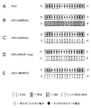
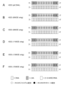
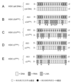
- 4 shows the structure of the nucleic acid used in Example 1.
- FIG. 4A shows the structure of ASO targeting the Mapt gene.
- FIG. 4B shows the structure of HDO (all RNA).
- FIG. 4C shows the structure of HDO (all DNA).
- FIG. 4D shows the structure of HDO (6MOE wing).
- FIG. 4E shows the structure of HDO (all MOE).
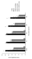
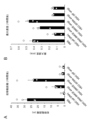
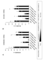
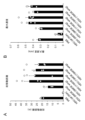
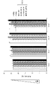
- FIG. 5 shows acute tolerability scores 30 minutes to 4 hours after administration of mice intracerebroventricularly administered with various nucleic acid agents. Error bars indicate standard error.
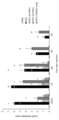
- FIG. 6 shows the motor function of mice intracerebroventricularly administered with various nucleic acid agents 1 hour after administration.
- Figure 6A shows the total distance traveled for 5 minutes.
- FIG. 6B shows the maximum movement speed. Error bars indicate standard error.
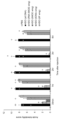
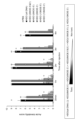
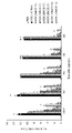
- FIG. 7 shows Mapt mRNA expression levels in the hippocampus of mice to which various nucleic acid agents were intracerebroventricularly administered. Error bars indicate standard error.
- 8 shows the structure of the nucleic acid used in Example 2.
- FIG. FIG. 8A shows the structure of ASO targeting the BACE1 gene.
- Figure 8B shows the structure of HDO (all RNA).
- FIG. 8C shows the structure of HDO (all DNA).
- FIG. 8D shows the structure of HDO (5MOE wing).
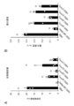
- FIG. 9 shows acute tolerability scores 30 minutes to 4 hours after administration of mice intracerebroventricularly administered with various nucleic acid agents. Error bars indicate standard error.
- FIG. 10 shows the motor function of mice intracerebroventricularly administered with various nucleic acid agents 1 hour after administration.
- FIG. 10A shows the total distance traveled for 5 minutes.
- FIG. 10B shows maximum movement speed. Error bars indicate standard error.
- 11 shows the structure of the nucleic acid used in Example 3.
- FIG. 11A shows the structure of ASO targeting the Malat1 gene.
- Figure 11B shows the structure of HDO (all RNA).
- FIG. 11C shows the structure of HDO (all DNA).
- FIG. 11D shows the structure of HDO (10 MOE wing).
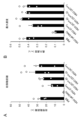
- FIG. 12 shows acute tolerability scores 30 minutes to 4 hours after administration of mice intracerebroventricularly administered with various nucleic acid agents. Error bars indicate standard error.
- FIG. 13 shows the motor function of mice intracerebroventricularly administered with various nucleic acid agents 1 hour after administration.
- Figure 13A shows the total distance traveled for 5 minutes.
- FIG. 13B shows maximum movement speed. Error bars indicate standard error.
- 14 shows the structure of the nucleic acid used in Example 4.
- FIG. 14A shows the structure of HDO (all DNA) containing ASOs targeting the Mapt gene.
- FIG. 14B shows the structure of HDO (RNA 6MOE wing).
- FIG. 14C shows the structure of HDO (6MOE wing).
- FIG. 14D shows the structure of HDO (6OMe wing).
- FIG. 14A shows the structure of HDO (all DNA) containing ASOs targeting the Mapt gene.
- FIG. 14B shows the structure of HDO (RNA 6MOE wing).
- FIG. 14E shows the structure of HDO (6F wing).
- FIG. 15 shows acute tolerability scores 30 minutes to 4 hours after administration of mice intracerebroventricularly administered with various nucleic acid agents. Error bars indicate standard error.
- FIG. 16 shows the motor function of mice intracerebroventricularly administered with various nucleic acid agents 1 hour after administration. Figure 16A shows the total distance traveled for 5 minutes. FIG. 16B shows maximum movement speed. Error bars indicate standard error.
- FIG. 17 shows the motor function of mice intracerebroventricularly administered with various nucleic acid agents 3 hours after administration. Figure 17A shows the total distance traveled for 5 minutes. FIG. 17B shows the maximum movement speed. Error bars indicate standard error. 18 shows the structure of the nucleic acid used in Example 5.
- FIG. 15 shows acute tolerability scores 30 minutes to 4 hours after administration of mice intracerebroventricularly administered with various nucleic acid agents. Error bars indicate standard error.
- FIG. 16 shows the motor function of mice intracerebroventricularly administered with various
- FIG. 18A shows the structure of an ASO targeting the Mapt gene.
- FIG. 18B shows the structure of HDO (6MOE wing).
- FIG. 18C shows the structure of HDO (G MOE ).
- FIG. 18D shows the structure of HDO (G RNA ).
- Figure 18E shows the structure of HDO (inosine).
- FIG. 19 shows acute tolerability scores 30 minutes to 4 hours after administration of mice intracerebroventricularly administered with various nucleic acid agents. Error bars indicate standard error.
- 20 shows the structure of the nucleic acid used in Example 6.
- FIG. 20A shows the structure of HDO (all DNA) containing ASOs targeting the Mapt gene.
- FIG. 20B shows the structure of HDO (6MOE-5'&3').
- FIG. 20C shows the structure of HDO (6MOE-5').
- Figure 20D shows the structure of HDO (6MOE-3').
- Figure 20E shows the structure of HDO (10MOE-5').
- FIG. 20F shows the structure of HDO(10MOE-3′).
- FIG. 21 shows acute tolerability scores 30 minutes to 4 hours after administration of mice intracerebroventricularly administered with various nucleic acid agents. Error bars indicate standard error.
- FIG. 22 shows the motor function of mice intracerebroventricularly administered with various nucleic acid agents 1 hour after administration.
- Figure 22A shows the total distance traveled for 5 minutes.
- FIG. 22B shows the maximum movement speed. Error bars indicate standard error.
- FIG. 23 shows the motor function of mice intracerebroventricularly administered with various nucleic acid agents 3 hours after administration.
- FIG. 23A shows the total distance traveled for 5 minutes.
- FIG. 23B shows the maximum movement speed. Error bars indicate standard error.
- FIG. 24 shows Mapt mRNA expression levels in the hippocampus of mice to which various nucleic acid agents were intracerebroventricularly administered. Error bars indicate standard error.
- 25 shows the structure of the nucleic acid used in Example 7.
- FIG. 25A shows the structure of HDO (all DNA) containing ASOs targeting the BACE1 gene.
- Figure 25B shows the structure of HDO (5MOE-5').
- Figure 25C shows the structure of HDO (5MOE-3').
- Figure 25D shows the structure of HDO (8MOE-5').
- Figure 25E shows the structure of HDO (8MOE-3').
- Figure 25F shows the structure of HDO (10MOE-5').
- FIG. 25G shows the structure of HDO (10MOE-3').
- FIG. 26 shows acute tolerability scores 30 minutes to 4 hours after administration of mice intracerebroventricularly administered with various nucleic acid agents. Error bars indicate standard error.
- FIG. 27 shows the motor function of mice intracerebroventricularly administered with various nucleic acid agents 1 hour after administration.
- Figure 27A shows the total distance traveled for 5 minutes.
- FIG. 27B shows the maximum movement speed. Error bars indicate standard error.
- 28 shows the structure of the nucleic acid used in Example 8.
- FIG. 28A shows the structure of HDO (all DNA) containing ASOs targeting the Mapt gene.
- FIG. 28B shows the structure of HDO (6MOE wing).
- FIG. 28C shows the structure of HDO (9MOE wing).
- FIG. 28D shows the structure of HDO (11MOE wing).
- FIG. 28E shows the structure of HDO (13MOE wing).
- FIG. 28F shows the structure of HDO (15 MOE wing).
- FIG. 29 shows acute tolerability scores 30 minutes to 4 hours after administration of mice intracerebroventricularly administered with various nucleic acid agents. Error bars indicate standard error.
- FIG. 30 shows the motor function of mice intracerebroventricularly administered with various nucleic acid agents 1 hour after administration.
- Figure 27A shows the total distance traveled for 5 minutes.
- FIG. 27B shows the maximum movement speed. Error bars indicate standard error.
- 31 shows the structure of the nucleic acid used in Example 9.
- FIG. 31A shows the structure of HDO (all DNA) containing ASOs targeting the Mapt gene.
- FIG. 31B shows the structure of HDO (A MOE ).
- FIG. 31C shows the structure of HDO (G MOE ).
- FIG. 31D shows the structure of HDO ( CMOE ).
- FIG. 31E shows the structure of HDO( TMOE ).
- FIG. 32 shows acute tolerability scores 30 minutes to 4 hours after administration of mice intracerebroventricularly administered with various nucleic acid agents. Error bars indicate standard error.
- FIG. 33 shows the motor function of mice intracerebroventricularly administered with various nucleic acid agents 1 hour after administration.
- Figure 33A shows the total distance traveled for 5 minutes.
- FIG. 33B shows maximum movement speed. Error bars indicate standard error.
- 34 shows the structure of the nucleic acid used in Example 10.
- FIG. 34A shows the structure of HDO (all DNA) containing ASOs targeting the Mapt gene.
- FIG. 34B shows the structure of HDO ( CMOE ).
- Figure 34C shows the structure of HDO (2C MOE -5).
- FIG. 34D shows the structure of HDO (2C MOE -3).
- FIG. 34E shows the structure of HDO (3C MOE ).
- FIG. 35 shows acute tolerability scores 30 minutes to 4 hours after administration of mice intracerebroventricularly administered with various nucleic acid agents. Error bars indicate standard error.
- FIG. 36 shows the motor function of mice intracerebroventricularly administered with various nucleic acid agents 1 hour after administration.
- Figure 36A shows the total distance traveled for 5 minutes.
- FIG. 36B shows maximum movement speed. Error bars indicate standard error.
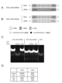
- Figure 37 shows the stability of various nucleic acid agents in human cerebrospinal fluid.
- 37A and B show the structure of the nucleic acid used in Example 11.
- FIG. Figure 37A shows the structure of an ASO targeting the Mapt gene.
- Figure 37B shows the structure of HDO (ASO/cRNA).
- FIG. 37C shows the results of examining stability by electrophoresis after mixing various nucleic acid agents with human cerebrospinal fluid (Human CSF; hCSF) for 10 minutes or 6 hours.
- FIG. 37D shows the results of quantification of band intensity of HDO duplex in HDO (ASO/cRNA).
- FIG. 38 shows the stability of various nucleic acid agents in human and rat cerebrospinal fluid.
- FIG. 38A and 38B show the structure of the nucleic acid used in Example 11.
- FIG. Figure 38A shows the structure of an HDO containing an ASO targeting the Mapt gene (ASO/cRNA).
- Figure 38B shows the structure of HDO (ASO/cDNA).
- FIG. 38C shows the results of examining stability by electrophoresis after mixing various nucleic acid agents with human or rat cerebrospinal fluid for 6 hours.
- FIG. 38D shows the stability of the second nucleic acid strand (cRNA and cDNA) in HDO (ASO/cRNA) and HDO (ASO/cDNA) in human and rat cerebrospinal fluid.
- Figure 39 shows the stability of various nucleic acid agents in mouse, rat, monkey, and human cerebrospinal fluid.
- FIG. 39A and 39B show the structure of the nucleic acid used in Example 12.
- FIG. 39A shows the structure of HDO (all RNA) containing ASOs targeting the Mapt gene.
- Figure 39B shows the structure of HDO (all DNA).
- FIG. 39C shows the results of examining the stability by electrophoresis after mixing various nucleic acid agents with mouse, rat, monkey, and human cerebrospinal fluid for 6 hours.
- FIG. 40 shows the stability of various nucleic acid agents in mouse, rat, monkey, and human cerebrospinal fluid.
- 40A and 40B show the structures of the nucleic acids used in Example 12.
- FIG. 40A shows the structure of HDO (cRNA 6MOE wing) containing an ASO targeting the Mapt gene.
- FIG. 40B shows the structure of HDO (cDNA 6MOE wing).
- FIG. 40C shows the results of mixing various nucleic acid agents with cerebrospinal fluid of mice, rats, monkeys and humans for 6 hours and then examining the stability by electrophoresis.
- 41 shows the structure of the nucleic acid used in Example 13.
- FIG. 41A shows the structure of HDO (all DNA) containing ASOs targeting the Mapt gene.
- FIG. 41B shows the structure of HDO(A RNA ).
- Figure 41C shows the structure of HDO (G RNA ).
- FIG. 41D shows the structure of HDO(C RNA ).
- FIG. 41E shows the structure of HDO(U RNA ).
- FIG. 42 shows the results of examining stability by electrophoresis after mixing various nucleic acid agents with human cerebrospinal fluid for 6 hours.
- 43 shows the structure of the nucleic acid used in Example 14.
- FIG. Figure 43A shows the structure of HDO (all DNA) containing ASOs targeting the Mapt gene.
- FIG. 43B shows the structure of HDO (GA RNA ).
- FIG. 43C shows the structure of HDO (CU RNA ).
- FIG. 43D shows the structure of HDO(C RNA ).
- FIG. 43E shows the structure of HDO(U RNA ).
- FIG. 44 shows the results of examining stability by electrophoresis after mixing various nucleic acid agents with human cerebrospinal fluid for 1 hour or 6 hours.
- 45 shows the structure of the nucleic acid used in Example 15.
- FIG. FIG. 45A shows the structure of HDO (all DNA) containing ASOs targeting the Malat1 gene.
- FIG. 45B shows the structure of HDO(A RNA ).
- Figure 45C shows the structure of HDO (G RNA ).
- FIG. 45D shows the structure of HDO(C RNA ).
- Figure 45E shows the structure of HDO (U RNA ).
- FIG. 46 shows the results of examining stability by electrophoresis after mixing various nucleic acid agents with human cerebrospinal fluid for 6 hours.
- FIG. 47 shows the results of evaluating the central nervous system toxicity of nucleic acid agents in monkeys.
- 47A-C show the structures of the nucleic acids used in Example 16.
- FIG. Figure 47A shows the structure of an ASO targeting the Mapt gene.
- FIG. 47B shows the structure of HDO (RNA-MOE).
- Figure 47C shows the structure of HDO (DNA-MOE).
- FIG. 47D shows the procedure for evaluating central nervous system toxicity in monkeys in Example 16.
- FIG. 47E shows the results of evaluating central nervous system toxicity of various nucleic acid agents in monkeys.
- 48 shows the structure of the nucleic acid used in Example 17.
- FIG. Figure 48A shows the structure of HDO (all DNA).
- FIG. 48B shows the structure of HDO (all MOE).
- FIG. 48C shows the structure of HDO (bulge1).
- FIG. 48D shows the structure of HDO (bulge2).
- FIG. 49 shows Mapt mRNA expression levels and LDH activity in human neuroblastoma-derived cells transfected with various nucleic acid agents.
- FIG. 49A shows relative Mapt mRNA levels.
- Figure 49B shows relative LDH release levels in the supernatant. Error bars indicate standard error.
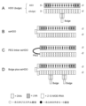
- 50 shows the structure of the nucleic acid used in Example 18.
- FIG. 50A shows the structure of HDO(bulge).
- FIG. 50B shows the structure of HDO (ssHDO).
- FIG. 50C shows the structure of PEG linker ssHDO.
- FIG. 50D shows the structure of Bulge plus ssHDO.
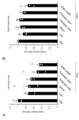
- FIG. 51 shows the results of evaluating motor function in mice to which various nucleic acid agents were intracerebroventricularly administered 1 hour after administration.
- FIG. 51A shows the total distance traveled for 5 minutes.
- FIG. 51B shows the maximum movement speed. Error bars indicate standard error.
- FIG. 51A shows the total distance traveled for 5 minutes.
- FIG. 51B shows the maximum movement speed. Error bars indicate standard error.
- FIG. 51A shows the total distance traveled for 5 minutes.
- FIG. 52 shows the expression levels of Malat1 RNA in the brains of mice to which various nucleic acid agents were intracerebroventricularly administered.
- FIG. 52A shows results for the left frontal cortex.
- FIG. 52B shows results for the right frontal cortex. Error bars indicate standard error.
- FIG. 53 shows the results of incubating various nucleic acid agents in brain tissue homogenate for 7 days and evaluating the efficiency of dissociation of double strands by electrophoresis.
- FIG. 54 shows acute phase tolerability scores 30 minutes to 4 hours after administration of mice intracerebroventricularly administered with various nucleic acid agents. Error bars indicate standard error.
- FIG. 55 shows the results of evaluating motor function in mice to which various nucleic acid agents were intracerebroventricularly administered 1 hour after administration.
- FIG. 55A shows the total distance traveled for 5 minutes.
- FIG. 55B shows maximum movement speed. Error bars indicate standard error.
- FIG. 56 shows acute phase tolerability scores 30 minutes to 4 hours after administration of mice intracerebroventricularly administered with various nucleic acid agents. Error bars indicate standard error.
- FIG. 57 shows the results of evaluating motor function in mice to which various nucleic acid agents were intracerebroventricularly administered 1 hour after administration.
- Figure 57A shows the total distance traveled for 5 minutes.
- Figure 57B shows the maximum movement speed. Error bars indicate standard error.
- FIG. 58 shows the results of measuring the body weight of mice to which various nucleic acid agents were intracerebroventricularly administered. Error bars indicate standard error. Error bars indicate standard error.
- FIG. 59 shows the results of evaluation of motor function in mice to which various nucleic acid agents were intracerebroventricularly administered one day after administration.
- FIG. 60 shows the expression level of Mapt mRNA in the right frontal lobe of mice to which various nucleic acid agents were intracerebroventricularly administered. Error bars indicate standard error.
- FIG. 61 shows the results of incubating various nucleic acid agents in a brain tissue homogenate for 7 days and evaluating the efficiency of dissociation of double strands by electrophoresis.
- FIG. 62 shows acute phase tolerability scores 30 minutes to 4 hours after administration of mice intracerebroventricularly administered with various nucleic acid agents. Error bars indicate standard error.
- FIG. 63 shows the results of evaluating motor function in mice to which various nucleic acid agents were intracerebroventricularly administered 1 hour after administration.
- Figure 63A shows the total distance traveled for 5 minutes.
- FIG. 63B shows maximum movement speed. Error bars indicate standard error.
- FIG. 64 shows LDH activity and Bace1 mRNA expression levels in mouse neuroblastoma-derived cells (Neuro2a cell line) introduced with various nucleic acid agents.
- Figure 64A shows relative LDH release levels in the supernatant.
- Figure 64B shows relative Bace1 mRNA levels. Error bars indicate standard error.
- FIG. 65 shows a modified FOB score evaluation method.
- FIG. 66 shows the results of evaluation of modified FOB scores in monkeys intrathecally administered with various nucleic acid agents.
- FIG. 67 shows the results of measurement of spontaneous movement time and number of jumps by 3-minute video animation in monkeys to which various nucleic acid agents were intrathecally administered.
- Double-stranded nucleic acid complex 1-1 A first aspect of the present invention is a double-stranded nucleic acid complex.
- a double-stranded nucleic acid complex of the present invention comprises a first nucleic acid strand and a second nucleic acid strand, and comprises one or more 2'-modified nucleosides.
- the double-stranded nucleic acid complex of the present invention is stable in the cerebrospinal fluid of primates including humans, and has reduced toxicity such as central nervous system toxicity.
- a "transcript" of a target gene refers to any RNA that is directly targeted by the nucleic acid complex of the present invention and synthesized by RNA polymerase. Specifically, mRNA transcribed from target genes (including mature mRNA, pre-mRNA, and mRNA that has not undergone base modification), non-coding RNA such as miRNA (non-coding RNA, ncRNA), long non-coding RNA RNA (lncRNA), which may include natural antisense RNA.
- target genes including mature mRNA, pre-mRNA, and mRNA that has not undergone base modification
- non-coding RNA such as miRNA (non-coding RNA, ncRNA), long non-coding RNA RNA (lncRNA), which may include natural antisense RNA.
- target gene refers to the function of a transcription product or translation product whose expression level can be suppressed or enhanced by the antisense effect of the double-stranded nucleic acid complex of the present invention. can be inhibited, or steric blocking, splicing switch, RNA editing, exon skipping or exon inclusion can be induced.
- the type of target gene is not particularly limited as long as it is expressed in vivo.
- genes derived from organisms into which the double-stranded nucleic acid complex of the present invention is introduced such as genes whose expression is increased in various diseases. is mentioned.
- scavenger receptor B1 (herein often referred to as "SR-B1”) gene
- metastasis associated lung adenocarcinoma transcript 1 (herein often referred to as abbreviated as "Malat1”) gene
- microtubule-associated protein tau in this specification, often abbreviated as "Mapt” gene
- ⁇ -secretase 1 (beta-secretase 1: in this specification , often referred to as "BACE1” gene
- DMPK dystrophia myotonica-protein kinase
- target transcript refers to any RNA that is directly targeted by the nucleic acid complex of the present invention and synthesized by RNA polymerase.
- transcription products of target genes correspond. Specifically, mRNA transcribed from target genes (including mature mRNA, pre-mRNA, and mRNA that has not undergone base modification), non-coding RNA such as miRNA (non-coding RNA, ncRNA), long non-coding RNA RNA (lncRNA), which may include natural antisense RNA.
- target gene transcripts examples include SR-B1 mRNA, which is the transcript of the SR-B1 gene, Mapt mRNA, which is the transcript of the Mapt gene, BACE1 mRNA, which is the transcript of the BACE1 gene, and transcript of the Malat1 gene.
- Mapt mRNA which is the transcript of the Mapt gene
- BACE1 mRNA which is the transcript of the BACE1 gene
- Malat1 non-coding RNA Malat1 non-coding RNA
- DMPK mRNA which is a transcription product of the DMPK gene
- dystrophin mRNA which is a transcription product of the dystrophin gene, or its precursor (pre-mRNA).
- target transcripts include the exon 23/intron 23 boundary region of Dystrophin pre-mRNA (GenBank Accession Number: NC_000086.7), such as positions 83803482 to 83803566, such as positions 83803512 to 83803536.
- SEQ ID NO: 7 shows the nucleotide sequence of mouse DMPK mRNA
- SEQ ID NO: 8 shows the nucleotide sequence of human DMPK mRNA. In each of SEQ ID NOs: 7 to 8, the base sequence of mRNA is replaced with the base sequence of DNA.
- the nucleotide sequence information of these genes and transcripts can be obtained from known databases such as the NCBI (National Center for Biotechnology Information) database.
- antisense oligonucleotides or “antisense nucleic acids” are capable of hybridizing (i.e., complementary a) refers to a single-stranded oligonucleotide containing a base sequence and capable of producing an antisense effect on a target transcript.
- the first nucleic acid strand functions as an ASO, and its target region is 3'UTR, 5'UTR, exon, intron, coding region, translation initiation region, translation termination region, or Any other nucleic acid region may be included.
- the target region of the target transcript is at least 8 bases long, e.g. It can be 16-22 bases long, or 16-20 bases long.
- Antisense effect refers to the effect of regulating the expression or editing of a target transcript (eg, RNA sense strand) by hybridizing the ASO to the target transcript.
- “Modulating the expression or editing of a target transcript” refers to the expression of a target gene or the expression level of a target transcript (herein, “target transcript expression level” is often referred to as “target transcript level”). expression), inhibition of translation, RNA editing, splicing function modification effects (eg, splicing switch, exon inclusion, exon skipping, etc.), or degradation of transcripts.
- RNA oligonucleotide when introduced into a cell as an ASO, the ASO forms a partial duplex by annealing with mRNA, the transcript of the target gene. This partial double strand serves as a cover to prevent translation by the ribosome, thereby inhibiting the expression of the target protein encoded by the target gene at the translational level (steric blocking).
- oligonucleotides containing DNA as ASO are introduced into cells, partial DNA-RNA heteroduplexes are formed. Recognition of this heteroduplex structure by RNase H results in degradation of target gene mRNA and inhibition of expression of the protein encoded by the target gene at the expression level.
- antisense effects can also be produced by targeting introns in pre-mRNAs.
- antisense effects can also be produced by targeting miRNAs.
- the functional inhibition of the miRNA can increase the expression of genes whose expression is normally controlled by the miRNA.
- modulating expression of the target transcript may be reduction in target transcript abundance.
- the antisense effect is measured, for example, by administering a test nucleic acid compound to a subject (eg, a mouse), and, for example, several days later (eg, 2 to 7 days later), the expression is regulated by the antisense effect provided by the test nucleic acid compound. It can be carried out by measuring the expression level of the target gene or the level (amount) of the target transcript (for example, the amount of mRNA or RNA such as microRNA, the amount of cDNA, the amount of protein, etc.).
- the measured target gene expression level or target transcript level is at least 10%, at least 20%, at least 25%, at least 30%, or at least 40% compared to a negative control (e.g., vehicle administration) If so, it indicates that the test nucleic acid compound is capable of producing an antisense effect (eg, reduction in target transcript abundance).
- a negative control e.g., vehicle administration
- the number, type and position of non-natural nucleotides in the nucleic acid chain can affect the antisense effect provided by the nucleic acid complex.
- the selection of modification may differ depending on the sequence of the target gene, etc., but a person skilled in the art should refer to the literature related to the antisense method (for example, WO 2007/143315, WO 2008/043753, and WO 2008/049085).
- the preferred embodiment can be determined by reference.
- the measured value thus obtained is not significantly lower than the measured value of the nucleic acid complex before modification (e.g., When the measured value obtained after modification is 70% or more, 80% or more, or 90% or more of the measured value of the nucleic acid complex before modification), the relevant modification can be evaluated.
- target gene translation product refers to any polypeptide or protein synthesized by translation of the target transcript or target gene transcript that is the direct target of the nucleic acid complex of the present invention. say.
- aptamer refers to a nucleic acid molecule that specifically binds to a specific target molecule intracellularly, on the cell membrane, or extracellularly, for example, on the cell membrane or extracellularly. Aptamers can be produced by methods known in the art, for example, in vitro selection using the SELEX (systematic evolution of ligands by exponential enrichment) method.
- the term “decoy” refers to a nucleic acid having a binding site sequence of a transcription factor (for example, NF-kB) or a similar sequence.
- a transcription factor for example, NF-kB
- a substance that suppresses the action represses transcription if it is a transcription activator, or promotes transcription if it is a transcription repressor.
- a decoy nucleic acid can be easily designed based on the information of the binding sequence of the target transcription factor.
- bait refers to a nucleic acid molecule that specifically binds to a specific target molecule in cells and modifies the function of the target molecule.
- a target that interacts with a bait is also called a "prey”.
- nucleic acid or “nucleic acid molecule” may refer to a monomeric nucleotide or nucleoside, or may refer to an oligonucleotide composed of more than one monomer, or if a polymer means a polynucleotide.
- Natural nucleic acid refers to a nucleic acid that occurs in nature. Natural nucleic acids include natural nucleosides, natural nucleotides, and the like, which will be described later.
- Non-natural nucleic acid or “artificial nucleic acid” refers to any nucleic acid other than a naturally occurring nucleic acid. Non-natural nucleic acids or artificial nucleic acids include non-natural nucleosides, non-natural nucleotides, and the like, which will be described later.
- nucleic acid strand or simply “strand” means two or more nucleosides linked by internucleoside linkages, and may be, for example, oligonucleotides or polynucleotides. Nucleic acid strands can be produced full length or partial strands by chemical synthetic methods, for example, using automated synthesizers, or by enzymatic processes using polymerases, ligases, or restriction reactions. A nucleic acid strand may comprise naturally occurring and/or non-naturally occurring nucleotides.
- Nucleoside generally refers to a molecule consisting of a combination of a base and a sugar.
- the sugar moiety of a nucleoside is usually, but not limited to, composed of pentofuranosyl sugars, specific examples of which include ribose and deoxyribose.
- the base portion (nucleobase) of a nucleoside is usually a heterocyclic base portion. Non-limiting examples include adenine, cytosine, guanine, thymine, or uracil, as well as other modified nucleobases (modified bases).
- Nucleotide refers to a molecule in which a phosphate group is covalently bonded to the sugar moiety of the nucleoside. Nucleotides containing a pentofuranosyl sugar typically have a phosphate group attached to the hydroxyl group at the 2', 3', or 5' position of the sugar.
- Oligonucleotide refers to a linear oligomer formed by covalently linking several to several tens of hydroxyl groups and phosphate groups of sugar moieties between adjacent nucleotides.
- polynucleotide refers to a linear polymer formed by linking several tens or more, preferably several hundred or more nucleotides by covalent bonds, which is larger than that of oligonucleotides.
- phosphate groups are commonly considered to form internucleoside linkages.
- Natural nucleoside refers to a nucleoside that exists in nature. Examples thereof include ribonucleosides composed of ribose and a base such as adenine, cytosine, guanine, or uracil, and deoxyribonucleosides composed of deoxyribose and a base such as adenine, cytosine, guanine, or thymine.
- ribonucleosides found in RNA and deoxyribonucleosides found in DNA are often referred to as "DNA nucleosides" and "RNA nucleosides", respectively.
- naturally occurring nucleotide refers to a naturally occurring nucleotide in which a phosphate group is covalently bonded to the sugar moiety of the natural nucleoside.
- ribonucleotides known as structural units of RNA, in which a phosphate group is bound to a ribonucleoside
- deoxyribonucleotides known as a structural unit of DNA, in which a phosphate group is bound to a deoxyribonucleoside.
- non-natural nucleotide refers to any nucleotide other than natural nucleotides, including modified nucleotides and nucleotide mimetics.
- modified nucleotide means a nucleotide having any one or more of modified sugar moieties, modified internucleoside linkages, and modified nucleobases.
- nucleotide mimetics include structures that are used to replace nucleosides and linkages at one or more positions of an oligomeric compound.
- Peptide Nucleic Acid is a nucleotide mimetic with a backbone of N-(2-aminoethyl)glycine instead of sugar linked by amide bonds.
- Nucleic acid strands comprising unnatural oligonucleotides, as used herein, are often characterized by, for example, enhanced cellular uptake, enhanced affinity for nucleic acid targets, increased stability in the presence of nucleases, or increased inhibitory activity. has the desired properties of Therefore, it is preferred over natural nucleotides.
- non-natural nucleoside refers to any nucleoside other than natural nucleosides. Examples include modified nucleosides and nucleoside mimetics. As used herein, “modified nucleoside” means a nucleoside having a modified sugar moiety and/or modified nucleobase.
- mimetics refers to functional groups that replace sugars, nucleobases, and/or internucleoside linkages. In general, mimetics are used in place of the sugar or sugar-internucleoside linkage combination so that the nucleobase is maintained for hybridization to a target of choice.
- nucleoside mimetic refers to substitution of a sugar at one or more positions of an oligomeric compound, substitution of a sugar and a base, or binding between monomer subunits constituting an oligomeric compound. Contains the structure used to replace.
- oligomeric compound is meant a polymer of linked monomeric subunits capable of hybridizing to at least a region of a nucleic acid molecule.
- Nucleoside mimetics include, for example, morpholino, cyclohexenyl, cyclohexyl, tetrahydropyranyl, bicyclic or tricyclic sugar mimetics, eg, nucleoside mimetics having non-furanose sugar units.
- Modified sugar refers to a sugar that has a substitution and/or any change from the natural sugar moiety (i.e., sugar moieties found in DNA (2'-H) or RNA (2'-OH));
- a “sugar modification” refers to a substitution and/or any change from a naturally occurring sugar moiety.
- Nucleic acid strands may optionally contain one or more modified nucleosides, including modified sugars.
- a “sugar modified nucleoside” refers to a nucleoside having a modified sugar moiety. Such sugar-modified nucleosides may confer enhanced nuclease stability, increased binding affinity, or some other beneficial biological property to nucleic acid chains.
- nucleosides include chemically modified ribofuranose ring moieties.
- chemically modified ribofuranose rings include, but are not limited to, addition of substituents (including 5' and 2' substituents), bridging of non-geminal ring atoms to form bicyclic nucleic acids (bridged nucleic acids, BNA ), S, N(R), or C(R1)(R2) of the ribosyl ring oxygen atom (R, R1 and R2 are each independently H, C1 - C12 alkyl, or a protecting group ), and combinations thereof.
- sugar modified nucleosides include, but are not limited to, 5'-vinyl, 5'-methyl (R or S), 5'-allyl (R or S), 4'-S, 2'-F ( 2'-fluoro group), 2'- OCH3 (2'-O-Me group or 2'-O-methyl group), 2'-O-[2-(N-methylcarbamoyl)ethyl] (2'- O-MCE group), and nucleosides containing 2'-O-methoxyethyl (2'-O- MOE or 2-O( CH2 ) 2OCH3 ) substituents.
- a "2'-modified sugar” means a furanosyl sugar modified at the 2' position. Nucleosides containing 2'-modified sugars are sometimes referred to as "2'-modified nucleosides" or "2'-sugar modified nucleosides.”
- Bicyclic nucleoside refers to a modified nucleoside containing a bicyclic sugar moiety. Nucleic acids containing bicyclic sugar moieties are commonly referred to as bridged nucleic acid (BNA). Nucleosides containing a bicyclic sugar moiety are sometimes referred to as “bridged nucleosides,” “bridged non-natural nucleosides,” or “BNA nucleosides.” Some examples of crosslinked nucleic acids are shown in FIG.
- a bicyclic sugar may be a sugar in which the 2' and 4' carbon atoms are bridged by two or more atoms. Examples of bicyclic sugars are known to those of skill in the art.
- One subgroup of bicyclic sugar containing nucleic acids (BNA) or BNA nucleosides are 4'-( CH2 ) p -O-2', 4'-( CH2 ) p - CH2-2 ', 4 '-( CH2 ) p -S-2', 4'-( CH2 ) p -OCO-2', 4'-( CH2 ) n -N( R3 )-O-( CH2 ) m- 2' [wherein p, m and n represent an integer of 1 to 4, an integer of 0 to 2, and an integer of 1 to 3, respectively; and R 3 is a hydrogen atom, an alkyl group, an alkenyl group, a cyclo Represents an alkyl group, an aryl group,
- R 1 and R 2 are typically is a hydrogen atom, which may be the same or different, and is also a hydroxyl group-protecting group for nucleic acid synthesis, an alkyl group, an alkenyl group, a cycloalkyl group, an aryl group, an aralkyl group, an acyl a sulfonyl group, a silyl group, a phosphate group, a phosphate group protected by a protecting group for nucleic acid synthesis, or P(R 4 )R 5 [wherein R 4 and R 5 are a hydroxyl group, a hydroxyl group protected by a protecting group for nucleic acid synthesis, a mercapto group, a mercapto group protected by a protecting group for nucleic acid synthesis, an amino group, 1
- Non-limiting examples of such BNA nucleosides include methyleneoxy(4'- CH2 -O-2') BNA nucleosides (LNA nucleosides, also known as 2',4'-BNA nucleosides) (e.g.
- amine BNA nucleosides as 2'-Amino-LNA nucleosides
- 2'-O,4'-C-spirocyclopropylene bridged nucleosides also known as scpBNA nucleosides
- other BNA nucleosides known to those skilled in the art.
- a "cationic nucleoside” refers to, at a certain pH (e.g., human physiological pH (about 7.4), pH of a delivery site (e.g., organelle, cell, tissue, organ, organism, etc.)) A modified nucleoside that exists as a cationic form as compared to a neutral form (such as the neutral form of a ribonucleoside).
- Cationic nucleosides may contain one or more cationic modifying groups at any position of the nucleoside.
- the cationic nucleosides are 2'-Amino-LNA nucleosides (e.g.
- Bicyclic nucleosides with a methyleneoxy (4'- CH2 -O-2') bridge are sometimes referred to as LNA nucleosides.
- modified internucleoside linkage refers to an internucleoside linkage that has a substitution or any change from a naturally occurring internucleoside linkage (ie, a phosphodiester linkage).
- Modified internucleoside linkages include internucleoside linkages containing a phosphorus atom and internucleoside linkages without a phosphorus atom.
- Representative phosphorus-containing internucleoside linkages include phosphodiester linkages, phosphorothioate linkages, phosphorodithioate linkages, phosphotriester linkages (methylphosphotriester and ethylphosphotriester linkages described in US Pat. No.
- alkyl Phosphonate linkages e.g., methylphosphonate linkages described in U.S. Pat. Nos. 5,264,423 and 5,286,717, methoxypropylphosphonate linkages described in WO 2015/168172
- alkylthiophosphonate linkages e.g., methylthiophosphonate linkages, boranophosphate linkages, cyclic guanidine moieties
- Internucleoside linkages containing e.g., the substructure represented by the following formula (I):
- an internucleoside linkage comprising a guanidine moiety substituted with 1 to 4 C 1-6 alkyl groups e.g., a tetramethylguanidine (TMG) moiety
- TMG tetramethylguanidine
- a phosphorothioate linkage refers to an internucleoside linkage in which a sulfur atom replaces the non-bridging oxygen atom of a phosphodiester linkage.
- Methods for preparing phosphorus-containing and non-phosphorus-containing linkages are well known.
- Modified internucleoside linkages are preferably linkages that are more resistant to nucleases than naturally occurring internucleoside linkages.
- the internucleoside linkage When the internucleoside linkage has a chiral center, the internucleoside linkage may be chirally controlled. "Chirally controlled” is intended to exist in a single diastereomer about a chiral center, eg, a chiral bound phosphorus. Chirally controlled internucleoside linkages can be completely chirally pure or highly chiral pure, e.g. It may have a chiral purity of %de, 99.9% de, or higher.
- chiral purity refers to the proportion of one diastereomer in a mixture of diastereomers, expressed as diastereomeric excess (% de), (diastereomer of interest minus other diastereomer defined as (stereomers)/(total diastereomers) ⁇ 100 (%).
- internucleoside linkages may be phosphorothioate linkages chiral controlled to the Rp or Sp configuration, guanidine moieties substituted with 1-4 C 1-6 alkyl groups (e.g., tetramethylguanidine (TMG) moieties; e.g. Alexander A. Lomzov et al., Biochem Biophys Res Commun., 2019, 513(4), 807-811) and/or an internucleoside linkage comprising a cyclic guanidine moiety.
- TMG tetramethylguanidine
- nucleobase or “base” used herein is a base component (heterocyclic moiety) that constitutes a nucleic acid, and adenine, guanine, cytosine, thymine, and uracil are mainly known.
- nucleobase or “base” includes both modified and unmodified nucleobases (bases) unless otherwise specified.
- the purine base can be either modified or unmodified purine base.
- the pyrimidine base may be a modified or unmodified pyrimidine base.
- Modified nucleobase or “modified base” means any nucleobase other than adenine, cytosine, guanine, thymine, or uracil.
- Unmodified nucleobases or “unmodified bases” (natural nucleobases) include the purine bases adenine (A) and guanine (G), and the pyrimidine bases thymine (T), cytosine (C), and means uracil (U).
- modified nucleobases include hypoxanthine, 5-methylcytosine, 5-fluorocytosine, 5-bromocytosine, 5-iodocytosine or N4-methylcytosine; N6-methyladenine or 8-bromoadenine; thymine; and N2-methylguanine or 8-bromoguanine, but are not limited to these.
- the modified nucleobase is preferably 5-methylcytosine.
- the term “complementary” means that the nucleobases can form so-called Watson-Crick base pairs (natural base pairs) or non-Watson-Crick base pairs (Hoogsteen bases) through hydrogen bonding. It means a relationship that can form an equality).
- the antisense oligonucleotide region in the first nucleic acid strand does not necessarily have to be completely complementary to at least part of the target transcript (for example, the transcript of the target gene). Complementarity of at least 70%, preferably at least 80%, even more preferably at least 90% (eg, 95%, 96%, 97%, 98%, or 99% or more) is acceptable.
- the antisense oligonucleotide region in the first nucleic acid strand is complementary in base sequence (typically when the base sequence is complementary to the base sequence of at least a portion of the target transcript), It can hybridize to a target transcript.
- the region of complementarity in the second nucleic acid strand need not necessarily be completely complementary to at least a portion of the first nucleic acid strand, but has a base sequence of at least 70%, preferably at least 80%, and even More preferably, at least 90% (eg, 95%, 96%, 97%, 98%, or 99% or more) complementarity is acceptable.
- the complementary regions in the second nucleic acid strand can anneal when they are complementary in base sequence to at least a portion of the first nucleic acid strand.
- Complementarity of base sequences can be determined by using the BLAST program or the like.
- a person skilled in the art can easily determine the conditions (temperature, salt concentration, etc.) under which two strands can anneal or hybridize, taking into consideration the degree of complementarity between the strands.
- a person skilled in the art can easily design an antisense nucleic acid complementary to the target transcript, for example, based on information on the base sequence of the target gene.
- Hybridization conditions may be various stringent conditions such as low stringent conditions and high stringent conditions.
- Low stringent conditions may be relatively low temperature and high salt conditions, eg, 30° C., 2 ⁇ SSC, 0.1% SDS.
- Highly stringent conditions may be relatively high temperature and low salt conditions, eg, 65° C., 0.1 ⁇ SSC, 0.1% SDS.
- Hybridization stringency can be adjusted by varying conditions such as temperature and salt concentration.
- 1 ⁇ SSC contains 150 mM sodium chloride and 15 mM sodium citrate.
- toxicity includes, for example, death, pain, tremor, convulsion, movement disorder, cognitive impairment, consciousness disturbance, general malaise, fatigue, nausea, vomiting, dizziness, numbness, lightheadedness, etc. An action that causes unfavorable objective or subjective symptoms or functional abnormalities in a specimen. Toxicity can be toxicity in any organ. The toxicity may be neurotoxic.
- neurotoxicity refers to an action that causes damage to nerve tissue, including central nerve tissue and peripheral nerve tissue, and interferes with normal activity of the nervous system.
- Neurotoxicity includes death, respiratory abnormalities, cardiovascular abnormalities, headache, nausea or vomiting, unresponsiveness or hyporesponsiveness, altered consciousness, psychiatric disturbances, personality changes, hallucinations, delusions, cognitive impairment, postural abnormalities, involuntary movements, and tremors. , convulsions, hyperactive motor dysfunction, paralysis, sensory disturbances or autonomic dysfunction.
- the neurotoxicity may be acute neurotoxicity.
- Acute neurotoxicity can be neurotoxicity that occurs within 1, 3, 6, 9, 12, 24 or 48 hours of administration. Toxicity can be evaluated, for example, by acute phase tolerability score, adverse event rate, mortality rate, etc., as described in the Examples below.
- central neurotoxicity refers to an action that causes damage to at least the central nervous system among nerve tissues and interferes with normal activity of the nervous system.
- the term "subject” refers to a subject to which the double-stranded nucleic acid complex or pharmaceutical composition of the present invention is applied.
- Subjects include organs, tissues, and cells as well as individuals.
- any animal including humans can be applicable.
- various livestock, poultry, pets, experimental animals, and the like can be mentioned.
- a subject may be an individual in need of decreased expression of a target transcript or an individual in need of treatment or prevention of a disease.
- the double-stranded nucleic acid complex of the present invention comprises a first nucleic acid strand and a second nucleic acid strand. Specific configurations of each nucleic acid strand are shown below.
- the first nucleic acid strand can hybridize to at least a portion of the target gene or its transcript and has an antisense effect on the target gene or its transcript.
- the second nucleic acid strand comprises a base sequence complementary to the first nucleic acid strand and contains one or more 2′-modified nucleosides.
- the number of 2'-modified nucleosides contained in the second nucleic acid strand is at least one, and is equal to or less than the number of all nucleosides constituting the second nucleic acid strand (that is, the base length of the second nucleic acid strand).
- the specific number of 2′-modified nucleosides contained in the second nucleic acid strand is, for example, 2 or more, 3 or more, 4 or more, 5 or more, 6 or more, 7 or more, 8 or more, 9 30 or less, 25 or less, 20 or less, 15 or less, 10 or less, 9 or less, 8 or less, 7 or less, 6 or less, 5 It may be 1 or less, 4 or less, 3 or less, or 2 or less.
- the number of 2'-modified nucleosides in the second nucleic acid strand is 1 to 30, 1 to 25, 1 to 24, 1 to 23, 1 to 22, 1 to 21, 1 to 20. , 1-19, 1-18, 1-17, 1-16, 1-15, 1-14, 1-13, 1-12, 1-11, 1-10 , 1-9, 1-8, 1-7, or 1-6.
- it may be 1, 2, 3, 4, 5, or 6.
- 1% or more, 2% or more, 3% or more, 4% or more, 5% or more, 10% or more, 15% or more, 20% or more, 25% or more, 30% of the nucleosides in the second nucleic acid strand % or more, 35% or more, 40% or more, 45% or more, 50% or more, 55% or more, 60% or more, 65% or more, 70% or more, 75% or more, 80% or more, 85% or more, 90% or more , 95% or more, or 99% or more, and/or 100% or less, 95% or less, 90% or less, 85% or less, 80% or less, 75% or less, 70% or less, 65% or less, 60% or less, 55 % or less, 50% or less, 45% or less, 40% or less, 35% or less, 30% or less, 25% or less, 20% or less, 15% or less, 10% or less, or 5% or less are 2'-modified nucleosides be. For example, 10%-90%, 20%-80%, 30%-70%,
- the nucleosides other than the 2'-modified nucleosides contained in the second nucleic acid strand may be natural nucleosides, non-natural nucleosides such as crosslinked nucleosides, or any combination thereof.
- the number of nucleosides other than the 2'-modified nucleosides contained in the second nucleic acid strand is not limited, but is, for example, 1 or more, 2 or more, 3 or more, 4 or more, 5 or more, 6 or more, 7 1 or more, 8 or more, 9 or more, or 10 or more, for example 1 to 40, 1 to 30, 1 to 20, 1 to 15, 1 to 12, 1 to 10 1, 1 to 8, or 1 to 6, such as 1 to 5, such as 1, 2, 3, 4, or 5.
- the second nucleic acid strand contains one 2'-modified nucleoside. In another embodiment, all of the nucleosides making up the second nucleic acid strand are 2'-modified nucleosides. For example, in the second nucleic acid strand, all nucleosides in the region consisting of a base sequence complementary to the first nucleic acid strand may be 2'-modified nucleosides. In a further embodiment, the second nucleic acid strand comprises one or more but less than all 2'-modified nucleosides.
- the second nucleic acid strand "containing less than all" of 2'-modified nucleosides means that the second nucleic acid strand contains at least one arbitrary nucleoside other than a 2'-modified nucleoside.
- the second nucleic acid strand comprises one or more contiguous 2'-modified nucleosides located at the 5' terminus.
- “comprising multiple consecutive 2'-modified nucleosides” means comprising multiple 2'-modified nucleosides linked by any internucleoside linkage.
- the second nucleic acid strand has one located at the 5' end, or consecutive 2 to 12, 2 to 10, 2 to 8, 2 to 6, 2 to 5, 2 to 4, Alternatively, it may contain 2-3, eg, 2, 3, or 4, 2'-modified nucleosides.
- the second nucleic acid strand comprises one or more consecutive 2'-modified nucleosides located at the 3' terminus.
- the second nucleic acid strand has one located at the 3′ end, or consecutive 2 to 12, 2 to 10, 2 to 8, 2 to 6, 2 to 5, 2 to 4, Alternatively, it may contain 2-3, eg, 2, 3, or 4, 2'-modified nucleosides.
- the second nucleic acid strand comprises one or more contiguous 2'-modified nucleosides located at the 5' terminus and one or more contiguous 2'-modified nucleosides located at the 3' terminus.
- the second nucleic acid strand contains 2'-modified nucleosides at positions other than the 5' and 3' ends.
- the second nucleic acid strand "containing a 2'-modified nucleoside at a position other than the 5' end and the 3' end" means that one or more consecutive 2' -modified nucleosides and 2'-modified nucleosides at positions other than one or more consecutive 2'-modified nucleosides located at the 3'-end.
- the second nucleic acid strand has 1 to 30, 1 to 25, 1 to 20, 1 to 15, 1 to 12, 1 to 10, 1 9, 1-8, 1-7, 1-6, 1-5, 1-4, or 1-3, such as 1 or 2, 2'-modified nucleosides.
- the second nucleic acid strand contains one or more nucleosides other than 2'-modified nucleosides. In a further embodiment, all of the nucleosides other than 2'-modified nucleosides in the second nucleic acid strand (eg, in the region consisting of the base sequence complementary to the first nucleic acid strand) can be deoxyribonucleosides.
- the 2'-modified nucleosides are 2'-O-methoxyethyl modified nucleosides and/or 2'-O-methyl modified nucleosides.
- the 2'-O-methoxyethyl modified nucleoside has the following formula (III): is indicated by 2'-O-methyl modified nucleosides may also be represented by the following formula (IV): is indicated by
- the 2'-modified nucleosides are 2'-O-methyl modified nucleosides.
- the 2'-modified nucleoside is a 2'-O-methoxyethyl modified nucleoside.
- all of the nucleosides in the first nucleic acid strand may be non-natural nucleosides or modified nucleosides.
- all of the nucleosides in the first nucleic acid strand may be 2'-modified nucleosides.
- all of the nucleosides may be 2'-O-methoxyethyl modified nucleosides.
- the first nucleic acid strand can contain at least 4, at least 5, at least 6, or at least 7 contiguous nucleosides that are recognized by RNase H when hybridized to the target transcript. Generally, any region containing consecutive nucleosides of 4 to 20 bases, 5 to 16 bases, or 6 to 12 bases may be used. As a nucleoside recognized by RNase H, for example, natural deoxyribonucleoside can be used. Suitable nucleosides containing modified deoxyribonucleosides and other bases are well known in the art. It is also known that nucleosides having a hydroxy group at the 2'-position, such as ribonucleosides, are unsuitable as the nucleosides.
- the suitability of nucleosides for use in this region containing "at least 4 contiguous nucleosides" can readily be determined.
- the first nucleic acid strand may comprise at least 4 contiguous deoxyribonucleosides.
- the nucleosides of the first nucleic acid strand comprise or consist of deoxyribonucleosides, e.g., 70% or more, 80% or more, 90% or more, or 95% or more of the nucleosides of the first nucleic acid strand are deoxyribonucleosides. be.
- the first nucleic acid strand may be a gapmer.
- the term "gapmer” basically refers to a central region (DNA gap region) and wing regions located directly at its 5' and 3' ends (5' wing region and 3' wing region, respectively). It refers to a single-stranded nucleic acid consisting of The length of the DNA gap region is 13 to 22 bases, 16 to 22 bases, or 16 to 20 bases, or 4 to 20 bases, 5 to 18 bases, 6 to 16 bases, or 7 to 14 bases. long, or 8-12 bases long.
- the central region in the gapmer contains at least 3 or at least 4 contiguous deoxyribonucleosides and the wing regions contain at least one non-natural nucleoside.
- non-natural nucleosides contained in the wing regions usually have higher binding strength to RNA than natural nucleosides and have high resistance to nucleolytic enzymes (nucleases and the like).
- the non-naturally occurring nucleosides making up the wing regions comprise or consist of bridging nucleosides
- said gapmers are specifically referred to as "BNA/DNA gapmers”.
- the number of bridging nucleosides contained in the 5' wing region and the 3' wing region is at least 1, and may be, for example, 2 or 3.
- the bridging nucleosides contained in the 5' wing region and the 3' wing region may be present continuously or non-contiguously within the 5' wing region and the 3' wing region.
- Bridging nucleosides can further include modified nucleobases (eg, 5-methylcytosine).
- modified nucleobases eg, 5-methylcytosine
- the bridging nucleosides are LNA nucleosides
- said gapmers are referred to as "LNA/DNA gapmers”.
- the non-natural nucleosides making up the 5' and 3' wing regions comprise or consist of peptide nucleic acids
- said gapmers are specifically referred to as "peptide nucleic acid gapmers”.
- the unnatural nucleosides making up the 5' and 3' wing regions comprise or consist of morpholino nucleic acids
- said gapmers are specifically referred to as "morpholino nucleic acid gapmers”.
- the base lengths of the 5' wing region and the 3' wing region may each independently be at least 2 bases long, for example, 2 to 10 bases long, 2 to 7 bases long, or 3 to 5 bases long.
- the 5′ wing region and/or the 3′ wing region may contain at least one non-natural nucleoside, and may further contain natural nucleosides.
- the 5' and 3' wing regions are, for example, non-natural nucleosides linked by modified internucleoside linkages such as phosphorothioate linkages, bridged nucleosides such as LNA nucleosides, and 2' -It may be a modified nucleoside.
- the central region contains at least 3 or at least 4 consecutive deoxyribonucleosides, and the wing regions (5' wing region and 3' wing region) contain at least one unnatural nucleoside.
- the central region can be functionally defined as the region that can be recognized by RNase H1.
- RNase H1 means that when the gapmer binds to the target RNA, the sequence in the target RNA paired with the gapmer can be cleaved by RNase H1.
- the boundary positions can be determined with the region that can be recognized by RNase H1 in the gapmer as the central region and the region that is not recognized by RNase H1 as the wing regions (5' wing region and 3' wing region).
- nucleosides adjacent to the central region in the 5' wing region and 3' wing region are defined as non-natural nucleosides
- nucleosides adjacent to the 5' wing region or 3' wing region in the central region are defined as natural nucleosides. .
- the first nucleic acid strand constituting the gapmer is, in order from the 5′ end, a bridging nucleoside of 2 to 7 bases or 3 to 5 bases (for example, 2 or 3 bases), 4 to 15 bases or 8 to 12 bases. It may be composed of ribonucleosides or deoxyribonucleosides having a base length (eg, 8 or 10 base lengths) and bridging nucleosides having a length of 2 to 7 bases or 3 to 5 bases (eg, 2 or 3 base lengths).
- a nucleic acid strand having a wing region only on either the 5'-end side or the 3'-end side is called a "hemigapmer" in the art. shall be included.
- the second nucleic acid strand contains 2'-modified nucleosides in a region consisting of a base sequence complementary to the 5' wing region and/or 3' wing region of the first nucleic acid strand.
- the second nucleic acid strand may have 2'-modified nucleosides for all nucleosides in a region consisting of a base sequence complementary to the 5' wing region and/or the 3' wing region of the first nucleic acid strand.
- 2'-modified nucleosides may be, for example, 2'-O-methoxyethyl modified nucleosides or 2'-O-methyl modified nucleosides.
- the nucleoside containing a purine base in the region consisting of the base sequence complementary to the central region of the first nucleic acid strand may be a ribonucleoside.
- all nucleosides containing purine bases can be ribonucleosides in the region consisting of the nucleotide sequence complementary to the central region of the first nucleic acid strand.
- the nucleosides containing pyrimidine bases in the region consisting of the base sequence complementary to the central region of the first nucleic acid strand may be deoxyribonucleosides.
- all nucleosides containing pyrimidine bases in the region consisting of the nucleotide sequence complementary to the central region of the first nucleic acid strand can be deoxyribonucleosides.
- all nucleosides containing purine bases are ribonucleosides and all nucleosides containing pyrimidine bases in the region consisting of a nucleotide sequence complementary to the central region of the first nucleic acid strand.
- the second nucleic acid strand has 2′-modified nucleosides (e.g., 2′- O-methoxyethyl-modified nucleoside or 2'-O-methyl-modified nucleoside), and in the second nucleic acid strand, all the purine bases in the region consisting of the base sequence complementary to the central region of the first nucleic acid strand.
- the nucleosides are ribonucleosides and all nucleosides containing a pyrimidine base can be deoxyribonucleosides.
- nucleoside in the first nucleic acid strand may be a 2'-modified nucleoside.
- nucleosides in the first nucleic acid strand may be 2'-modified nucleosides.
- At least one guanosine nucleoside in the 3' wing region and/or 5' wing region of the first nucleic acid strand is a 2'-modified nucleoside.
- nucleosides in the 3' wing region and/or 5' wing region of the first nucleic acid strand may be a further embodiment, (i) all guanosine nucleosides in the 3' wing region and/or 5' wing region of the first nucleic acid strand, (ii) the nucleosides 5' adjacent to said guanosine nucleosides, (iii) said Even if the nucleoside adjacent to the 3'-terminal side of the guanosine nucleoside or (iv) the nucleoside in the second nucleic acid strand that is complementary to any combination of (i) to (iii) above is a 2'-modified nucleoside good.
- nucleosides in the region consisting of a base sequence complementary to the central region of the first nucleic acid strand are (a) deoxyribonucleosides, (b) deoxyribonucleosides and ribonucleosides, (c ) deoxyribonucleosides and 2'-modified nucleosides, or (d) ribonucleosides and 2'-modified nucleosides, or (e) deoxyribonucleosides, ribonucleosides, and 2'-modified nucleosides.
- nucleosides in a region consisting of base sequences complementary to the 5' wing region and 3' wing region of the first nucleic acid strand are 2'-modified nucleosides, and All nucleosides in the region consisting of complementary nucleotide sequences can be deoxyribonucleosides.
- At least one of the nucleosides containing a pyrimidine base in the second nucleic acid strand may be a 2'-modified nucleoside and/or deoxyribonucleoside.
- the second nucleic acid strand does not contain natural ribonucleosides containing pyrimidine bases, for example all of the nucleosides containing pyrimidine bases in the second nucleic acid strand may be 2'-modified nucleosides and/or deoxyribonucleosides. .
- the second nucleic acid strand may contain modified nucleosides other than 2'-modified nucleosides in addition to 2'-modified nucleosides.
- R represents a hydrogen atom or a methyl group.
- the crosslinked non-natural nucleoside represented by formula (V) above is a 2'-O,4'-C-spirocyclopropylene crosslinked nucleic acid, and is mainly referred to as "scpBNA" in this specification.
- R may be either a hydrogen atom or a methyl group.
- R may be either a hydrogen atom or a methyl group.
- a methyl group can also be written as AmNA[N-Me].
- the first nucleic acid strand may be a mixmer.
- the term "mixmer” refers to a nucleic acid chain that contains alternating natural and unnatural nucleosides of periodic or random segment length and is free of four or more contiguous deoxyribonucleosides and ribonucleosides. .
- Mixmers in which the unnatural nucleoside is a bridged nucleoside and the natural nucleoside is a deoxyribonucleoside are particularly referred to as "BNA/DNA mixmers".
- the bridged nucleoside may be a bridged non-natural nucleoside represented by formula (V) or formula (VI) above.
- Mixmers in which the non-natural nucleoside is a peptide nucleic acid and the natural nucleoside is a deoxyribonucleoside are particularly referred to as "peptide nucleic acid/DNA mixmers".
- Mixmers in which the unnatural nucleoside is a morpholinonucleic acid and the natural nucleoside is a deoxyribonucleoside are particularly referred to as "morpholinonucleic acid/DNA mixmers”.
- Mixmers are not limited to containing only two nucleosides.
- Mixmers can include any number of types of nucleosides, whether natural or modified nucleosides or nucleoside mimetics.
- bridging nucleoside e.g., LNA nucleoside or a bridging non-natural nucleoside represented by formula (V) or formula (VI) above.
- Bridging nucleosides may further include modified nucleobases (eg, 5-methylcytosine).
- the second nucleic acid strand comprises at least 4 contiguous ribonucleosides complementary to the at least 4 contiguous nucleosides (e.g., deoxyribonucleosides) in the central region of the first nucleic acid strand. good too. This is so that the second nucleic acid strand forms a partial DNA-RNA heteroduplex with the first nucleic acid strand and is recognized and cleaved by RNaseH. At least four consecutive ribonucleosides in the second nucleic acid strand are preferably linked by naturally occurring internucleoside linkages, ie phosphodiester linkages.
- the second nucleic acid strand may further comprise at least 2 consecutive deoxyribonucleosides in addition to the at least 4 consecutive ribonucleosides described above.
- the at least two contiguous deoxyribonucleosides are complementary to the first nucleic acid strand and may be included in a region complementary to the central region of the first nucleic acid strand.
- the at least two consecutive deoxyribonucleosides may be positioned on either the 5' side or the 3' side of the at least four consecutive ribonucleosides, and may be positioned on both the 5' and 3' sides.
- the at least two consecutive deoxyribonucleosides may be 2, 3, 4, 5, 6 or more consecutive deoxyribonucleosides.
- Modified nucleosides may include modified sugars and/or modified nucleobases.
- Modified sugars may be 2'-modified sugars (eg, sugars containing a 2'-O-methyl group).
- a modified nucleobase can also be 5-methylcytosine.
- the second nucleic acid strand is, in order from the 5' end, a modified nucleoside (e.g., a modified nucleoside containing a 2'-modified sugar) having a length of 2 to 7 bases or a length of 3 to 5 bases (for example, 2 or 3 bases), 4 to ribonucleosides or deoxyribonucleosides (optionally linked by modified internucleoside linkages) 15 bases long or 8-12 bases long (eg 8 or 10 bases long) and 2-7 bases long or 3-5 bases long ( (eg, 2 or 3 bases long) modified nucleosides (eg, modified nucleosides containing 2'-modified sugars).
- the first nucleic acid strand may be a gapmer.
- the first nucleic acid strand and/or the second nucleic acid strand may contain nucleoside mimics or nucleotide mimics in whole or in part.
- Nucleotide mimetics may be peptide nucleic acids and/or morpholino nucleic acids.
- the second nucleic acid strand may contain non-complementary bases and/or insertions and/or deletions of one or more bases relative to the first nucleic acid strand.
- the number of non-complementary bases in the second nucleic acid strand is not limited, but for example, 1 to 10, 1 to 9, 1 to 8, 1 to 7, 1 to 6, 1 to 5, 1 to It may be 4, 1-3, 1-2, or 1 or 2.
- a sequence consisting of non-complementary bases may form a bulge structure, which will be described later.
- the number of bases in the insertion sequence in the second nucleic acid strand is not limited, but is, for example, 1 to 10, 1 to 9, 1 to 8, 1 to 7, 1 to 6, 1 to 5, 1 to It may be 4, 1-3, 1-2, or 1 or 2.
- the insertion sequence may be a sequence that forms a bulge structure as described below.
- the length of consecutive bases to be deleted in the second nucleic acid strand is not limited, but for example, 1 to 10, 1 to 9, 1 to 8, 1 to 7, 1 to 6, 1 to 5, 1 It may be ⁇ 4, 1-3, 1-2, or 1 or 2.
- the second nucleic acid strand may contain a bulge structure described later at the deletion position.
- the base lengths of the first nucleic acid strand and the second nucleic acid strand are not particularly limited, but are at least 8 bases long, at least 9 bases long, at least 10 bases long, at least 11 bases long, at least 12 bases long, at least 13 bases long, at least It may be 14 bases long, or at least 15 bases long.
- the base lengths of the first nucleic acid strand and the second nucleic acid strand are 35 base lengths or less, 30 base lengths or less, 25 base lengths or less, 24 base lengths or less, 23 base lengths or less, 22 base lengths or less, and 21 base lengths or less. , 20 bases or less, 19 bases or less, 18 bases or less, 17 bases or less, or 16 bases or less.
- the first nucleic acid strand and the second nucleic acid strand may have the same length or different lengths (eg, one may be 1 to 3 bases shorter or longer).
- the length of the second nucleic acid strand is shorter than the length of the first nucleic acid strand.
- the position at which the second nucleic acid strand can bind to the first nucleic acid strand does not matter. For example, it may bind to any of the 5'-side region, the central region, or the 3'-side region in the first nucleic acid strand.
- the second nucleic acid strand may be at least 8 bases long.
- the double-stranded structure formed by the first nucleic acid strand and the second nucleic acid strand may contain a bulge.
- the choice of length can be determined by the balance between the strength of the antisense effect and the specificity of the nucleic acid strand for its target, among other factors such as cost, synthetic yield, and the like.
- the second nucleic acid strand may contain at least one overhang region located on one or both of its 5' and 3' ends.
- the "overhang region” is a region adjacent to a region complementary to the first nucleic acid strand in the second nucleic acid strand, and when the first nucleic acid strand and the second nucleic acid strand anneal to form a double-stranded structure, the 5' end of the second nucleic acid strand extends beyond the 3' end of the first nucleic acid strand and/or the 3' end of the second nucleic acid strand extends beyond the 5' end of the first nucleic acid strand, i.e.
- the overhang region in the second nucleic acid strand may be located on the 5'-terminal side or the 3'-terminal side of the complementary region.
- the overhang regions in the second nucleic acid strand may be located on the 5'-end side and the 3'-end side of the complementary region.
- the length of the overhang region is not limited, but may be 1 to 20 bases long, for example, 2 to 15 bases long, 2 to 12 bases long, 2 to 10 bases long, 2 to 8 bases long, 2 to It may be 6 bases long, 2-5 bases long, 2-4 bases long, or 2-3 bases long.
- the type of nucleosides constituting the overhang region is not limited. For example, it may be composed of natural nucleosides (eg deoxyribonucleosides) or non-natural nucleosides (eg bridged nucleosides such as LNA nucleosides). Also, all or part of the internucleoside linkages in the overhang region may be modified internucleoside linkages.
- Modified internucleoside linkages may be, for example, phosphorothioate linkages.
- the overhang regions are preferably protein-binding, lipophilic and/or nuclease-resistant and may be composed of, for example, deoxyribonucleosides or LNA nucleosides linked by phosphorothioate linkages.
- the nucleotide sequence of the overhang region may be a sequence unrelated to the nucleotide sequence of the target gene.
- At least 1, at least 2 (eg, 2), at least 3, or at least 4 nucleosides from the end (5' end, 3' end, or both ends) of the second nucleic acid strand are non-natural nucleosides (modified nucleoside).
- Modified nucleosides may include modified sugars and/or modified nucleobases.
- Modified sugars may be 2'-modified sugars (eg, sugars containing a 2'-O-methyl group).
- a modified nucleobase can also be 5-methylcytosine.
- the second nucleic acid strand is 1 to 8, 1 to 7, 1 to 6, 1 ⁇ 5, 1-4, or 1-3 (e.g., 1-2, or 1) non-complementary bases, 1-8, 1-7, 1-6, 1-5 1 to 4, or 1 to 3 (eg, 1 to 2, or 1) deleted bases, and/or 1 to 20 (eg, 1 to 15, 1 to 12, 1 ⁇ 10, 1-8, 1-6, 1-4, 1-3, or 1) inserted bases.
- a sequence region consisting of inserted bases may form a bulge structure.
- the two nucleic acid strands contain at least one bulge structure consisting of a non-complementary base sequence to the first nucleic acid strand.
- the term “bulge structure” refers to a portion of a double-stranded nucleic acid in which part of the nucleic acid of any one of the nucleic acid strands constituting the double strand protrudes from the double-stranded structure without base pairing.
- the base length of the bulge structure is not limited. For example, it is 1 to 50 bases long, 1 to 40 bases long, 1 to 30 bases long, 1 to 20 bases long, 1 to 15 bases long, preferably 1 to 10 bases long.
- the bulge structure comprises sugar-unmodified nucleosides. In a further embodiment, all nucleosides in the bulge structure are sugar unmodified nucleosides.
- all nucleosides other than the bulge structure may be 2'-modified nucleosides.
- the internucleoside linkages in the first nucleic acid strand and the second nucleic acid strand may be naturally occurring internucleoside linkages and/or modified internucleoside linkages. At least 1, at least 2, or at least 3 internucleoside linkages from the end (5′ end, 3′ end, or both ends) of the first nucleic acid strand and/or the second nucleic acid strand are modified internucleoside linkages, but are not limited to A bond is preferred.
- two internucleoside bonds from the end of a nucleic acid chain mean the internucleoside bond closest to the end of the nucleic acid chain and the internucleoside bond adjacent thereto and located on the opposite side of the end. Modified internucleoside linkages in terminal regions of nucleic acid strands are preferred because they can reduce or inhibit unwanted degradation of the nucleic acid strand.
- all or part of the internucleoside linkages of the first nucleic acid strand and/or the second nucleic acid strand may be modified internucleoside linkages.
- the first nucleic acid strand and/or the second nucleic acid strand each has 1, 2, 3, 4, 5, 6, 7, 8, 9 modified internucleoside linkages. 10, 11, 12, 13, 14, 15, 16, 17, 18, 19, 20, 21, 22, 23, 24, 25, It may contain 26, 27, 28, 29, 30, 35, 40, 45, 50 or more.
- the first nucleic acid strand and/or the second nucleic acid strand each have at least 5%, at least 10%, at least 20%, at least 30%, at least 40%, at least 50%, at least 60%, at least 70%, at least 80%, at least 90%, at least 93%, at least 95%, at least 98%, or 100%.
- the modified internucleoside linkage may be a phosphorothioate linkage or a boranophosphate linkage.
- all or part of the internucleoside linkages in the first nucleic acid strand may be phosphorothioate linkages.
- the first nucleic acid strand and/or the second nucleic acid strand each have 1, 2, 3, 4, 5, 6, 7, 8 chirally controlled internucleoside linkages. 9, 10, 11, 12, 13, 14, 15, 16, 17, 18, 19, 20, 21, 22, 23, 24, It may contain 25, 26, 27, 28, 29, 30, 35, 40, 45, 50 or more.
- the first nucleic acid strand and/or the second nucleic acid strand each have at least 5%, at least 10%, at least 20%, at least 30%, at least 40%, at least 50% chirally controlled internucleoside linkages. %, at least 60%, at least 70%, at least 80%, at least 90%, or more.
- the first nucleic acid strand and/or the second nucleic acid strand each have 1, 2, 3, 4 non-negatively charged internucleoside linkages (preferably neutral internucleoside linkages), 5, 6, 7, 8, 9, 10, 11, 12, 13, 14, 15, 16, 17, 18, 19, 20, 21 , 22, 23, 24, 25, 26, 27, 28, 29, 30, 35, 40, 45, 50 or more.
- the first nucleic acid strand and/or the second nucleic acid strand each have at least 5%, at least 10%, at least 20%, at least 30%, at least 40%, at least 50% non-negatively charged internucleoside linkages. , at least 60%, at least 70%, at least 80%, at least 90%, or more.
- At least 1, at least 2, or at least 3 internucleoside linkages from the 5' end of the second nucleic acid strand may be modified internucleoside linkages. At least 1, at least 2, or at least 3 internucleoside linkages from the 3′ end of the second nucleic acid strand are replaced with modified internucleoside linkages, such as phosphorothioate linkages, 1-4 C 1-6 alkyl groups may be an internucleoside linkage comprising a modified guanidine moiety (eg, a TMG moiety) and/or an internucleoside linkage comprising a cyclic guanidine moiety. Modified internucleoside linkages may be chirally controlled to the Rp or Sp configuration.
- At least one (eg, three) internucleoside linkage from the 3' end of the second nucleic acid strand may be a modified internucleoside linkage such as a highly RNase-resistant phosphorothioate linkage.
- the double-stranded nucleic acid complex has an improved gene suppressing activity, which is preferable.
- the modified internucleoside linkages of the first nucleic acid strand and/or the second nucleic acid strand are controlled at a pH (e.g., human physiological pH (about 7.4), a delivery site (e.g., an organelle, cell, tissue, organ).
- a pH e.g., human physiological pH (about 7.4)
- a delivery site e.g., an organelle, cell, tissue, organ.
- the modified internucleoside linkage is in an anionic form (e.g., -OP(O)(O - )-O- (anionic form of natural phosphate linkage), -OP(O) (S ⁇ )—O— (anionic form of phosphorothioate linkages, etc.)), including non-negatively charged (neutral or cationic, respectively) internucleoside linkages that exist as neutral or cationic forms.
- the modified internucleoside linkages of the first and/or second nucleic acid strand comprise neutral internucleoside linkages.
- the modified internucleoside linkages of the first and/or second nucleic acid strand comprise cationic internucleoside linkages.
- a non-negatively charged internucleoside linkage e.g., a neutral internucleoside linkage
- the non-negatively charged internucleoside linkage is, for example, a methylphosphonate linkage as described in U.S. Patent Nos.
- the non-negatively charged internucleoside linkage comprises a triazole or alkyne moiety.
- the non-negatively charged internucleoside linkage comprises a cyclic guanidine moiety and/or a guanidine moiety (preferably a TMG moiety) substituted with 1-4 C 1-6 alkyl groups.
- a modified internucleoside linkage comprising a cyclic guanidine moiety has a substructure represented by Formula (I).
- the guanidine moiety substituted with 1-4 C 1-6 alkyl groups has a substructure represented by Formula (II).
- neutral internucleoside linkages comprising cyclic guanidine moieties and/or guanidine moieties substituted with 1-4 C 1-6 alkyl groups are chirally controlled.
- the disclosure relates to a composition
- a composition comprising an oligonucleotide comprising at least one neutral internucleoside linkage and at least one phosphorothioate internucleoside linkage.
- neutral internucleotide linkages have properties compared to equivalent nucleic acids that do not contain neutral internucleotide linkages. and/or have improved activity, e.g., improved delivery, improved resistance to exonucleases and endonucleases, improved cellular uptake, improved endosomal escape, and/or improved nuclear uptake, etc.
- the second nucleic acid strand may be bound to a ligand (also referred to herein as a binder).
- Ligands include small molecules (small molecule ligands), medium molecules (medium molecule ligands), macromolecules (macromolecular ligands), peptides (peptide ligands), lipids (lipid ligands), and aptamers (e.g., nucleic acid aptamers).
- peptide refers to an amino acid polymer having one or more peptide bonds.
- a “peptide” is not limited by the number of amino acid residues it contains.
- peptide includes oligopeptides containing a few amino acid residues, such as dipeptides and tripeptides, to polypeptides (proteins) containing many amino acid residues.
- Peptides can be linear, branched or cyclic peptides.
- Peptide ligands may bind to molecules present on the cell surface or inside cells, or in body fluids.
- the peptide may be an antibody or active fragment thereof.
- antibodies include monoclonal antibodies, polyclonal antibodies, recombinant antibodies such as chimeric antibodies and humanized antibodies, Fab, F(ab') 2 , Fab', VHH and the like.
- active fragments of antibodies include scFv (single chain fragment of variable region: single chain antibody), diabody, triabody, tetrabody and the like.
- Lipids include tocopherols, cholesterol, fatty acids, phospholipids and their analogues; folic acid, vitamin C, vitamin B1, vitamin B2; estradiol, androstane and their analogues; steroids and their analogues; SRBI or LRP1/2 ligands; FK-506, and cyclosporin; lipids described in PCT/JP2019/012077, PCT/JP2019/010392 and PCT/JP2020/035117, but are not limited thereto.
- Lipids also include tocopherol or analogues thereof and/or cholesterol or analogues thereof , substituted or unsubstituted C 1-30 alkyl groups, substituted or unsubstituted C 2-30 It may be a 30 alkenyl group or a substituted or unsubstituted C 1-30 alkoxy group.
- tocopherol is a methylated derivative of tocorol, a fat-soluble vitamin (vitamin E) with a cyclic structure called chroman. Tocorol has a strong antioxidant action, and therefore, in vivo, as an antioxidant substance, it has the function of quenching free radicals generated by metabolism and protecting cells from injury.
- tocopherol Several different forms of tocopherol are known, consisting of ⁇ -tocopherol, ⁇ -tocopherol, ⁇ -tocopherol, and ⁇ -tocopherol, based on the position of the methyl group that binds to the chroman.
- a tocopherol herein can be any tocopherol.
- Analogues of tocopherol include various unsaturated analogues of tocopherol, such as ⁇ -tocotrienol, ⁇ -tocotrienol, ⁇ -tocotrienol and ⁇ -tocotrienol.
- the tocopherol is ⁇ -tocopherol.
- cholesterol is a type of sterol, also known as steroidal alcohol, and is particularly abundant in animals. Cholesterol plays an important role in metabolic processes in vivo, and in animal cells, it is also a major constituent of the cell membrane system together with phospholipids. Analogs of cholesterol also refer to various cholesterol metabolites and analogs, which are alcohols having a sterol backbone, including, but not limited to, cholestanol, lanosterol, cerebrosterol, dehydrocholesterol, and coprostanol. etc.
- analog refers to compounds having the same or similar basic skeleton and having similar structures and properties. Analogs include, for example, biosynthetic intermediates, metabolites, compounds with substituents, and the like. A person skilled in the art can determine whether a compound is an analog of another compound based on common general technical knowledge.
- Cholesterol analogs refer to various cholesterol metabolites and analogs, etc., which are alcohols with a sterol backbone, including, but not limited to, cholestanol, lanosterol, cerebrosterol, dehydrocholesterol, and coprostanol. include.
- the second nucleic acid strand may be bound with tocopherol or cholesterol or analogs thereof.
- the second nucleic acid strand bound to cholesterol or its analogue may have a group represented by the following general formula (VII).
- R c represents an optionally substituted alkylene group having 4 to 18 carbon atoms, preferably 5 to 16 carbon atoms (wherein the substituent is a halogen atom or a hydroxy an alkyl group having 1 to 3 carbon atoms which may be substituted with a group such as a hydroxymethyl group, and non-adjacent carbon atoms of the alkylene group may be substituted with oxygen atoms).
- Rc is, but is not limited to, -( CH2 ) 3 -O-( CH2 ) 2 -O-( CH2 ) 2- O-( CH2 ) 2- O-( CH2 ) 2- , -( CH2 ) 3 -O-( CH2 ) 2- O-( CH2 ) 2- O-( CH2 ) 2- O- CH2 -CH( CH2OH )- or -( CH2 ) 6- may be
- the group represented by the above general formula (VII) can be bound to the 5' end or 3' end of the second nucleic acid strand via a phosphate ester bond.
- a ligand such as cholesterol or an analogue thereof may be bound to the 5' end, 3' end, or both ends of the second nucleic acid strand.
- a ligand such as cholesterol or an analogue thereof may also be attached to an internal nucleotide of the second nucleic acid strand.
- the second nucleic acid strand contains multiple cholesterols or analogues thereof, they may be the same or different. For example, this applies to the case where the second nucleic acid strand has one cholesterol bound to the 5′ end and one other cholesterol analogue bound to the 3′ end. With respect to the binding position, the cholesterol or analogue thereof may be bound at multiple positions and/or at one position as a group on the second nucleic acid strand. Cholesterol or analogs thereof may be linked to the 5' end and the 3' end of the second nucleic acid strand, respectively.
- the binding between the second nucleic acid strand and the ligand may be direct binding or indirect binding mediated by another substance.
- the second nucleic acid strand When the second nucleic acid strand directly binds to the ligand, it may be bound to the second nucleic acid strand via, for example, covalent bond, ionic bond, hydrogen bond, or the like.
- a covalent bond is preferred in view of the fact that a more stable bond can be obtained.
- the second nucleic acid strand is not bound to a ligand.
- "not bound to a ligand” means that a ligand such as tocopherol or cholesterol is not bound.
- the double-stranded nucleic acid complex of the invention is unliganded, ie neither the first nor the second nucleic acid strand is bound with a ligand.
- linker When the second nucleic acid strand and the ligand are indirectly bound, they may be bound via a linking group (herein often referred to as "linker").
- a linker can be either a cleavable linker or an uncleavable linker.
- “Cleavable linker” means a linker that can be cleaved under physiological conditions, for example, intracellularly or in an animal body (eg, human body). Cleavable linkers are selectively cleaved by endogenous enzymes such as nucleases. Cleavable linkers include, but are not limited to, amides, esters, phosphodiester esters or both, phosphate esters, carbamates, and disulfide bonds, as well as natural DNA linkers. As an example, cholesterol or an analogue thereof may be linked via a disulfide bond.
- Non-cleavable linker is meant a linker that is not cleaved under physiological conditions, eg, in cells or in animals (eg, in humans).
- Non-cleavable linkers include, but are not limited to, phosphorothioate linkages, and linkers composed of modified or unmodified deoxyribonucleosides or modified or unmodified ribonucleosides linked by phosphorothioate linkages.
- the linker is a nucleic acid such as DNA or an oligonucleotide
- the chain length is not particularly limited, but usually it may be 2 to 20 bases, 3 to 10 bases, or 4 to 6 bases.
- a specific example of the linker is a linker represented by formula (VIII) below.
- L 2 is a substituted or unsubstituted C 1 -C 12 alkylene group (e.g., propylene, hexylene, dodecylene), a substituted or unsubstituted C 3 -C 8 cycloalkylene group, (e.g., cyclohexylene), -( CH2 ) 2- O-( CH2 ) 2- O-( CH2 ) 2- O-( CH2 ) 3- , -( CH2 ) 2- O-(CH 2 ) 2- O-( CH2 ) 2- O-( CH2 ) 2- O-(CH2) 3- or CH(CH2 - OH )-CH2 - O-( CH2 ) 2- O -( CH2 ) 2- O-( CH2 ) 2- O-( CH2 ) 3- , L3 represents -NH- or a bond, L4 is substituted or unsubstituted C1 - C12 alkylene groups (e.g.
- C3-8 cycloalkylene groups e.g. cyclohexylene
- the linker represented by formula (VIII) is such that L 2 is an unsubstituted C 3 -C 6 alkylene group (eg, propylene, hexylene), —(CH 2 ) 2 —O—( CH2 ) 2- O-( CH2 ) 2- O-( CH2 ) 3- or -( CH2 ) 2- O-(CH2) 2- O-( CH2 ) 2- O- (CH 2 ) 2 -O-( CH2 ) 3- , L3 is -NH-, and L4 and L5 are a bond.
- C 3 -C 6 alkylene group eg, propylene, hexylene
- L3 is -NH-
- L4 and L5 are a bond.
- linker is a linker represented by the following general formula (IX).
- n 0 or 1;
- the first nucleic acid strand and/or the second nucleic acid strand may further contain at least one functional moiety bound to the polynucleotides constituting the nucleic acid strand.
- a “functional portion” refers to a portion that imparts a desired function to a double-stranded nucleic acid complex and/or a nucleic acid strand to which the functional portion is bound. Desired functions include, for example, labeling functions, purification functions, and the like. Examples of moieties that provide a labeling function include compounds such as fluorescent proteins and luciferase.
- moieties that impart a purification function include compounds such as biotin, avidin, His-tag peptides, GST-tag peptides and FLAG-tag peptides.
- a functional moiety is added to the second nucleic acid strand.
- a molecule having an activity of delivering a double-stranded nucleic acid complex in certain embodiments to a target site is bound.
- moieties that provide targeted delivery functions include lipids, antibodies, aptamers, ligands for specific receptors, and the like.
- the first nucleic acid strand and/or the second nucleic acid strand (preferably the second nucleic acid strand) is associated with a functional moiety.
- the binding between the second nucleic acid strand and the functional moiety may be direct binding or indirect binding via another substance. It is preferable that the second nucleic acid strand and the functional moiety are directly bonded by, for example, a hydrogen bond, and a covalent bond is more preferable from the viewpoint of obtaining a more stable bond.
- the first nucleic acid strand and the second nucleic acid strand may be linked via a linker.
- the first nucleic acid strand and the second nucleic acid strand can be linked via a linker to form a single strand.
- the double-stranded nucleic acid complex in this case can be called hinge nucleic acid, single-stranded HDO, ssHDO, or the like.
- the functional region has the same configuration as that of the double-stranded nucleic acid complex even in that case, such a single-stranded nucleic acid is also included in the present specification as an embodiment of the double-stranded nucleic acid complex of the present invention. do.
- the linker joins the 5' end of the first nucleic acid strand and the 3' end of the second nucleic acid strand. In another embodiment, a linker joins the 3' end of the first nucleic acid strand and the 5' end of said second nucleic acid strand. In a further embodiment, the linker joins the 5' end of the first nucleic acid strand and the 3' end of the second nucleic acid strand, and connects the 3' end of the first nucleic acid strand and the 5' end of said second nucleic acid strand. Join. In this case, the double-stranded nucleic acid complex has a circular structure.
- the linker can be any polymer.
- linkers that connect the second nucleic acid strand and the ligand as described above can also be used.
- it can be composed of, for example, natural nucleotides and nucleosides such as DNA and RNA, or non-natural nucleotides and nucleosides such as peptide nucleic acids and morpholino nucleic acids.
- it may be composed of a polyether such as polyethylene glycol.
- the chain length of the linker is at least 1 base, such as 2 to 50 bases, 2 to 40 bases, 2 to 30 bases, 2 to 20 bases, 2 to 15 bases, 2 to 12 bases, 3 to It can have a chain length of 10 bases or 4-6 bases. A chain length of 4 bases is preferred.
- the position of the linker can be either on the 5' side or the 3' side of the first nucleic acid strand. , the 5′ end of the first nucleic acid strand and the 3′ end of the second nucleic acid strand are linked via the linker.
- Linkers may be either cleavable or uncleavable.
- first nucleic acid strand and the second nucleic acid strand are linked via a linker, and the second nucleic acid strand has at least a bulge structure consisting of a non-complementary base sequence to the first nucleic acid strand.
- the antisense effect of the first nucleic acid strand on the target transcript can be measured by a method known in the art. For example, after introducing a double-stranded nucleic acid complex into a cell or the like, measurement may be performed using a known technique such as Northern blotting, quantitative PCR, or Western blotting. By measuring the expression level of target genes or the level of target transcripts (e.g., the amount of mRNA or RNA such as microRNA, the amount of cDNA, the amount of protein, etc.) in specific tissues, double-stranded nucleic acid complexes can be detected at those sites. It can be determined whether target gene expression is repressed by the body. For exon skipping, for example, the effect can be determined by comparing the product produced by exon skipping with the product produced without exon skipping.
- a known technique such as Northern blotting, quantitative PCR, or Western blotting.
- the double-stranded nucleic acid complex of the present invention is not limited to the above exemplary embodiments.
- the double-stranded nucleic acid complex of the present invention can be produced by a person skilled in the art by appropriately selecting a known method. Although not limited, it usually starts with designing and manufacturing each of the first nucleic acid strand and the second nucleic acid strand that constitute the double-stranded nucleic acid complex.
- the first nucleic acid strand is designed based on information on the nucleotide sequence of the target transcript (for example, the nucleotide sequence of the target gene), and the second nucleic acid strand is designed as its complementary strand.
- each nucleic acid chain may be synthesized using, for example, a commercially available automatic nucleic acid synthesizer from GE Healthcare, Thermo Fisher Scientific, Beckman Coulter, and the like. After that, the obtained oligonucleotide can be purified using a reverse phase column or the like.
- the first nucleic acid strand may be produced according to the above method.
- the functional moiety-bound second nucleic acid strand can be produced by performing the above-described synthesis and purification using a nucleic acid species to which a functional moiety has been bound in advance.
- a nucleic acid species with pre-bound cholesterol or an analog thereof may be used to produce a second nucleic acid strand by performing the synthesis and purification described above.
- cholesterol or an analogue thereof may be bound to the second nucleic acid strand produced by carrying out the synthesis and purification described above by a known method.
- the first and second nucleic acid strands are annealed to produce a double-stranded nucleic acid complex to which the desired functional moiety is bound.
- the nucleic acids are mixed in a suitable buffer solution, denatured at about 90° C.-98° C. for several minutes (eg, 5 minutes), and then the nucleic acids are annealed at about 30° C.-70° C. for about 1-8 hours. to produce one of the double-stranded nucleic acid complexes of the present invention.
- Methods for linking functional moieties to nucleic acids are well known in the art. Nucleic acid chains can also be obtained by ordering from various manufacturers (eg, Gene Design Co., Ltd.) specifying the nucleotide sequence, modification site and type.
- the double-stranded nucleic acid complex of the present invention has at least one of the following effects: an effect of suppressing or enhancing the expression level of a transcription product or translation product of a target gene, a target Those intended for the action of inhibiting the function of gene transcription or translation products, the action of regulating RNA splicing, and the action of inhibiting the binding of target genes to proteins, such as exon skipping good.
- the double-stranded nucleic acid complex of the present invention may have at least one of the following actions: exon skipping, exon inclusion, steric blocking, and RNA expression enhancement.
- the double-stranded nucleic acid complex of the present invention exerts the above action in specific tissues such as brain, spinal cord, kidney, liver, lung, intestinal tract, spleen, adrenal gland, eye, retina, skin, peripheral nerves such as brain.
- the brain may be any of the cerebrum, diencephalon, brainstem, and cerebellum, and may be, for example, any one or more of the cerebrum (cerebral cortex, etc.), brainstem, cerebellum, hippocampus, and striatum.
- the double-stranded nucleic acid complex of the present invention may be one for exerting the above-mentioned action on muscle tissue including cardiac muscle and skeletal muscle.
- the double-stranded nucleic acid complex of the present invention is for disease or prevention.
- the disease may be skeletal muscle dysfunction or cardiac dysfunction.
- diseases include muscular dystrophy (Duchenne muscular dystrophy, myotonic dystrophy type 1 (DM1), Fukuyama muscular dystrophy, facioscapulohumeral muscular dystrophy, limb girdle muscular dystrophy, etc.), congenital myopathy, primary age-related Tauopathy (PART), Alzheimer's disease (AD), progressive supranuclear palsy (PSP), corticobasal degeneration/corticobasal ganglia syndrome (CBD), Pick's disease, frontotemporal dementia, neuronal inclusions disease, spinal muscular atrophy (SMA), amyotrophic lateral sclerosis (ALS), Parkinson's disease, Huntington's disease, hereditary spinocerebellar ataxia (SCA), multiple system atrophy, hereditary spastic paraplegia, multiple Severe sclerosis, cerebral infarction,
- toxicity such as central nervous system toxicity of the double-stranded nucleic acid complex can be reduced or eliminated without impairing the efficacy of the double-stranded nucleic acid complex. That is, compared with conventional double-stranded nucleic acid complexes, the central nervous system toxicity is reduced without impairing the antisense effect on the target gene.
- a second aspect of the present invention is a pharmaceutical composition.
- the pharmaceutical composition of the present invention contains the double-stranded nucleic acid complex of the first aspect as an active ingredient.
- the pharmaceutical composition of the present invention has reduced central nervous system toxicity and can be administered intrathecally or intracerebroventricularly without side effects.
- Each component that can be included in the pharmaceutical composition of the present invention is specifically described below.
- the pharmaceutical composition of the present invention includes at least the double-stranded nucleic acid complex according to the first aspect as an active ingredient.
- the pharmaceutical composition of the present invention may contain two or more types of double-stranded nucleic acid complexes.
- the amount (content) of the double-stranded nucleic acid complex contained in the pharmaceutical composition depends on the type of the double-stranded nucleic acid complex, the delivery site, the dosage form of the pharmaceutical composition, the dosage of the pharmaceutical composition, and the carrier described later. depends on the type of Therefore, it is sufficient to determine them as appropriate in consideration of each condition.
- a single dose of the pharmaceutical composition is adjusted to include an effective amount of the double-stranded nucleic acid complex.
- Effective amount means an amount necessary for the double-stranded nucleic acid complex to exhibit its function as an active ingredient, and an amount that imparts little or no harmful side effects to the living body to which it is applied. Say.
- Subject information is various individual information of a living body to which the pharmaceutical composition is applied. For example, if the subject is a human, the information includes age, weight, sex, dietary habits, health condition, disease progression and severity, drug sensitivity, presence or absence of concomitant drugs, and the like.
- Carrier A pharmaceutical composition of the present invention can comprise a pharmaceutically acceptable carrier.
- “Pharmaceutically acceptable carrier” refers to additives commonly used in the field of formulation technology. For example, solvents, vegetable oils, bases, emulsifiers, suspending agents, surfactants, pH adjusters, stabilizers, flavorants, fragrances, excipients, vehicles, preservatives, binders, diluents, Tonicity agents, soothing agents, bulking agents, disintegrating agents, buffering agents, coating agents, lubricants, coloring agents, sweetening agents, thickening agents, flavoring agents, solubilizing agents, and other additives.
- the solvent may be, for example, water or other pharmaceutically acceptable aqueous solution, or a pharmaceutically acceptable organic solvent.
- Aqueous solutions include, for example, physiological saline, isotonic solutions containing glucose and other adjuvants, phosphate buffers, and sodium acetate buffers.
- auxiliary agents include D-sorbitol, D-mannose, D-mannitol, sodium chloride, low-concentration nonionic surfactants, polyoxyethylene sorbitan fatty acid esters, and the like.
- the carrier is used to avoid or suppress in vivo degradation of the double-stranded nucleic acid complex, which is an active ingredient, by enzymes, etc., facilitate formulation and administration methods, and maintain the dosage form and efficacy. and can be used as needed.
- the dosage form of the pharmaceutical composition of the present invention is such that the active ingredient, the double-stranded nucleic acid complex according to the first aspect, is delivered to the target site without being inactivated by degradation or the like, and is effective in vivo.
- the form can exhibit the pharmacological effect of the component (antisense effect on target gene expression).
- Specific dosage forms differ depending on the administration method and/or prescription conditions. Since administration methods can be broadly classified into parenteral administration and oral administration, dosage forms suitable for each administration method may be used.
- the preferred dosage form is a liquid formulation that can be administered directly to the target site or systemically administered via the circulatory system.
- liquid formulations include injections. Injections are appropriately combined with the aforementioned excipients, elixirs, emulsifiers, suspending agents, surfactants, stabilizers, pH adjusters, etc., and mixed in a unit dosage form generally accepted for pharmaceutical practice. It can be formulated by Other formulations include ointments, plasters, cataplasms, transdermal formulations, lotions, inhalants, aerosols, eye drops, and suppositories.
- preferred dosage forms may be solids or liquids, such as tablets, capsules, drops, lozenges, pills, granules, powders, powders, and liquids for internal use. , emulsions, syrups, pellets, sublingual agents, peptizers, buccal preparations, pastes, suspensions, elixirs, coatings, ointments, plasters, cataplasms, oral Skins, lotions, inhalants, aerosols, eye drops, injections, and suppositories.
- a solid formulation it is optionally made into a dosage form with a coating known in the art, such as a sugar-coated tablet, a gelatin-coated tablet, an enteric-coated tablet, a film-coated tablet, a double tablet, and a multilayer tablet. be able to.
- a coating known in the art, such as a sugar-coated tablet, a gelatin-coated tablet, an enteric-coated tablet, a film-coated tablet, a double tablet, and a multilayer tablet.
- each dosage form described above are not particularly limited as long as they are within the range of dosage forms known in the art for each dosage form.
- the method for producing the pharmaceutical composition of the present invention it may be formulated according to a conventional method in the technical field.
- the double-stranded nucleic acid complex of the present invention has excellent solubility in water, Japanese Pharmacopoeia Dissolution Test 2nd Fluid, or Japanese Pharmacopoeia Disintegration Test 2nd Fluid, drug half-life, brain penetration, metabolic stability, CYP inhibition) and low toxicity (e.g., acute toxicity, chronic toxicity, genotoxicity, reproductive toxicity, cardiotoxicity, drug interaction, carcinogenicity, phototoxicity) It is superior as a drug in terms of, etc.) and has excellent properties as a drug, such as less side effects (for example, suppression of sedation and avoidance of lamellar necrosis).
- parenteral administration there is no particular limitation herein on the preferred dosage form of the pharmaceutical composition.
- oral administration or parenteral administration may be used.
- specific examples of parenteral administration include intramuscular administration, intravenous administration, intraarterial administration, intraperitoneal administration, subcutaneous administration (including continuous subcutaneous implanted administration), intradermal administration, tracheal/bronchial administration, rectal administration, and by blood transfusion.
- Intrathecal administration may be, for example, an occipital fovea puncture or a lumbar puncture.
- the dosage or ingestion is, for example, 0.00001 mg/kg/day to 10000 mg/kg/day, or 0.001 mg/kg/day of the included double-stranded nucleic acid complex. It should be between 100mg/kg/day.
- the pharmaceutical composition may be single-dose or multi-dose. In the case of multiple administrations, it can be administered daily or at appropriate time intervals (eg, at intervals of 1 day, 2 days, 3 days, 1 week, 2 weeks, 1 month), for example, 2 to 20 doses.
- a single dose of the double-stranded nucleic acid complex is, for example, 0.001 mg/kg or more, 0.005 mg/kg or more, 0.01 mg/kg or more, 0.25 mg/kg or more, 0.5 mg/kg or more, 1 mg/kg or more, kg or more, 2.5 mg/kg or more, 0.5 mg/kg or more, 1.0 mg/kg or more, 2.0 mg/kg or more, 3.0 mg/kg or more, 4.0 mg/kg or more, 5 mg/kg or more, 10 mg/kg or more, 20 mg /kg or more, 30mg/kg or more, 40mg/kg or more, 50mg/kg or more, 75mg/kg or more, 100mg/kg or more, 150mg/kg or more, 200mg/kg or more, 300mg/kg or more, 400mg/kg or more, or 500 mg/kg or more, for example, any amount within the range of 0.001 mg/kg to 500 mg/kg (e.g., 0.001 mg/kg,
- the double-stranded nucleic acid complex of the present invention may be administered at a dose of 0.01 to 10 mg/kg (eg, about 6.25 mg/kg) twice a week for 4 times.
- the double-stranded nucleic acid complex is administered at a dose of 0.05 to 30 mg/kg (eg, about 25 mg/kg) at a frequency of 1 to 2 times a week, 2 to 4 times, for example, twice a week. good too.
- Employment of such a dosing regimen can reduce toxicity (eg, avoid platelet depletion) and reduce burden on the subject compared to single administration of higher doses.
- the pharmaceutical composition has an additive inhibitory effect in cells even after repeated administration.
- the pharmaceutical composition of this aspect is administered intracerebroventricularly or intrathecally.
- 0.01 mg or more, 0.1 mg or more, or 1 mg or more, for example, 2 mg or more, 3 mg or more, 4 mg or more, or 5 mg or more is administered to monkeys or humans.
- 1 mg to 20 mg may be administered, and in the case of mice, 1 ⁇ g or more may be administered.
- the pharmaceutical composition of this aspect is administered intravenously or subcutaneously.
- the diseases to which the pharmaceutical composition is applied are not limited.
- the antisense effect of the double-stranded nucleic acid complex of the present invention can suppress or enhance the expression level of the transcription product or translation product, inhibit the function of the transcription product or translation product, or steric blocking or splicing switch. , RNA editing, exon skipping or exon inclusion-inducible genes may be of interest. Specific examples of diseases conform to the description in “1-5. Use of double-stranded nucleic acid complex”.
- the pharmaceutical composition of this aspect can be used to treat central nervous system disease in a subject.
- the central nervous system disease to which the pharmaceutical composition of this aspect is applied is not particularly limited, but examples thereof include brain tumor, Alzheimer's disease, Parkinson's disease, amyotrophic lateral sclerosis, multiple sclerosis, Huntington's disease, and the like. be done.
- the pharmaceutical composition of the present invention has reduced central nervous system toxicity. Therefore, the pharmaceutical composition of the present invention can achieve preventive or therapeutic effects without side effects by intracerebroventricular or intrathecal administration.
- treatment of neurological diseases such as Alzheimer's disease requires administration of high doses of nucleic acid agents, and there is a risk of side effects.
- the pharmaceutical composition of the present invention can significantly reduce such side effects. becomes possible.
- a method for treating and/or preventing a disease such as a central nervous system disease comprises administering the double-stranded nucleic acid complex or pharmaceutical composition described above to a subject.
- double-stranded nucleic acid complex of the present invention in the manufacture of a medicament for treating and/or preventing diseases.
- Example 1 MOE modification of HDO targeting Mapt gene>
- a heteroduplex oligonucleotide comprising a first nucleic acid strand consisting of an antisense nucleic acid (hereinafter referred to as "ASO”) targeting the Mapt gene and a second nucleic acid strand having a base sequence complementary to the first nucleic acid strand (hereinafter referred to as "HDO”) is administered intracerebroventricularly, and the toxicity-reducing effect based on MOE modification is verified by in vivo experiments.
- ASO antisense nucleic acid
- HDO second nucleic acid strand having a base sequence complementary to the first nucleic acid strand
- ASO used in this example is an LNA/DNA gapmer-type antisense nucleic acid that targets mouse microtubule-associated protein tau (Mapt) mRNA, and has a base sequence complementary to a portion of Mapt mRNA. , 3 LNA nucleosides at the 5' end, 3 LNA nucleosides at the 3' end, and 10 DNA nucleosides therebetween linked by phosphorothioate bonds.
- HDO (all RNA), HDO (all DNA), HDO (6MOE wing), and HDO (all MOE) used in this example all contain the above ASO as the first nucleic acid strand, and the second nucleic acid strand It has a sequence complementary to one nucleic acid strand.
- the second nucleic acid strand (c(all RNA)) of HDO(all RNA) has a structure in which RNA nucleosides are linked by phosphodiester bonds.
- the second nucleic acid strand (c(all DNA)) of HDO(all DNA) has a structure in which DNA nucleosides are linked by phosphodiester bonds.
- the second nucleic acid strand of HDO(6MOE wing) (c(6MOE wing)) consists of three 2'-O-MOE-RNA nucleosides at the 5' end and three 2'-O-MOE- It has a structure in which RNA nucleosides and 10 DNA nucleosides between them are linked by phosphodiester bonds.
- the second nucleic acid strand (c(all MOE)) of HDO(all MOE) has a structure in which 2'-O-MOE-RNA nucleosides are linked by phosphodiester bonds.
- the 2'-O-MOE-RNA nucleoside used in the examples of the present specification has the following formula (III): is a non-natural nucleoside represented by
- the first and second nucleic acid strands were mixed in equimolar amounts and the solution was heated to 95°C for 5 minutes and then to 37°C. The mixture was cooled and held for 1 hour to anneal the nucleic acid strands and prepare a double-stranded nucleic acid complex. Annealed nucleic acids were stored at 4°C or on ice. All oligonucleotides were custom synthesized by Gene Design (Osaka, Japan).
- Each category contains two behavioral endpoints.
- Each behavioral evaluation item is evaluated on a 5-point scale of 0 to 5 (Fig. 3, score 0 to 5), with 0 for normal and higher scores for higher toxicity.
- score 0 to 5 For each category, the score for the higher of the two behavioral measures is taken as the score for that category.
- the acute phase tolerability score (0-20 points) is the sum of the five category scores.
- Quantitative RT-PCR was performed using the resulting cDNA as a template to measure the expression levels of Mapt mRNA and Actb mRNA (internal standard genes). Quantitative RT-PCR was performed by TaqMan (Roche Applied Science). Primers used in quantitative RT-PCR were products designed and manufactured by Thermo Fisher Scientific (formerly Life Technologies Corp). Amplification conditions (temperature and time) were as follows: 95° C. for 15 seconds, 60° C. for 30 seconds, and 72° C. for 1 second (1 cycle) repeated for 40 cycles.
- the ratio of the expression level of Mapt mRNA to the expression level of Actb mRNA was calculated, and the value normalized to the value of the PBS administration group was taken as the relative Mapt mRNA level.
- FIG. 5 shows the results of evaluation of central nervous system toxicity in mice to which various nucleic acid agents were intracerebroventricularly administered. At 30 minutes, 1 hour, 2 hours, 3 hours, and 4 hours after administration of the nucleic acid agent, ASO, Compared to the HDO(all RNA) and HDO(all DNA) administration groups, the acute tolerability score was remarkably decreased. These results indicated that HDO containing 2'-O-MOE-RNA nucleosides significantly reduced central nervous system toxicity.
- Figure 6 shows the results of evaluating motor function in mice intracerebroventricularly administered with various nucleic acid agents 1 hour after administration.
- total movement distance Fig. 6A
- maximum movement velocity Figure 6B
- Fig. 7 shows Mapt mRNA expression levels in the hippocampus 7 days after intracerebroventricular administration of various nucleic acid agents.
- the gene suppression effect was lower than in the ASO, HDO (all RNA), and HDO (all DNA) administration groups.
- the HDO (6MOE wing) administration group showed gene suppression effects equivalent to those of the ASO, HDO (all RNA), and HDO (all DNA) administration groups.
- Example 2 MOE modification of HDO targeting BACE1 gene> (the purpose) MOE modification for central neurotoxicity observed when intracerebroventricularly administered HDO containing a first nucleic acid strand consisting of an ASO targeting the BACE1 gene and a second nucleic acid strand having a nucleotide sequence complementary to the first nucleic acid strand To verify the toxicity reduction effect based on the in vivo experiment.
- Table 2 and FIG. 8 show the base sequences and chemical modifications of the first and second nucleic acid strands constituting the ASO and HDO used in this example.
- ASO used in this example is an LNA/DNA gapmer-type antisense nucleic acid that targets mouse ⁇ -secretase 1 (BACE1) mRNA, and has a base complementary to a part of BACE1 mRNA. It has a structure in which 2 LNA nucleosides at the 5' end, 3 LNA nucleosides at the 3' end, and 8 DNA nucleosides therebetween are linked by phosphorothioate bonds.
- HDO (all RNA), HDO (all DNA), and HDO (5MOE wing) used in this example all contain the above ASO as the first nucleic acid strand, and the second nucleic acid strand is complementary to the first nucleic acid strand. array.
- the second nucleic acid strand (c(all RNA)) of HDO(all RNA) has a structure in which RNA nucleosides are linked by phosphodiester bonds.
- the second nucleic acid strand (c(all DNA)) of HDO(all DNA) has a structure in which DNA nucleosides are linked by phosphodiester bonds.
- the second nucleic acid strand (c(5MOE wing)) of HDO(5MOE wing) consists of three 2'-O-MOE-RNA nucleosides at the 5' end and two 2'-O-MOE- It has a structure in which RNA nucleosides and eight DNA nucleosides between them are linked by phosphodiester bonds.
- HDO all RNA
- HDO all DNA
- HDO all DNA
- HDO all DNA
- HDO 5MOE wing
- FIG. 9 shows the results of evaluation of central nervous system toxicity in mice to which various nucleic acid agents were intracerebroventricularly administered. At 30 minutes, 1 hour, 2 hours, 3 hours, and 4 hours after administration of the nucleic acid agent, in the HDO (5MOE wing) administration group, ASO, HDO (all RNA), and The acute tolerability score was significantly decreased compared to the HDO (all DNA) administration group. These results indicated that HDO containing 2'-O-MOE-RNA nucleosides significantly reduced central nervous system toxicity.
- Fig. 10 shows the results of evaluating motor function in mice intracerebroventricularly administered with various nucleic acid agents 1 hour after administration.
- the HDO(5MOE wing) administration group significantly improved the total moving distance (Fig. 10A) and the maximum moving speed (Fig. 10B) compared to the ASO, HDO(all RNA), and HDO(all DNA) administration groups.
- Fig. 10A the total moving distance
- Fig. 10B maximum moving speed
- HDO containing 2'-O-MOE-RNA nucleosides had a very small inhibitory effect on motor function and a very low toxicity.
- Example 3 MOE modification of HDO targeting Malat1 gene> (the purpose) MOE modification for central neurotoxicity observed when intracerebroventricularly administered HDO containing a first nucleic acid strand consisting of an ASO targeting the Malat1 gene and a second nucleic acid strand having a nucleotide sequence complementary to the first nucleic acid strand To verify the toxicity reduction effect based on the in vivo experiment.
- Table 3 and FIG. 11 show the base sequences and chemical modifications of the first and second nucleic acid strands that constitute the ASO and HDO used in this example.
- the ASO used in this example is a 2'-O-MOE-RNA/DNA gapmer-type anti-RNA that targets metastasis associated lung adenocarcinoma transcript 1 (Malat1) non-coding RNA in mice. It is a sense nucleic acid, has a base sequence complementary to a part of Malat1 ncRNA, has five 2'-O-MOE-RNA nucleosides at the 5' end and five 2'-O-MOE at the 3' end - RNA nucleosides and 10 DNA nucleosides between them linked by phosphorothioate bonds.
- Malat1 metastasis associated lung adenocarcinoma transcript 1
- HDO (all RNA), HDO (all DNA), and HDO (10MOE wing) used in this example all contain the above ASO as the first nucleic acid strand, and the second nucleic acid strand is complementary to the first nucleic acid strand. array.
- the second nucleic acid strand (c(all RNA)) of HDO(all RNA) has a structure in which RNA nucleosides are linked by phosphodiester bonds.
- the second nucleic acid strand (c(all DNA)) of HDO(all DNA) has a structure in which DNA nucleosides are linked by phosphodiester bonds.
- the second nucleic acid strand (c(10MOE wing)) of HDO(10MOE wing) consists of five 2'-O-MOE-RNA nucleosides at the 5' end and five 2'-O-MOE- It has a structure in which RNA nucleosides and 10 DNA nucleosides between them are linked by phosphodiester bonds.
- HDO all RNA
- HDO all DNA
- HDO (10MOE wing) listed in Table 3 the same methods as in Example 1 were used to prepare nucleic acids, perform in vivo experiments, and evaluate central nervous system toxicity. Motor function evaluation was performed. However, in this example, the dose of the nucleic acid agent per mouse was 13.86 nmol/mouse.
- FIG. 12 shows the results of evaluating central nervous system toxicity in mice to which various nucleic acid agents were intracerebroventricularly administered.
- ASO and HDO all DNA
- the acute phase tolerability score decreased significantly compared to the group.
- the HDO (5MOE wing) administration group had a higher acute phase tolerability score than the HDO (all RNA) administration group. was low.
- Fig. 13 shows the results of evaluating the motor function of mice to which various nucleic acid agents were intracerebroventricularly administered 1 hour after administration.
- the HDO (10 MOE wing) administration group significantly improved the total moving distance (Fig. 13A) and the maximum moving speed (Fig. 13B) compared to the ASO, HDO (all RNA), and HDO (all DNA) administration groups. .
- Example 4 Comparison of MOE modification, 2'OMe modification, and 2'F modification> (the purpose) MOE modification for central neurotoxicity observed when intracerebroventricularly administered HDO containing a first nucleic acid strand consisting of an ASO targeting the Mapt gene and a second nucleic acid strand having a base sequence complementary to the first nucleic acid strand , 2'OMe modification, and 2'F modification are compared by in vivo experiments.
- Table 4 and FIG. 14 show the base sequences and chemical modifications of the first and second nucleic acid strands that constitute the ASO and HDO used in this example.
- the ASO used in this example is an LNA/DNA gapmer-type antisense nucleic acid that targets Mapt mRNA, has a nucleotide sequence complementary to a part of Mapt mRNA, and has three LNAs at the 5' end. It has a structure in which a nucleoside, 3 LNA nucleosides at the 3' end, and 10 DNA nucleosides therebetween are linked by phosphorothioate bonds.
- the second nucleic acid strand has a sequence complementary to the first nucleic acid strand.
- the second nucleic acid strand (c(all DNA)) of HDO(all DNA) has a structure in which DNA nucleosides are linked by phosphodiester bonds.
- the second nucleic acid strand (c(RNA 6MOE wing)) of HDO (RNA 6MOE wing) consists of three 2'-O-MOE-RNA nucleosides at the 5' end and three 2'-O- It has a structure in which MOE-RNA nucleosides and 10 RNA nucleosides between them are linked by phosphodiester bonds.
- the second nucleic acid strand of HDO(6MOE wing) (c(6MOE wing)) consists of three 2'-O-MOE-RNA nucleosides at the 5' end and three 2'-O-MOE- It has a structure in which RNA nucleosides and 10 DNA nucleosides between them are linked by phosphodiester bonds.
- the second nucleic acid strand (c(6OMe wing)) of HDO(6OMe wing) consists of three 2'-O-Me-RNA nucleosides at the 5' end and three 2'-O-Me- It has a structure in which RNA nucleosides and 10 DNA nucleosides between them are linked by phosphodiester bonds.
- the second nucleic acid strand (c(6F wing)) of HDO(6F wing) consists of three 2'F-RNA nucleosides at the 5' end, three 2'F-RNA nucleosides at the 3' end, and their It has a structure in which 10 DNA nucleosides in between are linked by phosphodiester bonds.
- RNA 6MOE wing RNA 6MOE wing
- HDO (6MOE wing) HDO (6OMe wing)
- HDO (6F wing HDO (6F wing) listed in Table 4, preparation of nucleic acids, in vivo experiments, and Central nervous system toxicity evaluation and motor function evaluation were performed. However, in this example, the dose of the nucleic acid agent per mouse was 19 nmol/mouse.
- FIG. 15 shows the results of evaluating central nervous system toxicity in mice to which various nucleic acid agents were intracerebroventricularly administered.
- HDO RNA 6MOE wing
- HDO (6MOE wing) RNA 6MOE wing
- 6MOE wing HDO Acute phase tolerability scores were significantly reduced compared to the (all DNA), HDO (6OMe wing), and HDO (6F wing) administration groups.
- the HDO (6MOE wing) administration group showed a lower acute tolerability score than the HDO (RNA 6MOE wing) administration group. From these results, HDOs containing 2'-O-MOE-RNA nucleosides can achieve lower CNS toxicity than HDOs containing 2'-O-Me-RNA and 2'F-RNA nucleosides. was shown.
- Figures 16 and 17 show the results of evaluating motor function in mice intracerebroventricularly administered with various nucleic acid agents 1 hour and 3 hours after administration.
- the total distance traveled Fig. 16A, Fig. 17A
- maximum movement speed Fig. 16B, Fig. 17B
- HDOs containing 2'-O-MOE-RNA nucleosides are more effective in suppressing motor function than HDOs containing 2'-O-Me-RNA and 2'F-RNA nucleosides. It was shown to be extremely small and of extremely low toxicity.
- Example 5 Substitution of guanosine nucleoside> (the purpose) Regarding central nervous toxicity observed when intracerebroventricularly administered HDO containing a first nucleic acid strand consisting of an ASO targeting the Mapt gene and a second nucleic acid strand having a base sequence complementary to the first nucleic acid strand, the second To investigate the toxicity-reducing effect of guanosine nucleoside substitution in nucleic acid chains by in vivo experiments.
- Table 5 and FIG. 18 show the base sequences and chemical modifications of the first and second nucleic acid strands that constitute the ASO and HDO used in this example.
- ASO used in this example is an LNA/DNA gapmer-type antisense nucleic acid that targets Mapt mRNA, has a nucleotide sequence complementary to a part of Mapt mRNA, and has three LNAs at the 5' end. It has a structure in which a nucleoside, 3 LNA nucleosides at the 3' end, and 10 DNA nucleosides therebetween are linked by phosphorothioate bonds.
- HDO (6MOE wing), HDO (G MOE ), HDO (G RNA ), and HDO (inosine) used in this example all contain the above ASO as the first nucleic acid strand, and the second nucleic acid strand is the first It has a sequence complementary to a nucleic acid strand.
- the second nucleic acid strand (c(6MOE wing)) of HDO(6MOE wing) consists of three 2'-O-MOE-RNA nucleosides at the 5' end and three 2' It has a structure in which -O-MOE-RNA nucleosides and 10 DNA nucleosides therebetween are linked by phosphodiester bonds.
- a DNA nucleoside containing a guanine base is replaced with a 2′-O-MOE-RNA nucleoside in c(6MOE wing).
- a DNA nucleoside containing a guanine base is replaced with an RNA nucleoside in c(6MOE wing).
- guanine bases contained in DNA nucleosides are substituted with inosine bases in c(6MOE wing).
- HDO 6MOE wing
- HDO G MOE
- HDO G RNA
- HDO inosine
- FIG. 19 shows the results of evaluating central nervous system toxicity in mice to which various nucleic acid agents were intracerebroventricularly administered. At 30 minutes, 1 hour, 2 hours, 3 hours, and 4 hours after administration of the nucleic acid agent, the HDO (G MOE ) and HDO (G RNA ) administration groups 6MOE wing) showed an acute phase tolerability score equivalent to or lower than the administration group.
- Example 6 Various MOE modifications in HDO targeting the Mapt gene> (the purpose) Various studies have been conducted on the central nervous system toxicity observed when intracerebroventricularly administered HDO containing a first nucleic acid strand consisting of an ASO targeting the Mapt gene and a second nucleic acid strand having a nucleotide sequence complementary to the first nucleic acid strand. We will verify the toxicity-reducing effect based on MOE modification by in vivo experiments.
- Table 6 and FIG. 20 show the base sequences and chemical modifications of the first and second nucleic acid strands constituting the HDO used in this example.
- the ASO used in this example is an LNA/DNA gapmer-type antisense nucleic acid that targets Mapt mRNA, has a nucleotide sequence complementary to a part of Mapt mRNA, and has three LNAs at the 5' end. It has a structure in which a nucleoside, 3 LNA nucleosides at the 3' end, and 10 DNA nucleosides therebetween are linked by phosphorothioate bonds.
- HDO (all DNA), HDO (6MOE-5'&3'), HDO (6MOE-5'), HDO (6MOE-3'), HDO (10MOE-5'), and HDO (10MOE) used in this example -3') all contain the above ASO as the first nucleic acid strand, and the second nucleic acid strand has a sequence complementary to the first nucleic acid strand.
- the second nucleic acid strand (c(all DNA)) of HDO(all DNA) has a structure in which DNA nucleosides are linked by phosphodiester bonds.
- the second nucleic acid strand of HDO(6MOE-5'&3') (c(6MOE-5'&3')) consists of three 2'-O-MOE-RNA nucleosides at the 5' end, three 2'-O-MOE-RNA nucleosides and 10 DNA nucleosides between them linked by phosphodiester bonds.
- the second nucleic acid strand of HDO(6MOE-5') (c(6MOE-5')) consists of 6 2'-O-MOE-RNA nucleosides at the 5' end and 10 DNA nucleosides at the 3' end. have a structure in which are linked by phosphodiester bonds.
- the second nucleic acid strand (c(6MOE-3')) of HDO(6MOE-3') consists of 10 DNA nucleosides at the 5' end and 6 2'-O-MOE-RNA nucleosides at the 3' end. have a structure in which are linked by phosphodiester bonds.
- the second nucleic acid strand (c(10MOE-5')) of HDO(10MOE-5') consists of 10 2'-O-MOE-RNA nucleosides at the 5' end and 6 DNA nucleosides at the 3' end. have a structure in which are linked by phosphodiester bonds.
- the second nucleic acid strand (c(10MOE-3')) of HDO(10MOE-3') consists of 6 DNA nucleosides at the 5' end and 10 2'-O-MOE-RNA nucleosides at the 3' end. have a structure in which are linked by phosphodiester bonds.
- HDO(all DNA), HDO(6MOE-5'&3'), HDO(6MOE-5'), HDO(6MOE-3'), HDO(10MOE-5'), and HDO(10MOE) listed in Table 6 -3′) was subjected to nucleic acid preparation, in vivo experiments, central nervous system toxicity evaluation, motor function evaluation, and gene suppression effect evaluation in the same manner as in Example 1.
- the dose of the nucleic acid agent per mouse was 18.86 nmol/mouse.
- FIG. 21 shows the results of evaluation of central nervous system toxicity in mice to which various nucleic acid agents were intracerebroventricularly administered.
- HDO(6MOE-5'&3'), HDO(6MOE-5'), HDO(6MOE-3'), HDO(10MOE-5'), and HDO(10MOE-3') significantly reduced acute tolerability scores compared to HDO(all DNA) bottom.
- the acute tolerability score in the HDO (10MOE-5') and HDO (10MOE-3') administration groups was the lowest in the HDO administration group.
- Figures 22 and 23 show the results of evaluating motor function in mice intracerebroventricularly administered with various nucleic acid agents 1 hour and 3 hours after administration.
- HDO(all DNA) significantly improved the total moving distance (Figs. 22A and 23A) and the maximum moving speed (Figs. 22B and 23B) compared to the administration group.
- the improvement effect in the HDO (10MOE-5') and HDO (10MOE-3') administration groups was greatest in the HDO administration group.
- Three hours after administration the improvement effect in the HDO (6MOE-5'&3') administration group was the greatest in the HDO administration group.
- Fig. 24 shows Mapt mRNA expression levels in the hippocampus 7 days after intracerebroventricular administration of various nucleic acid agents.
- Administration of HDO(all DNA), HDO(6MOE-5'&3'), HDO(6MOE-5'), HDO(6MOE-3'), HDO(10MOE-5'), and HDO(10MOE-3') The group showed a significant reduction in Mapt mRNA expression levels compared to negative controls that received PBS alone.
- Example 7 Various MOE modifications in HDO targeting the BACE1 gene> (the purpose) Various studies have been conducted on the central nervous system toxicity observed when intracerebroventricularly administered HDO containing a first nucleic acid strand consisting of an ASO targeting the BACE1 gene and a second nucleic acid strand having a nucleotide sequence complementary to the first nucleic acid strand. We will verify the toxicity-reducing effect based on MOE modification by in vivo experiments.
- Table 7 and FIG. 25 show the base sequences and chemical modifications of the first and second nucleic acid strands that constitute the HDO used in this example.
- ASO used in this example is an LNA/DNA gapmer-type antisense nucleic acid that targets BACE1 mRNA, has a nucleotide sequence complementary to a part of BACE1 mRNA, and has two LNAs at the 5' end. It has a structure in which a nucleoside, 3 LNA nucleosides at the 3' end, and 8 DNA nucleosides therebetween are linked by phosphorothioate bonds.
- HDO (all DNA), HDO (5MOE-5'), HDO (5MOE-3'), HDO (8MOE-5'), HDO (8MOE-3'), HDO (10MOE-5') used in this example ), and HDO(10MOE-3′) both contain the above ASO as the first nucleic acid strand, and the second nucleic acid strand has a sequence complementary to the first nucleic acid strand.
- the second nucleic acid strand (c(all DNA)) of HDO(all DNA) has a structure in which DNA nucleosides are linked by phosphodiester bonds.
- the second nucleic acid strand (c(5MOE-5')) of HDO(5MOE-5') consists of five 2'-O-MOE-RNA nucleosides at the 5' end and eight DNA nucleosides at the 3' end. have a structure in which are linked by phosphodiester bonds.
- the second nucleic acid strand (c(5MOE-3')) of HDO(5MOE-3') consists of 8 DNA nucleosides at the 5' end and 5 2'-O-MOE-RNA nucleosides at the 3' end. have a structure in which are linked by phosphodiester bonds.
- the second nucleic acid strand (c(8MOE-5')) of HDO(8MOE-5') consists of 8 2'-O-MOE-RNA nucleosides at the 5' end and 5 DNA nucleosides at the 3' end. have a structure in which are linked by phosphodiester bonds.
- the second nucleic acid strand (c(8MOE-3')) of HDO(8MOE-3') consists of 5 DNA nucleosides at the 5' end and 8 2'-O-MOE-RNA nucleosides at the 3' end. have a structure in which are linked by phosphodiester bonds.
- the second nucleic acid strand (c(10MOE-5')) of HDO(10MOE-5') consists of 10 2'-O-MOE-RNA nucleosides at the 5' end and 3 DNA nucleosides at the 3' end. have a structure in which are linked by phosphodiester bonds.
- the second nucleic acid strand (c(10MOE-3')) of HDO(10MOE-3') consists of 3 DNA nucleosides at the 5' end and 10 2'-O-MOE-RNA nucleosides at the 3' end. have a structure in which are linked by phosphodiester bonds.
- FIG. 26 shows the results of evaluating central nervous system toxicity in mice to which various nucleic acid agents were intracerebroventricularly administered.
- HDO(5MOE-5'), HDO(5MOE-3'), HDO( 8MOE-5'), HDO(8MOE-3'), HDO(10MOE-5'), and HDO(10MOE-3') showed better acute phase tolerance than HDO(all DNA). Tolerability score decreased significantly.
- Fig. 27 shows the results of evaluating the motor function of mice to which various nucleic acid agents were intracerebroventricularly administered 1 hour after administration.
- HDO(5MOE-5'), HDO(5MOE-3'), HDO(8MOE-5'), HDO(8MOE-3'), HDO(10MOE-5'), and HDO(10MOE-3') Compared with the HDO(all DNA) administration group, the total distance traveled (Fig. 27A) and the maximum movement speed (Fig. 27B) were significantly improved in the administration group.
- Example 8 Various MOE modifications in HDO targeting the Mapt gene> (the purpose) Various studies have been conducted on the central nervous system toxicity observed when intracerebroventricularly administered HDO containing a first nucleic acid strand consisting of an ASO targeting the Mapt gene and a second nucleic acid strand having a nucleotide sequence complementary to the first nucleic acid strand. We will verify the toxicity-reducing effect based on MOE modification by in vivo experiments.
- Table 8 and FIG. 28 show the base sequences and chemical modifications of the first and second nucleic acid strands that constitute the HDO used in this example.
- the ASO used in this example is an LNA/DNA gapmer-type antisense nucleic acid that targets Mapt mRNA, has a nucleotide sequence complementary to a part of Mapt mRNA, and has three LNAs at the 5' end. It has a structure in which a nucleoside, 3 LNA nucleosides at the 3' end, and 10 DNA nucleosides therebetween are linked by phosphorothioate bonds.
- the second nucleic acid strand has a sequence complementary to the first nucleic acid strand.
- the second nucleic acid strand (c(all DNA)) of HDO(all DNA) has a structure in which DNA nucleosides are linked by phosphodiester bonds.
- the second nucleic acid strand of HDO(6MOE wing) (c(6MOE wing)) consists of three 2'-O-MOE-RNA nucleosides at the 5' end and three 2'-O-MOE- It has a structure in which RNA nucleosides and 10 DNA nucleosides between them are linked by phosphodiester bonds.
- the second nucleic acid strand (c(9MOE wing)) of HDO(9MOE wing) consists of five 2'-O-MOE-RNA nucleosides at the 5' end and four 2'-O-MOE- It has a structure in which RNA nucleosides and seven DNA nucleosides between them are linked by phosphodiester bonds.
- the second nucleic acid strand (c(11MOE wing)) of HDO(11MOE wing) consists of six 2'-O-MOE-RNA nucleosides at the 5' end and five 2'-O-MOE- It has a structure in which RNA nucleosides and five DNA nucleosides between them are linked by phosphodiester bonds.
- the second nucleic acid strand (c(13MOE wing)) of HDO(13MOE wing) consists of seven 2'-O-MOE-RNA nucleosides at the 5' end and six 2'-O-MOE- It has a structure in which an RNA nucleoside and three DNA nucleosides therebetween are linked by phosphodiester bonds.
- the second nucleic acid strand (c(15MOE wing)) of HDO(15MOE wing) consists of eight 2'-O-MOE-RNA nucleosides at the 5' end and seven 2'-O-MOE- It has a structure in which RNA nucleosides and one DNA nucleoside between them are linked by phosphodiester bonds.
- HDO all DNA
- HDO (6 MOE wing) HDO (9 MOE wing)
- HDO 11 MOE wing
- HDO 13 MOE wing
- HDO 15 MOE wing
- the same Preparation of nucleic acids, in vivo experiments, evaluation of central nervous system toxicity and motor function, and evaluation of gene suppression effects were carried out according to the method.
- the dose of the nucleic acid agent per mouse was 18.86 nmol/mouse.
- FIG. 29 shows the results of evaluating central nervous system toxicity in mice to which various nucleic acid agents were intracerebroventricularly administered.
- HDO (6MOE wing), HDO (9MOE wing), HDO (11MOE wing), HDO (11MOE wing), In the HDO (13MOE wing) and HDO (15MOE wing) administration groups, the acute phase tolerability score significantly decreased compared to the HDO (all DNA) administration group.
- Fig. 30 shows the results of evaluating the motor function of mice to which various nucleic acid agents were intracerebroventricularly administered 1 hour after administration.
- HDO (6 MOE wing), HDO (9 MOE wing), HDO (11 MOE wing), HDO (13 MOE wing), and HDO (15 MOE wing) treatment groups compared with HDO (all DNA) treatment group Distance (Fig. 30A) and maximum movement speed (Fig. 30B) improved significantly.
- Example 9 Comparison of base species in MOE modification> (the purpose) Regarding central nervous toxicity observed when intracerebroventricularly administered HDO containing a first nucleic acid strand consisting of an ASO targeting the Mapt gene and a second nucleic acid strand having a base sequence complementary to the first nucleic acid strand, the second In vivo experiments are used to compare the toxicity-reducing effects of different types of nucleosides (nucleosides containing an adenine base, a guanine base, a cytosine base, or a thymine base) into which MOE modifications are introduced in the nucleic acid chain.
- nucleosides nucleosides containing an adenine base, a guanine base, a cytosine base, or a thymine base
- Table 9 and FIG. 31 show the base sequences and chemical modifications of the first and second nucleic acid strands that constitute the ASO and HDO used in this example.
- ASO used in this example is an LNA/DNA gapmer-type antisense nucleic acid that targets Mapt mRNA, has a nucleotide sequence complementary to a part of Mapt mRNA, and has three LNAs at the 5' end. It has a structure in which a nucleoside, 3 LNA nucleosides at the 3' end, and 10 DNA nucleosides therebetween are linked by phosphorothioate bonds.
- HDO (all DNA), HDO (A MOE ), HDO (G MOE ), HDO (C MOE ), and HDO (T MOE ) used in this example all contain the ASO as the first nucleic acid strand
- the second nucleic acid strand has a sequence complementary to the first nucleic acid strand.
- the second nucleic acid strand (c(all DNA)) of HDO(all DNA) has a structure in which DNA nucleosides are linked by phosphodiester bonds.
- the second nucleic acid strand (a(A MOE )) of HDO(A MOE ) has a structure in which all DNA nucleosides containing adenine bases are replaced with 2′-O-MOE-RNA nucleosides.
- HDO( CMOE ) have guanine bases
- HDO( CMOE ) have cytosine bases
- HDO( TMOE ) have structures in which DNA nucleosides containing thymine bases are all substituted with 2′-O-MOE-RNA nucleosides.
- FIG. 32 shows the results of evaluating central nervous system toxicity in mice to which various nucleic acid agents were intracerebroventricularly administered.
- HDO A MOE
- HDO C MOE
- HDO T MOE
- the acute phase tolerability score decreased significantly compared to the HDO (all DNA ) administration group
- Tolerability score decreased slightly.
- FIG. 33 shows the results of evaluating motor function in mice to which various nucleic acid agents were intracerebroventricularly administered 1 hour and 3 hours after administration.
- the total distance traveled ( Figure 33A) and the maximum travel speed ( Figure 33B) was markedly improved, and the HDO(G MOE )-administered group had slightly improved total moving distance (FIG. 33A) and maximum moving speed (FIG. 33B) compared to the HDO(all DNA)-administered group.
- Example 10 Comparison of adjacent nucleic acids in MOE modification> (the purpose) Regarding central nervous toxicity observed when intracerebroventricularly administered HDO containing a first nucleic acid strand consisting of an ASO targeting the Mapt gene and a second nucleic acid strand having a base sequence complementary to the first nucleic acid strand, the second Toxicity-reducing effects by introducing MOE modifications to nucleosides containing cytosine bases and nucleosides adjacent to the 5' and/or 3' sides thereof in nucleic acid chains are compared by in vivo experiments.
- Table 10 and FIG. 34 show the base sequences and chemical modifications of the first and second nucleic acid strands constituting the ASO and HDO used in this example.
- ASO used in this example is an LNA/DNA gapmer-type antisense nucleic acid that targets Mapt mRNA, has a nucleotide sequence complementary to a part of Mapt mRNA, and has three LNAs at the 5' end. It has a structure in which a nucleoside, 3 LNA nucleosides at the 3' end, and 10 DNA nucleosides therebetween are linked by phosphorothioate bonds.
- HDO(all DNA), HDO(C MOE ), HDO(2C MOE ⁇ 5), HDO(2C MOE ⁇ 3) and HDO(3C MOE ) used in this example are all and the second nucleic acid strand has a sequence complementary to the first nucleic acid strand.
- the second nucleic acid strand (c(all DNA)) of HDO(all DNA) has a structure in which DNA nucleosides are linked by phosphodiester bonds.
- the second nucleic acid strand (c( CMOE )) of HDO( CMOE ) has a structure in which all DNA nucleosides containing cytosine bases are replaced with 2'-O-MOE-RNA nucleosides.
- the second nucleic acid strand (c(2C MOE -5)) of HDO(2C MOE -5) converts a DNA nucleoside containing a cytosine base and its 5'-adjacent DNA nucleoside into 2'-O-MOE-RNA nucleosides.
- the second nucleic acid strand (c(2C MOE -3)) of HDO(2C MOE -3) replaces all DNA nucleosides containing cytosine bases and its 3' adjacent DNA nucleosides with 2'-O-MOE-RNA nucleosides. It has a structure that The second nucleic acid strand (c(3C MOE )) of HDO(3C MOE ) converts a DNA nucleoside containing a cytosine base and its 5′ and 3′ flanking DNA nucleosides into 2′-O-MOE-RNA nucleosides. It has a permuted structure.
- HDO (all DNA), HDO (C MOE ), HDO (2C MOE -5), HDO (2C MOE -3) and HDO (3C MOE ) listed in Table 10 were subjected to nucleic acid preparation in the same manner as in Example 1. preparation, in vivo experiments, central nervous system toxicity evaluation and motor function evaluation. However, in this example, the dose of the nucleic acid agent per mouse was 19 nmol/mouse.
- FIG. 35 shows the results of evaluating central nervous system toxicity in mice to which various nucleic acid agents were intracerebroventricularly administered.
- FIG. 36 shows the results of evaluating motor function in mice to which various nucleic acid agents were intracerebroventricularly administered 1 hour and 3 hours after administration.
- the total distance traveled (Fig. 36A) and maximum locomotion velocity (Fig. 36B) were significantly improved.
- Fig. 36A the total distance traveled
- Fig. 36B maximum locomotion velocity
- HDO stability in cerebrospinal fluid (the purpose) HDO containing a first nucleic acid strand consisting of an ASO targeting the Mapt gene and a second nucleic acid strand having a base sequence complementary to the first nucleic acid strand was incubated in human or rat cerebrospinal fluid, and its stability was evaluated. think about.
- the ASO used in this example is an LNA/DNA gapmer-type antisense nucleic acid that targets Mapt mRNA, has a nucleotide sequence complementary to a part of Mapt mRNA, and has three LNAs at the 5' end. It has a structure in which a nucleoside, 3 LNA nucleosides at the 3' end, and 10 DNA nucleosides therebetween are linked by phosphorothioate bonds.
- Both HDO (ASO/cRNA) and HDO (ASO/cDNA) used in this example contain the above ASO as the first nucleic acid strand, and the second nucleic acid strand has a sequence complementary to the first nucleic acid strand.
- the second nucleic acid strand (cRNA) of HDO has three phosphorothioate bonds from the 5' end, three phosphorothioate bonds from the 3' end, and three phosphorothioate bonds between them. It has a structure linked by nine phosphodiester bonds.
- the second nucleic acid strand (cDNA) of HDO (ASO/cDNA) consists of three phosphorothioate bonds from the 5' end, three phosphorothioate bonds from the 3' end, and nine phosphodiester bonds between them. It has a bond-linked structure.
- a 16% acrylamide gel (1 x TBE) was prepared and produced. 6 ⁇ L of the above sample was loaded onto the gel and run at 100 V for 80 minutes. As a control, ASO alone and cRNA alone were electrophoresed simultaneously. After that, a solution was prepared by diluting GelRed ( ⁇ 10000) aqueous solution (Biotium) with 1 ⁇ TBE to a concentration of 1/10000. The gel was permeated with the solution for 10 minutes. Gels were then imaged with a ChemiDoc Touch imaging system (BioRad).
- FIG. 37C shows electrophoresis results after incubating HDO (ASO/cRNA) in human cerebrospinal fluid (Human CSF) for 10 minutes and 6 hours.
- FIG. 37D shows the results of quantifying the band intensity of the HDO double-stranded band in the electrophoresis results of FIG. 37C.
- HDO(ASO/cRNA) was mostly degraded after 10 minutes of incubation in human cerebrospinal fluid and completely degraded after 6 hours.
- FIG. 38C shows the results of electrophoresis after incubating HDO (ASO/cRNA) or HDO (ASO/cDNA) in human cerebrospinal fluid or rat cerebrospinal fluid for 6 hours.
- HDO ASO/cRNA
- HDO ASO/cDNA
- Example 12 HDO stability in cerebrospinal fluid> (the purpose) An HDO comprising a first nucleic acid strand consisting of an ASO targeting the Mapt gene and a second nucleic acid strand having a base sequence complementary to the first nucleic acid strand was incubated in mouse, rat, monkey, or human cerebrospinal fluid. , to consider its stability.
- the ASO used in this example is an LNA/DNA gapmer-type antisense nucleic acid that targets Mapt mRNA, has a nucleotide sequence complementary to a part of Mapt mRNA, and has three LNAs at the 5' end. It has a structure in which a nucleoside, 3 LNA nucleosides at the 3' end, and 10 DNA nucleosides therebetween are linked by phosphorothioate bonds.
- HDO (all RNA), HDO (all DNA), HDO (cRNA 6MOE wing), and HDO (cDNA 6MOE wing) used in this example all contain the above ASO as the first nucleic acid strand, and the second nucleic acid strand has a sequence complementary to the first nucleic acid strand.
- the second nucleic acid strand (cRNA) of HDO (all RNA) has a structure in which RNA nucleosides are linked by phosphodiester bonds.
- the second nucleic acid strand (cDNA) of HDO (all DNA) has a structure in which DNA nucleosides are linked by phosphodiester bonds.
- the second nucleic acid strand of HDO(cRNA 6MOE wing) (cRNA(6MOE wing)) consists of three 2'-O-MOE-RNA nucleosides at the 5' end and three 2'-O-MOE at the 3' end. - RNA nucleosides and 10 RNA nucleosides between them are linked by phosphodiester bonds.
- the second nucleic acid strand of HDO (cDNA 6MOE wing) (cDNA (6MOE wing)) consists of three 2'-O-MOE-RNA nucleosides at the 5' end and three 2'-O-MOE at the 3' end. - RNA nucleosides and 10 DNA nucleosides between them linked by phosphodiester bonds.
- HDO all RNA
- HDO all DNA
- HDO cRNA 6MOE wing
- HDO cDNA 6MOE wing
- FIG. 39C shows electrophoresis results after incubation of HDO(all RNA) and HDO(all DNA) in mouse, rat, monkey and human cerebrospinal fluid for 6 hours.
- HDO (all RNA) was stable and not degraded in mouse and rat cerebrospinal fluids, while it was degraded and unstable in monkey and human cerebrospinal fluids.
- HDO(all DNA) was stable without degradation in the cerebrospinal fluids of mice, rats, monkeys and humans.
- FIG. 40C shows the results of electrophoresis after incubating HDO (cRNA 6MOE wing) and HDO (cDNA 6MOE wing) in mouse, rat, monkey, and human cerebrospinal fluid for 6 hours.
- HDO cRNA 6MOE wing
- HDO cDNA 6MOE wing
- FIG. 40C shows the results of electrophoresis after incubating HDO (cRNA 6MOE wing) and HDO (cDNA 6MOE wing) in mouse, rat, monkey, and human cerebrospinal fluid for 6 hours.
- HDO cRNA 6MOE wing
- HDO cDNA 6MOE wing
- HDO stability in cerebrospinal fluid (the purpose) In an HDO comprising a first nucleic acid strand consisting of an ASO targeting the Mapt gene and a second nucleic acid strand having a base sequence complementary to the first nucleic acid strand, part of the DNA nucleosides in the second nucleic acid strand are replaced with RNA nucleosides.
- ASO used in this example is an LNA/DNA gapmer-type antisense nucleic acid that targets Mapt mRNA, has a nucleotide sequence complementary to a part of Mapt mRNA, and has three LNAs at the 5' end. It has a structure in which a nucleoside, 3 LNA nucleosides at the 3' end, and 10 DNA nucleosides therebetween are linked by phosphorothioate bonds.
- HDO(all DNA), HDO(A RNA ), HDO(G RNA ), HDO(C RNA ), and HDO(U RNA ) used in this example all contain the above ASO as the first nucleic acid strand
- the second nucleic acid strand has a sequence complementary to the first nucleic acid strand.
- the second nucleic acid strand (c(all DNA)) of HDO(all DNA) has a structure in which DNA nucleosides are linked by phosphodiester bonds.
- the second nucleic acid strand (c(A RNA )) of HDO(A RNA ) has a structure in which all DNA nucleosides containing adenine bases in c(all DNA) are replaced with RNA nucleosides.
- the second nucleic acid strand (c(G RNA )) of HDO(G RNA ) has a structure in which all DNA nucleosides containing guanine bases in c(all DNA) are replaced with RNA nucleosides.
- the second nucleic acid strand (c(C RNA )) of HDO(C RNA ) has a structure in which all DNA nucleosides containing cytosine bases in c(all DNA) are replaced with RNA nucleosides.
- the second nucleic acid strand (c(U RNA )) of HDO(U RNA ) has a structure in which all DNA nucleosides containing thymine bases in c(all DNA) are replaced with RNA nucleosides containing uracil bases.
- HDO all DNA
- HDO A RNA
- HDO G RNA
- HDO C RNA
- HDO U RNA
- Figure 42 shows the results of electrophoresis after incubating HDO (all DNA), HDO (A RNA ), HDO (G RNA ), HDO (C RNA ), and HDO (U RNA ) in human cerebrospinal fluid for 6 hours. show. HDO(all DNA), HDO(A RNA ), and HDO(G RNA ) were stable and not degraded in human cerebrospinal fluid. On the other hand, HDO(C RNA ) and HDO(U RNA ) were degraded and unstable in human cerebrospinal fluid.
- Example 14 HDO stability in cerebrospinal fluid> (the purpose) In an HDO comprising a first nucleic acid strand consisting of an ASO targeting the Mapt gene and a second nucleic acid strand having a base sequence complementary to the first nucleic acid strand, part of the DNA nucleosides in the second nucleic acid strand are replaced with RNA nucleosides. The stability in cerebrospinal fluid when replaced with
- ASO used in this example is an LNA/DNA gapmer-type antisense nucleic acid that targets Mapt mRNA, has a nucleotide sequence complementary to a part of Mapt mRNA, and has three LNAs at the 5' end. It has a structure in which a nucleoside, 3 LNA nucleosides at the 3' end, and 10 DNA nucleosides therebetween are linked by phosphorothioate bonds.
- HDO (all DNA), HDO (GA RNA ), HDO (CU RNA ), HDO (C RNA ), and HDO (U RNA ) used in this example all contain the ASO as the first nucleic acid strand
- the second nucleic acid strand has a sequence complementary to the first nucleic acid strand.
- the second nucleic acid strand (c(all DNA)) of HDO(all DNA) has a structure in which DNA nucleosides are linked by phosphodiester bonds.
- the second nucleic acid strand (c(GA RNA )) of HDO(GA RNA ) has a structure in which all DNA nucleosides containing guanine bases and DNA nucleosides containing adenine bases in c(all DNA) are replaced with RNA nucleosides.
- all DNA nucleosides containing cytosine bases in c(all DNA) are replaced with RNA nucleosides, and all DNA nucleosides containing thymine bases are replaced with uracil bases. It has a structure in which RNA nucleosides containing are substituted.
- the second nucleic acid strand (c(C RNA )) of HDO(C RNA ) has a structure in which all DNA nucleosides containing cytosine bases in c(all DNA) are replaced with RNA nucleosides.
- the second nucleic acid strand (c(U RNA )) of HDO(U RNA ) has a structure in which all DNA nucleosides containing thymine bases in c(all DNA) are replaced with RNA nucleosides containing uracil bases.
- HDO all DNA
- HDO GA RNA
- CU RNA HDO
- C RNA HDO
- U RNA U RNA
- Figure 44 shows the electrical activity of HDO(all DNA), HDO(GA RNA ), HDO(CU RNA ), HDO(C RNA ), and HDO(U RNA ) after incubation in human cerebrospinal fluid for 1 hour and 6 hours. The results of electrophoresis are shown. HDO(all DNA) and HDO(GA RNA ) were stable and not degraded in human cerebrospinal fluid. On the other hand, HDO(CU RNA ), HDO(C RNA ), and HDO(U RNA ) were degraded and unstable in human cerebrospinal fluid.
- Example 15 HDO stability in cerebrospinal fluid> (the purpose) In an HDO comprising a first nucleic acid strand consisting of an ASO targeting the Malat1 gene and a second nucleic acid strand having a base sequence complementary to the first nucleic acid strand, part of the DNA nucleosides in the second nucleic acid strand are replaced with RNA nucleosides. The stability in cerebrospinal fluid when replaced with
- the ASO used in this example is a 2'-O-MOE-RNA/DNA gapmer-type antisense nucleic acid targeting Malat1 ncRNA, and has a base sequence complementary to a part of Malat1 ncRNA.
- the 5 2'-O-MOE-RNA nucleosides at the 'end, the 5 2'-O-MOE-RNA nucleosides at the 3' end, and the 10 DNA nucleosides in between were linked with phosphorothioate linkages. have a structure.
- HDO(all DNA), HDO(A RNA ), HDO(G RNA ), HDO(C RNA ), and HDO(U RNA ) used in this example all contain the above ASO as the first nucleic acid strand
- the second nucleic acid strand has a sequence complementary to the first nucleic acid strand.
- the second nucleic acid strand (c(all DNA)) of HDO(all DNA) has a structure in which DNA nucleosides are linked by phosphodiester bonds.
- the second nucleic acid strand (c(A RNA )) of HDO(A RNA ) has a structure in which all DNA nucleosides containing adenine bases in c(all DNA) are replaced with RNA nucleosides.
- the second nucleic acid strand (c(G RNA )) of HDO(G RNA ) has a structure in which all DNA nucleosides containing guanine bases in c(all DNA) are replaced with RNA nucleosides.
- the second nucleic acid strand (c(C RNA )) of HDO(C RNA ) has a structure in which all DNA nucleosides containing cytosine bases in c(all DNA) are replaced with RNA nucleosides.
- the second nucleic acid strand (c(U RNA )) of HDO(U RNA ) has a structure in which all DNA nucleosides containing thymine bases in c(all DNA) are replaced with RNA nucleosides containing uracil bases.
- HDO all DNA
- HDO A RNA
- HDO G RNA
- HDO C RNA
- HDO U RNA
- Figure 46 shows the results of electrophoresis after incubating HDO (all DNA), HDO (A RNA ), HDO (G RNA ), HDO (C RNA ), and HDO (U RNA ) in human cerebrospinal fluid for 6 hours. show. HDO(all DNA), HDO(A RNA ), and HDO(G RNA ) were stable and not degraded in human cerebrospinal fluid. On the other hand, HDO(C RNA ) and HDO(U RNA ) were degraded and unstable in human cerebrospinal fluid.
- Nucleic acids were prepared in the same manner as in Example 1 for ASO, HDO (RNA-MOE), and HDO (DNA-MOE) listed in Table 16.
- HDO having a bulge structure> (the purpose) An HDO comprising a first nucleic acid strand consisting of an ASO targeting the Mapt gene and a second nucleic acid strand having a base sequence complementary to the first nucleic acid strand is introduced into a neuronal cell line. To verify the toxicity-reducing effect and gene-suppressing effect by introducing a bulge structure into the second nucleic acid strand by in vitro experiments.
- the ASO used in this example is an LNA/DNA gapmer-type antisense nucleic acid that targets Mapt mRNA, has a nucleotide sequence complementary to a part of Mapt mRNA, and has three LNAs at the 5' end. It has a structure in which a nucleoside, 3 LNA nucleosides at the 3' end, and 10 DNA nucleosides therebetween are linked by phosphorothioate bonds.
- HDO (all DNA), HDO (all MOE), HDO (bulge1), and HDO (bulge2) used in this example all contain the ASO as the first nucleic acid strand, and the second nucleic acid strand is the first nucleic acid. It contains a sequence complementary to the strand.
- the second nucleic acid strand (c(all DNA)) of HDO(all DNA) has a structure in which DNA nucleosides are linked by phosphodiester bonds.
- the second nucleic acid strand (c(all MOE)) of HDO(all MOE) has a structure in which 2'-O-MOE-RNA nucleosides are linked by phosphodiester bonds.
- the second nucleic acid strand (c(bulge1)) of HDO(bulge1) contains a complementary region consisting of a base sequence complementary to the entire length of the first nucleic acid strand, and a bulge structure located in the center of the complementary region.
- the complementary region has a structure in which 2'-O-MOE-RNA nucleosides are linked by phosphodiester bonds.
- a bulge structure has a structure in which two DNA nucleosides are linked by a phosphodiester bond.
- the second nucleic acid strand (c(bulge2)) of HDO(bulge2) includes a complementary region consisting of a base sequence complementary to the first nucleic acid strand and a bulge structure located in the center of the complementary region.
- the region has a single base deletion relative to the first nucleic acid strand, and a bulge structure is located at the site of the deletion.
- the complementary region has a structure in which 2'-O-MOE-RNA nucleosides are linked by phosphodiester bonds.
- the bulge structure has a structure in which three DNA nucleosides are linked by phosphodiester bonds.
- Nucleic acids were prepared in the same manner as in Example 1 for HDO (all DNA), HDO (all MOE), HDO (bulge1), and HDO (bulge2) listed in Table 17.
- the ratio of the Mapt mRNA expression level to the Actb mRNA (internal standard gene) expression level was calculated, and the value normalized to the value of the PBS administration group was taken as the relative Mapt mRNA level.
- LDH activity in the cell supernatant was measured as evaluation of neuronal cytotoxicity.
- LDH activity was measured using Cytotoxicity LDH Assay Kit-WST (Dojindo Laboratories) according to the attached protocol.
- a value normalized to the LDH activity in the PBS-administered group was defined as the relative LDH release level.
- FIG. 49A shows the results of evaluation of target gene inhibitory effects of various nucleic acid agents in human neuroblastoma-derived cells (BE(2)-M17 cell line).
- the suppressive effect was significantly attenuated compared to HDO (all DNA).
- the HDO(bulge1) and HDO(bulge2) administration groups exhibited a strong gene-silencing effect equivalent to that of HDO(all DNA).
- Fig. 49B shows the results of evaluating the LDH activity in the supernatant as cytotoxicity when various nucleic acid agents were introduced into cells.
- the HDO (all DNA) administration group showed an increase in LDH activity
- the HDO (all MOE), HDO (bulge1), and HDO (bulge2) groups showed LDH activity equal to or lower than that of the PBS administration group, resulting in cytotoxicity. decreased significantly.
- HDO body1
- HDO body2
- Example 18 Single-stranded HDO> (the purpose) Regarding central nervous toxicity observed when intracerebroventricularly administered HDO containing a first nucleic acid strand consisting of an ASO targeting the Mapt gene and a second nucleic acid strand having a base sequence complementary to the first nucleic acid strand, the second Introduction of a bulge structure into the nucleic acid strand, linking the first and second nucleic acid strands with a linker, and toxicity-reducing effects and gene-suppressing effects of both are verified by in vivo experiments.
- HDO (bulge), ssHDO, PEG linker ssHDO, and bulge plus ssHDO used in this example all contain a first nucleic acid strand and a second nucleic acid strand.
- the first nucleic acid strands of HDO(bulge), ssHDO, PEG linker ssHDO, and Bulge plus ssHDO are all LNA/DNA gapmer-type antisense nucleic acids, having a base sequence complementary to a portion of Mapt mRNA, It has a structure in which 3 LNA nucleosides at the 5' end, 3 LNA nucleosides at the 3' end, and 10 DNA nucleosides therebetween are linked by phosphorothioate bonds.
- the first and second nucleic acid strands of HDO are not linked by a linker, but in ssHDO, PEG linker ssHDO, and Bulge plus ssHDO, the 3' end of the second nucleic acid strand is 5 ' is bound to the end via a linker.
- the second nucleic acid strand (c(bulge)) of HDO(bulge) includes a complementary region consisting of a base sequence complementary to the first nucleic acid strand and a bulge structure.
- the complementary region has a structure in which 2'-O-MOE-RNA nucleosides are linked by phosphodiester bonds.
- the bulge structure has a structure in which three DNA nucleosides are linked by phosphodiester bonds.
- the second nucleic acid strand of ssHDO has a sequence complementary to the first nucleic acid strand and contains three 2'-O-MOE-RNA nucleosides, ten DNA nucleosides, and three 2'-O-MOE -Have a structure in which RNA nucleosides are sequentially linked by phosphodiester bonds from the 5' end.
- the linker that connects the first and second nucleic acid strands in ssHDO consists of three DNA nucleosides linked by phosphodiester bonds.
- the configuration of the second nucleic acid strand of PEG linker ssHDO is the same as that of the second nucleic acid strand of ssHDO described above.
- the linker that connects the first nucleic acid strand and the second nucleic acid strand consists of PEG (polyethylene glycol).
- the second nucleic acid strand of Bulge plus ssHDO includes a complementary region consisting of a base sequence complementary to the first nucleic acid strand and two bulge structures.
- the complementary region has a structure in which 2'-O-MOE-RNA nucleosides are linked by phosphodiester bonds.
- Each of the two bulge structures has a structure in which three DNA nucleosides are linked by phosphodiester bonds.
- the linker connecting the first and second nucleic acid strands in Bulge plus ssHDO consists of three DNA nucleosides linked by phosphodiester bonds.
- HDO(bulge) ssHDO, PEG linker ssHDO, and Bulge plus ssHDO were intracerebroventricularly administered to mice, and compared with HDO(all DNA)-administered mice used in Example 1, 30 minutes to 4 minutes after administration. There is a marked decrease in post-hour acute tolerability scores.
- mice intracerebroventricularly administered HDO(bulge), ssHDO, PEG linker ssHDO, and Bulge plus ssHDO compared with mice etc. administered with HDO (all DNA) used in Example 1, 1 hour after administration Subsequent motor functions, such as total distance traveled and maximum speed of travel, are significantly improved.
- ⁇ Example 19 Examination of nucleic acid species to be introduced into the gap region of the second nucleic acid strand> (the purpose) Regarding the central nervous system toxicity observed when HDO targeting the Malat1 gene is intracerebroventricularly administered, a region consisting of a nucleotide sequence complementary to the central region (gap region) of the first nucleic acid strand in the second nucleic acid strand (hereinafter referred to as the second nucleic acid strand) The influence of the type of nucleic acid to be introduced into the "gap region” (also referred to as a "gap region" in two nucleic acid strands) is examined.
- HDO in which all nucleosides are RNA nucleosides HDO in which all nucleosides are DNA nucleosides, and RNA nucleosides are arranged at the positions of adenine base and guanine base, and cytosine
- the toxicity of HDOs with DNA nucleosides placed at base and thymine base positions is compared by in vivo experiments.
- Table 19 shows the base sequences and chemical modifications of the first and second nucleic acid strands constituting the ASOs and HDOs used in this example.
- HDO (all DNA), HDO (cRNA 10MOE), HDO (cDNA 10MOE), and HDO (agRNA 10MOE) used in this example contain the common ASO as the first nucleic acid strand, and the second nucleic acid strand It has a sequence complementary to a nucleic acid strand.
- the second nucleic acid strand (c(all DNA)) of HDO(all DNA) has a structure in which DNA nucleosides are linked by phosphodiester bonds.
- the second nucleic acid strand c (RNA 10MOE) of HDO has two regions consisting of base sequences complementary to the wing regions of the first nucleic acid strand (hereinafter also referred to as "wing regions" in the second nucleic acid strand).
- the second nucleic acid strand c (DNA 10MOE) of HDO (cDNA 10MOE) is composed of '-O-MOE-RNA nucleosides and the gap region is composed of RNA nucleosides;
- the second nucleic acid strand c (agRNA 10MOE) of HDO (agRNA 10MOE) is composed of RNA nucleosides, and the gap region is composed of DNA nucleosides;
- the wing region is composed of 2'-O-MOE-RNA nucleosides.
- nucleosides with adenine and guanine bases are RNA nucleosides
- nucleosides with cytosine and thymine bases are DNA nucleosides.
- nucleic acid molecules shown in Table 19 preparation of nucleic acids, in vivo experiments, evaluation of central nervous system toxicity, and evaluation of motor function were performed in the same manner as in Example 1. However, in this example, the dose of the nucleic acid agent per mouse was 14 nmol/mouse.
- FIG. 51 shows the results of evaluating motor function in mice to which various nucleic acid agents were intracerebroventricularly administered 1 hour after administration.
- HDO all DNA
- HDO cRNA 10MOE
- HDO cDNA 10MOE
- HDO agRNA 10MOE
- the total distance traveled Fig. 51A
- the average speed of movement Fig. 51B
- the total moving distance and average moving speed were improved compared to the HDO (all DNA) administration group.
- FIG. 52 shows Malat1 RNA expression levels in the right and left frontal lobes 7 days after intracerebroventricular administration of various nucleic acid agents.
- the HDO (agRNA 10MOE)-administered group showed a marked effect of suppressing the Malat1 RNA expression level compared to the ASO-administered group and other HDO-administered groups. From this result, HDO containing the second nucleic acid strand in which the positions of the adenine base and the guanine base in the gap region are RNA nucleosides, and the positions of the cytosine base and the thymine base are DNA nucleosides, all nucleosides in the gap region are DNA nucleosides. It was shown that an excellent gene expression-suppressing effect can be achieved compared to HDO containing a second nucleic acid strand that is an RNA nucleoside or an RNA nucleoside.
- Example 20 Evaluation of double strand dissociation efficiency in brain tissue> (the purpose)
- the first nucleic acid strand is dissociated from the second nucleic acid strand in the target tissue and can exert an antisense effect on the target gene or its transcript. Therefore, HDOs containing the first and second nucleic acid strands are incubated in mouse brain tissue homogenate to examine the efficiency of duplex dissociation.
- a solution was prepared by diluting GelRed ( ⁇ 10000) aqueous solution (Biotium) with 1 ⁇ TBE to a concentration of 1/10000. The gel was permeated with the solution for 10 minutes. The gel was then imaged with a ChemiDoc Touch imaging system (BioRad).
- Figure 53 shows electrophoresis results after incubation of ASO, HDO (cRNA 10MOE), HDO (cDNA 10MOE) and HDO (agRNA 10MOE) in mouse brain tissue homogenate for 7 days.
- HDO agRNA 10MOE
- An HDO comprising a second nucleic acid strand in which the positions of adenine and guanine bases in the gap region are RNA nucleosides and the positions of cytosine and thymine bases are DNA nucleosides, all nucleosides in the gap region are either DNA nucleosides or RNA nucleosides.
- the double-strand dissociation efficiency was extremely high in brain tissue, which strongly supported the result of marked suppression of target gene expression in Example 19 above.
- Example 21 Examination of nucleic acid modification to be introduced into the wing region of the second nucleic acid strand> (the purpose) Regarding the central neurotoxicity observed when HDO targeting the Malat1 gene is administered intracerebroventricularly, the effect of introduction of MOE modified nucleic acid or natural nucleic acid (RNA or DNA) in the wing region and gap region of the second nucleic acid strand is investigated in vivo. Compare by experiment.
- Table 20 shows the base sequences and chemical modifications of the first and second nucleic acid strands constituting the HDO used in this example.
- the second nucleic acid strand has a sequence complementary to the first nucleic acid strand.
- the second nucleic acid strand of HDO (all DNA) has a structure in which DNA nucleosides are linked by phosphodiester bonds.
- the wing region is composed of 2'-O-MOE-RNA nucleosides;
- the gap region is composed of RNA nucleoside structures;
- the second nucleic acid strand of HDO (gapMOE RNA) , the region corresponding to the wing region is composed of RNA nucleosides, and the region corresponding to the gap region is composed of 2'-O-MOE-RNA nucleoside structures;
- the wing region is composed of 2'-O-MOE-RNA nucleosides, and the gap structure is composed of DNA nucleoside structures;
- the gap region is composed of 2'-O-MOE-RNA nucleoside structures;
- the second nucleic acid strand of HDO (agRNA 10MOE) is composed of 2'-O-MOE-RNA nucleosides in the wing region
- nucleosides with adenine and guanine bases are RNA nucleosides, and nucleosides with adenine and gu
- nucleic acid molecules shown in Table 21 preparation of nucleic acids, in vivo experiments, evaluation of central nervous system toxicity, and evaluation of motor function were performed in the same manner as in Example 1. However, in this example, the dose of the nucleic acid agent per mouse was 28 nmol/mouse.
- FIG. 54 shows the results of evaluation of central nervous system toxicity in mice to which various nucleic acid agents were intracerebroventricularly administered.
- the HDO (cRNA MOEwing), HDO (gapMOE RNA), HDO (cDNA MOEwing), HDO (gapDNA MOE) and HDO (agRNA 10MOE) administration groups had better acute phase tolerance than the HDO (all DNA) administration group. Tolerability score decreased significantly.
- the HDO (gapMOE RNA), HDO (cDNA MOEwing), HDO (gapDNA MOE), and HDO (agRNA 10MOE) administration groups had higher acute phase tolerability scores than the HDO (cDNA MOEwing) administration group. decreased further.
- FIG. 55 shows the results of evaluating motor function in mice to which various nucleic acid agents were intracerebroventricularly administered 1 hour after administration.
- the total distance traveled was (Fig. 55A) and mean locomotion speed (Fig. 55B) were significantly improved.
- the HDO (gapMOE RNA), HDO (cDNA MOEwing), HDO (gapDNA MOE) and HDO (agRNA 10MOE) administration groups compared with the HDO (cDNA MOEwing) administration group, total moving distance and average moving speed improved.
- Example 22 Examination of nucleic acid species to be introduced into the gap region of the second nucleic acid strand: comparison of acute/delayed neurotoxicity> (the purpose) After intracerebroventricular administration of HDO containing a first nucleic acid strand consisting of an ASO targeting the Mapt gene and a MOE wing-modified second nucleic acid strand having a nucleotide sequence complementary to the first nucleic acid strand Acute neurotoxicity within 1 day and delayed neurotoxicity after 1 day after administration will be compared by in vivo experiments.
- Table 21 shows the base sequences and chemical modifications of the first and second nucleic acid strands constituting the ASOs and HDOs used in this example.
- HDO (all DNA), HDO (cRNA 6MOE), HDO (cDNA 6MOE), and HDO (agRNA 6MOE) used in this example contain the ASO as a common first nucleic acid strand, and the second nucleic acid strand It has a sequence complementary to one nucleic acid strand.
- the second nucleic acid strand (c(all DNA)) of HDO(all DNA) has a structure in which DNA nucleosides are linked by phosphodiester bonds.
- the second nucleic acid strand of HDO (cRNA 6MOE) consists of three 2'-O-MOE-RNA nucleosides at the 5' end, three 2'-O-MOE-RNA nucleosides at the 3' end, and has a structure in which 10 RNA nucleosides are linked by phosphodiester bonds.
- the second nucleic acid strand of HDO (cDNA 6MOE) consists of three 2'-O-MOE-RNA nucleosides at the 5' end, three 2'-O-MOE-RNA nucleosides at the 3' end, and has a structure in which 10 DNA nucleosides are linked by phosphodiester bonds.
- the second nucleic acid strand of HDO (agRNA 6MOE) consists of three 2'-O-Me-RNA nucleosides at the 5' end, three 2'-O-Me-RNA nucleosides at the 3' end, and In the gap region, nucleosides having adenine and guanine bases are RNA nucleosides, and nucleosides having cytosine and thymine bases are DNA nucleosides.
- HDO all DNA
- HDO cRNA 6MOE
- HDO cDNA 6MOE
- HDO agRNA 6MOE
- preparation of nucleic acids in vivo experiments, and In addition, central nervous system toxicity evaluation and motor function evaluation were performed.
- the dose of the nucleic acid agent per mouse was 28 nmol/mouse.
- FIG. 56 shows the results of evaluating acute central nervous system toxicity within 1 day of administration in mice to which various nucleic acid agents were intracerebroventricularly administered.
- HDO cRNA 6MOE
- HDO cDNA 6MOE
- HDO agRNA 6MOE
- the acute phase tolerability score was significantly decreased in the ASO and HDO (all DNA) administration groups.
- the HDO (agRNA 6MOE) administration group showed a lower acute phase tolerability score than the HDO (cRNA 6MOE) and HDO (cDNA 6MOE) administration groups.
- FIG. 57 shows the results of evaluating motor function in mice to which various nucleic acid agents were intracerebroventricularly administered 1 hour after administration. Total distance traveled (Fig. 57A) and maximum speed Figure 57B) improved.
- HDO agRNA 6MOE
- cDNA 6MOE a nucleoside having adenine base and a guanine base
- a nucleoside having a cytosine base and a thymine base is a DNA nucleoside in the gap region of the second nucleic acid strand.
- FIG. 58 shows the results of evaluation of body weight loss in mice to which various nucleic acid agents were intracerebroventricularly administered as delayed central nervous system toxicity after 1 day after administration. Body weight was measured on days 0, 7, 14, and 21 after administration.
- HDO (cRNA 6MOE), HDO (cDNA 6MOE), and HDO (agRNA 6MOE) administration groups showed reduced weight loss effects compared to ASO and HDO (all DNA) administration groups.
- FIG. 59 shows the results of evaluation of motor function in mice intracerebroventricularly administered with various nucleic acid agents as delayed central nervous system toxicity after one day of administration.
- HDO (cRNA 6MOE), HDO (cDNA 6MOE), and HDO (agRNA 6MOE) groups were compared with ASO and HDO (all DNA) groups at any time point 7, 14, or 21 days after administration. As a result, the reduction effect on maximum movement speed has been reduced.
- FIG. 60 shows Mapt mRNA expression levels in the right frontal lobe 21 days after intracerebroventricular administration of various nucleic acid agents.
- the HDO (agRNA 6MOE)-administered group showed a marked effect of suppressing the Mapt mRNA expression level compared to the HDO (cRNA 6MOE) and HDO (cDNA 6MOE)-administered groups.
- the nucleoside having an adenine base and a guanine base in the gap region of the second nucleic acid strand is an RNA nucleoside
- the nucleoside having a cytosine base and a thymine base is a DNA nucleoside
- the second nucleic acid strand is a DNA nucleoside or It was shown that a superior effect of suppressing gene expression can be achieved compared to HDOs composed of RNA nucleosides.
- FIG. 61 shows HDO (cRNA 6MOE), HDO (cDNA 6MOE), and HDO (agRNA 6MOE) using methods similar to Example 20 for evaluating the duplex dissociation efficiency of nucleic acid agents in brain tissue homogenates. shows the results of electrophoresis after incubation for 7 days in mouse brain tissue. HDO (agRNA 6MOE) showed higher duplex dissociation efficiency in brain tissue homogenates compared to HDO (cRNA 6MOE) and HDO (cDNA 6MOE).
- the second nucleic acid strand is a DNA nucleoside or Compared to HDO consisting of RNA nucleosides, the double-strand dissociation efficiency was extremely high in the brain tissue, which strongly supported the result that the expression of the target gene was remarkably suppressed in the above result 3.
- Example 23 Introduction of MOE modification into second nucleic acid strand of HDO containing all MOE-modified ASOs> (the purpose)
- an HDO comprising, as the first nucleic acid strand, an ASO in which all nucleosides are MOE-modified nucleosides (hereinafter referred to as "all-MOE-modified ASO") capable of regulating splicing by targeting the Mecp2 gene. target.
- all-MOE-modified ASO capable of regulating splicing by targeting the Mecp2 gene.
- Table 22 shows the base sequences and chemical modifications of the first and second nucleic acid strands constituting the ASOs and HDOs used in this example.
- ASO used in this example is a non-gapmer type antisense nucleic acid that targets MeCP2 (methyl-CpG binding protein 2) pre-mRNA, and has a base sequence complementary to a part of MECP2 pre-mRNA. and all nucleosides are 2'-O-MOE-RNA nucleosides linked by phosphorothioate linkages.
- HDO (all DNA) and HDO (cRDNA MOE) used in this example contain the above ASO as a common first nucleic acid strand, and the second nucleic acid strand has a sequence complementary to the first nucleic acid strand.
- the second nucleic acid strand of HDO (all DNA) has a structure in which DNA nucleosides are linked by phosphodiester bonds.
- the second nucleic acid strand of HDO (cRDNA MOE) consists of three 2'-O-MOE-RNA nucleosides at the 5' end, three 2'-O-MOE-RNA nucleosides at the 3' end, and In the gap region, nucleosides having adenine and guanine bases are RNA nucleosides, and nucleosides having cytosine and thymine bases are DNA nucleosides.
- ASO, HDO (all DNA), and HDO (cRDNA MOE) listed in Table 22 were subjected to nucleic acid preparation, in vivo experiments, central nervous system toxicity evaluation, and motor function evaluation in the same manner as in Example 1. .
- the dose of the nucleic acid agent per mouse was 28 nmol/mouse.
- FIG. 62 shows the results of evaluating acute central nervous system toxicity in mice to which various nucleic acid agents were intracerebroventricularly administered.
- ASO administration in the HDO (all DNA) and HDO (cRDNA MOE) administration groups The acute phase tolerability score decreased significantly compared to the group.
- the HDO(cRDNA MOE)-administered group showed a lower acute-phase tolerability score than the HDO(all DNA)-administered group.
- the wing region of the second nucleic acid strand is composed of 2'-O-MOE-RNA nucleosides, and in the gap region, nucleosides having adenine bases and guanine bases are RNA nucleosides, and have cytosine bases and thymine bases. It was shown that HDOs in which the nucleoside is a DNA nucleoside can achieve lower central nervous system toxicity than other HDOs.
- FIG. 63 shows the results of evaluating motor function in mice to which various nucleic acid agents were intracerebroventricularly administered 1 hour after administration. In the HDO (all DNA) and HDO (cRDNA MOE) administration groups, the total moving distance (Fig. 63A) and the maximum moving speed (Fig.
- the wing region of the second nucleic acid strand is composed of 2'-O-MOE-RNA nucleosides, and in the gap region, nucleosides having adenine bases and guanine bases are RNA nucleosides, and have cytosine bases and thymine bases. It was shown that HDO, in which the nucleoside is a DNA nucleoside, has a very small effect of suppressing motor function and a very low toxicity compared with other HDOs.
- Example 24 Single-chain HDO> (the purpose) The neurocytotoxicity of single-stranded HDO (ssHDO), in which the first nucleic acid strand consisting of ASO targeting the Bace1 gene and the second nucleic acid strand having a complementary base sequence to the first nucleic acid strand are linked by a linker, was investigated in It is verified by in vitro experiments.
- ssHDO single-stranded HDO
- HDO prepared in (1) was introduced into human neuroblastoma-derived cells (Neuro2a cell line) using the lipofection method (lipofectamine2000). 48 hours after introduction of HDO, RNA was extracted from the cells using the IsogenI kit (Genedesign Inc.). cDNA was synthesized using Transcriptor Universal cDNA Master, DNase (Roche Diagnostics) according to the protocol. Next, quantitative RT-PCR was performed using the resulting cDNA as a template to measure the expression levels of Bace1 mRNA and Actb mRNA (internal standard genes). Quantitative RT-PCR was performed by TaqMan (Roche Applied Science).
- Primers used in quantitative RT-PCR were products designed and manufactured by Thermo Fisher Scientific (formerly Life Technologies Corp). Amplification was performed by repeating 40 cycles of 95°C for 15 seconds, 60°C for 30 seconds, and 72°C for 1 second (one cycle). The ratio of the Bace1 mRNA expression level to the Actb mRNA (internal standard gene) expression level was calculated, and the value normalized to the value of the PBS-administered group was defined as the relative Bace1 mRNA level.
- LDH activity in the cell supernatant was measured as evaluation of neuronal cytotoxicity.
- LDH activity was measured using Cytotoxicity LDH Assay Kit-WST (Dojindo Laboratories) according to the attached protocol.
- a value normalized to the LDH activity in the PBS-administered group was defined as the relative LDH release level.
- FIG. 64B shows the results of evaluating target gene inhibitory effects of various nucleic acid agents in mouse neuroblastoma-derived cells (Neuro2a cell line).
- ssHDO-administered group a strong gene-silencing effect equivalent to that of HDO (all DNA) was obtained.
- FIG. 64A shows the results of evaluating LDH activity in the supernatant as cytotoxicity when various nucleic acid agents were introduced into cells.
- Cells treated with ssHDO at 5 nM and 25 nM exhibited a suppressed increase in LDH activity and markedly decreased cytotoxicity compared to cells treated with HDO (all DNA) at 5 nM and 25 nM, respectively.
- ssHDO containing 2'-O-MOE-RNA nucleosides significantly reduced cytotoxicity.
- Example 25 Evaluation of central nervous system toxicity in monkeys> (the purpose) HDO containing MOE modification is administered intrathecally to monkeys to assess central neurotoxicity.
- HDO all DNA
- HDO DNA MOE
- HDO RNA MOE
- HDO DNA RNA MOE
- FIG. 66 shows the results of evaluating acute central nervous system toxicity in monkeys intrathecally administered with various nucleic acid agents.
- Administration of HDO (all DNA), HDO (DNA MOE), HDO (RNA MOE), and HDO (DNA RNA MOE) at any of 1 hour, 2 hours, and 3 hours after administration of the nucleic acid agent The modified FOB score significantly decreased in the group compared with the ASO-treated group.
- the modified FOB score decreased in the HDO (DNA MOE) and HDO (DNA RNA MOE) administration groups compared to the HDO (all DNA) administration group.
- FIG. 67 shows the results of measuring the spontaneous exercise time and the number of jumps using a 3-minute video animation.
- HDO (all DNA), HDO (DNA MOE), HDO (RNA MOE), and HDO (DNA RNA MOE) administration groups suppressed the effect of reducing the spontaneous exercise time or the number of jumps compared to the ASO administration group. was done. Furthermore, in the HDO (DNA MOE) and HDO (DNA RNA MOE) administration groups, compared with the HDO (all DNA) administration group, the effect of reducing spontaneous exercise time or the number of jumps was suppressed. All publications, patents and patent applications cited herein are hereby incorporated by reference in their entirety.
Landscapes
- Health & Medical Sciences (AREA)
- Life Sciences & Earth Sciences (AREA)
- Engineering & Computer Science (AREA)
- Genetics & Genomics (AREA)
- Chemical & Material Sciences (AREA)
- Biomedical Technology (AREA)
- Bioinformatics & Cheminformatics (AREA)
- Organic Chemistry (AREA)
- Molecular Biology (AREA)
- General Health & Medical Sciences (AREA)
- Biotechnology (AREA)
- General Engineering & Computer Science (AREA)
- Wood Science & Technology (AREA)
- Zoology (AREA)
- Biochemistry (AREA)
- Animal Behavior & Ethology (AREA)
- Pharmacology & Pharmacy (AREA)
- Medicinal Chemistry (AREA)
- Public Health (AREA)
- Veterinary Medicine (AREA)
- Physics & Mathematics (AREA)
- Biophysics (AREA)
- Plant Pathology (AREA)
- Microbiology (AREA)
- Chemical Kinetics & Catalysis (AREA)
- Epidemiology (AREA)
- Nuclear Medicine, Radiotherapy & Molecular Imaging (AREA)
- General Chemical & Material Sciences (AREA)
- Neurosurgery (AREA)
- Neurology (AREA)
- Virology (AREA)
- Pharmaceuticals Containing Other Organic And Inorganic Compounds (AREA)
Abstract
Description
(2)前記第1核酸鎖がギャップマーである、(1)に記載の二本鎖核酸複合体。
(3)前記第1核酸鎖において、ヌクレオシドの全てが修飾ヌクレオシドである、(1)に記載の二本鎖核酸複合体。
(4)前記第1核酸鎖において、ヌクレオシドの全てが2'-修飾ヌクレオシドである、(3)に記載の二本鎖核酸複合体。
(5)前記第1核酸鎖において、ヌクレオシドの全てが2'-O-メトキシエチル修飾ヌクレオシドである、(4)に記載の二本鎖核酸複合体。
(6)前記第2核酸鎖が、5’末端に位置する1個又は連続する2~10個の2'-修飾ヌクレオシド、及び/又は3’末端に位置する1個又は連続する2~10個の2'-修飾ヌクレオシドを含む、(1)~(5)のいずれかに記載の二本鎖核酸複合体。
(7)前記第2核酸鎖が、5’末端及び3’末端以外の位置に、1~7個の2'-修飾ヌクレオシドを含む、(1)~(6)のいずれかに記載の二本鎖核酸複合体。
(8)前記第1核酸鎖が、
(1)少なくとも4個の連続するデオキシリボヌクレオシドを含む中央領域、
(2)前記中央領域の5'末端側に配置された、非天然ヌクレオシドを含む5’ウイング領域、及び
(3)前記中央領域の3'末端側に配置された、非天然ヌクレオシドを含む3’ウイング領域を含む、(2)に記載の二本鎖核酸複合体。
(9)前記第2核酸鎖が、前記第1核酸鎖の5’ウイング領域及び/又は3’ウイング領域に相補的な塩基配列からなる領域に2'-修飾ヌクレオシドを含む、(8)に記載の二本鎖核酸複合体。
(10)前記第2核酸鎖において、前記第1核酸鎖の5’ウイング領域及び/又は3’ウイング領域に相補的な塩基配列からなる領域の全てのヌクレオシドが2'-修飾ヌクレオシドである、(9)に記載の二本鎖核酸複合体。
(11)前記第2核酸鎖における前記第1核酸鎖の中央領域に相補的な塩基配列からなる領域において、プリン塩基を含む全てのヌクレオシドがリボヌクレオシドである、(10)に記載の二本鎖核酸複合体。
(12)前記第2核酸鎖における前記第1核酸鎖の中央領域に相補的な塩基配列からなる領域において、ピリミジン塩基を含む全てのヌクレオシドがデオキシリボヌクレオシドである、(11)に記載の二本鎖核酸複合体。
(13)前記第2核酸鎖において、前記2'-修飾ヌクレオシドが、2'-O-メトキシエチル修飾ヌクレオシド又は2'-O-メチル修飾ヌクレオシドである、(12)に記載の二本鎖核酸複合体。
(14)前記第2核酸鎖において、前記第1核酸鎖の中央領域に相補的な塩基配列からなる領域の全てのヌクレオシドが、
(a)デオキシリボヌクレオシド、
(b)デオキシリボヌクレオシド及びリボヌクレオシド、
(c)デオキシリボヌクレオシド及び2'-修飾ヌクレオシド、
(d)リボヌクレオシド及び2'-修飾ヌクレオシド、又は
(e)デオキシリボヌクレオシド、リボヌクレオシド、及び2'-修飾ヌクレオシド
である、(8)~(10)のいずれかに記載の二本鎖核酸複合体。
(15)前記第2核酸鎖において、前記第1核酸鎖の5’ウイング領域及び3’ウイング領域に相補的な塩基配列からなる領域の全てのヌクレオシドが2'-修飾ヌクレオシドであり、かつ前記第1核酸鎖の中央領域に相補的な塩基配列からなる領域の全てのヌクレオシドがデオキシリボヌクレオシドである、(14)に記載の二本鎖核酸複合体。
(16)前記第2核酸鎖が、前記第1核酸鎖の前記中央領域における少なくとも4個の連続するデオキシリボヌクレオシドに相補的な、少なくとも4個の連続するリボヌクレオシドを含む、(8)~(10)のいずれかに記載の二本鎖核酸複合体。
(17)(i)前記第1核酸鎖における少なくとも1個のグアノシンヌクレオシド、
(ii)前記グアノシンヌクレオシドの5'末端側に隣接するヌクレオシド、
(iii)前記グアノシンヌクレオシドの3'末端側に隣接するヌクレオシド、又は
(iv) (i)~(iii)の任意の組み合わせ
に相補的である前記第2核酸鎖におけるヌクレオシドが、2'-修飾ヌクレオシドである、(1)~(16)のいずれかに記載の二本鎖核酸複合体。
(18)(i)前記第1核酸鎖の5'ウイング領域における少なくとも1個のグアノシンヌクレオシド、
(ii)前記グアノシンヌクレオシドの5'末端側に隣接するヌクレオシド、
(iii)前記グアノシンヌクレオシドの3'末端側に隣接するヌクレオシド、又は
(iv) (i)~(iii)の任意の組み合わせ
に相補的である前記第2核酸鎖におけるヌクレオシドが、2'-修飾ヌクレオシドである、(1)~(17)のいずれかに記載の二本鎖核酸複合体。
(19)前記第2核酸鎖において、アデニン塩基又はピリミジン塩基を含むヌクレオシドの少なくとも1個が2'-修飾ヌクレオシド及び/又はデオキシリボヌクレオシドである、(1)~(18)のいずれかに記載の二本鎖核酸複合体。
(20)前記第2核酸鎖においてピリミジン塩基を含むヌクレオシドの少なくとも1個が2'-修飾ヌクレオシド及び/又はデオキシリボヌクレオシドである、(1)~(19)のいずれかに記載の二本鎖核酸複合体。
(21)前記第2核酸鎖が、ピリミジン塩基を含む天然リボヌクレオシドを含まない、(1)~(20)のいずれかに記載の二本鎖核酸複合体。
(22)前記第2核酸鎖においてピリミジン塩基を含むヌクレオシドの全てが2'-修飾ヌクレオシド及び/又はデオキシリボヌクレオシドである、(21)に記載の二本鎖核酸複合体。
(23)前記第2核酸鎖において、ヌクレオシドの20%以上が、2'-修飾ヌクレオシドである、(1)~(22)のいずれかに記載の二本鎖核酸複合体。
(24)前記第2核酸鎖において、2'-修飾ヌクレオシド以外の全てのヌクレオシドがデオキシリボヌクレオシドである、(1)~(23)のいずれかに記載の二本鎖核酸複合体。
(25)前記第2核酸鎖において、第1核酸鎖に相補的な塩基配列からなる領域の全てのヌクレオシドが2'-修飾ヌクレオシドである、(1)~(24)のいずれかに記載の二本鎖核酸複合体。
(26)前記第2核酸鎖において、前記2'-修飾ヌクレオシドが、2'-O-メトキシエチル修飾ヌクレオシド及び/又は2'-O-メチル修飾ヌクレオシドである、(1)~(25)のいずれかに記載の二本鎖核酸複合体。
(27)前記第2核酸鎖が、2'-O-メチル修飾ヌクレオシド、scpBNAヌクレオシド、AmNAヌクレオシド、又はBNA-NCを少なくとも1個含む、(1)~(26)のいずれかに記載の二本鎖核酸複合体。
(28)前記第1核酸鎖がミックスマーである、(1)に記載の二本鎖核酸複合体。
(29)前記第2核酸鎖が、前記第1核酸鎖に対して非相補的な塩基配列からなるバルジ構造を少なくとも1つ含む、(1)~(28)のいずれかに記載の二本鎖核酸複合体。
(30)前記第2核酸鎖が、前記第1核酸鎖に対して、非相補的塩基、及び/又は1塩基以上の、挿入配列及び/又は欠失を含む、(1)~(29)のいずれかに記載の二本鎖核酸複合体。
(31)前記第2核酸鎖が、前記非相補的塩基を1~3個含む、(30)に記載の二本鎖核酸複合体。
(32)前記挿入配列が1~8塩基からなる、(30)又は(31)に記載の二本鎖核酸複合体。
(33)前記欠失が連続する1~4塩基からなる、(30)~(32)のいずれかに記載の二本鎖核酸複合体。
(34)前記第2核酸鎖の前記非相補的塩基がバルジ構造を形成する、又は前記第2核酸鎖の前記欠失位置に、前記第1核酸鎖に対して非相補的な塩基配列からなるバルジ構造を含む、(30)~(33)のいずれかに記載の二本鎖核酸複合体。
(35)前記バルジ構造が糖非修飾ヌクレオシドを含む、又は前記バルジ構造において全てのヌクレオシドが糖非修飾ヌクレオシドである、(29)又は(34)に記載の二本鎖核酸複合体。
(36)前記バルジ構造が1~10塩基長である、(29)、(34)、又は(35)に記載の二本鎖核酸複合体。
(37)前記第2核酸鎖において、前記バルジ構造以外の全てのヌクレオシドが、2'-修飾ヌクレオシドである、(29)及び(34)~(36)のいずれかに記載の二本鎖核酸複合体。
(38)前記第2核酸鎖が少なくとも8塩基長である、(1)~(37)のいずれかに記載の二本鎖核酸複合体。
(39)前記第2核酸鎖の塩基長が第1核酸鎖の塩基長よりも短い、(1)~(38)のいずれかに記載の二本鎖核酸複合体。
(40)前記第2核酸鎖が、前記第1核酸鎖に相補的な塩基配列からなる領域の5'末端側及び/又は3'末端側に位置する少なくとも1つのオーバーハング領域を含む、(1)~(39)のいずれかに記載の二本鎖核酸複合体。
(41)前記オーバーハング領域が1~20塩基長である、(40)に記載の二本鎖核酸複合体。
(42)前記オーバーハング領域が、少なくとも1個のデオキシリボヌクレオシド及び/又は非天然ヌクレオシドを含む、(40)又は(41)に記載の二本鎖核酸複合体。
(43)前記第1核酸鎖と前記第2核酸鎖とがリンカーを介して結合している、(1)~(42)のいずれかに記載の二本鎖核酸複合体。
(44)前記リンカーが、前記第1核酸鎖の5'末端と前記第2核酸鎖の3'末端とを結合する、及び/又は前記第1核酸鎖の3'末端と前記第2核酸鎖の5'末端とを結合する、(43)に記載の二本鎖核酸複合体。
(45)前記リンカーが切断性(cleavable)又は非切断(uncleavable)リンカーである、(43)又は(44)に記載の二本鎖核酸複合体。
(46)前記リンカーが、天然ヌクレオシド及び/若しくは非天然ヌクレオシドを含む核酸、又はポリエチレングリコールからなる、(43)~(45)のいずれかに記載の二本鎖核酸複合体。
(47)前記核酸からなるリンカーが2~10塩基長である、(46)に記載の二本鎖核酸複合体。
(48)前記第2核酸鎖がリガンドに結合している、(1)~(47)のいずれかに記載の二本鎖核酸複合体。
(49)前記リガンドが、低分子、ペプチド、脂質、及び核酸アプタマーからなる群から選択されるいずれか1以上である、(48)に記載の二本鎖核酸複合体。
(50)前記脂質がコレステロール若しくはその類縁体、又はトコフェロール若しくはその類縁体である、(49)に記載の二本鎖核酸複合体。
(51)前記リガンドが前記第2核酸鎖の5’末端及び/又は3’末端に結合している、(48)~(50)のいずれかに記載の二本鎖核酸複合体。
(52)リガンドと結合していない、(1)~(47)のいずれかに記載の二本鎖核酸複合体。
(53)前記第1核酸鎖及び/又は前記第2核酸鎖のヌクレオシド間結合の全部又は一部が修飾ヌクレオシド間結合である、(1)~(52)のいずれかに記載の二本鎖核酸複合体。
(54)前記修飾ヌクレオシド間結合がホスホロチオエート結合である、(53)に記載の二本鎖核酸複合体。
(56)被検体の中枢神経系疾患を治療するための、(55)に記載の医薬組成物。
(57)脳室内投与又は髄腔内投与される、(55)又は(56)に記載の医薬組成物。
(58)前記髄腔内投与が後頭窩穿刺又は腰椎穿刺である、(57)に記載の医薬組成物。
(59)前記二本鎖核酸複合体が0.1mg~200mg投与される、(57)又は(58)に記載の医薬組成物。
(60)静脈内投与又は皮下投与される、(55)又は(56)に記載の医薬組成物。
(61)前記二本鎖核酸複合体が0.1mg/kg~100mg/kg投与される、(60)に記載の医薬組成物。
(62)中枢神経毒性が低減されている、(55)~(61)のいずれかに記載の医薬組成物。
本明細書は本願の優先権の基礎となる日本国特許出願番号2021-150310号の開示内容を包含する。
1-1.概要
本発明の第1態様は二本鎖核酸複合体である。本発明の二本鎖核酸複合体は第1核酸鎖及び第2核酸鎖を含み、2'-修飾ヌクレオシドを1個以上含む。本発明の二本鎖核酸複合体は、ヒトを含む霊長類の髄液中で安定であり、中枢神経毒性等の毒性が低減されている。
本明細書において、標的遺伝子の「転写産物」とは、本発明の核酸複合体の直接的な標的となり、かつRNAポリメラーゼによって合成される任意のRNAをいう。具体的には、標的遺伝子から転写されるmRNA(成熟mRNA、mRNA前駆体、塩基修飾を受けていないmRNA等を含む)、miRNA等のノンコーディングRNA(non-coding RNA、ncRNA)、ロングノンコーディングRNA(lncRNA)、ナチュラルアンチセンスRNAを含み得る。


本発明の二本鎖核酸複合体は、第1核酸鎖と第2核酸鎖とを含む。各核酸鎖の具体的な構成を以下に示す。
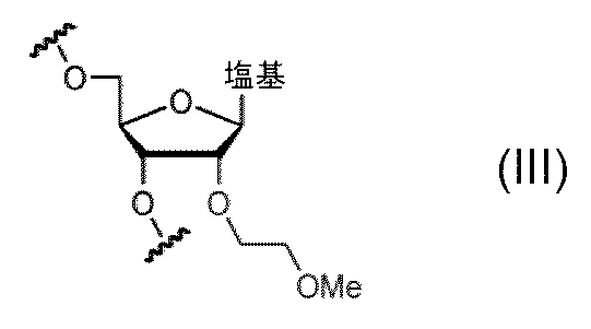
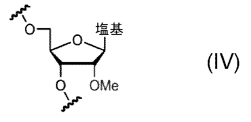
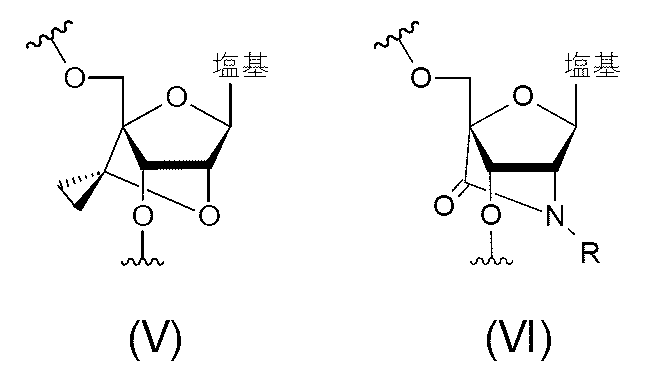
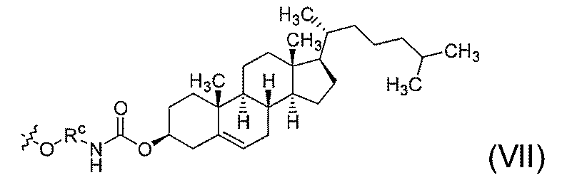
リンカーの一具体例として、以下の式(VIII)で表されるリンカーが挙げられる。


本発明の二本鎖核酸複合体は、当業者であれば、公知の方法を適切に選択することによって製造することができる。限定はしないが、通常は、まず二本鎖核酸複合体を構成する第1核酸鎖及び第2核酸鎖のそれぞれを設計し、製造するところから始まる。例えば、標的転写産物の塩基配列(例えば、標的遺伝子の塩基配列)情報に基づいて第1核酸鎖を設計し、その相補鎖として第2核酸鎖を設計する。続いて、設計した塩基配列情報に基づいて、それぞれの核酸鎖を、例えば、GE Healthcare社、Thermo Fisher Scientific社、Beckman Coulter社等の市販の自動核酸合成装置を使用して合成すればよい。その後は、得られたオリゴヌクレオチドに逆相カラム等を使用して精製するもできる。
一実施形態において、本発明の二本鎖核酸複合体は、以下の少なくとも一つの作用:標的遺伝子の転写産物又は翻訳産物の発現量を抑制若しくは亢進する作用、標的遺伝子の転写産物又は翻訳産物の機能を阻害する作用、RNAスプライシングを制御する作用、及び標的遺伝子のタンパク質への結合を阻害する作用を用途とするもの、例えばエクソンスキッピングのためのものであってもよい。例えば、本発明の二本鎖核酸複合体は、以下の少なくとも一つの作用:エクソンスキッピング、エクソンインクルージョン、ステリックブロッキング、及びRNA発現亢進のためのものであってもよい。本発明の二本鎖核酸複合体は、特定の組織、例えば脳、脊髄、腎臓、肝臓、肺、腸管、脾臓、副腎、眼、網膜、皮膚、末梢神経、例えば脳において上記作用を発揮するためのものであってもよい。脳は大脳、間脳、脳幹、小脳のいずれであってもよく、例えば、大脳(大脳皮質等)、脳幹、小脳、海馬、及び線状体のいずれか一つ以上であってもよい。また、本発明の二本鎖核酸複合体は、心筋及び骨格筋を含む筋組織において上記作用を発揮するためのものであってもよい。
本発明の二本鎖核酸複合体によれば、二本鎖核酸複合体の有効性が損なうことなく、二本鎖核酸複合体の中枢神経毒性等の毒性を軽減又は消失することができる。すなわち、従来の二本鎖核酸複合体と比較して、標的遺伝子に対するアンチセンス効果を損なうことなく、中枢神経毒性が低減されている。
2-1.概要
本発明の第2態様は医薬組成物である。本発明の医薬組成物は、前記第1態様の二本鎖核酸複合体を有効成分として含む。本発明の医薬組成物は、中枢神経毒性が軽減されており、副作用を伴うことなく髄腔内投与又は脳室内投与することができる。
以下、本発明の医薬組成物が包含し得る各成分について具体的に説明をする。
2-2-1.有効成分
本発明の医薬組成物は、有効成分として少なくとも第1態様に記載の二本鎖核酸複合体を包含する。本発明の医薬組成物は、二本鎖核酸複合体を二種以上含んでもよい。
本発明の医薬組成物は、薬学的に許容可能な担体を含むことができる。「薬学的に許容可能な担体」とは、製剤技術分野において通常使用する添加剤をいう。例えば、溶媒、植物性油、基剤、乳化剤、懸濁化剤、界面活性剤、pH調整剤、安定化剤、香味料、香料、賦形剤、ビヒクル、防腐剤、結合剤、希釈剤、等張化剤、鎮静剤、増量剤、崩壊剤、緩衝剤、コーティング剤、滑沢剤、着色剤、甘味剤、増粘剤、矯味剤、溶解助剤、及び他の添加剤が挙げられる。
本発明の医薬組成物の剤形は、有効成分である第1態様に記載の二本鎖核酸複合体を分解等により不活化させることなく、標的部位まで送達し、生体内でその有効成分の薬理効果(標的遺伝子の発現に対するアンチセンス効果)を発揮し得る形態であれば特に限定しない。
本明細書において、医薬組成物の好ましい投与形態には特定の限定はない。例えば、経口投与又は非経口投与であればよい。非経口投与の具体例として、筋肉内投与、静脈内投与、動脈内投与、腹腔内投与、皮下投与(埋め込み型持続皮下投与を含む)、皮内投与、気管/気管支投与、直腸投与、輸血による投与、脳室内投与、髄腔内投与、経鼻投与、及び筋肉内投与が挙げられる。髄腔内投与は、例えば後頭窩穿刺又は腰椎穿刺であってもよい。
医薬組成物の適用対象となる疾患は、限定しない。本発明の二本鎖核酸複合体のアンチセンス効果により、その転写産物又は翻訳産物の発現量が抑制若しくは亢進され得る、その転写産物又は翻訳産物の機能が阻害され得る、又はステリックブロッキング、スプライシングスイッチ、RNA編集、エクソンスキッピング若しくはエクソンインクルージョンが誘導され得る遺伝子が関係し得る疾患を対象とすることができる。疾患の具体例は「1-5.二本鎖核酸複合体の用途」の記載に準じる。
本発明の医薬組成物は、中枢神経毒性が軽減されている。それ故、本発明の医薬組成物は、脳室内投与や髄腔内投与により副作用を伴うことなく予防効果又は治療効果を達成することができる。
(目的)
Mapt遺伝子を標的とするアンチセンス核酸(以下、「ASO」と称する)からなる第1核酸鎖、及び第1核酸鎖に相補的な塩基配列を有する第2核酸鎖を含むヘテロ二重鎖オリゴヌクレオチド(以下、「HDO」と称する)を脳室内投与する場合に見られる中枢神経毒性について、MOE修飾に基づく毒性軽減効果をin vivo実験により検証する。
(1)核酸の調製
本実施例で用いたASO、並びにHDOを構成する第1核酸鎖及び第2核酸鎖の塩基配列と化学修飾を表1及び図4に示す。

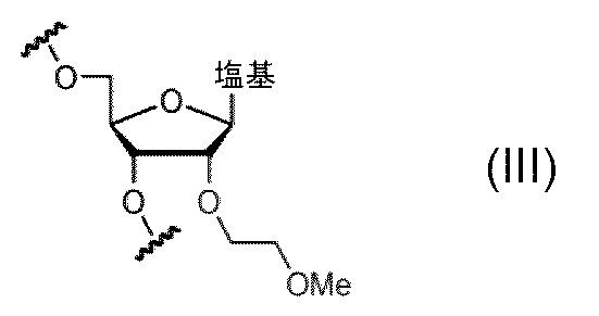
7週齢雌のICRマウスを、2.5~4%イソフルレン麻酔下にて脳定位固定装置に固定した。その後、耳間に前後2~3cmで皮膚を切開し、ブレグマ(bregma)の1mm左方かつ0.2mm後方に1mm径ドリルで穿孔した。ハミルトン(Hamilton)シリンジ内に核酸剤を充填した。穿孔部より針を3mm程度刺入し、2~3μl/分の速度で、マウス1匹当たり19nmol/匹の用量で核酸剤を左側脳室内投与し(n=4~7)、ナイロン糸で皮膚縫合した。また、陰性対照群としてPBSのみを投与したマウスも作製した。
各種核酸剤投与後のマウスにおいて、中枢神経毒性評価及び運動機能評価を行った。
中枢神経毒性評価として、核酸剤の投与から30分後、1時間後、2時間後、3時間後、及び4時間後に、図3に示すスコアリングシステムを用いて行動評価を行った。
各種核酸剤投与から7日後のマウスから海馬を摘出した。IsogenIキット(株式会社ジーンデザイン)を使用して、摘出した左海馬からRNAを抽出した。cDNAは、Transcriptor Universal cDNA Master, DNase (ロシュ・ダイアグノスティックス社(Roche Diagnostics))を使用してプロトコルに従って合成した。
図5は、各種核酸剤を脳室内投与したマウスを対象として中枢神経毒性を評価した結果を示す。核酸剤の投与から30分後、1時間後、2時間後、3時間後、及び4時間後のいずれの時点においても、HDO(6MOE wing)及びHDO(all MOE)の投与群では、ASO、HDO(all RNA)、及びHDO(all DNA)の投与群と比較して、急性期忍容性スコア(acute tolerability score)が著しく減少した。この結果から、2'-O-MOE-RNAヌクレオシドを含むHDOでは中枢神経毒性が顕著に低下することが示された。
(目的)
BACE1遺伝子を標的とするASOからなる第1核酸鎖、及び第1核酸鎖に相補的な塩基配列を有する第2核酸鎖を含むHDOを脳室内投与する場合に見られる中枢神経毒性について、MOE修飾に基づく毒性軽減効果をin vivo実験により検証する。
本実施例で用いたASO、並びにHDOを構成する第1核酸鎖及び第2核酸鎖の塩基配列と化学修飾を表2及び図8に示す。
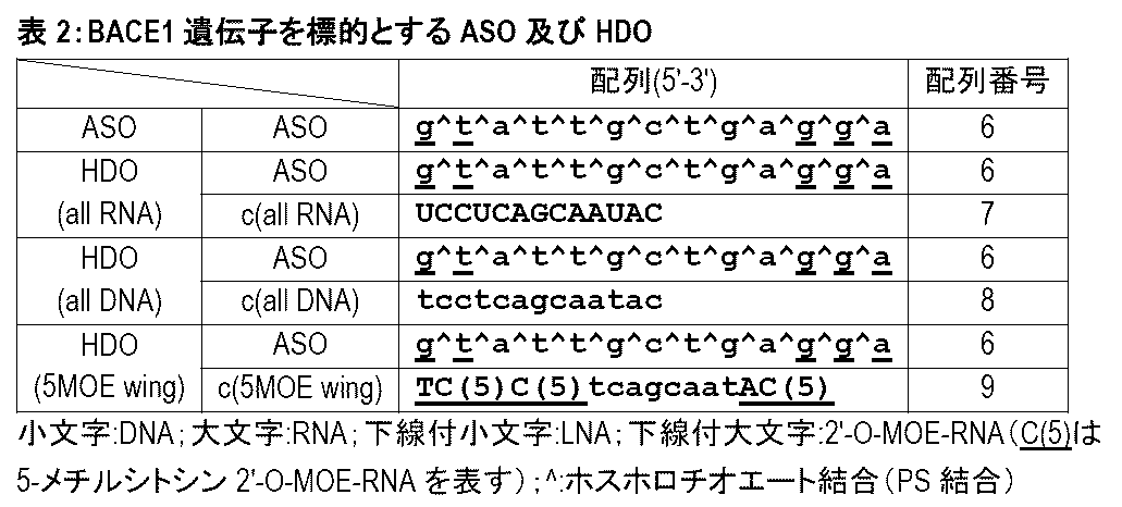
図9は、各種核酸剤を脳室内投与したマウスを対象として中枢神経毒性を評価した結果を示す。核酸剤の投与から30分後、1時間後、2時間後、3時間後、及び4時間後のいずれの時点においても、HDO(5MOE wing)投与群では、ASO、HDO(all RNA)、及びHDO(all DNA)の投与群と比較して、急性期忍容性スコアが著しく減少した。この結果から、2'-O-MOE-RNAヌクレオシドを含むHDOでは中枢神経毒性が顕著に低下することが示された。
(目的)
Malat1遺伝子を標的とするASOからなる第1核酸鎖、及び第1核酸鎖に相補的な塩基配列を有する第2核酸鎖を含むHDOを脳室内投与する場合に見られる中枢神経毒性について、MOE修飾に基づく毒性軽減効果をin vivo実験により検証する。
本実施例で用いたASO、並びにHDOを構成する第1核酸鎖及び第2核酸鎖の塩基配列と化学修飾を表3及び図11に示す。
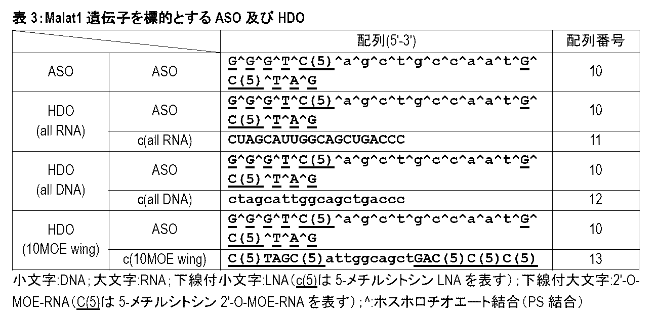
図12は、各種核酸剤を脳室内投与したマウスを対象として中枢神経毒性を評価した結果を示す。核酸剤の投与から30分後、1時間後、2時間後、3時間後、及び4時間後のいずれの時点においても、HDO(5MOE wing)投与群では、ASO及びHDO(all DNA)の投与群と比較して、急性期忍容性スコアが著しく減少した。また、核酸剤の投与から30分後、1時間後、及び2時間後の時点において、HDO(5MOE wing)投与群では、HDO(all RNA)投与群と比較して、急性期忍容性スコアが低かった。この結果から、2'-O-MOE-RNAヌクレオシドを含むHDOでは中枢神経毒性が顕著に低下することが示された。
(目的)
Mapt遺伝子を標的とするASOからなる第1核酸鎖、及び第1核酸鎖に相補的な塩基配列を有する第2核酸鎖を含むHDOを脳室内投与する場合に見られる中枢神経毒性について、MOE修飾、2'OMe修飾、及び2'F修飾による毒性軽減効果をin vivo実験により比較する。
本実施例で用いたASO、並びにHDOを構成する第1核酸鎖及び第2核酸鎖の塩基配列と化学修飾を表4及び図14に示す。

図15は、各種核酸剤を脳室内投与したマウスを対象として中枢神経毒性を評価した結果を示す。核酸剤の投与から30分後、1時間後、2時間後、3時間後、及び4時間後のいずれの時点においても、HDO(RNA 6MOE wing)及びHDO(6MOE wing)の投与群では、HDO(all DNA)、HDO(6OMe wing)、及びHDO(6F wing)の投与群と比較して、急性期忍容性スコアが著しく減少した。さらに、HDO(6MOE wing)投与群は、HDO(RNA 6MOE wing)投与群と比較して低い急性期忍容性スコアを示した。この結果から、2'-O-MOE-RNAヌクレオシドを含むHDOは、2'-O-Me-RNAや2'F-RNAヌクレオシドヌクレオシドを含むHDOと比較しても低い中枢神経毒性を達成し得ることが示された。
(目的)
Mapt遺伝子を標的とするASOからなる第1核酸鎖、及び第1核酸鎖に相補的な塩基配列を有する第2核酸鎖を含むHDOを脳室内投与する場合に見られる中枢神経毒性について、第2核酸鎖におけるグアノシンヌクレオシドの置換による毒性軽減効果をin vivo実験により検討する。
本実施例で用いたASO、並びにHDOを構成する第1核酸鎖及び第2核酸鎖の塩基配列と化学修飾を表5及び図18に示す。

図19は、各種核酸剤を脳室内投与したマウスを対象として中枢神経毒性を評価した結果を示す。核酸剤の投与から30分後、1時間後、2時間後、3時間後、及び4時間後のいずれの時点においても、HDO(GMOE)及びHDO(GRNA)の投与群は、HDO(6MOE wing)投与群に対して同等かそれより低い急性期忍容性スコアを示した。
(目的)
Mapt遺伝子を標的とするASOからなる第1核酸鎖、及び第1核酸鎖に相補的な塩基配列を有する第2核酸鎖を含むHDOを脳室内投与する場合に見られる中枢神経毒性について、様々なMOE修飾に基づく毒性軽減効果をin vivo実験により検証する。
本実施例で用いたHDOを構成する第1核酸鎖及び第2核酸鎖の塩基配列と化学修飾を表6及び図20に示す。

図21は、各種核酸剤を脳室内投与したマウスを対象として中枢神経毒性を評価した結果を示す。核酸剤の投与から30分後、1時間後、2時間後、3時間後、及び4時間後のいずれの時点においても、HDO(6MOE-5'&3')、HDO(6MOE-5')、HDO(6MOE-3')、HDO(10MOE-5')、及びHDO(10MOE-3')の投与群では、HDO(all DNA)投与群と比較して、急性期忍容性スコアが著しく減少した。特にHDO(10MOE-5')及びHDO(10MOE-3')の投与群における急性期忍容性スコアがHDO投与群で最小となった。
(目的)
BACE1遺伝子を標的とするASOからなる第1核酸鎖、及び第1核酸鎖に相補的な塩基配列を有する第2核酸鎖を含むHDOを脳室内投与する場合に見られる中枢神経毒性について、様々なMOE修飾に基づく毒性軽減効果をin vivo実験により検証する。
本実施例で用いたHDOを構成する第1核酸鎖及び第2核酸鎖の塩基配列と化学修飾を表7及び図25に示す。

図26は、各種核酸剤を脳室内投与したマウスを対象として中枢神経毒性を評価した結果を示す。核酸剤の投与から30分後、1時間後、2時間後、3時間後、及び4時間後のいずれの時点においても、HDO(5MOE-5')、HDO(5MOE-3')、HDO(8MOE-5')、HDO(8MOE-3')、HDO(10MOE-5')、及びHDO(10MOE-3')の投与群では、HDO(all DNA)投与群と比較して、急性期忍容性スコアが著しく減少した。
(目的)
Mapt遺伝子を標的とするASOからなる第1核酸鎖、及び第1核酸鎖に相補的な塩基配列を有する第2核酸鎖を含むHDOを脳室内投与する場合に見られる中枢神経毒性について、様々なMOE修飾に基づく毒性軽減効果をin vivo実験により検証する。
本実施例で用いたHDOを構成する第1核酸鎖及び第2核酸鎖の塩基配列と化学修飾を表8及び図28に示す。

図29は、各種核酸剤を脳室内投与したマウスを対象として中枢神経毒性を評価した結果を示す。核酸剤の投与から30分後、1時間後、2時間後、3時間後、及び4時間後のいずれの時点においても、HDO(6MOE wing)、HDO(9MOE wing)、HDO(11MOE wing)、HDO(13MOE wing)、及びHDO(15MOE wing)の投与群では、HDO(all DNA)投与群と比較して、急性期忍容性スコアが著しく減少した。
(目的)
Mapt遺伝子を標的とするASOからなる第1核酸鎖、及び第1核酸鎖に相補的な塩基配列を有する第2核酸鎖を含むHDOを脳室内投与する場合に見られる中枢神経毒性について、第2核酸鎖においてMOE修飾を導入するヌクレオシドの種類(アデニン塩基、グアニン塩基、シトシン塩基、又はチミン塩基を含むヌクレオシド)による毒性軽減効果の違いについてin vivo実験により比較する。
本実施例で用いたASO、並びにHDOを構成する第1核酸鎖及び第2核酸鎖の塩基配列と化学修飾を表9及び図31に示す。
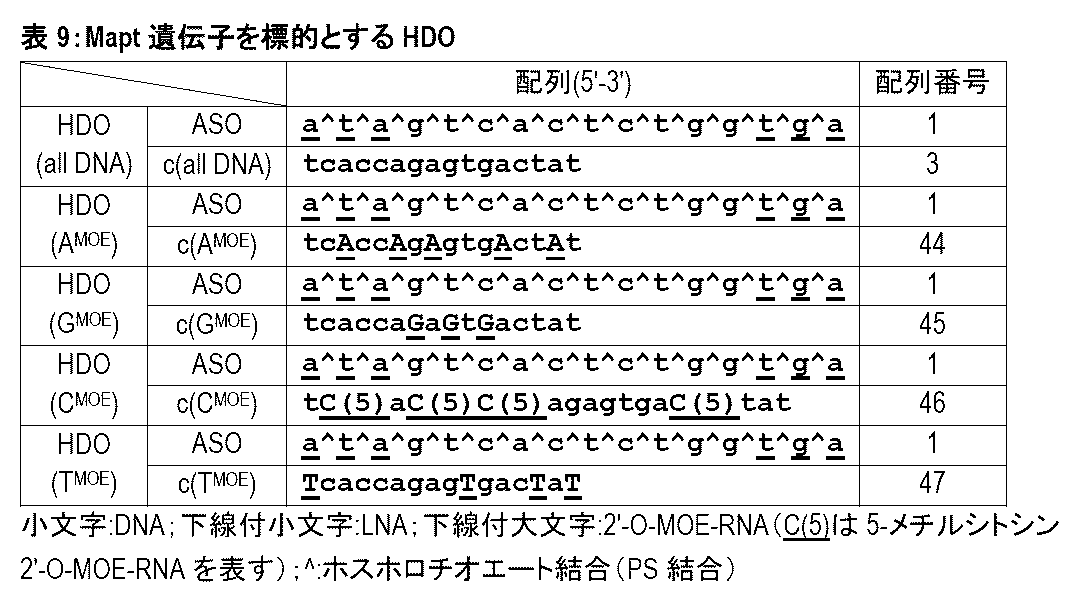
図32は、各種核酸剤を脳室内投与したマウスを対象として中枢神経毒性を評価した結果を示す。核酸剤の投与から30分後、1時間後、2時間後、3時間後、及び4時間後のいずれの時点においても、HDO(AMOE)、HDO(CMOE)、及びHDO(TMOE)の投与群では、HDO(all DNA)投与群と比較して、急性期忍容性スコアが著しく減少し、HDO(GMOE)投与群は、HDO(all DNA)投与群と比較して急性期忍容性スコアがわずかに減少した。この結果から、第2核酸鎖においてアデニン塩基、シトシン塩基、又はチミン塩基を含むヌクレオシドを2'-O-MOE-RNAヌクレオシドに置換したHDOは、第2核酸鎖がDNAヌクレオシドのみからなるHDOと比較して中枢神経毒性を軽減し得ることが示された。
(目的)
Mapt遺伝子を標的とするASOからなる第1核酸鎖、及び第1核酸鎖に相補的な塩基配列を有する第2核酸鎖を含むHDOを脳室内投与する場合に見られる中枢神経毒性について、第2核酸鎖においてシトシン塩基を含むヌクレオシド並びにその5'側及び/又は3'側に隣接するヌクレオシドへのMOE修飾の導入による毒性軽減効果をin vivo実験により比較する。
本実施例で用いたASO、並びにHDOを構成する第1核酸鎖及び第2核酸鎖の塩基配列と化学修飾を表10及び図34に示す。

図35は、各種核酸剤を脳室内投与したマウスを対象として中枢神経毒性を評価した結果を示す。核酸剤の投与から30分後、1時間後、2時間後、3時間後、及び4時間後のいずれの時点においても、HDO(2CMOE-5)、HDO(2CMOE-3)及びHDO(3CMOE)の投与群では、HDO(all DNA)及びHDO(CMOE)の投与群と比較して、急性期忍容性スコアが著しく減少した。この結果から、第2核酸鎖においてシトシン塩基を含むヌクレオシド及びその隣接するヌクレオシドを2'-O-MOE-RNAヌクレオシドに置換したHDOは、第2核酸鎖がDNAヌクレオシドからなるHDOと比較して中枢神経毒性が軽減されることが示された。
(目的)
Mapt遺伝子を標的とするASOからなる第1核酸鎖、及び第1核酸鎖に相補的な塩基配列を有する第2核酸鎖を含むHDOをヒト又はラットの髄液中でインキュベートし、その安定性を検討する。
(1)核酸の調製
本実施例で用いたHDOを構成する第1核酸鎖及び第2核酸鎖の塩基配列と化学修飾を表11、図37A、図37B、図38A、及び図38Bに示す。
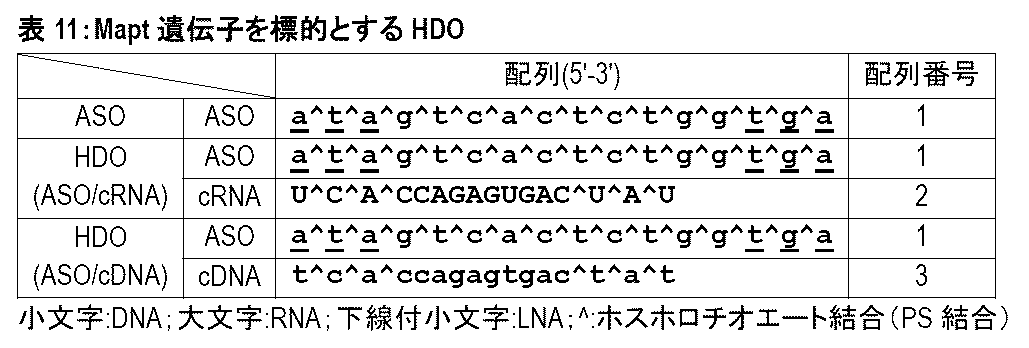
10μMのHDO(ASO/cRNA)及びHDO(ASO/cDNA)各4μLを用意した。HDO 4μLに対して、ヒト髄液又はラット髄液を16μL混合し、37℃ incubatorでインキュベートした。10分後、1時間後、及び6時間後の各時点で、液体窒素に投入して反応を停止させた。
図37Cは、HDO(ASO/cRNA)をヒト髄液(Human CSF)中で10分間及び6時間インキュベートした後の電気泳動結果を示す。図37Dは、図37Cの泳動結果におけるHDO 2本鎖バンドのバンド強度を定量した結果を示す。HDO(ASO/cRNA)は、ヒト髄液中での10分間のインキュベートで大部分が分解され、6時間で全部が分解された。
(目的)
Mapt遺伝子を標的とするASOからなる第1核酸鎖、及び第1核酸鎖に相補的な塩基配列を有する第2核酸鎖を含むHDOをマウス、ラット、サル、又はヒトの髄液中でインキュベートし、その安定性を検討する。
(1)核酸の調製
本実施例で用いたHDOを構成する第1核酸鎖及び第2核酸鎖の塩基配列と化学修飾を表12、図39A、図39B、図40A、及び図40Bに示す。
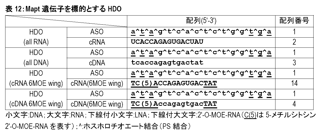
図39Cは、HDO(all RNA)及びHDO(all DNA)をマウス、ラット、サル、及びヒトの髄液中で6時間インキュベートした後の電気泳動結果を示す。HDO(all RNA)は、マウス及びラットの髄液中では分解されず安定であった一方、サル及びヒトの髄液中では分解され、不安定であった。これに対して、HDO(all DNA)は、マウス、ラット、サル、及びヒトのいずれの髄液中でも分解されず安定であった。
(目的)
Mapt遺伝子を標的とするASOからなる第1核酸鎖、及び第1核酸鎖に相補的な塩基配列を有する第2核酸鎖を含むHDOにおいて、第2核酸鎖中の一部のDNAヌクレオシドをRNAヌクレオシドに置換した場合の髄液中での安定性を検討する。
(1)核酸の調製
本実施例で用いたHDOを構成する第1核酸鎖及び第2核酸鎖の塩基配列と化学修飾を表13及び図41に示す。

図42は、HDO(all DNA)、HDO(ARNA)、HDO(GRNA)、HDO(CRNA)、及びHDO(URNA)をヒト髄液中で6時間インキュベートした後の電気泳動結果を示す。HDO(all DNA)、HDO(ARNA)、及びHDO(GRNA)は、ヒト髄液中で分解されず安定であった。一方、HDO(CRNA)及びHDO(URNA)は、ヒト髄液中で分解され、不安定であった。
(目的)
Mapt遺伝子を標的とするASOからなる第1核酸鎖、及び第1核酸鎖に相補的な塩基配列を有する第2核酸鎖を含むHDOにおいて、第2核酸鎖中の一部のDNAヌクレオシドをRNAヌクレオシドに置換した場合の髄液中での安定性を検討する。
(1)核酸の調製
本実施例で用いたHDOを構成する第1核酸鎖及び第2核酸鎖の塩基配列と化学修飾を表14及び図43に示す。

図44は、HDO(all DNA)、HDO(GARNA)、HDO(CURNA)、HDO(CRNA)、及びHDO(URNA)をヒト髄液中で1時間及び6時間インキュベートした後の電気泳動結果を示す。HDO(all DNA)及びHDO(GARNA)は、ヒト髄液中で分解されず安定であった。一方、HDO(CURNA)、HDO(CRNA)、及びHDO(URNA)は、ヒト髄液中で分解され、不安定であった。
(目的)
Malat1遺伝子を標的とするASOからなる第1核酸鎖、及び第1核酸鎖に相補的な塩基配列を有する第2核酸鎖を含むHDOにおいて、第2核酸鎖中の一部のDNAヌクレオシドをRNAヌクレオシドに置換した場合の髄液中での安定性を検討する。
(1)核酸の調製
本実施例で用いたHDOを構成する第1核酸鎖及び第2核酸鎖の塩基配列と化学修飾を表15及び図45に示す。
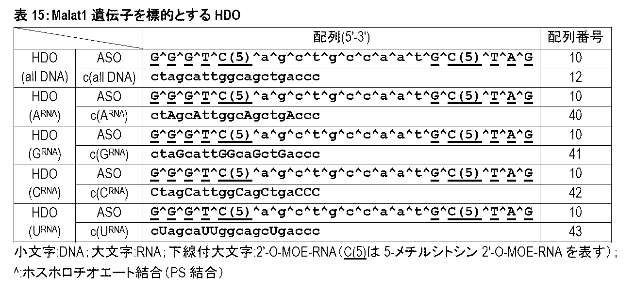
図46は、HDO(all DNA)、HDO(ARNA)、HDO(GRNA)、HDO(CRNA)、及びHDO(URNA)をヒト髄液中で6時間インキュベートした後の電気泳動結果を示す。HDO(all DNA)、HDO(ARNA)、及びHDO(GRNA)は、ヒト髄液中で分解されず安定であった。一方、HDO(CRNA)及びHDO(URNA)は、ヒト髄液中で分解され、不安定であった。
(目的)
MOE修飾を含むHDOをサルに髄腔内投与して、中枢神経毒性を評価する。
(1)核酸の調製
本実施例で用いたASO、並びにHDOを構成する第1核酸鎖及び第2核酸鎖の塩基配列と化学修飾を表16及び図47A-Cに示す。
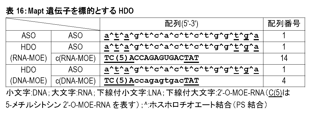
雄のカニクイザル(3歳齢、2kg)を対象として、ASO、HDO(RNA-MOE)、及びHDO(DNA-MOE)を投与した。投与方法としては、ASO 5mg又はHDO 0.94μmolを髄腔内投与した。
髄腔内投与から2時間後のサルについて、行動観察を行った(図47D)。
結果を図47Eに示す。ASOを髄腔内投与したサルは重篤な四肢麻痺を示し、強い中枢神経毒性を示した。HDO(RNA-MOE)を髄腔内投与したサルは、下肢に中程度の麻痺を示したもの、ASOの髄腔内投与と比較して中枢神経系毒性は軽減していた。HDO(DNA-MOE)を髄腔内投与したサルは、麻痺を示さず、正常な意識状態及び運動機能を示した。
(目的)
Mapt遺伝子を標的とするASOからなる第1核酸鎖、及び第1核酸鎖に相補的な塩基配列を有する第2核酸鎖を含むHDOを神経細胞株に導入する。第2核酸鎖へのバルジ構造の導入による毒性軽減効果及び遺伝子抑制効果をin vitro実験により検証する。
(1)核酸の調製
本実施例で用いるHDOを構成する第1核酸鎖及び第2核酸鎖の塩基配列と化学修飾を表17及び図48に示す。
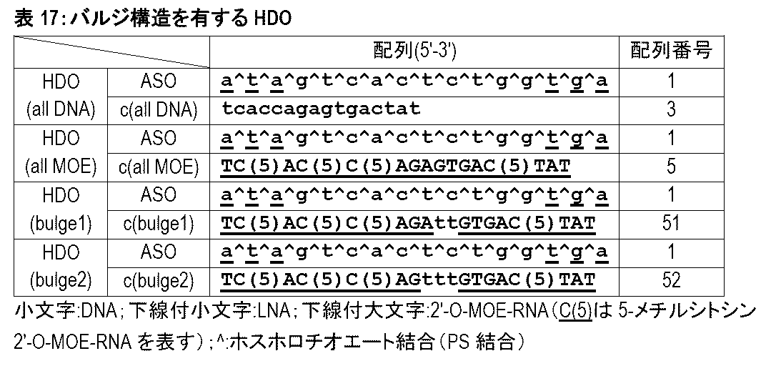
(1)で調製したHDOをヒト神経芽細胞腫由来細胞(BE(2)-M17細胞株)にリポフェクション法(lipofectamine2000)を用いて導入した。
導入48時間後に神経細胞毒性の評価として、細胞上清中の乳酸脱水素酵素(LDH)活性を測定した。LDH活性測定は、Cytotoxicity LDH Assay Kit-WST(同仁化学研究所)を使用して、添付プロトコルに従って実施した。PBS投与群のLDH活性に対して標準化した値を相対的LDH放出レベルとした。
図49Aは、ヒト神経芽細胞腫由来細胞(BE(2)-M17細胞株)において各種核酸剤による標的遺伝子抑制効果を評価した結果を示す。HDO(all MOE)の投与群ではHDO(all DNA)に比して抑制効果が著しく減弱した。これに対して、HDO(bulge1)及びHDO(bulge2)の投与群ではHDO(all DNA)と同等の強い遺伝子抑制効果が得られた。
(目的)
Mapt遺伝子を標的とするASOからなる第1核酸鎖、及び第1核酸鎖に相補的な塩基配列を有する第2核酸鎖を含むHDOを脳室内投与する場合に見られる中枢神経毒性について、第2核酸鎖へのバルジ構造の導入、第1核酸鎖と第2核酸鎖とのリンカーによる連結、及びその両方による毒性軽減効果及び遺伝子抑制効果をin vivo実験により検証する。
(1)核酸の調製
本実施例で用いたHDOの塩基配列と化学修飾を表18及び図50に示す。
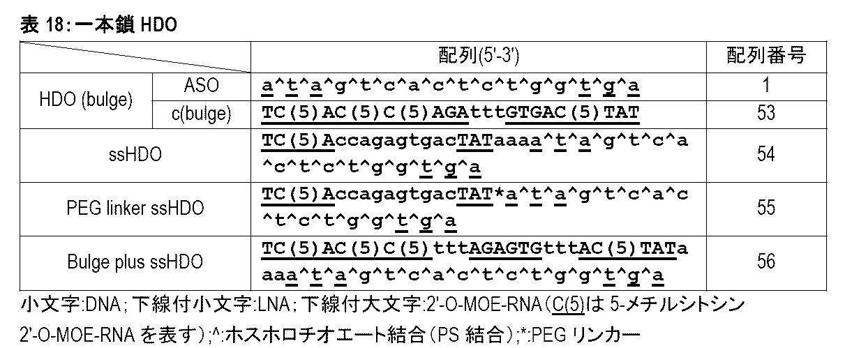
HDO(bulge)、ssHDO、PEG linker ssHDO、及びBulge plus ssHDOを脳室内投与したマウスでは、実施例1で用いたHDO(all DNA)を投与したマウス等と比較して、投与から30分~4時間後の急性期忍容性スコアが著しく減少する。また、HDO(bulge)、ssHDO、PEG linker ssHDO、及びBulge plus ssHDOを脳室内投与したマウスでは、実施例1で用いたHDO(all DNA)を投与したマウス等と比較して、投与から1時間後の運動機能、例えば総移動距離や最大移動速度が著しく改善する。さらに、HDO(bulge)、ssHDO、PEG linker ssHDO、及びBulge plus ssHDOを脳室内投与したマウスでは、実施例1で用いたHDO(all MOE)を投与したマウス等と比較して、投与から7日後の海馬においてMapt mRNA発現レベルが低下し、標的遺伝子抑制効果の向上が判明する。以上の結果から、第2核酸鎖にバルジ構造を導入すること、及びHDOの第1核酸鎖と第2核酸鎖をリンカーで連結することにより、中枢神経毒性の軽減と優れた遺伝子抑制効果の両方を達成できるすることが示される。また、HDOにバルジ構造とリンカーの両方を導入することにより、中枢神経毒性の軽減と優れた遺伝子抑制効果の両方を達成できるすることが示される。
(目的)
Malat1遺伝子を標的とするHDOを脳室内投与する場合に見られる中枢神経毒性について、第2核酸鎖において第1核酸鎖の中央領域(ギャップ領域)に相補的な塩基配列からなる領域(以下、第2核酸鎖においても「ギャップ領域」という)に導入する核酸種の影響を検討する。具体的には、第2核酸鎖のギャップ領域において、全てのヌクレオシドがRNAヌクレオシドであるHDO、全てのヌクレオシドがDNAヌクレオシドであるHDO、並びにアデニン塩基及びグアニン塩基の位置にRNAヌクレオシドを配置し、シトシン塩基及びチミン塩基の位置にDNAヌクレオシドを配置したHDOについて、毒性をin vivo実験により比較する。
本実施例で用いたASO、並びにHDOを構成する第1核酸鎖及び第2核酸鎖の塩基配列と化学修飾を表19に示す。

表19に記載される核酸分子について、実施例1と同様の方法により核酸の調製、in vivo実験、並びに中枢神経毒性評価及び運動機能評価を行った。但し、本実施例ではマウス1匹当たりの核酸剤投与量は、14nmol/匹とした。
図51は、各種核酸剤を脳室内投与したマウスにおいて投与から1時間後に運動機能を評価した結果を示す。HDO(all DNA)、HDO(cRNA 10MOE)、HDO(cDNA 10MOE)及びHDO(agRNA 10MOE)の投与群では、ASO投与群と比較して、総移動距離(図51A)及び平均移動速度(図51B)を著しく改善させた。また、HDO(cRNA 10MOE)、HDO(cDNA 10MOE)及びHDO(agRNA 10MOE)の投与群では、総移動距離及び平均移動速度がHDO(all DNA)の投与群と比較して改善した。更にHDO(agRNA 10MOE)の投与群では、総移動距離及び平均移動速度がHDO(cRNA 10MOE)及びHDO(cDNA 10MOE)の投与群と比較して改善した。この結果から、第2核酸鎖のギャップ領域においてアデニン塩基及びグアニン塩基の位置がRNAヌクレオシドであり、シトシン塩基及びチミン塩基の位置がDNAヌクレオシドであるHDOは、1本鎖ASO及び他のHDOと比較して中枢神経毒性を顕著に軽減し得ることが示された。
図52は、各種核酸剤の脳室内投与から7日後において右左の前頭葉におけるMalat1 RNA発現レベルを示す。HDO(agRNA 10MOE)投与群はASO投与群及び他のHDO投与群と比較して、Malat1 RNA発現レベルを顕著に抑制する効果を示した。この結果から、ギャップ領域においてアデニン塩基及びグアニン塩基の位置がRNAヌクレオシドであり、シトシン塩基及びチミン塩基の位置がDNAヌクレオシドである第2核酸鎖を含むHDOが、ギャップ領域において全てのヌクレオシドがDNAヌクレオシド又はRNAヌクレオシドである第2核酸鎖を含むHDOと比較して、優れた遺伝子発現抑制効果を達成し得ることが示された。
(目的)
標的組織において第1核酸鎖は第2核酸鎖から解離し、標的遺伝子やその転写産物に対するアンチセンス効果を発揮し得る。そのため、第1核酸鎖及び第2核酸鎖を含むHDOをマウスの脳組織ホモジネート中でインキュベートし、その2本鎖解離効率を検討する。
(1)核酸の調製
本実施例では、実施例19と同一のASO、HDO(cRNA 10MOE)、HDO(cDNA 10MOE)、及びHDO(agRNA 10MOE)を使用した。ASO及びHDOは、実施例19と同様の方法により調製した。
25μMのHDO(ASO/cRNA)及びHDO(ASO/cDNA)各5.2μLに対して、マウス脳ホモジネート溶液を16.7μL及びPBSを28.1μL混合し、37℃ incubatorでインキュベートした。インキュベーション後、プロテインキナーゼKとの混合により反応を停止させた。
16%アクリルアミドゲル(1×TBE)を準備、作製した。上記サンプル9.6μLをゲルにロードし、100V 120分で泳動した。対照としてASO単独を同時に泳動した。その後、1×TBEでGelRed(×10000)水溶液(Biotium)を1/10000の濃度に希釈した溶液を作製した。ゲルを10分間その溶液で浸透した。その後、ChemiDoc Touchイメージングシステム(BioRad社)でゲルを撮影した。
図53は、ASO、HDO(cRNA 10MOE)、HDO(cDNA 10MOE)及びHDO(agRNA 10MOE)をマウス脳組織ホモジネート中で7日間インキュベートした後の電気泳動結果を示す。HDO(agRNA 10MOE)は、HDO(cRNA 10MOE)及びHDO(cDNA 10MOE)と比較して、7日間インキュベート後の2本鎖核酸(図53、矢印)が著しく減少し、脳組織内で極めて優れた2本鎖分離能力を有することが明らかになった。ギャップ領域においてアデニン塩基及びグアニン塩基の位置がRNAヌクレオシドであり、シトシン塩基及びチミン塩基の位置がDNAヌクレオシドである第2核酸鎖を含むHDOは、ギャップ領域において全てのヌクレオシドがDNAヌクレオシド又はRNAヌクレオシドである第2核酸鎖を含むHDOと比較して脳組織内において2本鎖解離効率が極めて高く、上記の実施例19において標的遺伝子の発現を顕著に抑制した結果を強く裏付けた。
(目的)
Malat1遺伝子を標的とするHDOを脳室内投与する場合に見られる中枢神経毒性について、第2核酸鎖のウイング領域及びギャップ領域におけるMOE修飾核酸又は天然核酸(RNA又はDNA)の導入による影響をin vivo実験により比較する。
本実施例で用いたHDOを構成する第1核酸鎖及び第2核酸鎖の塩基配列と化学修飾を表20に示す。
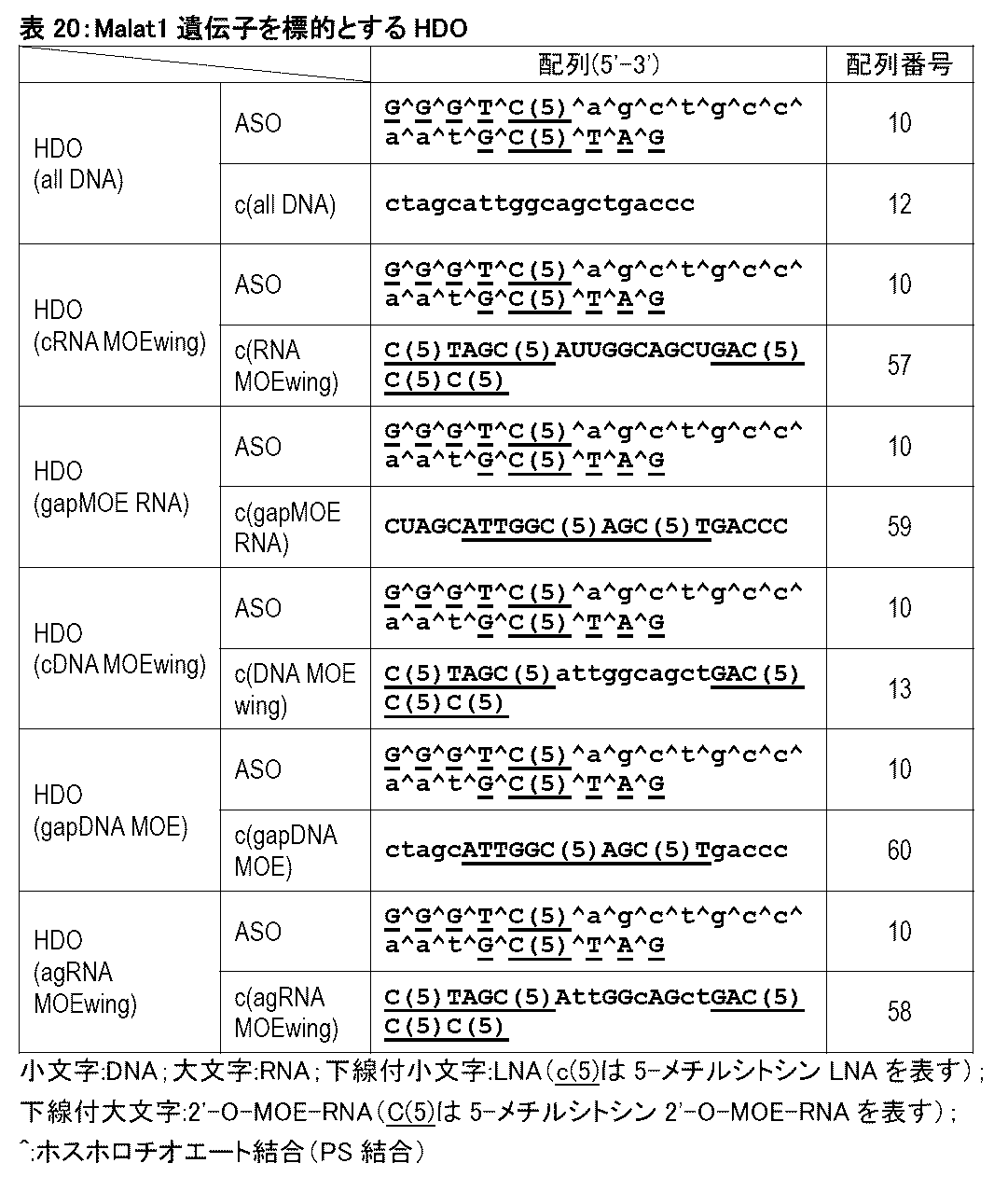
表21に記載される核酸分子について、実施例1と同様の方法により核酸の調製、in vivo実験、並びに中枢神経毒性評価及び運動機能評価を行った。但し、本実施例ではマウス1匹当たりの核酸剤投与量は、28nmol/匹とした。
図54は、各種核酸剤を脳室内投与したマウスを対象として中枢神経毒性を評価した結果を示す。HDO(cRNA MOEwing)、HDO(gapMOE RNA)、HDO(cDNA MOEwing)、HDO(gapDNA MOE)及びHDO(agRNA 10MOE)の投与群では、HDO(all DNA)の投与群と比較して、急性期忍容性スコアが著しく減少した。また、HDO(gapMOE RNA)、HDO(cDNA MOEwing)、HDO(gapDNA MOE)及びHDO(agRNA 10MOE)の投与群では、HDO(cDNA MOEwing)の投与群と比較して、急性期忍容性スコアが更に減少した。
この結果から、第2核酸鎖においてMOE修飾をウイング領域又はギャップ領域に含むHDOは、第2核酸鎖がDNAヌクレオシドからなるHDOと比較して、中枢神経毒性の軽減を達成し得ることが示された。
(目的)
Mapt遺伝子を標的とするASOからなる第1核酸鎖、及び第1核酸鎖に相補的な塩基配列を有するMOE wing修飾型第2核酸鎖を含むHDOを脳室内投与する場合に見られる、投与後1日以内の急性神経毒性、及び投与1日後以降の遅発性神経毒性について、毒性軽減効果をin vivo実験により比較する。
本実施例で用いたASO、並びにHDOを構成する第1核酸鎖及び第2核酸鎖の塩基配列と化学修飾を表21に示す。
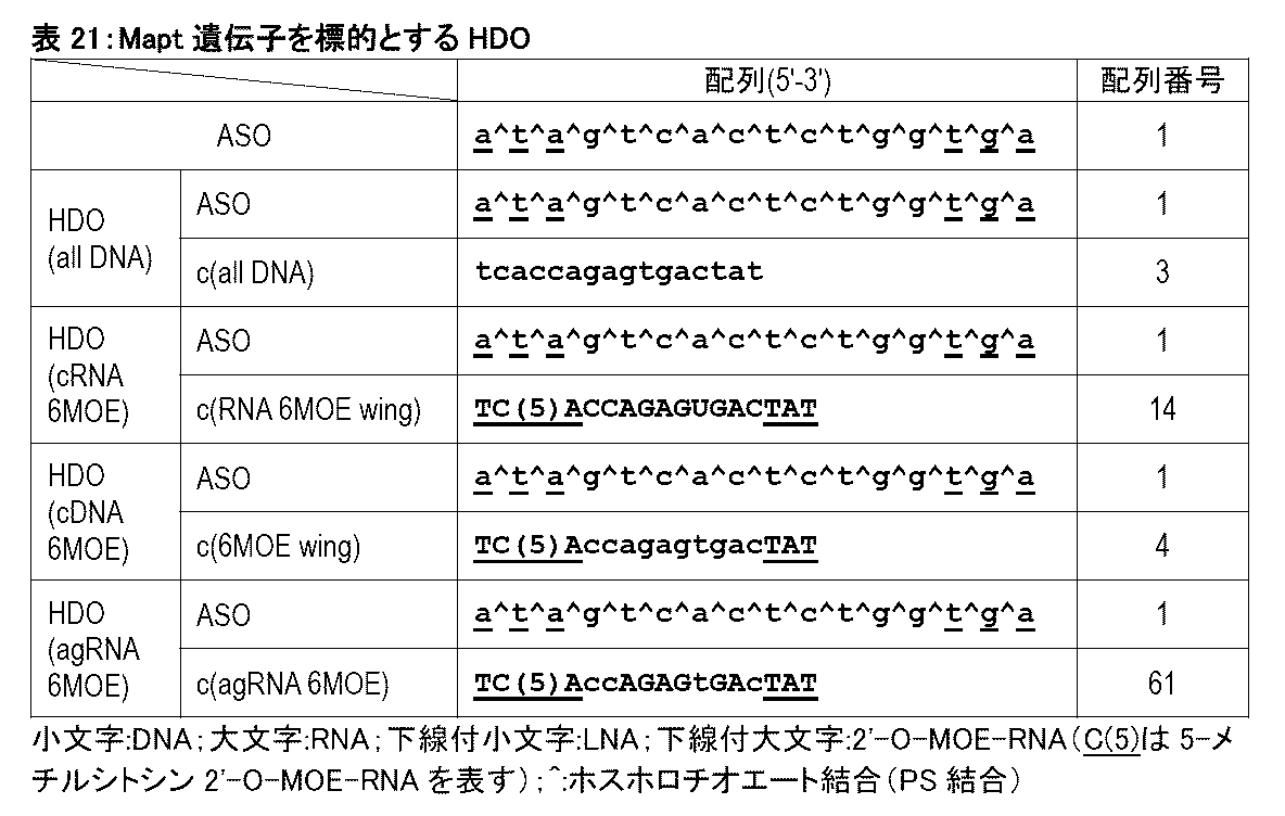
表21に記載されるASO、HDO(all DNA)、HDO(cRNA 6MOE)、HDO(cDNA 6MOE)、及びHDO(agRNA 6MOE)について、実施例1と同様の方法により核酸の調製、in vivo実験、並びに中枢神経毒性評価及び運動機能評価を行った。但し、本実施例ではマウス1匹当たりの核酸剤投与量は、28nmol/匹とした。
図56は、各種核酸剤を脳室内投与したマウスを対象として投与1日以内の急性の中枢神経毒性を評価した結果を示す。核酸剤の投与から30分後、1時間後、2時間後、3時間後、及び4時間後のいずれの時点においても、HDO(cRNA 6MOE)、HDO(cDNA 6MOE)、及びHDO(agRNA 6MOE)の投与群では、ASO及びHDO(all DNA)の投与群と比較して、急性期忍容性スコアが著しく減少した。さらに、HDO(agRNA 6MOE)投与群は、HDO(cRNA 6MOE)及びHDO(cDNA 6MOE)の投与群と比較して低い急性期忍容性スコアを示した。この結果から、第2核酸鎖のギャップ領域においてはアデニン塩基及びグアニン塩基を有するヌクレオシドがRNAヌクレオシドであり、シトシン塩基及びチミン塩基を有するヌクレオシドがDNAヌクレオシドであるHDOは、その他のHDOと比較しても中枢神経毒性を顕著に軽減する効果を達成し得ることが示された。
図57は、各種核酸剤を脳室内投与したマウスにおいて投与から1時間後に運動機能を評価した結果を示す。HDO(cRNA 6MOE)、HDO(cDNA 6MOE)、及びHDO(agRNA 6MOE)の投与群では、ASO及びHDO(all DNA)の投与群と比較して、総移動距離(図57A)及び最大移動速度(図57B)が改善した。更にHDO(agRNA 6MOE)投与群では、HDO(cRNA 6MOE)及びHDO(cDNA 6MOE)投与群と比較して運動機能が著しく改善した。この結果から、第2核酸鎖のギャップ領域においてはアデニン塩基及びグアニン塩基を有するヌクレオシドがRNAヌクレオシドであり、シトシン塩基及びチミン塩基を有するヌクレオシドがDNAヌクレオシドであるHDOは、その他のHDOと比較しても、運動機能に与える影響が極めて小さく、毒性が極めて低いことが示された。
図58は、各種核酸剤を脳室内投与したマウスを対象として投与1日後以降の遅発性の中枢神経毒性としてマウスの体重減少を評価した結果を示す。投与0、7、14、及び21日後に体重を測定したところ、投与7、14、及び21日のいずれの時点においてもASO及びHDO(all DNA)の投与群はPBS投与群と比較して体重減少を示し、HDO(cRNA 6MOE)、HDO(cDNA 6MOE)、及びHDO(agRNA 6MOE)の投与群では、ASO及びHDO(all DNA)の投与群と比較して、体重減少効果の軽減を示した。
図59は、各種核酸剤を脳室内投与したマウスを対象として投与1日後以降の遅発性の中枢神経毒性としてマウスの運動機能を評価した結果を示す。投与から7、14、21日後のいずれの時点においててもHDO(cRNA 6MOE)、HDO(cDNA 6MOE)、及びHDO(agRNA 6MOE)の投与群では、ASO及びHDO(all DNA)の投与群と比較して、最大移動速度に対する減少効果が軽減された。
図60は、各種核酸剤の脳室内投与から21日後において右前頭葉におけるMapt mRNA発現レベルを示す。HDO(agRNA 6MOE)投与群は、HDO(cRNA 6MOE)及びHDO(cDNA 6MOE)の投与群と比較して、Mapt mRNA発現レベルを顕著に抑制する効果を示した。この結果から、第2核酸鎖のギャップ領域においてアデニン塩基及びグアニン塩基を有するヌクレオシドがRNAヌクレオシドであり、シトシン塩基及びチミン塩基を有するヌクレオシドがDNAヌクレオシドであるHDOが、第2核酸鎖がDNAヌクレオシド又はRNAヌクレオシドからなるHDOと比較して優れた遺伝子発現抑制効果を達成し得ることが示された。
図61は、脳組織ホモジネート中における核酸剤の2本鎖解離効率評価のために実施例20と同様の方法を用いて、HDO(cRNA 6MOE)、HDO(cDNA 6MOE)、及びHDO(agRNA 6MOE)をマウス脳組織中で7日間インキュベートした後の電気泳動結果を示す。HDO(agRNA 6MOE)は、HDO(cRNA 6MOE)及びHDO(cDNA 6MOE)と比較して脳組織ホモジネート中で高い2本鎖解離効率を示した。この結果から、第2核酸鎖のギャップ領域においてアデニン塩基及びグアニン塩基を有するヌクレオシドがRNAヌクレオシドであり、シトシン塩基及びチミン塩基を有するヌクレオシドがDNAヌクレオシドであるHDOが、第2核酸鎖がDNAヌクレオシド又はRNAヌクレオシドからなるHDOと比較して脳組織内において2本鎖解離効率が極めて高く、上記の結果3において標的遺伝子の発現を顕著に抑制した結果を強く裏付けた。
(目的)
本実施例では、Mecp2遺伝子を標的としてスプライシングを制御することができる、全てのヌクレオシドがMOE修飾ヌクレオシドで構成されるASO(以下、「全MOE修飾型ASO」という)を第1核酸鎖として含むHDOを対象とする。当該HDOを脳室内投与する場合に見られる中枢神経毒性について、第2核酸鎖にMOE修飾を導入することによって毒性が軽減され得ることをin vivo実験により検証する。
本実施例で用いたASO、並びにHDOを構成する第1核酸鎖及び第2核酸鎖の塩基配列と化学修飾を表22に示す。
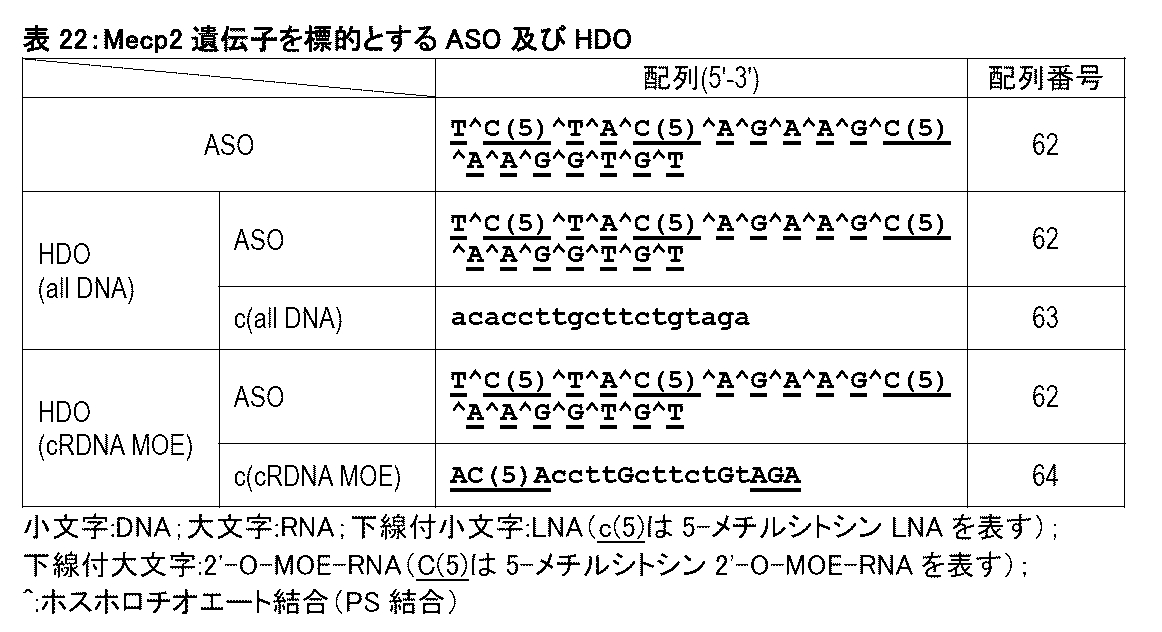
本実施例で用いたHDO(all DNA)及びHDO(cRDNA MOE)は、上記ASOを共通の第1核酸鎖として含み、第2核酸鎖は第1核酸鎖に相補的な配列を有する。具体的には、HDO(all DNA)の第2核酸鎖はDNAヌクレオシドがホスホジエステル結合で連結された構造を有する。HDO(cRDNA MOE)の第2核酸鎖は、5’末端の3個の2'-O-MOE-RNAヌクレオシド、3’末端の3個の2'-O-MOE-RNAヌクレオシド、及びそれらの間のギャップ領域から構成され、ギャップ領域においてはアデニン塩基及びグアニン塩基を有するヌクレオシドがRNAヌクレオシドであり、シトシン塩基及びチミン塩基を有するヌクレオシドがDNAヌクレオシドである。
表22に記載されるASO、HDO(all DNA)、及びHDO(cRDNA MOE)について、実施例1と同様の方法により核酸の調製、in vivo実験、並びに中枢神経毒性評価及び運動機能評価を行った。但し、本実施例ではマウス1匹当たりの核酸剤投与量は、28nmol/匹とした。
図62は、各種核酸剤を脳室内投与したマウスを対象として急性の中枢神経毒性を評価した結果を示す。核酸剤の投与から30分後、1時間後、2時間後、3時間後、及び4時間後のいずれの時点においても、HDO(all DNA)及びHDO(cRDNA MOE)の投与群では、ASO投与群と比較して、急性期忍容性スコアが著しく減少した。さらに、HDO(cRDNA MOE)投与群は、HDO(all DNA)投与群と比較して低い急性期忍容性スコアを示した。この結果から、第2核酸鎖のウイング領域が2'-O-MOE-RNAヌクレオシドで構成され、ギャップ領域においてはアデニン塩基及びグアニン塩基を有するヌクレオシドがRNAヌクレオシドであり、シトシン塩基及びチミン塩基を有するヌクレオシドがDNAヌクレオシドであるHDOは、その他のHDOと比較しても低い中枢神経毒性を達成し得ることが示された。
図63は、各種核酸剤を脳室内投与したマウスにおいて投与から1時間後に運動機能を評価した結果を示す。HDO(all DNA)及びHDO(cRDNA MOE)の投与群では、ASO投与群と比較して、総移動距離(図63A)及び最大移動速度(図63B)が改善した。更にHDO(cRDNA MOE)投与群は、HDO(all DNA)投与群と比較して総移動距離及び最大移動速度を改善した。この結果から、第2核酸鎖のウイング領域が2'-O-MOE-RNAヌクレオシドで構成され、ギャップ領域においてはアデニン塩基及びグアニン塩基を有するヌクレオシドがRNAヌクレオシドであり、シトシン塩基及びチミン塩基を有するヌクレオシドがDNAヌクレオシドであるHDOは、その他のHDOと比較しても、運動機能を抑制する効果が極めて小さく、毒性が極めて低いことが示された。
(目的)
Bace1遺伝子を標的とするASOからなる第1核酸鎖と第1核酸鎖に相補的な塩基配列を有する第2核酸鎖とをリンカーにより連結した一本鎖HDO(ssHDO)の神経細胞毒性について、in vitro実験により検証する。
(1)核酸の調製
本実施例で用いたHDO及びssHDOの塩基配列と化学修飾を表23に示す。

(1)で調製したHDOをヒト神経芽細胞腫由来細胞(Neuro2a細胞株)にリポフェクション法(lipofectamine2000)を用いて導入した。
HDO導入から48時間後にIsogenIキット(株式会社ジーンデザイン)を使用して、細胞からRNAを抽出した。cDNAは、Transcriptor Universal cDNA Master, DNase(ロシュ・ダイアグノスティックス社(Roche Diagnostics))を使用してプロトコルに従って合成した。次に、得られたcDNAを鋳型として定量RT-PCRを行うことにより、Bace1 mRNA及びActb mRNA (内部標準遺伝子)の発現量を測定した。定量RT-PCRは、TaqMan(ロシュ・アプライドサイエンス社(Roche Applied Science))により実施した。定量RT-PCRにおいて使用したプライマーとして、サーモ・フィッシャー・サイエンティフィック社(Thermo Fisher Sceientific、旧ライフ・テクノロジーズ社(Life Technologies Corp))によって設計及び製造された製品を用いた。増幅は、95℃15秒、60℃30秒、及び72℃1秒(1サイクル)を40サイクル繰り返すことにより行った。
Actb mRNA(内部標準遺伝子)発現量に対するBace1 mRNA発現量の比率を計算し、PBS投与群の値に対して標準化した値を相対的Bace1 mRNAレベルとした。
導入48時間後に神経細胞毒性の評価として、細胞上清中の乳酸脱水素酵素(LDH)活性を測定した。LDH活性測定は、Cytotoxicity LDH Assay Kit-WST(同仁化学研究所)を使用して、添付プロトコルに従って実施した。PBS投与群のLDH活性に対して標準化した値を相対的LDH放出レベルとした。
図64Bは、マウス神経芽細胞腫由来細胞(Neuro2a細胞株)において各種核酸剤による標的遺伝子抑制効果を評価した結果を示す。ssHDOの投与群ではHDO(all DNA)と同等の強い遺伝子抑制効果が得られた。
図64Aは、各種核酸剤を細胞内に導入した際の細胞毒性として、上清中のLDH活性を評価した結果を示す。ssHDOを5nM及び25nMで投与した細胞では、それぞれHDO(all DNA)を5nM及び25nMで投与した細胞と比較して、LDH活性の上昇が抑制されており、細胞毒性が著しく減少した。この結果から、2'-O-MOE-RNAヌクレオシドを含むssHDOでは細胞毒性が顕著に低下することが示された。
以上の結果から、ssHDOによって、神経細胞毒性の軽減と強力な遺伝子抑制効果の両方が達成されることが示された。
(目的)
MOE修飾を含むHDOをサルに髄腔内投与して、中枢神経毒性を評価する。
(1)核酸の調製
本実施例で用いたASO、並びにHDOを構成する第1核酸鎖及び第2核酸鎖の塩基配列と化学修飾を表24に示す。

雄のカニクイザル(3歳齢、2kg)2頭を対象として、ASO、HDO(all DNA)、HDO(DNA MOE)、HDO(RNA MOE)、及びHDO(DNARNA MOE)を投与した。投与方法としては、ASO又はHDO を0.94μmolを腰椎穿刺のうえ髄腔内投与した。
髄腔内投与から1、2、及び3時間後のサルについて、中枢神経毒性を評価するために図65に示す修正FOBスコア及び3分間のビデオ動画による自発的運動時間またはジャンプ回数の測定を、それぞれ盲検化された評価者による行動評価を行った。
図66は、各種核酸剤を髄腔内投与したサルを対象として急性の中枢神経毒性を評価した結果を示す。核酸剤の投与から1時間後、2時間後、及び3時間後のいずれの時点においても、HDO(all DNA)、HDO(DNA MOE)、HDO(RNA MOE)、及びHDO(DNARNA MOE)の投与群では、ASO投与群と比較して、修正FOBスコアが著しく減少した。更に、HDO(DNA MOE)及びHDO(DNARNA MOE)の投与群では、HDO(all DNA)投与群と比較して、修正FOBスコアが減少した。
また、3分間のビデオ動画による自発的運動時間及びジャンプ回数を測定した結果を図67に示す。HDO(all DNA)、HDO(DNA MOE)、HDO(RNA MOE)、及びHDO(DNARNA MOE)の投与群では、ASO投与群と比較して、自発的運動時間又はジャンプ回数が減少する効果が抑制された。更に、HDO(DNA MOE)及びHDO(DNARNA MOE)の投与群では、HDO(all DNA)投与群と比較して、自発的運動時間又はジャンプ回数が減少する効果が抑制された。
本明細書で引用した全ての刊行物、特許及び特許出願はそのまま引用により本明細書に組み入れられるものとする。
Claims (62)
- 第1核酸鎖と第2核酸鎖とを含む二本鎖核酸複合体であって、
前記第1核酸鎖は、標的遺伝子又はその転写産物の少なくとも一部にハイブリダイズすることができ、前記標的遺伝子又はその転写産物に対してアンチセンス効果を有し、
前記第2核酸鎖は、前記第1核酸鎖に相補的な塩基配列を含み、かつ2'-修飾ヌクレオシドを1個以上含む、前記二本鎖核酸複合体。 - 前記第1核酸鎖がギャップマーである、請求項1に記載の二本鎖核酸複合体。
- 前記第1核酸鎖において、ヌクレオシドの全てが修飾ヌクレオシドである、請求項1に記載の二本鎖核酸複合体。
- 前記第1核酸鎖において、ヌクレオシドの全てが2'-修飾ヌクレオシドである、請求項3に記載の二本鎖核酸複合体。
- 前記第1核酸鎖において、ヌクレオシドの全てが2'-O-メトキシエチル修飾ヌクレオシドである、請求項4に記載の二本鎖核酸複合体。
- 前記第2核酸鎖が、5’末端に位置する1個又は連続する2~10個の2'-修飾ヌクレオシド、及び/又は3’末端に位置する1個又は連続する2~10個の2'-修飾ヌクレオシドを含む、請求項1に記載の二本鎖核酸複合体。
- 前記第2核酸鎖が、5’末端及び3’末端以外の位置に、1~7個の2'-修飾ヌクレオシドを含む、請求項1に記載の二本鎖核酸複合体。
- 前記第1核酸鎖が、
(1)少なくとも4個の連続するデオキシリボヌクレオシドを含む中央領域、
(2)前記中央領域の5'末端側に配置された、非天然ヌクレオシドを含む5’ウイング領域、及び
(3)前記中央領域の3'末端側に配置された、非天然ヌクレオシドを含む3’ウイング領域を含む、請求項2に記載の二本鎖核酸複合体。 - 前記第2核酸鎖が、前記第1核酸鎖の5’ウイング領域及び/又は3’ウイング領域に相補的な塩基配列からなる領域に2'-修飾ヌクレオシドを含む、請求項8に記載の二本鎖核酸複合体。
- 前記第2核酸鎖において、前記第1核酸鎖の5’ウイング領域及び/又は3’ウイング領域に相補的な塩基配列からなる領域の全てのヌクレオシドが2'-修飾ヌクレオシドである、請求項9に記載の二本鎖核酸複合体。
- 前記第2核酸鎖における前記第1核酸鎖の中央領域に相補的な塩基配列からなる領域において、プリン塩基を含む全てのヌクレオシドがリボヌクレオシドである、請求項10に記載の二本鎖核酸複合体。
- 前記第2核酸鎖における前記第1核酸鎖の中央領域に相補的な塩基配列からなる領域において、ピリミジン塩基を含む全てのヌクレオシドがデオキシリボヌクレオシドである、請求項11に記載の二本鎖核酸複合体。
- 前記第2核酸鎖において、前記2'-修飾ヌクレオシドが、2'-O-メトキシエチル修飾ヌクレオシド又は2'-O-メチル修飾ヌクレオシドである、請求項12に記載の二本鎖核酸複合体。
- 前記第2核酸鎖において、前記第1核酸鎖の中央領域に相補的な塩基配列からなる領域の全てのヌクレオシドが、
(a)デオキシリボヌクレオシド、
(b)デオキシリボヌクレオシド及びリボヌクレオシド、
(c)デオキシリボヌクレオシド及び2'-修飾ヌクレオシド、
(d)リボヌクレオシド及び2'-修飾ヌクレオシド、又は
(e)デオキシリボヌクレオシド、リボヌクレオシド、及び2'-修飾ヌクレオシド
である、請求項8に記載の二本鎖核酸複合体。 - 前記第2核酸鎖において、
前記第1核酸鎖の5’ウイング領域及び3’ウイング領域に相補的な塩基配列からなる領域の全てのヌクレオシドが2'-修飾ヌクレオシドであり、かつ
前記第1核酸鎖の中央領域に相補的な塩基配列からなる領域の全てのヌクレオシドがデオキシリボヌクレオシドである、請求項14に記載の二本鎖核酸複合体。 - 前記第2核酸鎖が、前記第1核酸鎖の前記中央領域における少なくとも4個の連続するデオキシリボヌクレオシドに相補的な、少なくとも4個の連続するリボヌクレオシドを含む、請求項8に記載の二本鎖核酸複合体。
- (i)前記第1核酸鎖における少なくとも1個のグアノシンヌクレオシド、
(ii)前記グアノシンヌクレオシドの5'末端側に隣接するヌクレオシド、
(iii)前記グアノシンヌクレオシドの3'末端側に隣接するヌクレオシド、又は
(iv) (i)~(iii)の任意の組み合わせ
に相補的である前記第2核酸鎖におけるヌクレオシドが、2'-修飾ヌクレオシドである、請求項1に記載の二本鎖核酸複合体。 - (i)前記第1核酸鎖の5'ウイング領域における少なくとも1個のグアノシンヌクレオシド、
(ii)前記グアノシンヌクレオシドの5'末端側に隣接するヌクレオシド、
(iii)前記グアノシンヌクレオシドの3'末端側に隣接するヌクレオシド、又は
(iv) (i)~(iii)の任意の組み合わせ
に相補的である前記第2核酸鎖におけるヌクレオシドが、2'-修飾ヌクレオシドである、請求項1に記載の二本鎖核酸複合体。 - 前記第2核酸鎖において、アデニン塩基又はピリミジン塩基を含むヌクレオシドの少なくとも1個が2'-修飾ヌクレオシド及び/又はデオキシリボヌクレオシドである、請求項1に記載の二本鎖核酸複合体。
- 前記第2核酸鎖においてピリミジン塩基を含むヌクレオシドの少なくとも1個が2'-修飾ヌクレオシド及び/又はデオキシリボヌクレオシドである、請求項1に記載の二本鎖核酸複合体。
- 前記第2核酸鎖が、ピリミジン塩基を含む天然リボヌクレオシドを含まない、請求項1に記載の二本鎖核酸複合体。
- 前記第2核酸鎖においてピリミジン塩基を含むヌクレオシドの全てが2'-修飾ヌクレオシド及び/又はデオキシリボヌクレオシドである、請求項18に記載の二本鎖核酸複合体。
- 前記第2核酸鎖において、ヌクレオシドの20%以上が、2'-修飾ヌクレオシドである、請求項1に記載の二本鎖核酸複合体。
- 前記第2核酸鎖において、2'-修飾ヌクレオシド以外の全てのヌクレオシドがデオキシリボヌクレオシドである、請求項1に記載の二本鎖核酸複合体。
- 前記第2核酸鎖において、第1核酸鎖に相補的な塩基配列からなる領域の全てのヌクレオシドが2'-修飾ヌクレオシドである、請求項1に記載の二本鎖核酸複合体。
- 前記第2核酸鎖において、前記2'-修飾ヌクレオシドが、2'-O-メトキシエチル修飾ヌクレオシド及び/又は2'-O-メチル修飾ヌクレオシドである、請求項1に記載の二本鎖核酸複合体。
- 前記第2核酸鎖が、2'-O-メチル修飾ヌクレオシド、scpBNAヌクレオシド、AmNAヌクレオシド、又はBNA-NCを少なくとも1個含む、請求項1に記載の二本鎖核酸複合体。
- 前記第1核酸鎖がミックスマーである、請求項1に記載の二本鎖核酸複合体。
- 前記第2核酸鎖が、前記第1核酸鎖に対して非相補的な塩基配列からなるバルジ構造を少なくとも1つ含む、請求項1に記載の二本鎖核酸複合体。
- 前記第2核酸鎖が、前記第1核酸鎖に対して、非相補的塩基、及び/又は1塩基以上の、挿入配列及び/又は欠失を含む、請求項1に記載の二本鎖核酸複合体。
- 前記第2核酸鎖が、前記非相補的塩基を1~3個含む、請求項27に記載の二本鎖核酸複合体。
- 前記挿入配列が1~8塩基からなる、請求項30に記載の二本鎖核酸複合体。
- 前記欠失が連続する1~4塩基からなる、請求項30に記載の二本鎖核酸複合体。
- 前記第2核酸鎖の前記非相補的塩基がバルジ構造を形成する、又は
前記第2核酸鎖の前記欠失位置に、前記第1核酸鎖に対して非相補的な塩基配列からなるバルジ構造を含む、請求項30に記載の二本鎖核酸複合体。 - 前記バルジ構造が糖非修飾ヌクレオシドを含む、又は前記バルジ構造において全てのヌクレオシドが糖非修飾ヌクレオシドである、請求項29に記載の二本鎖核酸複合体。
- 前記バルジ構造が1~10塩基長である、請求項29に記載の二本鎖核酸複合体。
- 前記第2核酸鎖において、前記バルジ構造以外の全てのヌクレオシドが、2'-修飾ヌクレオシドである、請求項29に記載の二本鎖核酸複合体。
- 前記第2核酸鎖が少なくとも8塩基長である、請求項1に記載の二本鎖核酸複合体。
- 前記第2核酸鎖の塩基長が第1核酸鎖の塩基長よりも短い、請求項1に記載の二本鎖核酸複合体。
- 前記第2核酸鎖が、前記第1核酸鎖に相補的な塩基配列からなる領域の5'末端側及び/又は3'末端側に位置する少なくとも1つのオーバーハング領域を含む、請求項1に記載の二本鎖核酸複合体。
- 前記オーバーハング領域が1~20塩基長である、請求項40に記載の二本鎖核酸複合体。
- 前記オーバーハング領域が、少なくとも1個のデオキシリボヌクレオシド及び/又は非天然ヌクレオシドを含む、請求項40に記載の二本鎖核酸複合体。
- 前記第1核酸鎖と前記第2核酸鎖とがリンカーを介して結合している、請求項1に記載の二本鎖核酸複合体。
- 前記リンカーが、
前記第1核酸鎖の5'末端と前記第2核酸鎖の3'末端とを結合する、及び/又は
前記第1核酸鎖の3'末端と前記第2核酸鎖の5'末端とを結合する、請求項43に記載の二本鎖核酸複合体。 - 前記リンカーが切断性(cleavable)又は非切断(uncleavable)リンカーである、請求項43に記載の二本鎖核酸複合体。
- 前記リンカーが、天然ヌクレオシド及び/若しくは非天然ヌクレオシドを含む核酸、又はポリエチレングリコールからなる、請求項43に記載の二本鎖核酸複合体。
- 前記核酸からなるリンカーが2~10塩基長である、請求項46に記載の二本鎖核酸複合体。
- 前記第2核酸鎖がリガンドに結合している、請求項1に記載の二本鎖核酸複合体。
- 前記リガンドが、低分子、ペプチド、脂質、及び核酸アプタマーからなる群から選択されるいずれか1以上である、請求項48に記載の二本鎖核酸複合体。
- 前記脂質がコレステロール若しくはその類縁体、又はトコフェロール若しくはその類縁体である、請求項49に記載の二本鎖核酸複合体。
- 前記リガンドが前記第2核酸鎖の5’末端及び/又は3’末端に結合している、請求項45に記載の二本鎖核酸複合体。
- リガンドと結合していない、請求項1に記載の二本鎖核酸複合体。
- 前記第1核酸鎖及び/又は前記第2核酸鎖のヌクレオシド間結合の全部又は一部が修飾ヌクレオシド間結合である、請求項1に記載の二本鎖核酸複合体。
- 前記修飾ヌクレオシド間結合がホスホロチオエート結合である、請求項53に記載の二本鎖核酸複合体。
- 請求項1~54のいずれか一項に記載の二本鎖核酸複合体を有効成分として含む医薬組成物。
- 被検体の中枢神経系疾患を治療するための、請求項55に記載の医薬組成物。
- 脳室内投与又は髄腔内投与される、請求項55に記載の医薬組成物。
- 前記髄腔内投与が後頭窩穿刺又は腰椎穿刺である、請求項57に記載の医薬組成物。
- 前記二本鎖核酸複合体が0.1mg~200mg投与される、請求項57に記載の医薬組成物。
- 静脈内投与又は皮下投与される、請求項55に記載の医薬組成物。
- 前記二本鎖核酸複合体が0.1mg/kg~100mg/kg投与される、請求項60に記載の医薬組成物。
- 中枢神経毒性が低減されている、請求項55に記載の医薬組成物。
Priority Applications (3)
| Application Number | Priority Date | Filing Date | Title |
|---|---|---|---|
| US18/690,457 US20250179486A1 (en) | 2021-09-15 | 2022-09-15 | Heteronucleic acid including 2'-modified nucleoside |
| JP2023548500A JPWO2023042876A1 (ja) | 2021-09-15 | 2022-09-15 | |
| EP22870023.3A EP4403191A4 (en) | 2021-09-15 | 2022-09-15 | HETERONUCLEIC ACID COMPRISING A 2'-MODIFIED NUCLEOSIDE |
Applications Claiming Priority (2)
| Application Number | Priority Date | Filing Date | Title |
|---|---|---|---|
| JP2021-150310 | 2021-09-15 | ||
| JP2021150310 | 2021-09-15 |
Publications (1)
| Publication Number | Publication Date |
|---|---|
| WO2023042876A1 true WO2023042876A1 (ja) | 2023-03-23 |
Family
ID=85602964
Family Applications (1)
| Application Number | Title | Priority Date | Filing Date |
|---|---|---|---|
| PCT/JP2022/034531 Ceased WO2023042876A1 (ja) | 2021-09-15 | 2022-09-15 | 2'-修飾ヌクレオシドを含むヘテロ核酸 |
Country Status (4)
| Country | Link |
|---|---|
| US (1) | US20250179486A1 (ja) |
| EP (1) | EP4403191A4 (ja) |
| JP (1) | JPWO2023042876A1 (ja) |
| WO (1) | WO2023042876A1 (ja) |
Citations (11)
| Publication number | Priority date | Publication date | Assignee | Title |
|---|---|---|---|---|
| US5264423A (en) | 1987-03-25 | 1993-11-23 | The United States Of America As Represented By The Department Of Health And Human Services | Inhibitors for replication of retroviruses and for the expression of oncogene products |
| US5286717A (en) | 1987-03-25 | 1994-02-15 | The United States Of America As Represented By The Department Of Health And Human Services | Inhibitors for replication of retroviruses and for the expression of oncogene products |
| US5955599A (en) | 1995-06-01 | 1999-09-21 | Hybridon, Inc. | Process for making oligonucleotides containing o- and s- methylphosphotriester internucleoside linkages |
| WO2007143315A2 (en) | 2006-05-05 | 2007-12-13 | Isis Pharmaceutical, Inc. | Compounds and methods for modulating expression of pcsk9 |
| WO2008043753A2 (en) | 2006-10-09 | 2008-04-17 | Santaris Pharma A/S | Rna antagonist compounds for the modulation of pcsk9 |
| WO2008049085A1 (en) | 2006-10-18 | 2008-04-24 | Isis Pharmaceuticals, Inc. | Antisense compounds |
| WO2013089283A1 (en) | 2011-12-16 | 2013-06-20 | National University Corporation Tokyo Medical And Dental University | Chimeric double-stranded nucleic acid |
| WO2014192310A1 (en) | 2013-05-30 | 2014-12-04 | National University Corporation Tokyo Medical And Dental University | Double-stranded agents for delivering therapeutic oligonucleotides |
| WO2015168172A1 (en) | 2014-04-28 | 2015-11-05 | Isis Pharmaceuticals, Inc. | Linkage modified oligomeric compounds |
| WO2016081600A1 (en) | 2014-11-18 | 2016-05-26 | Zata Pharmaceuticals, Inc. | Phosphoramidite synthones for the synthesis of self-neutralizing oligonucleotide compounds |
| JP2021150310A (ja) | 2020-03-16 | 2021-09-27 | キオクシア株式会社 | 半導体記憶装置 |
Family Cites Families (2)
| Publication number | Priority date | Publication date | Assignee | Title |
|---|---|---|---|---|
| JP7333079B2 (ja) * | 2018-03-19 | 2023-08-24 | 国立大学法人 東京医科歯科大学 | 毒性が軽減した核酸 |
| WO2019182109A1 (ja) * | 2018-03-22 | 2019-09-26 | 国立大学法人東京医科歯科大学 | ヘテロ核酸のbbb通過脂質リガンド |
-
2022
- 2022-09-15 WO PCT/JP2022/034531 patent/WO2023042876A1/ja not_active Ceased
- 2022-09-15 EP EP22870023.3A patent/EP4403191A4/en active Pending
- 2022-09-15 JP JP2023548500A patent/JPWO2023042876A1/ja active Pending
- 2022-09-15 US US18/690,457 patent/US20250179486A1/en active Pending
Patent Citations (12)
| Publication number | Priority date | Publication date | Assignee | Title |
|---|---|---|---|---|
| US5264423A (en) | 1987-03-25 | 1993-11-23 | The United States Of America As Represented By The Department Of Health And Human Services | Inhibitors for replication of retroviruses and for the expression of oncogene products |
| US5286717A (en) | 1987-03-25 | 1994-02-15 | The United States Of America As Represented By The Department Of Health And Human Services | Inhibitors for replication of retroviruses and for the expression of oncogene products |
| US5955599A (en) | 1995-06-01 | 1999-09-21 | Hybridon, Inc. | Process for making oligonucleotides containing o- and s- methylphosphotriester internucleoside linkages |
| WO2007143315A2 (en) | 2006-05-05 | 2007-12-13 | Isis Pharmaceutical, Inc. | Compounds and methods for modulating expression of pcsk9 |
| WO2008043753A2 (en) | 2006-10-09 | 2008-04-17 | Santaris Pharma A/S | Rna antagonist compounds for the modulation of pcsk9 |
| WO2008049085A1 (en) | 2006-10-18 | 2008-04-24 | Isis Pharmaceuticals, Inc. | Antisense compounds |
| WO2013089283A1 (en) | 2011-12-16 | 2013-06-20 | National University Corporation Tokyo Medical And Dental University | Chimeric double-stranded nucleic acid |
| JP2015502134A (ja) * | 2011-12-16 | 2015-01-22 | 国立大学法人 東京医科歯科大学 | キメラ2重鎖核酸 |
| WO2014192310A1 (en) | 2013-05-30 | 2014-12-04 | National University Corporation Tokyo Medical And Dental University | Double-stranded agents for delivering therapeutic oligonucleotides |
| WO2015168172A1 (en) | 2014-04-28 | 2015-11-05 | Isis Pharmaceuticals, Inc. | Linkage modified oligomeric compounds |
| WO2016081600A1 (en) | 2014-11-18 | 2016-05-26 | Zata Pharmaceuticals, Inc. | Phosphoramidite synthones for the synthesis of self-neutralizing oligonucleotide compounds |
| JP2021150310A (ja) | 2020-03-16 | 2021-09-27 | キオクシア株式会社 | 半導体記憶装置 |
Non-Patent Citations (16)
| Title |
|---|
| "GenBank", Database accession no. NC 000086.7 |
| ALEXANDER A. LOMZOV ET AL., BIOCHEM BIOPHYS RES COMMUN., vol. 513, no. 4, 2019, pages 807 - 811 |
| ANASTASIA KHVOROVA ET AL., NAT. BIOTECHNOL., vol. 35, no. 3, 2017, pages 238 - 248 |
| ASAMI Y ET AL.: "Drug delivery system of therapeutic oligonucleotides", DRUG DISCOVERIES & THERAPEUTICS, vol. 10, no. 5, 2016, pages 256 - 262, XP055634473, DOI: 10.5582/ddt.2016.01065 |
| ASAMI YUTARO, NAGATA TETSUYA, YOSHIOKA KOTARO, KUNIEDA TAIKI, YOSHIDA-TANAKA KIE, BENNETT C. FRANK, SETH PUNIT P., YOKOTA TAKANORI: "Efficient Gene Suppression by DNA/DNA Double-Stranded Oligonucleotide In Vivo", MOLECULAR THERAPY, ELSEVIER INC., US, vol. 29, no. 2, 1 February 2021 (2021-02-01), US , pages 838 - 847, XP055834158, ISSN: 1525-0016, DOI: 10.1016/j.ymthe.2020.10.017 * |
| HARRISON R.K.: "Phase II and phase III failures: 2013-2015", NATURE REVIEWS DRUG DISCOVERY, vol. 15, 2016, pages 817 - 818 |
| NAOKI IWAMOTO ET AL., ANGEW. CHEM. INT. ED. ENGL., vol. 48, no. 3, 2009, pages 496 - 9 |
| NAOKI IWAMOTO ET AL., NAT. BIOTECHNOL, vol. 35, no. 9, 2017, pages 845 - 851 |
| NATSUHISA OKA ET AL., J. AM. CHEM. SOC., vol. 125, 2003, pages 8307 - 8317 |
| NATSUHISA OKA ET AL., J. AM. CHEM. SOC., vol. 130, 2008, pages 16031 - 16037 |
| NISHIKAWA MAKIYA, YUKITAKE YOSHIOKA, MAKOTO NAGAOKA, KOSUKE KUSAMORI: "Development of Oligonucleotide Therapeutics: Tissue Distribution and Drug Delivery System", DDS. DRUG DELIVERY SYSTEM, vol. 36, no. 1, 25 January 2021 (2021-01-25), pages 40 - 50, XP093006747, DOI: 10.2745/dds.36.40 * |
| NISHINA K: "DNA/RNA heteroduplex oligonucleotide for highly efficient gene silencing", NATURE COMMUNICATION, vol. 6, 2015, pages 7969, XP055834307, DOI: 10.1038/ncomms8969 |
| See also references of EP4403191A4 |
| TAKAO INOUE, KIYOMI SASAKI, TOKUYUKI YOSHIDA: "Current status of development of oligonucleotide therapeutics", DRUG DELIVERY SYSTEM., NIHON D D S GAKKAI, JAPAN, vol. 34, no. 2, 25 March 2019 (2019-03-25), Japan , pages 86 - 98, XP009539425, ISSN: 0913-5006, DOI: 10.2745/dds.34.86 * |
| YOHEI NUKAGA ET AL., J. ORG. CHEM., vol. 77, 2012, pages 7913 - 7922 |
| YOHEI NUKAGA ET AL., J. ORG. CHEM., vol. 81, 2016, pages 2753 - 2762 |
Also Published As
| Publication number | Publication date |
|---|---|
| US20250179486A1 (en) | 2025-06-05 |
| JPWO2023042876A1 (ja) | 2023-03-23 |
| EP4403191A1 (en) | 2024-07-24 |
| EP4403191A4 (en) | 2026-03-18 |
Similar Documents
| Publication | Publication Date | Title |
|---|---|---|
| JP7554488B2 (ja) | 血液脳関門通過型ヘテロ2本鎖核酸 | |
| JP7687682B2 (ja) | 筋疾患治療用医薬組成物 | |
| EP4389891A1 (en) | Modified heteronucleic acid containing morpholino nucleic acid | |
| US11260134B2 (en) | Double-stranded nucleic acid complex having overhang | |
| JP7394392B2 (ja) | 核酸複合体 | |
| JP7262816B2 (ja) | ヘテロ核酸のbbb通過脂質リガンド | |
| JP7333079B2 (ja) | 毒性が軽減した核酸 | |
| JP2025156522A (ja) | 修飾ヘテロ核酸 | |
| JP7737667B2 (ja) | 核酸複合体 | |
| EP4123023A1 (en) | Heteronucleic acid containing morpholino nucleic acid | |
| WO2021157730A1 (ja) | 核酸医薬とその使用 | |
| EP4596694A1 (en) | Nucleic acid molecule containing 5'-cyclopropylene modification | |
| WO2024106545A1 (ja) | 髄腔内投与のためのヘテロ核酸 | |
| JP6934695B2 (ja) | 核酸医薬とその使用 | |
| EP4403191A1 (en) | Heteronucleic acid including 2'-modified nucleoside | |
| JP7863880B2 (ja) | モルホリノ核酸を含むヘテロ核酸 | |
| WO2022250050A1 (ja) | scpBNA又はAmNAを含むヘテロ核酸 | |
| WO2024005156A1 (ja) | 核酸医薬の毒性軽減剤 | |
| WO2024071362A1 (ja) | 神経系送達促進剤 | |
| WO2023127848A1 (ja) | Ebv関連疾患を標的とする核酸医薬 | |
| WO2024228398A1 (ja) | グアニン四重鎖構造を誘導するための核酸剤 | |
| WO2023080225A1 (ja) | 脊髄小脳失調症治療用医薬組成物 |
Legal Events
| Date | Code | Title | Description |
|---|---|---|---|
| 121 | Ep: the epo has been informed by wipo that ep was designated in this application |
Ref document number: 22870023 Country of ref document: EP Kind code of ref document: A1 |
|
| DPE2 | Request for preliminary examination filed before expiration of 19th month from priority date (pct application filed from 20040101) | ||
| WWE | Wipo information: entry into national phase |
Ref document number: 2023548500 Country of ref document: JP |
|
| WWE | Wipo information: entry into national phase |
Ref document number: 2022870023 Country of ref document: EP |
|
| NENP | Non-entry into the national phase |
Ref country code: DE |
|
| ENP | Entry into the national phase |
Ref document number: 2022870023 Country of ref document: EP Effective date: 20240415 |
|
| WWP | Wipo information: published in national office |
Ref document number: 18690457 Country of ref document: US |