WO2023096065A1 - Procédé de diagnostic de sensibilité au rayonnement - Google Patents
Procédé de diagnostic de sensibilité au rayonnement Download PDFInfo
- Publication number
- WO2023096065A1 WO2023096065A1 PCT/KR2022/010503 KR2022010503W WO2023096065A1 WO 2023096065 A1 WO2023096065 A1 WO 2023096065A1 KR 2022010503 W KR2022010503 W KR 2022010503W WO 2023096065 A1 WO2023096065 A1 WO 2023096065A1
- Authority
- WO
- WIPO (PCT)
- Prior art keywords
- radiation
- sensitivity
- cancer
- cell
- diagnosing
- Prior art date
- Legal status (The legal status is an assumption and is not a legal conclusion. Google has not performed a legal analysis and makes no representation as to the accuracy of the status listed.)
- Ceased
Links
Images









Classifications
-
- C—CHEMISTRY; METALLURGY
- C12—BIOCHEMISTRY; BEER; SPIRITS; WINE; VINEGAR; MICROBIOLOGY; ENZYMOLOGY; MUTATION OR GENETIC ENGINEERING
- C12Q—MEASURING OR TESTING PROCESSES INVOLVING ENZYMES, NUCLEIC ACIDS OR MICROORGANISMS; COMPOSITIONS OR TEST PAPERS THEREFOR; PROCESSES OF PREPARING SUCH COMPOSITIONS; CONDITION-RESPONSIVE CONTROL IN MICROBIOLOGICAL OR ENZYMOLOGICAL PROCESSES
- C12Q1/00—Measuring or testing processes involving enzymes, nucleic acids or microorganisms; Compositions therefor; Processes of preparing such compositions
- C12Q1/02—Measuring or testing processes involving enzymes, nucleic acids or microorganisms; Compositions therefor; Processes of preparing such compositions involving viable microorganisms
-
- C—CHEMISTRY; METALLURGY
- C12—BIOCHEMISTRY; BEER; SPIRITS; WINE; VINEGAR; MICROBIOLOGY; ENZYMOLOGY; MUTATION OR GENETIC ENGINEERING
- C12N—MICROORGANISMS OR ENZYMES; COMPOSITIONS THEREOF; PROPAGATING, PRESERVING, OR MAINTAINING MICROORGANISMS; MUTATION OR GENETIC ENGINEERING; CULTURE MEDIA
- C12N5/00—Undifferentiated human, animal or plant cells, e.g. cell lines; Tissues; Cultivation or maintenance thereof; Culture media therefor
- C12N5/06—Animal cells or tissues; Human cells or tissues
- C12N5/0602—Vertebrate cells
- C12N5/0693—Tumour cells; Cancer cells
-
- C—CHEMISTRY; METALLURGY
- C12—BIOCHEMISTRY; BEER; SPIRITS; WINE; VINEGAR; MICROBIOLOGY; ENZYMOLOGY; MUTATION OR GENETIC ENGINEERING
- C12N—MICROORGANISMS OR ENZYMES; COMPOSITIONS THEREOF; PROPAGATING, PRESERVING, OR MAINTAINING MICROORGANISMS; MUTATION OR GENETIC ENGINEERING; CULTURE MEDIA
- C12N13/00—Treatment of microorganisms or enzymes with electrical or wave energy, e.g. magnetism, sonic waves
-
- G—PHYSICS
- G01—MEASURING; TESTING
- G01N—INVESTIGATING OR ANALYSING MATERIALS BY DETERMINING THEIR CHEMICAL OR PHYSICAL PROPERTIES
- G01N33/00—Investigating or analysing materials by specific methods not covered by groups G01N1/00 - G01N31/00
- G01N33/48—Biological material, e.g. blood, urine; Haemocytometers
- G01N33/50—Chemical analysis of biological material, e.g. blood, urine; Testing involving biospecific ligand binding methods; Immunological testing
-
- G—PHYSICS
- G01—MEASURING; TESTING
- G01N—INVESTIGATING OR ANALYSING MATERIALS BY DETERMINING THEIR CHEMICAL OR PHYSICAL PROPERTIES
- G01N33/00—Investigating or analysing materials by specific methods not covered by groups G01N1/00 - G01N31/00
- G01N33/48—Biological material, e.g. blood, urine; Haemocytometers
- G01N33/50—Chemical analysis of biological material, e.g. blood, urine; Testing involving biospecific ligand binding methods; Immunological testing
- G01N33/5005—Chemical analysis of biological material, e.g. blood, urine; Testing involving biospecific ligand binding methods; Immunological testing involving human or animal cells
- G01N33/5008—Chemical analysis of biological material, e.g. blood, urine; Testing involving biospecific ligand binding methods; Immunological testing involving human or animal cells for testing or evaluating the effect of chemical or biological compounds, e.g. drugs, cosmetics
- G01N33/5011—Chemical analysis of biological material, e.g. blood, urine; Testing involving biospecific ligand binding methods; Immunological testing involving human or animal cells for testing or evaluating the effect of chemical or biological compounds, e.g. drugs, cosmetics for testing antineoplastic activity
-
- C—CHEMISTRY; METALLURGY
- C12—BIOCHEMISTRY; BEER; SPIRITS; WINE; VINEGAR; MICROBIOLOGY; ENZYMOLOGY; MUTATION OR GENETIC ENGINEERING
- C12N—MICROORGANISMS OR ENZYMES; COMPOSITIONS THEREOF; PROPAGATING, PRESERVING, OR MAINTAINING MICROORGANISMS; MUTATION OR GENETIC ENGINEERING; CULTURE MEDIA
- C12N2513/00—3D culture
-
- G—PHYSICS
- G01—MEASURING; TESTING
- G01N—INVESTIGATING OR ANALYSING MATERIALS BY DETERMINING THEIR CHEMICAL OR PHYSICAL PROPERTIES
- G01N2800/00—Detection or diagnosis of diseases
- G01N2800/40—Disorders due to exposure to physical agents, e.g. heat disorders, motion sickness, radiation injuries, altitude sickness, decompression illness
-
- G—PHYSICS
- G01—MEASURING; TESTING
- G01N—INVESTIGATING OR ANALYSING MATERIALS BY DETERMINING THEIR CHEMICAL OR PHYSICAL PROPERTIES
- G01N2800/00—Detection or diagnosis of diseases
- G01N2800/52—Predicting or monitoring the response to treatment, e.g. for selection of therapy based on assay results in personalised medicine; Prognosis
Definitions
- the present invention relates to a method for diagnosing radiation sensitivity, and more particularly, to diagnose radiation sensitivity by culturing cancer tissue from a patient's lesion tissue in an environment similar to a living body outside the body, and irradiating the cultured cancer cells with radiation of various radiation doses.
- the present invention relates to a radiation sensitivity diagnosis method for predicting and diagnosing radiation sensitivity and prognosis of a patient before radiation treatment using a radiation sensitivity diagnosis kit.
- cancer is treated by various drug therapies including chemotherapy, radiation therapy and antibody-based drugs depending on the type treatment is performed by Among these, radiation therapy is performed for carcinomas where surgical operation is impossible or anticancer drugs are ineffective, and rather than the purpose of completely removing the ultimate cause, cancer cells, by removing cancer cells while minimizing damage to normal tissues, It is implemented for the purpose of maintaining high quality and prolonging life.
- drug therapies including chemotherapy, radiation therapy and antibody-based drugs depending on the type treatment is performed by Among these, radiation therapy is performed for carcinomas where surgical operation is impossible or anticancer drugs are ineffective, and rather than the purpose of completely removing the ultimate cause, cancer cells, by removing cancer cells while minimizing damage to normal tissues, It is implemented for the purpose of maintaining high quality and prolonging life.
- radiation therapy is performed for the purpose of conservative treatment in which a planned radiation dose is accurately administered to the target cancer, treatment of symptoms, and improvement of the disease is expected.
- a conservative treatment strategy using radiation therapy is recognized as an effective treatment method for head and neck cancer, digestive cancer, urinary cancer, esophageal cancer, brain tumor, lung cancer, liver cancer, rectal cancer, anal cancer, and cervical cancer.
- the present invention considers radiation response factors and cell growth factors at the same time, so that among various factors that have been previously studied, the most effective factors for diagnosing radiation sensitivity can accurately determine the radiation sensitivity of an individual patient. It is an object of the present invention to provide a method for diagnosing radiation sensitivity.
- an object of the present invention is to provide a diagnosis method that can effectively reduce the cost and time required for radiation sensitivity diagnosis by irradiating different radiation doses to each of a plurality of cancer organoids disposed in a radiation sensitivity diagnosis kit. .
- the present invention aims to provide information necessary for determining whether or not to perform radiation treatment for the purpose of conservative treatment by precisely predicting the radiation treatment effect by determining radiation sensitivity using cell experiment-based data derived from individual patients. to be
- the radiation sensitivity diagnosis method is a radiation sensitivity diagnosis method using a radiation sensitivity diagnosis kit including a radiation irradiation device and a control unit, (a) Dispensing one or more cancer cells isolated from the subject on the CRS plate of the diagnostic kit; (b) forming one or more cancer organoids by filling the CRS plate with culture medium and culturing the one or more cancer cells; (c) irradiating the CRS plate with radiation passing through a radiation dose control unit from a radiation generating unit of the radiation irradiation device; (d) measuring the cell activity of the one or more cancer organoids; (e) a data acquisition step of obtaining cell experiment-based data based on the measured cell activity; and (f) diagnosing, by the control unit, radiation sensitivity of the subject based on the cell experiment-based data.
- each of the plurality of cancer organoids placed in the diagnostic kit can be irradiated with different amounts of total radiation dose for the same period of time, effectively reducing the cost and time required for radiation sensitivity diagnosis, and based on longitudinal experiments.
- the radiation treatment effect can be predicted, and thus, information necessary for determining whether or not to perform radiotherapy, which is a conservative treatment strategy, can be provided.
- the present invention it is possible to predict the effect of radiation treatment in advance by effectively diagnosing the radiation sensitivity of cancer cells of a patient, and thus, head and neck cancer, lung cancer, esophageal cancer, rectal cancer, anal cancer, and uterus suitable for conservative treatment strategies. Whether or not to perform radiation therapy for cancer such as cervical cancer can be accurately and effectively determined.
- a radiation sensitivity diagnosis method capable of accurately determining the radiation sensitivity of an individual patient with the most effective factors for diagnosis of radiation sensitivity among various factors that have been previously studied by simultaneously considering radiation response factors and cell growth factors. is provided.
- a small number of samples and a limited amount of time can be obtained using a characteristic sensitivity diagnostic kit configured so that the total amount of radiation irradiated to each of the plurality of cancer organoids is different from each other due to a change in the relative position of the plate portion and the radiation shielding film.
- Data required for radiation sensitivity diagnosis can be obtained by overcoming limitations.
- FIG. 1 is a flowchart of a method for diagnosing radiation sensitivity according to an embodiment of the present invention.
- FIG. 2 is a diagram for explaining a radiation sensitivity diagnosis kit according to an embodiment of the present invention.
- 3A and 3B are views for explaining a method for diagnosing radiation sensitivity according to an embodiment of the present invention.
- FIGS. 4 to 7 are views for explaining a radiation dose control unit of a radiation sensitivity diagnostic kit according to various embodiments of the present invention.
- FIG. 8 is a view for explaining the measurement of cell activity of irradiated cancer organoids according to an embodiment of the present invention.
- 9 and 10 are views for explaining a radiation response factor according to an embodiment of the present invention.
- FIG. 11 is a diagram for explaining a radiation sensitivity diagnosis module according to an embodiment of the present invention.
- FIGS. 12 and 13 are views for explaining a method for diagnosing radiation sensitivity based on a radiation response factor and a cell growth factor according to an embodiment of the present invention.
- 14a and 14b are views for explaining a radiation susceptibility index according to an embodiment of the present invention.
- 15a and 15b show diagnostic results of the radiation sensitivity diagnostic kit according to an embodiment of the present invention.
- 16 exemplarily illustrates a radiation sensitive diagnosis system according to an embodiment of the present invention.
- first, second, etc. are used to describe various components, these components are not limited by these terms. These terms are only used to distinguish one component from another. Therefore, the first component mentioned below may also be the second component within the technical spirit of the present invention.
- ordinal expression expressions such as first and second are intended to describe objects that are equivalent and independent of each other, and in the order of main / sub or master / slave It should be understood as having no meaning.
- An object may mean any subject whose radiation sensitivity is to be diagnosed.
- a subject may have ovarian cancer, breast cancer, squamous cell carcinoma, uterine cancer, cervical cancer, prostate cancer, head and neck cancer, pancreatic cancer, brain tumor, liver cancer, skin cancer, esophageal cancer, testicular cancer, kidney cancer, colorectal cancer, rectal cancer, stomach cancer,
- the subject may be an individual having at least one carcinoma among kidney cancer, bladder cancer, bile duct cancer, and gallbladder cancer.
- the subject may be a subject having head and neck cancer, but is not limited thereto.
- Radiation sensitivity may mean sensitivity to radiation and may be a criterion for diagnosing whether or not a treatment response is displayed according to radiation. Radiation sensitivity may be used interchangeably with radiation treatment responsiveness, radiation responsiveness, and the like. Accordingly, according to the diagnosis of radiation sensitivity, the individual may be determined as a treatment positive individual or a negative treatment response individual for radiation treatment. In addition, an individual having a relatively high sensitivity to radiation may be determined to have a positive treatment response, and an individual having a relatively low sensitivity to radiation may be determined to have a negative treatment response.
- cancer organoid-based radiosensitivity diagnosis measures the radiation response of organoids by dispensing cancer patient's tissue cells and extracellular matrix into a culture plate and irradiating various radiation doses, and the result is a decisive aid in the patient's radiation treatment decision. can be used as a resource.
- Cancer cells in the human body constitute a microenvironment surrounded by an extracellular matrix, and a model that predicts radiation treatment needs a living body model that can mimic this. Therefore, according to one embodiment of the present invention, after forming an extracellular matrix on a pillar chip, culturing cancer cells therein to implement a microenvironment, and measuring the activity of cancer patient-derived cells by transmitting radiation thereto, the sensitivity to radiation is measured. can measure In this embodiment, experiments were conducted on head and neck cancer, but as mentioned above, cancers suitable for conservative treatment strategies, such as digestive cancer, urinary cancer, esophageal cancer, brain tumor, lung cancer, liver cancer, rectal cancer, anal cancer, and cervical cancer All patient-derived cells can be applied to the present invention.
- the diagnostic kit for example, cancer patient-derived cells such as head and neck cancer, according to various radiation doses on a single plate using a radiation dose control unit capable of selectively shielding/controlling radiation dose. activity can be measured.
- the present invention developed and used a radiation-sensitive diagnostic kit capable of irradiating cells with various radiation doses with a single radiation dose in a manner in which radiation dose can be freely adjusted within one diagnostic kit, and used for diagnosis.
- the radiation sensitivity diagnostic kit 100 according to an embodiment of the present invention will be described first with reference to FIGS. 2 to 7 .
- the radiation sensitivity diagnosis kit 100 includes a radiation irradiation device 140 and a control unit 150, and the control unit 150 includes a storage unit 110, a processor 120, a communication unit 130 and A radiation diagnosis module 135 may be included.
- the radiation sensitivity diagnosis kit 100 may further include an imaging device 147 for confirming an image of a cancer organoid, for example, a fluorescence microscope.
- the radiation irradiation device 140 may include, for example, a radiation generating unit 145, a radiation dose control unit 143, and a CRS plate (141, Cell Ratio-Sensitivity Plate).
- the radiation generating unit 145 is a device that generates radiation by accelerating charged particles, and the generated radiation passes through the radiation dose control unit 143 to one or more cancer organoids 1041 in the CRS plate 141. At least a portion may be irradiated with radiation doses different from each other.
- the radiation dose control unit 143 transmits radiation through a slit 243-1 that moves relative to the position of the CRS plate 141.
- a slit-type radiation dose modulator 243 that controls the dose a shield-type radiation dose modulator 443 that adjusts the irradiation time for each cancer organoid by moving the shielding film in a linear motion, and the pattern, color, and material of the radiation-transmitting shielding plate ( metal), a density control-type radiation shielding film 343 configured to selectively adjust the radiation dose according to the radiation transmittance controlled by the density of the material, or a laminated radiation shielding film in which different numbers of radiation protection shielding plates are stacked according to the radiation dose to be irradiated (543).
- the CRS plate 141 includes pillars capable of dispensing cells, is capable of culturing cells, and is a plate necessary for confirming radiation reactivity by a staining reagent after culturing.
- the CRS plate 141 may have various shapes as can be seen in FIGS. 3A and 3B. In the case of FIG.
- the CRS plate 141 is a form including wells that are accommodating spaces for the culture medium 1042, and a plurality of pillars protruding in the form of columns in which cancer organoids generated by cancer cells, which are culture objects, are placed ( 141-1) may protrude from the lower surface of the accommodation space in an upper direction (direction toward the radiation generating unit 145).
- the plurality of pillar portions 141-1 protrude from the bottom portion 141-2 of the CRS plate 141 in a downward direction (in a direction opposite to the direction toward the radiation generating unit 145). It is accommodated in the well 141-3 and configured to be immersed in the culture medium 1042.
- the present invention is not limited thereto, and any structure is possible as long as the cancer cells dispensed on the CRS plate 141 can be immersed in the culture medium.
- the radiation dose control unit 143 may include a slit-type radiation dose modulator 243 having a radiation protection plate 243-2 having a slit 243-1 formed thereon.
- radiation emitted from the radiation generating unit 145 is irradiated to the cancer organoids of the CRS plate 141 through the slit 243-1.
- the radiation protection plate 243-2 serves as a radiation shield, and the slit 243-1 may be an opening opened in the radiation protection plate 243-2 in an elongated form. Meanwhile, the relative position of the CRS plate 141 and the slit 243-1 may be moved in a direction perpendicular to the elongated direction of the slit 243-1.
- the CRS plate 141 moves in the embodiment of FIG. 4 , it is also possible to configure the slit 243-1 to move.
- the relative position of the slit 243-1 and the CRS plate 141 may be set in advance, and thus the overlapping area of the slit 243-1 and the CRS plate 141 may be changed for a preset time.
- the CRS plate 141 may move in a second direction (D2, a direction perpendicular to the elongated direction of the slit 243-1) for a preset period of time.
- the preset time may include a first time interval, a second time interval, and a third time interval, and during the first time interval among the preset times, the CRS plate 141 is positioned as shown in FIG.
- the organoid 1041c and the slit 243-1 may overlap. Accordingly, radiation may be irradiated only to the third organoid 1041c.
- the CRS plate moves in the second direction (D2) from the position of FIG. 4 (a) and is positioned as shown in FIG. may overlap.
- radiation may be irradiated only to the second organoid 1041b.
- the CRS plate 141 moves again in the second direction (D2) from the position of FIG. 4 (b) and is positioned as shown in FIG. (243-1) can be nested. Accordingly, radiation may be irradiated only to the third organoid 1041c.
- the amount of radiation irradiated to each of the plurality of cancer organoids for a preset time may be proportional to the time during which each of the plurality of cancer organoids overlaps the slit 243-1. That is, the total radiation dose irradiated to the first organoid 1041a may be proportional to the first time period, and the total radiation dose irradiated to the second organoid 1041b may be proportional to the second time period. The total radiation dose irradiated to the third organoid 1041c may be proportional to the third time period.
- the radiation sensitivity diagnosis kit 100 moves the CRS plate 141 in the second direction (D2) to adjust the overlapping time between each cancer organoid and the slit 243-1, so that each cancer organoid
- the total radiation dose for a preset period of time to be irradiated can be adjusted.
- the radiation generating unit 145 and the slit 243-1 are fixed, and the radiation irradiation area and the radiation amount are constant, so that the radiation dose can be accurately adjusted by simply adjusting the time.
- radiation irradiation time may take a little longer.
- the radiation irradiation device 140 includes a radiation generating unit 145, a radiation dose control unit 143 including a density controllable radiation shielding film 343, a first plate 341a, and a second
- the CRS plate 141 including the plate 341b may be included.
- the density-controlled radiation shielding film 343 includes a radiation transmission shielding plate 343-1 and a radiation protection plate 343-2 accommodating the radiation transmission shielding plate 343-1.
- the amount of radiation applied to the cancer organoid 1041 may be selectively adjusted by controlling the radiation transmittance by changing the pattern, color, material (metal), density of the material, etc. of the transmission shielding plate 343-1.
- the radiation transmission shielding plate 343-1 is manufactured to fit into an opening of a specific shape created in the radiation protection plate 343-2. In this case, the radiation transmission shielding plate 343-1 of the density controllable radiation shielding film 343 may be detachable from the radiation protection plate 343-2.
- the radiation protection plate 343-2 may be configured to use a film that completely shields radiation or to only physically support the radiation transmission shield plate 343-1 by using a film that transmits radiation. When the radiation protection plate 343-2 completely shields radiation, a hole is drilled at a portion supporting the radiation transmission shielding plate 343-1 to selectively transmit radiation.
- the amount (degree) of radiation passing through the density controllable radiation shielding film 343 may vary according to the type and radiation transmittance of the radiation transmitting shielding plate 343-1. That is, the amount of radiation reaching the cancer organoid through the density controllable radiation shielding film 343 can be adjusted for a predetermined time depending on which type of radiation transmission shielding plate 343-1 is attached to the radiation dose controller. there is. Accordingly, the total amount of radiation reaching at least a portion of the plurality of cancer organoids 1041 disposed on the CRS plate 141 during a preset time is the amount of the radiation transmission shielding plate 343-1 overlapping with the cancer organoids. Depending on the type (classified according to the radiation transmittance), it may be adjusted differently from each other.
- the CRS plate 141 is, for example, a unit in which cancer organoids (or cancer cells) are disposed under the radiation dose control unit 143, for example, the first plate 341a in which cancer organoids are disposed. , and a second plate 341b accommodating a culture medium 1042 for growing cancer organoids.
- the first plate 341a may have a pillar structure
- the second plate 341b may be a well having a diameter wider than that of the first plate 341a to accommodate the first plate 341a.
- the radiation dose control unit 143 may include a shielding film movable radiation dose modulator 443 .
- the CRS plate 141 is disposed below the radiation protection plate 443-2 of the shielding film movable radiation dose modulator 443, for example.
- a radiation protection plate 443-2 may be disposed between the CRS plate 141 and the radiation generating unit 145 in a state where the radiation CRS plate 141 is combined with the rest of the shielding film movable radiation dose modulator 443. .
- the radiation protection plate 443-2 of the shielding film movable radiation dose modulator 443 is disposed between the CRS plate 141 and the radiation generating unit 145, and slides and moves over time while the radiation generating unit 145 ), radiation emitted toward the CRS plate 141 may be shielded positionally and selectively over time.
- the radiation protection plate 443-2 of the shielding film movable radiation dose modulator 443 connects the radiation generating unit 145 and the CRS plate 141 in the first direction D1, which is the extending direction of the CRS plate 141.
- the sliding operation of the shielding film movable radiation dose modulator 443 may be performed by a shielding film driving gear.
- the radiation When radiation is irradiated from the radiation generating unit 145 toward the CRS plate 141, the radiation may be shielded in an area where the radiation protection plate 443-2 and the CRS plate 141 overlap, and the radiation protection plate ( 443-2) and the CRS plate 141 do not overlap, radiation may be irradiated to the CRS plate 141 without being shielded. Therefore, as the position of the radiation protection plate 443-2 is moved along the first direction D1, the plurality of cancer organoids 1041 (first to third organoids) disposed on the CRS plate 141 ) can be adjusted so that radiation is selectively irradiated to any one of them. Accordingly, at least some of the radiation doses irradiated to each of the one or more cancer organoids (the first organoid 1041a to the third organoid 1041c) may be different from each other.
- the radiation generating unit 145 may continuously radiate radiation toward the CRS plate 141 for a preset time. At this time, for a preset time, the radiation protection plate 443-2 of the shielding film movable radiation dose modulator 443 gradually slides in the first state overlapped with all cancer organoids as shown in FIG. As in d), it can be changed to a second state that does not overlap with all cancer organoids.
- the first organoid 1041a, the second organoid 1041b, and the third organoid 1041c overlap with the radiation protection plate 443-2 so that the radiation emitted from the radiation generating unit 145 may not be able to reach
- the radiation protection plate 443-2 in the first state may be moved in the first direction D1 as shown in (b) of FIG. 6A, and thus, the first organoid 1041a and the second organoid 1041b overlaps with the radiation protection plate 443-2 so that radiation does not reach it, and the third organoid 1041c does not overlap with the radiation protection plate 443-2 so that radiation emitted from the radiation generating unit 145 reaches can do.
- the radiation protection plate 443-2 in the state of (c) of FIG. 6A may be further moved in the first direction D1 to be in the second state as shown in (d) of FIG. 6A, and thus, the first state All of the organoid 1041a, the second organoid 1041b, and the third organoid 1041c do not overlap with the radiation protection plate 443-2 so that radiation can reach them.
- the location and/or area where radiation is irradiated to the CRS plate 141 may be changed for a preset time.
- the radiation protection plate 443-2 of the shielding film movable radiation dose modulator 443 slides and moves in the first direction D1 for a preset time, the area to which radiation is irradiated to the CRS plate 141 may increase. there is.
- the total radiation dose irradiated to the first organoid 1041a of the CRS plate 141 for a preset period of time may be smaller than the total radiation dose irradiated to the second organoid 1041b for a preset period of time, and A total radiation dose irradiated to the second organoid 1041b for a preset time may be smaller than a total radiation dose irradiated to the third organoid 1041c for a preset time.
- the radiation protection plate 443-2 of the shielding film movable radiation dose modulator 443 slides along the first direction D1 over time, and the relative position between the CRS plate 141 and the radiation protection plate 443-2 is changed, and total radiation doses irradiated to at least some of the plurality of organoids disposed on the CRS plate 141 for a preset time period may be adjusted to be different from each other.
- the total amount of radiation irradiated to the plurality of cancer organoids 1041 may be different, and thus, a small number of radiation irradiation devices 140 Even if the experiment is performed for a limited time (preset time) using , there is an effect of obtaining different cell experiment-based data with a different total irradiated radiation dose.
- the shielding film movable radiation dose modulator 443 operates the shielding film driving gear 443-1 and the radiation protection plate 443-2.
- the shielding film movable radiation dose modulator 443 may detach the CRS plate 141 as shown in FIG. 6C.
- the shielding film drive gear 443-1 is a linear actuator that linearly moves the radiation protection plate 443-2, and the radiation protection plate 443-2 exposes a specific area of the CRS plate 141 for a preset time. or perform linear motion to shield radiation.
- the shielding film movable radiation dose modulator 443 first removes the cover 443-3 (see (a) and (b) of FIG. 6c), and then the CRS The plate 141 may be mounted in the side space of the shield driving gear 443-1. ((c) and (d) in Fig. 6c)
- the radiation dose control unit 143 may include a laminated radiation shielding film 543 .
- the laminated radiation shielding film 543 includes a radiation-transmitting shielding plate 543-1 and a radiation protection plate 543-2 accommodating the radiation-transmitting shielding plate 543-1, and emits radiation from the radiation generating unit 145. It is a configuration that selectively shields radiation, that is, transmits only a part of it without transmitting the whole.
- the radiation protection plate 543-2 may use a film that completely shields radiation, or may be configured to only physically support the radiation transmission shielding film 543-1 by using a film that transmits radiation. When the radiation protection plate 543-2 uses a film that completely shields radiation, a hole is drilled in a portion supporting the radiation transmission shielding plate 543-1 to selectively transmit radiation.
- the radiation transmission shielding plate 543-1 of the multilayer radiation shielding film 543 may be made of, for example, a material containing tungsten, and thus, radiation may be selectively shielded.
- Each of the plurality of female organoids 1041 of the CRS plate 141 may have a different number of stacked radiation-transmitting shielding plates 543-1, corresponding to the number of stacked radiation-transmitting shielding plates 543-1.
- the size of the irradiated radiation dose may be determined.
- the first organoid 1041a may have one radiation transmission shielding plate 543-1 stacked, and the second organoid 1041b may have two radiation transmission shielding plates.
- the plates 543-1 may be stacked, and the third organoid 1041c may be stacked with three radiation-transmitting shielding plates 543-1.
- the intensity of radiation irradiated to the organoid may be decreased.
- the radiation dose per minute (unit Gy) irradiated to the cancer organoid 1041 decreases.
- the total radiation dose (unit Gy) irradiated for a certain time (pre-specified time) may decrease as the number of 1 mm-thick radiation-transmitting shielding plates 543-1 (eg, tungsten sheets) increases. .
- the thickness of the radiation-transmitting shielding plate 543-1 can be changed by the user's selection as needed, and as the thickness is reduced, the difference in the total radiation dose irradiated to each cancer organoid 1041 can be more precisely can be adjusted At this time, the thickness of all the radiation transmission shielding plates 543-1 may be constant, for example, 1 mm. However, it is not limited thereto, and the thicknesses of all the radiation transmission shielding plates 543-1 may not be the same.
- the total amount of radiation irradiated to each of the plurality of cancer organoids 1041 for a preset period of time by adjusting the number of stacked radiation-transmitting shielding plates 543-1 in the overlapping region of each of the plurality of cancer organoids 1041. can be adjusted.
- the radiation generating unit 145 radiates radiation of a constant intensity toward the CRS plate 141, and changes the number of the radiation transmission shielding plates 543-1 as needed, so that a plurality of arms are
- the total amount of radiation irradiated to each of the noids 1041 can be controlled.
- control unit 150 may include a storage unit 110, a processor 120, a communication unit 130, and a radiation sensitivity diagnosis module 135 that is a radiation diagnosis software module.
- a radiation diagnosis software module In the embodiment of the present invention, it is implemented as software installed in the control unit 150, which is a computer, but is not limited thereto.
- the radiation sensitivity diagnostic module 135 can be implemented in various ways, such as a software module, a hardware module, or a combination module of software and hardware.
- the storage unit 110 may store various data generated while diagnosing the degree of radiation sensitivity of the subject.
- the storage unit 110 may be configured to store diagnosis results of cell experiment-based data extracted by the radiation diagnosis module 135, radiation response factors, cell growth factors, and sensitivity levels. Stored cell experiment-based data may be applied as reference data.
- the storage unit 110 is a flash memory type, a hard disk type, a multimedia card micro type, a card type memory (eg SD or XD memory, etc.), RAM, SRAM, ROM, EEPROM, PROM, magnetic memory.
- a magnetic disk, and an optical disk may include at least one type of storage medium.
- the communication unit 130 connects the radiation sensitivity diagnosis kit 100 and an external system to enable communication.
- the communication unit 130 is connected to the user terminal 200 and further to the database providing server 300 using wired/wireless communication to transmit/receive various data.
- the communication unit 130 may receive data from the radiation irradiation device 140 or receive reference data that is a cell experiment-based data or clinical result of another entity from the database providing server 300 .
- the communication unit 130 may transmit a diagnosis result to the user terminal 200 .
- the processor 120 is operably connected to the storage unit 110, the communication unit 130, and the irradiation device 140, and obtains cell experiment-based data on cancer cells by executing the radiation sensitivity diagnosis module 135. It is possible to perform various commands for controlling to do so, analyzing experiment-based data to extract related susceptibility features, and determining the degree of susceptibility to radiation based on this.
- the radiation diagnosis module 135 is executed by a processor, can control the radiation irradiation device 140, the radiation generating unit 145, and the radiation control unit 143, and stores cell experiment-based data in the storage unit 110. You can control what to save. A specific implementation of the radiation diagnosis module 135 will be described later with reference to FIG. 11 .
- FIGS. 1, 3a and 3b a method for diagnosing radiation sensitivity according to an embodiment of the present invention will be described in detail with reference to FIGS. 1, 3a and 3b.
- the method for diagnosing radiation sensitivity includes (a) a dispensing step of dispensing one or more cancer cells isolated from a subject on the CRS plate of the diagnostic kit (S111); (b) a culture step (S112) of forming one or more cancer organoids by culturing the one or more cancer cells by filling the plate part with a culture medium; (c) irradiating radiation by a radiation dose control unit of the radiation irradiation device; (S120) (d) measuring cell activity of the one or more cancer organoids; (S130) (e) calculating a radiation response factor and a cell growth factor of each of the one or more cancer organoids based on the measured cell activity; (S140) and (f) the control unit diagnosing radiation sensitivity of the individual based on the radiation response factor and cell growth factor of each of the one or more cancer organoids (S150).
- the method for diagnosing radiation sensitivity includes dispensing cancer cells isolated from an individual (eg, patient) onto a CRS plate 141 . (S111).
- cancer cells are prepared as follows, for example. First, cancer tissue is extracted from a patient, and the extracted cancer tissue is enzymatically treated to form a single cancer cell. Then, blood cells (red blood cells/leukocytes/platelets, etc.) are removed from the enzyme-treated single cancer cells, and the single cancer cells from which the blood cells are removed are counted and mixed with the extracellular matrix. Then, single cancer cells are dispensed on the CRS plate 141 .
- the scope of the present invention is not limited thereto, and those skilled in the art can understand that the method of dispensing cancer cells may be performed by various methods.
- cancer cells prepared as described above are seeded at the ends of the plurality of pillar portions 141-1.
- cancer cells can be dispensed by, for example, a 3D bio-printer (eg, ASFA TM Spotter).
- the cancer cells are cultured by filling the CRS plate 141 or the separate well 141-3 with the culture medium. (S112).
- the CRS plate 141 may include a well (see FIG. 3A) or may require a separate well (see FIG. 3B).
- step (b) of FIG. 3A the culture solution is put into the CRS plate 141 having cell-divided pillars.
- step (b) of FIG. 3B the CRS plate 141 is immersed in the well 141-3 containing the culture medium.
- cancer organoids 1041 are generated while the cancer cells are cultured.
- the cancer cells seeded on the CRS plate 141 may be, for example, a first cancer organoid, a second cancer organoid, and a third cancer organoid in order from the left. .
- taking out the CRS plate 141 is not essential, and depending on the structure of the irradiation device 140, it is also possible to acquire an image of the cancer organoid 1041 without taking out the CRS plate 141.
- the CRS plate 141 is taken out, an image is acquired by the imaging device 147, and the image is analyzed to determine cell activity can be calculated.
- the radiation generating unit 145 adjusts the amount of radiation irradiated to one or more cancer organoids by the radiation dose control unit 143 to vary (e.g., one or more cancer organoids). Radiation is irradiated by controlling the amount of radiation applied to a part of the cancer organoid to be different from each other.
- the radiation dose control unit 143 may be configured in various forms.
- the radiation dose control unit 143 is, for example, the slit-type modulator 243 that controls the radiation dose through the CRS plate 141 and the relatively moving slit 243-1, which moves in a linear motion.
- the shielding film movable radiation modulator 443 adjusts the radiation irradiation time for each cancer organoid by using a shielding film, and the radiation dose is selectively controlled for a specific area by controlling the radiation transmittance by differentiating the pattern, material, density or color of the material, etc. of the radiation-transmitting shielding plate It may include a density controllable radiation shielding film 343 or a laminated radiation shielding film 543 in which the number of radiation transmission shielding plates is varied according to the radiation dose to be controlled.
- radiation of different radiation doses may be irradiated to the plurality of cancer organoids 1041, for example, the first cancer organoid, the second cancer organoid, and the third cancer organoid. . Also, one of the plurality of cancer organoids 1041 may not be irradiated with radiation.
- the radiosensitivity diagnosis kit 100 measures the cell activity of the irradiated cancer organoids.
- Cell activity can be measured, for example, by staining live cells to measure the total intensity/size, selected intensity/size, etc. of a fluorescent substance, or by staining dead cells to obtain apoptosis and conversely measuring cell activity. .
- the cell activity may include measuring the cell activity by measuring the transformation of the cell or a solution containing the cell using a chemical element such as MTT or APT.
- the initial value of the cell activity of the cancer organoid 1041 is measured before being irradiated with a plurality of radiation doses.
- the resultant value of cell activity during the culture period is measured. Accordingly, the radiation response of the cancer organoid 1041 may be confirmed by obtaining cell activity according to time and irradiated radiation dose.
- an initial value of cell activity of a cancer organoid 1041 irradiated with a plurality of radiation doses is measured.
- the culture time is preferably about 5 to 7 days.
- the result value of cell activity during the culture period is measured.
- the radiation sensitivity diagnostic kit 100 may obtain cell experiment-based data by calculating cell growth factors and radiation response factors based on the measured cell activity.
- Cell experiment-based data may refer to cell experiment-based test result data or test condition data conducted by the irradiation device 140 using cancer cells (cancer organoids) isolated from a subject.
- the cell experiment-based data is raw data associated with the type of radiation, the intensity of radiation, cell activity, radiation response factor and cell growth factor, cell activity according to radiation treatment of cancer cells, and further radiation treatment conditions can be data.
- cell experiment-based data is not limited to the foregoing.
- cell experiment-based data should be understood as a concept encompassing not only data acquired through clinical trials but also data acquired through treatment or diagnosis.
- Cell growth factors include quantitative indicators that indicate the growth of cancer cells over time.
- the cell growth factor may cover all factors that may indicate cell proliferation, such as cell growth rate, or Z scores (statistical indicators) of factors.
- cell growth factors may be obtained by measuring cell activity over time.
- the cell growth factor may be a cell activity increase rate, a cell growth rate, or a cell size increase rate.
- the cell growth rate may mean a change in cell volume, size, and number for a predetermined time.
- the radiation response factor is a measure for quantitatively measuring a cell's response to radiation and may include cell activity according to a change in radiation dose.
- Radiation response factor is a concept that encompasses all factors that can indicate radiation efficacy, such as RT 50 , %RT 50 , RT AUC (area of the radiation response curve), cell activity, and rate of change in cell activity, or Z scores (statistical indicators) of factors.
- RT 50 is a radiation response factor corresponding to a radiation dose that reduces the cellular activity of cancer cells by 50% compared to the cell activity when no radiation is irradiated
- RT AUC is a cell activity (degree) curve according to radiation dose is the radiation response factor corresponding to the area of (integrated area, that is, the area of the portion surrounded by the X-axis, Y-axis and cell activity (degree) curve).
- radiation response factors of RT 50 and RT AUC values may be provided as radiation response factors.
- RT 50 may mean a radiation response factor corresponding to a radiation dose at which the activity of cancer cells is reduced by 50%.
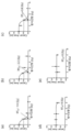
- 9 (a) to (e) and FIG. 10 (a) show examples of RT 50 radiation response factors in various cases.
- RT 50 was calculated based on the cell activity (Cancer Area) according to the radiation dose.
- the RT 50 value which is the radiation dose at which the activity of cancer cells is reduced by 50%, is 1.9Gy
- the RT 50 value is 2.2Gy
- the RT 50 is the case of 3.8Gy.
- the radiation generating unit 145 of the embodiment of the present invention is a device capable of generating a radiation dose of 0 Gy to 8 Gy, so the RT 50 value This is a case where can not be measured, and in this case, the radiation sensitivity can be said to be lower than (a), (b) and (c) in FIG.
- RT AUC may mean a radiation response factor corresponding to the area of a cell activity curve (integrated area, that is, the area of the X-axis, Y-axis and the area surrounded by the cell activity curve) according to the radiation dose.
- RT AUC which is the integral area of the area surrounded by the cell activity curve, may be 4.5.
- a radiation sensitivity diagnosis model is generated based on the calculated cell growth factor and radiation response factor, and the generated sensitivity prediction model is used. It can be used to diagnose radiation sensitivity.
- the radiation sensitivity diagnosis module 135 of FIG. 11 may generate a radiation sensitivity diagnosis model and diagnose radiation sensitivity of an individual patient or individual sample based on the generated radiation sensitivity diagnosis model.
- the radiation sensitivity diagnosis module 135 may be stored in the storage unit 110 and executed by the processor 120 as a software module, for example. Meanwhile, functionally, the radiation sensitivity diagnosis module 135 may generate a radiation sensitivity diagnosis model by extracting sensitivity characteristics using cell experiment-based data. In addition, the radiation sensitivity diagnosis module 135 may determine the radiation sensitivity (or prediction of a radiation treatment response) of an individual (individual patient or experimental sample) as positive or negative based on the radiation sensitivity diagnosis model.
- the radiation sensitivity diagnosis module 135 receives, for example, cell experiment-based data related to radiation response factors and cell growth factors of an individual and/or other individuals as reference data 432, and thereby It can be learned to extract susceptibility features from and create a radiation sensitivity diagnosis model.
- the radiation sensitivity diagnosis module 135 receives reference data 432, which is cell experiment-based data that is a clinical result and/or measurement result of another patient or other object, and cell experiment-based data of a specific individual, and determines the sensitivity characteristics and reference data. It can be configured to diagnose the radiation sensitivity of a specific subject based on the.
- the radiation sensitivity diagnosis module 135 receives cell experiment-based data of the specific individual after a user who wants to know the sensitivity of the specific individual is authenticated through login ( 412) is input to the susceptibility diagnosis module 135. At this time, cell experiment-based data 412 of a specific individual may be input to the input module 422 of the sensitivity diagnosis module 135 . Meanwhile, the input module 422 may further receive reference data 432 .
- sensitivity characteristics may be extracted from the cell experiment-based data 412 of the specific individual by the analysis module 424, and radiation sensitivity may be determined and output based on the sensitivity characteristics by the output module 426.
- the output module 426 may output a radiation sensitivity diagnosis result for a specific individual using the reference data 432 together with the sensitivity characteristics extracted from the cell experiment-based data 412 of the specific individual.
- the analysis module 424 and the output module 426 may output a radiation sensitivity diagnosis result as a treatment response positive or a treatment response negative based on multiple factors of a radiation response factor and a cell growth factor. Things related to this will be described later with reference to FIGS. 12 to 14 .
- the analysis module 424 and the output module 426 may determine the radiation sensitivity (radiation treatment response degree) of the subject as high, medium, or low, and provide the analysis result 428 .
- the analysis module 424 calculates the radiation sensitivity index from the cell experiment-based data 412 of a specific individual, and the output module 426 calculates the radiation sensitivity index according to a predetermined criterion, for example, from -100 to - When the sensitivity index is -10 to +10%, the treatment response degree is determined as 'medium', and when the sensitivity index is +10 to +100%, the treatment response is determined as 'low'. It can be configured to determine the degree as 'above'.
- the analysis module 424 and the output module 426 may probabilistically output treatment responsiveness. Furthermore, the output module 426 may further determine a radiation efficacy diagnosis chart representing a degree of sensitivity to radiation as a chart.
- the radiation sensitivity diagnosis module 135 provides a diagnosis result for the radiation sensitivity of the object to the user terminal, and terminates the user's authentication.
- the radiation sensitivity diagnosis module 135 provides a user with a radiation efficacy diagnosis chart representing the degree of sensitivity to radiation (high, medium, or low) of the subject, whether or not the treatment response is positive, and furthermore, the degree of sensitivity to radiation as a graph. can provide
- diagnosis result 428 of FIG. 11 may be transmitted to the user terminal by the output module 426 and provided through a display unit of the user terminal.
- user authentication termination may be performed after the step in which information on radiation sensitivity is provided.
- FIGS. 12 to 14 are views for explaining a radiation sensitivity diagnosis model according to an embodiment of the present invention.
- the inventors of the present invention have conducted extensive studies on which of the various factors of cancer cells, such as cell growth factors and radiation response factors, are related to radiation treatment sensitivity of patients, among which , it was noted that the prediction of radiation treatment effect is most accurate when cell growth factors and radiation response factors are considered simultaneously.
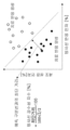
- a graph in which radiation response factors and cell growth factors are set as X-axis and Y-axis, respectively, may be provided as a radiation sensitivity diagnosis model.
- the radiation sensitivity diagnostic model may determine radiation sensitivity based on sensitivity characteristics including a sensitivity baseline according to radiation.
- the radiation sensitivity is not determined only by the radiation response factor, but based on the radiation sensitivity baseline made of the radiation response factor and the cell growth factor.
- RT 50 or RT AUC the radiation response factor
- radiation sensitivity is positive (with therapeutic effect) and negative (with therapeutic effect) according to the radiation sensitivity baseline in which cell growth factor (cell growth rate) is considered. ) can be judged differently.
- the radiation sensitivity baseline (fitting line) that distinguishes the positive group from the negative group of radiation reactivity may be formed such that the cell growth factor (%) decreases as the radiation response factor (RT 50 value or RT AUC value) increases. there is. Therefore, when the radiation response factor (RT 50 value or RT AUC value) increases, the cell growth factor (%) that distinguishes the positive group from the negative radiation reactive group may decrease. Therefore, even if they have the same radiation response factor (RT 50 value), depending on the cell growth factor of the subject (sample), the subject is positive (has a therapeutic effect) or negative (has little or no therapeutic effect) for radiation treatment. can be classified.
- the radiation sensitivity baseline of the sensitivity characteristics may be a fitting line generated by performing curve fitting on cell experiment-based data including cell growth factors and radiation response factors.
- the curve fitting was performed using multifactor regression analysis.
- curve fitting is not limited to this, and in the step of diagnosing susceptibility to radiation (S150), curve fitting is LR (LogistRT regression), PR (Probit regression), QuadratRT classifiers, Kernel estimation, LVQ (Learning vector quantization) , ANN(ArtifRTial neural networks), RF(random forest), Bagging(bootstrap aggregating), AdaBoost, Gradient Boosting, XGBoost, SVM(support vector machine), LASSO(least absolute shrinkage and selection operator), Ridge(ridge regression), and It can be performed by at least one of ElastRT Net.
- the degree of radiation sensitivity may be determined by a fitting line determined by curve fitting.
- the radiation sensitivity diagnosis model is a model for diagnosing treatment response based on the radiation response factor and the cell growth factor (FIG. 13(a)), and based on the reciprocal of the cell growth factor and the radiation response factor. It can be selected among the models (Fig. 13 (b)) for diagnosing treatment response. That is, based on the graph shown in (a) of FIG. 13 in which the baseline radiation sensitivity is inversely proportional according to the relationship between the cell growth factor (%) and the radiation response factor (%) as shown in FIG.
- the radiation sensitivity index is determined by the radiosensitivity baseline or the fitting line. Based on the shortest distance of the new data, it may be calculated by Equation 1 below.
- the radiation sensitivity index may be determined according to the shortest distance between the position of the object according to the cell growth factor and the radiation response factor and the predicted dividing line (fitting line).
- the standard is the distance from 100% of the x-axis and 100% of the y-axis
- the half distance of (value multiplied by 1/2) can be used as the maximum distance.
- the shortest distance from the predicted dividing line The ratio [%] to can be determined as the radiation susceptibility index.
- the shortest distance from the prediction dividing line may be a positive number in the area where treatment-positive subjects are arranged based on the prediction dividing line (the area adjacent to the intersection of the X-axis and Y-axis), and the area where treatment-negative subjects are arranged based on the prediction dividing line. (the area spaced from the intersection of the X and Y axes) may be a negative number.
- the radiation sensitivity index may be 0 at the predicted dividing line, may be a positive number in a treatment response positive region based on the predicted dividing line, and may be a negative number in a treatment response negative region based on the predicted dividing line.
- the radiation sensitivity index may have a value of -100% to +100%.
- the half distance between the x-intercept and the y-intercept of the radiation sensitivity baseline can be used as the maximum distance, or half the distance connecting the maximum values of the x-axis and the y-axis in the data range (in FIG. 13a ) can be used.
- the radiation sensitivity index in a model for diagnosing treatment response based on a radiation response factor (drug response factor) and a cell growth factor can be calculated by Equation 2 below based on the distance (distance to the reference point) from the reference point (origin, which is the intersection of the cell growth factor axis and the drug response factor axis).
- the reference point may be the origin of (0,0), and may have other values depending on the case.
- the radiation sensitivity index may be determined according to the position of the object according to the cell growth factor and the radiation response factor and the shortest distance from the reference point.
- the radiation sensitivity diagnosis module 135 may be configured to determine whether a patient belongs to a positive treatment response region, a treatment response negative region, or an uncertain region according to the radiation sensitivity index obtained as described above.
- the uncertain region may be a region corresponding to an object that does not belong to either of the positive region and the negative region.
- the uncertain region may be a region in which the shortest distance to the prediction dividing line is equal to or less than a preset distance. That is, regardless of which side it belongs to based on the prediction dividing line, if the shortest distance to the prediction dividing line is less than a preset distance, it can be judged as an uncertain object without being judged as positive or negative. Accordingly, the object can be determined as either positive or negative. It is not judged on the side either, so it can be excluded from diagnosis. Thus, the accuracy of overall treatment response prediction can be increased.
- the radiation sensitivity diagnosis module 135 determines the degree of treatment response as 'low' when the radiation sensitivity index is -100% to -10%, and the radiation sensitivity index is -10% to + If it is 10%, the degree of treatment response may be determined as 'medium', and if the sensitivity index is +10% to +100%, the degree of treatment response may be determined as 'high'. That is, the sensitivity diagnosis may be performed by determining the radiation sensitivity index as high/medium/low according to a predetermined criterion.
- the radiation sensitivity diagnosis kit 100 can predict and provide radiation sensitivity with high accuracy. Accordingly, the radiosensitivity diagnostic kit 100 according to the present invention allows the medical staff to quickly select an appropriate treatment radiation dose according to the diagnosis result, so the radiation sensitivity diagnostic system of the present invention has a remarkable effect that can contribute to early treatment and good treatment prognosis.
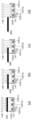
- the table of FIG. 15A is a table showing radiosensitivity diagnostic results and clinical parameters of head and neck cancer patients.
- the vertical axis of the table represents the clinical results (patients (#15 to #37) who are included in the items marked with thick lines are less likely to be treated due to their low radiation sensitivity), and the horizontal axis of the table represents the sensitivity index of each patient with head and neck cancer.
- Clinical results 1 to 8 refer to clinical results that can determine the patient's prognosis, such as, for example, recurrence or undifferentiation after surgery.
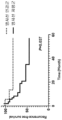
- the survival rate of head and neck cancer patients according to the radiation index according to the radiation-sensitive patient group (radiation sensitive group, treatment response positive group) and other radiation-insensitive patient groups (radiation resistance group, treatment response negative group) indicates In the radiation sensitivity test, when the survival rate was compared for 60 months in the patient group classified as the radiation sensitive group, the survival rate was statistically significantly higher.
- a sensitivity index based on radiation response factors and/or cell growth factors may be an index for diagnosing radiation sensitivity.
- the radiation sensitivity diagnosis system 1000 according to an embodiment of the present invention including the radiation sensitivity diagnosis kit 100 will be described in detail.
- the radiation sensitivity diagnosis system 1000 generates cell experiment-based data on cancer cells, determines the radiation sensitivity of cancer cells by using the data, and provides related information. It is a system that
- the radiation sensitivity diagnosis system 1000 includes a radiation sensitivity diagnosis kit 100 configured to determine the degree of radiation sensitivity of an individual based on cell experiment-based data, and a user terminal 200 that receives information on radiation sensitivity. ) and a database providing server 300 that provides cell experiment-based data and/or clinical result reference data.
- the radiation sensitivity diagnostic kit 100 includes a radiation irradiation device 140 and a control unit 150, and at this time, the control unit 150 controls the radiation irradiation device 140 and/or the database providing server 300. It may include a general-purpose computer, laptop, and/or data server that performs various calculations to diagnose the degree of radiation sensitivity based on cell experiment-based data and/or reference data of a specific user provided from ).
- the radiation sensitivity diagnosis kit 100 may provide diagnosis information on radiation sensitivity of an object to the user terminal 200 .
- Data provided from the radiation sensitivity diagnostic kit 100 may be provided as a web page through a web browser installed in the user terminal 200, or may be provided in the form of an application or program. In various embodiments, this data may be provided in a form incorporated into the platform in a client-server environment.
- the user terminal 200 is an electronic system that provides a user interface for requesting information on the sensitivity of radiation to an object and displaying diagnosis result data, such as a smartphone, a tablet PC (Personal Computer), a laptop and/or or at least one of a PC and the like.
- the user terminal 200 may be a terminal used by a user such as a medical staff or a patient corresponding to an object.
- the user terminal 200 may include a communication unit, a display unit, a storage unit, and a processor.
- the communication unit of the user terminal 200 may be configured to allow the user terminal 200 to communicate with an external system.
- the communication unit may be connected to the radiation sensitivity diagnostic kit 100 using wired/wireless communication to transmit various data related to radiation sensitivity.
- the communication unit determines the diagnosis result related to the radiation sensitivity of the subject from the radiation sensitivity diagnosis kit 100, for example, the degree of sensitivity to radiation of the subject (high, medium or low), whether the treatment response is positive, and furthermore, It is possible to receive a radiation efficacy diagnosis chart representing the degree of sensitivity as a chart.
- the display unit of the user terminal 200 may display various interface screens for displaying diagnosis results related to radiation sensitivity of an object.
- the display unit may display and provide a radiation efficacy diagnosis chart representing the degree of sensitivity to radiation (high, medium, or low) of the subject, whether or not the treatment response is positive, and further, the degree of sensitivity to radiation as a graph. .
- the display unit may include a touch screen, and for example, a touch using an electronic pen or a part of a user's body, a gesture, a proximity, a drag, and a swipe may be performed. Alternatively, a hovering input or the like may be received.
- CRS plate 143 radiation control unit
- the present invention it is possible to predict the effect of radiation treatment in advance by effectively diagnosing the radiation sensitivity of cancer cells of a patient, and thus, head and neck cancer, lung cancer, esophageal cancer, rectal cancer, anal cancer, and uterus suitable for conservative treatment strategies. Whether or not to perform radiation therapy for cancer such as cervical cancer can be accurately and effectively determined.
- a radiation sensitivity diagnosis method capable of accurately determining the radiation sensitivity of an individual patient with the most effective factors for diagnosis of radiation sensitivity among various factors that have been previously studied by simultaneously considering radiation response factors and cell growth factors. is provided.
- a small number of samples and a limited amount of time can be obtained using a characteristic sensitivity diagnostic kit configured so that the total amount of radiation irradiated to each of the plurality of cancer organoids is different from each other due to a change in the relative position of the plate portion and the radiation shielding film.
- Data required for radiation sensitivity diagnosis can be obtained by overcoming limitations.
Landscapes
- Health & Medical Sciences (AREA)
- Engineering & Computer Science (AREA)
- Life Sciences & Earth Sciences (AREA)
- Chemical & Material Sciences (AREA)
- Biomedical Technology (AREA)
- Organic Chemistry (AREA)
- Wood Science & Technology (AREA)
- Zoology (AREA)
- Bioinformatics & Cheminformatics (AREA)
- Genetics & Genomics (AREA)
- Biotechnology (AREA)
- Biochemistry (AREA)
- General Health & Medical Sciences (AREA)
- Microbiology (AREA)
- Immunology (AREA)
- General Engineering & Computer Science (AREA)
- Molecular Biology (AREA)
- Cell Biology (AREA)
- Hematology (AREA)
- Urology & Nephrology (AREA)
- Physics & Mathematics (AREA)
- Analytical Chemistry (AREA)
- Proteomics, Peptides & Aminoacids (AREA)
- Oncology (AREA)
- Food Science & Technology (AREA)
- Medicinal Chemistry (AREA)
- General Physics & Mathematics (AREA)
- Pathology (AREA)
- Biophysics (AREA)
- Toxicology (AREA)
- Tropical Medicine & Parasitology (AREA)
- Radiation-Therapy Devices (AREA)
Abstract
Priority Applications (2)
| Application Number | Priority Date | Filing Date | Title |
|---|---|---|---|
| US17/915,067 US20240218417A1 (en) | 2021-11-24 | 2022-07-19 | Radiation sensitivity diagnosis method |
| EP22898750.9A EP4439063A4 (fr) | 2021-11-24 | 2022-07-19 | Procédé de diagnostic de sensibilité au rayonnement |
Applications Claiming Priority (4)
| Application Number | Priority Date | Filing Date | Title |
|---|---|---|---|
| KR20210163284 | 2021-11-24 | ||
| KR10-2021-0163284 | 2021-11-24 | ||
| KR1020220088270A KR102656927B1 (ko) | 2021-11-24 | 2022-07-18 | 방사선 감수성 진단방법 |
| KR10-2022-0088270 | 2022-07-18 |
Publications (1)
| Publication Number | Publication Date |
|---|---|
| WO2023096065A1 true WO2023096065A1 (fr) | 2023-06-01 |
Family
ID=86539959
Family Applications (1)
| Application Number | Title | Priority Date | Filing Date |
|---|---|---|---|
| PCT/KR2022/010503 Ceased WO2023096065A1 (fr) | 2021-11-24 | 2022-07-19 | Procédé de diagnostic de sensibilité au rayonnement |
Country Status (3)
| Country | Link |
|---|---|
| US (1) | US20240218417A1 (fr) |
| EP (1) | EP4439063A4 (fr) |
| WO (1) | WO2023096065A1 (fr) |
Cited By (1)
| Publication number | Priority date | Publication date | Assignee | Title |
|---|---|---|---|---|
| CN118483404A (zh) * | 2024-04-29 | 2024-08-13 | 中山大学肿瘤防治中心(中山大学附属肿瘤医院、中山大学肿瘤研究所) | 一种放疗射线的相对生物效应的评估方法 |
Citations (3)
| Publication number | Priority date | Publication date | Assignee | Title |
|---|---|---|---|---|
| KR20130024904A (ko) * | 2010-05-26 | 2013-03-08 | 지방독립 행정법인 오사카 부립 병원 기구 | 암 조직 유래 세포괴 또는 암 세포 응집괴의 약제 또는 방사선 감수성 평가 방법 |
| KR20140039353A (ko) * | 2012-09-19 | 2014-04-02 | 고려대학교 산학협력단 | 컬리메이터 장치 |
| KR20200081296A (ko) * | 2018-12-26 | 2020-07-07 | 주식회사 넥스트앤바이오 | 암을 가진 대상체의 항암제 및/또는 방사선 내성 진단에 필요한 정보를 제공하는 방법 |
Family Cites Families (2)
| Publication number | Priority date | Publication date | Assignee | Title |
|---|---|---|---|---|
| JP4203208B2 (ja) * | 2000-03-28 | 2008-12-24 | 住友重機械工業株式会社 | 放射線のエネルギ分布調整機構、並びに、これを用いた放射線の照射装置 |
| CN109136188A (zh) * | 2017-06-15 | 2019-01-04 | 上海集技生物技术有限公司 | 一种活检肠道肿瘤类器官的培养、传代、冻存和复苏方法及其应用 |
-
2022
- 2022-07-19 US US17/915,067 patent/US20240218417A1/en active Pending
- 2022-07-19 EP EP22898750.9A patent/EP4439063A4/fr active Pending
- 2022-07-19 WO PCT/KR2022/010503 patent/WO2023096065A1/fr not_active Ceased
Patent Citations (3)
| Publication number | Priority date | Publication date | Assignee | Title |
|---|---|---|---|---|
| KR20130024904A (ko) * | 2010-05-26 | 2013-03-08 | 지방독립 행정법인 오사카 부립 병원 기구 | 암 조직 유래 세포괴 또는 암 세포 응집괴의 약제 또는 방사선 감수성 평가 방법 |
| KR20140039353A (ko) * | 2012-09-19 | 2014-04-02 | 고려대학교 산학협력단 | 컬리메이터 장치 |
| KR20200081296A (ko) * | 2018-12-26 | 2020-07-07 | 주식회사 넥스트앤바이오 | 암을 가진 대상체의 항암제 및/또는 방사선 내성 진단에 필요한 정보를 제공하는 방법 |
Non-Patent Citations (4)
| Title |
|---|
| "Cervical Cancer: Methods and Protocols", vol. 1249, 1 January 2015, HUMANA PRESS, US, ISBN: 978-1-4939-2012-9, article MAYIL VAHANAN BOSE, THANGARAJAN RAJKUMAR: "Assessment of the radiation sensitivity of cervical cancer cell lines", pages: 351 - 362, XP009546243, DOI: 10.1007/978-1-4939-2013-6_26 * |
| PASCH CHERI A., FAVREAU PETER F., YUEH ALEXANDER E., BABIARZ CHRISTOPHER P., GILLETTE AMANI A., SHARICK JOE T., KARIM MOHAMMAD REZ: "Patient-Derived Cancer Organoid Cultures to Predict Sensitivity to Chemotherapy and Radiation", CLINICAL CANCER RESEARCH, ASSOCIATION FOR CANCER RESEARCH, US, vol. 25, no. 17, 1 September 2019 (2019-09-01), US, pages 5376 - 5387, XP093068229, ISSN: 1078-0432, DOI: 10.1158/1078-0432.CCR-18-3590 * |
| See also references of EP4439063A4 * |
| YAO YE; XU XIAOYA; YANG LIFENG; ZHU JI; WAN JUEFENG; SHEN LIJUN; XIA FAN; FU GUOXIANG; DENG YUN; PAN MENGXUE; GUO QIANG; GAO XIAOX: "Patient-Derived Organoids Predict Chemoradiation Responses of Locally Advanced Rectal Cancer", CELL STEM CELL, ELSEVIER, CELL PRESS, AMSTERDAM, NL, vol. 26, no. 1, 21 November 2019 (2019-11-21), AMSTERDAM, NL , pages 17, XP085989337, ISSN: 1934-5909, DOI: 10.1016/j.stem.2019.10.010 * |
Cited By (2)
| Publication number | Priority date | Publication date | Assignee | Title |
|---|---|---|---|---|
| CN118483404A (zh) * | 2024-04-29 | 2024-08-13 | 中山大学肿瘤防治中心(中山大学附属肿瘤医院、中山大学肿瘤研究所) | 一种放疗射线的相对生物效应的评估方法 |
| CN118483404B (zh) * | 2024-04-29 | 2025-03-18 | 中山大学肿瘤防治中心(中山大学附属肿瘤医院、中山大学肿瘤研究所) | 一种放疗射线的相对生物效应的评估方法 |
Also Published As
| Publication number | Publication date |
|---|---|
| EP4439063A1 (fr) | 2024-10-02 |
| EP4439063A4 (fr) | 2025-10-22 |
| US20240218417A1 (en) | 2024-07-04 |
Similar Documents
| Publication | Publication Date | Title |
|---|---|---|
| Rakha et al. | New advances in molecular breast cancer pathology | |
| US20240161893A1 (en) | Determining a therapy efficacy | |
| Young et al. | Transscleral fine-needle aspiration biopsy of macular choroidal melanoma | |
| Yan et al. | Three-dimensional spatiotemporal modeling of colon cancer organoids reveals that multimodal control of stem cell self-renewal is a critical determinant of size and shape in early stages of tumor growth | |
| WO2023096065A1 (fr) | Procédé de diagnostic de sensibilité au rayonnement | |
| Calaf et al. | Ras protein expression as a marker for breast cancer | |
| Katz et al. | In-vivo optogenetics and pharmacology in deep intracellular recordings | |
| WO2022173232A2 (fr) | Procédé et système pour prédire le risque d'apparition d'une lésion | |
| Louia et al. | Artificial intelligence in cancer genomics: transforming Diagnosis, Treatment, and precision medicine | |
| Chen et al. | Single-cell panleukemia signatures of HSPC-like blasts predict drug response and clinical outcome | |
| KR102656927B1 (ko) | 방사선 감수성 진단방법 | |
| Theivendren et al. | Success stories: impactful applications of cancer biomarkers | |
| Hassanzadeh et al. | Technical and engineering considerations for designing therapeutics and delivery systems | |
| WO2017188517A1 (fr) | Inhibition de la cancérisation maligne induite par ras oncogène à l'aide d'un rayonnement à faible dose | |
| Pesce et al. | Designer drugs | |
| Mali et al. | Cancer management in terms of precision oncology | |
| CN113782130B (zh) | 一种基因组学数据管理与诊疗系统及方法 | |
| WO2021261625A1 (fr) | Procédé de fourniture d'informations nécessaires pour diagnostiquer la résistance d'un patient atteint de cancer à un agent et/ou à un rayonnement anticancéreux | |
| Zhou et al. | Evaluating third generation cervical Cancer intracavitary interstitial brachytherapy applicator efficacy and safety | |
| US20250027929A1 (en) | Method and system for predicting susceptibility to anticancer agent | |
| Malhotra et al. | Pharmacy and Medical Science | |
| Piccaluga | Molecular diagnostics of cancer | |
| Santiago-Gonzalez et al. | 295 Identification of Proteomic Biomarkers in Puerto Ricans with Pancreatic Cancer | |
| Nagasaka | Significance of targeting cancer stem cells in gynecologic cancer | |
| Zhu et al. | High-throughput strategy for targeting MDM2 in uveal melanoma to reverse radiation therapy resistance |
Legal Events
| Date | Code | Title | Description |
|---|---|---|---|
| WWE | Wipo information: entry into national phase |
Ref document number: 17915067 Country of ref document: US |
|
| 121 | Ep: the epo has been informed by wipo that ep was designated in this application |
Ref document number: 22898750 Country of ref document: EP Kind code of ref document: A1 |
|
| WWE | Wipo information: entry into national phase |
Ref document number: 2022898750 Country of ref document: EP |
|
| NENP | Non-entry into the national phase |
Ref country code: DE |
|
| ENP | Entry into the national phase |
Ref document number: 2022898750 Country of ref document: EP Effective date: 20240624 |