WO2024190857A1 - 動脈硬化性疾患を診断するための試薬および方法 - Google Patents
動脈硬化性疾患を診断するための試薬および方法 Download PDFInfo
- Publication number
- WO2024190857A1 WO2024190857A1 PCT/JP2024/009946 JP2024009946W WO2024190857A1 WO 2024190857 A1 WO2024190857 A1 WO 2024190857A1 JP 2024009946 W JP2024009946 W JP 2024009946W WO 2024190857 A1 WO2024190857 A1 WO 2024190857A1
- Authority
- WO
- WIPO (PCT)
- Prior art keywords
- neutrophil
- reagent
- antibody
- disease
- derived protein
- Prior art date
- Legal status (The legal status is an assumption and is not a legal conclusion. Google has not performed a legal analysis and makes no representation as to the accuracy of the status listed.)
- Ceased
Links
Images

Classifications
-
- G—PHYSICS
- G01—MEASURING; TESTING
- G01N—INVESTIGATING OR ANALYSING MATERIALS BY DETERMINING THEIR CHEMICAL OR PHYSICAL PROPERTIES
- G01N33/00—Investigating or analysing materials by specific methods not covered by groups G01N1/00 - G01N31/00
- G01N33/48—Biological material, e.g. blood, urine; Haemocytometers
- G01N33/50—Chemical analysis of biological material, e.g. blood, urine; Testing involving biospecific ligand binding methods; Immunological testing
- G01N33/53—Immunoassay; Biospecific binding assay; Materials therefor
- G01N33/573—Immunoassay; Biospecific binding assay; Materials therefor for enzymes or isoenzymes
Definitions
- the present invention relates to reagents and methods for diagnosing arteriosclerotic disease.
- Arteriosclerosis also known as atherosclerosis, is a condition in which the arterial lumen narrows and loses elasticity due to the accumulation of cholesterol in the blood under the intima of the arterial wall. It is most common in coronary arteries, cerebral blood vessels, carotid arteries, aorta, and popliteal arteries. Protruding lesions (plaques) on the inside of the blood vessel wall where cholesterol has been deposited can cause poor blood flow, and if the plaque ruptures, it is highly likely to cause dangerous, life-threatening diseases. Serious arteriosclerotic diseases include acute coronary syndrome, cerebrovascular disease, and aortic disease, and are not only major causes of death as lifestyle-related diseases, but also reduce quality of life (QOL) due to their occurrence. The formation of arteriosclerotic plaque is thought to be due to single or multiple causes, such as dyslipidemia, hypertension, and deterioration of lifestyle habits such as dietary habits, exercise habits, smoking, and drinking.
- QOL quality of life
- AMI acute myocardial infarction
- unstable angina and sudden cardiac death due to ischemia caused by coronary artery plaque rupture.
- NSTEI non-ST elevation myocardial infarction
- AMI is a disease caused by myocardial necrosis due to the interruption of blood flow to the myocardium caused by occlusion or sub-occlusion of the coronary artery due to plaque rupture in the coronary artery.
- Troponin is a marker for myocardial tissue damage, and cannot be used to grasp the state of plaque instability, which is the cause of AMI.
- blood levels of troponin are not specific to AMI, as they are also increased by myocardial damage caused by various conditions other than AMI.
- Patent Documents 1 and 2 Several attempts have been made to diagnose arteriosclerosis using biomarkers.
- a typical example is the measurement of blood cholesterol, which is related to the formation of arteriosclerosis (Patent Documents 1 and 2).
- blood cholesterol concentration is related to the gradual progression of arteriosclerosis, it is unable to grasp the state of unstable plaque that is easily ruptured.
- Patent Document 3 attempts to capture proteins that have fallen out of arteriosclerotic lesions into the blood using antibodies obtained from arteriosclerotic tissue itself as an antigen
- Patent Document 4 attempts to capture cardiovascular events using markers that increase with systemic acute inflammation
- Non-Patent Document 1 neutrophil extracellular traps
- Non-Patent Documents 4, 5, 6, 7 investigated the concentration of myeloperoxidase-DNA complexes released by neutrophils.
- Non-Patent Document 7 confirmed the relationship between neutrophil elastase levels and the onset of heart failure by blood sampling after the onset of myocardial infarction.
- An object of the present invention is to provide reagents and methods for diagnosing arteriosclerotic diseases, assessing their severity, predicting their onset or aggravation, or assessing the risk of onset or aggravation. It is another object of the present invention to provide reagents and methods for assessing the state of atherosclerosis.
- the present invention includes the following aspects.
- a reagent for use in diagnosing an arteriosclerotic disease, evaluating the severity, predicting the onset or aggravation of the disease, or evaluating the onset or aggravation risk of the disease comprising: A reagent comprising an antibody or antigen-binding fragment thereof against a complex bound to a neutrophil-derived protein or its corresponding binding molecule.
- a reagent comprising an antibody or antigen-binding fragment thereof against a complex bound to a neutrophil-derived protein or its corresponding binding molecule.
- the arteriosclerotic disease is at least one selected from the group consisting of acute coronary syndrome, cerebrovascular disease, and aortic disease.
- the reagent according to [1-2], wherein the acute coronary syndrome is at least one selected from the group consisting of acute myocardial infarction, unstable angina, and sudden cardiac death due to ischemia.
- the reagent according to [1-3], wherein the acute coronary syndrome is acute myocardial infarction.
- the reagent according to [1-4], wherein the acute myocardial infarction is non-ST elevation myocardial infarction.
- the reagent according to [1-2], wherein the cerebrovascular disease is at least one selected from the group consisting of cerebral aneurysm, lacunar infarction, atherothrombotic cerebral infarction, and cardiogenic cerebral embolism.
- [1-7] The reagent according to [1-2], wherein the aortic disease is at least one selected from the group consisting of aortic aneurysm and aortic dissection.
- [1-8] The reagent according to any one of [1-1] to [1-7], wherein the neutrophil-derived protein is neutrophil elastase.
- [1-9] The reagent according to any one of [1-1] to [1-8], wherein the reagent is an immunoassay reagent.
- the immunoassay reagent is a reagent for use in at least one immunoassay selected from the group consisting of enzyme immunoassay, fluorescent enzyme immunoassay, chemiluminescent enzyme immunoassay, chemiluminescent immunoassay, electrochemiluminescence immunoassay, fluorescent antibody method, radioimmunoassay, Western blot method, immunoblot method, latex agglutination method, immunochromatography method, and nephelometry method.
- the immunoassay reagent is a reagent for use in at least one immunoassay selected from the group consisting of enzyme immunoassay, fluorescent enzyme immunoassay, chemiluminescent enzyme immunoassay, chemiluminescent immunoassay, electrochemiluminescence immunoassay, fluorescent antibody method, radioimmunoassay, Western blot method, immunoblot method, latex agglutination method, immunochromatography method, and nephel
- a reagent for evaluating the state of atherosclerosis comprising: A reagent comprising an antibody or antigen-binding fragment thereof against a complex bound to a neutrophil-derived protein or its corresponding binding molecule.
- the immunoassay reagent is a reagent for use in at least one immunoassay selected from the group consisting of enzyme immunoassay, fluorescent enzyme immunoassay, chemiluminescent enzyme immunoassay, chemiluminescent immunoassay, electrochemiluminescence immunoassay, fluorescent antibody method, radioimmunoassay, Western blot method, immunoblot method, latex agglutination method, immunochromatography method, and nephelometry method.
- a method for diagnosing an arteriosclerotic disease, evaluating the severity thereof, predicting the onset or aggravation of the arteriosclerotic disease, or evaluating a risk of onset or aggravation of the arteriosclerotic disease comprising: (1) contacting the sample with an antibody or antigen-binding fragment thereof against a complex bound to a neutrophil-derived protein or its corresponding binding molecule; (2) measuring the binding state between the antibody or its antigen-binding fragment and a complex bound to the neutrophil-derived protein or its corresponding binding molecule.
- the arteriosclerotic disease is at least one selected from the group consisting of acute coronary syndrome, cerebrovascular disease, and aortic disease.
- the acute coronary syndrome is at least one selected from the group consisting of acute myocardial infarction, unstable angina, and sudden cardiac death due to ischemia.
- the acute coronary syndrome is acute myocardial infarction.
- the aortic disease is at least one selected from the group consisting of aortic aneurysm and aortic dissection.
- the neutrophil-derived protein is neutrophil elastase.
- [3-9] The method according to any one of [3-1] to [3-8], wherein the method for measuring the binding state is at least one immunological assay selected from the group consisting of enzyme immunoassay, fluorescent enzyme immunoassay, chemiluminescent enzyme immunoassay, chemiluminescent immunoassay, electrochemiluminescence immunoassay, fluorescent antibody method, radioimmunoassay, Western blot method, immunoblot method, latex agglutination method, immunochromatography method, and nephelometry method.
- the sample is at least one selected from the group consisting of plasma, serum, and whole blood.
- [4-1] A method for evaluating atherosclerosis, comprising: (1) contacting the sample with an antibody or antigen-binding fragment thereof against a complex bound to a neutrophil-derived protein or its corresponding binding molecule; (2) Measuring the binding state between the antibody or its antigen-binding fragment and a complex bound to the neutrophil-derived protein or its corresponding binding molecule.
- [4-2] The method according to [4-1], wherein the evaluation of the state of atherosclerosis is evaluation of the state of at least one type of plaque selected from the group consisting of vulnerable plaque, ruptured plaque, plaque prone to erosion, eroded plaque, plaque causing internal bleeding, plaque with nodular calcification, and plaque exhibiting severe stenosis in coronary artery plaque.
- [4-3] The method according to [4-1] or [4-2], wherein the neutrophil-derived protein is neutrophil elastase.
- [4-4] The method according to any one of [4-1] to [4-3], wherein the method for measuring the binding state is at least one immunological measurement method selected from the group consisting of enzyme immunoassay, fluorescent enzyme immunoassay, chemiluminescent enzyme immunoassay, chemiluminescent immunoassay, electrochemiluminescence immunoassay, fluorescent antibody method, radioimmunoassay, Western blot method, immunoblot method, latex agglutination method, immunochromatography method, and nephelometry method.
- the acute coronary syndrome is at least one selected from the group consisting of acute myocardial infarction, unstable angina, and sudden cardiac death due to ischemia.
- the acute coronary syndrome is acute myocardial infarction.
- the method according to [5-4] wherein the acute myocardial infarction is non-ST elevation myocardial infarction.
- the aortic disease is at least one selected from the group consisting of aortic aneurysm and aortic dissection.
- the neutrophil-derived protein is neutrophil elastase.
- [5-9] The method according to any one of [5-1] to [5-8], wherein the method for measuring the binding state is at least one immunological measurement method selected from the group consisting of enzyme immunoassay, fluorescent enzyme immunoassay, chemiluminescent enzyme immunoassay, chemiluminescent immunoassay, electrochemiluminescence immunoassay, fluorescent antibody method, radioimmunoassay, Western blot method, immunoblot method, latex agglutination method, immunochromatography method, and nephelometry method.
- the sample is at least one selected from the group consisting of plasma, serum, and whole blood.
- [6-1] A method for assisting in the assessment of atherosclerosis, comprising: (1) contacting the sample with an antibody or antigen-binding fragment thereof against a complex bound to a neutrophil-derived protein or its corresponding binding molecule; (2) Measuring the binding state between the antibody or its antigen-binding fragment and a complex bound to the neutrophil-derived protein or its corresponding binding molecule.
- the evaluation of the state of atherosclerosis is evaluation of the state of at least one type of plaque selected from the group consisting of vulnerable plaque, ruptured plaque, plaque prone to erosion, eroded plaque, plaque causing internal bleeding, plaque with nodular calcification, and plaque exhibiting severe stenosis in coronary artery plaque.
- [6-3] The method according to [6-1] or [6-2], wherein the neutrophil-derived protein is neutrophil elastase.
- [6-4] The method according to any one of [6-1] to [6-3], wherein the method for measuring the binding state is at least one immunological measurement method selected from the group consisting of enzyme immunoassay, fluorescent enzyme immunoassay, chemiluminescent enzyme immunoassay, chemiluminescent immunoassay, electrochemiluminescence immunoassay, fluorescent antibody method, radioimmunoassay, Western blot method, immunoblot method, latex agglutination method, immunochromatography method, and nephelometry method.
- the arteriosclerotic disease is at least one selected from the group consisting of acute coronary syndrome, cerebrovascular disease, and aortic disease.
- the acute coronary syndrome is at least one selected from the group consisting of acute myocardial infarction, unstable angina, and sudden cardiac death due to ischemia.
- the acute coronary syndrome is acute myocardial infarction.
- the aortic disease is at least one selected from the group consisting of aortic aneurysm and aortic dissection.
- the neutrophil-derived protein is neutrophil elastase.
- [7-9] The method according to any one of [7-1] to [7-8], wherein the method for measuring the binding state is at least one immunological measurement method selected from the group consisting of enzyme immunoassay, fluorescent enzyme immunoassay, chemiluminescent enzyme immunoassay, chemiluminescent immunoassay, electrochemiluminescence immunoassay, fluorescent antibody method, radioimmunoassay, Western blot method, immunoblot method, latex agglutination method, immunochromatography method, and nephelometry method.
- the sample is at least one selected from the group consisting of plasma, serum, and whole blood.
- [8-1] A method for collecting data for evaluating the state of atherosclerosis, comprising: (1) contacting the sample with an antibody or antigen-binding fragment thereof against a complex bound to a neutrophil-derived protein or its corresponding binding molecule; (2) Measuring the binding state between the antibody or its antigen-binding fragment and a complex bound to the neutrophil-derived protein or its corresponding binding molecule.
- [8-2] The method according to [8-1], wherein the evaluation of the state of atherosclerosis is evaluation of the state of at least one type of plaque selected from the group consisting of vulnerable plaque, ruptured plaque, plaque prone to erosion, eroded plaque, plaque causing internal bleeding, plaque with nodular calcification, and plaque exhibiting severe stenosis in coronary artery plaque.
- [8-3] The method according to [8-1] or [8-2], wherein the neutrophil-derived protein is neutrophil elastase.
- [8-4] The method according to any one of [8-1] to [8-3], wherein the method for measuring the binding state is at least one immunological measurement method selected from the group consisting of enzyme immunoassay, fluorescent enzyme immunoassay, chemiluminescent enzyme immunoassay, chemiluminescent immunoassay, electrochemiluminescence immunoassay, fluorescent antibody method, radioimmunoassay, Western blot method, immunoblot method, latex agglutination method, immunochromatography method, and nephelometry method.
- enzyme immunoassay fluorescent enzyme immunoassay, chemiluminescent enzyme immunoassay, chemiluminescent immunoassay, electrochemiluminescence immunoassay, fluorescent antibody method, radioimmunoassay, Western blot method, immunoblot method, latex a
- a method for detecting or quantifying a complex bound to a neutrophil-derived protein or its corresponding binding molecule in a sample derived from a subject for whom an evaluation of the state of atherosclerosis is required comprising: (1) contacting the sample with an antibody or antigen-binding fragment thereof against a complex bound to a neutrophil-derived protein or its corresponding binding molecule; (2) measuring the binding state between the antibody or its antigen-binding fragment and a complex bound to the neutrophil-derived protein or its corresponding binding molecule.
- the arteriosclerotic disease is at least one selected from the group consisting of acute coronary syndrome, cerebrovascular disease, and aortic disease.
- the acute coronary syndrome is at least one selected from the group consisting of acute myocardial infarction, unstable angina, and sudden cardiac death due to ischemia.
- the acute coronary syndrome is acute myocardial infarction.
- [9-9] The method according to any one of [9-1] to [9-8], wherein the method for measuring the binding state is at least one immunological measurement method selected from the group consisting of enzyme immunoassay, fluorescent enzyme immunoassay, chemiluminescent enzyme immunoassay, chemiluminescent immunoassay, electrochemiluminescence immunoassay, fluorescent antibody method, radioimmunoassay, Western blot method, immunoblot method, latex agglutination method, immunochromatography method, and nephelometry method.
- the sample is at least one selected from the group consisting of plasma, serum, and whole blood.
- a method for detecting or quantifying a complex bound to a neutrophil-derived protein or its corresponding binding molecule in a sample derived from a subject for whom an evaluation of the state of atherosclerosis is required comprising: (1) contacting the sample with an antibody or antigen-binding fragment thereof against a complex bound to a neutrophil-derived protein or its corresponding binding molecule; (2) measuring the binding state between the antibody or its antigen-binding fragment and a complex bound to the neutrophil-derived protein or its corresponding binding molecule.
- [10-2] The method according to [10-1], wherein the evaluation of the state of atherosclerosis is evaluation of the state of at least one type of plaque selected from the group consisting of vulnerable plaque, ruptured plaque, plaque prone to erosion, eroded plaque, plaque causing internal bleeding, plaque with nodular calcification, and plaque exhibiting severe stenosis in coronary artery plaque.
- the neutrophil-derived protein is neutrophil elastase.
- [10-4] The method according to any one of [10-1] to [10-3], wherein the method for measuring the binding state is at least one immunological measurement method selected from the group consisting of enzyme immunoassay, fluorescent enzyme immunoassay, chemiluminescent enzyme immunoassay, chemiluminescent immunoassay, electrochemiluminescence immunoassay, fluorescent antibody method, radioimmunoassay, Western blot method, immunoblot method, latex agglutination method, immunochromatography method, and nephelometry method.
- the sample is at least one selected from the group consisting of plasma, serum, and whole blood.
- [11-1] Use of an antibody or an antigen-binding fragment thereof against a complex bound to a neutrophil-derived protein or its corresponding binding molecule, in the manufacture of a reagent for diagnosing an arteriosclerotic disease, assessing its severity, predicting the onset or aggravation of the disease, or assessing the risk of onset or aggravation of the disease.
- the arteriosclerotic disease is at least one selected from the group consisting of acute coronary syndrome, cerebrovascular disease, and aortic disease.
- the cerebrovascular disease is at least one selected from the group consisting of cerebral aneurysm, lacunar infarction, atherothrombotic cerebral infarction, and cardiogenic cerebral embolism.
- [11-7] The use according to [11-2], wherein the aortic disease is at least one selected from the group consisting of aortic aneurysm and aortic dissection.
- [11-8] The use according to any one of [11-1] to [11-7], wherein the neutrophil-derived protein is neutrophil elastase.
- [11-9] The use according to any one of [11-1] to [11-8], wherein the reagent is a reagent for immunological measurement.
- the immunoassay reagent is a reagent for use in at least one immunoassay selected from the group consisting of enzyme immunoassay, fluorescent enzyme immunoassay, chemiluminescent enzyme immunoassay, chemiluminescent immunoassay, electrochemiluminescence immunoassay, fluorescent antibody method, radioimmunoassay, Western blot method, immunoblot method, latex agglutination method, immunochromatography method, and nephelometry method.
- [12-1] Use of an antibody or an antigen-binding fragment thereof against a complex bound to a neutrophil-derived protein or its corresponding binding molecule, for the manufacture of a reagent for evaluating the state of atherosclerosis.
- [12-2] The use according to [12-1], wherein the evaluation of the state of atherosclerosis is evaluation of the state of at least one type of plaque selected from the group consisting of vulnerable plaque, ruptured plaque, plaque prone to erosion, eroded plaque, plaque causing internal bleeding, plaque with nodular calcification, and plaque exhibiting severe stenosis in coronary artery plaque.
- [12-3] The use according to [12-1] or [12-2], wherein the neutrophil-derived protein is neutrophil elastase.
- the reagent is an immunoassay reagent.
- the immunoassay reagent is a reagent for use in at least one immunoassay selected from the group consisting of enzyme immunoassay, fluorescent enzyme immunoassay, chemiluminescent enzyme immunoassay, chemiluminescent immunoassay, electrochemiluminescence immunoassay, fluorescent antibody method, radioimmunoassay, Western blot method, immunoblot method, latex agglutination method, immunochromatography method, and nephelometry method.
- the present invention it is possible to diagnose arteriosclerotic diseases, evaluate their severity, predict the onset or aggravation of arteriosclerotic diseases, or evaluate the risk of onset or aggravation of arteriosclerotic diseases, and therefore it is possible to predict the onset and aggravation of arteriosclerotic diseases at an early stage. Furthermore, according to the present invention, the state of atherosclerosis can be evaluated, and therefore the presence or absence of unstable plaques that are prone to rupture can be confirmed early, particularly in patients with NSTEMI.
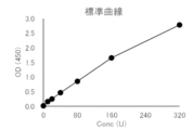
- FIG. 1 shows a standard curve in the neutrophil elastase ELISA measurement system constructed in Example 1.
- FIG. 2 shows the neutrophil elastase concentration in each serum sample measured in Example 2 using the reagent constructed in Example 1.
- neutrophil refers to a cell that has proteolytic enzymes such as myeloperoxidase (MPO), neutrophil elastase, and cathepsin G in its cytoplasmic granules, and that removes pathogens in response to stimulation through phagocytosis, degranulation, and production of reactive oxygen species (ROS) and inflammatory cytokines.
- MPO myeloperoxidase
- ROS reactive oxygen species
- NETs Neutrophil extracellular traps
- neutrophil-derived protein refers to a protein released from neutrophils into the blood.
- the protein includes proteins released into the body by NETs, specifically, neutrophil elastase, azurocidin, lactoferrin, myeloperoxidase, peptidylarginine deiminase 4 (hereinafter also referred to as "PAD4"), etc.
- neutrophil elastase is included.
- the term "complex formed by binding a neutrophil-derived protein to a corresponding binding molecule” refers to a complex formed by binding the corresponding binding molecule to the above-mentioned "neutrophil-derived protein.”
- Specific examples of the above complexes include complexes of neutrophil elastase and ⁇ 1- antitrypsin (A1AT), ⁇ 1- antichymotrypsin (ACT), or ⁇ 2- macroglobulin (A2M), particularly complexes of neutrophil elastase and ⁇ 1- antitrypsin (A1AT) or ⁇ 1 -antichymotrypsin (ACT).
- A1AT complexes of neutrophil elastase and ⁇ 1- antitrypsin
- ACT ⁇ 1-antichymotrypsin
- A2M macroglobulin
- the "neutrophil-derived protein” and the “complex of the neutrophil-derived protein bound to the corresponding binding molecule” will be collectively referred to
- the "antibody or antigen-binding fragment thereof against a complex bound to a neutrophil-derived protein or its corresponding binding molecule” is not particularly limited as long as it has a specific binding ability to the above-mentioned neutrophil-derived protein or a complex bound to its corresponding binding molecule.
- examples of antibodies or antigen-binding fragments thereof that have specific binding ability to a "neutrophil-derived protein” include antibodies or fragments thereof that are capable of recognizing and binding to a part of a neutrophil-derived protein as an epitope.
- the above-mentioned "antibody” may be a monoclonal antibody or a polyclonal antibody.
- the antibody may be a commercially available product or may be produced by using a neutrophil-derived protein or a part thereof as an antigen according to a known method such as cell fusion technology, gene recombination technology, or phage display technology.
- the above-mentioned "antigen-binding fragment of an antibody” refers to a fragment of the above-mentioned antibody that has the ability to bind to a neutrophil-derived protein, and examples thereof include a Fab fragment obtained by partially degrading the above-mentioned antibody with papain or the like, an F(ab')2 fragment obtained by partially degrading the above-mentioned antibody with pepsin or the like, and an Fab' fragment obtained by reducing the F(ab')2 fragment.
- the antibody is a monoclonal antibody or an antigen-binding fragment thereof.
- antibody and the above-mentioned “antigen-binding fragment of antibody” will be collectively referred to as “antibody”.
- arteriosclerotic disease includes arteriosclerosis itself, as well as ischemic heart disease such as myocardial infarction and angina pectoris, which are caused by vascular stenosis in the heart or brain due to arteriosclerosis, cerebrovascular disease, cerebral hemorrhage, aortic aneurysm, aortic dissection, nephrosclerosis in the renal artery and resulting renal failure, and arteriosclerosis obliterans in the peripheral arteries.
- ischemic heart disease such as myocardial infarction and angina pectoris, which are caused by vascular stenosis in the heart or brain due to arteriosclerosis, cerebrovascular disease, cerebral hemorrhage, aortic aneurysm, aortic dissection, nephrosclerosis in the renal artery and resulting renal failure, and arteriosclerosis obliterans in the peripheral arteries.
- arteriosclerosis cerebrovascular disease
- cerebral hemorrhage cerebral hemorrh
- the above-mentioned "acute coronary syndrome” includes, for example, at least one selected from the group consisting of acute myocardial infarction, unstable angina, and sudden cardiac death due to ischemia.
- acute myocardial infarction is included, and in particular, non-ST elevation myocardial infarction is included.
- cerebrovascular disease includes, for example, at least one selected from the group consisting of cerebral aneurysm, lacunar infarction, atherothrombotic cerebral infarction, and cardiogenic cerebral embolism.
- aortic disease includes, for example, at least one selected from the group consisting of aortic aneurysm and aortic dissection.
- plaques refers to a condition in which porridge-like protrusions (plaques) form on the inside of arteries, and can occur in any large or medium-sized artery, including major arteries such as coronary arteries, cerebral arteries, and the aorta.
- major arteries such as coronary arteries, cerebral arteries, and the aorta.
- plaques grow, blood flow becomes poor, and when plaques suddenly rupture, platelets gather there, forming blood clots and causing atherothrombosis, which blocks blood vessels.
- Examples of serious diseases caused by this series of mechanisms include the following: ⁇ Coronary artery plaque: Acute coronary syndrome (unstable angina, acute myocardial infarction, sudden cardiac death due to ischemia) - Cerebral artery plaque: Cerebrovascular disease (cerebral aneurysm, lacunar infarction, atherothrombotic cerebral infarction, cardiogenic cerebral embolism) - Aortic plaque: Aortic disease (aortic aneurysm, aortic dissection).
- the above-mentioned plaque may be at least one type selected from the group consisting of coronary artery plaques that are easily ruptured, ruptured plaques, plaques that are prone to erosion, plaques that have undergone erosion, plaques that have caused internal bleeding, plaques with nodular calcification, and plaques exhibiting severe stenosis.
- diagnosis refers to determining the current or future condition of a disease.
- severeness includes mild, moderate, severe, etc.
- aggravation includes progression from mild to moderate or severe, and from moderate to severe.
- onset refers to the appearance of symptoms of a disease.
- assessment of the state of atherosclerosis refers to obtaining information related to the onset and aggravation of acute disease due to arterial occlusion or subocclusion by evaluating the progression of atherosclerosis, which is characterized by patchy intimal plaques growing toward the lumen of medium-sized and large arteries, including coronary arteries, carotid arteries, cerebral arteries, aorta, aortic branches, and major arteries of the limbs, from the concentration of blood protein markers.
- Such evaluation includes, for example, evaluation of the state of coronary artery plaque, cerebral artery plaque, and aortic plaque, and in particular evaluation of the state of at least one type of plaque selected from the group consisting of vulnerable plaque, ruptured plaque, plaque prone to erosion, plaque that has eroded, plaque that has caused internal bleeding, plaque with nodular calcification, and plaque that exhibits severe stenosis.
- the term "subject” refers to a living organism that is the subject of a test, particularly an animal (e.g., a mammal such as a human, mouse, rat, hamster, guinea pig, monkey, cow, pig, horse, rabbit, sheep, goat, cat, or dog), and particularly a human (in this case, also referred to as a "test subject”).
- "subject” in the case of a human, “test subject” includes a subject (patient) suffering from a disease, a subject (test subject) suspected of suffering from a disease, a subject (test subject) at risk of suffering from a disease, a subject (test subject) not suffering from a disease (healthy subject), etc.
- sample in this specification is not particularly limited as long as it is a sample that can contain the above-mentioned neutrophil-derived proteins, and examples include blood samples prepared from blood collected from human subjects.
- blood sample refers to a sample that contains at least a portion of blood components, and may be any of whole blood, serum, and plasma, or any of these diluted forms.
- the blood sample is preferably serum or plasma.
- the blood sample can be prepared by known methods.
- the present invention includes a reagent for use in diagnosing an arteriosclerotic disease, assessing the severity, predicting the onset or aggravation, or assessing the risk of onset or aggravation, the reagent comprising an antibody or an antigen-binding fragment thereof against a complex bound to the above-mentioned neutrophil-derived protein or its corresponding binding molecule.
- the composition, shape, state, etc. of the reagent are not particularly limited.
- the above-mentioned antibody or antigen-binding fragment thereof may be one type or two or more types.
- two or more types of antibodies or antigen-binding fragments thereof are used, they may be, for example, two or more types of antibodies or antigen-binding fragments thereof that bind to different epitopes, or two or more types of antibodies or antigen-binding fragments thereof that bind to the same epitope.
- a neutrophil-derived protein when a neutrophil-derived protein may be present in a sample both in a free form and in a complex, at least one of the two or more types of antibodies may be an antibody or antigen-binding fragment thereof against the complex.
- examples include a combination of two or more types of antibodies or antigen-binding fragments thereof that can recognize and bind to different portions of a neutrophil-derived protein as an epitope; a combination of at least one antibody that can recognize and bind to a portion of a neutrophil-derived protein as an epitope and an antigen-binding fragment of at least one antibody that can recognize and bind to an epitope different from the epitope of the antibody; a combination of at least one antibody or antigen-binding fragment thereof that can recognize and bind to a portion of a neutrophil-derived protein as an epitope and at least one antibody or antigen-binding fragment thereof that can recognize and bind to a portion of a complex as part or all of an epitope; and the like.
- the neutrophil-derived protein is human neutrophil elastase and the binding molecule is human ⁇ 1 antitrypsin (A1AT)
- A1AT human ⁇ 1 antitrypsin
- an example of the combination is an anti-human neutrophil elastase antibody and an anti-human A1AT antibody.
- the antibody may be immobilized on a support.
- the support can be appropriately selected depending on the method in which the reagent is used. Examples of the support include well plates (e.g., 96-well microplates, etc.), membranes (e.g., nitrocellulose membranes, polyvinylidene fluoride membranes, etc.), slide glasses, magnetic beads, latex particles, etc.
- the antibody can be immobilized on the support by a known method selected depending on the material of the support.
- the antibody may be labeled.
- labeling substance there are no particular limitations on the labeling substance used, and any known substance may be used.
- labeling substances include enzyme labels such as peroxidase and alkaline phosphatase; fluorescent labels such as fluorescein isothiocyanate (FITC); radioisotope labels such as iodine-125; electrochemiluminescence labels such as ruthenium complexes; biotin; and metal nanoparticles.
- the antibody can be labeled by a known method selected according to the type of labeling substance.
- the above reagent may be in the form of a kit.
- the kit may contain other elements in addition to the above antibody. Examples of the other elements include a detection reagent for the labeled substance, a standard sample of a neutrophil-derived protein, a reagent for preparing a blood sample, a diluent, buffers, a support, and instructions for use.
- reagents for use in immunoassays include reagents for use in immunoassays (reagents for immunoassays), specifically reagents for use in enzyme immunoassays, fluorescent enzyme immunoassays, chemiluminescent enzyme immunoassays, chemiluminescent immunoassays, electrochemiluminescent immunoassays, fluorescent antibody assays, radioimmunoassays, Western blots, immunoblots, latex agglutination, immunochromatography, nephelometry, etc.
- reagents for use in latex agglutination or immunochromatography may be commercially available or may be prepared according to known methods.
- examples of reagents for use in the latex agglutination method include reagents comprising: (i) an antibody-sensitized latex liquid containing latex particles to which at least one type of antibody or antigen-binding fragment thereof is bound that is capable of recognizing and binding to a portion of a neutrophil-derived protein (e.g., neutrophil elastase) as an epitope; and (ii) an antibody-sensitized latex liquid containing latex particles to which an antibody or antigen-binding fragment thereof is bound that is capable of recognizing and binding to a portion of a binding molecule (e.g., ⁇ 1- antitrypsin) as a part or all of an epitope.
- a neutrophil-derived protein e.g., neutrophil elastase
- an antibody-sensitized latex liquid containing latex particles to which an antibody or antigen-binding fragment thereof is bound that is capable of recognizing and binding to a portion of
- the above reagent is capable of detecting neutrophil-derived proteins in a sample (particularly a blood sample) and measuring the amount (concentration) of neutrophil-derived proteins.
- the blood concentration of the neutrophil-derived protein shows a positive correlation with the progression of atherosclerotic disease from the onset to the aggravation of the disease, i.e., the blood concentration of the neutrophil-derived protein increases as the atherosclerotic disease progresses from the onset to the aggravation of the disease. Therefore, the blood concentration of neutrophil-derived proteins can be used as an index for diagnosing arteriosclerotic diseases. Therefore, the above reagent can be used for diagnosing arteriosclerotic diseases, assisting in the diagnosis, and collecting data for the diagnosis.
- the severity of arteriosclerotic diseases can be evaluated based on the blood concentration of neutrophil-derived proteins.
- a standard value or cutoff value pathological condition identification value
- standard value/cutoff value pathological condition identification value
- a value less than the first reference value/cutoff value can be evaluated as mild, a value between the first reference value/cutoff value or more and less than the second reference value/cutoff value as moderate, and a value equal to or more than the second reference value/cutoff value as severe.
- An upper reference limit determined by multiple healthy individuals can also be used as the first reference value/cutoff value, and an upper reference limit multiplied by a specific number can also be used as the second reference value/cutoff value. Therefore, the above reagent can be used for evaluating the severity of arteriosclerotic diseases, assisting in such evaluation, and collecting data for such evaluation.
- a standard value/cutoff value is determined by statistically processing data obtained from multiple healthy subjects and patients, and based on this value, it is possible to predict whether arteriosclerotic diseases will develop or whether arteriosclerotic diseases will become severe. For example, if the level is equal to or above a certain standard value/cutoff value, it can be predicted that an arteriosclerotic disease will develop, and if the level is below the standard value/cutoff value, it can be predicted that an arteriosclerotic disease will not develop.
- the above reagent can be used for predicting the onset or aggravation of arteriosclerotic diseases, assisting in such prediction, and collecting data for making such prediction.
- the presence or absence, or the level of the risk of developing or becoming severe of arteriosclerotic diseases can be determined based on the blood concentration of neutrophil-derived proteins.
- a standard value/cutoff value can be determined by statistically processing data obtained from multiple patients, and the presence or absence, or the level of the risk of developing or becoming severe of arteriosclerotic diseases can be determined based on this value. For example, if the value is equal to or greater than a predetermined standard value/cutoff value, it can be determined that the risk of developing arteriosclerotic disease exists or is high, and conversely, if the value is less than the standard value/cutoff value, it can be determined that such risk does not exist or is low.
- the above-mentioned reagent can be used for evaluating the risk of developing or aggravating arteriosclerotic diseases, assisting in such evaluation, and collecting data for such evaluation.
- the sample to be measured using the above-mentioned reagent may be a blood sample prepared from blood taken from a human subject (subject), and the subject may or may not be confirmed to have an arteriosclerotic disease.
- a disease can be confirmed by a known method, for example, an antibody test, a PCR test, a biomarker, a doctor's diagnosis, or the like.
- the present invention provides a method for diagnosing an arteriosclerotic disease, evaluating the severity thereof, predicting the onset or aggravation of the arteriosclerotic disease, or evaluating a risk of onset or aggravation of the arteriosclerotic disease, comprising: (1) contacting the sample with an antibody or antigen-binding fragment thereof against a complex bound to a neutrophil-derived protein or its corresponding binding molecule; (2) A method comprising measuring the binding state between a complex bound to the neutrophil-derived protein or its corresponding binding molecule and an antibody or an antigen-binding fragment thereof against the complex bound to the neutrophil-derived protein or its corresponding binding molecule.
- the present invention provides a method for assisting in the diagnosis of an arteriosclerotic disease, the assessment of the severity thereof, the prediction of the onset or aggravation of the arteriosclerotic disease, or the assessment of a risk of onset or aggravation of the arteriosclerotic disease, comprising: (1) contacting the sample with an antibody or antigen-binding fragment thereof against a complex bound to a neutrophil-derived protein or its corresponding binding molecule; (2) A method comprising measuring the binding state between the antibody or its antigen-binding fragment and a complex bound to the neutrophil-derived protein or its corresponding binding molecule.
- the present invention provides a method for collecting data for diagnosing an arteriosclerotic disease, assessing the severity thereof, predicting the onset or aggravation of the arteriosclerotic disease, or assessing a risk of onset or aggravation of the arteriosclerotic disease, comprising: (1) contacting the sample with an antibody or antigen-binding fragment thereof against a complex bound to a neutrophil-derived protein or its corresponding binding molecule; (2) A method comprising measuring the binding state between the antibody or its antigen-binding fragment and a complex bound to the neutrophil-derived protein or its corresponding binding molecule.
- the samples in the above-mentioned methods for diagnosing arteriosclerotic diseases, methods for assisting diagnosis, and methods for collecting data for diagnosis include samples that are to be subjected to measurement using the above-mentioned reagents.
- the antibody used in the above method is the same as the above reagent, and may be one type or two or more types.
- the above method may also be carried out using one or more of the above reagents.
- the above step (1) is a step of contacting a sample with an antibody, thereby binding the neutrophil-derived protein contained in the sample to the antibody. Therefore, there are no particular limitations on the step, so long as the step is carried out under conditions that allow the neutrophil-derived protein to bind to the antibody. For example, the step can be carried out in accordance with known immunological assay methods.
- the step (2) is a step of measuring the state of binding between the neutrophil-derived protein obtained in the step (1) and the antibody.
- the measurement of the state of binding is not particularly limited, and may be a known qualitative or quantitative method.
- Specific examples of the method include known immunological measurement methods, such as enzyme immunoassay, fluorescent enzyme immunoassay, chemiluminescent enzyme immunoassay, chemiluminescent immunoassay, electrochemiluminescent immunoassay, fluorescent antibody method, radioimmunoassay, Western blot method, immunoblot method, latex agglutination method, immunochromatography method, and nephelometry method.
- the above method for diagnosing arteriosclerotic diseases may include a step (3) of diagnosing arteriosclerotic diseases, assessing their severity, predicting the onset or aggravation of arteriosclerotic diseases, or assessing the risk of onset or aggravation of arteriosclerotic diseases based on the amount (concentration) of the neutrophil-derived protein obtained in step (2).
- Step (3) includes, for example, diagnosing using the amount (concentration) of the neutrophil-derived protein as an index, as explained in the above reagent, or determining a reference value/cutoff value and assessing the severity, predicting the onset or aggravation of arteriosclerotic diseases, or assessing the risk of onset or aggravation of arteriosclerotic diseases based on the reference value/cutoff value.
- the above-mentioned method for assisting in the diagnosis, etc., of arteriosclerotic diseases may further include steps such as determining a reference value/cutoff value that serves as an indicator for the diagnosis, etc., based on the amount (concentration) of the neutrophil-derived protein obtained in step (2), for example, as described for the above-mentioned reagent, and providing a result of comparison with the reference value/cutoff value.
- the method of collecting data for diagnosing or otherwise treating arteriosclerotic diseases may further include steps of collecting data for determining a reference value/cutoff value that serves as an indicator for the diagnosis or the like, based on the amount (concentration) of the neutrophil-derived protein obtained in step (2), for example, as described above for the reagent, and providing data for comparison with the reference value/cutoff value.
- the present invention provides a method for detecting or quantifying a complex bound to a neutrophil-derived protein or its corresponding binding molecule in a sample derived from a subject for whom an assessment of the state of atherosclerosis is required, comprising the steps of: (1) contacting the sample with an antibody or antigen-binding fragment thereof against a complex bound to a neutrophil-derived protein or its corresponding binding molecule; (2) A method comprising measuring the binding state between the antibody or its antigen-binding fragment and a complex bound to the neutrophil-derived protein or its corresponding binding molecule.
- the sample in this method includes the above-mentioned reagents used for the diagnosis of arteriosclerotic diseases and samples to be measured by the method.
- the antibody used in this method is the same as the reagent and method used in the diagnosis and the like of arteriosclerotic diseases described above, and may be one type or two or more types.
- the method can be carried out using one or more of the above-mentioned reagents used for the diagnosis, etc. of arteriosclerotic diseases.
- steps (1) and (2) in this method are the same as steps (1) and (2) in the above-mentioned method for diagnosing arteriosclerotic diseases.
- the present invention includes the use of an antibody or an antigen-binding fragment thereof against a complex bound to a neutrophil-derived protein or its corresponding binding molecule, for the manufacture of a reagent for diagnosing an arteriosclerotic disease, assessing its severity, predicting the onset or progression of the disease, or assessing the risk of onset or progression of the disease.
- the antibody in this embodiment is the same as the reagent used for the diagnosis of arteriosclerotic diseases, and may be one type or two or more types.
- the present invention includes a reagent for assessing atherosclerosis status, comprising an antibody or antigen-binding fragment thereof against a complex bound to a neutrophil-derived protein or its corresponding binding molecule.
- the composition, form, condition, etc. of the reagent are the same as those of the reagent used for the diagnosis of arteriosclerotic diseases.
- the above reagent is capable of detecting neutrophil-derived proteins in a sample (particularly a blood sample) and measuring the amount (concentration) of neutrophil-derived proteins.
- the blood concentration of neutrophil-derived proteins shows a positive correlation with the progression of atherosclerosis, that is, the blood concentration of neutrophil-derived proteins increases as atherosclerosis progresses. Therefore, the blood concentration of neutrophil-derived proteins can be used as an indicator for diagnosing diseases associated with atherosclerosis (eg, arteriosclerotic diseases).
- the reagent can be used to assess the state of atherosclerosis, aid in such assessment, collect data for such assessment, and the like.
- the progression of atherosclerosis can be evaluated based on the blood concentration of neutrophil-derived proteins.
- a standard value/cutoff value can be determined by statistically processing data obtained from multiple healthy subjects and patients, and the progression of atherosclerosis can be evaluated based on this value.
- a value less than the first reference value/cutoff value can be evaluated as mild, a value between the first reference value/cutoff value or more and less than the second reference value/cutoff value as moderate, and a value equal to or more than the second reference value/cutoff value as severe.
- An upper reference limit determined by multiple healthy subjects can also be used as the first reference value/cutoff value, and an upper reference limit multiplied by a specific number can also be used as the second reference value/cutoff value. Therefore, the above reagent can be used for evaluating the degree of progression of atherosclerosis, aiding in such evaluation, collecting data for such evaluation, and the like.
- the severity of atherosclerosis-related diseases can be evaluated based on the blood concentration of neutrophil-derived proteins.
- a standard value/cutoff value pathological condition identification value
- a value less than the first reference value/cutoff value can be evaluated as mild, a value between the first reference value/cutoff value or more and less than the second reference value/cutoff value as moderate, and a value equal to or more than the second reference value/cutoff value as severe.
- an upper reference limit determined by multiple healthy individuals can also be used as the first reference value/cutoff value, and an upper reference limit multiplied by a specific number can also be used as the second reference value/cutoff value. Therefore, the above-mentioned reagent can be used for assessing the severity of atherosclerosis-related diseases, aiding in such assessment, collecting data for such assessment, and the like.
- a standard value/cutoff value can be determined by statistically processing data obtained from multiple healthy subjects or patients, and based on this value, it is possible to predict whether or not the disease will develop, or whether or not the disease will become severe. For example, if the level is equal to or greater than a certain standard value/cutoff value, it can be predicted that the disease will develop, and if the level is below the standard value/cutoff value, it can be predicted that the disease will not develop.
- the value when the value is equal to or greater than a predetermined reference value/cutoff value, it can be predicted that the disease will become more severe, and when the value is less than the reference value/cutoff value, it can be predicted that the disease will not become more severe.
- An upper reference limit determined by a plurality of healthy subjects can also be used as the first reference value/cutoff value, and the upper reference limit multiplied by a specific number can also be used as the second reference value/cutoff value. Therefore, the above-mentioned reagent can be used for predicting the onset or aggravation of atherosclerosis-related diseases, assisting in such prediction, and collecting data for making such prediction.
- the presence or absence, or the level of the risk of developing or becoming severe in atherosclerosis-related diseases can be determined based on the blood concentration of neutrophil-derived proteins.
- a standard value/cutoff value can be determined by statistically processing data obtained from multiple healthy subjects or patients, and the presence or absence, or the level of the risk of developing or becoming severe in the disease can be determined based on this value. For example, if the value is equal to or greater than a predetermined standard value/cutoff value, it can be determined that the risk of developing the disease is present or is high, and conversely, if the value is less than the standard value/cutoff value, it can be determined that such risk is absent or is low.
- the above-mentioned reagent can be used for evaluating the risk of developing or aggravating atherosclerosis-related disease, assisting in such evaluation, and collecting data for such evaluation.
- the sample to be measured using the above-mentioned reagent may be a blood sample prepared from blood collected from a human subject, who may or may not have been confirmed to have atherosclerosis. Also, the subject may or may not have been confirmed to have atherosclerosis-related disease.
- the presence of a disease can be confirmed by a known method, for example, an antibody test, a PCR test, a biomarker, a doctor's diagnosis, or the like.
- the present invention also provides, in one embodiment, a method for assessing an atherosclerosis condition, comprising: (1) contacting the sample with an antibody or antigen-binding fragment thereof against a complex bound to a neutrophil-derived protein or its corresponding binding molecule; (2) A method comprising measuring the binding state between a complex bound to the neutrophil-derived protein or its corresponding binding molecule and an antibody or an antigen-binding fragment thereof against the complex bound to the neutrophil-derived protein or its corresponding binding molecule.
- the present invention also provides, in one embodiment, a method for assisting in the assessment of an atherosclerotic condition, comprising: (1) contacting the sample with an antibody or antigen-binding fragment thereof against a complex bound to a neutrophil-derived protein or its corresponding binding molecule; (2) A method comprising measuring the binding state between the antibody or its antigen-binding fragment and a complex bound to the neutrophil-derived protein or its corresponding binding molecule.
- the present invention further provides, in one embodiment, a method for collecting data for assessing atherosclerosis status, comprising the steps of: (1) contacting the sample with an antibody or antigen-binding fragment thereof against a complex bound to a neutrophil-derived protein or its corresponding binding molecule; (2) A method comprising measuring the binding state between the antibody or its antigen-binding fragment and a complex bound to the neutrophil-derived protein or its corresponding binding molecule.
- the samples in the above-mentioned methods for evaluating the state of atherosclerosis, the methods for assisting the evaluation, and the methods for collecting data for evaluation include samples that are to be subjected to measurement using the above-mentioned reagents.
- the antibody used in the above method is the same as the above reagent, and may be one type or two or more types.
- the above method may also be carried out using one or more of the above reagents.
- the above step (1) is a step of contacting a sample with an antibody, thereby binding the neutrophil-derived protein contained in the sample to the antibody. Therefore, there are no particular limitations on the step, so long as the step is carried out under conditions that allow the neutrophil-derived protein to bind to the antibody. For example, the step can be carried out in accordance with known immunological assay methods.
- the step (2) is a step of measuring the state of binding between the neutrophil-derived protein obtained in the step (1) and the antibody.
- the measurement of the state of binding is not particularly limited, and may be a known qualitative or quantitative method.
- Specific examples of the method include known immunological measurement methods, such as enzyme immunoassay, fluorescent enzyme immunoassay, chemiluminescent enzyme immunoassay, chemiluminescent immunoassay, electrochemiluminescent immunoassay, fluorescent antibody method, radioimmunoassay, Western blot method, immunoblot method, latex agglutination method, immunochromatography method, and nephelometry method.
- the above method for evaluating the state of atherosclerosis may include a step (3) of evaluating the degree of progression of atherosclerosis based on the amount (concentration) of the neutrophil-derived protein obtained in the step (2).
- the step (3) may include, for example, evaluating the degree of progression of atherosclerosis using the amount (concentration) of the neutrophil-derived protein as an index, as explained in the above reagent, or determining a reference value/cutoff value and evaluating the degree of progression of atherosclerosis based on the reference value/cutoff value.
- the above-mentioned method for evaluating the state of atherosclerosis may also include a step (3) of diagnosing an atherosclerosis-related disease, assessing its severity, predicting the onset or aggravation of the disease, or assessing the risk of onset or aggravation of the disease based on the amount (concentration) of the neutrophil-derived protein obtained in step (2).
- Step (3) may include, for example, diagnosing the disease using the amount (concentration) of the neutrophil-derived protein as an index, as explained in the above-mentioned reagent, or determining a reference value/cutoff value and assessing the severity, predicting the onset or aggravation of the disease, or assessing the risk of onset or aggravation of the disease based on the reference value/cutoff value.
- the above-mentioned method for assisting in the evaluation of the state of atherosclerosis may further include steps such as determining a reference value/cutoff value that serves as an indicator for evaluating the degree of progression, etc., based on the amount (concentration) of the neutrophil-derived protein obtained in step (2), as described above for the reagent, and providing a result of comparison with the reference value/cutoff value.
- the method of collecting data for evaluating the state of atherosclerosis may further include steps of collecting data for determining a reference value/cutoff value that serves as an index for evaluating the degree of progression, etc., based on the amount (concentration) of the neutrophil-derived protein obtained in step (2), for example, as described in the above-mentioned reagent, and providing data for comparison with the reference value/cutoff value.
- the present invention provides a method for detecting or quantifying a complex bound to a neutrophil-derived protein or its corresponding binding molecule in a sample derived from a subject for whom an assessment of the state of atherosclerosis is required, comprising the steps of: (1) contacting the sample with an antibody or antigen-binding fragment thereof against a complex bound to a neutrophil-derived protein or its corresponding binding molecule; (2) A method comprising measuring the binding state between the antibody or its antigen-binding fragment and a complex bound to the neutrophil-derived protein or its corresponding binding molecule.
- the sample in this method includes the above-mentioned reagents used in the evaluation of the state of atherosclerosis and samples to be measured by the method.
- the antibody used in this method is the same as the reagent and method used in the evaluation of the atherosclerosis state described above, and may be one type or two or more types.
- the method may be carried out using one or more of the above-mentioned reagents for assessing an atherosclerotic condition.
- steps (1) and (2) in this method are similar to steps (1) and (2) in the above-mentioned method for evaluating the state of atherosclerosis.
- the present invention includes, as one embodiment, the use of an antibody or an antigen-binding fragment thereof against a complex bound to a neutrophil-derived protein or its corresponding binding molecule for the manufacture of a reagent for assessing the state of atherosclerosis.
- the antibody in this embodiment is the same as the reagent used for evaluating the state of atherosclerosis described above, and may be one type or two or more types.
- Antibody A and Antibody B were obtained according to the following procedure.
- Human leukocyte-derived purified elastase (Elastin Products, Cat. No. CK828) was suspended in an adjuvant (Complete Freund's adjuvant: CFA), and BALB/c mice (5 weeks old, female) were immunized at 2-week intervals. Partial blood was collected during the immunization, and the degree of titer increase at the antiserum level was confirmed using the reactivity with the immunogen, human leukocyte-derived purified elastase, as an index.
- CFA Complete Freund's adjuvant
- antibody-producing cells were collected from the spleen and fused with myeloma (P3X63Ag8.653, ECACC). Cell fusion was performed using the PEG method, and the fused cells were seeded on a culture plate. The cells were then cultured in a carbon dioxide incubator at 37°C. Next, the hybridoma culture supernatant was collected, and the antibody titer was confirmed based on the reactivity with purified elastase derived from human leukocytes. The antibody-producing clone cells were then subcultured and cloned by limiting dilution.
- the cells derived from the resulting single colony were obtained as anti-human neutrophil elastase monoclonal antibody-producing hybridomas.
- Anti-human A1AT antibody-producing hybridomas were also obtained by the same method using human plasma-derived ⁇ 1- antitrypsin (Merck, Cat. No. A6150-25MG).
- the two obtained hybridomas producing monoclonal antibodies were cultured in large quantities.
- Each hybridoma was intraperitoneally administered to BALB/c mice that had been pre-bred with pristane administered intraperitoneally. The mice were then bred for 10 to 25 days, and the accumulation of ascites was allowed to continue.
- antibody A which is an anti-human neutrophil elastase monoclonal antibody
- antibody B which is an anti-human A1AT antibody
- Enzyme immunoassay was performed by sandwich enzyme immunoassay method using a kit composed of the following reagents.
- Standard Reagent Antibody A and antibody B were bound to HiTrap NHS-activated HP Columns (Cytiva, Cat. No. 17071601) to obtain an antibody A-bound affinity column and an antibody B-bound affinity column.
- 30 mL of healthy subject plasma was diluted 10-fold with 20 mM phosphate buffer, and the filtrate was passed through a 0.45 ⁇ m filter to recover the filtrate.
- the filtrate was passed through the antibody A-bound affinity column, and the adsorbed protein components were eluted with 100 mM Glycine-HCl Buffer pH 2.7 to obtain an eluted fraction.
- the eluted fraction was buffer-substituted with 20 mM phosphate buffer, then passed through an antibody B binding affinity column, and the adsorbed protein components were eluted with 100 mM Glycine-HCl Buffer pH 2.7 to obtain an eluted fraction.
- the eluted fraction was buffer-substituted with 20 mM phosphate buffer to obtain a standard reagent stock solution.
- a dilution series was prepared with 1% BSA-containing PBS buffer, and the buffer containing only 1% BSA-containing PBS buffer was designated as standard reagent 0.
- Sample dilution solution PBS buffer containing 1% BSA. Use as is.
- Enzyme-labeled antibody solution This solution is prepared by diluting enzyme-labeled antibody B labeled with horseradish peroxidase (HRP) 16,000-fold with PBS buffer containing 1% BSA. This solution is used as is.
- Reaction plate A 96-well microplate is used in which antibody A is immobilized and then blocked. One well is used for each measurement.
- Washing solution 0.05% Tween 20, PBS buffer. Ready to use.
- Substrate solution A solution containing tetramethylbenzidine (3,3',5,5'-tetramethylbenzidine stabilized substrate: TMB, Agilent). The substrate solution is used as is. 100 ⁇ L of the substrate solution is used for one measurement.
- Reaction stop solution 3N sulfuric acid. Use as is. Use 100 ⁇ L of reaction stop solution for one measurement.
- a standard curve was created according to the following procedure. First, the standard reagent was appropriately diluted with the specimen dilution solution to prepare neutrophil elastase solutions of various concentrations. Then, 100 ⁇ L of each of these diluted standard solutions was added to the reaction plate, stirred in a mixer, and incubated at room temperature for 1 hour. Then, the reaction solution was removed from each well using an ELISA washer, and 0.3 mL of a washing solution prepared by diluting the washing stock solution was added to each well for washing. After repeating this washing three times, the washing solution remaining in the well was removed with a paper towel or the like. Next, 100 ⁇ L of an enzyme-labeled antibody solution was added, and incubation was performed at room temperature for 1 hour.
- reaction solution was removed from each well using an ELISA washer, and 0.3 mL of a washing solution prepared by diluting the washing stock solution was added to each well for washing. After repeating this washing three times, the washing solution remaining in the well was removed with a paper towel or the like. Next, 100 ⁇ L of substrate solution was added, and incubation was performed at room temperature for exactly 30 minutes in the dark. After that, 100 ⁇ L of reaction stop solution was added and the mixture was quickly stirred in a mixer to stop the enzyme reaction. The absorbance at 450 nm was measured for each well, and a standard curve was created ( Figure 1).
- Example 2 ⁇ Measurement of human serum samples using an ELISA measurement system> The neutrophil elastase concentrations in 11 serum samples collected from patients diagnosed with acute coronary syndrome (unstable angina: 2 cases, non-ST elevation myocardial infarction: 4 cases), cerebrovascular disease (cerebral aneurysm: 3 cases), and aortic disease (thoracic aortic aneurysm rupture: 2 cases) as disease groups with unstable plaque, and 3 serum samples collected from healthy subjects (Table 1 below) were measured using the reagent constructed in Example 1. The results are shown in Figure 2. If the upper limit of the measurement results from samples from healthy subjects is taken as the upper limit of reference, all patients with unstable plaque showed values more than twice the upper limit of reference.
Landscapes
- Health & Medical Sciences (AREA)
- Life Sciences & Earth Sciences (AREA)
- Immunology (AREA)
- Engineering & Computer Science (AREA)
- Molecular Biology (AREA)
- Biomedical Technology (AREA)
- Chemical & Material Sciences (AREA)
- Hematology (AREA)
- Urology & Nephrology (AREA)
- Biotechnology (AREA)
- Microbiology (AREA)
- Cell Biology (AREA)
- Food Science & Technology (AREA)
- Medicinal Chemistry (AREA)
- Physics & Mathematics (AREA)
- Analytical Chemistry (AREA)
- Biochemistry (AREA)
- General Health & Medical Sciences (AREA)
- General Physics & Mathematics (AREA)
- Pathology (AREA)
- Investigating Or Analysing Biological Materials (AREA)
Abstract
Description
本発明は、動脈硬化性疾患を診断するための試薬および方法に関する。
また、本発明の別の目的は、アテローム性動脈硬化の状態を評価するための試薬および方法を提供することである。
すなわち、本発明は、以下の態様を含む。
[1-1] 動脈硬化性疾患の診断、重症度の評価、発症もしくは重症化の予測、または発症リスクもしくは重症化リスクの評価に用いるための試薬であって、
好中球由来のタンパク質またはその対応する結合分子と結合した複合体に対する抗体またはその抗原結合フラグメントを含む、試薬。
[1-2] 前記動脈硬化性疾患が、急性冠症候群、脳血管疾患、および大動脈疾患からなる群から選択される少なくとも1種である、[1-1]に記載の試薬。
[1-3] 前記急性冠症候群が、急性心筋梗塞、不安定狭心症、および虚血による心臓突然死からなる群から選択される少なくとも1種である、[1-2]に記載の試薬。
[1-4] 前記急性冠症候群が、急性心筋梗塞である、[1-3]に記載の試薬。
[1-5] 前記急性心筋梗塞が、非ST上昇型心筋梗塞である、[1-4]に記載の試薬。
[1-6] 前記脳血管疾患が、脳動脈瘤、ラクナ梗塞、アテローム血栓性脳梗塞、および心原性脳塞栓症からなる群から選択される少なくとも1種である、[1-2]に記載の試薬。
[1-7] 前記大動脈疾患が、大動脈瘤、および大動脈解離からなる群から選択される少なくとも1種である、[1-2]に記載の試薬。
[1-8] 前記好中球由来のタンパク質が、好中球エラスターゼである、[1-1]~[1-7]のいずれかに記載の試薬。
[1-9] 前記試薬が、免疫学的測定用試薬である、[1-1]~[1-8]のいずれかに記載の試薬。
[1-10] 前記免疫学的測定用試薬が、酵素免疫測定法、蛍光酵素免疫測定法、化学発光酵素免疫測定法、化学発光免疫測定法、電気化学発光測免疫測定法、蛍光抗体法、ラジオイムノアッセイ、ウェスタンブロット法、イムノブロット法、ラテックス凝集法、イムノクロマトグラフィー法、およびネフェロメトリー法からなる群から選択される少なくとも1種の免疫学的測定法に用いるための試薬である、[1-9]に記載の試薬。
[2-1] アテローム性動脈硬化の状態を評価するための試薬であって、
好中球由来のタンパク質またはその対応する結合分子と結合した複合体に対する抗体またはその抗原結合フラグメントを含む、試薬。
[2-2] 前記アテローム性動脈硬化の状態の評価が、冠動脈プラークの易破綻性プラーク、破綻したプラーク、びらんを起こし易いプラーク、びらんを起こしたプラーク、内出血を来たしたプラーク、小結節上石灰化を有するプラーク、および高度狭窄を呈するプラークからなる群から選択される少なくとも1種のプラークの状態の評価である、[2-1]に記載の試薬。
[2-3] 前記好中球由来のタンパク質が、好中球エラスターゼである、[2-1]または[2-2]に記載の試薬。
[2-4] 前記試薬が、免疫学的測定用試薬である、[2-1]~[2-3]のいずれかに記載の試薬。
[2-5] 前記免疫学的測定用試薬が、酵素免疫測定法、蛍光酵素免疫測定法、化学発光酵素免疫測定法、化学発光免疫測定法、電気化学発光測免疫測定法、蛍光抗体法、ラジオイムノアッセイ、ウェスタンブロット法、イムノブロット法、ラテックス凝集法、イムノクロマトグラフィー法、およびネフェロメトリー法からなる群から選択される少なくとも1種の免疫学的測定法に用いるための試薬である、[2-4]に記載の試薬。
[3-1] 動脈硬化性疾患を診断、その重症度を評価、その発症もしくは重症化を予測、またはその発症リスクもしくは重症化リスクを評価するための方法であって、
(1)試料と、好中球由来のタンパク質またはその対応する結合分子と結合した複合体に対する抗体またはその抗原結合フラグメントを接触させること、
(2)該好中球由来のタンパク質またはその対応する結合分子と結合した複合体と、該抗体またはその抗原結合フラグメントとの結合状態を測定すること
を含む、方法。
[3-2] 前記動脈硬化性疾患が、急性冠症候群、脳血管疾患、および大動脈疾患からなる群から選択される少なくとも1種である、[3-1]に記載の方法。
[3-3] 前記急性冠症候群が、急性心筋梗塞、不安定狭心症、および虚血による心臓突然死からなる群から選択される少なくとも1種である、[3-2]に記載の方法。
[3-4] 前記急性冠症候群が、急性心筋梗塞である、[3-3]に記載の方法。
[3-5] 前記急性心筋梗塞が、非ST上昇型心筋梗塞である、[3-4]に記載の方法。
[3-6] 前記脳血管疾患が、脳動脈瘤、ラクナ梗塞、アテローム血栓性脳梗塞、および心原性脳塞栓症からなる群から選択される少なくとも1種である、[3-2]に記載の方法。
[3-7] 前記大動脈疾患が、大動脈瘤、および大動脈解離からなる群から選択される少なくとも1種である、[3-2]に記載の方法。
[3-8] 前記好中球由来のタンパク質が、好中球エラスターゼである、[3-1]~[3-7]のいずれかに記載の方法。
[3-9] 前記結合状態の測定方法が、酵素免疫測定法、蛍光酵素免疫測定法、化学発光酵素免疫測定法、化学発光免疫測定法、電気化学発光測免疫測定法、蛍光抗体法、ラジオイムノアッセイ、ウェスタンブロット法、イムノブロット法、ラテックス凝集法、イムノクロマトグラフィー法、およびネフェロメトリー法からなる群から選択される少なくとも1種の免疫学的測定法である、[3-1]~[3-8]のいずれかに記載の方法。
[3-10] 前記試料が、血漿、血清、および全血からなる群から選択される少なくとも1種である、[3-1]~[3-9]のいずれかに記載の方法。
[4-1] アテローム性動脈硬化の状態を評価するための方法であって、
(1)試料と、好中球由来のタンパク質またはその対応する結合分子と結合した複合体に対する抗体またはその抗原結合フラグメントを接触させること、
(2)該好中球由来のタンパク質またはその対応する結合分子と結合した複合体と、該抗体またはその抗原結合フラグメントとの結合状態を測定すること
を含む、方法。
[4-2] 前記アテローム性動脈硬化の状態の評価が、冠動脈プラークの易破綻性プラーク、破綻したプラーク、びらんを起こし易いプラーク、びらんを起こしたプラーク、内出血を来たしたプラーク、小結節上石灰化を有するプラーク、および高度狭窄を呈するプラークからなる群から選択される少なくとも1種のプラークの状態の評価である、[4-1]に記載の方法。
[4-3] 前記好中球由来のタンパク質が、好中球エラスターゼである、[4-1]または[4-2]に記載の方法。
[4-4] 前記結合状態の測定方法が、酵素免疫測定法、蛍光酵素免疫測定法、化学発光酵素免疫測定法、化学発光免疫測定法、電気化学発光測免疫測定法、蛍光抗体法、ラジオイムノアッセイ、ウェスタンブロット法、イムノブロット法、ラテックス凝集法、イムノクロマトグラフィー法、およびネフェロメトリー法からなる群から選択される少なくとも1種の免疫学的測定法である、[4-1]~[4-3]のいずれかに記載の方法。
[4-5] 前記試料が、血漿、血清、および全血からなる群から選択される少なくとも1種である、[4-1]~[4-4]のいずれかに記載の方法。
[5-1] 動脈硬化性疾患を診断、その重症度を評価、その発症もしくは重症化を予測、またはその発症リスクもしくは重症化リスクの評価を補助する方法であって、
(1)試料と、好中球由来のタンパク質またはその対応する結合分子と結合した複合体に対する抗体またはその抗原結合フラグメントを接触させること、
(2)該好中球由来のタンパク質またはその対応する結合分子と結合した複合体と、該抗体またはその抗原結合フラグメントとの結合状態を測定すること
を含む、方法。
[5-2] 前記動脈硬化性疾患が、急性冠症候群、脳血管疾患、および大動脈疾患からなる群から選択される少なくとも1種である、[5-1]に記載の方法。
[5-3] 前記急性冠症候群が、急性心筋梗塞、不安定狭心症、および虚血による心臓突然死からなる群から選択される少なくとも1種である、[5-2]に記載の方法。
[5-4] 前記急性冠症候群が、急性心筋梗塞である、[5-3]に記載の方法。
[5-5] 前記急性心筋梗塞が、非ST上昇型心筋梗塞である、[5-4]に記載の方法。
[5-6] 前記脳血管疾患が、脳動脈瘤、ラクナ梗塞、アテローム血栓性脳梗塞、および心原性脳塞栓症からなる群から選択される少なくとも1種である、[5-2]に記載の方法。
[5-7] 前記大動脈疾患が、大動脈瘤、および大動脈解離からなる群から選択される少なくとも1種である、[5-2]に記載の方法。
[5-8] 前記好中球由来のタンパク質が、好中球エラスターゼである、[5-1]~[5-7]のいずれかに記載の方法。
[5-9] 前記結合状態の測定方法が、酵素免疫測定法、蛍光酵素免疫測定法、化学発光酵素免疫測定法、化学発光免疫測定法、電気化学発光測免疫測定法、蛍光抗体法、ラジオイムノアッセイ、ウェスタンブロット法、イムノブロット法、ラテックス凝集法、イムノクロマトグラフィー法、およびネフェロメトリー法からなる群から選択される少なくとも1種の免疫学的測定法である、[5-1]~[5-8]のいずれかに記載の方法。
[5-10] 前記試料が、血漿、血清、および全血からなる群から選択される少なくとも1種である、[5-1]~[5-9]のいずれかに記載の方法。
[6-1] アテローム性動脈硬化の状態の評価を補助する方法であって、
(1)試料と、好中球由来のタンパク質またはその対応する結合分子と結合した複合体に対する抗体またはその抗原結合フラグメントを接触させること、
(2)該好中球由来のタンパク質またはその対応する結合分子と結合した複合体と、該抗体またはその抗原結合フラグメントとの結合状態を測定すること
を含む、方法。
[6-2] 前記アテローム性動脈硬化の状態の評価が、冠動脈プラークの易破綻性プラーク、破綻したプラーク、びらんを起こし易いプラーク、びらんを起こしたプラーク、内出血を来たしたプラーク、小結節上石灰化を有するプラーク、および高度狭窄を呈するプラークからなる群から選択される少なくとも1種のプラークの状態の評価である、[6-1]に記載の方法。
[6-3] 前記好中球由来のタンパク質が、好中球エラスターゼである、[6-1]または[6-2]に記載の方法。
[6-4] 前記結合状態の測定方法が、酵素免疫測定法、蛍光酵素免疫測定法、化学発光酵素免疫測定法、化学発光免疫測定法、電気化学発光測免疫測定法、蛍光抗体法、ラジオイムノアッセイ、ウェスタンブロット法、イムノブロット法、ラテックス凝集法、イムノクロマトグラフィー法、およびネフェロメトリー法からなる群から選択される少なくとも1種の免疫学的測定法である、[6-1]~[6-3]のいずれかに記載の方法。
[6-5] 前記試料が、血漿、血清、および全血からなる群から選択される少なくとも1種である、[6-1]~[6-4]のいずれかに記載の方法。
[7-1] 動脈硬化性疾患を診断、その重症度を評価、その発症もしくは重症化を予測、またはその発症リスクもしくは重症化リスクを評価するためのデータの収集方法であって、
(1)試料と、好中球由来のタンパク質またはその対応する結合分子と結合した複合体に対する抗体またはその抗原結合フラグメントを接触させること、
(2)該好中球由来のタンパク質またはその対応する結合分子と結合した複合体と、該抗体またはその抗原結合フラグメントとの結合状態を測定すること
を含む、方法。
[7-2] 前記動脈硬化性疾患が、急性冠症候群、脳血管疾患、および大動脈疾患からなる群から選択される少なくとも1種である、[7-1]に記載の方法。
[7-3] 前記急性冠症候群が、急性心筋梗塞、不安定狭心症、および虚血による心臓突然死からなる群から選択される少なくとも1種である、[7-2]に記載の方法。
[7-4] 前記急性冠症候群が、急性心筋梗塞である、[7-3]に記載の方法。
[7-5] 前記急性心筋梗塞が、非ST上昇型心筋梗塞である、[7-4]に記載の方法。
[7-6] 前記脳血管疾患が、脳動脈瘤、ラクナ梗塞、アテローム血栓性脳梗塞、および心原性脳塞栓症からなる群から選択される少なくとも1種である、[7-2]に記載の方法。
[7-7] 前記大動脈疾患が、大動脈瘤、および大動脈解離からなる群から選択される少なくとも1種である、[7-2]に記載の方法。
[7-8] 前記好中球由来のタンパク質が、好中球エラスターゼである、[7-1]~[7-7]のいずれかに記載の方法。
[7-9] 前記結合状態の測定方法が、酵素免疫測定法、蛍光酵素免疫測定法、化学発光酵素免疫測定法、化学発光免疫測定法、電気化学発光測免疫測定法、蛍光抗体法、ラジオイムノアッセイ、ウェスタンブロット法、イムノブロット法、ラテックス凝集法、イムノクロマトグラフィー法、およびネフェロメトリー法からなる群から選択される少なくとも1種の免疫学的測定法である、[7-1]~[7-8]のいずれかに記載の方法。
[7-10] 前記試料が、血漿、血清、および全血からなる群から選択される少なくとも1種である、[7-1]~[7-9]のいずれかに記載の方法。
[8-1] アテローム性動脈硬化の状態を評価するためのデータの収集方法であって、
(1)試料と、好中球由来のタンパク質またはその対応する結合分子と結合した複合体に対する抗体またはその抗原結合フラグメントを接触させること、
(2)該好中球由来のタンパク質またはその対応する結合分子と結合した複合体と、該抗体またはその抗原結合フラグメントとの結合状態を測定すること
を含む、方法。
[8-2] 前記アテローム性動脈硬化の状態の評価が、冠動脈プラークの易破綻性プラーク、破綻したプラーク、びらんを起こし易いプラーク、びらんを起こしたプラーク、内出血を来たしたプラーク、小結節上石灰化を有するプラーク、および高度狭窄を呈するプラークからなる群から選択される少なくとも1種のプラークの状態の評価である、[8-1]に記載の方法。
[8-3] 前記好中球由来のタンパク質が、好中球エラスターゼである、[8-1]または[8-2]に記載の方法。
[8-4] 前記結合状態の測定方法が、酵素免疫測定法、蛍光酵素免疫測定法、化学発光酵素免疫測定法、化学発光免疫測定法、電気化学発光測免疫測定法、蛍光抗体法、ラジオイムノアッセイ、ウェスタンブロット法、イムノブロット法、ラテックス凝集法、イムノクロマトグラフィー法、およびネフェロメトリー法からなる群から選択される少なくとも1種の免疫学的測定法である、[8-1]~[8-3]のいずれかに記載の方法。
[8-5] 前記試料が、血漿、血清、および全血からなる群から選択される少なくとも1種である、[8-1]~[8-4]のいずれかに記載の方法。
[9-1] アテローム性動脈硬化の状態を評価することが必要とされる被験者に由来する試料中の好中球由来のタンパク質またはその対応する結合分子と結合した複合体を検出または定量する方法であって、
(1)試料と、好中球由来のタンパク質またはその対応する結合分子と結合した複合体に対する抗体またはその抗原結合フラグメントを接触させること、
(2)該好中球由来のタンパク質またはその対応する結合分子と結合した複合体と、該抗体またはその抗原結合フラグメントとの結合状態を測定すること
を含む、方法。
[9-2] 前記動脈硬化性疾患が、急性冠症候群、脳血管疾患、および大動脈疾患からなる群から選択される少なくとも1種である、[9-1]に記載の方法。
[9-3] 前記急性冠症候群が、急性心筋梗塞、不安定狭心症、および虚血による心臓突然死からなる群から選択される少なくとも1種である、[9-2]に記載の方法。
[9-4] 前記急性冠症候群が、急性心筋梗塞である、[9-3]に記載の方法。
[9-5] 前記急性心筋梗塞が、非ST上昇型心筋梗塞である、[9-4]に記載の方法。
[9-6] 前記脳血管疾患が、脳動脈瘤、ラクナ梗塞、アテローム血栓性脳梗塞、および心原性脳塞栓症からなる群から選択される少なくとも1種である、[9-2]に記載の方法。
[9-7] 前記大動脈疾患が、大動脈瘤、および大動脈解離からなる群から選択される少なくとも1種である、[9-2]に記載の方法。
[9-8] 前記好中球由来のタンパク質が、好中球エラスターゼである、[9-1]~[9-7]のいずれかに記載の方法。
[9-9] 前記結合状態の測定方法が、酵素免疫測定法、蛍光酵素免疫測定法、化学発光酵素免疫測定法、化学発光免疫測定法、電気化学発光測免疫測定法、蛍光抗体法、ラジオイムノアッセイ、ウェスタンブロット法、イムノブロット法、ラテックス凝集法、イムノクロマトグラフィー法、およびネフェロメトリー法からなる群から選択される少なくとも1種の免疫学的測定法である、[9-1]~[9-8]のいずれかに記載の方法。
[9-10] 前記試料が、血漿、血清、および全血からなる群から選択される少なくとも1種である、[9-1]~[9-9]のいずれかに記載の方法。
[10-1] アテローム性動脈硬化の状態を評価することが必要とされる被験者に由来する試料中の好中球由来のタンパク質またはその対応する結合分子と結合した複合体を検出または定量する方法であって、
(1)試料と、好中球由来のタンパク質またはその対応する結合分子と結合した複合体に対する抗体またはその抗原結合フラグメントを接触させること、
(2)該好中球由来のタンパク質またはその対応する結合分子と結合した複合体と、該抗体またはその抗原結合フラグメントとの結合状態を測定すること
を含む、方法。
[10-2] 前記アテローム性動脈硬化の状態の評価が、冠動脈プラークの易破綻性プラーク、破綻したプラーク、びらんを起こし易いプラーク、びらんを起こしたプラーク、内出血を来たしたプラーク、小結節上石灰化を有するプラーク、および高度狭窄を呈するプラークからなる群から選択される少なくとも1種のプラークの状態の評価である、[10-1]に記載の方法。
[10-3] 前記好中球由来のタンパク質が、好中球エラスターゼである、[10-1]または[10-2]に記載の方法。
[10-4] 前記結合状態の測定方法が、酵素免疫測定法、蛍光酵素免疫測定法、化学発光酵素免疫測定法、化学発光免疫測定法、電気化学発光測免疫測定法、蛍光抗体法、ラジオイムノアッセイ、ウェスタンブロット法、イムノブロット法、ラテックス凝集法、イムノクロマトグラフィー法、およびネフェロメトリー法からなる群から選択される少なくとも1種の免疫学的測定法である、[10-1]~[10-3]のいずれかに記載の方法。
[10-5] 前記試料が、血漿、血清、および全血からなる群から選択される少なくとも1種である、[10-1]~[10-4]のいずれかに記載の方法。
[11-1] 動脈硬化性疾患を診断、その重症度を評価、その発症もしくは重症化を予測、またはその発症リスクもしくは重症化リスクを評価するための試薬を製造するための、好中球由来のタンパク質またはその対応する結合分子と結合した複合体に対する抗体またはその抗原結合フラグメントの使用。
[11-2] 前記動脈硬化性疾患が、急性冠症候群、脳血管疾患、および大動脈疾患からなる群から選択される少なくとも1種である、[11-1]に記載の使用。
[11-3] 前記急性冠症候群が、急性心筋梗塞、不安定狭心症、および虚血による心臓突然死からなる群から選択される少なくとも1種である、[11-2]に記載の使用。
[11-4] 前記急性冠症候群が、急性心筋梗塞である、[11-3]に記載の使用。
[11-5] 前記急性心筋梗塞が、非ST上昇型心筋梗塞である、[11-4]に記載の使用。
[11-6] 前記脳血管疾患が、脳動脈瘤、ラクナ梗塞、アテローム血栓性脳梗塞、および心原性脳塞栓症からなる群から選択される少なくとも1種である、[11-2]に記載の使用。
[11-7] 前記大動脈疾患が、大動脈瘤、および大動脈解離からなる群から選択される少なくとも1種である、[11-2]に記載の使用。
[11-8] 前記好中球由来のタンパク質が、好中球エラスターゼである、[11-1]~[11-7]のいずれかに記載の使用。
[11-9] 前記試薬が、免疫学的測定用試薬である、[11-1]~[11-8]のいずれかに記載の使用。
[11-10] 前記免疫学的測定用試薬が、酵素免疫測定法、蛍光酵素免疫測定法、化学発光酵素免疫測定法、化学発光免疫測定法、電気化学発光測免疫測定法、蛍光抗体法、ラジオイムノアッセイ、ウェスタンブロット法、イムノブロット法、ラテックス凝集法、イムノクロマトグラフィー法、およびネフェロメトリー法からなる群から選択される少なくとも1種の免疫学的測定法に用いるための試薬である、[11-9]に記載の使用。
[12-1] アテローム性動脈硬化の状態を評価するための試薬を製造するための、好中球由来のタンパク質またはその対応する結合分子と結合した複合体に対する抗体またはその抗原結合フラグメントの使用。
[12-2] 前記アテローム性動脈硬化の状態の評価が、冠動脈プラークの易破綻性プラーク、破綻したプラーク、びらんを起こし易いプラーク、びらんを起こしたプラーク、内出血を来たしたプラーク、小結節上石灰化を有するプラーク、および高度狭窄を呈するプラークからなる群から選択される少なくとも1種のプラークの状態の評価である、[12-1]に記載の使用。
[12-3] 前記好中球由来のタンパク質が、好中球エラスターゼである、[12-1]または[12-2]に記載の使用。
[12-4] 前記試薬が、免疫学的測定用試薬である、[12-1]~[12-3]のいずれかに記載の使用。
[12-5] 前記免疫学的測定用試薬が、酵素免疫測定法、蛍光酵素免疫測定法、化学発光酵素免疫測定法、化学発光免疫測定法、電気化学発光測免疫測定法、蛍光抗体法、ラジオイムノアッセイ、ウェスタンブロット法、イムノブロット法、ラテックス凝集法、イムノクロマトグラフィー法、およびネフェロメトリー法からなる群から選択される少なくとも1種の免疫学的測定法に用いるための試薬である、[12-4]に記載の使用。
また、本発明によれば、アテローム性動脈硬化の状態を評価することができるので、特にNSTEMI患者において不安定で破綻し易いプラークの有無を早期に確認することができる。
また、本明細書における「好中球由来のタンパク質の対応する結合分子と結合した複合体」は、上記「好中球由来のタンパク質」に、その対応する結合分子が結合した複合体を意味する。
上記複合体の具体例としては、例えば、好中球エラスターゼと、α1アンチトリプシン(A1AT)、α1アンチキモトリプシン(ACT)、またはα2マクログロブリン(A2M)との複合体が挙げられ、特に好中球エラスターゼと、α1アンチトリプシン(A1AT)またはα1アンチキモトリプシン(ACT)との複合体が挙げられる。
以下、特に言及しない限り、「好中球由来のタンパク質」および「好中球由来のタンパク質の対応する結合分子と結合した複合体」を、まとめて「好中球由来のタンパク質」と称する。
ここで、「好中球由来のタンパク質」に特異的結合能を有する抗体またはその抗原結合フラグメントとしては、好中球由来のタンパク質の一部をエピトープとして認識して結合可能である抗体またはそのフラグメントなどが挙げられる。
また、好中球由来のタンパク質と結合分子との「複合体」に特異的結合能を有する抗体またはその抗原結合フラグメントとしては、複合体の状態で表面に表れている好中球由来のタンパク質の一部をエピトープとして認識して結合可能である抗体またはその抗原結合フラグメント、結合分子の一部をエピトープの一部または全部として認識して結合可能である抗体またはその抗原結合フラグメントなどが挙げられる。
上記「抗体」は、モノクローナル抗体でも、ポリクローナル抗体でもよい。また、当該抗体は、市販品でもよく、好中球由来のタンパク質またはその一部分を抗原として用いて細胞融合技術、遺伝子組換え技術、ファージディスプレイ等の既知の方法にしたがって生産したものでもよい。
また、上記「抗体の抗原結合フラグメント」は、上記抗体のフラグメントであって、好中球由来のタンパク質に結合能を有するフラグメントを意味し、例えば、上記抗体をパパイン等で部分分解して得られるFabフラグメント、ペプシン等で部分分解して得られるF(ab’)2フラグメント、F(ab’)2フラグメントを還元処理して得られるFab’フラグメント等が挙げられる。
好ましくは、モノクローナル抗体またはその抗原結合フラグメントが挙げられる。
以下、特に言及しない限り、上記「抗体」および上記「抗体の抗原結合フラグメント」を、まとめて「抗体」と称する。
好ましくは、急性冠症候群、脳血管疾患、および大動脈疾患からなる群から選択される少なくとも1種が挙げられる。
上記「急性冠症候群」としては、例えば、急性心筋梗塞、不安定狭心症、および虚血による心臓突然死からなる群から選択される少なくとも1種が挙げられる。好ましくは急性心筋梗塞が挙げられ、特に非ST上昇型心筋梗塞が挙げられる。
上記「脳血管疾患」としては、例えば、脳動脈瘤、ラクナ梗塞、アテローム血栓性脳梗塞、および心原性脳塞栓症からなる群から選択される少なくとも1種が挙げられる。
上記「大動脈疾患」としては、例えば、大動脈瘤、および大動脈解離からなる群から選択される少なくとも1種が挙げられる。
これら一連の機序で引き起こされる重大な疾患の例としては、例えば、以下の疾患が挙げられる:
・冠動脈プラーク:急性冠症候群(不安定狭心症、急性心筋梗塞、虚血による心臓突然死)
・脳動脈プラーク:脳血管疾患(脳動脈瘤、ラクナ梗塞、アテローム血栓性脳梗塞、心原性脳塞栓症)
・大動脈プラーク:大動脈疾患(大動脈瘤、大動脈解離)。
また、上記プラークとしては、例えば、冠動脈プラークの易破綻性プラーク、破綻したプラーク、びらんを起こし易いプラーク、びらんを起こしたプラーク、内出血を来たしたプラーク、小結節上石灰化を有するプラーク、および高度狭窄を呈するプラークからなる群から選択される少なくとも1種が挙げられる。
本明細書において「重症度」としては、軽症、中等症、重症等が挙げられ、「重症化」としては、軽症から中等症または重症への重症化、中等症から重症への重症化が挙げられる。
本明細書において「発症」とは、疾患の症状が現れることをいう。
本発明は、一実施態様として、動脈硬化性疾患の診断、重症度の評価、発症もしくは重症化の予測、または発症リスクもしくは重症化リスクの評価に用いるための試薬であって、上記好中球由来のタンパク質またはその対応する結合分子と結合した複合体に対する抗体またはその抗原結合フラグメントを含む、試薬を含む。
また、上記試薬において、上記抗体またはその抗原結合フラグメントは、1種でもよく、2種以上でもよい。2種以上の抗体またはその抗原結合フラグメントを用いる場合、それらは、例えば、異なるエピトープに結合する2種以上の抗体またはその抗原結合フラグメントであってもよく、同じエピトープに結合する2種以上の抗体またはその抗原結合フラグメントであってもよい。また、好中球由来のタンパク質が遊離体としても複合体としても試料中に存在し得る場合、2種以上の抗体のうち、少なくとも1種は、複合体に対する抗体またはその抗原結合フラグメントであってもよい。
例えば、好中球由来のタンパク質の異なる部分をエピトープとして認識して結合可能である2種以上の抗体またはその抗原結合フラグメントの組合せ;好中球由来のタンパク質の一部をエピトープとして認識して結合可能である少なくとも1種の抗体と、その抗体のエピトープとは異なるエピトープを認識して結合可能である少なくとも1種の抗体の抗原結合フラグメントとの組合せ;好中球由来のタンパク質の一部をエピトープとして認識して結合可能である少なくとも1種の抗体またはその抗原結合フラグメントと、複合体の一部をエピトープの一部または全部として認識して結合可能である少なくとも1種の抗体またはその抗原結合フラグメントの組合せ;好中球由来のタンパク質の一部をエピトープとして認識して結合可能である少なくとも1種の抗体またはその抗原結合フラグメントと、結合分子の一部をエピトープの一部または全部として認識して結合可能である抗体またはその抗原結合フラグメントなどが挙げられる。
より具体的には、例えば、好中球由来のタンパク質がヒト好中球エラスターゼであり、その結合分子がヒトα1アンチトリプシン(A1AT)である場合、抗ヒト好中球エラスターゼ抗体と、抗ヒトA1AT抗体との組合せが挙げられる。
より具体的には、例えば、ラテックス凝集法に用いるための試薬としては、(i)好中球由来のタンパク質(例えば、好中球エラスターゼ)の一部をエピトープとして認識して結合可能である少なくとも1種の抗体またはその抗原結合フラグメントを結合させたラテックス粒子を含む抗体感作ラテックス液と、(ii)結合分子(例えば、α1アンチトリプシン)の一部をエピトープの一部または全部として認識して結合可能である抗体またはその抗原結合フラグメントを結合させたラテックス粒子を含む抗体感作ラテックス液を含む、試薬などが挙げられる。
ここで、好中球由来のタンパク質の血中濃度は、動脈硬化性疾患の発症から重症化の進行と正の相関を示す。すなわち、好中球由来のタンパク質の血中濃度は、動脈硬化性疾患の発症から重症化が進行するほど高くなる。
したがって、好中球由来のタンパク質の血中濃度を、動脈硬化性疾患を診断するための指標として用いることができる。
よって、上記試薬は、動脈硬化性疾患の診断、その診断の補助、その診断を行うためのデータの収集等に用いることができる。
例えば、第一の基準値/カットオフ値未満を軽症、第一の基準値/カットオフ値以上から第二の基準値/カットオフ値未満を中等症、第二の基準値/カットオフ値以上を重症と評価することができる。第一の基準値/カットオフ値として複数の健常者により決定された基準上限値、第二の基準値/カットオフ値として基準上限値に特定の数を掛けたものを用いることもできる。
したがって、上記試薬は、動脈硬化性疾患の重症度の評価、その評価の補助、その評価を行うためのデータの収集等に用いることができる。
例えば、所定の基準値/カットオフ値以上である場合、動脈硬化性疾患が発症すると予測することができ、当該基準値/カットオフ値未満である場合、動脈硬化性疾患は発症しないと予測することができる。
同様に、所定の基準値/カットオフ値以上である場合、動脈硬化性疾患の重症化が進行すると予測することができ、当該基準値/カットオフ値未満である場合、動脈硬化性疾患の重症化は進行しないと予測することができる。
したがって、上記試薬は、動脈硬化性疾患の発症または重症化の予測、その予測の補助、その予測を行うためのデータの収集等に用いることができる。
例えば、所定の基準値/カットオフ値以上である場合、動脈硬化性疾患の発症リスクが存在するまたは高いと判定することができ、逆に当該基準値/カットオフ値未満である場合、そのようなリスクが存在しないまたは低いと判定することができる。
同様に、所定の基準値/カットオフ値以上である場合、動脈硬化性疾患の重症化リスクが存在するまたは高いと判定することができ、逆に当該基準値/カットオフ値未満である場合、そのようなリスクが存在しないまたは低いと判定することができる。
したがって、上記試薬は、動脈硬化性疾患の発症リスクまたは重症化リスクの評価、その評価の補助、その評価を行うためのデータの収集等に用いることができる。
ここで、疾患の罹患の確認は、既知の方法で行うことができる。例えば、抗体検査、PCR検査、バイオマーカー、医師の診断などで確認することができる。
(1)試料と、好中球由来のタンパク質またはその対応する結合分子と結合した複合体に対する抗体またはその抗原結合フラグメントを接触させること、
(2)該好中球由来のタンパク質またはその対応する結合分子と結合した複合体と、該好中球由来のタンパク質またはその対応する結合分子と結合した複合体に対する抗体またはその抗原結合フラグメントとの結合状態を測定すること
を含む、方法を含む。
(1)試料と、好中球由来のタンパク質またはその対応する結合分子と結合した複合体に対する抗体またはその抗原結合フラグメントを接触させること、
(2)該好中球由来のタンパク質またはその対応する結合分子と結合した複合体と、該抗体またはその抗原結合フラグメントとの結合状態を測定すること
を含む、方法を含む。
(1)試料と、好中球由来のタンパク質またはその対応する結合分子と結合した複合体に対する抗体またはその抗原結合フラグメントを接触させること、
(2)該好中球由来のタンパク質またはその対応する結合分子と結合した複合体と、該抗体またはその抗原結合フラグメントとの結合状態を測定すること
を含む、方法を含む。
上記方法で使用する抗体は、上記試薬と同様であり、また、1種でもよく、2種以上でもよい。
また、上記方法は、1種または2種以上の上記試薬を用いて行うことができる。
結合の状態の測定は、特に限定されず、既知の定性的または定量的方法が挙げられる。具体的には、例えば、既知の免疫学的測定法が挙げられ、具体的には、酵素免疫測定法、蛍光酵素免疫測定法、化学発光酵素免疫測定法、化学発光免疫測定法、電気化学発光測免疫測定法、蛍光抗体法、ラジオイムノアッセイ、ウェスタンブロット法、イムノブロット法、ラテックス凝集法、イムノクロマトグラフィー法、ネフェロメトリー法などが挙げられる。
上記測定方法によって、例えば、試料中の好中球由来のタンパク質を検出することができ、試料中の好中球由来のタンパク質の量(濃度)を定量することができる。
(1)試料と、好中球由来のタンパク質またはその対応する結合分子と結合した複合体に対する抗体またはその抗原結合フラグメントを接触させること、
(2)該好中球由来のタンパク質またはその対応する結合分子と結合した複合体と、該抗体またはその抗原結合フラグメントとの結合状態を測定すること
を含む、方法を含む。
当該方法における試料は、上記動脈硬化性疾患の診断等に用いる試薬および方法の測定対象となる試料が挙げられる。
当該方法で使用する抗体は、上記動脈硬化性疾患の診断等に用いる試薬および方法と同様であり、また、1種でもよく、2種以上でもよい。
当該方法は、1種または2種以上の上記動脈硬化性疾患の診断等に用いる試薬を用いて行うことができる。
また、当該方法における工程(1)および(2)は、上記動脈硬化性疾患を診断等する方法の工程(1)および(2)と同様である。
当該態様における抗体は、上記動脈硬化性疾患の診断等に用いる試薬と同様であり、また、1種でもよく、2種以上でもよい。
本発明は、一実施態様として、アテローム性動脈硬化の状態を評価するための試薬であって、好中球由来のタンパク質またはその対応する結合分子と結合した複合体に対する抗体またはその抗原結合フラグメントを含む、試薬を含む。
当該試薬の構成、形態、状態等は、上記動脈硬化性疾患の診断等に用いる試薬と同様である。
ここで、好中球由来のタンパク質の血中濃度は、アテローム性動脈硬化の進展と正の相関を示す。すなわち、好中球由来のタンパク質の血中濃度は、アテローム性動脈硬化が進展するほど高くなる。
したがって、好中球由来のタンパク質の血中濃度を、アテローム性動脈硬化に関連する疾患(例えば、動脈硬化性疾患)を診断するための指標として用いることができる。
よって、上記試薬は、アテローム性動脈硬化の状態の評価、その評価の補助、その評価を行うためのデータの収集等に用いることができる。
例えば、第一の基準値/カットオフ値未満を軽度、第一の基準値/カットオフ値以上から第二の基準値/カットオフ値未満を中等度、第二の基準値/カットオフ値以上を重度と評価することができる。第一の基準値/カットオフ値として複数の健常者により決定された基準上限値、第二の基準値/カットオフ値として基準上限値に特定の数を掛けたものを用いることもできる。
したがって、上記試薬は、アテローム性動脈硬化の進行度の評価、その評価の補助、その評価を行うためのデータの収集等に用いることができる。
例えば、第一の基準値/カットオフ値未満を軽症、第一の基準値/カットオフ値以上から第二の基準値/カットオフ値未満を中等症、第二の基準値/カットオフ値以上を重症と評価することができる。第一の基準値/カットオフ値として複数の健常者により決定された基準上限値、第二の基準値/カットオフ値として基準上限値に特定の数を掛けたものを用いることもできる。
したがって、上記試薬は、アテローム性動脈硬化に関連する疾患の重症度の評価、その評価の補助、その評価を行うためのデータの収集等に用いることができる。
例えば、所定の基準値/カットオフ値以上である場合、当該疾患が発症すると予測することができ、当該基準値/カットオフ値未満である場合、当該疾患は発症しないと予測することができる。
同様に、所定の基準値/カットオフ値以上である場合、当該疾患の重症化が進行すると予測することができ、当該基準値/カットオフ値未満である場合、当該疾患の重症化は進行しないと予測することができる。第一の基準値/カットオフ値として複数の健常者により決定された基準上限値、第二の基準値/カットオフ値として基準上限値に特定の数を掛けたものを用いることもできる。
したがって、上記試薬は、アテローム性動脈硬化に関連する疾患の発症または重症化の予測、その予測の補助、その予測を行うためのデータの収集等に用いることができる。
例えば、所定の基準値/カットオフ値以上である場合、当該疾患の発症リスクが存在するまたは高いと判定することができ、逆に当該基準値/カットオフ値未満である場合、そのようなリスクが存在しないまたは低いと判定することができる。
同様に、所定の基準値/カットオフ値以上である場合、当該疾患の重症化リスクが存在するまたは高いと判定することができ、逆に当該基準値/カットオフ値未満である場合、そのようなリスクが存在しないまたは低いと判定することができる。
したがって、上記試薬は、アテローム性動脈硬化に関連する疾患の発症リスクまたは重症化リスクの評価、その評価の補助、その評価を行うためのデータの収集等に用いることができる。
ここで、疾患の罹患の確認は、既知の方法で行うことができる。例えば、抗体検査、PCR検査、バイオマーカー、医師の診断などで確認することができる。
(1)試料と、好中球由来のタンパク質またはその対応する結合分子と結合した複合体に対する抗体またはその抗原結合フラグメントを接触させること、
(2)該好中球由来のタンパク質またはその対応する結合分子と結合した複合体と、該好中球由来のタンパク質またはその対応する結合分子と結合した複合体に対する抗体またはその抗原結合フラグメントとの結合状態を測定すること
を含む、方法を含む。
(1)試料と、好中球由来のタンパク質またはその対応する結合分子と結合した複合体に対する抗体またはその抗原結合フラグメントを接触させること、
(2)該好中球由来のタンパク質またはその対応する結合分子と結合した複合体と、該抗体またはその抗原結合フラグメントとの結合状態を測定すること
を含む、方法を含む。
(1)試料と、好中球由来のタンパク質またはその対応する結合分子と結合した複合体に対する抗体またはその抗原結合フラグメントを接触させること、
(2)該好中球由来のタンパク質またはその対応する結合分子と結合した複合体と、該抗体またはその抗原結合フラグメントとの結合状態を測定すること
を含む、方法を含む。
上記方法で使用する抗体は、上記試薬と同様であり、また、1種でもよく、2種以上でもよい。
また、上記方法は、1種または2種以上の上記試薬を用いて行うことができる。
結合の状態の測定は、特に限定されず、既知の定性的または定量的方法が挙げられる。具体的には、例えば、既知の免疫学的測定法が挙げられ、具体的には、酵素免疫測定法、蛍光酵素免疫測定法、化学発光酵素免疫測定法、化学発光免疫測定法、電気化学発光測免疫測定法、蛍光抗体法、ラジオイムノアッセイ、ウェスタンブロット法、イムノブロット法、ラテックス凝集法、イムノクロマトグラフィー法、ネフェロメトリー法などが挙げられる。
上記測定方法によって、例えば、試料中の好中球由来のタンパク質を検出することができ、試料中の好中球由来のタンパク質の量(濃度)を定量することができる。
(1)試料と、好中球由来のタンパク質またはその対応する結合分子と結合した複合体に対する抗体またはその抗原結合フラグメントを接触させること、
(2)該好中球由来のタンパク質またはその対応する結合分子と結合した複合体と、該抗体またはその抗原結合フラグメントとの結合状態を測定すること
を含む、方法を含む。
当該方法における試料は、上記アテローム性動脈硬化の状態の評価に用いる試薬および方法の測定対象となる試料が挙げられる。
当該方法で使用する抗体は、上記アテローム性動脈硬化の状態の評価に用いる試薬および方法と同様であり、また、1種でもよく、2種以上でもよい。
当該方法は、1種または2種以上の上記アテローム性動脈硬化の状態の評価に用いる試薬を用いて行うことができる。
また、当該方法における工程(1)および(2)は、上記アテローム性動脈硬化の状態を評価する方法の工程(1)および(2)と同様である。
当該態様における抗体は、上記アテローム性動脈硬化の状態の評価に用いる試薬と同様であり、また、1種でもよく、2種以上でもよい。
<モノクローナル抗体の作製>
以下の手順にしたがって、抗体A、抗体Bを得た。
ヒト白血球由来精製エラスターゼ(Elastin Products社、Cat.No.CK828)をアジュバント(Complete Freund’s adjuvant:CFA)に懸濁し、BALB/cマウス(5週齢、雌)に2週間間隔で免疫した。途中で部分採血を行い、抗血清レベルでの力価上昇度を免疫原であるヒト白血球由来精製エラスターゼとの反応性を指標に確認した。複数回の免疫にて抗体力価が十分上昇していることを確認した上で、脾臓より抗体産生細胞を採取し、ミエローマ(P3X63Ag8.653、ECACC)と細胞融合した。細胞融合はPEG法で行い、融合細胞を培養プレートに播種した。その後、37℃の炭酸ガスインキュベーター内で培養した。次に、ハイブリドーマ培養上清を採取し、抗体力価をヒト白血球由来精製エラスターゼとの反応性を指標に確認した。そして抗体産生クローンの細胞を継代培養し、限界希釈法によりクローニングを行った。得られた単一コロニー由来の細胞を抗ヒト好中球エラスターゼモノクローナル抗体産生ハイブリドーマとして取得した。抗ヒトA1AT抗体産生ハイブリドーマについても、ヒト血漿由来α1アンチトリプシン(Merck社、Cat.No.A6150-25MG)を用い、同様な方法により所得した。
次に得られた2種のモノクローナル抗体産生ハイブリドーマを大量培養した。予め腹腔内にプリスタンを投与し、予備飼育したBALB/cマウスに対し、それぞれハイブリドーマを腹腔内投与した。そして10~25日間飼育し、腹水の貯留を待った。その後、腹水を採取し、得られた腹水抗体についてアフィニティー精製を行うことにより抗ヒト好中球エラスターゼモノクローナル抗体である抗体A、抗ヒトA1AT抗体である抗体Bを取得した。
以下の試薬で構成したキットを用いて、サンドイッチ型酵素免疫測定法による酵素免疫測定を行った。
(1)標準試薬:HiTrap NHS-activated HP Columns(Cytiva社、Cat.No.17071601)に、抗体Aおよび抗体Bをそれぞれ結合させ、抗体A結合アフェニティカラム、抗体B結合アフェニティカラムを得た。次に、健常者血漿30mLを20mMリン酸緩衝液10倍希釈後、0.45μmフィルターを通しろ液を回収した。ろ液を抗体A結合アフェニティカラムへ通し、吸着したタンパク質成分を100mMGlycine-HCl Buffer pH2.7で溶出し、溶出画分を得た。溶出画分を20mMリン酸緩衝液でバッファー置換後、抗体B結合アフェニティカラムへ通し、吸着したタンパク質成分を100mMGlycine-HCl Buffer pH2.7で溶出し、溶出画分を得た。溶出画分を20mMリン酸緩衝液でバッファー置換し標準試薬原液を得た。標準試薬原液を1%BSA含有PBS緩衝液で希釈系列を作成し、1%BSA含有PBS緩衝液のみの緩衝液を標準試薬0とする。
(2)検体希釈溶液:1%BSA含有PBS緩衝液。そのまま使用する。
(3)酵素標識抗体液:西洋ワサビペルオキシダーゼ(HRP)標識された酵素標識抗体Bを、1%BSA含有PBS緩衝液にて16,000倍に希釈したものからなる。そのまま使用する。
(4)反応プレート:抗体Aを96穴マイクロプレートに固相化後、ブロッキングを施したものを用いる。一回の測定に1ウェルを使用する。
(5)洗浄液:0.05%Tween20、PBS緩衝液。そのまま使用する。
(6)基質液:テトラメチルベンジジン(3,3’,5,5’-tetramethyl benzidine stabilized substrate:TMB、Agilent)を含む溶液からなる。基質液はそのまま使用する。一回の測定では基質液を100μL使用する。
(7)反応停止液:3N硫酸。そのまま使用する。一回の測定では反応停止液を100μL使用する。
<ヒト血清検体のELISA測定系での測定>
不安定プラークを有する疾患群として、急性冠症候群患者(不安定狭心症:2例、非ST上昇型心筋梗塞:4例)、脳血管疾患患者(脳動脈瘤:3例)、大動脈疾患患者(胸部大動脈瘤破裂:2例)と診断された患者から採取された血清検体11検体と、健常者から採取された血清検体3検体(下表1)を、実施例1で構築した試薬を用いて、検体中の好中球エラスターゼ濃度を測定した。その結果を図2に示す。
仮に健常者検体の測定結果上限値を基準上限値とした場合、不安定プラークを有する患者全てで、基準上限値の2倍以上の値を示した。
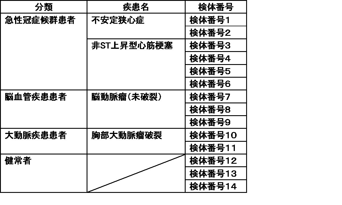
Claims (17)
- 動脈硬化性疾患の診断、重症度の評価、発症もしくは重症化の予測、または発症リスクもしくは重症化リスクの評価に用いるための試薬であって、
好中球由来のタンパク質またはその対応する結合分子と結合した複合体に対する抗体またはその抗原結合フラグメントを含む、試薬。 - 前記動脈硬化性疾患が、急性冠症候群、脳血管疾患、および大動脈疾患からなる群から選択される少なくとも1種である、請求項1に記載の試薬。
- 前記急性冠症候群が、急性心筋梗塞、不安定狭心症、および虚血による心臓突然死からなる群から選択される少なくとも1種である、請求項2に記載の試薬。
- 前記急性冠症候群が、急性心筋梗塞である、請求項3に記載の試薬。
- 前記急性心筋梗塞が、非ST上昇型心筋梗塞である、請求項4に記載の試薬。
- 前記脳血管疾患が、脳動脈瘤、ラクナ梗塞、アテローム血栓性脳梗塞、および心原性脳塞栓症からなる群から選択される少なくとも1種である、請求項2に記載の試薬。
- 前記大動脈疾患が、大動脈瘤、および大動脈解離からなる群から選択される少なくとも1種である、請求項2に記載の試薬。
- アテローム性動脈硬化の状態を評価するための試薬であって、
好中球由来のタンパク質またはその対応する結合分子と結合した複合体に対する抗体またはその抗原結合フラグメントを含む、試薬。 - 前記好中球由来のタンパク質が、好中球エラスターゼである、請求項1~8のいずれかに記載の試薬。
- 前記試薬が、免疫学的測定用試薬である、請求項1~9のいずれかに記載の試薬。
- 前記免疫学的測定用試薬が、酵素免疫測定法、蛍光酵素免疫測定法、化学発光酵素免疫測定法、化学発光免疫測定法、電気化学発光測免疫測定法、蛍光抗体法、ラジオイムノアッセイ、ウェスタンブロット法、イムノブロット法、ラテックス凝集法、イムノクロマトグラフィー法、およびネフェロメトリー法からなる群から選択される少なくとも1種の免疫学的測定法に用いるための試薬である、請求項10に記載の試薬。
- 動脈硬化性疾患を診断、その重症度を評価、その発症もしくは重症化を予測、またはその発症リスクもしくは重症化リスクを評価するための方法であって、
(1)試料と、好中球由来のタンパク質またはその対応する結合分子と結合した複合体に対する抗体またはその抗原結合フラグメントを接触させること、
(2)該好中球由来のタンパク質またはその対応する結合分子と結合した複合体と、該抗体またはその抗原結合フラグメントとの結合状態を測定すること
を含む、方法。 - 前記動脈硬化性疾患が、急性冠症候群、脳血管疾患、および大動脈疾患からなる群から選択される少なくとも1種である、請求項12に記載の方法。
- アテローム性動脈硬化の状態を評価するための方法であって、
(1)試料と、好中球由来のタンパク質またはその対応する結合分子と結合した複合体に対する抗体またはその抗原結合フラグメントを接触させること、
(2)該好中球由来のタンパク質またはその対応する結合分子と結合した複合体と、該抗体またはその抗原結合フラグメントとの結合状態を測定すること
を含む、方法。 - 前記好中球由来のタンパク質が、好中球エラスターゼである、請求項12~14のいずれかに記載の方法。
- 前記結合状態の測定方法が、酵素免疫測定法、蛍光酵素免疫測定法、化学発光酵素免疫測定法、化学発光免疫測定法、電気化学発光測免疫測定法、蛍光抗体法、ラジオイムノアッセイ、ウェスタンブロット法、イムノブロット法、ラテックス凝集法、イムノクロマトグラフィー法、およびネフェロメトリー法からなる群から選択される少なくとも1種の免疫学的測定法である、請求項12~15のいずれかに記載の方法。
- 前記試料が、血漿、血清、および全血からなる群から選択される少なくとも1種である、請求項12~16のいずれかに記載の方法。
Priority Applications (1)
| Application Number | Priority Date | Filing Date | Title |
|---|---|---|---|
| JP2025506921A JPWO2024190857A1 (ja) | 2023-03-15 | 2024-03-14 |
Applications Claiming Priority (2)
| Application Number | Priority Date | Filing Date | Title |
|---|---|---|---|
| JP2023-040611 | 2023-03-15 | ||
| JP2023040611 | 2023-03-15 |
Publications (1)
| Publication Number | Publication Date |
|---|---|
| WO2024190857A1 true WO2024190857A1 (ja) | 2024-09-19 |
Family
ID=92755264
Family Applications (1)
| Application Number | Title | Priority Date | Filing Date |
|---|---|---|---|
| PCT/JP2024/009946 Ceased WO2024190857A1 (ja) | 2023-03-15 | 2024-03-14 | 動脈硬化性疾患を診断するための試薬および方法 |
Country Status (2)
| Country | Link |
|---|---|
| JP (1) | JPWO2024190857A1 (ja) |
| WO (1) | WO2024190857A1 (ja) |
Citations (8)
| Publication number | Priority date | Publication date | Assignee | Title |
|---|---|---|---|---|
| JP2002048790A (ja) * | 2000-08-03 | 2002-02-15 | Ikagaku:Kk | 動脈硬化性病変の診断用キット |
| JP2004520598A (ja) * | 2001-05-04 | 2004-07-08 | バイオサイト インコーポレイテッド | 急性冠状動脈症候群の診断マーカーおよびその使用方法 |
| WO2010018870A1 (ja) * | 2008-08-15 | 2010-02-18 | 藤倉化成株式会社 | 動脈硬化診断用ポリペプチドマーカー、及び該マーカー等を用いる動脈硬化の検出方法、並びに動脈硬化診断用キット |
| US20110059103A1 (en) * | 2007-09-10 | 2011-03-10 | Ericus Anna Leonardus Biessen | Future cardiac event biomarkers |
| JP2011528792A (ja) * | 2008-07-23 | 2011-11-24 | ベー.エル.アー.ハー.エム.エス ゲゼルシャフト ミット ベシュレンクテル ハフツング | 心臓病性疾患におけるマーカーとしての好アズール性顆粒 |
| JP2016505847A (ja) * | 2012-12-28 | 2016-02-25 | 株式会社Lsiメディエンス | リスク層別化、診断、及び予後のためのsCD14又はその断片若しくは誘導体の使用 |
| US20170219608A1 (en) * | 2014-05-22 | 2017-08-03 | Wenbo HU | Kit for rapidly testing myocardial infarction and a preparation method and an application thereof |
| WO2017167763A1 (en) * | 2016-03-29 | 2017-10-05 | INSERM (Institut National de la Santé et de la Recherche Médicale) | Methods for diagnosis of haemorrhagic atherothrombotic plaques |
-
2024
- 2024-03-14 WO PCT/JP2024/009946 patent/WO2024190857A1/ja not_active Ceased
- 2024-03-14 JP JP2025506921A patent/JPWO2024190857A1/ja active Pending
Patent Citations (8)
| Publication number | Priority date | Publication date | Assignee | Title |
|---|---|---|---|---|
| JP2002048790A (ja) * | 2000-08-03 | 2002-02-15 | Ikagaku:Kk | 動脈硬化性病変の診断用キット |
| JP2004520598A (ja) * | 2001-05-04 | 2004-07-08 | バイオサイト インコーポレイテッド | 急性冠状動脈症候群の診断マーカーおよびその使用方法 |
| US20110059103A1 (en) * | 2007-09-10 | 2011-03-10 | Ericus Anna Leonardus Biessen | Future cardiac event biomarkers |
| JP2011528792A (ja) * | 2008-07-23 | 2011-11-24 | ベー.エル.アー.ハー.エム.エス ゲゼルシャフト ミット ベシュレンクテル ハフツング | 心臓病性疾患におけるマーカーとしての好アズール性顆粒 |
| WO2010018870A1 (ja) * | 2008-08-15 | 2010-02-18 | 藤倉化成株式会社 | 動脈硬化診断用ポリペプチドマーカー、及び該マーカー等を用いる動脈硬化の検出方法、並びに動脈硬化診断用キット |
| JP2016505847A (ja) * | 2012-12-28 | 2016-02-25 | 株式会社Lsiメディエンス | リスク層別化、診断、及び予後のためのsCD14又はその断片若しくは誘導体の使用 |
| US20170219608A1 (en) * | 2014-05-22 | 2017-08-03 | Wenbo HU | Kit for rapidly testing myocardial infarction and a preparation method and an application thereof |
| WO2017167763A1 (en) * | 2016-03-29 | 2017-10-05 | INSERM (Institut National de la Santé et de la Recherche Médicale) | Methods for diagnosis of haemorrhagic atherothrombotic plaques |
Non-Patent Citations (4)
| Title |
|---|
| BERNADET PH., BONNET J., COUFFINHAL TH., TOURTOULOU V., BENCHIMOL D., DROUILLET F., CROCKETT R., BRICAUD H.: "Evaluation of Plasmatic Leucocyte Elastase Levels in Coronary Artery Disease.", JAPANESE HEART JOURNAL., JAPANESE HEART JOURNAL ASSOCIATION., JP, vol. 33, no. 2, 1 January 1992 (1992-01-01), JP , pages 159 - 168, XP093212462, ISSN: 0021-4868, DOI: 10.1536/ihj.33.159 * |
| KORAI MASAAKI, PURCELL JAMES, KAMIO YOSHINOBU, MITSUI KAZUHA, FURUKAWA HAJIME, YOKOSUKA KIMIHIKO, MIYAMOTO TAKESHI, SATO HITOMI, S: "Neutrophil Extracellular Traps Promote the Development of Intracranial Aneurysm Rupture", HYPERTENSION, LIPPINCOTT WILLIAMS & WILKINS, US, vol. 77, no. 6, 1 June 2021 (2021-06-01), US , pages 2084 - 2093, XP093212452, ISSN: 0194-911X, DOI: 10.1161/HYPERTENSIONAHA.120.16252 * |
| MURATA, NOBUHIRO: "Etiology, pathology, and pathogenesis of acute myocardial infarction", JAPANESE JOURNAL OF CLINICAL MEDICINE, NIPPON-RINSHO CO., OSAKA., JP, vol. 74, no. Special issue 6, 1 January 2016 (2016-01-01), JP , pages 88 - 92, XP009558715, ISSN: 0047-1852 * |
| PLANA EMMA, OTO JULIA, MEDINA PILAR, FERNÁNDEZ-PARDO ÁLVARO, MIRALLES MANUEL: "Novel contributions of neutrophils in the pathogenesis of abdominal aortic aneurysm, the role of neutrophil extracellular traps: A systematic review", THROMBOSIS RESEARCH, ELSEVIER, AMSTERDAM, NL, vol. 194, 1 October 2020 (2020-10-01), AMSTERDAM, NL , pages 200 - 208, XP093212455, ISSN: 0049-3848, DOI: 10.1016/j.thromres.2020.07.039 * |
Also Published As
| Publication number | Publication date |
|---|---|
| JPWO2024190857A1 (ja) | 2024-09-19 |
Similar Documents
| Publication | Publication Date | Title |
|---|---|---|
| JP5031928B2 (ja) | 糖タンパク質の測定方法、肝疾患の検査方法、糖タンパク質定量用試薬および肝疾患病態指標糖鎖マーカー糖タンパク質 | |
| JP4423375B2 (ja) | 急性腎障害及び予後推定用バイオマーカー並びにその用途 | |
| US8603829B2 (en) | Atherosclerosis marker and use thereof | |
| EP2295966B1 (en) | IgA NEPHROPATHY DETECTION METHOD AND DETECTION KIT | |
| JP2009020049A (ja) | 脳血管疾患の診断方法 | |
| WO2018029214A1 (en) | Histones and/or proadm as markers indicating an adverse event | |
| WO2009090882A1 (ja) | 非アルコール性脂肪性肝疾患の診断方法 | |
| WO2018029213A1 (en) | Histones and/or proadm as markers indicating organ dysfunction | |
| WO2013183596A1 (ja) | 腎疾患のバイオマーカー及びその用途 | |
| Piedrafita et al. | A universal predictive and mechanistic urinary peptide signature in acute kidney injury | |
| AU2011311765B2 (en) | A new method for diagnosing hypertension as well as cardiomyopathies | |
| JP5280214B2 (ja) | 炎症性腸疾患の診断方法 | |
| US20110143382A1 (en) | IgA Nephropathy Testing Method And Test Kit | |
| JP6606552B2 (ja) | 特異的に精製された抗プレセプシン抗体 | |
| WO2024190857A1 (ja) | 動脈硬化性疾患を診断するための試薬および方法 | |
| WO2013050731A1 (en) | Prognostic method for diabetes | |
| WO2017001841A1 (en) | Method of detecting or monitoring an igg4-related disease by detecting igg4 kappa/lambda hybrid antibodies | |
| WO2015064348A1 (ja) | 糖鎖不全ヒトIgA1ヒンジ部を認識するモノクローナル抗体及びその用途 | |
| WO2023132348A1 (ja) | 急性呼吸窮迫症候群を診断するための試薬および方法 | |
| JP2009288219A (ja) | 心血管イベント発症リスクの診断方法 | |
| JP2009014521A (ja) | 睡眠時無呼吸症候群の診断方法 | |
| JP2024039982A (ja) | 急性腎障害の発症リスクの評価方法 | |
| JP5593502B2 (ja) | ケマリン濃度の測定方法 | |
| WO2017067821A1 (en) | A method for determining presence or risk of hemostasis disorder | |
| JP2011257147A (ja) | データ収集方法、キット及び腫瘍マーカー |
Legal Events
| Date | Code | Title | Description |
|---|---|---|---|
| 121 | Ep: the epo has been informed by wipo that ep was designated in this application |
Ref document number: 24770960 Country of ref document: EP Kind code of ref document: A1 |
|
| ENP | Entry into the national phase |
Ref document number: 2025506921 Country of ref document: JP Kind code of ref document: A |
|
| WWE | Wipo information: entry into national phase |
Ref document number: 2025506921 Country of ref document: JP |
|
| NENP | Non-entry into the national phase |
Ref country code: DE |
|
| 122 | Ep: pct application non-entry in european phase |
Ref document number: 24770960 Country of ref document: EP Kind code of ref document: A1 |