WO2024201691A1 - Procédé de criblage de composé induisant l'autophagie - Google Patents
Procédé de criblage de composé induisant l'autophagie Download PDFInfo
- Publication number
- WO2024201691A1 WO2024201691A1 PCT/JP2023/012419 JP2023012419W WO2024201691A1 WO 2024201691 A1 WO2024201691 A1 WO 2024201691A1 JP 2023012419 W JP2023012419 W JP 2023012419W WO 2024201691 A1 WO2024201691 A1 WO 2024201691A1
- Authority
- WO
- WIPO (PCT)
- Prior art keywords
- dead
- rna helicase
- box rna
- autophagy
- compound
- Prior art date
- Legal status (The legal status is an assumption and is not a legal conclusion. Google has not performed a legal analysis and makes no representation as to the accuracy of the status listed.)
- Ceased
Links
Images


Classifications
-
- C—CHEMISTRY; METALLURGY
- C12—BIOCHEMISTRY; BEER; SPIRITS; WINE; VINEGAR; MICROBIOLOGY; ENZYMOLOGY; MUTATION OR GENETIC ENGINEERING
- C12N—MICROORGANISMS OR ENZYMES; COMPOSITIONS THEREOF; PROPAGATING, PRESERVING, OR MAINTAINING MICROORGANISMS; MUTATION OR GENETIC ENGINEERING; CULTURE MEDIA
- C12N9/00—Enzymes; Proenzymes; Compositions thereof; Processes for preparing, activating, inhibiting, separating or purifying enzymes
- C12N9/90—Isomerases (5.)
-
- A—HUMAN NECESSITIES
- A61—MEDICAL OR VETERINARY SCIENCE; HYGIENE
- A61P—SPECIFIC THERAPEUTIC ACTIVITY OF CHEMICAL COMPOUNDS OR MEDICINAL PREPARATIONS
- A61P35/00—Antineoplastic agents
-
- C—CHEMISTRY; METALLURGY
- C12—BIOCHEMISTRY; BEER; SPIRITS; WINE; VINEGAR; MICROBIOLOGY; ENZYMOLOGY; MUTATION OR GENETIC ENGINEERING
- C12N—MICROORGANISMS OR ENZYMES; COMPOSITIONS THEREOF; PROPAGATING, PRESERVING, OR MAINTAINING MICROORGANISMS; MUTATION OR GENETIC ENGINEERING; CULTURE MEDIA
- C12N15/00—Mutation or genetic engineering; DNA or RNA concerning genetic engineering, vectors, e.g. plasmids, or their isolation, preparation or purification; Use of hosts therefor
- C12N15/09—Recombinant DNA-technology
- C12N15/11—DNA or RNA fragments; Modified forms thereof; Non-coding nucleic acids having a biological activity
- C12N15/52—Genes encoding for enzymes or proenzymes
-
- C—CHEMISTRY; METALLURGY
- C12—BIOCHEMISTRY; BEER; SPIRITS; WINE; VINEGAR; MICROBIOLOGY; ENZYMOLOGY; MUTATION OR GENETIC ENGINEERING
- C12Q—MEASURING OR TESTING PROCESSES INVOLVING ENZYMES, NUCLEIC ACIDS OR MICROORGANISMS; COMPOSITIONS OR TEST PAPERS THEREFOR; PROCESSES OF PREPARING SUCH COMPOSITIONS; CONDITION-RESPONSIVE CONTROL IN MICROBIOLOGICAL OR ENZYMOLOGICAL PROCESSES
- C12Q1/00—Measuring or testing processes involving enzymes, nucleic acids or microorganisms; Compositions therefor; Processes of preparing such compositions
- C12Q1/02—Measuring or testing processes involving enzymes, nucleic acids or microorganisms; Compositions therefor; Processes of preparing such compositions involving viable microorganisms
- C12Q1/24—Methods of sampling, or inoculating or spreading a sample; Methods of physically isolating an intact microorganisms
-
- C—CHEMISTRY; METALLURGY
- C12—BIOCHEMISTRY; BEER; SPIRITS; WINE; VINEGAR; MICROBIOLOGY; ENZYMOLOGY; MUTATION OR GENETIC ENGINEERING
- C12Q—MEASURING OR TESTING PROCESSES INVOLVING ENZYMES, NUCLEIC ACIDS OR MICROORGANISMS; COMPOSITIONS OR TEST PAPERS THEREFOR; PROCESSES OF PREPARING SUCH COMPOSITIONS; CONDITION-RESPONSIVE CONTROL IN MICROBIOLOGICAL OR ENZYMOLOGICAL PROCESSES
- C12Q1/00—Measuring or testing processes involving enzymes, nucleic acids or microorganisms; Compositions therefor; Processes of preparing such compositions
- C12Q1/25—Measuring or testing processes involving enzymes, nucleic acids or microorganisms; Compositions therefor; Processes of preparing such compositions involving enzymes not classifiable in groups C12Q1/26 - C12Q1/66
-
- G—PHYSICS
- G01—MEASURING; TESTING
- G01N—INVESTIGATING OR ANALYSING MATERIALS BY DETERMINING THEIR CHEMICAL OR PHYSICAL PROPERTIES
- G01N2500/00—Screening for compounds of potential therapeutic value
- G01N2500/02—Screening involving studying the effect of compounds C on the interaction between interacting molecules A and B (e.g. A = enzyme and B = substrate for A, or A = receptor and B = ligand for the receptor)
-
- G—PHYSICS
- G01—MEASURING; TESTING
- G01N—INVESTIGATING OR ANALYSING MATERIALS BY DETERMINING THEIR CHEMICAL OR PHYSICAL PROPERTIES
- G01N2500/00—Screening for compounds of potential therapeutic value
- G01N2500/04—Screening involving studying the effect of compounds C directly on molecule A (e.g. C are potential ligands for a receptor A, or potential substrates for an enzyme A)
-
- G—PHYSICS
- G01—MEASURING; TESTING
- G01N—INVESTIGATING OR ANALYSING MATERIALS BY DETERMINING THEIR CHEMICAL OR PHYSICAL PROPERTIES
- G01N2500/00—Screening for compounds of potential therapeutic value
- G01N2500/10—Screening for compounds of potential therapeutic value involving cells
Definitions
- the present invention relates to a method for screening compounds that induce autophagy and a method for producing compounds that induce the degradation of target intracellular molecules or organelles by autophagic processes.
- Autophagy is one of the mechanisms by which intracellular molecules are broken down within cells, and is a mechanism found in eukaryotes from yeast to humans.
- autophagy When autophagy is induced, a membrane vesicle called an autophagosome is formed, which then fuses with a lysosome, resulting in the degradation of the molecules taken up into the autophagosome.
- autophagy is induced when cells enter a state of starvation, and it has become clear that autophagy is also involved in physiological functions such as development and differentiation, as well as infection defense functions such as the elimination of viruses that have invaded cells.
- Non-Patent Document 1 The mechanism by which autophagy is induced has also been elucidated through various reports.
- Mohammad Omar Faruk et al. report in detail the mechanism of selective autophagy, which contributes to cellular homeostasis by degrading specific soluble proteins, supramolecular complexes, liquid-phase separated droplets, abnormal or excessive organelles, pathogenic invading bacteria, etc.
- p62 binds to ubiquitinated proteins, causing liquid-phase separation, forming droplets in which p62 and ubiquitinated proteins are condensed, and these droplets recruit other proteins to form isolation membranes/phagocytic membranes, and p62 and other proteins in the droplets are degraded in lysosomes.
- Non-Patent Document 2 Lin Ma et al. suggest that knockdown of DDX5 suppresses endoplasmic reticulum stress and promotes recovery of autophagic flux in esophageal squamous cell carcinoma (ESCC) (Non-Patent Document 2).
- DDX5 stimulates autophagy and suppresses the development of liver cancer by interacting with the autophagy receptor p62 (Non-Patent Document 3). They also reported that DDX5 promotes the degradation of p62 and significantly reduces its half-life.
- Non-Patent Document 4 Yongsang Jo et al., in a report on intrinsically disordered protein-protein interactions in membrane-free protein droplets, disclosed that they formed DDX4 droplets by adding streptavidin.
- degraders protein degradation targeting chimeras
- SNIPERs specific non-genetic IAP-dependent protein eliminators
- UPS ubiquitin-proteasome system
- [6] A method for producing a compound that induces degradation of a target intracellular molecule or organelle by autophagic degradation, comprising linking, via a linker, a ligand that specifically binds to the target intracellular molecule or organelle to the compound obtained by the method according to any one of [1] to [5].
- [7] A method for inducing autophagic degradation of a target intracellular molecule or organelle by contacting a compound obtained by the method according to [6] with a DEAD-box RNA helicase in a cell in the presence of p62.
- an autophagy-inducing compound can be efficiently obtained, and by linking this as a degradation tag moiety via a linker to a ligand that specifically binds to a target intracellular molecule, a compound that induces the degradation of a target molecule or organelle by autophagic degradation can be synthesized.
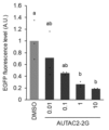
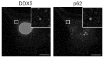
- Examples 1 to 4 the amount of light emitted 10 minutes after the start of luminescence measurement is shown. Shown are EGFP fluorescence levels when cells transfected with pEGFP-FKBP12 were cultured in fresh DMEM containing 0.01 mM, 0.1 mM, 1 mM, and 10 mM AUTAC2-2G, respectively, and in fresh DMEM containing DMSO as a control. HeLa cells transiently expressing GFP-FKBP F36V were treated with SLF'-AUTAC, fixed with paraformaldehyde, and immunostained with anti-DDX5 or anti-p62 antibodies, and images were observed under a fluorescent microscope.
- FIG. 4A shows an image observed with a fluorescent microscope when HeLa cells transiently expressing GFP-FKBP12 and RFP-DDX5 were treated with AUTAC2-2G
- FIG. 4B shows an image observed with a fluorescent microscope when HeLa cells transiently expressing GFP-FKBP12 and RFP-p62 were treated with AUTAC2-2G.
- the screening method according to the present invention screens for autophagy-inducing compounds by utilizing at least one of the following properties: (1) It has high binding affinity to DEAD-box RNA helicase. (2) It brings DEAD-box RNA helicase and p62 into close proximity to promote their interaction. (3) Promotes droplet formation of DEAD-box RNA helicase and p62. As demonstrated in the Examples described below, compounds having autophagy-inducing activity have these properties, and therefore, autophagy-inducing compounds can be obtained by screening for compounds having at least one of these properties.
- DEAD-box RNA helicase is a superfamily of RNA helicases that have the motif DEAD (Asp-Glu-Ala-Asp). DEAD-box RNA helicases are known to be involved in the disassembly and formation of double-stranded RNA and RNA-protein complexes using the energy of ATP hydrolysis, and to play important roles in transcription, splicing, nuclear export and degradation of RNA, translation, and ribosome biogenesis.
- DEAD-box RNA helicases include DDX4 (DEAD box protein 4), DDX5 (DEAD box protein 5) and DDX17 (DEAD box protein 17), with DEAD-Box Helicase 5 (DDX5) also known as P68.
- amino acid sequences of DDX5 and DDX17 are as follows:
- Method for measuring binding affinity Measurement can be performed by surface plasmon resonance assay.
- an autophagy-inducing compound is immobilized on a thin gold film of a sensor chip, and a purified DEAD-box RNA helicase protein solution is poured onto the surface of the sensor chip, and the SPR signal that changes depending on the binding state of the two can be measured by BIAcore.
- measurement can also be performed by detecting the change in thermal stability of the DEAD-box RNA helicase protein due to binding with an autophagy-inducing compound using a thermal shift assay called CETSA.
- p62 p62 is a receptor protein that induces specific proteins and organelles into autophagy. p62 is localized diffusely in the cytoplasm and nucleus, but has a half-life of about 10 hours and is basically degraded in an autophagy-dependent manner.
- p62 has many domains, such as the N-terminal Phox1 and Bem1p domain (Phox1 and Bem1p: PB1), zinc finger (ZZ), TRAF6-binding domain (TB), LC3-interacting region (LIR, consisting of the DDDWTHL sequence), Keap1-interacting region (KIR), and C-terminal ubiquitin-associated domain (UBA), and interacts with LC3, an autophagosome-localized protein, at the LC3-interacting region (LIR) to induce autophagy (see Non-Patent Document 13).
- the amino acid sequence of p62 or the nucleic acid sequence of the SQSTM1 gene encoding it is as follows:
- Method for detecting the interaction (proximation) between DEAD-box RNA helicase and p62 there is no particular limitation on the method for detecting the interaction (proximation) between DEAD-box RNA helicase and p62, and any available method may be used.
- the method can be performed using NanoLuc (registered trademark) Binary Technology Assays (NanoBiT (registered trademark) Assays) according to the reaction scheme shown below.
- the term "droplet” refers to a liquid-like aggregate in which DEAD-box RNA helicase, a compound associated therewith, and p62 are concentrated and are separated into a liquid-liquid phase from the outside.
- Such "droplets” can be confirmed by labeling DEAD-box RNA helicase and/or p62 with a fluorescent label or the like, introducing a test compound into a system in which they exist, preferably treating cells expressing at least one of them with the test compound, and observing the presence of a substance visualized by the label using a fluorescent microscope, etc.
- a compound that induces autophagic degradation of a target intracellular molecule can be produced by linking, via a linker, a ligand that specifically binds to a target intracellular molecule or organelle to a compound obtained by the above-mentioned screening method.
- target intracellular molecule or organelle refers to an intracellular molecule or organelle targeted for degradation by autophagy.
- Intracellular molecule refers to a biomolecule at least a portion of which is present within the cell, and includes not only naturally occurring biomolecules present within the cell, but also molecules artificially introduced or expressed within the cell.
- organelle includes not only naturally occurring organelles present within the cell (e.g., dysfunctional mitochondria), but also foreign viruses and microorganisms that have invaded the cell.
- intracellular molecules include lipids, glycolipids, proteins, glycoproteins, etc.
- Intracellular proteins present within cells, and are typically proteins present within cells, including protein aggregates such as amyloid beta (A ⁇ ), tau protein, and ⁇ -synuclein.
- a ⁇ amyloid beta
- tau protein tau protein
- ⁇ -synuclein ⁇ -synuclein
- intracellular proteins intracellular proteins associated with pathology (particularly, intracellular proteins associated with human pathology) are cited as practical targets, and many reports have been published. Intracellular proteins associated with such pathological conditions can be identified, for example, by UniProt or Human Protein Atlas.
- Some examples include A ⁇ , tau protein, ⁇ -synuclein or aggregates thereof (involved in neurodegenerative diseases such as Alzheimer's disease and Parkinson's disease), BRD4 (involved in multiple myeloma, acute myeloid leukemia, etc.), Ras (involved in colon cancer, pancreatic cancer, etc.), calcineurin, CyP, FRB, FKBP, PyL, ABI1, GID1, GA1, FKBP F36V, IAA17 TIR1, EGFR, and BTK (B.Z. Stanton et al. Science; 359; eaao5902 (2016), N. Shindo et al. Bioorg. Med.
- Intracellular molecules also include intracellular proteins that can bind to a ligand, as described below, by fusing a specific tag to any target intracellular molecule or organelle using tagging techniques such as HaloTag (registered trademark), SNAP Tag (registered trademark), and CLIP Tag (registered trademark).
- tagging techniques such as HaloTag (registered trademark), SNAP Tag (registered trademark), and CLIP Tag (registered trademark).
- An intracellular protein fused with HaloTag (registered trademark) is fused with a tag having the F272H mutant of dehalogenase DhaA derived from Rhodococcus rhodochrous
- an intracellular protein fused with SNAP Tag (registered trademark) is fused with a tag having the hATG mutant of human-derived O 6 -alkylguanine-DNA alkyltransferase
- an intracellular protein fused with CLIP Tag registered trademark
- is fused with a tag having the E30R mutant of a SNAP tagged protein are outlined, for example, in Halo SNAP CLIP for Protein Labeling - Bitesize Bio, the contents of which are incorporated herein by reference.
- intracellular proteins tagged with mutant FKBPs such as ⁇ -synuclein or aggregates thereof, can also be targeted intracellular molecules for autophagy.
- ligand that specifically binds to an intracellular molecule or organelle means a structural unit that constitutes part of a compound that induces the degradation of a target intracellular molecule or organelle by autophagy and has specific binding activity to a target intracellular molecule (including the above-mentioned tagged fusion protein) or organelle. Therefore, the substance that constitutes the ligand is a substance that has specific binding activity to an intracellular molecule or organelle, and there have been many reports on such substances.
- BindingDB https://www.bindingdb.org/rwd/bind/index.jsp
- FK506 calcineurin
- CyP cyclosporin A
- FKCsA CyP, FKBP
- rapamycin FRB, FKBP
- FK1012 FKBP
- abscisic acid PyI, ABI1
- gibberellin 3 GID1, GA1
- AP1903/AP20187 FKBP F36V
- Auxin IAA17 TIR1
- afatinib EGFR
- osimertinib EGFR
- dacomitinib EGFR
- neratinib neratinib
- sotrasib ibrutinib
- BK acalabrutinib
- zanubrutinib BTK
- tirabrutinib ibrutinib
- examples of ligands for BRD4 include Examples of the ligand for FKBP12 include Examples of the ligand for MetAP2 include Examples include:
- ligand compounds for Ras include ligands described in the following documents: Nature, 2013, 503, 548-551; International Publication No. 2013/155223; Science, 2016, 351, 604-608; International Publication No. WO2014152588; International Publication No. 2015/054572; International Publication No. 2016/049524; Nat. Rev. Drug Discov., 2014, 13, 828-851; Chem. Soc. Rev., 2016, advance article (DOI: 10.1039/C5CS00911A). Therefore, the ligand for Ras can be composed of these compounds. Examples of ligands for Ras include, Examples include:
- Ligands for intracellular proteins fused with Halotag include, for example, halogenated C 1-12 alkoxy groups (particularly, C 1-6 alkoxy groups with a halogenated terminal carbon), preferably halogenated (particularly, halogenated terminal carbon) hexyloxy groups (particularly, chlorinated hexyloxy groups).
- An example of a ligand for an intracellular protein fused with a SNAP tag (registered trademark) is O 6 -benzylguanine.
- An example of a ligand for an intracellular protein fused with a CLIP tag® is O 6 -benzylcytosine.
- Ligands for intracellular proteins fused with tags are generally described in Halo SNAP CLIP for Protein Labeling--Bitsize Bio, the contents of which are incorporated herein by reference.
- examples of ligands for target intracellular molecules tagged with mutant FKBP include: Examples include:
- linker refers to an atomic group that links the "ligand” portion of the compound of the present invention to the tag portion that induces autophagy.
- the linker structure can be a known linker structure that is used to link molecules with different functions.
- Japanese Patent No. 7161760 and Non-Patent Documents 5, 9, and 10 provide detailed descriptions of linker structures that link intracellular molecules to tag portions that induce autophagy, and the structures described in these documents can be used.
- linker can be linked to the ligand and the tag moiety using known reactions.
- Patent No. 7161760 Non-Patent Documents 5, 9, and 10; 5th Edition Experimental Chemistry Lectures, Vol. 13-19 (edited by the Chemical Society of Japan); New Experimental Chemistry Lectures, Vol. 14-15 (edited by the Chemical Society of Japan); Precision Organic Chemistry Revised 2nd Edition (L. F. Tietz, Th.
- Reactions that can be used include protection or deprotection reactions of functional groups, reduction reactions, oxidation reactions, aromatic nucleophilic substitution reactions, azidation reactions of alcohols, alkyl halides, and sulfonate esters, reductive amination reactions, Mitsunobu reactions, esterification reactions, amidation reactions, urea reactions, coupling reactions, Wohl-Ziegler reactions, sulfone esterification reactions, hydrolysis reactions, bromination reactions, N-alkylation reactions, hydroxylation reactions via diazotization, deamination reactions, cyclization reactions, carbamate reactions, amidation by carbon monoxide insertion reactions, esterification, click reactions, and the like, and the reagents and reaction conditions necessary for these reactions are well known to those skilled in the art to which the present invention pertains.
- the compounds obtained by the above manufacturing method have a ligand that specifically binds to a target molecule or organelle and a tag moiety that induces autophagy linked via a linker, and have the advantage that they can target a wide range of substrates by replacing the ligand that binds to them depending on the target molecule or organelle.
- the compounds of the present invention are useful as preventive or therapeutic agents for diseases involving targeted intracellular molecules or organelles.
- the compounds of the present invention may be effective in preventing or treating any disease involving targeted intracellular molecules or organelles.
- they are expected to be applied to the treatment or prevention of cancer, inflammatory diseases, autoimmune diseases, and bone and joint degenerative diseases.
- p62 cDNA was synthesized from HeLa total RNA using oligo(dT) 15 primer, amplified using oligo primers (5'-agcggtaggcatggcgtcgctcaccg-3' and 5'-agaagatctgctagtacaacggcgggatgc-3') and inserted into the vector using the In-fusion cloning system (the underlined 15 nt is the In-fusion arm).
- the pBiT2.1-N [TK/SmBiT] Vector (Promega #N2014) was linearized by polymerase chain reaction using oligo primers (5'-ggtggctttaccaacagtaccg-3' and 5'- gctagcagatcttctagtcgg-3').
- DDX5 cDNA was synthesized from HeLa total RNA using oligo(dT) 15 primer, amplified using oligo primers (5'-agcggtggcatgtcgggattcgagtgaccg-3' and 5'- agaagatctgctagcttattgggaatcctgttggcattg-3') and inserted into the vector using the In-fusion cloning system (the underlined 15 nt is the In-fusion arm).
- nucleic acid sequences of pLgBiT-p62 and pSmBiT-DDX5 carried by each vector are shown below.
- HeLa cells RIKEN BRC Cell Bank #RCB0007
- DMEM Fujifilm Wako #043-30085
- FBS Thermo Fisher Scientific #10437028
- Non-essential amino acids Fujifilm/Wako #139-15651
- Opti-MEM I Thermo Fisher Scientific #11058-021 96-well culture plate.
- Promega #E5650 Lipofectamine3000 transfection reagent Thermo Fisher Scientific #L3000008 Nano-Glo® Live Cell Reagent: Promega #N2014 GloMax Discover Microplate Reader:Promega #GM3000 PrimeSTAR Max DNA polymerase: TaKaRa #R045A TRIzol reagent (for total RNA extraction):Thermo Fisher Scientific #15596026 PrimeScriptTM RT reagent Kit (for cDNA synthesis): TaKaRa #RR037A In-Fusion HD Cloning Kit: TaKaRa #Z9649N
- Test Compounds The following compounds were used to verify the screening method of the present invention.
- AUTAC4-2G and 8-nitro-cGMP are compounds and their tag moieties disclosed in Japanese Patent No. 7,161,760
- AUTAC2-2G and FBnG are compounds and their tag moieties that were newly synthesized to verify whether compounds screened by the method of the present invention actually induce autophagy (the synthesis schemes of FBnG and AUTAC2-2G are as follows).
- Test procedure [Example 1] HeLa cells grown in 96-well plates containing growth medium were transfected with pLgBiT-p62 and pSmBiT-DDX5 using Lipofectamine3000® transfection reagent (Thermo Fisher Scientific Inc.) as described in the accompanying documentation. 24 hours after the addition of the reagent, AUTAC2-2G was added to the wells at a concentration of 10 ⁇ M and the transfected cells were treated with AUTAC2-2G for 12 hours. The growth medium was then replaced with 100 ⁇ L of Opti-MEM® I (Thermo Fisher Scientific Inc.).
- Nano-Glo containing the substrate furimazine was added to the wells at a concentration of 10 ⁇ M and the cells were then treated with AUTAC2-2G. 25 ⁇ L of Live Cell Reagent was added, and luminescence was immediately measured using a GloMax® Discover Microplate Reader (Promega Corporation).
- Example 2 The cells were treated and luminescence was measured in the same manner as in Example 1, except that AUTAC4-2G was added to the wells at a concentration of 10 ⁇ M and the transfected cells were treated with AUTAC4-2G for 12 hours.
- Example 3 The cells were treated and luminescence was measured as in Example 1, except that 8-nitro-cGMP was added to the wells to a concentration of 10 ⁇ M and the transfected cells were treated with 8-nitro-cGMP for 24 hours.
- Example 4 The cells were treated and luminescence was measured in the same manner as in Example 1, except that FBnG was added to the wells at a concentration of 10 ⁇ M and the transfected cells were treated with FBnG for 24 hours.
- Example 1 The cells were treated and luminescence was measured in the same manner as in Example 1, except that DMSO (dimethyl sulfoxide) was added to the wells to a concentration of 10 ⁇ M and the transfected cells were treated with DMSO for 24 hours.
- DMSO dimethyl sulfoxide
- Figure 1 shows the amount of luminescence 10 minutes after the start of luminescence measurement in Examples 1 to 4.
- the data are average values from three independent experiments.
- the graph shows an upper limit of 107, and in fact, the amount of luminescence was higher than that. Error bars indicate standard deviation.
- Statistical analysis was performed using the Turkey-Kramer multiple comparison test. A P value of ⁇ 0.01 was considered to be statistically significant.
- Cells treated with AUTAC2-2G, AUTAC4-2G, 8-nitro-cGMP, and FBnG showed 1,000-fold more luminescence than cells treated with DMSO, demonstrating that the presence of these compounds brought p62 and DDX5 into close proximity, indicating that this method can be used to screen for compounds that bring p62 and DDX5 into close proximity.
- pEGFP-FKBP12 was linearized by polymerase chain reaction using oligo primers (5'- cgcaaatctagacttgtacagctcgtcc-3' and 5'- ggccgcttcgagcagatg-3').
- FKBP12 cDNA was synthesized from HeLa cell total RNA using oligo(dT) 15 primer, amplified with oligo primers (5'-aagtctagatttgcgatgggagtgcaggtgg-3' and 5'-ctgctcgaagcggcctcattccagttagaagc-3'), and inserted into a vector using the infusion cloning system (the underlined 15 nt is the infusion arm).
- HeLa cells RIKEN BRC Cell Bank #RCB0007 Dulbecco's Modified Eagle Medium (DMEM): Fujifilm Wako #043-30085
- FBS Thermo Fisher Scientific #10437028
- Non-essential amino acids Fujifilm/Wako #139-15651
- Opti-MEM I Thermo Fisher Scientific #11058-021 96-well culture plate.
- Thermo Fisher Scientific #165305 Lipofectamine3000 transfection reagent Thermo Fisher Scientific #L3000008 Live Cell Imaging Solution: Thermo Fisher Scientific #A14291DJ Fluorescence microscope: Keyence #BZ-X800 PrimeSTAR Max DNA polymerase: TaKaRa #R045A TRIzol reagent (for total RNA extraction): Thermo Fisher Scientific #15596026 PrimeScriptTM RT reagent Kit (for cDNA synthesis): TaKaRa #RR037A In-Fusion HD Cloning Kit: TaKaRa #Z9649N
- Test procedure HeLa cells were grown on 96-well plates and transfected with pEGFP-FKBP12 using Lipofectamine 3000 transfection reagent. After 24 h, the transfected cells were incubated for an additional 24 h in fresh DMEM containing 0.01 mM AUTAC2-2G, as well as in fresh DMEM containing DMSO as a control. The medium was then removed and replaced with 100 ⁇ L of live cell imaging solution. EGFP-FKBP12 levels were analyzed by measuring EGFP fluorescence.
- Example 6 Cells were treated and EGFP fluorescence was measured as in Example 5, except that the transfected cells were incubated for an additional 24 hours in fresh DMEM containing 0.1 mM AUTAC2-2G.
- Example 7 Cells were treated and EGFP fluorescence was measured as in Example 5, except that the transfected cells were incubated for an additional 24 hours in fresh DMEM containing 1 mM AUTAC2-2G.
- Example 8 Cells were treated and EGFP fluorescence was measured as in Example 5, except that the transfected cells were incubated for an additional 24 hours in fresh DMEM containing 10 mM AUTAC2-2G.
- Test procedure HeLa cells transiently expressing GFP tagged with mutant FKBP (GFP-FKBP F36V ) were treated with 1 ⁇ M SLF'-AUTAC for 12 hours. The treated cells were fixed with 4% paraformaldehyde, permeabilized with 0.1% TritonX-100/PBS, blocked with 1% BSA/PBS, and incubated overnight at 4°C in PBS containing 1% each of rabbit anti-DDX5 antibody and mouse anti-p62 antibody. Then, the cells were incubated at room temperature for 1 hour in PBS containing 1% each of AlexaFluor555-labeled anti-rabbit IgG and AlexaFluor555-labeled anti-mouse IgG. The cells after immunostaining were observed under a fluorescent microscope.
- FIG. 3 shows images observed with a fluorescent microscope (scale bar: 10 ⁇ m).
- AUTAC2-2G was used to test whether it induces the formation of droplets containing DDX5 and p62.
- Test procedure HeLa cells transiently expressing GFP-FKBP12 and RFP-DDX5, or GFP-FKBP12 and RFP-p62 were treated with AUTAC2-2G at a concentration of 10 ⁇ M for 13 hours. The treated cells were observed live under a fluorescent microscope, and the fluorescence derived from GFP and RFP was continuously observed. During the course of the observation, 5% 1,6-hexanediol was added 5 minutes after the start of observation, and it was observed whether the dot-shaped structures in the field of view disappeared. 1,6-hexanediol is known to have the ability to dissolve droplets, making it possible to confirm that dot-like structures in the microscope field of view are droplets.
- Figure 4 shows an image observed with a fluorescent microscope when HeLa cells transiently expressing GFP-FKBP12 and RFP-DDX5 were treated with AUTAC2-2G
- Figure 4B shows an image observed with a fluorescent microscope when HeLa cells transiently expressing GFP-FKBP12 and RFP-p62 were treated with AUTAC2-2G.
- droplets containing GFP-FKBP12 and RFP-DDX5, and dot-like structures containing GFP-FKBP12 and RFP-p62 were observed, which were confirmed to be droplets because they disappeared after the addition of 1,6-hexanediol.
Landscapes
- Chemical & Material Sciences (AREA)
- Health & Medical Sciences (AREA)
- Life Sciences & Earth Sciences (AREA)
- Organic Chemistry (AREA)
- Engineering & Computer Science (AREA)
- Genetics & Genomics (AREA)
- Zoology (AREA)
- Wood Science & Technology (AREA)
- Bioinformatics & Cheminformatics (AREA)
- Biotechnology (AREA)
- Molecular Biology (AREA)
- Proteomics, Peptides & Aminoacids (AREA)
- General Engineering & Computer Science (AREA)
- General Health & Medical Sciences (AREA)
- Biochemistry (AREA)
- Microbiology (AREA)
- Physics & Mathematics (AREA)
- Biophysics (AREA)
- Biomedical Technology (AREA)
- Immunology (AREA)
- Analytical Chemistry (AREA)
- Medicinal Chemistry (AREA)
- Chemical Kinetics & Catalysis (AREA)
- Nuclear Medicine, Radiotherapy & Molecular Imaging (AREA)
- Pharmacology & Pharmacy (AREA)
- Animal Behavior & Ethology (AREA)
- Public Health (AREA)
- Veterinary Medicine (AREA)
- General Chemical & Material Sciences (AREA)
- Plant Pathology (AREA)
- Pathology (AREA)
- Measuring Or Testing Involving Enzymes Or Micro-Organisms (AREA)
Abstract
L'invention fournit un procédé de criblage destiné à chercher un nouveau composé induisant l'autophagie. L'invention a pour but de chercher un nouveau composé induisant l'autophagie en mettant en œuvre au moins une caractéristique parmi les suivantes : (1) pocéssion d'une affinité de liaison élevée vis-à-vis d'un ARN-hélicase type DEAD-box, (2) stimulation d'interactions par rapprochement de l'ARN-hélicase type DEAD-box et d'un p62, et (3) stimulation de la formation de gouttes d'ARN-hélicase type DEAD-box et p62.
Priority Applications (6)
| Application Number | Priority Date | Filing Date | Title |
|---|---|---|---|
| PCT/JP2023/012419 WO2024201691A1 (fr) | 2023-03-28 | 2023-03-28 | Procédé de criblage de composé induisant l'autophagie |
| CN202480022512.XA CN121002194A (zh) | 2023-03-28 | 2024-03-28 | 诱导自噬的化合物的筛选方法 |
| PCT/JP2024/012899 WO2024204648A1 (fr) | 2023-03-28 | 2024-03-28 | Procédé de criblage de composé induisant l'autophagie |
| KR1020257031424A KR20250169534A (ko) | 2023-03-28 | 2024-03-28 | 오토파지를 유도하는 화합물의 스크리닝 방법 |
| JP2025511227A JPWO2024204648A1 (fr) | 2023-03-28 | 2024-03-28 | |
| EP24780746.4A EP4692367A1 (fr) | 2023-03-28 | 2024-03-28 | Procédé de criblage de composé induisant l'autophagie |
Applications Claiming Priority (1)
| Application Number | Priority Date | Filing Date | Title |
|---|---|---|---|
| PCT/JP2023/012419 WO2024201691A1 (fr) | 2023-03-28 | 2023-03-28 | Procédé de criblage de composé induisant l'autophagie |
Publications (1)
| Publication Number | Publication Date |
|---|---|
| WO2024201691A1 true WO2024201691A1 (fr) | 2024-10-03 |
Family
ID=92904278
Family Applications (2)
| Application Number | Title | Priority Date | Filing Date |
|---|---|---|---|
| PCT/JP2023/012419 Ceased WO2024201691A1 (fr) | 2023-03-28 | 2023-03-28 | Procédé de criblage de composé induisant l'autophagie |
| PCT/JP2024/012899 Ceased WO2024204648A1 (fr) | 2023-03-28 | 2024-03-28 | Procédé de criblage de composé induisant l'autophagie |
Family Applications After (1)
| Application Number | Title | Priority Date | Filing Date |
|---|---|---|---|
| PCT/JP2024/012899 Ceased WO2024204648A1 (fr) | 2023-03-28 | 2024-03-28 | Procédé de criblage de composé induisant l'autophagie |
Country Status (5)
| Country | Link |
|---|---|
| EP (1) | EP4692367A1 (fr) |
| JP (1) | JPWO2024204648A1 (fr) |
| KR (1) | KR20250169534A (fr) |
| CN (1) | CN121002194A (fr) |
| WO (2) | WO2024201691A1 (fr) |
Citations (2)
| Publication number | Priority date | Publication date | Assignee | Title |
|---|---|---|---|---|
| WO2018143403A1 (fr) * | 2017-02-03 | 2018-08-09 | 国立大学法人東北大学 | Composé hétérocyclique |
| WO2019013181A1 (fr) * | 2017-07-10 | 2019-01-17 | 国立大学法人東北大学 | Agent de dégradation utilisant un mécanisme autophagique de mitochondries endommagées |
Family Cites Families (5)
| Publication number | Priority date | Publication date | Assignee | Title |
|---|---|---|---|---|
| CN104379563B (zh) | 2012-04-10 | 2018-12-21 | 加利福尼亚大学董事会 | 用于治疗癌症的组合物和方法 |
| TW201524952A (zh) | 2013-03-15 | 2015-07-01 | Araxes Pharma Llc | Kras g12c之共價抑制劑 |
| JP6559123B2 (ja) | 2013-10-10 | 2019-08-14 | アラクセス ファーマ エルエルシー | Krasg12cの阻害剤 |
| JP2017528498A (ja) | 2014-09-25 | 2017-09-28 | アラクセス ファーマ エルエルシー | Kras g12c変異体タンパク質のインヒビター |
| WO2024054876A1 (fr) | 2022-09-07 | 2024-03-14 | Arvinas Operations, Inc. | Composés de dégradation de kinase à séquence répétée riche en leucine 2 (lrrk2) et méthodes d'utilisation associées |
-
2023
- 2023-03-28 WO PCT/JP2023/012419 patent/WO2024201691A1/fr not_active Ceased
-
2024
- 2024-03-28 WO PCT/JP2024/012899 patent/WO2024204648A1/fr not_active Ceased
- 2024-03-28 CN CN202480022512.XA patent/CN121002194A/zh active Pending
- 2024-03-28 EP EP24780746.4A patent/EP4692367A1/fr active Pending
- 2024-03-28 KR KR1020257031424A patent/KR20250169534A/ko active Pending
- 2024-03-28 JP JP2025511227A patent/JPWO2024204648A1/ja active Pending
Patent Citations (2)
| Publication number | Priority date | Publication date | Assignee | Title |
|---|---|---|---|---|
| WO2018143403A1 (fr) * | 2017-02-03 | 2018-08-09 | 国立大学法人東北大学 | Composé hétérocyclique |
| WO2019013181A1 (fr) * | 2017-07-10 | 2019-01-17 | 国立大学法人東北大学 | Agent de dégradation utilisant un mécanisme autophagique de mitochondries endommagées |
Non-Patent Citations (1)
| Title |
|---|
| ARIMOTO, HIROKAZU; TAKAHASHI, DAIKI; ITTO, KAORI: "4SY24-5 Targeted degradation of cytosolic materials using selective autophagy", ANNUAL MEETING OF THE JAPAN SOCIETY FOR BIOSCIENCE, BIOTECHNOLOGY, AND AGROCHEMISTRY (JSBBA 2018), vol. 2018, 5 March 2018 (2018-03-05), pages 2101, XP009558719 * |
Also Published As
| Publication number | Publication date |
|---|---|
| KR20250169534A (ko) | 2025-12-03 |
| WO2024204648A1 (fr) | 2024-10-03 |
| JPWO2024204648A1 (fr) | 2024-10-03 |
| CN121002194A (zh) | 2025-11-21 |
| EP4692367A1 (fr) | 2026-02-11 |
Similar Documents
| Publication | Publication Date | Title |
|---|---|---|
| Helma et al. | Direct and dynamic detection of HIV-1 in living cells | |
| Foglieni et al. | Split GFP technologies to structurally characterize and quantify functional biomolecular interactions of FTD-related proteins | |
| Lafarga et al. | Widespread displacement of DNA‐and RNA‐binding factors underlies toxicity of arginine‐rich cell‐penetrating peptides | |
| Vu et al. | Defining the Caprin-1 interactome in unstressed and stressed conditions | |
| Yamashiro et al. | Armitage determines Piwi− piRISC processing from precursor formation and quality control to inter‐organelle translocation | |
| Bacon et al. | Quantitative yeast–yeast two hybrid for the discovery and binding affinity estimation of protein–protein interactions | |
| Romano et al. | Multi-omic profiling reveals the ataxia protein sacsin is required for integrin trafficking and synaptic organization | |
| Mumford et al. | Simple visualization of submicroscopic protein clusters with a phase-separation-based fluorescent reporter | |
| US20220365070A1 (en) | Compositions, systems, and methods for identifying a compound that modulates one or more characteristics associated with a rbm20 condensate and/or a rbm20 polypeptide | |
| Pírez-Schirmer et al. | Oriented functionalization of magnetic beads with in vivo biotinylated nanobodies for rapid MALDI-TOF MS ultrasensitive quantitation of microcystins in biological samples | |
| US20150105274A1 (en) | Methods for the Characterisation of Interaction Sites on Target Proteins | |
| Shen et al. | A general method for chemogenetic control of peptide function | |
| Bressendorff et al. | Importance of an N-terminal structural switch in the distinction between small RNA-bound and free ARGONAUTE | |
| KR101190157B1 (ko) | Rab5를 이용한 세포내 단백질 상호 작용 시각화 방법 | |
| Rawlinson et al. | Conformational dynamics, RNA binding, and phase separation regulate the multifunctionality of rabies virus P protein | |
| Sowa et al. | Preparation of screening assays for ADP-ribosyl readers and erasers using the GAP-tag as a binding probe | |
| Bell et al. | An electrostatic repulsion model of centromere organisation | |
| Maurel et al. | Critical impact of lysine 136 in TDP-43 phase separation, compartmentalization, and aggregation in living vertebrates | |
| WO2024204648A1 (fr) | Procédé de criblage de composé induisant l'autophagie | |
| Meyer et al. | Manipulating condensation of thermo-sensitive SUF4 protein tunes flowering time in Arabidopsis thaliana | |
| CA2620934C (fr) | Compositions et procedes pour determiner les interactions de proteines al'aide d'une sequence de signal tat et d'une proteine marqueur | |
| González Montoro et al. | Specificity of transmembrane protein palmitoylation in yeast | |
| EP3172563B1 (fr) | Procédés pour détecter des interactions dans des cellules eucaryotes au moyen de structures de microtubules et dynamiques | |
| Kettel et al. | Stress-induced clustering of the UPR sensor IRE1α is driven by disordered regions within its ER lumenal domain | |
| Guise et al. | Approaches for studying the subcellular localization, interactions, and regulation of histone deacetylase 5 (HDAC5) |
Legal Events
| Date | Code | Title | Description |
|---|---|---|---|
| 121 | Ep: the epo has been informed by wipo that ep was designated in this application |
Ref document number: 23930331 Country of ref document: EP Kind code of ref document: A1 |
|
| NENP | Non-entry into the national phase |
Ref country code: DE |
|
| 122 | Ep: pct application non-entry in european phase |
Ref document number: 23930331 Country of ref document: EP Kind code of ref document: A1 |