WO2024242155A1 - Programme informatique, dispositif d'évaluation d'électrocardiogramme intracardiaque, système d'évaluation d'électrocardiogramme intracardiaque, procédé d'évaluation d'électrocardiogramme intracardiaque et procédé de génération de modèle d'apprentissage - Google Patents
Programme informatique, dispositif d'évaluation d'électrocardiogramme intracardiaque, système d'évaluation d'électrocardiogramme intracardiaque, procédé d'évaluation d'électrocardiogramme intracardiaque et procédé de génération de modèle d'apprentissage Download PDFInfo
- Publication number
- WO2024242155A1 WO2024242155A1 PCT/JP2024/018932 JP2024018932W WO2024242155A1 WO 2024242155 A1 WO2024242155 A1 WO 2024242155A1 JP 2024018932 W JP2024018932 W JP 2024018932W WO 2024242155 A1 WO2024242155 A1 WO 2024242155A1
- Authority
- WO
- WIPO (PCT)
- Prior art keywords
- electrocardiogram
- intracardiac
- electrocardiogram data
- data
- learning model
- Prior art date
- Legal status (The legal status is an assumption and is not a legal conclusion. Google has not performed a legal analysis and makes no representation as to the accuracy of the status listed.)
- Ceased
Links
Images




Classifications
-
- G—PHYSICS
- G16—INFORMATION AND COMMUNICATION TECHNOLOGY [ICT] SPECIALLY ADAPTED FOR SPECIFIC APPLICATION FIELDS
- G16H—HEALTHCARE INFORMATICS, i.e. INFORMATION AND COMMUNICATION TECHNOLOGY [ICT] SPECIALLY ADAPTED FOR THE HANDLING OR PROCESSING OF MEDICAL OR HEALTHCARE DATA
- G16H40/00—ICT specially adapted for the management or administration of healthcare resources or facilities; ICT specially adapted for the management or operation of medical equipment or devices
- G16H40/60—ICT specially adapted for the management or administration of healthcare resources or facilities; ICT specially adapted for the management or operation of medical equipment or devices for the operation of medical equipment or devices
- G16H40/63—ICT specially adapted for the management or administration of healthcare resources or facilities; ICT specially adapted for the management or operation of medical equipment or devices for the operation of medical equipment or devices for local operation
-
- A—HUMAN NECESSITIES
- A61—MEDICAL OR VETERINARY SCIENCE; HYGIENE
- A61B—DIAGNOSIS; SURGERY; IDENTIFICATION
- A61B5/00—Measuring for diagnostic purposes; Identification of persons
- A61B5/24—Detecting, measuring or recording bioelectric or biomagnetic signals of the body or parts thereof
- A61B5/316—Modalities, i.e. specific diagnostic methods
- A61B5/318—Heart-related electrical modalities, e.g. electrocardiography [ECG]
- A61B5/346—Analysis of electrocardiograms
-
- G—PHYSICS
- G16—INFORMATION AND COMMUNICATION TECHNOLOGY [ICT] SPECIALLY ADAPTED FOR SPECIFIC APPLICATION FIELDS
- G16H—HEALTHCARE INFORMATICS, i.e. INFORMATION AND COMMUNICATION TECHNOLOGY [ICT] SPECIALLY ADAPTED FOR THE HANDLING OR PROCESSING OF MEDICAL OR HEALTHCARE DATA
- G16H20/00—ICT specially adapted for therapies or health-improving plans, e.g. for handling prescriptions, for steering therapy or for monitoring patient compliance
- G16H20/10—ICT specially adapted for therapies or health-improving plans, e.g. for handling prescriptions, for steering therapy or for monitoring patient compliance relating to drugs or medications, e.g. for ensuring correct administration to patients
-
- G—PHYSICS
- G16—INFORMATION AND COMMUNICATION TECHNOLOGY [ICT] SPECIALLY ADAPTED FOR SPECIFIC APPLICATION FIELDS
- G16H—HEALTHCARE INFORMATICS, i.e. INFORMATION AND COMMUNICATION TECHNOLOGY [ICT] SPECIALLY ADAPTED FOR THE HANDLING OR PROCESSING OF MEDICAL OR HEALTHCARE DATA
- G16H40/00—ICT specially adapted for the management or administration of healthcare resources or facilities; ICT specially adapted for the management or operation of medical equipment or devices
- G16H40/60—ICT specially adapted for the management or administration of healthcare resources or facilities; ICT specially adapted for the management or operation of medical equipment or devices for the operation of medical equipment or devices
- G16H40/67—ICT specially adapted for the management or administration of healthcare resources or facilities; ICT specially adapted for the management or operation of medical equipment or devices for the operation of medical equipment or devices for remote operation
-
- G—PHYSICS
- G16—INFORMATION AND COMMUNICATION TECHNOLOGY [ICT] SPECIALLY ADAPTED FOR SPECIFIC APPLICATION FIELDS
- G16H—HEALTHCARE INFORMATICS, i.e. INFORMATION AND COMMUNICATION TECHNOLOGY [ICT] SPECIALLY ADAPTED FOR THE HANDLING OR PROCESSING OF MEDICAL OR HEALTHCARE DATA
- G16H50/00—ICT specially adapted for medical diagnosis, medical simulation or medical data mining; ICT specially adapted for detecting, monitoring or modelling epidemics or pandemics
- G16H50/20—ICT specially adapted for medical diagnosis, medical simulation or medical data mining; ICT specially adapted for detecting, monitoring or modelling epidemics or pandemics for computer-aided diagnosis, e.g. based on medical expert systems
-
- G—PHYSICS
- G16—INFORMATION AND COMMUNICATION TECHNOLOGY [ICT] SPECIALLY ADAPTED FOR SPECIFIC APPLICATION FIELDS
- G16H—HEALTHCARE INFORMATICS, i.e. INFORMATION AND COMMUNICATION TECHNOLOGY [ICT] SPECIALLY ADAPTED FOR THE HANDLING OR PROCESSING OF MEDICAL OR HEALTHCARE DATA
- G16H50/00—ICT specially adapted for medical diagnosis, medical simulation or medical data mining; ICT specially adapted for detecting, monitoring or modelling epidemics or pandemics
- G16H50/30—ICT specially adapted for medical diagnosis, medical simulation or medical data mining; ICT specially adapted for detecting, monitoring or modelling epidemics or pandemics for calculating health indices; for individual health risk assessment
-
- G—PHYSICS
- G16—INFORMATION AND COMMUNICATION TECHNOLOGY [ICT] SPECIALLY ADAPTED FOR SPECIFIC APPLICATION FIELDS
- G16H—HEALTHCARE INFORMATICS, i.e. INFORMATION AND COMMUNICATION TECHNOLOGY [ICT] SPECIALLY ADAPTED FOR THE HANDLING OR PROCESSING OF MEDICAL OR HEALTHCARE DATA
- G16H50/00—ICT specially adapted for medical diagnosis, medical simulation or medical data mining; ICT specially adapted for detecting, monitoring or modelling epidemics or pandemics
- G16H50/70—ICT specially adapted for medical diagnosis, medical simulation or medical data mining; ICT specially adapted for detecting, monitoring or modelling epidemics or pandemics for mining of medical data, e.g. analysing previous cases of other patients
Definitions
- the present invention relates to a computer program, an intracardiac electrocardiogram evaluation device, an intracardiac electrocardiogram evaluation system, an intracardiac electrocardiogram evaluation method, and a learning model generation method.
- Atrial fibrillation is an arrhythmia that can cause irreversible diseases such as cerebral infarction and heart failure, which have a large impact on society, and has a high prevalence among the elderly. For this reason, there is an increasing need for arrhythmia diagnosis in order to reduce medical costs across society and aim for a healthy, long-lived society in which each individual maintains their productivity.
- Patent Document 1 discloses an electrocardiogram data processing device that automatically detects parts of a 12-lead electrocardiogram with high accuracy.
- the present application includes multiple means for solving the above problem, and as one example, a computer program causes a computer to execute a process of acquiring surface electrocardiogram data of a subject, inputting the acquired surface electrocardiogram data into a first learning model that outputs an index for evaluating intracardiac electrocardiogram data when the surface electrocardiogram data is input, and outputting an index for evaluating the intracardiac electrocardiogram data of the subject.
- the present invention makes it possible to predict with high accuracy the presence or absence and mechanism of arrhythmia from a surface electrocardiogram.
- FIG. 1 illustrates an example of an atrial tachycardia pattern.
- FIG. 1 is a diagram showing an example of an atrioventricular reciprocating tachycardia pattern.
- FIG. 13 is a diagram showing an example of visualization of prediction grounds by Grad-CAM. A figure showing an example of the configuration of a second learning model.
- FIG. 11 is a diagram showing an example of arrhythmia diagnosis information.
- FIG. 2 is a diagram showing an example of a processing procedure performed by the intracardiac electrocardiogram evaluation device. A diagram showing an example of a processing procedure for generating a first learning model.
- FIG. 1 is a diagram showing an example of the configuration of an intracardiac electrocardiogram evaluation system.
- the intracardiac electrocardiogram evaluation system comprises an intracardiac electrocardiogram evaluation device 50 and an electrocardiogram examination device 10.
- the intracardiac electrocardiogram evaluation device 50 is connected to the electrocardiogram examination device 10 by wired or wireless communication.
- the intracardiac electrocardiogram evaluation device 50 is also connected to a data server 20 via a communication network 1.
- the electrocardiogram examination device 10 records the surface electrocardiogram data of the subject (e.g., a patient) and outputs the surface electrocardiogram data to the intracardiac electrocardiogram evaluation device 50.
- the electrocardiogram examination device 10 may be a 12-lead electrocardiogram examination device, a Holter electrocardiogram monitor, a portable electrocardiogram monitor, a wearable terminal, or the like.
- the function of the intracardiac electrocardiogram evaluation device 50 may be incorporated into the electrocardiogram examination device 10.
- Figure 2 shows an example of surface electrocardiogram data.
- Figure 2 shows 12-lead electrocardiogram data as an example of surface electrocardiogram data.
- Electricity generated in the sinoatrial node flows from the atrium to the atrioventricular node to the ventricle (right and left bundle branch), and the surface electrocardiogram is the time-series data detected from the body surface that records the potential difference at each site.
- the atrium and ventricle contract during depolarization, and expand during repolarization.
- a 12-lead electrocardiogram is an observation of the heart from 12 different directions on the body surface through this electrical activity. There are limb leads and chest leads as lead methods for 12-lead electrocardiogram examinations.
- Chest leads reflect the electrical direction in the horizontal plane, and by attaching six electrodes at specific locations on the chest, six waveforms, "V1, V2, V3, V4, V5, and V6," can be obtained from the six electrodes.
- the control unit 51 is configured by incorporating the required number of CPUs (Central Processing Units), MPUs (Micro-Processing Units), GPUs (Graphics Processing Units), GPGPUs (General-purpose computing on graphics processing units), etc.
- the control unit 51 may also be configured by combining DSPs (Digital Signal Processors), FPGAs (Field-Programmable Gate Arrays), etc.
- the display unit 54 can be configured with a liquid crystal display or an organic EL display. Note that instead of the display unit 54, an external display device may be connected to the intracardiac electrocardiogram evaluation device 50.
- the operation unit 55 is composed of, for example, a keyboard, a mouse, a touchpad, or a touch panel, and can accept operations on the information displayed on the display unit 54.
- the storage unit 60 can be configured, for example, with a hard disk or semiconductor memory, and can store a computer program (program product) 61, a first learning model 62, a second learning model 63, and required information.
- the first learning model 62 and the second learning model 63 include a model before learning, a model in the middle of learning, or a model that has already been learned.
- Computer program 61 is deployed in memory 53 and executed by control unit 51.
- Computer program 61 may be downloaded from an external device via communication unit 52 and stored in storage unit 60.
- computer program 61 recorded on a recording medium e.g., an optically readable disk storage medium such as a CD-ROM
- Computer program 61 may be deployed to be executed on a single computer, or on multiple computers located at one site, or distributed across multiple sites and interconnected by a communications network.
- the evaluation index generating unit 56 and the electrocardiogram feature extracting unit 57 can execute their functions by performing processing using a first learning model 62 with a computer program 61. Furthermore, the arrhythmia diagnostic information generating unit 58 can execute its functions by performing processing using a second learning model 63 with a computer program 61.
- FIG. 3 is a diagram showing an example of the configuration of the first learning model 62.
- the first learning model 62 includes an input layer 621, a convolutional layer 622, and an output layer 623.
- the input layer 621 has a predetermined number of channels.
- the input surface electrocardiogram data is 12-lead electrocardiogram data
- 12 pieces of time series data of the 12-lead electrocardiogram data are input to the one-dimensional 12 channels of the input layer 621.
- the convolutional layer 622 has multiple layers, extracts surface ECG features, which are features of the surface ECG data, and outputs the extracted surface ECG features to the output layer 623.
- the surface ECG features are extracted as features by combining, for example, the shape of each waveform in the surface ECG data, the order in which waves (e.g., P waves, QRS waves, T waves, etc.), the intervals between characteristic parts of the waveform, etc.
- the output layer 623 can be configured, for example, as a fully connected layer, and outputs the probability indicating which label the input surface ECG data belongs to by combining the surface ECG features output from the convolutional layer 622 into a one-dimensional numerical value.
- the label is an index (evaluation index) for evaluating the intracardiac ECG data.
- the evaluation index is information that has been abstracted from intracardiac electrocardiogram data, and is, for example, artificially labeled by an arrhythmia specialist using intracardiac electrocardiogram data.
- the first learning model 62 can be configured, for example, with models such as EfficientNet, ResNet, DenseNet, and Vision Transformer. As described above, by performing processing using the first learning model 62, the function of the evaluation index generating unit 56 that generates an evaluation index for intracardiac electrocardiogram data based on the surface electrocardiogram data, and the function of the electrocardiogram feature extracting unit 57 that extracts surface electrocardiogram features, which are features of the surface electrocardiogram data, based on the surface electrocardiogram data, are performed.
- the function of the evaluation index generating unit 56 that generates an evaluation index for intracardiac electrocardiogram data based on the surface electrocardiogram data
- the function of the electrocardiogram feature extracting unit 57 that extracts surface electrocardiogram features, which are features of the surface electrocardiogram data, based on the surface electrocardiogram data, are performed.
- Figure 4 shows an example of intracardiac electrocardiogram data.
- Sources of intracardiac electrocardiogram data that serve as evaluation indices for intracardiac electrocardiogram data include, for example, pulmonary vein potential, coronary sinus potential, right ventricular potential, and ablation catheter potential.
- Different intracardiac electrocardiogram data sources can be used depending on the type of arrhythmia being treated (e.g., paroxysmal atrial fibrillation, persistent atrial fibrillation, postoperative recurrent atrial fibrillation, paroxysmal supraventricular tachycardia, ventricular tachycardia, ventricular premature contractions, atrial tachycardia, ventricular fibrillation, WPS syndrome, etc.).
- FIG. 4 shows intracardiac electrocardiogram data obtained from electrode catheters inserted into the right atrium (MAP), coronary sinus (CS), and pulmonary vein (SPV).
- An arrhythmia specialist can abstract intracardiac electrocardiogram data such as that shown in Figure 4 and artificially classify it as an evaluation index (label).
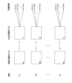
- FIG. 5 is a diagram showing an example of training data creation for generating the first learning model 62.
- Patients with arrhythmia P1, P2, ..., Px
- the surface electrocardiogram data (B1, B2, ..., Bx) and intracardiac electrocardiogram data (I1, I2, ..., Ix) of the patients are collected, and the intracardiac electrocardiogram data of each patient is abstracted and labeled.
- labels are 1, 2, ..., N.
- the labels for different patients may be different or the same label. In this way, the abstracted intracardiac electrocardiogram is prepared as the correct label.
- the training data can be created using an external device (e.g., a personal computer, etc.).
- the training data created by the external device can be stored in the data server 20.
- FIG. 6 is a diagram showing an example of a method for generating the first learning model 62.
- the control unit 51 acquires training data including indices (correct labels) for evaluating the surface electrocardiogram data and intracardiac electrocardiogram data from the data server 20.
- the control unit 51 receives the surface electrocardiogram data of a subject based on the acquired training data, it can generate the first learning model 62 so as to output an index for evaluating the intracardiac electrocardiogram data of the subject.
- surface electrocardiogram data is input as learning input data into the first learning model 62.
- the parameters in the first learning model 62 are adjusted so that the label (evaluation index) output by the first learning model 62 matches the correct label as training data.
- the parameters of the first learning model 62 can be stored in the memory unit 60 and the first learning model 62 can be generated.
- FIG. 7 is a diagram showing a first example of labels.
- Examples of labels that abstract intracardiac electrocardiogram data can be classified as follows: label 1 has pulmonary vein potential, label 2 has no pulmonary vein potential, label 3 has coronary sinus potential, label 4 has no coronary sinus potential, label 5 has right ventricular potential, label 6 has no right ventricular potential, label 7 has ablation catheter potential, label 8 has no ablation catheter potential, etc. Note that the label numbers are given for convenience and are not limited to the example in FIG. 7.
- the evaluation index includes the presence or absence of a pulmonary vein potential.
- the evaluation index may also include at least one of the presence or absence of a coronary sinus potential, the presence or absence of a right ventricular potential, and the presence or absence of an ablation catheter potential.
- the control unit 51 acquires the subject's surface electrocardiogram data, and when the surface electrocardiogram data is input, the control unit 51 can input the acquired surface electrocardiogram data to the first learning model 62, which outputs an index (evaluation index, label) for evaluating the intracardiac electrocardiogram data, and output an index for evaluating the intracardiac electrocardiogram data of the subject.
- Atrial fibrillation is an arrhythmia disorder that occurs when electrical signals are generated uncontrollably near a blood vessel called the "pulmonary vein" in the left atrium of the heart. It is thought that aging causes cells with abnormal electrical currents to appear in the pulmonary vein, and that a strong electrical current from these cells flows into the atrium, causing it to lose balance and enter a state of fibrillation.
- the first learning model 62 when the subject's surface electrocardiogram data is input to the first learning model 62, if the first learning model 62 outputs label 1, it can be predicted with a high degree of accuracy that the subject is at risk of arrhythmia due to atrial fibrillation and the associated risk of serious events such as cerebral infarction. In this way, according to this embodiment, the presence or absence and mechanism of arrhythmia disease can be predicted with a high degree of accuracy from the surface electrocardiogram.
- FIG. 8 is a diagram showing a second example of labels.
- label 11 is potential pattern aaa1 of site AAA
- label 12 is potential pattern aaa2 of site AAA
- ... label 21 is potential pattern bbb1 of site BBB
- label 22 is potential pattern bbb2 of site BBB
- ... label numbers, sites, and potential patterns are given for convenience and are not limited to the example in FIG. 8.
- the evaluation index can include clusters classified by clustering based on the potential patterns observed at multiple electrodes placed at required locations on the heart. A correct label can be assigned to each cluster classified by clustering.
- Figure 9 shows an example of a coronary sinus potential pattern.
- Location AAA is the coronary sinus
- label 11 is the normal label
- label 12 is the abnormal label.
- the electrodes CS CS1 and 2 are on the left side of the heart
- CS9 and 10 are on the right side of the heart.
- the potential pattern flows from CS9 and 10 to CS1 and 2 (i.e., from the right side to the left side of the heart) over time.
- the abnormal label label
- the potential pattern flows from CS1 and 2 to CS9 and 10 (i.e., from the left side to the right side of the heart) over time.
- the first learning model 62 when the subject's surface electrocardiogram data is input to the first learning model 62, if the first learning model 62 outputs label 12, it can be predicted with a high degree of accuracy that the subject is at risk of arrhythmia due to an imbalance in electrical potential in the coronary sinus, and the associated risk of serious events such as cerebral infarction. In this way, according to this embodiment, the presence or absence and mechanism of arrhythmia can be predicted with a high degree of accuracy from the surface electrocardiogram.
- FIG. 10 is a diagram showing a third example of labels.
- Intracardiac electrocardiogram data is collected from multiple patients with arrhythmia, and the intracardiac electrocardiogram data is clustered using the sequence of intracardiac electrocardiogram waveforms at site XXX and the intracardiac electrocardiogram waveforms as classification axes.
- Site XXX may include multiple sites.
- the sequence of intracardiac electrocardiogram waveforms is the time order of the waveforms at each electrode.
- the intracardiac electrocardiogram waveforms include the shape of the waveform at each electrode, the magnitude of the potential, and the interval between characteristic points of the waveform.
- the data is classified into four clusters, labeled 31, 32, 33, and 34. Note that clustering may be performed using either the sequence of intracardiac electrocardiogram waveforms or the intracardiac electrocardiogram waveform as a classification axis.
- the evaluation index may include clusters classified by clustering based on at least one of multiple intracardiac electrocardiogram waveforms and a sequence of multiple intracardiac electrocardiogram waveforms observed with multiple electrodes placed at required locations on the heart.
- FIG. 11 shows an example of an atrial tachycardia pattern.
- FIG. 11 shows intracardiac electrocardiogram data obtained from electrode catheters inserted into predetermined locations in the right atrium (RA) and coronary sinus (CS).
- the waveform and waveform arrangement indicated by symbol A show the case of sinus rhythm (normal pattern).
- the waveform and waveform arrangement indicated by symbol B show the case of an atrial tachycardia pattern.
- Atrial tachycardia is a type of tachycardia that occurs when abnormally excitable cardiomyocytes are present in the atrium and these cardiomyocytes excite the heart at a frequency higher than that of the sinus node.
- the label (evaluation index) of the atrial tachycardia pattern is label 31 and the subject's surface electrocardiogram data is input to the first learning model 62, and the first learning model 62 outputs label 31, it can be predicted with a high degree of accuracy that the subject is at risk of arrhythmia due to atrial tachycardia and the associated risk of serious events such as cerebral infarction. In this way, according to this embodiment, the presence or absence and mechanism of arrhythmia disease can be predicted with a high degree of accuracy from the surface electrocardiogram.
- Figure 12 shows an example of an atrioventricular reciprocating tachycardia pattern.
- Figure 12 shows intracardiac electrocardiogram data obtained from an electrode catheter inserted into a specified location in the coronary sinus (CS). The waveform and waveform arrangement in the framed time period show an atrioventricular reciprocating tachycardia pattern.
- Atrioventricular reciprocating tachycardia (WPS syndrome) is a type of tachycardia that occurs when the stimulation conduction system connecting the atrium and ventricle has an accessory pathway in addition to the normal pathway, and the electrical signal circulates between the normal pathway and the accessory pathway.
- the label (evaluation index) of the atrioventricular reciprocating tachycardia pattern is label 41 and the subject's surface electrocardiogram data is input to the first learning model 62, and the first learning model 62 outputs label 41, it can be predicted with a high degree of accuracy that the subject is at risk of arrhythmia due to atrioventricular reciprocating tachycardia and the associated risk of serious events such as cerebral infarction.
- the presence or absence and mechanism of arrhythmia disease can be predicted with a high degree of accuracy from the surface electrocardiogram.
- Figure 13 is a diagram showing an example of visualization of prediction grounds using Grad-CAM.
- Grad-CAM inputs surface electrocardiogram data into the first learning model 62, extracts a feature map from the first learning model 62, calculates weights for each channel of the extracted feature map, and adds up the weighted channels to generate a saliency map. The more important the location in the surface electrocardiogram data, the greater the saliency, making it possible to visualize the prediction grounds.
- FIG. 14 is a diagram showing an example of the configuration of the second learning model 63.
- the second learning model 63 includes an input layer 631, an intermediate layer 632, and an output layer 633.
- the intermediate layer 632 may be a convolutional layer, but does not have to be a convolutional layer.
- the second learning model 63 outputs arrhythmia diagnosis information.
- control unit 51 acquires the subject's surface electrocardiogram features, and when the control unit 51 inputs the surface electrocardiogram features, it can input the acquired surface electrocardiogram features to the second learning model 63, which outputs arrhythmia diagnosis information, and output arrhythmia diagnosis information for the subject.
- the function of the arrhythmia diagnosis information generating unit 58 which generates arrhythmia diagnosis information based on the surface electrocardiogram features, is performed.
- the second learning model 63 may be generated as follows. That is, the control unit 51 acquires training data including surface electrocardiogram features and arrhythmia diagnosis information, and based on the acquired training data, when the surface electrocardiogram features of a subject are input, the control unit 51 can generate the second learning model 63 so as to output arrhythmia diagnosis information of the subject.
- the training data may be stored in advance in the data server 20.
- the second learning model 63 may be generated as follows. That is, the control unit 51 may acquire training data including surface electrocardiogram data and arrhythmia diagnosis information, and generate the second learning model 63 based on the acquired training data so that when the subject's surface electrocardiogram data is input, the second learning model 63 is generated so as to output arrhythmia diagnosis information for the subject.
- the training data may be stored in the data server 20 in advance.
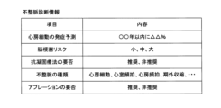
- FIG. 15 is a diagram showing an example of arrhythmia diagnosis information.
- the predicted occurrence of atrial fibrillation is, for example, ⁇ % within XX years.
- the risk of cerebral infarction is, for example, small, medium, or large.
- the necessity of anticoagulant therapy is, for example, recommended or not recommended.
- the type of arrhythmia is, for example, atrial fibrillation, ventricular tachycardia, atrial tachycardia, premature ventricular contraction, ....
- the necessity of ablation is, for example, recommended or not recommended.
- Ablation is a treatment for atrial fibrillation using pulmonary vein isolation (PVI).
- PV pulmonary vein isolation
- Different second learning models 63 may be used for different items of arrhythmia diagnosis information, or the same second learning model 63 may be used for multiple items.
- the second learning model 63 medical information such as surface electrocardiogram data, diagnosis information, treatment information, and intracardiac electrocardiogram data of multiple patients with arrhythmia is collected for each patient. Based on the medical information of multiple patients, for example, the second learning model 63 is trained to learn the correlation between the time when surface electrocardiogram data was acquired and the time when atrial fibrillation occurred, making it possible to predict the onset of atrial fibrillation in a subject from the subject's surface electrocardiogram data.
- the second learning model 63 learn, for example, the correlation between the time when surface electrocardiogram data was acquired and the presence or absence of cerebral infarction based on the medical information of multiple patients, it becomes possible to predict the risk of cerebral infarction of a subject from the subject's surface electrocardiogram data.
- the second learning model 63 learns the correlation between, for example, surface electrocardiogram data and treatment information on whether or not anticoagulant therapy was required based on the medical information of multiple patients, it becomes possible to determine whether or not a subject requires anticoagulant therapy based on the subject's surface electrocardiogram data.
- the second learning model 63 learn, for example, the correlation between surface electrocardiogram data and the type of arrhythmia based on intracardiac electrocardiogram data based on the medical information of multiple patients, it becomes possible to determine the type of arrhythmia of a subject from the subject's surface electrocardiogram data.
- the second learning model 63 learn, for example, the correlation between surface electrocardiogram data and symptoms after ablation based on the medical information of multiple patients, it becomes possible to determine whether or not a subject needs ablation based on the subject's surface electrocardiogram data.
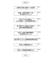
- FIG. 16 is a diagram showing an example of a processing procedure by the intracardiac electrocardiogram evaluation device 50. For convenience, the processing will be described as being performed by the control unit 51.
- the control unit 51 acquires the subject's surface electrocardiogram data (S11) and inputs the acquired surface electrocardiogram data to the first learning model 62 (S12).
- the control unit 51 acquires an evaluation index for the intracardiac electrocardiogram data from the output layer 623 of the first learning model 62 (S13).
- the control unit 51 extracts surface electrocardiogram features from the convolutional layer 622 of the first learning model 62 (S14).
- the control unit 51 inputs the extracted surface electrocardiogram features to the second learning model 63 (S15).
- the control unit 51 acquires arrhythmia diagnosis information from the second learning model 63 (S16).
- the control unit 51 outputs the acquired evaluation index (S17), outputs the acquired arrhythmia diagnosis information (S18), and ends the process.
- the arrhythmia diagnosis information may be displayed on the display unit 54 or an external display device.
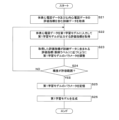
- FIG. 17 is a diagram showing an example of a generation process procedure for the first learning model 62.
- the control unit 51 acquires training data including evaluation indices (correct labels) for the surface electrocardiogram data and the intracardiac electrocardiogram data (S21).
- the control unit 51 inputs the surface electrocardiogram data as learning input data into the first learning model 62 and acquires the evaluation indices (output labels) output by the first learning model 62 (S22).
- the control unit 51 adjusts the parameters of the first learning model 62 so that the acquired evaluation index (output label) approaches the evaluation index (correct label) included in the training data (S23).
- the control unit 51 determines whether the prediction accuracy of the first learning model 62 is within an acceptable range (S24), and if the prediction accuracy is not within the acceptable range (NO in S24), continues the processing from step S22 onwards.
- control unit 51 stores the parameters of the first learning model 62 in the storage unit 60 (S25), generates the first learning model 62 (S26), and ends the process.
- the pulmonary vein potential (the small potential confirmed by the intracardiac electrocardiogram), which is believed to be the cause of atrial fibrillation, can be detected at the appropriate time phase from the surface electrocardiogram data, and can be used as a fundamental technology for using a clinical arrhythmia prediction model.
- the computer program acquires surface electrocardiogram data of a subject, and when the surface electrocardiogram data is input, causes the computer to execute a process of inputting the acquired surface electrocardiogram data into a first learning model that outputs an index for evaluating the intracardiac electrocardiogram data of the subject, and outputs an index for evaluating the intracardiac electrocardiogram data of the subject.
- Appendix 2 In the computer program of Appendix 1, the indicators include the presence or absence of a pulmonary vein potential.
- the indicators include at least one of the presence or absence of coronary sinus potential, the presence or absence of right ventricular potential, and the presence or absence of ablation catheter potential.
- the index includes clusters classified by clustering based on potential patterns observed at multiple electrodes placed at required locations on the heart.
- the index includes clusters classified by clustering based on at least one of a plurality of intracardiac electrocardiogram waveforms observed with a plurality of electrodes placed at required locations of the heart and a sequence of the plurality of intracardiac electrocardiogram waveforms.
- appendix 6 The computer program in any one of appendices 1 to 5 causes a computer to execute a process of extracting electrocardiogram features based on the acquired surface electrocardiogram data, and when the electrocardiogram features are input, inputting the extracted electrocardiogram features into a second learning model that outputs arrhythmia diagnosis information, and outputting arrhythmia diagnosis information for the subject.
- the arrhythmia diagnosis information includes at least one of atrial fibrillation risk, cerebral infarction risk, and type of arrhythmia.
- the intracardiac electrocardiogram evaluation device includes a control unit and a first learning model that outputs an index for evaluating the intracardiac electrocardiogram data when surface electrocardiogram data is input, and the control unit acquires the subject's surface electrocardiogram data, inputs the acquired surface electrocardiogram data into the first learning model, and outputs an index for evaluating the subject's intracardiac electrocardiogram data.
- the intracardiac electrocardiogram evaluation system includes the intracardiac electrocardiogram evaluation device and an electrocardiogram examination device, and the intracardiac electrocardiogram evaluation device acquires the subject's surface electrocardiogram data from the electrocardiogram examination device.
- the intracardiac electrocardiogram evaluation method acquires a subject's surface electrocardiogram data, and when the surface electrocardiogram data is input, a first learning model outputs an index for evaluating the intracardiac electrocardiogram data by inputting the acquired surface electrocardiogram data into the first learning model, which outputs an index for evaluating the intracardiac electrocardiogram data of the subject.
- the learning model generation method acquires training data including indices for evaluating surface electrocardiogram data and intracardiac electrocardiogram data, and generates a first learning model based on the acquired training data so that, when a subject's surface electrocardiogram data is input, an indices for evaluating the subject's intracardiac electrocardiogram data are output.
- Electrocardiogram examination device 20
- Data server 50
- Intracardiac electrocardiogram evaluation device 51
- Control unit 52
- Memory 54
- Display unit 55
- Operation unit 56
- Evaluation index generation unit 57
- Electrocardiogram feature extraction unit 58
- Arrhythmia diagnosis information generation unit 60
- Storage unit 61
- Computer program 62
- First learning model 63 Second learning model
Landscapes
- Health & Medical Sciences (AREA)
- Engineering & Computer Science (AREA)
- Medical Informatics (AREA)
- Public Health (AREA)
- Biomedical Technology (AREA)
- General Health & Medical Sciences (AREA)
- Epidemiology (AREA)
- Primary Health Care (AREA)
- Pathology (AREA)
- Data Mining & Analysis (AREA)
- Life Sciences & Earth Sciences (AREA)
- Databases & Information Systems (AREA)
- Business, Economics & Management (AREA)
- General Business, Economics & Management (AREA)
- Cardiology (AREA)
- Animal Behavior & Ethology (AREA)
- Chemical & Material Sciences (AREA)
- Bioinformatics & Cheminformatics (AREA)
- Medicinal Chemistry (AREA)
- Veterinary Medicine (AREA)
- Surgery (AREA)
- Molecular Biology (AREA)
- Heart & Thoracic Surgery (AREA)
- Biophysics (AREA)
- Physics & Mathematics (AREA)
- Measurement And Recording Of Electrical Phenomena And Electrical Characteristics Of The Living Body (AREA)
Abstract
Priority Applications (3)
| Application Number | Priority Date | Filing Date | Title |
|---|---|---|---|
| AU2024277885A AU2024277885A1 (en) | 2023-05-23 | 2024-05-23 | Computer program, intracardiac electrocardiogram evaluation device, intracardiac electrocardiogram evaluation system, intracardiac electrocardiogram evaluation method, and learning model generation method |
| JP2025522443A JPWO2024242155A1 (fr) | 2023-05-23 | 2024-05-23 | |
| EP24811161.9A EP4717175A1 (fr) | 2023-05-23 | 2024-05-23 | Programme informatique, dispositif d'évaluation d'électrocardiogramme intracardiaque, système d'évaluation d'électrocardiogramme intracardiaque, procédé d'évaluation d'électrocardiogramme intracardiaque et procédé de génération de modèle d'apprentissage |
Applications Claiming Priority (2)
| Application Number | Priority Date | Filing Date | Title |
|---|---|---|---|
| JP2023084755 | 2023-05-23 | ||
| JP2023-084755 | 2023-05-23 |
Publications (1)
| Publication Number | Publication Date |
|---|---|
| WO2024242155A1 true WO2024242155A1 (fr) | 2024-11-28 |
Family
ID=93589454
Family Applications (1)
| Application Number | Title | Priority Date | Filing Date |
|---|---|---|---|
| PCT/JP2024/018932 Ceased WO2024242155A1 (fr) | 2023-05-23 | 2024-05-23 | Programme informatique, dispositif d'évaluation d'électrocardiogramme intracardiaque, système d'évaluation d'électrocardiogramme intracardiaque, procédé d'évaluation d'électrocardiogramme intracardiaque et procédé de génération de modèle d'apprentissage |
Country Status (4)
| Country | Link |
|---|---|
| EP (1) | EP4717175A1 (fr) |
| JP (1) | JPWO2024242155A1 (fr) |
| AU (1) | AU2024277885A1 (fr) |
| WO (1) | WO2024242155A1 (fr) |
Citations (8)
| Publication number | Priority date | Publication date | Assignee | Title |
|---|---|---|---|---|
| JP2011072542A (ja) | 2009-09-30 | 2011-04-14 | Fukuda Denshi Co Ltd | 心電図データ処理装置、心電図データ処理方法および心電図データ処理プログラム |
| US20130345580A1 (en) * | 2009-04-06 | 2013-12-26 | Sorin Crm S.A.S. | Reconstruction of a surface electrocardiogram from an endocardial electrogram using non-linear filtering |
| US20150011903A1 (en) * | 2009-03-25 | 2015-01-08 | Sorin Crm Sas | Non-linear filtering for the reconstruction of a surface electrocardiogram from an endocardial electrogram |
| JP2019505249A (ja) * | 2015-12-01 | 2019-02-28 | サイマップ メディカル (スーチョウ),エルティーディー | 動脈の壁を支配する機能的な神経をマッピングするためのシステムと方法、そのための3dマッピング、およびカテーテル |
| JP2019058350A (ja) * | 2017-09-26 | 2019-04-18 | フクダ電子株式会社 | 心腔内除細動システム及び除細動器 |
| JP2021186685A (ja) * | 2020-06-04 | 2021-12-13 | バイオセンス・ウエブスター・(イスラエル)・リミテッドBiosense Webster (Israel), Ltd. | オートエンコーダを使用する心内心電図のノイズ削減、並びに深層学習訓練損失関数を使用する心内及び身体表面心電図の利用及び精緻化 |
| US20220400951A1 (en) * | 2015-09-07 | 2022-12-22 | Ablacon Inc. | Systems, Devices, Components and Methods for Detecting the Locations of Sources of Cardiac Rhythm Disorders in a Patient's Heart Using Improved Electrographic Flow (EGF) Methods |
| US20230017546A1 (en) * | 2021-07-19 | 2023-01-19 | GE Precision Healthcare LLC | Methods and systems for real-time cycle length determination in electrocardiogram signals |
-
2024
- 2024-05-23 WO PCT/JP2024/018932 patent/WO2024242155A1/fr not_active Ceased
- 2024-05-23 JP JP2025522443A patent/JPWO2024242155A1/ja active Pending
- 2024-05-23 EP EP24811161.9A patent/EP4717175A1/fr active Pending
- 2024-05-23 AU AU2024277885A patent/AU2024277885A1/en active Pending
Patent Citations (8)
| Publication number | Priority date | Publication date | Assignee | Title |
|---|---|---|---|---|
| US20150011903A1 (en) * | 2009-03-25 | 2015-01-08 | Sorin Crm Sas | Non-linear filtering for the reconstruction of a surface electrocardiogram from an endocardial electrogram |
| US20130345580A1 (en) * | 2009-04-06 | 2013-12-26 | Sorin Crm S.A.S. | Reconstruction of a surface electrocardiogram from an endocardial electrogram using non-linear filtering |
| JP2011072542A (ja) | 2009-09-30 | 2011-04-14 | Fukuda Denshi Co Ltd | 心電図データ処理装置、心電図データ処理方法および心電図データ処理プログラム |
| US20220400951A1 (en) * | 2015-09-07 | 2022-12-22 | Ablacon Inc. | Systems, Devices, Components and Methods for Detecting the Locations of Sources of Cardiac Rhythm Disorders in a Patient's Heart Using Improved Electrographic Flow (EGF) Methods |
| JP2019505249A (ja) * | 2015-12-01 | 2019-02-28 | サイマップ メディカル (スーチョウ),エルティーディー | 動脈の壁を支配する機能的な神経をマッピングするためのシステムと方法、そのための3dマッピング、およびカテーテル |
| JP2019058350A (ja) * | 2017-09-26 | 2019-04-18 | フクダ電子株式会社 | 心腔内除細動システム及び除細動器 |
| JP2021186685A (ja) * | 2020-06-04 | 2021-12-13 | バイオセンス・ウエブスター・(イスラエル)・リミテッドBiosense Webster (Israel), Ltd. | オートエンコーダを使用する心内心電図のノイズ削減、並びに深層学習訓練損失関数を使用する心内及び身体表面心電図の利用及び精緻化 |
| US20230017546A1 (en) * | 2021-07-19 | 2023-01-19 | GE Precision Healthcare LLC | Methods and systems for real-time cycle length determination in electrocardiogram signals |
Also Published As
| Publication number | Publication date |
|---|---|
| EP4717175A1 (fr) | 2026-04-01 |
| JPWO2024242155A1 (fr) | 2024-11-28 |
| AU2024277885A1 (en) | 2025-12-18 |
Similar Documents
| Publication | Publication Date | Title |
|---|---|---|
| CN105228508B (zh) | 一种测定用于分类的危险评分的系统 | |
| Yang et al. | Automated diagnosis of atrial fibrillation using ECG component-aware transformer | |
| US12458270B2 (en) | Electrocardiographic identification of non-ST elevation ischemic events | |
| Mannhart et al. | Clinical validation of an artificial intelligence algorithm offering cross-platform detection of atrial fibrillation using smart device electrocardiograms | |
| SippensGroenewegen et al. | Body surface mapping of counterclockwise and clockwise typical atrial flutter: a comparative analysis with endocardial activation sequence mapping | |
| Vardas et al. | The electrocardiogram endeavour: from the Holter single-lead recordings to multilead wearable devices supported by computational machine learning algorithms | |
| Deng et al. | ST-Net: Synthetic ECG tracings for diagnosing various cardiovascular diseases | |
| Armoundas et al. | Can microvolt T-wave alternans testing reduce unnecessary defibrillator implantation? | |
| Rodriguez et al. | Risk stratification in idiopathic dilated cardiomyopathy patients using cardiovascular coupling analysis | |
| WO2024242155A1 (fr) | Programme informatique, dispositif d'évaluation d'électrocardiogramme intracardiaque, système d'évaluation d'électrocardiogramme intracardiaque, procédé d'évaluation d'électrocardiogramme intracardiaque et procédé de génération de modèle d'apprentissage | |
| US9538930B2 (en) | Linear multi-domain electrocardiogram | |
| KR102886211B1 (ko) | 심전도 판독에 기반한 시각화 콘텐츠를 제공하는 방법, 프로그램 및 장치 | |
| CN103300847A (zh) | Twa测量装置和twa测量方法 | |
| Yao et al. | Arrhythmia classification from single lead ecg by multi-scale convolutional neural networks | |
| Rjoob et al. | Machine learning improves the detection of misplaced v1 and v2 electrodes during 12-lead electrocardiogram acquisition | |
| Manukova et al. | An approach to evaluation of clinically healthy people by preventive cardio control | |
| Myerburg et al. | Task force 2: training in electrocardiography, ambulatory electrocardiography, and exercise testing | |
| Gao et al. | Electrical inhomogeneity in left ventricular hypertrophy | |
| Moubarak et al. | Electrocardiographic abnormalities in centenarians and octogenarians: A case‐matched study | |
| TW202315574A (zh) | 輔助辨識病徵之方法 | |
| Chouvarda et al. | Time-domain analysis of the electrocardiogram | |
| Przystup et al. | ECG-based prediction of ventricular fibrillation by means of the PCA | |
| Heena | Analysis and Classification of Heart Abnormalities | |
| Prats-Boluda et al. | Identification of atrial fibrillation drivers by means of concentric ring electrodes | |
| Marcantoni et al. | Electrocardiographic alternans as an additional criterion for cardioverter defibrillator implantation in primary prevention of sudden cardiac death |
Legal Events
| Date | Code | Title | Description |
|---|---|---|---|
| 121 | Ep: the epo has been informed by wipo that ep was designated in this application |
Ref document number: 24811161 Country of ref document: EP Kind code of ref document: A1 |
|
| ENP | Entry into the national phase |
Ref document number: 2025522443 Country of ref document: JP Kind code of ref document: A |
|
| WWE | Wipo information: entry into national phase |
Ref document number: 2025522443 Country of ref document: JP |
|
| WWE | Wipo information: entry into national phase |
Ref document number: AU2024277885 Country of ref document: AU |
|
| ENP | Entry into the national phase |
Ref document number: 2024277885 Country of ref document: AU Date of ref document: 20240523 Kind code of ref document: A |
|
| WWE | Wipo information: entry into national phase |
Ref document number: 2024811161 Country of ref document: EP |
|
| NENP | Non-entry into the national phase |
Ref country code: DE |
|
| ENP | Entry into the national phase |
Ref document number: 2024811161 Country of ref document: EP Effective date: 20251223 |
|
| ENP | Entry into the national phase |
Ref document number: 2024811161 Country of ref document: EP Effective date: 20251223 |
|
| ENP | Entry into the national phase |
Ref document number: 2024811161 Country of ref document: EP Effective date: 20251223 |
|
| ENP | Entry into the national phase |
Ref document number: 2024811161 Country of ref document: EP Effective date: 20251223 |
|
| ENP | Entry into the national phase |
Ref document number: 2024811161 Country of ref document: EP Effective date: 20251223 |
|
| ENP | Entry into the national phase |
Ref document number: 2024811161 Country of ref document: EP Effective date: 20251223 |
|
| ENP | Entry into the national phase |
Ref document number: 2024811161 Country of ref document: EP Effective date: 20251223 |
|
| WWP | Wipo information: published in national office |
Ref document number: 2024811161 Country of ref document: EP |