WO2024247651A1 - Activateur cellulaire - Google Patents
Activateur cellulaire Download PDFInfo
- Publication number
- WO2024247651A1 WO2024247651A1 PCT/JP2024/017330 JP2024017330W WO2024247651A1 WO 2024247651 A1 WO2024247651 A1 WO 2024247651A1 JP 2024017330 W JP2024017330 W JP 2024017330W WO 2024247651 A1 WO2024247651 A1 WO 2024247651A1
- Authority
- WO
- WIPO (PCT)
- Prior art keywords
- kda
- cells
- fraction
- shed
- cell
- Prior art date
- Legal status (The legal status is an assumption and is not a legal conclusion. Google has not performed a legal analysis and makes no representation as to the accuracy of the status listed.)
- Ceased
Links
Images






Classifications
-
- A—HUMAN NECESSITIES
- A61—MEDICAL OR VETERINARY SCIENCE; HYGIENE
- A61P—SPECIFIC THERAPEUTIC ACTIVITY OF CHEMICAL COMPOUNDS OR MEDICINAL PREPARATIONS
- A61P43/00—Drugs for specific purposes, not provided for in groups A61P1/00-A61P41/00
-
- A—HUMAN NECESSITIES
- A61—MEDICAL OR VETERINARY SCIENCE; HYGIENE
- A61K—PREPARATIONS FOR MEDICAL, DENTAL OR TOILETRY PURPOSES
- A61K35/00—Medicinal preparations containing materials or reaction products thereof with undetermined constitution
- A61K35/12—Materials from mammals; Compositions comprising non-specified tissues or cells; Compositions comprising non-embryonic stem cells; Genetically modified cells
- A61K35/32—Bones; Osteocytes; Osteoblasts; Tendons; Tenocytes; Teeth; Odontoblasts; Cartilage; Chondrocytes; Synovial membrane
Definitions
- the present invention relates to a cell activation agent.
- MSCs mesenchymal stem cells
- cytokines growth factors and cytokines
- MSCs can be easily collected from various tissues such as bone marrow, dental pulp, adipose tissue, and umbilical cord, but in particular, deciduous tooth pulp stem cells, which are MSCs that can be collected from baby teeth, can be easily prepared from baby teeth that have traditionally been discarded, and have high proliferation capacity, making them particularly promising as useful MSCs.
- culture supernatant of deciduous dental pulp stem cells is useful for repairing damaged tissues such as those caused by cerebral infarction, and for protecting skin tissues (Patent Documents 1 and 2).
- components in the culture supernatant are the active ingredients.
- the culture supernatant may contain components that act in an inhibitory manner against the active ingredients, but these have not been clarified.
- the present invention was made with the aim of purifying the culture supernatant of deciduous dental pulp stem cells and providing a composition with excellent cell activation effects.
- the present invention provides a cell activation agent comprising a fraction isolated from the culture supernatant of deciduous dental pulp stem cells, the fraction not containing components less than 3.5 kDa.
- the present invention provides a use of a fraction isolated from a culture supernatant of deciduous dental pulp stem cells as a cell activating agent, the fraction not containing components less than 3.5 kDa.
- the fraction preferably contains components of 50 to 100 kDa and does not contain components of less than 30 kDa.
- the fraction preferably does not contain components greater than 100 kDa and/or components between 30 and 50 kDa.
- the deciduous tooth pulp stem cells may be primary cultured cells.
- deciduous dental pulp stem cells may be immortalized cells.
- the present invention provides a method for activating cells in a subject, the method comprising the step of administering the cell activation agent to the subject.
- the cell activation agent of the present invention can be easily and inexpensively prepared from culture supernatant of deciduous dental pulp stem cells, and does not contain any components that inhibit cell activation activity, making it highly effective and safe, and useful.
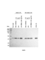
- FIG. 1 shows polyacrylamide gel electrophoretic images of SHED-CM and its fractions.
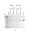
- FIG. 2 shows polyacrylamide gel electrophoretic images of IM-SHED-CM and its fractions.
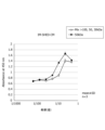
- FIG. 3 is a graph showing the results of a WST assay evaluating the intracellular dehydrogenase activity of NIH3T3 cells cultured with SHED-CM fractions at different concentration rates.
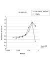
- FIG. 4 is a graph showing the results of a WST assay evaluating the intracellular dehydrogenase activity of NIH3T3 cells cultured with IM-SHED-CM fractions at different concentration rates.
- FIG. 1 shows polyacrylamide gel electrophoretic images of SHED-CM and its fractions.
- FIG. 2 shows polyacrylamide gel electrophoretic images of IM-SHED-CM and its fractions.
- FIG. 3 is a graph showing the results of a WST assay evaluating the intracellular dehydrogenase activity of NIH3T3 cells cultured with SHED-
- FIG. 5 is a graph showing the results of a WST assay evaluating the intracellular dehydrogenase activity of NIH3T3 cells cultured with dilutions of SHED-CM or fractions thereof adjusted based on protein concentration.
- FIG. 6 is a graph showing the results of a WST assay evaluating the intracellular dehydrogenase activity of NIH3T3 cells cultured with dilutions of IM-SHED-CM or fractions thereof adjusted based on protein concentration.
- FIG. 6 is a graph showing the results of a WST assay evaluating the intracellular dehydrogenase activity of NIH3T3 cells cultured with dilutions of IM-SHED-CM or fractions thereof adjusted based on protein concentration.
- FIG. 7 is a graph showing the results of a WST assay evaluating the intracellular dehydrogenase activity of NIH3T3 cells cultured with a dilution of the 50-100 kDa fraction derived from SHED-CM, the >100 kDa fraction, or the 30-50 kDa fraction.
- FIG. 8 is a graph showing the results of a WST assay evaluating the intracellular dehydrogenase activity of NIH3T3 cells cultured with a dilution of the 50-100 kDa fraction derived from IM-SHED-CM, the >100 kDa fraction, or the 30-50 kDa fraction.
- FIG. 8 is a graph showing the results of a WST assay evaluating the intracellular dehydrogenase activity of NIH3T3 cells cultured with a dilution of the 50-100 kDa fraction derived from IM-SHED-CM, the >100 kDa fraction, or the 30-50
- FIG. 9 shows the results of Western blot analysis comparing the expression levels and phosphorylation of MAPK in NIH3T3 cells cultured with SHED-CM/IM-SHED-CM or fractions thereof.
- FIG. 10 shows the results of Western blot analysis comparing the expression levels and phosphorylation of Akt in NIH3T3 cells cultured with SHED-CM/IM-SHED-CM or fractions thereof.
- FIG. 11 shows the results of Western blot analysis comparing the expression levels and phosphorylation of MAPK in NIH3T3 cells cultured with SHED-CM/IM-SHED-CM or fractions thereof at the same protein concentrations.
- FIG. 10 shows the results of Western blot analysis comparing the expression levels and phosphorylation of MAPK in NIH3T3 cells cultured with SHED-CM/IM-SHED-CM or fractions thereof at the same protein concentrations.
- FIG. 12 shows the results of Western blot analysis comparing the expression levels and phosphorylation of Akt in NIH3T3 cells cultured with SHED-CM/IM-SHED-CM or fractions thereof at the same protein concentrations.
- FIG. 13 shows the results of a scratch assay for NIH3T3 cells cultured with IM-SHED-CM or a fraction thereof.
- FIG. 14 is a graph showing the results of a WST assay evaluating the intracellular dehydrogenase activity of NIH3T3 cells cultured with IM-SHED-CM or its >3.5 kDa fraction.
- FIG. 15 shows the results of a scratch assay for NIH3T3 cells cultured with IM-SHED-CM or its >3.5 kDa fraction.
- FIG. 16 is a graph showing the results of a WST assay evaluating the intracellular dehydrogenase activity of HUVECs cultured with diluted solutions of SHED-CM or its 50-100 kDa fraction adjusted based on the protein concentration.
- FIG. 17 is a graph showing the results of a WST assay evaluating the intracellular dehydrogenase activity of HUVECs cultured with diluted solutions of IM-SHED-CM or its 50-100 kDa fraction adjusted based on the protein concentration.
- FIG. 17 is a graph showing the results of a WST assay evaluating the intracellular dehydrogenase activity of HUVECs cultured with diluted solutions of IM-SHED-CM or its 50-100 kDa fraction adjusted based on the protein concentration.
- FIG. 18 is a graph showing the intracellular dehydrogenase activity of HUVECs cultured with the 50-100 kDa fraction of SHED-CM, or a mixture of the >100 kDa, 50-100 kDa, and 30-50 kDa fractions.
- FIG. 19 is a graph showing the intracellular dehydrogenase activity of HUVECs cultured with the 50-100 kDa fraction of SHED-CM, or a mixture of the 50-100 kDa and ⁇ 30 kDa fractions.
- FIG. 19 is a graph showing the intracellular dehydrogenase activity of HUVECs cultured with the 50-100 kDa fraction of SHED-CM, or a mixture of the 50-100 kDa and ⁇ 30 kDa fractions.
- FIG. 20 is a graph showing the intracellular dehydrogenase activity of HUVECs cultured with the 50-100 kDa fraction of IM-SHED-CM, or a mixture of the >100 kDa, 50-100 kDa, and 30-50 kDa fractions.
- FIG. 21 is a graph showing the intracellular dehydrogenase activity of HUVECs cultured with the 50-100 kDa fraction of IM-SHED-CM, or a mixture of the 50-100 kDa and ⁇ 30 kDa fractions.
- FIG. 22 is a graph showing the results of a WST assay evaluating the inhibitory effect of IM-SHED-CM on cell damage caused by hydrogen peroxide treatment.
- FIG. 23 is a graph showing the results of a WST assay evaluating the inhibitory effect of the 50-100 kDa fraction of IM-SHED-CM on cell damage caused by hydrogen peroxide treatment.
- FIG. 24 is a graph showing the results of a WST assay evaluating the inhibitory effect of the >100 kDa fraction of IM-SHED-CM on cell damage caused by hydrogen peroxide treatment.
- FIG. 25 is a graph showing the results of a WST assay evaluating the inhibitory effect of the 30-50 kDa fraction of IM-SHED-CM on cell damage caused by hydrogen peroxide treatment.
- FIG. 26 is a graph showing the results of a quantitative analysis of the recovery of the scratches in FIGS.
- the present invention provides a cell activation agent comprising a fraction isolated from a culture supernatant of deciduous dental pulp stem cells, the fraction not containing any components less than 3.5 kDa.
- cell activation means normalizing or improving functions such as cell proliferation and metabolism. Therefore, in this embodiment, “cell activation” may include, for example, activation of enzymes related to cell proliferation and metabolism (various dehydrogenases, Akt, AMPK, etc.) and enzymes that suppress oxidative stress (superoxide dismutase (SOD), catalase (CAT), glutathione peroxidase (GPx)), etc.).
- the "cell” may be in any vertebrate, preferably a mammalian cell such as a mouse, rat, rabbit, pig, cow, monkey, or human, and particularly preferably a human cell.
- the type of cell is not particularly limited, and may be, for example, a fibroblast, endothelial cell, epithelial cell, nerve cell, stem cell, leukocyte, bone cell, muscle cell, adipocyte, etc., and preferably a fibroblast or endothelial cell.
- Deciduous tooth pulp stem cells are mesenchymal stem cells harvested from the dental pulp of a deciduous tooth.
- the deciduous tooth pulp stem cells may be derived from any vertebrate, preferably from a mammal such as mouse, rat, rabbit, pig, cow, monkey, or human, and particularly preferably from human.
- deciduous dental pulp stem cells have already been established (e.g., WO 2011/118795), and they can be isolated and prepared from deciduous dental pulp according to methods known in the art.
- the deciduous dental pulp stem cells may be primary cultured cells or immortalized cells.
- "Immortalized cells” means that the cells remain capable of proliferation even after a certain number of divisions, i.e., the cells have the ability to proliferate indefinitely. Methods for immortalizing cells have already been established, and known techniques can be used. For example, cells can be immortalized by introducing the SV40 T antigen gene or the telomerase reverse transcriptase (TERT) gene into the cells.
- deciduous dental pulp stem cells can be cultured according to a method known in the art.
- deciduous dental pulp stem cells may be cultured by seeding at a concentration of, for example, 5 ⁇ 10 4 to 2 ⁇ 10 5 cells/mL in a medium containing, as a basal medium, DMEM, IMDM, Ham's F- 12 , RPMI-1640, or a mixture thereof, appropriately supplemented with serum such as FBS or serum substitutes such as KSR, glucose, amino acids, vitamins, etc.
- the cell activating agent of this embodiment is preferably prepared from a culture supernatant obtained by culturing deciduous dental pulp stem cells in a medium containing no serum.
- the cell activator of this embodiment does not contain components of less than 3.5 kDa derived from the culture supernatant of deciduous dental pulp stem cells.
- “not containing” means that the components to be removed are not detected by a commonly used detection method (e.g., immunochemical method, etc.). Therefore, the cell activator of this embodiment may be one in which at least 85% by weight, 90% by weight or more, 95% by weight or more, 97% by weight or more, 98% by weight or more, or 99% by weight or more of the components of less than 3.5 kDa contained in the unpurified culture supernatant have been removed.
- the cell activator of this embodiment may contain up to 15% by weight, less than 10% by weight, less than 5% by weight, less than 3% by weight, less than 2% by weight, or less than 1% by weight of the components of less than 3.5 kDa contained in the unpurified culture supernatant.
- the cell activator of this embodiment shows an improved cell activation effect by removing the components of less than 3.5 kDa from the culture supernatant of deciduous dental pulp stem cells.
- “kDa” may be calculated, for example, based on SDS-PAGE or ultrafiltration, and when the culture supernatant is purified using a commercially available ultrafiltration filter unit, it may be the nominal molecular weight cutoff (NMWL) or molecular weight cutoff (MWCO) value indicated on the product.
- NMWL nominal molecular weight cutoff
- MWCO molecular weight cutoff
- the cell activator of this embodiment preferably contains a fraction containing a component of 50 to 100 kDa; on the other hand, it is preferable that it does not contain a fraction containing a component of less than 30 kDa. It is more preferable that the cell activator of this embodiment does not contain a fraction containing a component of more than 100 kDa and/or a fraction containing a component of 30 to 50 kDa. This is because components of 50 to 100 kDa have higher cell activating activity, while not only components of less than 3.5 kDa but also components of more than 100 kDa and components of 30 to 50 kDa may have a detrimental effect on the cell activating activity of the 50 to 100 kDa components.
- the cell activator of this embodiment can be prepared, for example, by combining ultrafiltration membranes with 100,000 MWCO, 50,000 MWCO, 30,000 MWCO, and 3,500 MWCO. For example, by removing components of more than 100 kDa from the culture supernatant using an ultrafiltration membrane with 100,000 MWCO, and concentrating the resulting filtrate using an ultrafiltration membrane with 50,000 MWCO, a cell activator can be prepared that contains 50-100 kDa components from the culture supernatant of deciduous dental pulp stem cells, but does not contain components of 30-50 kDa or components less than 30 kDa.
- the cell activation agent of this embodiment may be composed only of a fraction purified from the culture supernatant of deciduous dental pulp stem cells, but generally may further contain pharma- ceutically acceptable carriers and additives as optional components.
- the cell activator of this embodiment may be prepared as a liquid or solid, and may be formulated into various dosage forms, such as injections, jellies, sprays, tablets, and granules, but is preferably prepared as a liquid.
- sterile water, physiological saline, an aqueous glucose solution, and the like may be used as a carrier, and bactericides, isotonicity agents, stabilizers, and the like may be further blended as desired.
- additives such as starch, lactose, mannitol, and inorganic salts may be further blended as desired, and binders, disintegrants, lubricants, and the like may be further blended.
- the content (protein weight equivalent) of the 50-100 kDa component derived from the culture supernatant of deciduous dental pulp stem cells in the cell activator of this embodiment may be, for example, 60% by weight or more, 70% by weight or more, 80% by weight or more, or 90% by weight or more, preferably 60-70% by weight, when the total mass of the cell activator is taken as 100%.
- the cell activation agent of this embodiment contains components derived from the culture supernatant of deciduous dental pulp stem cells that have cell activation activity, but does not contain components that inhibit this activity. Therefore, the cell activation agent of this embodiment has a superior cell activation effect and is useful compared to unpurified culture supernatant of deciduous dental pulp stem cells.
- the present invention is a method for activating cells in a subject, the method comprising the step of administering the cell activation agent to the subject.
- activating cells and “cells” are defined as in the first embodiment.
- the "subject" in this embodiment may be any vertebrate, but is preferably a mammal such as a mouse, rat, rabbit, pig, cow, monkey, or human, and is particularly preferably a human.
- the subject may be of any age, including infants, juveniles, adolescents, adults, and geriatric subjects.
- the cell activator in the method of this embodiment may be formulated into various dosage forms as defined in the first embodiment, and may be administered by any administration method suitable for the dosage form, for example, oral administration, intravenous administration, subcutaneous administration, transdermal administration, intramuscular administration, nasal administration, intraperitoneal administration, direct injection into target tissue, inhalation administration, etc.
- the dosage of the cell activator may vary depending on the age, weight, health condition, etc. of the subject, and may be, for example, 0.1 to 10 mg/kg (body weight), preferably 0.5 to 2 mg/kg (body weight).
- the dosage may be administered once or multiple times.
- the present invention relates to the use of a fraction isolated from the culture supernatant of deciduous dental pulp stem cells as a cell activating agent, the fraction containing components of 50-100 kDa and not containing components of less than 30 kDa.
- a fraction derived from the culture supernatant of deciduous dental pulp stem cells containing components of 50-100 kDa and not containing components of less than 30 kDa can be used as a cell activating agent.
- NIH3T3 cells were purchased from JCRB Cell Bank.
- Human umbilical vein endothelial cells (HUVEC), EBM TM -2 basal medium (CC-3156) and EGM TM -2 supplement kit (CC-4176) were purchased from Lonza.
- Dulbecco's Modified Eagle Medium (DMEM) was purchased from GIBCO.
- Bovine Calf Serum (hereinafter referred to as "CS") and antimycotic solution (100x) were purchased from Sigma-Aldrich.
- Fetal bovine serum (FBS) and 0.05% trypsin/EDTA solution were purchased from Invitrogen.
- Charcoal (powder, activated) (hereinafter referred to as "activated charcoal”) was purchased from Nacalai Tesque.
- Dulbecco's phosphate buffer (PBS), RIPA buffer, and Quick CBB staining solution were purchased from Fujifilm Wako Pure Chemical Industries.
- Ultrafiltration units (Vivaspin Turbo 15 membrane 30,000, 50,000, 100,000 MWCO) were purchased from SARTORIUS.
- Millex-GV low protein binding durapore (PVDF) Membrane 0.22 ⁇ m was purchased from Merck Millipore.
- Spectra/Por TM 7 Dialysis Membrane (MWCO: 3.5 kD) was purchased from REPLIGEN.
- PD-10 columns were purchased from Cytiva.
- Micro BCA Protein Assay Kit was purchased from Thermo Fisher Scientific. Cell Counting Kit-8 was purchased from Dojindo Laboratories. Acrylamide, protein standard markers and Clarity Western ECL Substrate were purchased from Bio-Rad. Protease inhibitor complete (#11 697 498 001) was purchased from Roche. Anti-phospho p44/42 antibody (#9106) and anti-phospho Akt antibody (#9271) were purchased from Cell Signaling Technology. Anti-mouse IgG-HRP antibody (#62-6520) and anti-rabbit IgG-HRP antibody (#65-6120) were purchased from Invitrogen.
- the dental pulp tissue was suspended in DMEM containing 10% by volume FBS, and 2 mg/ml collagenase (Fujifilm Wako Pure Chemical Industries, Ltd.) and dispase (Fujifilm Wako Pure Chemical Industries, Ltd.) were added, and the mixture was incubated at 37°C for 1 hour. The mixture was centrifuged at 777 x g for 5 minutes, the supernatant was removed, and dental pulp cells were collected. The dental pulp cells were suspended in DMEM (4 mL) containing 10% by volume FBS and 1% by volume antibiotic-antimycotic (Gibco TM Antibiotic-Antimycotic), and seeded on a 6-well plate.
- the cells were cultured at 37°C and 5% CO2 until they became subconfluent. 0.05% trypsin/EDTA solution was added, incubated at 37°C for 5 minutes, the cells were collected, and seeded on a 10 cm dish for adherent cells (Violamo). After that, the cells were subcultured three times and allowed to grow to about 1 x 10⁷ cells. 0.05% trypsin/EDTA solution was added, incubated at 37°C for 5 minutes, and the collected cells were named "SHED (Stem cells from Human Exfoliated Deciduous Teeth)".
- CM Fractionation and concentration of culture supernatant
- the CM was centrifuged at room temperature at 1,000 rpm for 3 minutes; at 4°C at 500 ⁇ g for 30 minutes; and at 4°C at 2,000 ⁇ g for 30 minutes, and impurities such as cells were removed by filtration through a Millex-GV low protein binding durapore (PVDF) membrane 0.22 ⁇ m.
- PVDF Millex-GV low protein binding durapore
- the CM was then subjected to a 100,000 MWCO ultrafiltration unit and centrifuged at 4°C at 2,000 ⁇ g for 20 minutes.
- the filtrate (hereinafter referred to as "PT") was subjected to a 50,000 MWCO ultrafiltration unit and centrifuged at 4°C at 2,000 ⁇ g for 20 minutes.
- the PT was subjected to a 30,000 MWCO ultrafiltration unit and centrifuged at 2,000 xg for 20 minutes at 4°C.
- Activated carbon-treated CS (hereinafter referred to as "CH-CS") was prepared by the following procedure: 10 mg of activated carbon was added to 1 mL of CS, and gently vortexed for 15 seconds. After incubation at 37°C for 10 minutes, the mixture was centrifuged at 4°C and 12,000 rpm for 20 minutes, and the supernatant was passed through a 0.22 ⁇ m PVDF filter to obtain CH-CS. NIH3T3 cells were seeded (5,000 cells/well) in a 96-well plate (Violamo) and cultured overnight in 10% CS/DMEM.
- the medium was replaced with 0.4% CH-CS/DMEM, and the cells were cultured for 24 hours.
- the fraction prepared in 1 above was then diluted with 0.4% CH-CS/DMEM and added, and the cells were cultured for 44 hours.
- WST solution (10 ⁇ L/well) of Cell Counting Kit-8 was added, and the cells were further cultured for 4 hours, after which the absorbance at 450 nm was measured.
- NIH3T3 cells were seeded in 12-well plates ( 6x10⁴ cells/well) and cultured overnight in 10% CS/DMEM. To serum starve the cells, they were washed once with DMEM, and the medium was replaced with 0.4% CH-CS/DMEM and cultured for 24 hours. Then, the CM or a fraction thereof prepared in 1 above was diluted with 0.4% CH-CS/DMEM and added. For controls, 10% FBS or human TNF ⁇ (PeproTech) (50 ng/mL) was added. After 30 minutes of culture, the cells were washed twice with PBS and lysed with RIPA buffer supplemented with protease inhibitors.
- the cell lysate was collected in a microtube and shaken at 4°C for 30 minutes. The cells were centrifuged at 15,000 rpm for 20 minutes at 4°C, and the supernatant was collected. SDS-containing sample buffer was added, and the membrane was incubated at 95°C for 5 minutes, followed by electrophoresis on a 10% polyacrylamide gel. Proteins were transferred to a nitrocellulose membrane, and then blocked with 5% skim milk/TBST solution (room temperature, 1 hour). The nitrocellulose membrane was then incubated overnight at 4°C in a primary antibody solution. The primary antibody solution was removed, and the nitrocellulose membrane was washed three times with TBST, after which it was incubated at room temperature for 1 hour in a secondary antibody solution.
- Anti-phospho p44/42 antibody (1:2000) or anti-phospho Akt antibody (1:2000) was used as the primary antibody, and anti-mouse IgG-HRP antibody (1:5000) or anti-rabbit IgG-HRP antibody (1:5000) was used as the secondary antibody. Clarity Western ECL Substrate was used for the detection reaction.
- NIH3T3 cells were seeded in 48-well plates (3.75 x 10⁴ cells/well) and cultured overnight in 10% CS/DMEM. To serum starve the cells, they were washed once with DMEM, then the medium was replaced with 0.4% CH-CS/DMEM and cultured for 24 h. The cell monolayer was scratched linearly with a 200 ⁇ L pipette tip to create a scratch.
- CM diluted 1/2 with 0.4% CH-CS/DMEM
- 10% FBS/DMEM or fractions of >100 kDa, 50-100 kDa, or 30-50 kDa (diluted with 0.4% CH-CS/DMEM to a protein concentration 5-fold higher than the original solution) were added and cultured for 24 h.
- IM-SHED-CM IM-SHED-CM was packed in a Spectra/Por TM 7 dialysis membrane (MWCO: 3.5 kDa) and dialyzed against 100 times the amount of Milli-Q water for 72 hours. During this time, the Milli-Q water was exchanged 8 times. The dialyzed solution was lyophilized, dissolved in PBS (on ice for >30 min) in an amount of 1/10 of the starting volume, and filtered through a 0.22 ⁇ m PVDF filter. The activity of the resulting ⁇ 3.5 kDa fraction against intracellular dehydrogenases and cell migration was evaluated by the same procedures as in 2 and 5 above.
- the results of the WST assay are shown in Figure 14.
- the results of the scratch assay are shown in Figure 15, and the results of the quantitative analysis are shown in Figure 26.
- the ⁇ 3.5 kDa fraction significantly increased cell migration into the scratch.
- HUVEC Activation of cell proliferation and viability by SHED-CM/IM-SHED-CM fractions (2) Using HUVEC instead of NIH3T3 cells, the activation of intracellular dehydrogenase by the 50-100 kDa fraction purified from SHED-CM and IM-SHED-CM was evaluated by the same procedure as in 2 above. HUVEC were grown to 80-90% confluence in a 10 cm dish for adherent cells using EBM TM -2 basal medium (CC-3156) and a medium prepared with EGM TM -2 supplement kit (hereinafter referred to as "EGM-2 medium”), detached with 0.05 trypsin/EDTA solution, and collected.
- EBM TM -2 basal medium CC-3156
- EGM-2 medium a medium prepared with EGM TM -2 supplement kit
- EGM-2 medium was added to suspend the cells, and the cell number and cell viability were measured using Countess2 automatic cell counter and trypan blue, and the concentration of the cell suspension was adjusted to 25,000 cells/mL.
- Cells were seeded in a 96-well plate (100 ⁇ L/well). After 1 day, the medium was replaced with EGM-2 medium supplemented with CM or 50-100 kDa fraction. After culturing for 3 days, WST solution (10 ⁇ L/well) of Cell Counting Kit-8 was added, and absorbance at 450 nm was measured after culturing for 3 to 4 hours.
- HUVECs were grown in 10 cm dishes for adherent cells in EGM-2 medium to 80-90% confluence, detached with 0.05 trypsin/EDTA solution, and harvested.
- EGM-2 medium was added to suspend the cells, and the cell number and cell viability were measured using a Countess2 automated cell counter and trypan blue, and the concentration of the cell suspension was adjusted to 50,000 cells/mL.
- the cells were seeded in a 96-well plate (100 ⁇ L/well). After 1 day, the medium was replaced with serial dilutions of IM-SHED-CM and its fractions in EGM-2 medium and cultured for 3 days.
- Hydrogen peroxide (final concentration 0.005-0.00031%) was added to the cells and incubated at 37°C for 1 hour. The dilution solution was removed, and EGM-2 medium (100 ⁇ L/well) and WST solution of Cell Counting Kit-8 (10 ⁇ L/well) were added, and after 3 to 4 hours of culture, the absorbance at 450 nm was measured.
Landscapes
- Health & Medical Sciences (AREA)
- Life Sciences & Earth Sciences (AREA)
- General Health & Medical Sciences (AREA)
- Veterinary Medicine (AREA)
- Engineering & Computer Science (AREA)
- Chemical & Material Sciences (AREA)
- Medicinal Chemistry (AREA)
- Animal Behavior & Ethology (AREA)
- Public Health (AREA)
- Pharmacology & Pharmacy (AREA)
- Chemical Kinetics & Catalysis (AREA)
- Biomedical Technology (AREA)
- General Chemical & Material Sciences (AREA)
- Organic Chemistry (AREA)
- Biotechnology (AREA)
- Orthopedic Medicine & Surgery (AREA)
- Nuclear Medicine, Radiotherapy & Molecular Imaging (AREA)
- Rheumatology (AREA)
- Cell Biology (AREA)
- Bioinformatics & Cheminformatics (AREA)
- Developmental Biology & Embryology (AREA)
- Immunology (AREA)
- Virology (AREA)
- Zoology (AREA)
- Epidemiology (AREA)
- Medicines Containing Material From Animals Or Micro-Organisms (AREA)
Abstract
Priority Applications (2)
| Application Number | Priority Date | Filing Date | Title |
|---|---|---|---|
| EP24815135.9A EP4721749A1 (fr) | 2023-05-26 | 2024-05-10 | Activateur cellulaire |
| CN202480035212.5A CN121218999A (zh) | 2023-05-26 | 2024-05-10 | 细胞活化剂 |
Applications Claiming Priority (2)
| Application Number | Priority Date | Filing Date | Title |
|---|---|---|---|
| JP2023-086930 | 2023-05-26 | ||
| JP2023086930A JP2024170020A (ja) | 2023-05-26 | 2023-05-26 | 細胞賦活化剤 |
Publications (1)
| Publication Number | Publication Date |
|---|---|
| WO2024247651A1 true WO2024247651A1 (fr) | 2024-12-05 |
Family
ID=93657262
Family Applications (1)
| Application Number | Title | Priority Date | Filing Date |
|---|---|---|---|
| PCT/JP2024/017330 Ceased WO2024247651A1 (fr) | 2023-05-26 | 2024-05-10 | Activateur cellulaire |
Country Status (4)
| Country | Link |
|---|---|
| EP (1) | EP4721749A1 (fr) |
| JP (1) | JP2024170020A (fr) |
| CN (1) | CN121218999A (fr) |
| WO (1) | WO2024247651A1 (fr) |
Cited By (1)
| Publication number | Priority date | Publication date | Assignee | Title |
|---|---|---|---|---|
| WO2025159096A1 (fr) * | 2024-01-23 | 2025-07-31 | 株式会社U-Factor | Procédé de production de bfgf |
Citations (7)
| Publication number | Priority date | Publication date | Assignee | Title |
|---|---|---|---|---|
| WO2011118795A1 (fr) | 2010-03-26 | 2011-09-29 | 国立大学法人名古屋大学 | Composition pour le traitement d'une partie lésée |
| JP2014230685A (ja) * | 2013-05-30 | 2014-12-11 | 国立大学法人 東京医科歯科大学 | 歯周組織形成用材料 |
| CN109966317A (zh) * | 2017-12-01 | 2019-07-05 | 凯薾国际生医有限公司 | 神经保护组合物、其制备方法及其医学用途 |
| WO2020130038A1 (fr) * | 2018-12-20 | 2020-06-25 | 株式会社ジェネシス | Composition pour thérapie régénérative et procédé de production de composition pour thérapie régénérative |
| JP2020195363A (ja) * | 2019-05-29 | 2020-12-10 | パナジー株式会社 | 細胞増殖方法、細胞増殖剤および細胞増殖用培地 |
| JP2021155335A (ja) * | 2018-05-31 | 2021-10-07 | 国立大学法人徳島大学 | 変形性関節症の処置および/または予防方法 |
| KR20230161580A (ko) * | 2022-05-18 | 2023-11-28 | (주)셀앤매터 | 유치 줄기세포 배양액을 유효성분으로 포함하는 혈관 및 신경 재생용 조성물 |
-
2023
- 2023-05-26 JP JP2023086930A patent/JP2024170020A/ja active Pending
-
2024
- 2024-05-10 EP EP24815135.9A patent/EP4721749A1/fr active Pending
- 2024-05-10 WO PCT/JP2024/017330 patent/WO2024247651A1/fr not_active Ceased
- 2024-05-10 CN CN202480035212.5A patent/CN121218999A/zh active Pending
Patent Citations (7)
| Publication number | Priority date | Publication date | Assignee | Title |
|---|---|---|---|---|
| WO2011118795A1 (fr) | 2010-03-26 | 2011-09-29 | 国立大学法人名古屋大学 | Composition pour le traitement d'une partie lésée |
| JP2014230685A (ja) * | 2013-05-30 | 2014-12-11 | 国立大学法人 東京医科歯科大学 | 歯周組織形成用材料 |
| CN109966317A (zh) * | 2017-12-01 | 2019-07-05 | 凯薾国际生医有限公司 | 神经保护组合物、其制备方法及其医学用途 |
| JP2021155335A (ja) * | 2018-05-31 | 2021-10-07 | 国立大学法人徳島大学 | 変形性関節症の処置および/または予防方法 |
| WO2020130038A1 (fr) * | 2018-12-20 | 2020-06-25 | 株式会社ジェネシス | Composition pour thérapie régénérative et procédé de production de composition pour thérapie régénérative |
| JP2020195363A (ja) * | 2019-05-29 | 2020-12-10 | パナジー株式会社 | 細胞増殖方法、細胞増殖剤および細胞増殖用培地 |
| KR20230161580A (ko) * | 2022-05-18 | 2023-11-28 | (주)셀앤매터 | 유치 줄기세포 배양액을 유효성분으로 포함하는 혈관 및 신경 재생용 조성물 |
Non-Patent Citations (4)
| Title |
|---|
| KATO, MAKOTO ET AL.: "Examination of the effect of human deciduous tooth pulp stem cell culture supernatant on angiogenesis", TONYOBYO = JOURNAL OF THE JAPAN DIABETES SOCIETY, vol. 63, 1 January 2020 (2020-01-01), pages S338, XP009559912, ISSN: 1881-588X * |
| KATO, MAKOTO ET AL.: "W27-4 Examination of the effect of human deciduous dental pulp stem cell culture supernatant on vascular nerves", DIABETIC COMPLICATIONS, vol. 34, no. Suppl. 1, 1 January 2020 (2020-01-01), pages 259, XP009559911 * |
| MIURA‐YURA EMIRI, TSUNEKAWA SHIN, NARUSE KEIKO, NAKAMURA NOBUHISA, MOTEGI MIKIO, NAKAI‐SHIMODA HIROMI, ASANO SAEKO, KATO MAKOTO, Y: "Secreted factors from cultured dental pulp stem cells promoted neurite outgrowth of dorsal root ganglion neurons and ameliorated neural functions in streptozotocin‐induced diabetic mice", JOURNAL OF DIABETES INVESTIGATION, WILEY-BLACKWELL PUBLISHING ASIA, AUSTRALIA, vol. 11, no. 1, 1 January 2020 (2020-01-01), Australia , pages 28 - 38, XP093244674, ISSN: 2040-1116, DOI: 10.1111/jdi.13085 * |
| TERUNUMA, HIROSHI ET AL.: "Clinical research on the safety of diabetes treatment using immortalized somatic stem cell culture supernatants", REGENERATIVE MEDICINE, vol. 14, P-02-032, 2015, pages 327, XP009511359, ISSN: 1347-7919 * |
Cited By (1)
| Publication number | Priority date | Publication date | Assignee | Title |
|---|---|---|---|---|
| WO2025159096A1 (fr) * | 2024-01-23 | 2025-07-31 | 株式会社U-Factor | Procédé de production de bfgf |
Also Published As
| Publication number | Publication date |
|---|---|
| JP2024170020A (ja) | 2024-12-06 |
| CN121218999A (zh) | 2025-12-26 |
| EP4721749A1 (fr) | 2026-04-08 |
Similar Documents
| Publication | Publication Date | Title |
|---|---|---|
| JP7117020B2 (ja) | 臍帯間葉系幹細胞MSCsの培養法 | |
| Prasad et al. | Vitamin K3 (menadione) inhibits the growth of mammalian tumor cells in culture | |
| US20150064273A1 (en) | Mesenchymal Stem Cells Conditioned Medium and Methods of Generating and Using the Same | |
| JP5431146B2 (ja) | 免疫学的寛容および調節性前駆細胞 | |
| JP2010529987A5 (fr) | ||
| BR112021001757A2 (pt) | métodos para modificação genética de células hematopoiéticas | |
| WO2021185376A1 (fr) | Composition contenant des mitochondries et son utilisation pour réparer une lésion cartilagineuse ou améliorer l'arthrite dégénérative | |
| WO2024247651A1 (fr) | Activateur cellulaire | |
| CN113785068A (zh) | 杂合启动子及其在治疗中,特别是在治疗ii型胶原病中的用途 | |
| JP2023053044A (ja) | エリスロポエチン産生能が亢進された線維芽細胞 | |
| CN114901806A (zh) | 细胞群以及其取得方法 | |
| JP2004135672A (ja) | 動物細胞又は組織を培養するための、ポリエチレングリコールを含んで成る組成物 | |
| WO2005112959A2 (fr) | Utilisation de cellules souches de cordon ombilical pour traiter un evenement ischemique | |
| JP7506905B2 (ja) | 添加剤 | |
| WO2003082305A1 (fr) | Medicament renfermant des cellules mesenchymales d'origine placentaire et procede de production de vegf au moyen de ces cellules | |
| JP2025508482A (ja) | 無血清培地を用いた幹細胞の培養方法 | |
| JP2023091619A (ja) | 腫瘍を縮小、又は消失させるための組成物 | |
| WO2024036839A1 (fr) | Médicament cellulaire génétiquement modifié exprimant la pah, son procédé de préparation et son utilisation | |
| CN118785921A (zh) | 用于减缓听力损伤的组合物及其用途 | |
| AU2014211790B2 (en) | Use of allogeneic interstitial vessel-layer cell and allogeneic mesenchymal progenitor cell for preventing or treating osteoarthritis | |
| CN114561344A (zh) | 可提高干细胞免疫调控能力的无血清培养基及制备方法和应用 | |
| CN113373108A (zh) | 一种表皮干细胞扩增培养基及其制备方法和应用 | |
| JP7441447B2 (ja) | ヘルニア嚢から間葉系幹細胞を単離精製する方法、及び組織修復するためのヘルニア嚢由来の間葉系幹細胞の使用 | |
| EP0148770A2 (fr) | Composition pour la culture de cellules, sa préparation et son utilisation | |
| EP4316498A1 (fr) | Procédé d'inhibition de trypsine, et procédé de production d'une préparation cellulaire dans lequel intervient un tel procédé d'inhibition de trypsine |
Legal Events
| Date | Code | Title | Description |
|---|---|---|---|
| 121 | Ep: the epo has been informed by wipo that ep was designated in this application |
Ref document number: 24815135 Country of ref document: EP Kind code of ref document: A1 |
|
| NENP | Non-entry into the national phase |
Ref country code: DE |
|
| WWE | Wipo information: entry into national phase |
Ref document number: 2024815135 Country of ref document: EP |
|
| ENP | Entry into the national phase |
Ref document number: 2024815135 Country of ref document: EP Effective date: 20260102 |
|
| ENP | Entry into the national phase |
Ref document number: 2024815135 Country of ref document: EP Effective date: 20260102 |
|
| ENP | Entry into the national phase |
Ref document number: 2024815135 Country of ref document: EP Effective date: 20260102 |
|
| ENP | Entry into the national phase |
Ref document number: 2024815135 Country of ref document: EP Effective date: 20260102 |
|
| ENP | Entry into the national phase |
Ref document number: 2024815135 Country of ref document: EP Effective date: 20260102 |
|
| ENP | Entry into the national phase |
Ref document number: 2024815135 Country of ref document: EP Effective date: 20260102 |
|
| WWP | Wipo information: published in national office |
Ref document number: 2024815135 Country of ref document: EP |