WO2024256626A1 - A drug-eluting stent comprising a biocompatible thermoplastic polymer lining and a drug-containing coating - Google Patents
A drug-eluting stent comprising a biocompatible thermoplastic polymer lining and a drug-containing coating Download PDFInfo
- Publication number
- WO2024256626A1 WO2024256626A1 PCT/EP2024/066528 EP2024066528W WO2024256626A1 WO 2024256626 A1 WO2024256626 A1 WO 2024256626A1 EP 2024066528 W EP2024066528 W EP 2024066528W WO 2024256626 A1 WO2024256626 A1 WO 2024256626A1
- Authority
- WO
- WIPO (PCT)
- Prior art keywords
- drug
- stent
- lining
- structural scaffold
- eluting stent
- Prior art date
- Legal status (The legal status is an assumption and is not a legal conclusion. Google has not performed a legal analysis and makes no representation as to the accuracy of the status listed.)
- Ceased
Links
Classifications
-
- A—HUMAN NECESSITIES
- A61—MEDICAL OR VETERINARY SCIENCE; HYGIENE
- A61L—METHODS OR APPARATUS FOR STERILISING MATERIALS OR OBJECTS IN GENERAL; DISINFECTION, STERILISATION OR DEODORISATION OF AIR; CHEMICAL ASPECTS OF BANDAGES, DRESSINGS, ABSORBENT PADS OR SURGICAL ARTICLES; MATERIALS FOR BANDAGES, DRESSINGS, ABSORBENT PADS OR SURGICAL ARTICLES
- A61L31/00—Materials for other surgical articles, e.g. stents, stent-grafts, shunts, surgical drapes, guide wires, materials for adhesion prevention, occluding devices, surgical gloves, tissue fixation devices
- A61L31/14—Materials characterised by their function or physical properties, e.g. injectable or lubricating compositions, shape-memory materials, surface modified materials
- A61L31/16—Biologically active materials, e.g. therapeutic substances
-
- A—HUMAN NECESSITIES
- A61—MEDICAL OR VETERINARY SCIENCE; HYGIENE
- A61F—FILTERS IMPLANTABLE INTO BLOOD VESSELS; PROSTHESES; DEVICES PROVIDING PATENCY TO, OR PREVENTING COLLAPSING OF, TUBULAR STRUCTURES OF THE BODY, e.g. STENTS; ORTHOPAEDIC, NURSING OR CONTRACEPTIVE DEVICES; FOMENTATION; TREATMENT OR PROTECTION OF EYES OR EARS; BANDAGES, DRESSINGS OR ABSORBENT PADS; FIRST-AID KITS
- A61F2/00—Filters implantable into blood vessels; Prostheses, i.e. artificial substitutes or replacements for parts of the body; Appliances for connecting them with the body; Devices providing patency to, or preventing collapsing of, tubular structures of the body, e.g. stents
- A61F2/82—Devices providing patency to, or preventing collapsing of, tubular structures of the body, e.g. stents
- A61F2/86—Stents in a form characterised by the wire-like elements; Stents in the form characterised by a net-like or mesh-like structure
- A61F2/90—Stents in a form characterised by the wire-like elements; Stents in the form characterised by a net-like or mesh-like structure characterised by a net-like or mesh-like structure
-
- A—HUMAN NECESSITIES
- A61—MEDICAL OR VETERINARY SCIENCE; HYGIENE
- A61K—PREPARATIONS FOR MEDICAL, DENTAL OR TOILETRY PURPOSES
- A61K31/00—Medicinal preparations containing organic active ingredients
- A61K31/70—Carbohydrates; Sugars; Derivatives thereof
- A61K31/715—Polysaccharides, i.e. having more than five saccharide radicals attached to each other by glycosidic linkages; Derivatives thereof, e.g. ethers, esters
- A61K31/726—Glycosaminoglycans, i.e. mucopolysaccharides
- A61K31/727—Heparin; Heparan
-
- A—HUMAN NECESSITIES
- A61—MEDICAL OR VETERINARY SCIENCE; HYGIENE
- A61K—PREPARATIONS FOR MEDICAL, DENTAL OR TOILETRY PURPOSES
- A61K45/00—Medicinal preparations containing active ingredients not provided for in groups A61K31/00 - A61K41/00
- A61K45/06—Mixtures of active ingredients without chemical characterisation, e.g. antiphlogistics and cardiaca
-
- A—HUMAN NECESSITIES
- A61—MEDICAL OR VETERINARY SCIENCE; HYGIENE
- A61L—METHODS OR APPARATUS FOR STERILISING MATERIALS OR OBJECTS IN GENERAL; DISINFECTION, STERILISATION OR DEODORISATION OF AIR; CHEMICAL ASPECTS OF BANDAGES, DRESSINGS, ABSORBENT PADS OR SURGICAL ARTICLES; MATERIALS FOR BANDAGES, DRESSINGS, ABSORBENT PADS OR SURGICAL ARTICLES
- A61L31/00—Materials for other surgical articles, e.g. stents, stent-grafts, shunts, surgical drapes, guide wires, materials for adhesion prevention, occluding devices, surgical gloves, tissue fixation devices
- A61L31/04—Macromolecular materials
- A61L31/06—Macromolecular materials obtained otherwise than by reactions only involving carbon-to-carbon unsaturated bonds
-
- A—HUMAN NECESSITIES
- A61—MEDICAL OR VETERINARY SCIENCE; HYGIENE
- A61L—METHODS OR APPARATUS FOR STERILISING MATERIALS OR OBJECTS IN GENERAL; DISINFECTION, STERILISATION OR DEODORISATION OF AIR; CHEMICAL ASPECTS OF BANDAGES, DRESSINGS, ABSORBENT PADS OR SURGICAL ARTICLES; MATERIALS FOR BANDAGES, DRESSINGS, ABSORBENT PADS OR SURGICAL ARTICLES
- A61L31/00—Materials for other surgical articles, e.g. stents, stent-grafts, shunts, surgical drapes, guide wires, materials for adhesion prevention, occluding devices, surgical gloves, tissue fixation devices
- A61L31/08—Materials for coatings
- A61L31/10—Macromolecular materials
-
- C—CHEMISTRY; METALLURGY
- C08—ORGANIC MACROMOLECULAR COMPOUNDS; THEIR PREPARATION OR CHEMICAL WORKING-UP; COMPOSITIONS BASED THEREON
- C08L—COMPOSITIONS OF MACROMOLECULAR COMPOUNDS
- C08L27/00—Compositions of homopolymers or copolymers of compounds having one or more unsaturated aliphatic radicals, each having only one carbon-to-carbon double bond, and at least one being terminated by a halogen; Compositions of derivatives of such polymers
- C08L27/02—Compositions of homopolymers or copolymers of compounds having one or more unsaturated aliphatic radicals, each having only one carbon-to-carbon double bond, and at least one being terminated by a halogen; Compositions of derivatives of such polymers not modified by chemical after-treatment
- C08L27/12—Compositions of homopolymers or copolymers of compounds having one or more unsaturated aliphatic radicals, each having only one carbon-to-carbon double bond, and at least one being terminated by a halogen; Compositions of derivatives of such polymers not modified by chemical after-treatment containing fluorine atoms
- C08L27/18—Homopolymers or copolymers or tetrafluoroethene
-
- C—CHEMISTRY; METALLURGY
- C08—ORGANIC MACROMOLECULAR COMPOUNDS; THEIR PREPARATION OR CHEMICAL WORKING-UP; COMPOSITIONS BASED THEREON
- C08L—COMPOSITIONS OF MACROMOLECULAR COMPOUNDS
- C08L67/00—Compositions of polyesters obtained by reactions forming a carboxylic ester link in the main chain; Compositions of derivatives of such polymers
- C08L67/04—Polyesters derived from hydroxycarboxylic acids, e.g. lactones
-
- A—HUMAN NECESSITIES
- A61—MEDICAL OR VETERINARY SCIENCE; HYGIENE
- A61F—FILTERS IMPLANTABLE INTO BLOOD VESSELS; PROSTHESES; DEVICES PROVIDING PATENCY TO, OR PREVENTING COLLAPSING OF, TUBULAR STRUCTURES OF THE BODY, e.g. STENTS; ORTHOPAEDIC, NURSING OR CONTRACEPTIVE DEVICES; FOMENTATION; TREATMENT OR PROTECTION OF EYES OR EARS; BANDAGES, DRESSINGS OR ABSORBENT PADS; FIRST-AID KITS
- A61F2250/00—Special features of prostheses classified in groups A61F2/00 - A61F2/26 or A61F2/82 or A61F9/00 or A61F11/00 or subgroups thereof
- A61F2250/0058—Additional features; Implant or prostheses properties not otherwise provided for
- A61F2250/0067—Means for introducing or releasing pharmaceutical products into the body
-
- A—HUMAN NECESSITIES
- A61—MEDICAL OR VETERINARY SCIENCE; HYGIENE
- A61L—METHODS OR APPARATUS FOR STERILISING MATERIALS OR OBJECTS IN GENERAL; DISINFECTION, STERILISATION OR DEODORISATION OF AIR; CHEMICAL ASPECTS OF BANDAGES, DRESSINGS, ABSORBENT PADS OR SURGICAL ARTICLES; MATERIALS FOR BANDAGES, DRESSINGS, ABSORBENT PADS OR SURGICAL ARTICLES
- A61L2300/00—Biologically active materials used in bandages, wound dressings, absorbent pads or medical devices
- A61L2300/40—Biologically active materials used in bandages, wound dressings, absorbent pads or medical devices characterised by a specific therapeutic activity or mode of action
- A61L2300/416—Anti-neoplastic or anti-proliferative or anti-restenosis or anti-angiogenic agents, e.g. paclitaxel, sirolimus
Definitions
- a drug-eluting stent comprising a biocompatible thermoplastic polymer lining and a drug-containing coating
- the present disclosure relates generally to medical devices. More specifically, the present disclosure relates to a drug-eluting stent.
- Stents may be deployed in various body lumens for a variety of therapeutic purposes including the treatment of occlusions within the lumens.
- Stents have a support structure such as a metallic or polymeric structure to provide the strength required to maintain the patency of the vessel in which it is implanted.
- vascular stents One of the drawbacks of vascular stents is the potential for restenosis via the development of a scar tissue (neointima) that forms inside the lumen. Development of a neointima can re-occlude the vessel lumen (restenosis). Substantial improvements have been made in reduction of neointima after stent placement by the use of biocompatible materials, antiinflammatory drug-eluting stents, resorbable stents, and others.
- An approach consists in providing the stent with a biocompatible polymer (BP) coating to provide a biocompatible surface. Therefore, BP-covered stents such as polytetrafluoroethylene (PTFE)-covered stent may avoid stent dysfunction, including thrombosis, and abnormal pseudointimal proliferation.
- BP biocompatible polymer
- Drug-eluting stents are provided with a surface coating of a polymeric material wherein a therapeutic agent is loaded, what allows the delivery of the therapeutic agent locally to a lumen wall adjacent to the stent.
- DES are particularly useful in the treatment of atherosclerotic stenosis in arteries and blood vessels, being effective in reducing in-stent neointimal proliferation and, hence, the occurrence of restenosis.
- restenosis may occur at a higher rate near the outer borders of the stent (stent end stenosis), what is known as the "edge effect". That is, after a stent is implanted, the interior of the artery near its edges may continue to decrease in size, what could eventually cause restenosis and re-block the artery.
- the present inventors have found that by covering the central portion of the structural scaffold of a stent on the longitudinal direction with a lining of biocompatible thermoplastic polymer, that is leaving the end portions of the structural scaffold without the lining, and coating the two end portions of the structural scaffold with a drug-polymer coating, not only restenosis is avoided, but also stent end stenosis.
- the formation of a thin endothelial layer on the inside surface of the stent is allowed.
- any tissue growth closing the vessel lumen is avoided. This would permit that the stent is more consistent with the surrounding body lumens, thereby reducing turbulent blood flow, decreasing the risk of thrombosis, or the formation of blood clots.
- the formation of the endothelial layer can promote the healing at the region of the lumen in need thereof. Hence, in addition to ensuring vessel reperfusion, restenosis is avoided.
- the cover of biocompatible thermoplastic polymer in the central portion is not coated with the polymer containing the drug, the effect provided by the former is not altered.
- the edge effect is avoided while maintaining the effect of the biocompatible thermoplastic polymer.
- the amount of drug in the polymer coating can be diminished compared to drug-eluting stents completely coated with a drug-containing polymer coating (either in the abluminal surface, in the luminal surface, or both) without having a detrimental effect. Therefore, advantageously, secondary undesirable effects associated to the drug are reduced.
- a drug-eluting stent comprising:
- an expandable structural scaffold configured to resist radial compression when disposed in a lumen of a patient, the structural scaffold comprising a central portion arranged between a proximal end portion and a distal end portion,
- the lining comprises a biocompatible thermoplastic polymer and, optionally, a fibering agent
- proximal and the distal end portions are coated with a drug-containing coating, the coating comprising a biodegradable polymer and a Limus drug; wherein the central portion of the structural scaffold and the lining are free from the Limus drug.
- a second aspect of the present disclosure relates to a method of manufacturing a drugeluting stent, the method comprising: (a) providing an expandable structural scaffold configured to resist radial compression when disposed in a lumen of a patient, the structural scaffold comprising a central portion arranged between a proximal end portion and a distal end portion;
- Fig. 1 A shows of a drug-eluting stent 10 in accordance with an embodiment of the invention comprising a lining 30 comprising an inner lining layer 31 covering the luminal surface of the central portion of the structural scaffold 20.
- Fig. 1C represents a cross-sectional view along the line A-A’ of the drug-eluting stent 10 of Fig. 1A.
- Fig. 1 B shows of a drug-eluting stent 10 in accordance with another embodiment of the invention comprising a lining 30 comprising an outer lining layer 32 covering the abluminal surface of the central portion of the structural scaffold 20.
- Fig. 1 D represents a cross-sectional view along the line B-B' of the drug-eluting stent 10 of Fig. 1 B.
- Fig. 1 E represents a cross-sectional view along the line B-B' of a drug-eluting stent comprising a lining 30 comprising an inner lining layer 31 covering the luminal surface of the central portion of the structural scaffold 20 and an outer lining layer 32 covering the abluminal surface of the central portion of the structural scaffold 20.
- Fig. 2 shows a shrink tube positioned on a Solaris® stent before being shrunk.
- Fig. 3 shows a shrink tube conforming to a Solaris® stent.
- Fig. 4. shows the comparison between minimal vessel diameters measured by IVIIS after implantation of a Solaris DE® vascular stent and a Solaris® stent.
- Fig. 5. shows the comparison between mean stent areas measured by IVIIS after implantation of a Solaris DE® vascular stent and a Solaris® stent.
- Fig. 6. shows the comparison between mean stent diameters measured by IVIIS after implantation of a Solaris DE® vascular stent and a Solaris® stent.
- Fig. 7. shows the comparison between hyperplasia volumes measured by IVIIS after implantation of a Solaris DE® vascular stent and a Solaris® stent.
- Fig. 8. shows the comparison between Lumen volumes measured by IVIIS after implantation of a Solaris DE® vascular stent and a Solaris® stent.
- Fig. 9. shows the comparison between neointimal hyperplasia volumes measured through IVUS after implantation of a Solaris DE® vascular stent and a Solaris® stent.
- Fig. 10 shows the comparison between the percentages of obstruction by neointimal hyperplasia measured through IVUS after implantation of a Solaris DE® vascular stent and a Solaris® stent.
- Lumen refers to the inner open space or cavity of a blood vessel.
- drug drug
- therapeutic agent therapeutic agent
- active agent active agent
- the expression "therapeutically effective amount” with respect to a therapeutic agent refers to the amount of a therapeutic agent that, when administered to a human or veterinary patient, is sufficient to prevent the development of, or alleviate to some extent, one or more of the symptoms of the disease or abnormal condition to which is addressed.
- the particular dose of compound administered according to this invention will of course be determined by the particular circumstances surrounding the case, including the compound administered, the route of administration, the particular condition being treated, the particular circumstances of the individual subject to be treated, and similar considerations.
- Stents are typically composed of a metallic or polymeric structural scaffold that includes a pattern or network of interconnecting structural elements or struts.
- the structural scaffold can be provided with a polymeric cover and/or coating.
- structural scaffold refers to of scaffolding or stent frame, for example, laser-cut stent frames, polymeric stent frames, wire scaffolding, and so forth.
- a stent may consist of a wire shaped to form scaffolding.
- the structural scaffold of the stent can be formed in a variety of ways to provide a suitable intraluminal support structure having an outer surface (abluminal surface) for contact with the vessel wall upon implantation, and an inner surface (luminal surface) towards the lumen of the vessel.
- proximal end of a stent is defined as the end of the stent closest to the practitioner when the stent is disposed within a deployment device which is being used by the practitioner; and the term “distal end” of a stent is defined as the end opposite the proximal end, along the longitudinal direction of the stent.
- distal end is defined as the end opposite the proximal end, along the longitudinal direction of the stent.
- the “longitudinal direction" of the stent is the direction along the axis of a generally tubular stent.
- the term “covered” or “lining” refers to a tubular structure of biocompatible thermoplastic polymer covering a surface of the central portion of the structural scaffold.
- an “inner lining” is covering the luminal surface of the central portion of the structural scaffold; and an “outer lining” is covering the abluminal surface of the central portion of the structural scaffold.
- a first aspect of the present disclosure relates to a drug-eluting stent comprising an expandable structural scaffold, a lining arranged only at the central portion of the structural scaffold and comprising a biocompatible thermoplastic polymer, wherein the proximal and the distal end portions are coated with a drug-containing coating, the coating comprising a biodegradable polymer and a Limus drug; wherein the central portion of the structural scaffold and the lining are free from the Limus drug.
- the lining is drug-free.
- the structural scaffold (also known as stent frame) is made of a material selected from a cobalt-chrome alloy (such as MP35N, L605), a Nitinol alloy (a nickel-titanium alloy where the two elements are present in roughly equal atomic percentages, such as Nitinol 55 and Nitinol 60), magnesium alloys, and biodegradable synthetic polymers, such as poly-L-lactic acid (PLLA), poly glycolic acid (PGA), poly (lactide- co-glycolic acid) (PLGA), poly-lactide-co-caprolactone (PLC), and poly-e-caprolactone (PCL), and hyaluronic acid.
- a cobalt-chrome alloy such as MP35N, L605
- a Nitinol alloy a nickel-titanium alloy where the two elements are present in roughly equal atomic percentages, such as Nitinol 55 and Nitinol 60
- magnesium alloys and biodegradable synthetic
- the structural scaffold comprised in the present disclosure can have a variety of diameters and lengths depending on the specific stent application.
- the structural scaffold has a diameter from 5 mm to 10 mm such as of 5, 6, 7, 8,9 and 10 mm, and a length from 40 mm to 100 mm such as or 40, 60, 80, or 100 mm, prior to deployment.
- the drug-eluting stent of the present disclosure can be used in aortic applications.
- the structural scaffold has a diameter from 16 to 45 mm and length from 60 to 200 mm.
- Other product lines as aortic graft stents have extended sizes.
- the structural scaffold of the drug-eluting stent comprises a luminal surface and an abluminal surface.
- the lining can be disposed covering the luminal surface of the central portion of the structural scaffold, the abluminal surface of the central portion of the structural scaffold, or both.
- the lining comprises an inner lining layer covering the luminal surface of the central portion of the structural scaffold. This can allow the formation of an endothelial layer on the inside surface of the stent, which may be desirable for healing, biocompatibility, prevention of thrombosis, or reduction of the turbulent blood flow within the stent.
- the lining comprises an outer lining layer covering the abluminal surface of the central portion of the structural scaffold. This can permit the healing and the integration of the stent into the body lumen, for instance, by the growth of tissue of the surrounding lumen on the outer lining.
- the lining comprises an inner lining layer covering the luminal surface of the central portion of the structural scaffold, and an outer lining layer covering the abluminal surface of the central portion of the structural scaffold.
- the lining comprises an inner lining layer covering the middle portion of the luminal surface of the structural scaffold, and an outer lining layer covering the middle portion of the abluminal surface of the structural scaffold.
- the biocompatible thermoplastic polymer is selected from the group consisting of polytetrafluoroethylene (PTFE), polyvinylidene fluoride (PVDF), polyurethane, and a mixture thereof.
- PTFE polytetrafluoroethylene
- PVDF polyvinylidene fluoride
- the biocompatible thermoplastic polymer is PTFE, which due to its great tissue compatibility, provided a good matrix for neointimal coverage of the stent surface.
- the biocompatible thermoplastic polymer is electrospun PTFE or expanded polytetrafluoroethylene (ePTFE).
- the lining further comprises a fibering agent.
- fibering agents are disclosed, for instance, in W02010083530
- the fibering agent is polyethylene oxide (PEO).
- the lining comprises PTFE and PEO.
- the biocompatible thermoplastic polymer cover has thickness from 10 to 200 pm, particularly of 30 to 100 pm.
- the drug-containing coating comprises a biodegradable polymer and a Limus drug.
- the a biodegradable polymer is a poly a-hydroxy acid; optionally, wherein the poly a-hydroxy acid is selected from the group consisting of poly-L- lactic acid (PLLA), poly-D-lactic acid (PDLA), poly-lactic acid (PLA; poly-L,D-lactic acid in all their chiral ratios), polyglycolic acid (PGA), poly (lactide-co-glycolic acid) (PLGA; in all the chirality and ratios of the lactic and glycolic groups which form them), and combinations thereof.
- PLLA poly-L- lactic acid
- PDLA poly-D-lactic acid
- PLA poly-lactic acid
- PLA poly-lactic acid
- PLA poly-lactic acid
- PLA poly-lactic acid
- PLA poly-lactic acid
- PLA poly-lactic acid
- PLA poly-lactic acid
- PLA poly-lactic acid
- PLA poly-lactic acid
- PLA poly-lactic acid
- PLA poly-lactic acid
- PLA poly-lactic acid
- PLA poly-lactic acid
- the biodegradable polymer is PLA, in particular, PLA having a molecular weight (Mw) from 85,000 to 160,000 Da and a viscosity from 0.5 to 1.5 dl/g.
- the biodegradable polymer is PLGA (50:50), in particular PLGA (50:50) having a Mw from 30,000 to 60,000 Da and a viscosity from 0.3 to 1.0 dl/g.
- the biodegradable polymer is a mixture of PLA and PLGA.
- the weight ratio of PLA and PLGA is from 3:1 to 7:1 , particularly from 3:1 to 4:1.
- the drug-containing coating is arranged on the luminal surface, on the abluminal surface, or both, of the proximal and the distal end portions.
- the Limus drug is selected from the group consisting of sirolimus (rapamycin), pimecrolimus, tacrolimus, everolimus, zotarolimus, novolimus, myolimus, temsirolimus, deforolimus, biolimus or combinations thereof.
- the Limus drug is sirolimus.
- the Limus drug should be incorporated in the device in a therapeutically effective amount, that is, the Limus drug present in the coating should be in an amount that is effective to inhibit restenosis, particularly, stent end stenosis, when the stent is deployed and the drug is delivered to a wall of the vessel that is being treated by the stent.
- the level of a drug that will be therapeutically effective will vary with the specific drug in use, the implant site, the condition to be treated, the composition of the coating including the drug, and other potential factors.
- a skilled person in the art will know which amount of Limus drug will be a therapeutically effective amount in any circumstance.
- the Limus drug has a concentration from 1 to 10 pg/mm 2 of coated surface, in particular from 2 to 5 pg/mm 2 of coated surface.
- the weigh ratio of biodegradable polymer and Limus drug is from 1.7:1 to 3.5:1 , particularly from 1 :7 to 2:1.
- the drug-containing coating has a thickness of 20 pm.
- the drug-coating coating further comprises a second active agent, optionally, wherein the second active agent is an anticoagulant agent.
- the anticoagulant agent is heparin or a low molecular weight heparin (LMWH).
- LMWH low molecular weight heparin
- LMWHs are heparin salts having an average molecular weight of less than 8000 Da and for which at least 60% of all chains have a molecular weight less than 8000 Da.
- LMWHs can be obtained by various methods of fractionation or depolymerization of polymeric heparin well known in the art.
- the drug-containing coating is applied only on the proximal and distal end portions of structural scaffold, which are not covered with the lining of biocompatible thermoplastic polymer. Therefore, after deployment, the drug will be released from the scaffold struts coated with the drug-containing coating according to a drug release curve, the biodegradable polymer comprised in the drug-containing coating working as drug reservoir.
- any person skilled in the art will know how to obtain a drugcontaining coating to provide the sought controlled release of Limus drug depending on the circumstances.
- the length of the drug-coated proximal end and distal ends should be enough to allow that the drug-containing coating supplies the amount of drug needed to provide the sought effect of minimizing, or even avoiding, stent end stenosis.
- the lining can cover from 92.5% to 95.0% of the structural scaffold, either along the luminal surface, the abluminal surface, or both structural scaffold in its longitudinal direction. Hence, the sum of the length of the stent end portions not covered by the lining is from 5.0% to 7.5% of the structural scaffold.
- the drug-coated proximal and distal end portions have, independently, a length from 0.5 to 30 mm, particularly of 5 mm to 30 mm.
- the drug-coated proximal end portion, the drug coated distal end portion, or both have a length from 0.5 to 3 mm, such as of 2 mm, such as in arteriovenous fistulas (AVFs).
- the drug-coated proximal end portion, the drug coated distal end portion, or both have a length from 10 mm to 30 mm, such as of 15 mm, such as in stents for aortic uses.
- a method of manufacturing a drug-eluting stent comprising (a) providing an expandable structural scaffold as defined above; (b) covering only the central portion of the structural scaffold with a lining of a biocompatible thermoplastic polymer such that the proximal and distal end portions are free from the lining; and (c) coating the proximal and distal end portions with a coating solution comprising a biodegradable polymer, a Limus drug, a solvent, and, optionally, a second active agent as defined above.
- each one of the embodiments or features defined for the drugeluting stent can be considered applicable to the method of manufacture, when pertinent, whether or not they are explicitly discussed in the context of that other aspect.
- the biocompatible thermoplastic polymer, the biodegradable polymer, the Limus drug, the second active agent, or the structural features of the drug-eluting stent such as disposition of the cover or the coating in the luminal or abluminal surfaces of the central or the end portions, the concentration of drug, the length and diameter or the structural scaffold, the length of the drug-coated end portions, and so on
- such options also refer to the method for the manufacture of the drug-eluting stent, when applicable.
- a method for covering the central portion of the structural scaffold with a lining of a biocompatible thermoplastic polymer is electrospinning.
- Electrospinning is a method for the manufacture of ultra-thin synthetic fibers on charged surfaces. The use of electrospinning for stent coating allows obtaining durable, homogeneous coating while controlling the thickness, density, porosity, and other characteristics of the cover so formed.
- the pores of the cover formed allows the growth of endothelial cells, what reduce the likelihood of negative flow characteristics, thrombosis formation in the luminal surface of the stent, or of inflammatory response on the tissue contacting the abluminal surface of the stent.
- step (b) is performed by electrospinning.
- step (b) is performed by electrospinning and the biocompatible thermoplastic polymer is PTFE, namely electrospun PTFE.
- Electrospun PTFE consists of tubes, mats, or other shapes of PTFE formed from randomly deposited strings of PTFE. Electrospinning can be carried out by depositing a polymer on a surface of the stent in the presence of an electrostatic field. Electrospinning of PTFE is described, for instance, in W02010083530 and US2016296351.
- the drug-containing coating on the surface the proximal and/or distal end portions, which are not covered by the lining of biocompatible thermoplastic polymer, can be applied by any suitable method.
- the coating can be formed by preparing a coating solution containing the biodegradable polymer, the Limus drug, a solvent, and, optionally, a further active agent, and applying the coating solution by dipping or spraying.
- the coating of the ends of the stent can be performed by applying a solution comprising the Limus drug, the polymer carrier and, optionally, a stabilizer such as ethanol, in a suitable solvent by spray coating.
- Spray coating can be carried out with a conventional spray coating machine.
- the solution can be prepared and applied as disclosed in detail in the examples below.
- the solvent is selected from water, an organic solvent, and mixtures thereof.
- the organic solvent is a polar organic solvent.
- the organic solvent is selected from the group consisting of ethanol, methanol, hexane, heptane, dichloromethane, chloroform, toluene, benzyl alcohol, and mixtures thereof.
- the solvent is chloroform.
- the solvent is ethanol.
- the solvent is a mixture of chloroform and ethanol.
- the solvent is a mixture of water and ethanol.
- the lining covering the structural scaffold can be previously protected with a cover that, once finished the coating step (c), will be removed.
- cover is a shrink tube.
- the cover to protect the lining before coating the proximal and distal end portions can be made of a thermoplastic polymer selected from a polyolefin such as polyethylene including high-density polyethylene (HDPE) and low-density polyethylene (LDPE); fluorinated ethylene propylene; and a Pebax® elastomers (thermoplastic block copolymers made up of rigid polyamide blocks and soft polyether blocks).
- a polyolefin such as polyethylene including high-density polyethylene (HDPE) and low-density polyethylene (LDPE); fluorinated ethylene propylene; and a Pebax® elastomers (thermoplastic block copolymers made up of rigid polyamide blocks and soft polyether blocks).
- a drug-eluting stent 10 in accordance with an embodiment of the invention.
- Fig. 1C represents a cross-sectional view along the line A-A’ of the drug-eluting stent 10 of Fig. 1A.
- the stent 10 comprises an expandable structural scaffold 20 comprising a central portion 21 arranged between a proximal end portion 22 and a distal end portion 23.
- the structural scaffold 20 comprises a luminal surface 24 and an abluminal surface 25.
- the luminal surface 24 defines the inner surface of the structural scaffold
- the abluminal surface 25 defines the outer surface of the structural scaffold 20.
- a lining 30 comprising a biocompatible thermoplastic polymer is arranged covering only the central portion 21 so that the end portions 22 and 23 are free from the lining 30.
- the lining 30 of these figures comprises an inner lining layer 31 covering the luminal surface of the central portion of the structural scaffold 20.
- the central portion 21 of the structural scaffold 20 is thus covering the inner lining layer 31.
- the proximal end portion 22 and the distal end portion 23 are coated with a drug-containing coating 40 comprising a biodegradable polymer and a Limus drug.
- the central portion 21 of the structural scaffold 20 and the lining 30 are free from Limus drug.
- the drug-containing coating 40 can be arranged on the luminal surface, on the abluminal surface, or both, of the proximal and the distal end portions.
- the lining 30 comprises an outer lining layer 32 covering the abluminal surface of the central portion of the structural scaffold 20 as shown in Fig. 1 B.
- a cross- sectional view of the drug-eluting stent along the line B-B' is shown in Fig. 1 D.
- the proximal end portion 22 and the distal end portion 23 of Fig. 1 B and 1 D are not covered by the lining 30.
- Fig. 2E shows another example of a cross-sectional view of the central portion 21 of the structural scaffold 20.
- the lining comprises an inner lining layer 31 covering the luminal surface of the central portion of the structural scaffold 20 and an outer lining layer 32 covering the abluminal surface of the central portion of the structural scaffold 20.
- Stents may be deployed in various body lumens for a variety of purposes, for example, in the central venous system for a variety of therapeutic purposes including the treatment of occlusions within the lumens of that system. It will be appreciated that the current disclosure may be applicable to stents designed for the peripheral arterial disease (PAD) abdominal aortic aneurism (AAA), arteriovenous fistula and so on.
- PAD peripheral arterial disease
- AAA abdominal aortic aneurism
- arteriovenous fistula arteriovenous fistula
- the stent of the present disclosure can be used for treating a disorder in a blood vessel, in particular to dilate a constricted blood vessel, and more particularly, to treat or prevent stenosis or restenosis in the blood vessel, even more particularly, to prevent stent edge stenosis.
- the stent of the present disclosure can be used in the treatment of arteriovenous fistulas, obstructive diseases, aneurisms, vessel dissections.
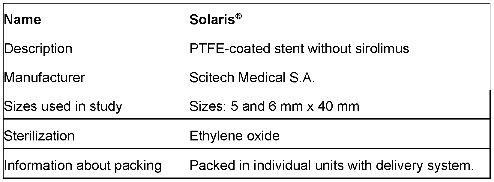
- the Solaris® is a flexible, self-expanding stent, comprised of a thin, multi-direction, durable electrospinning PTFE membrane encapsulating a Nitinol stent structure.
- the stent is laser cut from a metal tube of Nickel-titanium alloy (Nitinol) to form a metallic scaffold which is subsequently covered with polytetrafluorethylene (PTFE) layers.
- PTFE polytetrafluorethylene
- the delivery system is “Pull Back” type and OTW (Over the Wire) composed of concentric tubes that moves to allow external tube retraction on the stent implantation procedure.
- Solaris® stents of 5, 6, 7, 8 ,9 and 10 mm of diameter and a length of 40 to 100 mm, having uncovered end portions of 2 to 3 mm were used as starting material for the preparation of a Solaris DE stent.
- a Sirolimus coating was applied as explained below in order to obtain a Solaris DE stent.
- the coating formulation contained sirolimus drug as an active ingredient a biodegradable polymer matrix of PLA and PLGA.
- Drug-coating solutions containing PLA, PLGA, and rapamycin in chloroform were prepared according to the amounts shown in Table 1 below.
- the stents were washed with isopropyl alcohol.
- Shrink tubes of 20 mm ⁇ 5 mm, made of a HDPE, and having a length were used as protectors to avoid the drug-containing coating covers the PTFE-cover. A cut was mad on a side of each tube to ease the insertion of the stent inside. Shrink tubes were positioned at the ends of the stent, so that the tube overlapped the PTFE coating and the cut was pointed towards the center of the stent (Fig. 2).
- the assembly of the stent with the shrink tube was positioned in front of a hot air blowing machine, set at a temperature of at 130 °C ⁇ 10 °C, and the assembly was rotated while flowing hot are at a minimum value to promote conformation (Fig. 3.
- the conformation was made to allow a minimum movement of the shrink tube if necessary, and to protect the PTFE cover while leaving the structural scaffold end portions free.
- Coating of the ends of the stents was carried out by spray coating (Coating Machine 1.0- Scitech Medical).
- the process was carried out at a relative humidity of 60% to 65% and a temperature from 25 °C to 30 °C.
- the airbrush flow was calibrated to 0.5 ml in 35 s ⁇ 2s.
- the stent was fixed in a mandrel.
- the volumes of coating solutions shown in Table 2 were introduced in the airbrush container.
- *Other volumes can be used according to coating machine parameters; the variation can reach up to 200%.
- the process was repeated for every end of the stent, by repositioning the stent after coating one of the ends so that the uncoated end was in front of the airbrush.
- the ends of the stent were thus coated with a whitish and uniform coating.
- the shrink tubes were removed by tearing them by the cut ends, approximately 10 minutes after the coating process is finished.
- the 5 mm, 6 mm and 7 mm diameter stents were packed in cryogenic tubes, and the 8 mm and 9 mm diameter stents in amber glass tubes.
- the obtained Solaris DE were stored in Tyvek and then in a desiccator protected from light and moisture. A vacuum of at least 300 mmHg was also applied.
- Vascular response was determined in vivo by intravascular imaging and related histopathological evaluation.
- IVIIS intravascular imaging
- Porcine Sus scrofa domestica
- Sex Female or castrated male
- Animals included into the study had similar weights and ages. Pigs were acclimated to the facility seven days before the beginning of the study. After implantation, follow up was performed at 30 and 90 days (n 6/time-point). A total number of 12 animals were used for the study.
- the primary endpoint is lumen area and % stenosis. Both parameters are inversely proportional.
- Pigs included in the study had similar weights (40-43 Kg) and ages. They were acclimated to the facility three days before the start of the study.
- the animals were submitted to a 12-hour food fast and a 6-hour water fast, and were anesthetized according to a standard procedure before stent implantation.
- an 8F or 9F sheath was used (depending on the caliber of the stent delivery system) inserted into the femoral artery. 100-200 lll/kg of heparin was administered intravenously prior to selective catheterization for stent implantation.
- angiographies of the aorta and iliac arteries were performed with an appropriately sized multipurpose catheter, positioned in the topography of the distal aorta, just below the renal arteries. Then, under fluoroscopy, with the aid of a guide wire, the right and left common iliac arteries received one stent each (according to Table 4), respecting the maximum mean diameter of 0.5 mm to 1 mm in relation to the artery. The catheter was removed, and a post-implant image acquired. Finally, the catheter and guidewire were removed, and hemostasis was performed by suturing the access site. During the entire procedure, the animals remained in the supine position. Implant matrix used is shown in Table 5.
- Angiography will be performed shortly after stent implantation to assess the correct position of the stents.
- the animals evaluated after 60 days were also subjected to IVIIS evaluation of the stent segments to determine the occurrence of neointimal formation.
- the acquired images were analyzed using appropriate measurement software and provided measurements for luminal areas, stent areas, areas of neointimal hyperplasia and % neointimal hyperplasia.
- the vascular access site was observed and evaluated once daily for a minimum of 7 days for signs of infection or inflammation.
- NIH neointimal hyperplasia
- the tested SOLARIS DE product fulfilled all expectations of efficiency in this pre-clinical trial without raising any safety concerns.
- the results indicate that the test group showed an adequate safety profile in the preclinical mode, and that there is a tendency for the stent of the invention (Solaris DE stent) to have less hyperplasia that the control stent (Solaris non drug-eluting stent).
- Inspiron® DES (commercialized by Scitech Medical S.A.) is a stent made of a platform material of CoCr and thin struts (75 pm) and an abluminal coating composed of a mixture of PLA and PLGA polymers and a low Sirolimus dosage.
- Thrombosis with sirolimus-polymer was compared with controls with no coating and with only polymer coating with no drugs. Thrombosis was assessed clinically (sudden death of the animal after the procedure with autopsy showed thrombotic occlusion of the stent), by angiographic or histological analysis. The presence medial necrosis with aneurysm formation of polymer-coated stent with sirolimus compared with controls with no coating and polymer coating only with no drugs was determined by histology or angiography (aneurysm, defined as dilation 150% of the vascular diameter of reference).
- the occurrence of exacerbated neointimal formation of polymer-coated stent with sirolimus compared with controls with no coating and polymer coating only with no drugs was evaluated by histology (neointimal area by histomorphometry) or angiography (late lumen loss).
- the primary efficacy endpoint was the reduction of neointimal proliferation of polymer-coated stent with sirolimus compared with no drugs.
- the animals underwent implantation of three types of coronary stents.
- Comparative example 4 stent covered with polymer and sirolimus - Inspiron® DES, i.e. Cronus® stent coated with sirolimus in a polymer carrier at the concentration of 1.4 pg/mm 2 , polymer carrier: biodegradable polymer blend of PLLA and PLGA.
- the basal and control angiograms were analyzed quantitatively to assess the presence of stent migration, intraluminal negative image or involvement of the lateral branch.
- Quantitative angiographic analysis was performed using the CASS II system (Pie Medical, Maastricht, Netherlands). Were analyzed the in-stent segment, and 5 mm proximal and distal to the stent. The following parameters were measured at baseline and control angiograms in each segment (in-stent, proximal edge and distal edge), minimum luminal diameter (MLD), reference diameter (RD), luminal stenosis in diameter (ELD). Based on these measures the angiographic late loss, defined as control DLM - DLM basal, was calculated. Aneurysmal dilatation was defined as an increase in diameter in the treated area 150 % of reference diameter.
- the hearts were explanted and perfused for 30 minutes under pressure ( ⁇ 80 mm Hg) through the aortic root with 10% formalin.
- the arterial segments and the adjacent tissue dissected form the epicardial surface and dried in progressive ethanol solutions for inclusion in plastic resin.
- the stent was cut into segments of 5 mm. For each segment was obtained with 1-10-micron sections mm thick, stained for histological analysis with hematoxylin-eosin, van Gieson dye and Masson's trichrome.
- Sectional areas were measured internal elastic lamina (LEI) and the vascular lumen.
- LAI internal elastic lamina
- the neointima area was calculated as 100X (LAW - light area) /LAW.
- the thickness of the neointima was measured perpendicular to the light from each stent strut.
- the severity of vascular injury caused by stent implantation was measured according to the following criteria:
- the Solaris DE stent combines the advantages of Solaris stent and the advantages of drug-eluting stents (Inspiron®) with a lower amount of drug, which is applied only on the ends of the stent.
- Inspiron® drug-eluting stents
- a clinical significance was observed in the image tests with a lower volume of neointimal hyperplasia after 60 days of use and a greater volume of lumen for Solaris DE when compared to the control Solaris (Table 8).
Landscapes
- Health & Medical Sciences (AREA)
- Life Sciences & Earth Sciences (AREA)
- Chemical & Material Sciences (AREA)
- Public Health (AREA)
- Veterinary Medicine (AREA)
- Animal Behavior & Ethology (AREA)
- General Health & Medical Sciences (AREA)
- Epidemiology (AREA)
- Medicinal Chemistry (AREA)
- Vascular Medicine (AREA)
- Heart & Thoracic Surgery (AREA)
- Engineering & Computer Science (AREA)
- Biomedical Technology (AREA)
- Surgery (AREA)
- Chemical Kinetics & Catalysis (AREA)
- Pharmacology & Pharmacy (AREA)
- Molecular Biology (AREA)
- Organic Chemistry (AREA)
- Polymers & Plastics (AREA)
- Dermatology (AREA)
- Cardiology (AREA)
- Oral & Maxillofacial Surgery (AREA)
- Transplantation (AREA)
- Materials For Medical Uses (AREA)
Abstract
Description




Claims
Priority Applications (6)
| Application Number | Priority Date | Filing Date | Title |
|---|---|---|---|
| KR1020267001332A KR20260030817A (en) | 2023-06-15 | 2024-06-14 | Drug-eluting stent comprising a biocompatible thermoplastic polymer lining and a drug-containing coating |
| AU2024302945A AU2024302945A1 (en) | 2023-06-15 | 2024-06-14 | A drug-eluting stent comprising a biocompatible thermoplastic polymer lining and a drug-containing coating |
| EP24733178.8A EP4727608A1 (en) | 2023-06-15 | 2024-06-14 | A drug-eluting stent comprising a biocompatible thermoplastic polymer lining and a drug-containing coating |
| CN202480038168.3A CN121358509A (en) | 2023-06-15 | 2024-06-14 | Drug-eluting scaffold containing a biocompatible thermoplastic polymer liner and a drug-coated layer |
| IL325329A IL325329A (en) | 2023-06-15 | 2025-12-14 | A drug-eluting stent comprising a biocompatible thermoplastic polymer lining and a drug-containing coating |
| MX2025015108A MX2025015108A (en) | 2023-06-15 | 2025-12-15 | A drug-eluting stent comprising a biocompatible thermoplastic polymer lining and a drug-containing coating |
Applications Claiming Priority (2)
| Application Number | Priority Date | Filing Date | Title |
|---|---|---|---|
| EP23382599.1 | 2023-06-15 | ||
| EP23382599.1A EP4477240A1 (en) | 2023-06-15 | 2023-06-15 | A drug-eluting stent comprising a biocompatible thermoplastic polymer lining and a drug-containing coating |
Publications (1)
| Publication Number | Publication Date |
|---|---|
| WO2024256626A1 true WO2024256626A1 (en) | 2024-12-19 |
Family
ID=86861774
Family Applications (1)
| Application Number | Title | Priority Date | Filing Date |
|---|---|---|---|
| PCT/EP2024/066528 Ceased WO2024256626A1 (en) | 2023-06-15 | 2024-06-14 | A drug-eluting stent comprising a biocompatible thermoplastic polymer lining and a drug-containing coating |
Country Status (8)
| Country | Link |
|---|---|
| US (1) | US20240415678A1 (en) |
| EP (2) | EP4477240A1 (en) |
| KR (1) | KR20260030817A (en) |
| CN (1) | CN121358509A (en) |
| AU (1) | AU2024302945A1 (en) |
| IL (1) | IL325329A (en) |
| MX (1) | MX2025015108A (en) |
| WO (1) | WO2024256626A1 (en) |
Citations (4)
| Publication number | Priority date | Publication date | Assignee | Title |
|---|---|---|---|---|
| US20070078509A1 (en) * | 2003-09-24 | 2007-04-05 | Lotfy Wael M N | Valved balloon stent |
| US20100161032A1 (en) * | 2007-08-15 | 2010-06-24 | Francisco Avellanet | Biologically engineered stent |
| WO2010083530A2 (en) | 2009-01-16 | 2010-07-22 | Zeus Industrial Products, Inc. | Electrospinning of ptfe with high viscosity materials |
| US20160296351A1 (en) | 2009-01-16 | 2016-10-13 | Zeus Industrial Products, Inc. | Electrospun ptfe encapsulated stent and method of manufacture |
Family Cites Families (4)
| Publication number | Priority date | Publication date | Assignee | Title |
|---|---|---|---|---|
| US5464650A (en) * | 1993-04-26 | 1995-11-07 | Medtronic, Inc. | Intravascular stent and method |
| US6124523A (en) * | 1995-03-10 | 2000-09-26 | Impra, Inc. | Encapsulated stent |
| US20060210600A1 (en) * | 2003-07-07 | 2006-09-21 | Medtronic Vascular, Inc. | Coated stent with timed release of multiple therapeutic agents to inhibit restenosis adjacent to the stent ends |
| US20070179599A1 (en) * | 2006-01-31 | 2007-08-02 | Icon Medical Corp. | Vascular protective device |
-
2023
- 2023-06-15 EP EP23382599.1A patent/EP4477240A1/en not_active Withdrawn
- 2023-07-27 US US18/360,284 patent/US20240415678A1/en active Pending
-
2024
- 2024-06-14 EP EP24733178.8A patent/EP4727608A1/en active Pending
- 2024-06-14 AU AU2024302945A patent/AU2024302945A1/en active Pending
- 2024-06-14 KR KR1020267001332A patent/KR20260030817A/en active Pending
- 2024-06-14 WO PCT/EP2024/066528 patent/WO2024256626A1/en not_active Ceased
- 2024-06-14 CN CN202480038168.3A patent/CN121358509A/en active Pending
-
2025
- 2025-12-14 IL IL325329A patent/IL325329A/en unknown
- 2025-12-15 MX MX2025015108A patent/MX2025015108A/en unknown
Patent Citations (4)
| Publication number | Priority date | Publication date | Assignee | Title |
|---|---|---|---|---|
| US20070078509A1 (en) * | 2003-09-24 | 2007-04-05 | Lotfy Wael M N | Valved balloon stent |
| US20100161032A1 (en) * | 2007-08-15 | 2010-06-24 | Francisco Avellanet | Biologically engineered stent |
| WO2010083530A2 (en) | 2009-01-16 | 2010-07-22 | Zeus Industrial Products, Inc. | Electrospinning of ptfe with high viscosity materials |
| US20160296351A1 (en) | 2009-01-16 | 2016-10-13 | Zeus Industrial Products, Inc. | Electrospun ptfe encapsulated stent and method of manufacture |
Also Published As
| Publication number | Publication date |
|---|---|
| US20240415678A1 (en) | 2024-12-19 |
| KR20260030817A (en) | 2026-03-06 |
| EP4477240A1 (en) | 2024-12-18 |
| IL325329A (en) | 2026-02-01 |
| MX2025015108A (en) | 2026-04-01 |
| CN121358509A (en) | 2026-01-16 |
| EP4727608A1 (en) | 2026-04-22 |
| AU2024302945A1 (en) | 2026-01-29 |
Similar Documents
| Publication | Publication Date | Title |
|---|---|---|
| US8303650B2 (en) | Biodegradable self-expanding drug-eluting prosthesis | |
| JP6955553B2 (en) | Drug-eluting stents and their use to enable recovery of functional endothelial cell layers | |
| US6306166B1 (en) | Loading and release of water-insoluble drugs | |
| EP2316377B1 (en) | Drug-Delivery Endovascular Stent | |
| US8317857B2 (en) | Biodegradable self-expanding prosthesis | |
| JP2009525809A (en) | Implantable medical device with drug delivery coating with surface erodible polyester | |
| Uurto et al. | Biodegradable self-expanding poly-L/D-lactic acid vascular stent: a pilot study in canine and porcine iliac arteries | |
| Bünger et al. | Sirolimus-eluting biodegradable poly-L-lactide stent for peripheral vascular application: a preliminary study in porcine carotid arteries | |
| CN101631514A (en) | Multi-drug eluting coronary stent for percutaneous coronary intervention | |
| US20080167710A1 (en) | Medical Device Having Regions With Various Agents Dispersed Therein and a Method for Making the Same | |
| KR101595267B1 (en) | Consecutive Drug releaseing stent for restenosis and inflammatory regulation and manufacturing method thereof | |
| US20240415678A1 (en) | A Drug-Eluting Stent Comprising a Biocompatible Thermoplastic Polymer Lining and a Drug- Containing Coating | |
| US20050214343A1 (en) | Medical devices comprising a protein-tyrosine kinase inhibitor to inhibit restonosis | |
| WO2004009147A1 (en) | Medical devices comprising a protein-tyrosine kinase inhibitor to inhibit restonosis | |
| JP7641222B2 (en) | Covered stents for localized drug delivery | |
| KR20230150790A (en) | drug-eluting stent | |
| Edelman | Coronary stenting with M-Guard: feasibility and safety porcine trial | |
| Radeleff et al. | Comparison of a microporous thermoplastic polyurethane–covered stent with a self-expanding bare nitinol stent in a porcine iliac artery model | |
| Lim et al. | Optimal coating method for a dual-layer stent with sirolimus and alpha-lipoic acid in a porcine coronary restenosis model | |
| HK1259282B (en) | Drug eluting stent and method of use of the same for enabling restoration of functional endothelial cell layers | |
| Uurto | Preclinical Evaluation of New Drug-Eluting Biodegradable Poly-D/L-lactic Acid Vascular Stent | |
| HK1150229B (en) | Drug-delivery endovascular stent and method for treating restenosis |
Legal Events
| Date | Code | Title | Description |
|---|---|---|---|
| 121 | Ep: the epo has been informed by wipo that ep was designated in this application |
Ref document number: 24733178 Country of ref document: EP Kind code of ref document: A1 |
|
| WWE | Wipo information: entry into national phase |
Ref document number: 325329 Country of ref document: IL |
|
| WWE | Wipo information: entry into national phase |
Ref document number: P2025-04088 Country of ref document: AE Ref document number: MX/A/2025/015108 Country of ref document: MX |
|
| REG | Reference to national code |
Ref country code: BR Ref legal event code: B01A Ref document number: 112025027875 Country of ref document: BR |
|
| WWE | Wipo information: entry into national phase |
Ref document number: 202617002608 Country of ref document: IN |
|
| WWE | Wipo information: entry into national phase |
Ref document number: AU2024302945 Country of ref document: AU Ref document number: 828960 Country of ref document: NZ |
|
| ENP | Entry into the national phase |
Ref document number: 1020267001332 Country of ref document: KR Free format text: ST27 STATUS EVENT CODE: A-0-1-A10-A15-NAP-PA0105 (AS PROVIDED BY THE NATIONAL OFFICE) |
|
| WWE | Wipo information: entry into national phase |
Ref document number: 1020267001332 Country of ref document: KR |
|
| WWE | Wipo information: entry into national phase |
Ref document number: 2024733178 Country of ref document: EP Ref document number: 2026100699 Country of ref document: RU |
|
| NENP | Non-entry into the national phase |
Ref country code: DE |
|
| ENP | Entry into the national phase |
Ref document number: 2024733178 Country of ref document: EP Effective date: 20260115 |
|
| ENP | Entry into the national phase |
Ref document number: 2024733178 Country of ref document: EP Effective date: 20260115 |
|
| ENP | Entry into the national phase |
Ref document number: 2024302945 Country of ref document: AU Date of ref document: 20240614 Kind code of ref document: A |
|
| WWP | Wipo information: published in national office |
Ref document number: 828960 Country of ref document: NZ |
|
| ENP | Entry into the national phase |
Ref document number: 2024733178 Country of ref document: EP Effective date: 20260115 |
|
| WWP | Wipo information: published in national office |
Ref document number: 325329 Country of ref document: IL |
|
| ENP | Entry into the national phase |
Ref document number: 2024733178 Country of ref document: EP Effective date: 20260115 |
|
| WWP | Wipo information: published in national office |
Ref document number: 1020267001332 Country of ref document: KR |
|
| WWP | Wipo information: published in national office |
Ref document number: MX/A/2025/015108 Country of ref document: MX |
|
| WWP | Wipo information: published in national office |
Ref document number: 2024733178 Country of ref document: EP |