WO2024256627A1 - Novel innate lymphoid cells (ilcs) - Google Patents
Novel innate lymphoid cells (ilcs) Download PDFInfo
- Publication number
- WO2024256627A1 WO2024256627A1 PCT/EP2024/066529 EP2024066529W WO2024256627A1 WO 2024256627 A1 WO2024256627 A1 WO 2024256627A1 EP 2024066529 W EP2024066529 W EP 2024066529W WO 2024256627 A1 WO2024256627 A1 WO 2024256627A1
- Authority
- WO
- WIPO (PCT)
- Prior art keywords
- human
- ilcregs
- epithelial
- cells
- ilc
- Prior art date
- Legal status (The legal status is an assumption and is not a legal conclusion. Google has not performed a legal analysis and makes no representation as to the accuracy of the status listed.)
- Ceased
Links
Classifications
-
- C—CHEMISTRY; METALLURGY
- C12—BIOCHEMISTRY; BEER; SPIRITS; WINE; VINEGAR; MICROBIOLOGY; ENZYMOLOGY; MUTATION OR GENETIC ENGINEERING
- C12N—MICROORGANISMS OR ENZYMES; COMPOSITIONS THEREOF; PROPAGATING, PRESERVING, OR MAINTAINING MICROORGANISMS; MUTATION OR GENETIC ENGINEERING; CULTURE MEDIA
- C12N5/00—Undifferentiated human, animal or plant cells, e.g. cell lines; Tissues; Cultivation or maintenance thereof; Culture media therefor
- C12N5/06—Animal cells or tissues; Human cells or tissues
- C12N5/0602—Vertebrate cells
- C12N5/0634—Cells from the blood or the immune system
- C12N5/0636—T lymphocytes
-
- C—CHEMISTRY; METALLURGY
- C12—BIOCHEMISTRY; BEER; SPIRITS; WINE; VINEGAR; MICROBIOLOGY; ENZYMOLOGY; MUTATION OR GENETIC ENGINEERING
- C12N—MICROORGANISMS OR ENZYMES; COMPOSITIONS THEREOF; PROPAGATING, PRESERVING, OR MAINTAINING MICROORGANISMS; MUTATION OR GENETIC ENGINEERING; CULTURE MEDIA
- C12N2502/00—Coculture with; Conditioned medium produced by
- C12N2502/23—Gastro-intestinal tract cells
-
- C—CHEMISTRY; METALLURGY
- C12—BIOCHEMISTRY; BEER; SPIRITS; WINE; VINEGAR; MICROBIOLOGY; ENZYMOLOGY; MUTATION OR GENETIC ENGINEERING
- C12N—MICROORGANISMS OR ENZYMES; COMPOSITIONS THEREOF; PROPAGATING, PRESERVING, OR MAINTAINING MICROORGANISMS; MUTATION OR GENETIC ENGINEERING; CULTURE MEDIA
- C12N2513/00—3D culture
Definitions
- This invention relates to a novel human cell type, named a human regulatory innate lymphoid cell (ILCreg).
- the invention provides a human ILCreg.
- the invention also provides an in vitro population of human ILCregs, pharmaceutical compositions comprising the human ILCregs and uses thereof.
- ILCs Innate lymphoid cells
- NK cells cytotoxic Natural Killer cells
- ILCs T-bet + Eomes
- ILC1 T-bet + Eomes + cytotoxic Natural Killer cells
- NK cells T-bet + Eomes
- ILC1 T-bet + Eomes
- the second group comprises Roro + Gata3 + ILC2
- the third group encompasses RORyt + Lymphoid tissue inducer cells (LTi) as well as Natural Cytotoxicity Receptor (NCR) +/_ ILC3.
- LTi Lymphoid tissue inducer cells
- NCR Natural Cytotoxicity Receptor
- ILC1 drive epithelial cell proliferation through TGF-B1; ILC2 are activated and proliferate in response to Tuft-cell derived IL-25; and ILC3 drive Lgr5 + intestinal stem cell proliferation through IL-22; finally, foetal Lti mediate development of secondary lymphoid structures, whereas NK cells are circulatory and not specifically enriched in mucosa.
- ILC Intracellular Lymphoid Precursor
- CLP Common Lymphoid Precursor
- ILC restricted Lineage CD127 + Id2 + IL-7R + o4B7 + ILC precursor (ILCP) in mice.
- Sources of ILCP have been identified in the murine adult bone marrow, foetal liver, small intestine and lung (Bando et al., 2015).
- ILC precursors have also been identified in human bone marrow, tonsils foetal, paediatric and adult intestines, but these are less well characterised than their murine counterparts (Elmentaite et al., 2021).
- ILCreg regulatory ILC
- human regulatory ILCs or ILCregs. These are defined in more detail below and may be used to suppress inflammation, especially in the intestine.
- the invention provides a human regulatory innate lymphoid cell (ILCreg) which expresses a detectable level of FOXP3 and/or CTLA4.
- the invention also provides: an in vitro population of human ILCregs, wherein the population comprises at least about two human ILCregs of the invention; a pharmaceutical composition comprising a human ILCreg of the invention or an in vitro population of the invention and a pharmaceutically or physiologically acceptable diluent and/or carrier; a method of treating or preventing a disease in a subject, wherein the method comprises administering to the subject a human ILCreg of the invention, an in vitro population of the invention or a pharmaceutical composition of the invention; a human ILCreg of the invention, an in vitro population of the invention or a pharmaceutical composition of the invention for use in the treatment or prevention of a disease; a method for expanding a human ILCreg of the invention or an in vitro population of the invention, the method comprising co-culturing the human ILC
- the invention also provides a human regulatory ILC (ILCreg) which expresses a detectable level of FOXP3.
- the human ILCreg preferably further expresses detectable levels of one or more of CD25 (IL2RA), CD127 and CTLA4, such as CD25 (IL2RA), CD127, CTLA4, CD25 and CD127, CD25 and CTLA4, CD127 and CTLA4, or CD25, CD127 and CTLA4.
- the human ILCreg preferably further expresses detectable levels of one or more, such as 2, 3, 4, 5, 6, 7, 8, 9, 10, 11, 12, 13, 14, 15, 16, or 17, of the following genes: CCR5, CTLA4, FGGY, GATA3, GZMB, IL10, IL1R1, IL2RA, IL2RB, KAT2B, LGLS3, PIM1, PRDM1, RUNX1, SOX4, TNFRSF18 and TRAF1.
- the human ILCreg preferably expresses detectable levels of all of these genes. Gene expression is typically measured by measuring messenger RNA (mRNA) expression, for instance using RNA sequencing or single cell RNAseq (scRNAseq). Gene expression, especially of FOXP3, can also be measured by measuring protein in the cell.
- mRNA messenger RNA
- scRNAseq single cell RNAseq
- Protein expression can be measured using standard methods, such as flow cytometry, Western blotting, immunohistochemistry, or enzyme-linked immunosorbent assay (ELISA). It is worthy of note FOXP3, RUNX1, IL1R1 and CTLA4 are associated with human T- regulatory cells (Tregs).
- the human ILCreg preferably further secretes a detectable level of IL-10. This can be measured using a standard cytokine release assay and by intracellular flow cytometry.
- the human ILCreg preferably expresses detectable levels of one or more, such as 2 or 3, of the following cell surface markers: CD25 (IL2RA), CD127 and CTLA4.
- the human ILCreg preferably expresses detectable levels of CD25 (IL2RA), CD127, CTLA4, CD25 and CD127, CD25 and CTLA4, CD127 and CTLA4, or CD25, CD127 and CTLA4.
- IL2RA CD25
- CD127 and CTLA4 CD25 and CD127
- CD25 and CTLA4 CD127 and CTLA4
- CD25, CD127 and CTLA4 CD25, CD127 and CTLA4.
- Surface marker expression can be measured using standard methods, such as flow cytometry.
- the invention also provides a human regulatory ILC (ILCreg) which expresses detectable levels of CTLA4.
- ILCreg human regulatory ILC
- Surface expression of CTLA4 can be measured using standard methods, such as flow cytometry.
- the human ILCreg preferably further expresses a detectable level of FOXP3.
- the human ILCreg preferably further expresses detectable levels of CD25 (IL2RA), and/or CD127.
- the human ILCreg preferably further expresses detectable levels of one or more, such as 2, 3, 4, 5, 6, 7, 8, 9, 10, 11, 12, 13, 14, 15, 16 or 17, of the following genes: CCR5, FGGY, FOXP3, GATA3, GZMB, IL10, IL1R1, IL2RA, IL2RB, KAT2B, LGLS3, PIM1, PRDM1, RUNX1, SOX4, TNFRSF18 and TRAF1.
- the human ILCreg preferably expresses detectable levels of all of these genes. Gene expression is typically measured by measuring messenger RNA (mRNA) expression, for instance using RNA sequencing or single cell RNAseq (scRNAseq). Gene expression, especially of FOXP3, can also be measured by measuring protein in the cell.
- mRNA messenger RNA
- scRNAseq single cell RNAseq
- Protein expression can be measured using standard methods, such as flow cytometry, Western blotting, immunohistochemistry, or enzyme-linked immunosorbent assay (ELISA). It is worthy of note FOXP3, RUNX1, IL1R1 and CTLA4 are associated with human T-regulatory cells (Tregs). The human ILCreg preferably further secretes a detectable level of IL-10. This can be measured using a standard cytokine release assay and by intracellular flow cytometry.
- the invention also provides a human regulatory ILC (ILCreg) which expresses detectable levels of FOXP3 and CTLA4. Expression of FOXP3 and CTLA4 can be measured as described above.
- the human ILCreg preferably further expresses detectable levels of CD25 (IL2RA), and/or CD127.
- the human ILCreg preferably further expresses detectable levels of one or more, such as 2, 3, 4, 5, 6, 7, 8, 9, 10, 11, 12, 13, 14, 15, or 16, of the following genes: CCR5, FGGY, GATA3, GZMB, IL10, IL1R1, IL2RA, IL2RB, KAT2B, LGLS3, PIM1, PRDM1, RUNX1, SOX4, TNFRSF18 and TRAF1.
- the human ILCreg preferably expresses detectable levels of all of these genes. Gene expression can be measured as described above. It is worthy of note FOXP3, RUNX1, IL1R1 and CTLA4 are associated with human T-regulatory cells (Tregs). The human ILCreg preferably further secretes a detectable level of IL-10. This can be measured using a standard cytokine release assay and by intracellular flow cytometry.
- the invention also provides a human regulatory ILC (ILCreg) which expresses detectable levels of one or more, such as 2 or 3, of the following cell surface markers: CD25 (IL2RA), CD127 and CTLA4.
- the human ILCreg preferably expresses detectable levels of CD25 (IL2RA), CD127, CTLA4, CD25 and CD127, CD25 and CTLA4, CD127 and CTLA4, or CD25, CD127 and CTLA4.
- Surface marker expression can be measured using standard methods, such as flow cytometry.
- the human ILCreg preferably further expresses a detectable level of FOXP3.
- the human ILCreg preferably further expresses detectable levels of one or more, such as 2, 3, 4, 5, 6, 7, 8, 9, 10, 11, 12, 13, 14, or 15, of the following genes: CCR5, FGGY, GATA3, GZMB, IL10, IL1R1, IL2RB, KAT2B, LGLS3, PIM1, PRDM1, RUNX1, SOX4, TNFRSF18 and TRAF1.
- the human ILCreg preferably expresses detectable levels of all of these genes. Gene expression is typically measured by measuring messenger RNA (mRNA) expression, for instance using RNA sequencing or single cell RNAseq (scRNAseq).
- mRNA messenger RNA
- scRNAseq single cell RNAseq
- FOXP3, RUNX1, IL1R1 and CTLA4 are associated with human T-regulatory cells (Tregs).
- the human ILCreg preferably further secretes a detectable level of IL-10. This can be measured using a standard cytokine release assay and by intracellular flow cytometry.
- the human ILCreg preferably does not express detectable levels of one or more, such as 2, 3, 4, 5 or 6, of CD3, CD4, CD19, CD20, TCRap, and TCRy8.
- the human ILCreg preferably does not express detectable levels of one or more, such as 2, 3, 4 or 5, of CD3, CD4, CD19, CD20, and TCRap.
- the human ILCreg preferably does not express detectable levels of one or more these markers at the cell surface and/or at the RNA level.
- the human ILCreg preferably does not express extracellular detectable levels of CD3 and CD4.
- the human ILCreg preferably does not express extracellular detectable levels of any of these markers.
- the human ILCreg preferably comprises a CD3", CD4", CD19", CD20", TCRap-, TCRy8’ expression profile.
- the human ILCreg preferably comprises a CD3", CD4", CD19", CD20", TCRap- expression profile.
- the human ILCreg preferably expresses a detectable level of CD127.
- the ILCregs of the invention are human. It is straightforward for the skilled person to determine whether or not a cell is human, for instance by examining the number and size of the chromosomes present in the nucleus, by sequencing part of the genome or by identifying the presence of specific human markers. The source of the cell also helps the skilled person confirm the ILCreg is human.
- the invention also provides an in vitro population of human ILCregs, wherein the population comprises at least about two human ILCregs of the invention.
- the population preferably comprises at least about 5, at least about 10, at least about 50, at least about 100, at least about 1000 or at least about 5 x 10 3 human ILCregs of the invention.
- the population preferably comprises at least about 5, at least about 10, at least about 50, at least about 100, at least about 150, at least about 200, at least about 250, at least about 300, at least about 350, at least about 400, at least about 450, at least about 500, at least about 600, at least about 700, at least about 800, at least about 900, at least about 1000 or at least about 5 x 10 3 human ILCregs of the invention.
- the human ILCregs of the invention may be any of those defined above.
- an in vitro population includes a population of cells in a format suitable for administration to a subject. This may include a vial, bag or needles containing the cells. The cells may be in a liquid solution or frozen form.
- the population comprises at least about 5 x 10 3 human ILCregs, at least about 1 x 10 4 human ILCregs, at least about 5 xlO 4 human ILCregs, at least about 1 xlO 5 human ILCregs, at least about 5 xlO 5 human ILCregs, at least about IxlO 6 human ILCregs, at least about 5 xlO 6 human ILCregs, at least about IxlO 7 human ILCregs, at least about 5xl0 7 human ILCregs, at least about IxlO 8 human ILCregs, at least about 5xl0 8 human ILCregs, at least about IxlO 9 human ILCregs, at least about 5xl0 9 human ILCregs, at least about IxlO 10 human ILCregs, at least about 5xlO 10 human ILCregs, at least about IxlO 11 human ILCregs, at least about IxlO
- the population comprises at least about IxlO 20 human ILCregs. In some embodiments the population comprises at least about IxlO 100 human ILCregs. In some embodiments the population comprises at least about IxlO 500 human ILCregs. In some embodiments the population comprises at least about IxlO 1000 human ILCregs.
- the population comprises no more than about IxlO 10000 human ILCregs. In some embodiments the population comprises no more than about IxlO 500 human ILCregs. In some embodiments the population comprises of from about 5 x 10 4 human ILCregs to about IxlO 10000 human ILCregs. In some embodiments the population comprises of from about IxlO 200 human ILCregs to about IxlO 10000 human ILCregs.
- the in vitro population may comprise other ILCs in addition to the human ILCregs.
- the in vitro population of ILCs may comprise a plurality of Group 1 and Group 2 ILCs.
- the in vitro population of ILCs comprise a plurality of Group 1 and Group 3 ILCs.
- the in vitro population of ILCs may comprise a plurality of Group 2 and Group 3 ILCs.
- the in vitro population of ILCs comprise a plurality of Group 1, Group 2 and Group 3 ILCs.
- the in vitro population of ILCs comprises a plurality of Group 1 ILCs and human ILCregs.
- the in vitro population of ILCs comprises a plurality of human ILCregs and Group 2 ILCs. In some embodiments, the in vitro population of ILCs comprises a plurality of human ILCregs and Group 3 ILCs. In some embodiments, the in vitro population of ILCs comprises a plurality of Group 1 ILCs, Group 2 ILCs and human ILCregs. In some embodiments, the in vitro population of ILCs comprises a plurality of Group 1 ILCs, Group 3 ILCs and human ILCregs. In some embodiments, the in vitro population of ILCs comprises a plurality of Group 2 ILCs, Group 3 ILCs and human ILCregs. In some embodiments, the in vitro population of ILCs comprises a plurality of Group 1 ILCs, Group 2 ILCs, Group 3 ILCs and human ILCregs.
- the in vitro population of human ILCregs of the invention may comprise at least about 0.1%, at least about 0.2%, at least about 0.5%, at least about 0.8%, at least about 1%, at least about 2%, at least about 5%, at least about 10%, at least about 20%, at least about 25%, at least about 30%, at least about 40%, at least about 50%, at least about 60%, at least about 70%, at least about 80% or at least about 90% human ILCregs of the invention.
- the in vitro population of ILCs is a heterogenous population of ILCs. In other embodiments the in vitro population of ILCs is a homologous population of ILCs.
- At least about 10%, at least about 20%, at least about 30%, at least about 40%, at least about 50%, at least about 60%, at least about 70%, at least about 80% or at least about 90% of the human ILCregs express a detectable level of FOXP3.
- At least about 10%, at least about 20%, at least about 30%, at least about 40%, at least about 50%, at least about 60%, at least about 70%, at least about 80% or at least about 90% of the human ILCregs express a detectable level of CD25 (IL2RA).
- At least about 10%, at least about 20%, at least about 30%, at least about 40%, at least about 50%, at least about 60%, at least about 70%, at least about 80% or at least about 90% of the human ILCregs express a detectable level of CD127.
- At least about 10%, at least about 20%, at least about 30%, at least about 40%, at least about 50%, at least about 60%, at least about 70%, at least about 80% or at least about 90% of the human ILCregs express a detectable level of CTLA4.
- At least 50% of the in vitro population of human ILCs may co-express detectable levels of two or more cytokines.
- the in vitro population of human ILCregs may be a stimulated in vitro population of human ILCregs.
- the population may comprise a PMA and lonomycin stimulated in vitro population of human ILCregs.
- a stimulated in vitro population of human ILCregs expresses detectable levels of one or more cytokines as described above.
- the in vitro population of human ILCregs may have a tissue-specific imprint.
- tissuespecific imprint this will be understood to refer to a genetic signature and phenotype specific of in vivo immune cells of a particular organ.
- the in vitro population of human ILCregs comprises or consists of a population of tissue-specific human ILCregs.
- tissue-specific human ILCregs may have utility for the treatment of particular diseases and/or may have improved homing capacity to the specific tissue when administered to a subject.
- the in vitro population of human ILCregs comprise or consist of skin, intestinal, lung, thymic, thyroid, reproductive, bladder, kidney, pancreas, oral mucosal or liver specific human ILCregs.
- the in vitro population of human ILCregs comprises or consists of skin, intestinal, lung, thyroid, reproductive, bladder, kidney, pancreas, oral mucosal or liver specific human ILCregs. In some embodiments, the in vitro population of human ILCregs comprises or consists of intestinal, lung, reproductive or oral mucosal specific human ILCregs.
- Reproductive-specific human ILCregs may comprise fallopian tube specific human ILCregs, ovary specific human ILCregs, prostate specific human ILCregs, endometrium specific human ILCregs, cervix specific human ILCregs, vaginal specific human ILCregs and testes- specific human ILCregs.
- Reproductive-specific human ILCregs may be selected from fallopian tube specific human ILCregs, ovary specific human ILCregs, prostate specific human ILCregs and endometrium specific human ILCregs.
- Oral mucosal specific human ILCregs may comprise salivary gland taste bud specific ILCs, lingual region specific human ILCregs and oesophagus specific human ILCregs.
- the oral mucosal specific human ILCregs are oesophageal specific human ILCregs.
- the in vitro population of human ILCregs comprises or consists of intestinal-specific human ILCregs and/or lung-specific human ILCregs.
- the intestinal specific human ILCregs may be small intestinal-specific human ILCregs and/or lung specific human ILCregs.
- the in vitro population of human ILCregs may comprise or consist of epithelial cancer specific human ILCregs.
- the in vitro population of human ILCregs may comprise or consist of skin cancer specific human ILCregs, intestinal cancer specific human ILCregs, lung cancer specific human ILCregs, thymic cancer specific human ILCregs, thyroid cancer specific human ILCregs, reproductive cancer specific human ILCregs, bladder cancer specific human ILCregs, kidney cancer specific human ILCregs, pancreas cancer specific human ILCregs, oral mucosal cancer specific human ILCregs or liver cancer specific human ILCregs
- the in vitro population of human ILCregs may comprise or consist of skin cancer specific human ILCregs, intestinal cancer specific human ILCregs , lung cancer specific human ILCregs, thyroid cancer specific human ILCregs, reproductive cancer specific human ILCregs, bladder cancer specific human ILCregs, kidney cancer specific human ILC
- the human ILCregs are primary human ILCregs.
- a "primary cell” is a cell that is obtained from or is derived from a subject. Primary cells are not immortalised cells from a cell line.
- Primary human ILCregs can be produced from primary human ILC precursors, i.e., from human ILC precursors that have been obtained from a subject.
- the primary human ILCregs may be autologous.
- the primary human ILCregs may be allogeneic.
- the population comprises a mixture of allogeneic and autologous human ILCregs.
- the human ILCregs may comprise or consist of immortalised immune cells from a cell line.
- the human ILCregs may comprise or consist of immortalised human ILCregs or a human ILCreg cell line.
- At least about 10% of the in vitro population comprises an exogenous polynucleotide.
- exogenous polynucleotide this will be understood to refer to a polynucleotide which has been introduced into the human ILCreg, such that the ILCreg is genetically modified.
- the in vitro population of human ILCreg is a genetically modified in vitro population of human ILCreg.
- the exogenous polynucleotide is recombinant.
- the exogenous polynucleotide typically encodes an exogenous polypeptide.
- the exogenous polypeptide may be a polypeptide which is endogenous to the human ILCreg but is expressed in the cell at higher levels following introduction of the exogenous polynucleotide by genetic modification
- the exogenous polypeptide may be a polypeptide which is not naturally expressed in the human ILCreg.
- At least about 20%, at least about 30%, at least about 40%, at least about 50%, at least about 60%, at least about 70%, at least about 80% or at least about 90% of the cells in the in vitro population comprise an exogenous polynucleotide.
- At least about 10% of the cells in the in vitro population express the exogenous polypeptide encoded by the exogenous polynucleotide at a detectable level. In some embodiments at least about 20%, at least about 30%, at least about 40%, at least about 50%, at least about 60%, at least about 70%, at least about 80% or at least about 90% of the cells in the in vitro population express the exogenous polypeptide encoded by the exogenous polynucleotide at a detectable level.
- the exogenous polypeptide may comprise a marker protein or an immunotherapeutic molecule.
- the marker protein may otherwise be referred to as a reporter protein.
- Suitable marker proteins include, but are not necessarily limited to GFP, a MYC epitope tag or a FLAG epitope tag.
- the exogenous polypeptide further comprises a purification tag.
- a purification tag can assist with purification.
- purification tags include but are not necessarily limited to a His-tag, Arg-tag, T7-tag, Strep- tag, S-tag, aptamer-tag, V5 tag, or AviTagTM.
- Various other tags are well known in the art.
- the immunotherapeutic molecule may be any immunotherapeutic molecule which may further increase the immunotherapeutic use of the in vitro population of human ILCregs.
- the immunotherapeutic molecule may comprise an enzyme, an antibody, an antigen, a chimeric antigen receptor (CAR), MHC class II cell receptor, chemokine receptor and/or a cytokine.
- the MHC class II cell receptor may preferably comprise HLA-DR.
- the immunotherapeutic molecule comprises or consists of a chimeric antigen receptor (CAR).
- Chimeric antigen receptors are immune cell receptors which have been genetically engineered to confer the ability to target a specific antigen or antigens.
- chimeric antigen receptors are specific for one or more cancer-associated antigens. As such, chimeric antigen receptors are commonly used in the treatment of cancer.
- the CAR may comprise or consist of a first, second, third, or fourth generation CAR.
- First-generation CARs comprise or consist of a binding domain that is capable of specifically binding to an epitope on a target antigen, a transmembrane domain, and one or more intracellular signalling domains.
- the extracellular binding domain may comprise a singlechain variable fragment (scFv) from a monoclonal antibody.
- a first-generation CAR typically comprises a CD3 chain domain or a variant thereof as the intracellular signalling domain, which is the primary transmitter of signals.
- second-generation CARs also contain a co-stimulatory domain, such as CD28 and/or 4-1BB.
- a co-stimulatory domain improves T-cell proliferation, cytokine secretion, resistance to apoptosis, and in vivo persistence.
- the co-stimulatory domain of a second- generation CAR is typically in cis with and upstream of the one or more intracellular signalling domains.
- Third-generation CARs combine multiple co-stimulatory domains in cis with one or more intracellular signalling domains, to augment T-cell activity.
- a third-generation CAR may comprise co-stimulatory domains derived from CD28 and 41BB, together with an intracellular signalling domain derived from CD3 zeta.
- Other third-generation CARs may comprise co-stimulatory domains derived from CD28 and 0X40, together with an intracellular signalling domain derived from CD3 zeta.
- Fourth-generation CARs combine the features of a second-generation CAR with further factors to enhance anti-tumour activity (e.g., cytokines, co-stimulatory ligands, chemokines receptors or further chimeric receptors of immune regulatory or cytokine receptors).
- the factors may be in trans or in cis with the CAR, typically in trans with the CAR.
- the CAR is specific for a cancer antigen.
- the cancer antigen may be a solid tumour cancer antigen.
- specific in the context of the CAR, this will be understood to refer to being capable of specifically binding to a target antigen.
- Cancer antigens include, but are not necessarily limited to Erbbl, Erbb3, Erbb4, Erbb2, mucins, PSMA, carcinoembryonic antigen (CEA), mesothelin, GD2, MUC1, folate receptor, NKG2D ligands, ligands bound by other NK receptors such as NKp30, NKp44 or NKp46, GPC3, CAIX, FAP, NY-ESO-1, gplOO, PSCA, ROR1, PD-L1, PD-L2, EpCAM, EGFRvIII, CD19, CD20, CD22, GD3, CLL-1, ductal epithelial mucin, CA-125, GP36, TAG-72, glycosphingolipids, glioma-associated antigen, beta-hCG, AFP (alpha-fetoprotein) and lectinreactive AFP, thyroglobulin, receptor for advanced glycation end products (RAGE),
- the cancer antigen is selected from NYESO, GP100, PRAME, COL6A3, MR1, CDlc, HER2, SLCA2, CD19, PSMA, AFP, CEA, CA-125, MUC1, ETA, tyrosinase and MAGE.
- the CAR is an anti-CD19, anti-SLC3A2 or anti-PSMA CAR.
- the CAR is an anti-CD19 or anti-PSMA CAR.
- MAGE may be selected from MAGE Al, MAGE A2, MAGE A4 or MAGE A8.
- the CAR may be linked to a reporter protein, for example GFP, MYC epitope flag or a FLAG epitope tag.
- a reporter protein for example GFP, MYC epitope flag or a FLAG epitope tag.
- Other suitable reporter proteins will be known to those skilled in the art.
- the CAR comprises a second-generation CAR.
- Suitable CAR intracellular signalling domains may include any suitable signalling domain, including any region comprising an Immune-receptor-Tyrosine-based-Activation-Motif (ITAM), as reviewed for example by Love et al. Cold Spring Harbor Perspect. Biol 2010 2(6)1 a002485.
- the signalling domain comprises the intracellular domain of human CD3 [zeta] chain as described for example in US Patent No 7,446,190, or a variant thereof.
- co-stimulatory domains are known to engineer CAR cells.
- the CAR may comprise one or more of these domains.
- Suitable co-stimulatory domains include, but are not necessarily limited to members of the B7/CD28 family such as B7-1, B7-2, B7-H1, B7-H2, B7-H3, B7-H4, B7-H6, B7-H7, BTLA, CD28, CTLA-4, Gi24, ICOS, PD-1, PD-L2 or PDCD6; or ILT/CD85 family proteins such as LILRA3, LILRA4, LILRB1, LILRB2, LILRB3 or LILRB4; or tumour necrosis factor (TNF) superfamily members such as 4-1BB, BAFF, BAFF R, CD27, CD30, CD40, DR3, GITR, HVEM, LIGHT, Lymphotoxin-alpha, 0X40, RELT, TACI, TL1A, TNF- alpha or TNF RII
- the immunotherapeutic molecule comprises a MHC Class II cell surface receptor.
- the MHC Class II cell surface receptor comprises or consists of HLA-DR.
- exogenous expression of HLA-DR by the human ILCregs may aid immunoregulatory activity.
- the immunotherapeutic molecule comprises a cytokine.
- the cytokine may be an immunoregulatory cytokine, for example IL-10 or TGF-p.
- the expression of an immunoregulatory cytokine or receptor in the in vitro population of the human ILCregs may have particular use when the human ILCregs are used for the treatment of an autoimmune disease, for example inflammatory bowel disease (IBD) or multiple sclerosis, or an allergy. Further autoimmune diseases and specific allergies are described below.
- the cytokine is an inflammatory cytokine.
- exemplary inflammatory cytokines include, but are not necessarily limited to IL-22, IL-17A, IL-5, IL-4, Amphiregulin, IFN-y, IL-2, IL-1, IL-18, TNF-o and GM-CSF.
- the cytokine comprises one or more of IL-22, IL-17A, IL-5, IL-4, Amphiregulin, IFN-y, IL-2, IL-1, IL-18, TNF-o and GM-CSF.
- the cytokine comprises one or more of IL-22, IL- 17A, IL-5, IL-4, IFN-y, TNF-o and GM-CSF.
- the expression of such an inflammatory cytokine may have particular use when the human ILCregs are used for the treatment of a cancer.
- chemokine receptors are known in the art. Exemplary chemokine receptors include, but are not necessarily limited to CXC chemokine receptors, CC chemokine receptors, XCR1 and CX3CR1.
- the chemokine receptor comprises a CC chemokine receptor.
- the chemokine receptor may comprise one or more of CCR1, CCR.2, CCR.3, CCR4, CCR.5, CCR.6, CCR.7, CCR.8, CCR.9, CCR10 and CCR11.
- the chemokine receptor comprises or consists of CCR7 or CCR3.
- the chemokine receptor comprises or consists of CCR7.
- the present inventors believe that the expression of a chemokine receptor in the in vitro population of human ILCregs may assist with tissuespecific therapeutic targeting of the human ILCregs. This may further increase the therapeutic efficacy of the in vitro population of human ILCregs.
- a vector comprises the exogenous polynucleotide.
- the vector may be viral or non-viral.
- Various viral and non-viral vectors are known to those skilled in the art.
- Non-viral vectors include plasmids, episomal vectors, and human artificial chromosomes (see, e.g., Harrington et al., 1997, Nat Genet. 15:345).
- non-viral vectors useful for expression of the exogenous polypeptide in mammalian (e.g., human) cells include pThioHis A, B and C, pcDNA3.1/His, pEBVHis A, B and C, (Invitrogen, San Diego, Calif.), MPS V vectors, and numerous other vectors known in the art for expressing other proteins.
- Useful viral vectors include vectors based on retroviruses, adenoviruses, adeno-associated viruses, herpes viruses, vectors based on SV40, papilloma virus, HBP Epstein Barr virus, vaccinia virus vectors and Semliki Forest virus (SFV).
- retroviral, lentiviral, adenoviral or adeno-associated viral vectors are commonly used for expression in immune cells such as T-cells.
- retroviral, lentiviral, adenoviral or adeno-associated viral vectors are commonly used for expression in immune cells such as T-cells.
- retroviral, lentiviral, adenoviral or adeno-associated viral vectors include the SFG retroviral expression vector (see Riviere et al., 1995, Proc. Natl. Acad. Sci. (USA) 92:6733-6737).
- the vector is a retroviral or lentiviral vector.
- the vector is an SFG retroviral vector.
- the vector is a lentiviral vector. Lentiviral vectors include self-inactivating lentiviral vectors (so-called SIN vectors).
- Expression vectors for mammalian host cells can include expression control sequences, such as an origin of replication, a promoter, and an enhancer (see, e.g., Queen, et al., 1986, Immunol. Rev. 89:49-68), and necessary processing information sites, such as ribosome binding sites, RNA splice sites, polyadenylation sites, and transcriptional terminator sequences.
- expression control sequences such as an origin of replication, a promoter, and an enhancer (see, e.g., Queen, et al., 1986, Immunol. Rev. 89:49-68)
- necessary processing information sites such as ribosome binding sites, RNA splice sites, polyadenylation sites, and transcriptional terminator sequences.
- These vectors usually contain promoters derived from mammalian genes or from mammalian viruses. Suitable promoters may be constitutive, cell type- specific, stage-specific, and/or modulatable or regulatable.
- Useful promoters include, but are not limited to, the metallothionein promoter, the constitutive adenovirus major late promoter, the dexamethasone-inducible MMTV promoter, the SV40 promoter, the MRP polIII promoter, the constitutive MPS V promoter, the tetracycline-inducible CMV promoter (such as the human immediate-early CMV promoter), the constitutive CMV promoter, the EFl alpha promoter, the phosphoglycerate kinase (PGK) promoter and promoter-enhancer combinations known in the art.
- the metallothionein promoter the constitutive adenovirus major late promoter
- the dexamethasone-inducible MMTV promoter the SV40 promoter, the MRP polIII promoter, the constitutive MPS V promoter
- the tetracycline-inducible CMV promoter such as the human immediate-early CMV promoter
- the vector may further comprise a polynucleotide encoding a reporter gene.
- Suitable reporter genes include, but are not necessarily limited to HNIS, hNET and HSVtK.
- Cultures of transformed organisms can be expanded under non-inducing conditions without biasing the population for coding sequences whose expression products are better tolerated by the in vitro population of human ILCregs.
- promoters other regulatory elements may also be required or desired for efficient expression. These elements typically include an ATG initiation codon and adjacent ribosome binding site or other sequences.
- the efficiency of expression may be enhanced by the inclusion of enhancers appropriate to the cell system in use (see, e.g., Scharf et al., 1994, Results Probl. Cell Differ. 20: 125; and Bittner et al., 1987, Meth. Enzymol., 153:516).
- the SV40 enhancer or CMV enhancer may be used to increase expression in mammalian host cells.
- the genetic engineering of immune cells such as human ILCregs can be carried out according to standard cloning and expression techniques, which are known in the art (e.g., as described in Sambrook, J., Fritsh, E. F., and Maniatis, T. Molecular Cloning: A Laboratory Manual 2 nd , ed., Cold Spring Harbor Laboratory, Cold Spring Harbor Laboratory Press, Cold Spring Harbor, N.Y., 1989).
- the vector may be introduced into the in vitro population of human ILCregs using such techniques. Introduction may comprise transfection or transduction into the in vitro population of human ILCregs.
- transfection are intended to encompass a wide variety of techniques commonly used for the introduction of exogenous DNA into a prokaryotic or eukaryotic host cell, e.g., electroporation, calcium-phosphate precipitation, DEAE-dextran transfection and the like.
- polynucleotide refers to a polymer comprising two or more nucleotides.
- the polynucleotide comprises at least 30 nucleotides, at least 40 nucleotides, at least 50 nucleotides or at least 100 nucleotides.
- the nucleotides can be naturally occurring or artificial.
- a nucleotide typically contains a nucleobase, a sugar and at least one linking group, such as a phosphate, 2'O-methyl, 2' methoxy-ethyl, phosphoramidate, methylphosphonate or phosphorothioate group.
- the nucleobase is typically heterocyclic.
- Nucleobases include, but are not limited to, purines and pyrimidines and more specifically adenine (A), guanine (G), thymine (T), uracil (U) and cytosine (C).
- the sugar is typically a pentose sugar.
- Nucleotide sugars include, but are not limited to, ribose and deoxyribose.
- nucleosides include, but are not limited to, adenosine, guanosine, 5-methyluridine, uridine, cytidine, deoxyadenosine, deoxyguanosine, thymidine, deoxyuridine and deoxycytidine.
- the nucleosides may be adenosine, guanosine, uridine and cytidine.
- the nucleotides are typically ribonucleotides or deoxyribonucleotides.
- the nucleotides may be deoxyribonucleotides.
- the nucleotides typically contain a monophosphate, diphosphate or triphosphate. Phosphates may be attached on the 5' or 3' side of a nucleotide.
- Nucleotides include, but are not limited to, adenosine monophosphate (AMP), adenosine diphosphate (ADP), adenosine triphosphate (ATP), guanosine monophosphate (GMP), guanosine diphosphate (GDP), guanosine triphosphate (GTP), thymidine monophosphate (TMP), thymidine diphosphate (TDP), thymidine triphosphate (TTP), uridine monophosphate (UMP), uridine diphosphate (UDP), uridine triphosphate (UTP), cytidine monophosphate (CMP), cytidine diphosphate (CDP), cytidine triphosphate (CTP), 5-methylcytidine monophosphate, 5-methylcytidine diphosphate, 5-methylcytidine triphosphate, 5- hydroxy methylcytidine monophosphate, 5-hydroxymethylcytidine diphosphate, 5- hydroxy methylcytidine triphosphate,
- the nucleotides may be selected from AMP, UMP, GMP, CMP, dAMP, dTMP, dGMP or dCMP. In some embodiments, the nucleotides are selected from dAMP, dTMP, dGMP or dCMP.
- nucleotides may contain additional modifications.
- suitable modified nucleotides include, but are not limited to, 2'amino pyrimidines (such as 2'-amino cytidine and 2'-amino uridine), 2'-hyrdroxyl purines (such as , 2'-fluoro pyrimidines (such as 2'- fluorocytidine and 2'fluoro uridine), hydroxyl pyrimidines (such as 5'-o-P-borano uridine), 2'-O-methyl nucleotides (such as 2'-O-methyl adenosine, 2'-O-methyl guanosine, 2'-O- methyl cytidine and 2'-0-methyl uridine), 4'-thio pyrimidines (such as 4'-thio uridine and 4'- thio cytidine) and nucleotides have modifications of the nucleobase (such as 5-pentyl
- One or more nucleotides in the polynucleotide may be modified, for instance with a label or a tag.
- the label may be any suitable label which allows the nucleotides to be detected. Suitable labels include, but are not limited to, fluorescent molecules, radioisotopes, e.g. 125 I, 35 S, enzymes, antibodies, antigens, other polynucleotides and ligands such as biotin.
- the nucleotides in the exogenous polynucleotide may be attached to each other in any manner.
- the nucleotides may be linked by phosphate, 2'0-methyl, 2' methoxy-ethyl, phosphoramidate, methylphosphonate or phosphorothioate linkages.
- the nucleotides are typically attached by their sugar and phosphate groups.
- the nucleotides may be connected via their nucleobases as in pyrimidine dimers.
- the exogenous polynucleotide may comprise a deoxyribonucleic acid (DNA) or a ribonucleic acid (RIMA).
- the exogenous polynucleotide comprises DNA.
- the exogenous polynucleotide may be any synthetic nucleic acid known in the art, such as peptide nucleic acid (PNA), glycerol nucleic acid (GNA), threose nucleic acid (TNA), locked nucleic acid (LNA), morpholino nucleic acid or other synthetic polymers with nucleotide side chains.
- codon optimisation and codon wobble both of which are known to those skilled in the art.
- codon- optimised and codon-wobbled exogenous polynucleotide are also envisaged.
- the exogenous polynucleotide is codon-optimised for human expression.
- the exogenous polynucleotide can be produced by de novo solid-phase DNA synthesis or by PCR mutagenesis of an existing sequence.
- Direct chemical synthesis of polynucleotides can be accomplished by methods known in the art, such as the phosphotriester method of Narang et al., 1979, Meth. Enzymol. 68:90; the phosphodiester method of Brown et al., 1979, Meth. Enzymol. 68: 109; the diethylphosphoramidite method of Beaucage et al., 1981, Tetra. Lett., 22: 1859; and the solid support method of U.S. Pat. No. 4,458,066.
- PCR Technology Principles and Applications for DNA Amplification, H. A. Erlich (Ed.), Freeman Press, NY, N.Y., 1992; PCR Protocols: A Guide to Methods and Applications, Innis et al. (Ed.), Academic Press, San Diego, Calif, 1990; Mattila et al., 1991, Nucleic Acids Res. 19:967; and Eckert et al., 1991, PCR Methods and Applications 1 : 17.
- the invention also provides a pharmaceutical composition
- a pharmaceutical composition comprising a human ILCreg of the invention or an in vitro population of human ILCregs of the invention and a pharmaceutically or physiologically acceptable diluent and/or carrier.
- the carrier and/or diluent is generally selected to be suitable for the intended mode of administration and can include agents for modifying, maintaining, or preserving, for example, the pH, osmolarity, viscosity, clarity, colour, isotonicity, odour, sterility, stability, rate of dissolution or release, adsorption, or penetration of the composition.
- these carriers and/or diluents include aqueous or alcoholic/aqueous solutions, emulsions, or suspensions, including saline and/or buffered media.
- Suitable further agents for inclusion in the pharmaceutical compositions include, but are not limited to, amino acids (such as glycine, glutamine, asparagine, arginine, or lysine), antimicrobials, antioxidants (such as ascorbic acid, sodium sulphite, or sodium hydrogensulphite), buffers (such as borate, bicarbonate, Tris-HCI, citrates, phosphates, or other organic acids), bulking agents (such as mannitol or glycine), chelating agents (such as ethylenediamine tetraacetic acid (EDTA)), complexing agents (such as caffeine, polyvinylpyrrolidone, beta-cyclodextrin, or hydroxypropyl-beta-cyclodextrin), fillers, monosaccharides, disaccharides, and other carbohydrates (such as glucose, mannose, or dextrins), proteins (such as free serum albumin, gelatin, or immunoglobulins), colouring, flavouring and di
- the carrier and/or diluent may be a parenteral, optionally intravenous vehicle.
- suitable parenteral vehicles include sodium chloride solution, Ringer's dextrose, dextrose and sodium chloride and lactated Ringer's.
- Suitable physiologically-acceptable thickeners such as carboxymethylcellulose, polyvinylpyrrolidone, gelatin and alginates may be included.
- Intravenous vehicles include fluid and nutrient replenishers and electrolyte replenishers, such as those based on Ringer's dextrose.
- agents to adjust tonicity of the composition for example, sugars, polyalcohols such as mannitol, sorbitol, or sodium chloride in a pharmaceutical composition.
- the composition is substantially isotonic.
- Preservatives and other additives such as antimicrobials, antioxidants, chelating agents, and inert gases, may also be present.
- the precise formulation will depend on the route of administration. Additional relevant principle, methods and components for pharmaceutical formulations are well known (see, e.g., Allen, Loyd V. Ed, (2012) Remington's Pharmaceutical Sciences, 22 nd Edition).
- a pharmaceutical composition of the present invention can be administered by one or more routes of administration using one or more of a variety of methods known in the art. As will be appreciated by the skilled person, the route and/or mode of administration will vary depending upon the desired results. Routes of administration for pharmaceutical compositions of the invention include intravenous, intramuscular, intradermal, intraperitoneal, intrapleural, subcutaneous, intratumoural, spinal, or other parenteral routes of administration, for example by injection or infusion.
- parenteral administration means modes of administration other than enteral and topical administration, usually by injection, and includes, without limitation, intravenous, intramuscular, intraarterial, intrathecal, intracapsular, intraorbital, intracardiac, intradermal, intraperitoneal, transtracheal, subcutaneous, subcuticular, intraarticular, subcapsular, subarachnoid, intraspinal, epidural, intratumoural, intrapleural and intra-sternal injection and infusion.
- the pharmaceutical composition is administered intratumourally.
- administration is intrapleural or intraperitoneal.
- the pharmaceutical compositions are usually in the form of a sterile, pyrogen-free, parenterally acceptable composition.
- a particularly suitable vehicle for parenteral injection is a sterile, isotonic solution, properly preserved.
- the pharmaceutical composition can be in the form of a lyophilizate, such as a lyophilized cake.
- the pharmaceutical composition of the invention can be administered by a non-parenteral route, such as a topical, epidermal, or mucosal route of administration, for example, intranasally, orally, vaginally, rectally, sublingually, or topically.
- a non-parenteral route such as a topical, epidermal, or mucosal route of administration, for example, intranasally, orally, vaginally, rectally, sublingually, or topically.
- the pharmaceutical composition is for subcutaneous administration.
- the pharmaceutical compositions for subcutaneous administration contain suitable stabilizers (e.g., amino acids, such as methionine, and or saccharides such as sucrose), buffering agents and tonicifying agents.
- suitable stabilizers e.g., amino acids, such as methionine, and or saccharides such as sucrose
- buffering agents e.g., buffering agents and tonicifying agents.
- the pharmaceutical composition may be for intravenous administration.
- the invention also provides a kit comprising the human ILCreg of the invention, the in vitro population and/or the pharmaceutical composition as defined above.
- the kit may further comprise instructions for use.
- the human ILCreg, the in vitro population and/or the pharmaceutical composition is provided in an aqueous solution, optionally buffered solution and/or at a temperature of at least -20°C.
- a method of treating or preventing a disease in a subject wherein the method comprises administering to the subject the human ILCreg, the in vitro population and/or the pharmaceutical composition of the invention.
- the method typically comprises administering a therapeutically effective amount or a prophylactically effective amount of human ILCregs, the in vitro population and/or the pharmaceutical composition of the invention.
- a therapeutically effective amount is an amount which ameliorates one or more symptoms, such as all the symptoms, of the disease and/or abolishes one or more symptoms, such as all the symptoms, of the disease.
- the therapeutically effective amount preferably cures the disease.
- a prophylactically effective amount is an amount which prevents the onset of the disease and/or prevents the onset of one or more symptoms, such as all the symptoms, of the disease.
- the prophylactically effective amount preferably prevents the subject from developing the disease. Suitable amounts are discussed in more detail below.
- the human ILCreg, the in vitro population and/or the pharmaceutical composition of the invention may be administered to a subject that displays symptoms of disease.
- the human ILCreg, the in vitro population and/or the pharmaceutical composition of the invention may be administered to a subject that is asymptomatic, i.e. does not display symptoms of disease.
- the human ILCreg, the in vitro population and/or the pharmaceutical composition of the invention may be administered when the subject's disease status is unknown or the patient is expected not to have a disease.
- the human ILCreg, the in vitro population and/or the pharmaceutical composition of the invention may be administered to a subject that is predisposed, such as genetically predisposed, to developing the disease.
- the subject is typically human.
- Various diseases are suitable for treatment or prophylaxis by administration of the human ILCreg, the in vitro population and/or the pharmaceutical composition of the invention. Any disease which can be treated or prevented using immunotherapy is envisaged.
- the ILCregs of the invention are capable of reducing inflammation or an immune response, especially in the lung, intestine or any of the other specific tissue discussed above.
- the ILCregs of the invention are capable of reducing inflammation or an immune response associated with any of the diseases described below.
- the disease may be cancer, an infection, an autoimmune disease or an allergy.
- the disease is cancer, an autoimmune disease or an allergy.
- the disease is a cancer or an autoimmune disease.
- the disease is an autoimmune disease or an allergy.
- the disease is an allergy or cancer.
- the disease is a cancer.
- the disease is an autoimmune disease.
- the disease is an allergy.
- the disease is an inflammatory disease.
- Such diseases are discussed in more detail below.
- the disease comprises a chronic or acute inflammatory disease.
- the chronic or acute inflammatory disease may comprise a chronic or acute infection.
- the autoimmune disease may include, but not necessarily be limited to inflammatory bowel disease, eczema, rheumatoid arthritis, psoriasis, multiple sclerosis (MS), myasthenia gravis, type 1 diabetes mellitus, systemic lupus erythematosus (SLE or Lupus), Guillain-Barre syndrome, chronic inflammatory demyelinating polyneuropathy, Graves' disease, Hashimoto's thyroiditis and vasculitis.
- inflammatory bowel disease eczema
- psoriasis multiple sclerosis
- MS multiple sclerosis
- myasthenia gravis type 1 diabetes mellitus
- SLE or Lupus systemic lupus erythematosus
- Guillain-Barre syndrome chronic inflammatory demyelinating polyneuropathy
- Graves' disease Hashimoto's thyroiditis and vasculitis.
- the autoimmune disease is selected from inflammatory bowel disease, rheumatoid arthritis, psoriasis, multiple sclerosis (MS), type 1 diabetes mellitus and systemic lupus erythematosus (SLE or Lupus). Additional autoimmune diseases are discussed below.
- the autoimmune disease comprises inflammatory bowel disease. Exemplary inflammatory bowel diseases include Crohn's disease and ulcerative colitis.
- the cancer may include, but not necessarily be limited to, a solid tumour cancer, a soft tissue tumour, a metastatic lesion, and a haematological cancer.
- the cancer can be liver cancer, lung cancer, breast cancer, prostate cancer, lymphoid cancer, colon cancer, renal cancer, bone cancer, pancreatic cancer, skin cancer, cancer of the head or neck, such as squamous cell carcinoma of the head and neck (SCCHN), cutaneous or intraocular malignant melanoma, uterine cancer, ovarian cancer, rectal cancer, cancer of the anal region, stomach cancer, testicular cancer, uterine cancer, carcinoma of the fallopian tubes, carcinoma of the endometrium, carcinoma of the cervix, carcinoma of the vagina, carcinoma of the vulva, Hodgkin's Disease, non-Hodgkin's lymphoma, cancer of the oesophagus, cancer of the small intestine, cancer of the endocrine system, cancer of the thyroid gland, cancer of the parathyroid gland
- the cancer may be a solid tumour cancer.
- the cancer is selected from the group consisting of cancer of the head and/or neck, ovarian cancer, malignant mesothelioma, breast cancer, pancreatic cancer, colorectal cancer, lung cancer, gastric cancer, bladder cancer, prostate cancer, oesophageal cancer, endometrial cancer, hepatobiliary cancer, chronic or acute leukaemia including acute myeloid leukaemia, duodenal carcinoma, thyroid carcinoma, cancer of the central nervous system or renal cell carcinoma.
- the cancer is selected from ovarian cancer, breast cancer, optionally triple-negative breast cancer, pancreatic cancer, chronic or acute leukaemia including acute myeloid leukaemia, malignant mesothelioma, and combinations of said cancers.
- the subject may have been pre-treated with a chemotherapeutic agent.
- the disease is cancer or autoimmune disease and the subject has been pretreated with a chemotherapeutic agent.
- the administration of the human ILCreg, the in vitro population and/or the pharmaceutical composition of the invention to the subject may result in a decrease in tumour size of about 10%, about 20%, about 30%, about 40%, about 50%, about 60%, about 70%, about 80%, about 90%, or even about 100%, when compared to an untreated tumour.
- the allergy may include, but not necessarily be limited to allergic rhinitis (which may otherwise be referred to as hayfever), dust mite allergy, animal allergy, food allergy, insect bite/sting allergy, medicinal allergy, latex allergy, mould allergy, allogeneic rejection and/or graft versus host disease.
- allergic rhinitis which may otherwise be referred to as hayfever
- dust mite allergy animal allergy
- food allergy insect bite/sting allergy
- medicinal allergy e.g., latex allergy
- mould allergy e.g., allogeneic rejection and/or graft versus host disease.
- Common food allergies include, but are not necessarily limited to nut allergy, fruit allergy, shellfish allergy, cow's milk protein allergy, egg allergy and a lactose allergy.
- the nut allergy may be a peanut allergy.
- the fruit allergy may be a strawberry, rhubarb, pineapple, apple or pear allergy.
- the allergy is selected from allergic rhinitis, food allergy, allogeneic rejection and graft versus host disease.
- the disease is preferably an inflammatory disease, such as an autoimmune disease, an infection or cancer.
- the inflammatory disease may be chronic or acute as discussed above.
- the inflammatory disease may be present in the cells of any of the tissues discussed above, including skin, gastro-intestinal, lung, thymic, thyroid, reproductive, bladder, kidney, pancreas, or liver tissue.
- An inflammatory disease is a disease or infection which comprises the damage or destruction of healthy viable cells.
- inflammatory diseases include, but not limited to, an autoimmune disease, an allergy, asthma, coeliac disease, nephritis, hepatitis, reperfusion injury, graft versus host disease (GvHD), transplant rejection and an infection.
- fection this will be understood to infection with a pathogen.
- the inflammatory disease comprises an autoimmune disease, an infection, or cancer.
- An autoimmune disease may comprise rheumatoid arthritis, psoriasis, system lupus erythematosus (lupus), inflammatory bowel disease, multiple sclerosis, diabetes, Guillain- Barre syndrome, chronic inflammatory demyelinating polyneuropathy, Graves' disease, Hashimoto's thyroiditis, Myasthenia gravis, Aplastic Anaemia (AA), Vasculitis or combinations thereof.
- the autoimmune disease preferably comprises inflammatory bowel disease.
- Exemplary inflammatory bowel diseases include Crohn's disease and ulcerative colitis.
- the infection may be an infection with any pathogen.
- the pathogen may be a bacterium, an archaeon, a single cell eukaryote, such as an amoeba or a paramecium, a fungus, or a virus.
- the bacterium may be Gram negative or Gram positive.
- the Gram-positive bacterium is preferably from the genus Bacillus, Clostridium, Enterococcus, Mycobacterium, Staphylococcus or Streptococcus.
- the Gram-positive bacterium may be from the genus Pasteurella or Nocardia.
- the Gram negative bacterium is preferably from the genus Aggregatibacter, Bacteroides, Bartonella, Brucella, Campylobacter, Chylamidia, Enterbacter, Francisella, Haemophilus, Heliobacter, Klebsiella, Legionella, Moraxella, Neisseria, Porphyromonas, Pseudomonas, Salmonella, Serratia, Stenotrophomonas, Vibrio or Yersinia.
- the Gram negative bacterium may be from the genus Escherichia or Pseudomonas.
- the bacterium may be from the genus Borrelia, Chlamydophila, Listeria, Mycoplasma, Proteus or Treponema.
- the bacterium is preferably Aggregatibacter actinomycetemcomitans, Bacillus anthracis, Bacillus licheniformis, Bacteroides fragilis, Bartonella henselae, Bordetella pertussis, Borrelia burgdorferi, Brucella abortus, Campylobacter jejuni, Chlamydia trachomatis, Chlamydophila pneumoniae, Clostridium difficile, Clostridium perfringens, Enterobacter aerogenes, Enterococcus faecalis, Enterococcus faecium, Francisella tularensis, Haemophilus influenzae, Helicobacter pylori, Klebsiella oxytoca, Legionella pneumophila, Listeria monocytogenes,
- bacteria include, but are not limited, to Mycobacterium tuberculosis, Mycobacterium intracellilare, Mycobacterium kansaii, Mycobacterium gordonae, Streptococcus agalactiae, Streptococcus viridans group, Streptococcus faecalis, Streptococcus bovis, Streptococcus pneumoniae, Corynebacterium diptheriae, Erysipelothrix rhusiopathie, Clostridium tetani, Klebsiella pneumoniae, Pasteurella multocida, Fusobacterium nucleatum, Streptobacillus moniliformis, Treponema perum and Actinomyces israelii.
- the microbe is preferably a Mycobacterium species that are capable of causing tuberculosis.
- the microbe is preferably Mycobacterium tuberculosis (M. tuberculosis), Mycobacterium africanum (M. africanum), Mycobacterium orygis (M. orygis, which may otherwise be referred to as the oryx bacilli), Mycobacterium bovis (M. bovis), Mycobacterium microti (M. microti), Mycobacterium canetti (M. canetti), Mycobacterium caprae (M. caprae), Mycobacterium pinnipedii (M. pinnipedi), Mycobacterium suricattae (M. suricattae) or Mycobacterium mungi (M. mungi)
- the fungus is preferably from the genus Absidia, Acremonium, Aspergillus, Aureobasidium, Basidiobolus, Blastomyces, Blastoschizomyces, Candida, Cladosporium, Coccidioides, Cryptococcus, Cunninghamella, Curvularia, Debaryomyces, Exophiala, Exserohilum, Fonsecea, Fusarium, Geotrichum, Histoplasma, Issatchenkia, Kluyveromyces, Malezzesia, Mucor, Paracoccidioides, Paecilomyces, Penicillium, Pichia, Pneumocystis, Rhizomucor, Rhizopus, Rhodotorula, Saccharomyces, Scedosporium, Schizophyllum, Scopulariopsis, Sporothrix, Trichoderma, Trichophyton or Trichosporon.
- the fungus is preferably Aspergillus fumigatus, Aspergillus flavus, Aspergillus lentulus, Aspergillus terreus, Aspergillus nidulans, Aspergillus oryzae, Aspergillus niger, Candida albicans, Candida caribbica (Candida fermentati), Candida dubliniensis, Candida famata (Debaryomyces hansenii), Candida fukuyamaensis (Candida xestobii or Candida carpophila), Candida guilliermondii, Candida kefyr (Kluyveromyces marxianus), Candida krusei (Issatchenkia orientalis), Candida metapsilosis, Candida orthopsilosis, Candida parapsilosis, Candida parapsilosis, Candida pelliculosa, Candida psychrophila, Candida rugosa, Candida smithsonii, Candida tropicalis, Candida utilis,
- the virus may belong to the family Retroviridae, such as human deficiency viruses, such as HIV-I (also referred to as HTLV- III), HIV-II, LAC, IDLV-III/LAV, HIV-III or other isolates such as HIV-LP, the family Picornaviridae, such as poliovirus, hepatitis A, enteroviruses, human Coxsackie viruses, rhinoviruses, echoviruses, the family Calciviridae, such as viruses that cause gastroenteritis, the family Togaviridae, such as equine encephalitis viruses and rubella viruses, the family Flaviviridae, such as dengue viruses, encephalitis viruses and yellow fever viruses, the family Coronaviridae, such as coronaviruses (e.g., SARS-CoV or SARS-CoV-2/COVID-19), the family Rhabdoviridae, such as vesicular stomata viruses and rabies viruses, the
- the virus may be an unclassified virus, such as the etiologic agents of Spongiform encephalopathies, the agent of delta hepatitis, the agents of non-A, non-B hepatitis (class 1 enterally transmitted; class 2 parenterally transmitted such as Hepatitis C); Norwalk and related viruses and astroviruses.
- unclassified virus such as the etiologic agents of Spongiform encephalopathies, the agent of delta hepatitis, the agents of non-A, non-B hepatitis (class 1 enterally transmitted; class 2 parenterally transmitted such as Hepatitis C); Norwalk and related viruses and astroviruses.
- the cancer may be any of those discussed above.
- Administration of the human ILCregs of the invention, including in the population or pharmaceutical composition of the invention, to the subject may reduce symptoms by at least about 10%, about 20%, about 30%, about 40%, about 50%, about 60%, about 70%, about 80%, about 90%, or even about 100%, when compared to an untreated subject.
- Administration of the human ILCregs of the invention, including in the population or pharmaceutical composition of the invention, to the subject may reduce inflammation or an immune response associated with the disease by at least about 10%, about 20%, about 30%, about 40%, about 50%, about 60%, about 70%, about 80%, about 90%, or even about 100%, when compared to an untreated subject.
- the number of cells administered to the subject should take into account the route of administration, the disease being treated, the weight of the subject and/or the age of the subject. In general, from about 1 x 10 6 to about 1 x 10 11 human ILCregs are administered to the subject. In some embodiments, from about 1 x 10 7 to about 1 x 10 10 human ILCregs, or from about 1 x 10 8 to about 1 x 10 9 human ILCregs are administered to the subject.
- the invention also provides a human ILCreg of the invention, an in vitro population of human ILCregs of the invention and/or a pharmaceutical composition of the invention comprising human ILCregs of the invention for use in any of the therapeutic methods described above.
- a human ILCreg of the invention an in vitro population of human ILCregs of the invention and/or a pharmaceutical composition of the invention comprising human ILCregs of the invention for use in the treatment or prevention of a disease. This may otherwise be referred to for use in therapy.
- the invention provides a human ILCreg of the invention, an in vitro population of human ILCregs of the invention and/or a pharmaceutical composition of the invention comprising human ILCregs of the invention for use in the treatment or prevention of an inflammatory disease, such as such as an autoimmune disease, an infection or cancer.
- an inflammatory disease such as such as an autoimmune disease, an infection or cancer.
- a human ILCreg of the invention an in vitro population of human ILCregs of the invention and/or a pharmaceutical composition of the invention comprising human ILCregs of the invention for the manufacture of a medicament for the treatment or prevention of a disease.
- a human ILCreg of the invention an in vitro population of human ILCregs of the invention and/or a pharmaceutical composition of the invention comprising human ILCregs of the invention for the treatment or prevention of a disease.
- the disease is preferably an inflammatory disease, such as such as an autoimmune disease, an infection or cancer.
- a human ILCreg of the invention an in vitro population of human ILCregs of the invention and/or a pharmaceutical composition of the invention comprising human ILCregs of the invention for therapy.
- Human ILCregs of the invention can be produced by co-culturing human ILC precursors with at least one epithelial organoid, wherein the epithelial organoid comprises more epithelial cells than mesenchymal cells, to expand the human ILC precursors and differentiate the ILC precursors into the human ILCregs.
- the ILCregs of the invention can be expanded by coculturing them with at least one epithelial organoid, wherein the epithelial organoid comprises more epithelial cells than mesenchymal cells.
- organoid is a known term of the art, which refers to a plurality of cells which self-assemble in vitro to form a complex structure.
- the organoid is 3D and resembles an in vitro miniaturized version of an organ or section of an organ thereof.
- organoids are 3D.
- co-culture is with at least one whole epithelial organoid.
- culture is with a portion of at least one epithelial organoid.
- a portion may comprise a layer of cells, which will comprise all cell types comprised in the full organoid. In such embodiments, it will be appreciated that the portion may be obtained from mechanically or chemically breaking up the organoid.
- epithelial organoid refers to an organoid comprising epithelial cells.
- organoids can remain viable and stable in vitro for extended periods of time.
- organoids may remain viable and stable in vitro for at least two weeks, at least three weeks, at least four weeks, at least five weeks, at least six weeks, at least three months, at least six months, at least nine months, at least 12 months, at least 18 months or at least 24 months.
- the long-term stability of the organoid, together with its complex structure provides an in vitro culture environment which closely correlates to the in vivo environment. Such long-term stability also enables research of the organoid over an extended period of time which also more closely correlates to in vivo time periods.
- the present inventors have found that the co-culture of at least one epithelial organoid comprising more epithelial cells than mesenchymal cells and human ILCregs leads to significant expansion of the human ILCregs. Prior to this finding, it was believed that mesenchymal cells were essential to ensure maintenance and expansion of the immune cells. Therefore, the significant expansion achieved by the present method is entirely unexpected.
- the inventors have also surprisingly found that the co-culture of at least one epithelial organoid comprising more epithelial cells than mesenchymal cells and ILC precursors differentiates the ILC precursors into substantial numbers of human ILCregs.
- a mesenchymal cell is a stromal cell.
- a mesenchymal cell is a cell having plastic adherent properties under normal culture conditions and has a fibroblast-like morphology.
- Cultured mesenchymal cells may be CD90 positive.
- Cultured mesenchymal cells may be CD90 and CD105 positive.
- Cultured mesenchymal cells may be CD90, CD105 and CD73 positive.
- Cultured mesenchymal cells may be CD73, CD90, CD105, CD44, CD106 and CD166 positive.
- Cultured mesenchymal cells may be CDllb, CD14, CD19, CD34, CD45, CD79a and HLA- DR negative and CD73, CD90 and CD105 positive.
- Cultured mesenchymal cells may be CDllb, CD14, CD19, CD34, CD45, CD79a and HLA-DR negative and CD73, CD90, CD105, CD44, CD106 and CD166 positive.
- a cell is defined as positive for a particular marker, for example CD45, it will be appreciated that the cell comprises a detectable level of the marker. Conversely, if a cell is defined as negative for a particular marker, it will be appreciated that the cell comprises an undetectable level of the marker.
- Methods for measuring the presence of markers/proteins/mRNA are known in the art and discussed in more detail below.
- the terms “amount” and "level” are interchangeable.
- Mesenchymal cells may comprise multipotent mesenchymal cells. Multipotent mesenchymal cells are capable of differentiating into a plurality of different cell types.
- the mesenchymal cells comprise or consist of fibroblasts.
- the term “expansion” refers to the generation or production of human ILCregs.
- the term “expansion” comprises proliferation of the human ILCregs and/or differentiation of the ILC precursors into differentiated human ILCregs.
- the term “expansion” does not relate to activation of a specific sub-clone, such as a specific T-cell clone.
- the epithelial organoid comprises less than about 45% mesenchymal cells. In some embodiments, the epithelial organoid comprises less than about 44% mesenchymal cells, less than about 43% mesenchymal cells, less than about 42% mesenchymal cells, less than about 41% mesenchymal cells, less than about 40% mesenchymal cells, less than about 39% mesenchymal cells, less than about 38% mesenchymal cells, less than about 37% mesenchymal cells, less than about 36 % mesenchymal cells, less than about 35% mesenchymal cells, less than about 34% mesenchymal cells, less than about 33% mesenchymal cells, less than about 32% mesenchymal cells, less than about 31% mesenchymal cells, less than about 30% mesenchymal cells, less than about 29% mesenchymal cells, less than about 28% mesenchymal cells, less than about 27% mesenchymal
- the epithelial organoid comprises less than about 40% mesenchymal cells. In some embodiments, the epithelial organoid comprises less than about 35% mesenchymal cells. In some embodiments, the epithelial organoid comprises less than 30% mesenchymal cells. In some embodiments, the epithelial organoid comprises less than 25% mesenchymal cells. In some embodiments, the epithelial organoid comprises less than 24% mesenchymal cells. In some embodiments, the epithelial organoid comprises less than 23% mesenchymal cells. In some embodiments, the epithelial organoid comprises less than 22% mesenchymal cells.
- the epithelial organoid comprises less than 21% mesenchymal cells. In some embodiments, the epithelial organoid comprises less than 20% mesenchymal cells. In some embodiments, the epithelial organoid comprises less than 15% mesenchymal cells. In some embodiments, the epithelial organoid comprises less than 10% mesenchymal cells.
- the epithelial organoid comprises of from about 0.01% mesenchymal cells to about 45% mesenchymal cells. In some embodiments, the epithelial organoid comprises of from about 0.01% mesenchymal cells to about 30% mesenchymal cells. In some embodiments, the epithelial organoid comprises of from about 0.01% mesenchymal cells to about 25% mesenchymal cells. In some embodiments, the epithelial organoid comprises of from about 0.01% mesenchymal cells to about 20% mesenchymal cells. In some embodiments, the epithelial organoid comprises of from about 0.01% mesenchymal cells to about 15% mesenchymal cells. In some embodiments, the epithelial organoid comprises of from about 0.01% mesenchymal cells to about 10% mesenchymal cells.
- the epithelial organoid comprises of from about 0.1% mesenchymal cells to about 45% mesenchymal cells. In some embodiments, the epithelial organoid comprises of from about 0.1% mesenchymal cells to about 30% mesenchymal cells. In some embodiments, the epithelial organoid comprises of from about 0.1% mesenchymal cells to about 25% mesenchymal cells. In some embodiments, the epithelial organoid comprises of from about 0.1% mesenchymal cells to about 20% mesenchymal cells.
- the epithelial organoid comprises of from about 1% mesenchymal cells to about 45% mesenchymal cells. In some embodiments, the epithelial organoid comprises of from about 1% mesenchymal cells to about 30% mesenchymal cells. In some embodiments, the epithelial organoid comprises of from about 1% mesenchymal cells to about 25% mesenchymal cells. In some embodiments, the epithelial organoid comprises of from about 1% mesenchymal cells to about 20% mesenchymal cells.
- the epithelial organoid may not comprise a detectable level of mesenchymal cells.
- the epithelial organoid comprises an undetectable level of mesenchymal cells.
- the percentage or level of mesenchymal cells in the organoid may be detected by flow cytometry.
- Another suitable method of detection may comprise fluorescence microscopy, for example using a confocal microscope.
- Other suitable methods will be known to the skilled person. Detection of the percentage or level of mesenchymal cells may be prior to step (a). Alternatively, detection of the percentage or level of mesenchymal cells may be during or after step (a).
- the method may comprise a step of depleting mesenchymal cells from the epithelial organoid.
- the epithelial organoid of the invention may have undergone depletion of mesenchymal cells.
- depleting mesenchymal cells, this will be understood to refer to removal of mesenchymal cells from the epithelial organoid.
- undergone depletion will refer to an epithelial organoid which has already undergone removal of mesenchymal cells.
- the method may comprise a step of depleting mesenchymal cells from the epithelial organoid immediately before step (a) and/or after full maturation of the epithelium.
- An epithelial organoid which has undergone depletion of mesenchymal cells may comprise less than about 45% mesenchymal cells, less than about 44% mesenchymal cells, less than about 43% mesenchymal cells, less than about 42% mesenchymal cells, less than about 41% mesenchymal cells, less than about 40% mesenchymal cells, less than about 39% mesenchymal cells, less than about 38% mesenchymal cells, less than about 37% mesenchymal cells, less than about 36 % mesenchymal cells, less than about 35% mesenchymal cells, less than about 34% mesenchymal cells, less than about 33% mesenchymal cells, less than about 32% mesenchymal cells, less than about 31% mesenchymal cells, less than about 30% mesenchymal cells, less than about 29% mesenchymal cells, less than about 28% mesenchymal cells, less than about 27% mesenchymal cells, less than
- Depletion may comprise mechanical disruption of the epithelial organoid.
- mechanical disruption separates a mesenchymal fraction (if present) from the epithelial structure of the epithelial organoid.
- the mesenchymal fraction can then be removed, leaving the epithelial structure of the organoid.
- depletion comprises digestion of the epithelial organoid, for example digestion using collagenase.
- Depletion may be repeated two or more times. For example, depletion may be repeated three, four, five or six times.
- depletion comprises mechanical disruption of the epithelial organoid which is repeated three, four or five times. In other embodiments, depletion comprises i) mechanical disruption of the epithelial organoid and ii) digestion of the epithelial organoid using collagenase. Preferably, digestion of the epithelial organoid using collagenase is after mechanical disruption.
- the epithelial organoid may be a primary organoid or derived from stem cells.
- the stem cells may comprise or consist of induced pluripotent stem cells (iPSCs) or adult stem cells.
- iPSCs are a pluripotent stem cell obtained by genetic reprogramming of adult somatic cells into an embryonic state.
- primary organoid this will be understood to refer to an organoid obtained from a subject biopsy sample.
- the subject biopsy sample may have been obtained during an endoscopy.
- the subject biopsy sample is preferably a human biopsy sample.
- the subject biopsy sample is more preferably a human small intestine biopsy sample or a human colon biopsy sample.
- the subject biopsy sample is most preferably a human small intestine biopsy sample.
- the organoid is preferably a human small intestine biopsy derived organoid or a human colon biopsy derived organoid.
- the organoid is most preferably a human small intestine biopsy derived organoid. The effectiveness of such organoids is shown in Example 10.
- the subject biopsy sample is preferably from a surgical resection.
- the subject biopsy sample is preferably from a human surgical resection.
- the subject biopsy sample is more preferably from a human small intestine surgical resection or a human colon surgical resection.
- the subject biopsy sample is more preferably from a human small intestine surgical resection.
- the organoid is preferably a human small intestine surgical resection derived organoid or a human colon surgical resection derived organoid.
- the organoid is most preferably a human small intestine surgical resection derived organoid.
- the iPSCs may be obtained from the Human Induced Pluripotent Stem Cells Initiative (HipSci, https://www.hipsci.org).
- the method may comprise a step before step (a) of producing the iPSCs.
- Producing the iPSCs may comprise introducing a polynucleotide sequence encoding one or more of OCT3/4, SOX2, KLF4 and MYC into isolated primary cells.
- the polynucleotide sequence encodes OCT3/4, SOX2, KLF4 and MYC. More preferably, the polynucleotide sequence encodes human OCT3/4, human SOX2, human KLF4 and human MYC.
- Introduction may comprise transduction or transfection, typically transduction.
- a vector comprises the polynucleotide sequence.
- the vector may be a Sendai vector.
- the cells may be cultured in an iPS cell medium for at least five, at least ten, at least 20, at least 30 or at least 40 days. Culture may be on a feeder layer.
- the iPS cell medium may comprise advanced DMEM.
- the iPS cell medium may further comprise Knockout Serum Replacement (KSR), which is commercially available from Life Technologies.
- KSR Knockout Serum Replacement
- the iPS cell medium may comprise about 1%, about 2%, about 3%, about 4%, about 5%, about 6%, about 7%, about 8%, about 9%, about 10%, about 15% or about 20% KSR.
- the iPS cell medium comprises about 10% KSR.
- the iPS cell medium further comprises L-glutamine and/or Fibroblast Growth Factor-2.
- the iPS cell medium may comprise L-glutamine at a concentration of at least about 0.5 mM, about 1 mM, about 2 mM, about 3 mM, about 4 mM or about 5 mM.
- the iPS cell medium may comprise less than about 10 mM, less than about 7 mM, less than about 6 mM or less than about 5 mM L-glutamine.
- the iPS cell medium comprises of from about 1 mM to about 3 mM L-glutamine.
- the iPS cell medium comprises about 2 mM L-glutamine.
- the Fibroblast Growth Factor-2 may be Zebrafish Fibroblast Growth Factor-2. In some embodiments the Fibroblast Growth Factor-2 is recombinant.
- the iPS cell medium may comprise Fibroblast Growth Factor-2 at a concentration of at least about Ing/ml, about 2ng/ml, about 3ng/ml, about 4ng/ml, about 5ng/ml or at least about lOng/ml. In some embodiments the iPS cell medium comprises less than about 20ng/ml, less than about lOng/ml or less than about 5ng/ml Fibroblast Growth Factor-2. In some embodiments the iPS cell medium comprises about 4ng/ml Fibroblast Growth Factor-2.
- the iPS cell medium may further comprise an antibiotic, for example pen/strep.
- the iPS cell medium may comprise about 0.1%, about 0.2%, about 0.5%, about 1%, about 2%, about 3%, about 4% or about 5% pen/strep. In some embodiments the iPS cell medium comprises about 1% pen/strep.
- the iPS cell medium may further comprise 2-mercaptoethanol.
- the iPS cell medium may comprise about 0.001%, about 0.002%, about 0.005%, about 0.007%, about 0.01%, about 0.02%, about 0.03%, about 0.04% or about 0.05% 2-mercaptoethanol. In some embodiments the iPS cell medium comprises about 0.007% 2-mercaptoethanol.
- the method may comprise a step before step (a) of obtaining the primary epithelial organoid from a subject biopsy sample.
- the method may comprise a step before step (a) of obtaining the primary epithelial organoid from a human biopsy sample.
- Obtaining the primary epithelial organoid from a subject biopsy sample may comprise culturing a subject biopsy sample (or portions of the subject biopsy sample thereof) suspension in a matrix and a basal medium.
- matrix will be understood to refer to a cell culture matrix.
- Suitable matrices may include, but not necessarily be limited to Geltrex, Cultrex, Matrigel, Collagen I hydrogels, IV hydrogels and other synthetic hydrogels derived from crosslinking of functionalised polypeptides and/or polymers like Polyethylene Glycol (PEG) as described, for example, in Jowett et al. 2021.
- PEG Polyethylene Glycol
- the matrix comprises or consists of Matrigel.
- obtaining the primary epithelial organoid from a subject biopsy sample may comprise culturing a subject biopsy sample (or portions of the subject biopsy sample thereof) suspension in a hanging drop suspension.
- hanging drop suspension this will be understood to refer to suspension in a basal medium from a surface.
- obtaining the primary epithelial organoid from a subject biopsy sample comprises culturing a subject biopsy sample (or portions of the subject biopsy sample thereof) suspension in Matrigel and a basal medium.
- the basal medium may comprise Advanced DMEM/F12.
- Other suitable basal mediums will be known to those skilled in the art.
- the basal medium may further comprise L-glutamine, antibiotic, N2 supplement, B27 supplement, HEPES and/or N-acetylcysteine.
- the basal medium may comprise L-glutamine at a concentration of at least about 0.5 mM, about 1 mM, about 2 mM, about 3 mM, about 4 mM or about 5 mM.
- the basal medium may comprise less than about 10 mM, less than about 7 mM, less than about 6 mM or less than about 5 mM L-glutamine.
- the basal medium comprises of from about 1 mM to about 3 mM L-glutamine.
- the basal medium comprises about 2 mM L-glutamine.
- the basal medium may comprise HEPES at a concentration of at least about 1 mM, about 2 mM, about 3 mM, about 4 mM, about 5 mM, about 6 mM, about 7 mM, about 8 mM, about 9 mM, about 10 mM, about 12 mM, about 15 mM or at least about 20 mM.
- the basal medium may comprise less than about 50 mM, about 40 mM, about 30 mM, about 20 mM or about 10 mM HEPES.
- the basal medium comprises HEPES at a concentration of from about 5 mM to about 15 mM.
- the basal medium comprises about 10 mM HEPES.
- the basal medium may comprise N-acetylcysteine at a concentration of at least about 0.5 mM, about 1 mM, about 2 mM, about 3 mM, about 4 mM or about 5 mM.
- the basal medium may comprise less than about 10 mM, less than about 7 mM, less than about 6 mM or less than about 5 mM N-acetylcysteine.
- the basal medium comprises of from about 0.5 mM to about 3 mM N-acetylcysteine.
- the basal medium comprises about 1 mM N-acetylcysteine.
- Culturing the subject biopsy sample (or portions of the subject biopsy sample thereof) suspension may be for a period of at least 120 hours, at least 168 hours, at least two weeks, at least three weeks, at least four weeks, at least five weeks, at least six weeks, at least seven weeks or at least eight weeks. In some embodiments, culturing a subject biopsy sample (or portions of the subject biopsy sample thereof) suspension is for a period of from 120 hours to eight weeks.
- obtaining the primary epithelial organoid from a subject biopsy sample comprises culturing a subject biopsy sample (or portions of the subject biopsy sample thereof) suspension in a matrix and a basal medium for a period of at least 120 hours, at least 168 hours, at least two weeks, at least three weeks, at least four weeks, at least five weeks, at least six weeks, at least seven weeks or at least eight weeks.
- obtaining the primary epithelial organoid from a subject biopsy sample comprises culturing a subject biopsy sample (or portions of the subject biopsy sample thereof) suspension in a matrix and a basal medium for a period of from 120 hours to eight weeks.
- the basal medium comprises Noggin.
- the basal medium comprises R-Spondinl.
- the basal medium comprises EGF.
- the basal medium comprises Noggin, R-Spondinl and EGF.
- the basal medium may further comprise one or more of FGF10, CHIR and RhoK-inhibitor.
- the epithelial organoid is derived from iPSCs or adult stem cells. Most preferably, the epithelial organoid is derived from iPSCs.
- the derivation of an organoid from a stem cell allows the in vitro differentiation of the stem cells into a plurality of different cell types. This plurality of different cell types can self-assemble in vitro into the complex 3D structure of the organoid.
- the method before step (a) the method comprises an initial step of generating the organoid in vitro from iPSCs or adult stem cells.
- the method before step (a) the method comprises an initial step of generating the organoid in vitro from iPSCs.
- Generating the organoid in vitro from iPSCs may comprise culture of the iPSCs in an organoid differentiation medium.
- the method comprises: i) culturing the iPSCs in a plurality of different organoid differentiation media for a total time period of at least about 48 hours to form organoid colonies; ii) selecting organoid colonies; and optionally iii) culturing the organoid colonies.
- the iPSCs are human iPSCs.
- the organoid differentiation media may comprise RPMI medium or E8 medium supplemented with the appropriate supplements for differentiation.
- suitable mediums may comprise endoderm differentiation and mid/hindgut differentiation media.
- An exemplary endoderm differentiation medium may comprise RPMI medium, B27 and Activin A.
- An exemplary mid/hindgut differentiation medium may comprise RPMI medium, B27, FGF4 and CHIR.
- the endoderm or mid/hindgut differentiation medium may comprise at least about 0.1% B27, at least about 0.2% B27, at least about 0.3% B27, at least about 0.4% B27, at least about 0.5% B27, at least about 0.6% B27, at least about 0.7% B27, at least about 0.8% B27, at least about 0.9% B27, at least about 1% B27, at least about 1.5% B27, at least about 2% B27 or at least about 3% B27.
- the endoderm differentiation medium comprises less than about 10% B27, less than about 5% B27 or less than about 3% B27.
- the endoderm differentiation medium comprises about 0.2% B27.
- the endoderm differentiation medium comprises about 1% B27.
- the mid/hindgut differentiation medium comprises about 2% B27.
- the endoderm differentiation medium may comprise at least about 0.5pl/ml Activin A, at least about Ipl/ml Activin A, at least about 2pl/ml Activin A, at least about 3pl/ml Activin A or at least about 4pl/ml Activin A. In some embodiments the endoderm differentiation medium comprises less than about 5pl/ml Activin A. In some embodiments the endoderm differentiation medium comprises about 1 pl/ml Activin A.
- the mid/hindgut differentiation medium may comprise at least about 0.5pl/ml Fibroblast Growth Factor 4 (FGF4), at least about Ipl/ml FGF4, at least about 2pl/ml FGF4, at least about 3pl/ml FGF4 or at least about 4pl/ml FGF4. In some embodiments the mid/hindgut differentiation medium comprises less than about 5pl/ml FGF4. In some embodiments the mid/hindgut differentiation medium comprises about 1 pl/ml FGF4.
- FGF4 Fibroblast Growth Factor 4
- An exemplary organoid generation method for the generation of a human intestinal organoid is as follows:
- the method comprises an initial step of generating the organoid in vitro from adult stem cells, preferably murine adult stem cells.
- Generating the organoid in vitro from adult stem cells may comprise culture of the adult stem cells in an organoid differentiation medium for a period of at least about 4 hours.
- the method comprises: i) culturing the adult stem cells in a organoid differentiation medium for at least about 4 hours to form organoid colonies; ii) selecting organoid colonies; and optionally iii) culturing the organoid colonies.
- the organoid differentiation media may comprise RPMI medium or E8 medium supplemented with the appropriate supplements for differentiation.
- the organoid differentiation medium may comprise a basal medium as defined above.
- the organoid differentiation medium may comprise Advanced DMEM/F12, L-glutamine, antibiotic, N2 supplement, B27 supplement, R-spondin, EGF, Noggin, HEPES and/or N- a cetylcysteine.
- the basal medium may further comprise one or more of FGF10, CHIR and RhoK-inhibitor.
- the at least one epithelial organoid may be a plurality of epithelial organoids.
- the at least one epithelial organoid may be at least two epithelial organoids, at least three epithelial organoids, at least four epithelial organoids, at least five epithelial organoids, at least six epithelial organoids, at least seven epithelial organoids, at least eight epithelial organoids, at least nine epithelial organoids, at least ten epithelial organoids, at least 20 epithelial organoids, at least 30 epithelial organoids, at least 40 epithelial organoids, at least 50 epithelial organoids, at least 60 epithelial organoids, at least 70 epithelial organoids, at least 80 epithelial organoids, at least 90 epithelial organoids, at least 100 epithelial organoids, at least 150 epithelial organoids, at least 200 epithelial organoids or at least 500 epithelial organoids.
- the at least one epithelial organoid is less than 150 epithelial organoids, less than 100 epithelial organoids, less than 90 epithelial organoids, less than 80 epithelial organoids, less than 70 epithelial organoids, less than 60 epithelial organoids, less than 50 epithelial organoids, less than 40 epithelial organoids or less than 30 epithelial organoids.
- the at least one epithelial organoid is of from two organoids to 100 organoids. In some embodiments, the at least one epithelial organoid is of from 25 to 100 epithelial organoids, preferably, of from 25 to 50 epithelial organoids.
- the numbers of organoids described above may be in relation to one co-culture, for example one co-culture well of a tissue culture plate.
- the total numbers of organoids may be much greater when multiple co-culture wells are used and/or when the method is a high throughput method.
- the total number of organoids may be at least 10, at least 20, at least 30, at least 40, at least 50, at least 60, at least 70, at least 80, at least 90, at least 100, at least 150, at least 200, at least 250, at least 500, at least 1000, at least 10000, at least 50,000, at least 100,000, at least 200,000, at least 300,000, at least 400,000, at least 500,000, at least 1000000 or at least 5000000.
- the total number of organoids is less than 100000000 organoids. In some embodiments the total number of organoids is of from 10 to 100000000 organoids. The total number of organoids may be of from 20 to 500,000 organoids. In some embodiments, the total number of organoids is of from 20 to 10000 organoids, optionally of from 20 to 1000 organoids. In some embodiments, the total number of organoids is of from 20 to 500 organoids. The total number of organoids may be of from 20 to 250 organoids.
- the epithelial organoid is a human or murine epithelial organoid. More preferably, the epithelial organoid is a human epithelial organoid. In the context of the present invention, the term "murine" will be understood to encompass rat and mouse. Thus, in some embodiments, the epithelial organoid is a murine epithelial organoid, preferably a mouse epithelial organoid.
- the epithelial organoid may be a primary human epithelial organoid or derived from human iPSCs or human adult stem cells.
- the epithelial organoid may be a primary mouse epithelial organoid or derived from mouse iPSCs, mouse embryonic stem cells or mouse adult stem cells.
- the epithelial organoid is a primary mouse epithelial organoid
- the mouse from which the organoid is isolated may be germ-free or specific pathogen free.
- the present inventors have found that organoids from such germ-free or specific pathogen free mice are suitable for the present invention. This is surprising given the known role of microorganisms in organ differentiation and maintenance, particularly intestinal differentiation and maintenance.
- the plurality of epithelial organoids preferably comprise or consist of epithelial organoids from the same species.
- the plurality of epithelial organoids may comprise or consist of a plurality of epithelial human organoids.
- the plurality of epithelial organoids may comprise or consist of a plurality of epithelial murine organoids, for example a plurality of epithelial mouse organoids.
- the plurality of epithelial organoids may consist of epithelial organoids from at least two different species, for example human and mouse.
- the plurality of epithelial organoids comprise or consist of a plurality of murine and human epithelial organoids.
- the epithelial organoid is an in vitro miniaturized version of an organ or section of an organ thereof, it will be appreciated that any epithelial organoid can be used in the present invention.
- the epithelial organoid can be an in vitro miniaturized version of any organ (or section of an organ thereof) which comprises epithelial cells.
- the epithelial organoid may be a skin, gastro-intestinal, lung, thymic, thyroid, reproductive, bladder, kidney, pancreas or liver epithelial organoid.
- the epithelial organoid is a skin, gastro-intestinal, lung, thyroid, reproductive, bladder, kidney, pancreas, or liver epithelial organoid. In some embodiments, the epithelial organoid is a lung, reproductive or gastro-intestinal epithelial organoid. In some embodiments the epithelial organoid is a lung or gastro-intestinal epithelial organoid.
- Reproductive epithelial organoids may comprise fallopian tube epithelial organoids, ovary epithelial organoids, prostate epithelial organoids, endometrium epithelial organoids, cervix epithelial organoids, vaginal epithelial organoids and testes epithelial organoids (which may otherwise be referred to as gonadal epithelial organoids).
- Cervix epithelial organoids may comprise endocervical canal epithelial organoids and/or ectocervix epithelial organoids.
- Reproductive epithelial organoids may be selected from fallopian tube epithelial organoids, ovary epithelial organoids, prostate epithelial organoids and endometrium epithelial organoids.
- the term "gastro-intestinal” will be understood to refer to oral mucosal organs, stomach, intestine and anus.
- oral mucosal will be understood to refer to mucosal organs of the oral tract, such as the salivary gland, pharynx, taste buds, lingual region, gingival epithelial and oesophagus.
- Gastro-intestinal epithelial organoids may thus comprise stomach epithelial organoids, salivary gland epithelial organoids, taste bud epithelial organoids, lingual region epithelial organoids, ginigival, epithelial organoids, oesophagus epithelial organoids, pharynx epithelial organoids, intestinal epithelial organoids and anal epithelial organoids.
- the epithelial organoid is an oral mucosal epithelial organoid or an intestinal epithelial organoid.
- the epithelial organoid is an oral mucosal epithelial organoid.
- the oral mucosal epithelial organoid may be selected from a stomach epithelial organoid and oesophageal epithelial organoid.
- the epithelial organoid is an intestinal epithelial organoid.
- intestinal organoids comprise small intestinal organoids, large intestinal organoids and rectum organoids.
- the intestinal epithelial organoids are small intestinal epithelial organoids.
- the intestinal epithelial organoids are large intestinal epithelial organoids.
- Large intestinal epithelial organoids may comprise or consist of colon epithelial organoids.
- the intestinal epithelial organoids are rectum organoids.
- the epithelial organoid is an intestinal epithelial organoid or lung epithelial organoid.
- the epithelial organoid is an intestinal epithelial organoid. In some embodiments the epithelial organoid is a small intestinal epithelial organoid or a lung epithelial organoid. Alternatively, the epithelial organoid may be a lung epithelial organoid.
- the epithelial organoid may be an epithelial cancer organoid.
- epithelial cancer organoid this will be understood to refer to an organoid obtained from a cancerous tumour biopsy sample.
- the epithelial cancer organoid may be an epithelial skin cancer, intestinal cancer, lung cancer, thymic cancer, thyroid cancer, reproductive cancer, bladder cancer, kidney cancer, pancreas cancer, oral mucosal cancer or liver cancer organoid.
- the epithelial cancer organoid is an epithelial skin cancer, intestinal cancer, lung cancer, thyroid cancer, reproductive cancer, bladder cancer, kidney cancer, pancreas cancer, oral mucosal cancer or liver cancer organoid.
- the epithelial cancer organoid may be an epithelial head and neck cancer organoid. It will be appreciated that an epithelial head and neck cancer organoid originates from a head and neck cancerous tumour biopsy sample.
- the epithelial organoid is a primary skin, intestinal, lung, thymic, thyroid, reproductive, bladder, kidney, pancreas, oral mucosal or liver epithelial organoid. In some embodiments, the epithelial organoid is a primary skin, intestinal, lung, thyroid, reproductive, bladder, kidney, pancreas, oral mucosal or liver epithelial organoid. In some embodiments, the epithelial organoid is a primary intestinal, lung, reproductive or oral mucosal epithelial organoid.
- Primary epithelial organoids, as defined above, are organoids comprising epithelial cells obtained from a subject biopsy sample. A primary epithelial intestinal organoid may otherwise be referred to as an enteroid. A primary colon epithelial organoid may otherwise be referred to as a colonoid.
- human ILCregs obtained by the method of the present invention may be imprinted with a genetic signature and phenotype specific of in vivo human ILCregs in the primary organ.
- This advantageously allows the generation of tissue-specific human ILCregs which may have utility for the treatment of particular diseases and/or may have improved homing capacity to the specific tissue when administered to a subject.
- the epithelial organoid is an intestinal organoid
- the human ILCregs obtained may thus have an intestinal-specific genetic signature and phenotype.
- the epithelial organoid is a lung organoid
- the human ILCregs obtained may have a lung-specific genetic signature and phenotype.
- the tissue-specific genetic signatures and phenotypes are discussed in more detail herein.
- the human ILCregs or human ILC precursors and at least one epithelial organoid are co-cultured in a medium which supports the culture of both the human ILCregs or human ILC precursors and the epithelial organoid(s).
- a medium which supports the culture of both the human ILCregs or human ILC precursors and the epithelial organoid(s).
- the medium preferably comprises a basal medium comprising one or more of R-Spondin, Noggin, EGF, 2-mercaptoethanol, IL-2, and IL-7.
- Suitable basal mediums include, but are not necessarily limited to Advanced DMEM/F12 and Essential 8TM medium. Further suitable basal media are known to those skilled in the art.
- the medium comprises a basal medium comprising R-Spondin, Noggin, EGF, IL-2, and IL-7. In some embodiments the medium comprises a basal medium comprising R-Spondin, Noggin, EGF, 2-mercaptoethanol, IL-2, and IL-7. In some embodiments the medium comprises a basal medium comprising IL-2 and IL-7. In some embodiments the medium comprises a basal medium comprising IL-2, IL-7 and 2- mercaptoethanol. In some embodiments the medium comprises Advanced DMEM/F12 medium comprising R-Spondin, Noggin, EGF, IL-2, and IL-7.
- the medium comprises Essential 8TM medium comprising R-Spondin, Noggin, EGF, IL-2, and IL- 7.
- the basal medium further comprises IL-23.
- the basal medium further comprises IL-15.
- the basal medium further comprises IL-22.
- the basal medium further comprises TGF-p.
- the basal medium comprises IL-2, IL-7, IL-15 and 2- mercaptoethanol. In some embodiments the basal medium comprises R-Spondin, Noggin, EGF, IL-2, IL-7, IL-15 and 0-mercaptoethanol. In some embodiments the basal medium comprises Advanced DMEM/F12 medium comprising R-Spondin, Noggin, EGF, IL-2, IL-7, IL- 15 and [3-mercaptoethanol.
- the basal medium may comprise EGF at a concentration of at least about Ing/ml, at least about lOng/ml, at least about 20ng/ml, at least about 25ng/ml, at least about 30ng/ml, at least about 35ng/ml, at least about 40ng/ml, at least about 45ng/ml, at least about 50ng/ml, at least about 55ng/ml, at least about 60ng/ml, at least about 65ng/ml, at least about 70ng/ml, at least about 75ng/ml, at least about 80 ng/ml, at least about 85ng/ml, at least about 90ng/ml, at least about 95ng/ml or at least about lOOng/ml.
- the basal medium comprises EGF at a concentration of at least about lOng/ml.
- the basal medium comprises EGF at a concentration of at least about 25ng/ml, optionally at least about 50ng/ml.
- the basal medium may comprise EGF at a concentration of less than about lOOOng/ml, less than about 900ng/ml, less than about 800ng/ml, less than about 700ng/ml, less than about 600ng/ml, less than about 500ng/ml, less than about 400ng/ml, less than about 300ng/ml, less than about 200ng/ml, less than about lOOng/ml or less than about 50ng/ml.
- the basal medium comprises EGF at a concentration of less than about 50 ng/ml.
- the basal medium comprises EGF at a concentration of from about Ing/ml to about lOOOng/ml.
- the basal medium may comprise EGF at a concentration of from about lOng/ml to about 500ng/ml, optionally at a concentration of from about lOng/ml to about lOOng/ml. In some embodiments the basal medium comprises EGF at a concentration of about 50ng/ml.
- the EGF may be recombinant. In some embodiments, the EGF is murine or human.
- the basal medium may comprise R-spondin at a concentration of at least about lOOng/ml, at least about 200ng/ml, at least about 300ng/ml, at least about 400ng/ml, at least about 500ng/ml, at least about 600ng/ml, at least about 700ng/ml, at least about 800ng/ml, at least about 900ng/ml, at least about Ipg/ml, at least about 2pg/ml, at least about 3pg/ml, at least about 4pg/ml, at least about 5pg/ml, at least about 6pg/ml, at least about 7pg/ml, at least about 8pg/ml, at least about 9pg/ml, at least about lOpg/ml, at least about 15pg/ml or at least about 20pg/ml.
- the basal medium comprises R- spondin at a concentration of at least about 500ng/ml. In some embodiments the basal medium comprises R-spondin at a concentration of at least about 700ng/ml, optionally at least about 900ng/ml.
- the basal medium may comprise R-spondin at a concentration of less than about 25pg/ml, less than about 24pg/ml, less than about 23pg/ml, less than about 22pg/ml or less than about 21pg/ml. In some embodiments the basal medium comprises R-spondin at a concentration of less than about lOpg/ml.
- the basal medium comprises R-spondin at a concentration of from about lOOng/ml to about 20pg/ml.
- the basal medium may comprise R-spondin at a concentration of from about 200ng/ml to about lOpg/ml, optionally at a concentration of from about 500ng/ml to about 2pg/ml.
- the basal medium comprises R-spondin at a concentration of about Ipg/ml.
- the R-spondin may be recombinant.
- the R-spondin is murine or human.
- the R-spondin is an R-spondin-comprising supernatant. The supernatant may have been isolated from an R-spondin-producing cell line, of which various cell lines are available.
- the basal medium may comprise Noggin at a concentration of at least about Ing/ml, at least about 2 ng/ml, at least about 3ng/ml, at least about 4ng/ml, at least about 5ng/ml, at least about 6ng/ml, at least about 7ng/ml, at least about 8ng/ml, at least about 9ng/ml, at least about lOng/ml, at least about 1 Ing/ml, at least about 12ng/ml, at least about 13ng/ml, at least about 14ng/ml, at least about 15ng/ml, at least about 16ng/ml, at least about 17ng/ml, at least about 18ng/ml, at least about 19ng/ml, at least about 20ng/ml, at least about 21ng/ml, at least about 22ng/ml, at least about 23ng/ml, at least about 24ng/ml, at least about 25ng/ml, at least about 26ng/ml, at least
- the basal medium may comprise Noggin at a concentration of less than about lOOOng/ml, less than about 900ng/ml, less than about 800ng/ml, less than about 700ng/ml, less than about 600ng/ml, less than about 500ng/ml, less than about 400ng/ml, less than about 300ng/ml, less than about 200ng/ml, less than about lOOng/ml or less than about 50ng/ml.
- the basal medium comprises Noggin at a concentration of less than about 200ng/ml.
- the basal medium comprises Noggin at a concentration of from about Ing/ml to about lOOOng/ml.
- the basal medium may comprise Noggin at a concentration of from about lOng/ml to about 500ng/ml, optionally at a concentration of from about lOng/ml to about 200ng/ml.
- the basal medium comprises Noggin at a concentration of from about 50ng/ml to about 200ng/ml.
- the basal medium comprises Noggin at a concentration of about lOOng/ml.
- the Noggin may be recombinant.
- the Noggin is murine or human.
- the Noggin is a Noggin-comprising supernatant.
- the supernatant may have been isolated from a Noggin-producing cell line, of which various cell lines are available.
- the basal medium may comprise IL-2 at a concentration of at least about Ing/ml, at least about 2 ng/ml, at least about 3ng/ml, at least about 4ng/ml, at least about 5ng/ml, at least about 6ng/ml, at least about 7ng/ml, at least about 8ng/ml, at least about 9ng/ml, at least about lOng/ml, at least about llng/ml, at least about 12ng/ml, at least about 13ng/ml, at least about 14ng/ml, at least about 15ng/ml, at least about 16ng/ml, at least about 17ng/ml, at least about 18ng/ml, at least about 19ng/ml, at least about 20ng/ml, at least about 21ng/ml, at least about 22ng/ml, at least about 23ng/ml, at least about 24ng/ml, at least about 25ng/ml, at least about 26ng/ml, at least
- the basal medium comprises IL-2 at a concentration of at least about lOng/ml. In some embodiments the basal medium comprises IL-2 at a concentration of at least about 15ng/ml, optionally at least about 20ng/ml.
- the basal medium may comprise IL-2 at a concentration of less than about lOOOng/ml, less than about 900ng/ml, less than about 800ng/ml, less than about 700ng/ml, less than about 600ng/ml, less than about 500ng/ml, less than about 400ng/ml, less than about 300ng/ml, less than about 200ng/ml, less than about lOOng/ml or less than about 50ng/ml.
- the basal medium comprises IL-2 at a concentration of less than about 50 ng/ml.
- the basal medium comprises IL-2 at a concentration of from about Ing/ml to about lOOOng/ml.
- the basal medium may comprise IL-2 at a concentration of from about lOng/ml to about 500ng/ml, optionally at a concentration of from about lOng/ml to about lOOng/ml.
- the basal medium comprises IL-2 at a concentration of from about lOng/ml to about 50ng/ml.
- the basal medium comprises IL-2 at a concentration of about 20ng/ml.
- the IL-2 is recombinant.
- the IL-2 is murine or human. More preferably, the IL-2 is recombinant human IL-2.
- the basal medium may comprise IL-7 at a concentration of at least about Ing/ml, at least about 2 ng/ml, at least about 3ng/ml, at least about 4ng/ml, at least about 5ng/ml, at least about 6ng/ml, at least about 7ng/ml, at least about 8ng/ml, at least about 9ng/ml, at least about lOng/ml, at least about llng/ml, at least about 12ng/ml, at least about 13ng/ml, at least about 14ng/ml, at least about 15ng/ml, at least about 16ng/ml, at least about 17ng/ml, at least about 18ng/ml, at least about 19ng/ml, at least about 20ng/ml, at least about 21ng/ml, at least about 22ng/ml, at least about 23ng/ml, at least about 24ng/ml, at least about 25ng/ml, at least about 26ng/ml, at least
- the basal medium comprises IL-7 at a concentration of at least about lOng/ml. In some embodiments the basal medium comprises IL-7 at a concentration of at least about 15ng/ml, optionally at least about 20ng/ml.
- the basal medium may comprise IL-7 at a concentration of less than about lOOOng/ml, less than about 900ng/ml, less than about 800ng/ml, less than about 700ng/ml, less than about 600ng/ml, less than about 500ng/ml, less than about 400ng/ml, less than about 300ng/ml, less than about 200ng/ml, less than about lOOng/ml or less than about 50ng/ml.
- the basal medium comprises IL-7 at a concentration of less than about 50 ng/ml.
- the basal medium comprises IL-7 at a concentration of from about Ing/ml to about lOOOng/ml.
- the basal medium may comprise IL-7 at a concentration of from about lOng/ml to about 500ng/ml, optionally at a concentration of from about lOng/ml to about lOOng/ml.
- the basal medium comprises IL-7 at a concentration of from about lOng/ml to about 50ng/ml.
- the basal medium comprises IL-7 at a concentration of about 20ng/ml.
- the IL-7 is recombinant.
- the IL-7 is murine or human. More preferably, the IL-7 is recombinant murine IL-7.
- the basal medium comprises R-Spondin at a concentration of about Ipg/ml, Noggin at a concentration of about lOOng/ml, EGF at a concentration of about 50ng/ml, IL- 2 at a concentration of about 20ng/ml and IL-7 at a concentration of about 20ng/ml.
- the R-spondin may be recombinant.
- the R-spondin is murine or human.
- the R-spondin is a R-spondin-comprising supernatant. The supernatant may have been isolated from a R-spondin-producing cell line, of which various cell lines are available.
- the basal medium comprises IL-15 at a concentration of at least about 0. Ing/ml, at least about 0.2 ng/ml, at least about 0.3ng/ml, at least about 0.4ng/ml, at least about 0.5ng/ml, at least about 0.6ng/ml, at least about 0.7ng/ml, at least about 0.8ng/ml, at least about 0.9ng/ml, at least about Ing/ml, at least about 1.
- the basal medium may comprise IL-15 at a concentration of less than about 20ng/ml, less than about 15ng/ml, less than about 12ng/ml, less than about lOng/ml or less than about 5ng/ml.
- the basal medium comprises IL-15 at a concentration of from about 0. Ing/ml to about 5ng/ml.
- the basal medium comprises IL-15 at a concentration of from about 0.5ng/ml to about 2ng/ml.
- the basal medium comprises IL-15 at a concentration of about Ing/ml.
- the IL-15 may be recombinant.
- the basal medium comprises 2-mercaptoethanol.
- the basal medium may comprise at least ImM, at least 2mM, at least 3mM, at least 4mM, at least 5mM, at least 6mM, at least 7mM, at least 8mM, at least 9mM, at least lOmM, at least llmM, at least 12mM, at least 13mM, at least 14mM, at least 15mM, at least 16mM, at least 17mM, at least 18mM, at least 19mM, at least 20mM, at least 21mM, at least 22mM, at least 23mM, at least 24mM, at least 25mM, at least 26mM, at least 27mM, at least 28mM, at least 29mM, at least 30mM, at least 31mM, at least 32mM, at least 33mM, at least 34mM, at least 35mM, at least 36mM, at least 37mM, at least 38mM, at least 39m
- the basal medium comprises no more than 500mM, no more than 400mM, no more than 300mM, no more than 200mM, no more than lOOmM, no more than 90mM, no more than 80mM, no more than 70mM or no more than 60mM 2- mercaptoethanol.
- the basal medium comprises of from ImM to 500mM 2- mercaptoethanol. In some embodiments the basal medium comprises of from lOmM to lOOmM 2-mercaptoethanol. Optionally the basal medium comprises about 50mM 2- mercaptoethanol.
- the basal medium comprises R-Spondin at a concentration of about Ipg/ml, Noggin at a concentration of about lOOng/ml, EGF at a concentration of about 50ng/ml, IL- 2 at a concentration of about 20ng/ml, IL-7 at a concentration of about 20ng/ml, IL-15 at a concentration of about Ing/ml and 20 pm 2-mercaptoethanol.
- the medium/basal medium does not comprise a detectable level of one or more of IL-23, IL-113, IL-15, IL-12, IL-18, IL-25, and IL-33.
- the medium/basal medium not comprising a detectable level of a particular marker refers to the marker not being exogenously added or included in the medium.
- the medium/basal medium not comprising a detectable level of the marker is in relation to the medium at the start of the method, and does not exclude detectable levels of the marker produced by the immune cells and/or epithelial organoid into the medium during the method.
- the medium/basal medium does not comprise a detectable level of IL-15, IL-12 and IL-18. In some embodiments the medium/basal medium does not comprise a detectable level of IL-113, IL-15, IL-12, IL-18 and IL-25. In some embodiments the medium/basal medium does not comprise a detectable level of IL-113, IL-15, IL-12, IL-18, IL-25 and IL-33. In some embodiments the medium/basal medium does not comprise a detectable level of IL-23, IL-113, IL-15, IL-12, IL-18 and IL-25. In some embodiments the medium/basal medium does not comprise a detectable level of IL-23, IL-113, IL-15, IL-12, IL-18, IL-25, and IL-33.
- the medium is germ-free or specific pathogen free.
- the medium is medium A as disclosed in the Examples.
- the human ILCregs or human ILC precursors and the at least one epithelial organoid are co-cultured in a matrix.
- the matrix may comprise or consist of Geltrex, Cultrex or Matrigel. Other commercially available matrices, particularly synthetic hydrogels, are known to the skilled person.
- the matrix comprises or consists of Matrigel. More preferably, the human ILCregs or human ILC precursors and the at least one epithelial organoid are co-cultured in Matrigel in a medium as defined above, preferably a basal medium.
- the human ILCregs or human ILC precursors and the at least one epithelial organoid are co-cultured in a transwell.
- a permeable insert separates cell populations.
- the human ILCregs or human ILC precursors may be separated from the at least one epithelial organoid, preferably by a permeable insert.
- At least 10%, at least 20%, at least 30%, at least 40%, at least 50%, at least 60%, at least 70%, at least 80%, at least 90% or at least 99% of the medium is replaced of from about every 24 hours to about every 72 hours.
- At least 10%, at least 20%, at least 30%, at least 40%, at least 50%, at least 60%, at least 70%, at least 80%, at least 90% or at least 99% of the medium is replaced about every 24 hours. In some embodiments, at least 10%, at least 20%, at least 30%, at least 40%, at least 50%, at least 60%, at least 70%, at least 80%, at least 90% or at least 99% of the medium is replaced about every 48 hours. In some embodiments, at least 10%, at least 20%, at least 30%, at least 40%, at least 50%, at least 60%, at least 70%, at least 80%, at least 90% or at least 99% of the medium is replaced about every 72 hours.
- the human ILCregs or human ILC precursors and the at least one epithelial organoid are co-cultured at a temperature of at least about 20°C, at least about 25°C, at least about 30°C or at least about 35°C. More preferably, the human ILCregs or human ILC precursors and the at least one epithelial organoid are co-cultured at a temperature of about 37°C.
- the human ILCregs or human ILC precursors and at least one epithelial organoid are co-cultured in at least about 1%, at least about 2%, at least about 3%, at least about 4%, at least about 5%, at least about 6%, at least about 7%, at least about 8%, at least about 9%, at least about 10% or at least about 20% CO2.
- the human ILCregs or human ILC precursors and at least one epithelial organoid are co-cultured in of from about 1% to about 10% CO2. More preferably, the human ILCregs or human ILC precursors and at least one epithelial organoid are co-cultured in about 5% CO2.
- the epithelial organoid comprises a metabolite, for example a bacterial metabolite such as succinate or butyrate.
- the epithelial organoid may be injected with a metabolite, preferably succinate.
- the human ILCregs or human ILC precursors are primary human ILCregs or human ILC precursors. More preferably, the human ILCregs or human ILC precursors are primary human cells.
- primary cell this will be understood to refer to a cell that has been obtained from or is derived from a subject. Primary cells are not immortalised cells from a cell line.
- the primary human ILCregs or human ILC precursors may be autologous. Alternatively, the primary human ILCregs or human ILC precursors may be allogeneic. In some embodiments, the primary human ILCregs or human ILC precursors comprise a mixture of allogeneic and autologous human ILCregs or human ILC precursors.
- autologous cells are cells from the same subject, i.e. cells which have been obtained from a subject which will be administered back to the same subject.
- Allogeneic cells are cells obtained from a different subject to the subject to which the cells will be administered. The different subjects are typically from the same species. Allogenic cells are thus genetically different to the subject to which they are administered.
- human ILCregs or human ILC precursors may comprise or consist of immortalised human ILCregs or human ILC precursors from a cell line.
- the human ILCregs or human ILC precursors are primary human ILCregs or human ILC precursors and the epithelial organoid is a primary epithelial organoid.
- the human ILCregs or human ILC precursors are primary human ILCregs or human ILC precursors and the epithelial organoid is derived from iPSCs or adult stem cells.
- the human ILCregs or human ILC precursors are primary human human ILCregs or human ILC precursors and the epithelial organoid is a primary human epithelial organoid.
- the human ILCregs or human ILC precursors are primary human human ILCregs or human ILC precursors and the epithelial organoid is derived from human iPSCs or human adult stem cells.
- the human ILCregs or human ILC precursors are primary human human ILCregs or human ILC precursors and the epithelial organoid is a primary mouse epithelial organoid.
- the human ILCregs or human ILC precursors may be primary human human ILCregs or human ILC precursors and the epithelial organoid may be derived from mouse iPSCs or mouse adult stem cells.
- ILCs are innate counterparts of T cells and are capable of expressing cytokines at a detectable level. ILCs are typically found at mucosal barriers in vivo and can be identified by the skilled person.
- ILCs can be categorised into three groups: Group 1, Group 2 and Group 3 ILCs.
- Group 1 ILCs may comprise NK cells and ILC1 cells.
- the NK cells are preferably cytotoxic NK cells.
- Group 2 ILCs may comprise ILC2 cells, while group 3 may comprise ILC3 and Lymphoid tissue inducer (LTi) cells.
- Group 2 and Group 3 ILCs typically express CD127.
- Human regulatory ILCs (ILCregs) are a subset of ILCs identified by the inventors and these are defined in more detail above.
- Each subset of ILCs can be identified using various methods. Each subset of ILCs may be identified using flow cytometry and/or RNA sequencing to determine the expression profile.
- Human group 1 ILCs may comprise a Lin’, RORyt’, CD127 +/ ’, CD56 +/ ’, CD161 +/_ expression profile.
- Human group 1 ILCs preferably express detectable levels of one or more, such as 2, 3, 4, 5, 6, 7, 8, 9, 10, 11, 12, 13, 14, 15, 16, 17, 18, 19, 20, 21, 22, 23, 24, 25, 26, 27 or 28, of the following genes: AOAH, CCL3, CCL4, CCL5, CD244, CD247, CST7, EOMES, FCGR3A, FGR, GNLY, GZMB, GZMK, IFNG, IKZF3, IL12RB2, ITGAX, ITGB2, KLRC1, KLRD1, NCAM1, NCR1, NKG7, PRF1, SAMD3, TBX21, TIGIT and ZNF683. Human group 1 ILCs preferably express detectable levels of all these genes.
- Gene expression is typically measured by measuring messenger RNA (mRNA) expression, for instance using RNA sequencing or single cell RNAseq (scRNAseq) as shown in Example 8.
- mRNA messenger RNA
- scRNAseq single cell RNAseq
- human group 1 ILCs preferably comprise human NK cells and/or human ILC1 cells.
- Human group 2 ILCs may comprise a Lineage’, RORyt’, CD127 + , GATA-3 + , CRTH2 + , c-Kit +/ ’ expression profile.
- human group 2 ILCs may comprise a Lineage’, GATA-3 + , CRTH2 + , c-Kit +/ ’, ST2 + expression profile.
- Human group 2 ILCs preferably express detectable levels of one or more, such as 2, 3, 4, 5, 6, 7, 8, 9, 10, 11, 12, 13, 14, 15, 16, 17, 18, 19 or 20, of the following genes: ANXA1, BCL11B, CCR2, GATA3, HPGD, HPGDS, IL10RA, IL13, IL17RB, IL32, IL5, IL9R, KLRG1, LGALS1, MAF, MBOAT2, PPARG, PTGDR2, PTGER2 and TNFSF10.
- Human group 2 ILCs preferably express detectable levels of all of these genes. Gene expression is typically measured by measuring messenger RNA (mRNA) expression, for instance using RNA sequencing or single cell RNAseq (scRNAseq) as shown in Example 8.
- Murine group 3 ILCs may comprise a Lineage’, CD127 + , RORyt + , NK1.1 +/ ’, NKp46 +/ ’, CCR6 +/ , CD4 +/_ expression profile.
- Human group 3 ILCs may comprise a Lineage’, CD127 + , RORyt + , NKp44 +/ ’, c-Kit +/ ’, CCR6 +/ ’ HLA-DR +/ ’ expression profile.
- Human group 3 ILCs preferably express detectable levels of one or more, such as 2, 3, 4, 5, 6, 7, 8, 9, 10, 11, 12, 13, 14, 15, 16, 17, 18, 19, 20, 21 or 22, of the following genes: AHR, BCL2A1, CCL20, CD81, CSF2, CXCL8, ID2, IKZF2, IL1R1, IL23R, IL4I1, IRF4, KIT, LIF, LTA4H, NCR1, PECAM1, RBPJ, RORC, TNFRSF25, TNFSF4 and TOX2.
- Human group 3 ILCs preferably express detectable levels of all of these genes. Gene expression is typically measured by measuring messenger RNA (mRNA) expression, for instance using RNA sequencing or single cell RNAseq (scRNAseq) as shown in Example 8.
- mRNA messenger RNA
- scRNAseq single cell RNAseq
- Lineage as used herein in relation to human ILCs will be understood to refer to a CD3’ CD20’ CD14 CD19’ expression profile.
- ILC1 cells may comprise the expression profile RORyt’ Klrgl’ NK1.1 +/ ’ NKp46 + Eomes’ T- bet + .
- Human ILC1 cells may express detectable levels of one or more of the genes discussed above.
- NK cells may comprise the expression profile T-bet + Eomes + .
- NK cells may comprise the expression profile T-bet’ Eomes + .
- the NK cells are preferably cytotoxic NK cells.
- Human NK cells preferably express detectable levels of one or more of the genes discussed above.
- the immune cells comprise a plurality of different human ILCs.
- the immune cells comprise a plurality of human Group 1 ILCs and human ILCregs.
- the immune cells comprise a plurality of human ILCregs and human Group 2 ILCs.
- the immune cells comprise a plurality of human ILCregs and human Group 3 ILCs.
- the immune cells comprise a plurality of human Group 1 ILCs, human Group 2 ILCs and human ILCregs.
- the immune cells comprise a plurality of human Group 1 ILCs, human Group 3 ILCs and human ILCregs. In some embodiments, the immune cells comprise a plurality of human Group 2 ILCs, human Group 3 ILCs and human ILCregs. In some embodiments, the immune cells comprise a plurality of human Group 1 ILCs, human Group 2 ILCs, human Group 3 ILCs and human ILCregs.
- the method generates a heterogenous population of human ILCs.
- the heterogenous population of human ILCs may comprise or consist of human ILCregs, but have varying expression profiles.
- the heterogenous population of ILCs may comprise a heterogenous population of ILCs.
- the inventors believe that such a heterogenous population may have increased utility for in vitro research since it more accurately represents an in vivo population.
- the inventors also believe that such a heterogenous population may have improved viability and function given its more accurate representation of an in vivo population. This may have particular utility in therapeutic applications.
- the method generates a homologous population of human ILCregs.
- one or more of the different immune cell subsets in the plurality are enriched over time.
- the human ILCregs may be enriched over time.
- tissue-specific human ILCregs may be enriched over time. Tissuespecific human ILCregs are described herein. For example, when the epithelial organoid is an intestinal organoid, intestinal-specific human ILCregs may be enriched over time. When the epithelial organoid is a lung organoid, lung-specific human ILCregs may be enriched over time.
- human ILC precursors are progenitor cells of the human ILCregs.
- expansion and differentiation of the ILC precursors leads to the generation of the human ILCregs.
- the method comprises a precursor step of expanding an ILC precursor to generate a plurality of the ILC precursors.
- the method is for the production and expansion of human ILCregs and wherein the method comprises before step (a): culturing an human ILC precursor to generate human ILC precursors; and co-culturing the human ILC precursors and the epithelial organoid to expand the human ILC precursors and differentiate them into the human ILCregs.
- human ILC precursors may comprise a Lineage’, CD127 + expression profile.
- human ILC precursors comprise a Lineage’, CD127 + , CD7 +/ ’, c-Kit + , CRTh2’, KLRG1’, CD56’, NKp46’ expression profile.
- human ILC precursors comprise a Lineage’, CD127 + , c-Kit + expression profile.
- Human ILC precursors may comprise a Lineage’, CD34 + expression profile.
- human ILC precursors may comprise a Lineage’, CD127 + , CD45RA + , CD62L’ expression profile.
- Various methods can be used to determine the expression profile, for example flow cytometry, fluorescence microscopy, RT-PCR and RNA-sequencing.
- Lineage as used herein in relation to human ILCs precursors will be understood to refer to a CD3’, CD4’, CD19’, CD20’, TCRaP’, TCRy8’ expression profile.
- the human ILC precursors are primary human ILC precursors. More preferably, the human ILC precursors are primary human ILC precursors.
- primary cell this will be understood to refer to a cell that has been obtained from a subject. Primary cells are not immortalised cells from a cell line.
- the primary human ILC precursors may be autologous. Alternatively, the primary human ILC precursors may be allogeneic. In some embodiments, the primary human ILC precursors comprise a mixture of allogeneic and autologous human ILC precursors.
- the human ILC precursors may comprise or consist of immortalised human ILC precursors from a cell line.
- the human ILC precursors are primary human ILC precursors and the epithelial organoid is a primary epithelial organoid. In some embodiments, the human ILC precursors are primary human ILC precursors and the epithelial organoid is derived from stem cells, preferably iPSCs or adult stem cells. In some embodiments, the human ILC precursors are primary human ILC precursors and the epithelial organoid is a primary human epithelial organoid. In some embodiments, the human ILC precursors are primary human ILC precursors and the epithelial organoid is derived from human iPSCs or human adult stem cells.
- the ILC precursors are primary mouse ILC precursors and the epithelial organoid is a primary human epithelial organoid. In some embodiments the ILC precursors are primary mouse ILC precursors and the epithelial organoid is derived from human iPSCs or human adult stem cells. In some embodiments the human ILC precursors are primary human ILC precursors and the epithelial organoid is a primary mouse epithelial organoid. Alternatively, the human ILC precursors may be primary human ILC precursors and the epithelial organoid may be derived from mouse embryonic stem cells (ESC), mouse adult stem cells or mouse iPSCs, preferably derived from mouse ESCs or mouse adult stem cells.
- ESC mouse embryonic stem cells
- mouse adult stem cells or mouse iPSCs preferably derived from mouse ESCs or mouse adult stem cells.
- primary I human LCs or primary human ILC precursors are isolated primary human ILCs or primary human ILC precursors, given the in vitro nature of the method of the present invention.
- the primary human ILCs or primary human ILC precursors may have been isolated from blood, bone marrow, foetal liver, tonsils or intestine.
- the primary human ILCs or human ILC precursors are isolated blood or bone marrow primary human ILCs or primary human ILC precursors.
- the primary human ILCs or human ILC precursors are isolated blood primary human ILCs or primary human ILC precursors.
- the present method can be maintained for prolonged periods of time, which enables the ongoing and reliable production of a large number of human ILCregs.
- the immune cells and the epithelial organoid are co-cultured for at least about 72 hours.
- the human ILCregs or human ILC precursors and the epithelial organoid are co-cultured for at least about 96 hours, at least about 120 hours, at least about 144 hours, at least about 168 hours, at least about 192 hours, at least about 216 hours, at least about 240 hours, at least about 264 hours, at least about 288 hours, at least about 312 hours, at least about 336 hours, at least about 504 hours, at least about 672 hours or at least about 1008 hours.
- the human ILCregs or human ILC precursors and the epithelial organoid are co-cultured for no more than about 1440 hours, about 1008 hours, about 720 hours, about 672 hours or about 504 hours.
- the human ILCregs or human ILC precursors and the epithelial organoid are co-cultured for of from about 96 hours to about 504 hours.
- the human ILCregs or human ILC precursors and the epithelial organoid are co-cultured for of from about 120 hours to about 504 hours. More preferably, the human ILCregs or human ILC precursors and the epithelial organoid are co-cultured for of from about 168 hours to 336 hours.
- the human ILCregs or human ILC precursors preferably expand at a rate of at least 2-fold every about 24 hours. In some embodiments, the human ILCregs or human ILC precursors expand at a rate of at least 3-fold every about 24 hours. The human ILCregs or human ILC precursors may expand at a rate of at least 4-fold every about 48 hours, optionally at a rate of at least about 6 fold every about 48 hours.
- the human ILCregs or human ILC precursors expand at a rate of at least 256- fold every about 168 hours.
- the human ILCregs or human ILC precursors expand at a rate of at least 200-fold, at least 250-fold, at least 300-fold, at least 350-fold, at least 400-fold, at least 450-fold, at least 500-fold, at least 550-fold, at least 600-fold, at least 650-fold, at least 700-fold, at least 750-fold, at least 800-fold, at least 850-fold or at least 900-fold about every 336 hours of co-culture.
- the human ILCregs or human ILC precursors expand at a rate of no more than 5000-fold, no more than 2000-fold, no more than 1000-fold, or no more than 950-fold about every 336 hours of co-culture.
- the human ILCregs or human ILC precursors may expand at a rate of from 400-fold to 900-fold about every 336 hours of co-culture.
- the human ILCregs or human ILC precursors expand at a rate of from 500-fold to 1000-fold about every 336 hours of co-culture.
- the human ILCregs or human ILC precursors may expand at a rate of from 500-fold to 800-fold about every 336 hours of co-culture.
- the human ILCregs or human ILC precursors expand at a rate of about 750-fold about every 336 hours of co-culture.
- Figure 1 HIO promote proliferation and maturation of systemic human ILCP.
- c) Representative flow cytometry plot overlays of EpCAM + intestinal epithelial cells (E, green), double negative mesenchymal cells (M, magenta), and CD45 + ILC (blue) after 14 day co-cultures, highlighting Matrigel debris in grey, and quantified in (d) as count of EpCAM’ CD45 + Lineage’ ILC and (e) fold change expansion of ILC after 14 day co-culture relative to the number of ILCP seeded on day 1 (N 3
- Figure 2 Representative confocal image of a human intestinal organoid (HIO, apical actin ring magenta, hindgut expression of transcription factor CDX2 white, Nuclei stained with DAPI, cyan) in co-culture with ILCP (CD45, yellow arrows). Mesenchyme surrounds complete epithelial-mesenchymal HIO structures Comp. HIO) as CD45- CDX2- nuclei (blue arrows). Scale bar 50pm. b) Count of seeded ILCP (day 1), CD45 immune cells after 14 day co-culture with or without complete epithelial-mesenchymal HIO.
- Figure 3 Human epithelial cells, not mesenchymal cells, drive proliferation and maturation of functional human ILC. a) Frequency of Live, EpCAM’, CD45 + , Lineage’, RORyt + ILC after 14 day co-culture expressing markers CCR6, NKp44, and/or T-bet. b) Frequency of IL-22 + and IL-17A + MD-HIO and ED-FB derived ILC after 4h stimulation with PMA/Ionomycin.
- FIG. 6 Transfer of gut-matured ILC to HLO recapitulate tissue-specific human ILC2 phenotypes, a) Representative image of HIO and human lung organoids (HLO) showing E- cadherin + epithelium (magenta), CD45 + ILC (yellow), and nuclei (Hoechst, cyan) after 14- day co-culture (scale bars 50pm). b) Count of EpCAM’, CD45 + , LIN’ ILC after 14-day coculture with MD-HIO or MD-HLO, with corresponding count of EpCAM’ CD45’ fibroblasts/mesenchyme.
- HLO HIO and human lung organoids
- FIG. 7 Expression of HLA-DR on ILC3 generated from 13 days co-culture of ILC precursors (ILCP) on mesenchymal depleted human intestinal organoids (MD-HIO). Showing percentage of HLA-DR+ from the ILC3 population pre-gated on Live, CD45+, Lin- (CD3- CD20- CD14- CD19-), ckit + ILC3. Red: ILC3, blue: fluorescence minus one (FMO) control.
- ILCP ILC precursors
- MD-HIO mesenchymal depleted human intestinal organoids
- FIG. 8 Antigen processing by ILC via MHC-II shown by hydrolysis of DQ-BSA each leads to fluorescent signal, a) Expression of Dye Quenched-Bovine Serum Albumin (DQ-BSA) fluorescence in expanded ILCs followed by a 4 hour incubation with DQ-BSA at 37°C, on Ice or without DQ-BSA.
- ILCs were expanded from ILC precursors derived from healthy adult PBMCs. (CD45+, Lin- then ILCls are cKit- CRTH2-, CD56- and CD161+, ILC2s are CRTH2+ and ILC3s are cKit+, CRTH2-).
- FIG. 9 Single cell RNAseq (scRNAseq) on the organoid-generated ILCs.
- scRNAseq Single cell RNAseq
- A) UMAP visualisation of all cells which passed quality controls (n 7,446) color-coded based on annotated ILC subsets.
- ILCregs Whilst the gene expression of ILCregs has yet to be characterised within the human intestine, key genes used to differentiate these cells from the other ILC subsets in mice (Wang et al., 2017) were found to be highly expressed in this cluster, including IL10, S0X4 and ID3, alongside other genes characteristic of Tregs (e.g., F0XP3, CTLA4 and IL2RA).
- D UMAP visualisation of the density of TBX21 and EOMES expression. Density mapping was employed here due to the weak detected expression of these two transcription factors. At the resolution used here, ILC1 and NK cells do not form distinct clusters; however, the patterning of EOMES expression suggests that both cell types were successfully generated within this co-culture.
- E-F Density plot and UMAP visualisations of the enrichment scores of gene modules associated with human tissue specific ILC populations (Mazzurana et al., 2021) in ILC3s. The three modules examined here correspond to a circulatory ILC module (modll), a colonic ILC module (mod3) and a lung ILC module (mod34).
- ILC3s were examined as Mazzurana et al., did not identify any distinct clusters of ILCls or ILC2s in the colon. Despite ILC3s exhibiting greatest enrichment of a circulatory phenotype, this was not uniform across the ILC3 cluster (F) with the cluster dividing into those cells showing a greater enrichment for a circulatory ILC signature, which includes the putative ILCP cells, and those displaying a more colonic (mod3) enrichment. Notably, these ILC3s exhibited the weakest enrichment for the lung ILC gene module. This indicates that within the intestinal co-cultures, ILC3s may mature and differentiate towards a more intestinal tissue-specific phenotype.
- NK cells generated by co-culture of ILC precursors (ILCP) with intestinal organoids are cytotoxic. Percentage (%) of dead CFSE stained K562 target cells.
- CFSE cells target cells, T
- CD56 + NK cells effector cells, E
- E effector cells
- E expanded from ILCP in human intestinal organoid co-cultures. They were co-cultured at different effector-to -target (E:T) ratios from 5: 1, 2.5: 1 to 1.25: 1 with IL-2 supplementation (20ng/ml).
- E:T effector-to -target
- Three control samples were prepared, target cells alone, effector cells and a positive control for cell dead (targets cells treated with Tween 20). To determine cellular viability, all experimental groups were stained with LIVE/DEAD fixable UV blue stain.
- FIG 11. Generation of ILCs in human small intestine biopsy derived organoids. Generation of differentiated ILCs (including ILC1, ILC2, ILC3 and NK cells) from ILC precursors (ILCP) after 15 days in culture with human small intestine biopsy derived organoids in which 50% IntesticultTM (commercially available from StemCell Technologies) and 50% homemade organoid medium were used. A. Cell counts at the start and end of the culture. B. Visual representation (brightfield microscope) of the expanded ILCs in contact with human small intestinal organoids (dark areas). Figure 12. Intestinal organoid generated Foxp3+ Innate Lymphoid Cells (ILC).
- ILC precursors ILC precursors
- A) Flow cytometry plots showing intracellular staining for Foxp3+ in cells generated from 13 days co-culture of blood ILC precursors (ILCP) with mesenchymal depleted hIPSC-derived intestinal organoid. Cells pre-gated on single cells, live, CD45+, EpCAM-, Lin- (CD3, CD14, CD19, CD20), CD56-. Blood obtained from 3 donors.
- FIG. 13 CTLA-4 expression on intestinal organoid generated Foxp3+ Innate Lymphoid Cells (ILC).
- A) Flow cytometry plots showing intracellular staining for CTLA-4 in Foxp3+ cells generated from 13 days co-culture of blood ILC precursors (ILCP) with mesenchymal depleted hIPSC-derived intestinal organoid. Cells pre-gated on single cells, live, CD45+, EpCAM-, Lin- (CD3, CD14, CD19, CD20), CD56-Foxp3+. Blood obtained from 3 donors.
- B) Graphical representation of the percentage of CTLA-4 expressing cells within the LiveCD45+Lin+CD56-Foxp3+ population.
- HIO were derived following previously established protocols (McCracken et al., 2011, incorporated by reference), with substitution of CHIR99021 for recombinant Wnt3a. HIO were further matured through addition of 20ng/ml IL-2 to the expansion media. HIO were passaged and reseeded in Matrigel as whole structures every 10-14 days. Organoids were matured for 4-8 weeks before use in experiments.
- ILCP Systemic ILCP were isolated from leukocyte cones (NHS-BT) following established protocols (Lim et al., 2017a, herein incorporated by reference). Briefly, lymphocytes were purified from cones using FICOLL density gradient separation. A small aliquot from each cone was phenotyped using the hILCP panel to assess ILCP frequency and titre antibodies, and the remaining cone was distributed between 20-30 cryo-preserved vials of approximate equal hILCP frequency (10% DMSO in foetal calf serum, added dropwise and stored in liquid nitrogen after being frozen in a Mr Frosty Isopranol container), allowing for experiments to be performed from the same three donors to reduce variability and ensure that sufficient Lineage antibody was added to exclude non ILC subtypes.
- cryovials were thawed and rested in media containing 10%FCS and Fc block for Ih, rinsed with PBS for fixable Live/Dead staining, stained and sorted on a FACSARIA-III (BD biosciences).
- HIO/HLO were digested with 0.1 mg/ml Collagenase for 15 min at 37 °C, then mechanically disrupted until a cloudy mesenchyme-enriched fraction appeared.
- Epithelial structures were allowed to settle to the base of a 15 ml falcon tube, and the mesenchymerich fraction was removed, repeating this step 3-5 times or until the epithelial-enriched fraction became clear.
- Approximately 25-50 human organoid structures were transferred to
- Half media changes (50% media out, replenished with 60% of the remaining volume) were performed every 1-3 days, allowing for conditioned media to remain in the wells while supplementing with fresh 2X growth factors to ensure viability of organoids and ILC without disrupting ILC-epithelia I interactions.
- No small molecules, FGF7, or FGF10 were supplemented to lung organoid cultures to maintain consistency between conditions.
- ILCP+organoid co-cultures were dissociated with TryPLE on day 7, and live EpCAM-, CD45+, Lineage- whole populations were isolated from half of the culture by FACS, with the other half being used for flow cytometry analysis. This prevented epitopes from being blocked for secondary analysis on day 14.
- the organoid- matured ILC populations yielded by FACS were then re-seeded with the same or the opposite organoid cultures in fresh Matrigel, and the protocol was restarted as on day 1.
- day 7 or day 14 (swapped) co-cultures were either fixed in 4% PFA for immunocytochemistry or rinsed with PBS and dissociated with TrypLE (Gibco) and DNAse (due to potential dead epithelial cells) for 20 min to obtain single-cell suspension to be analysed by flow, or to FACS purify individual populations for RT-qPCR. Any suspension containing single epithelial cells were maintained in 2%FCS with 0.1 mM EDTA and ImM HEPES to reduce cell clumping. Co -culture analysis
- Flow cytometry data was acquired on a Fortessa II (BD Biosciences) using DIVA software and analysed using FlowJo 10.4.1.
- RIMA was extracted using the RNAeasy micro kit (Qiagen), with 10
- cDNA reverse transcription was performed following manufacturer's protocols with the RevertAid synthesis kit (ThermoFischer), using 0.5 pl random primer and 0.5 pl Oligo dTTT primer per lOpI reaction.
- RTqPCR were with primers ordered from Invitrogen with SYBR (Applied Bioscences) or with TAQ-FAM probes (ThermoFisher) with TAQ enzyme (Applied Biosciences) and run on a BioRad Real Time CFX384 Touch with CFX Maestro software, and resulting data were processed and normalised in Microsoft excel.
- Co-cultures were fixed in 4% PFA for 5-15 min at room temperature within 3D Matrigel bubbles to retain relative ILC-organoid localisation.
- Samples were either stained as whole organoids, or were cryo-preserved in 30% glucose, then embedded in OCT overnight at 4 °C to desiccate the Matrigel, then frozen and cryosectioned.
- Samples were permeabilised using 0.05% Triton-X, stained in primary antibody overnight at 4 °C, and stained in secondary antibody and Hoechst for Ih at room temperature (RT). Images were acquired on a Leica SP8 confocal microscope using LAX software, and resulting images were processed using FIJI (ImageJ).
- RNA-sequencing was performed by normalising raw FPKM values to the Geometric mean of housekeeping genes Actb/ACTB, Hprtl/HPRTl, and Gapdh/GAPDH, and the (logX+1, 2) of these values was analysed with multiple row t-tests.
- Log-q values were visualised as Volcano plots in GraphPad Prism 8, heatmaps were produced using heatmapper.ca/expression/, applying clustering to rows and columns (applying clustering dendrograms to columns) using centroid linkage and Eucledian distance measurement.
- Example 1 Human intestinal organoids promote maturation of human systemic ILCP
- PBMC peripheral blood mononuclear cells
- HIO human intestinal organoids
- Heterogeneous systemic ILCPs were co-cultured with these hiPSC-derived epithelial-mesenchymal HIO ( Figure lb). Co-culture with HIO significantly expanded ( ⁇ 20 fold) ILCPs relative to the number of cells seeded without HIO, and when compared to the number of ILCP seeded on day 1 (dl) ( Figure 2a, b). As in murine co-cultures, human ILCP developed as putative group 2 (Figure 2c), group 3 (Figure 2d), and group 1 ILC ( Figure 2e) when cultured with whole HIO supplemented with IL-2 and IL-7.
- Example 2 Epithelial cells, not mesenchyme, drive robust ILC expansion and maturation These data suggest the human intestinal organoid microenvironment provides stimuli for baseline maturation of ILC subsets from ILCP. However, unlike the murine primary epithelial-only organoids, hiPSC-derived hindgut organoids co-develop with rich and complex native mesenchyme. These cells not only contribute to the maturation of epithelial cells (Stallmach et al., 1989), but are a known source of ILC-survival factor and CD127- ligand IL-7 (Xu et al., 2015).
- the epithelial-enriched MD-HIO fraction promoted patterns of ILC-subset maturation that more closely resembled the distribution of mature ILC in the healthy human intestine (Kramer et al., 2017) than the epithelial-depleted mesenchyme fraction (Figure 1g).
- ILC reseeded without epithelial cells appeared to decrease their rate of proliferation, and while there was no difference in the expression of IL-13 (Figure 5e), the greater presence of mesenchyme resulted in a significant decrease in IL-5 expression by ILC2 ( Figure 5e).
- Example 3 Human mucosal epithelial identity contributes to tissue-specific ILC maturation
- ILCP were cultured either with hiPSC-derived small intestine or hiPSC-derived lung organoids.
- ILCP expanded in both co-cultures ( Figure 6b), in which the absolute number of fibroblasts post mesenchyme-depletion remaining sufficiently consistent between conditions.
- HLO demonstrated comparable capacity to yield mature group 1 and group 3 cells that expressed IL-22, IL-17A, and IFN-y ( Figure 6c). Both HIO and HLO yielded GATA3 + ILC that expressed IL-5 and IL-13 ( Figure 6d).
- HLO did not support maturation of IL-5 + as significantly as HLO.
- a recently described c-KIT + , CRTh2 l0W , IL-17A + ILC2 population was additionally present in HIO, but not in HLO cultures, suggesting that much like the murine system, the human intestinal microenvironment may favour group 3 maturation, even in inducing ex-ILC2 to ILC3 plasticity in mature MD-HIO-derived ILC2.
- co-culture with HIO or HLO alone was not sufficient to induce statistically significant differences in the frequency of CD25 and ST2 ILC within this GATA3+ putative ILC2 population (Figure 6e).
- the ILCs of the invention express HLA-DR, an MHC-II surface receptor ( Figure 7).
- the ability of the ILCs of the invention to process antigens via MHC-II was tested by hydrolysis of DQ-BSA (which leads to fluorescent signal). The positive results are shown in Figure 8.
- CD45+ Lin- cells were isolated via fluorescence activated cell sorting (FACS).
- FACS fluorescence activated cell sorting
- helper-like ILC cell types were identified via determining expression patterns of key genes previously described to distinguish the different ILC family members in single cell RNA sequencing studies using human samples (Bjbrklund et al., 2016; Celia et al., 2019; Liu et al., 2021; Mazzurana et al., 2021). The significant enrichment of these genes in each cluster was determined via unbiased differential gene expression analysis comparing expression of the genes within each cluster to that of all other clusters ( Figure 9B).
- the ILC1 population was identified via its enriched expression of key cytokines and chemokines including IFNG and CCL3 alongside their relatively high expression of genes such KLRC1, PRF1, and GNLY, previously shown to characterise cytotoxic populations of ILC1.
- the increased expression of the transcription factor GATA-3 (encoded via GATA3) and the cytokine signalling genes IL33 and IL17RB were used to identify the two ILC2 populations, alongside their upregulation of additional ILC2-associated genes such as HPGDS and KLRG1.
- ILC3s showed significantly greater enrichment of genes encoding their characteristic transcription factors (e.g., RORC), cell surface markers (e.g., KIT), and cytokine signalling pathways (e.g., IL23RA). These genes were found coupled to expression of markers previously shown to be highly expressed in both ILC3s and ILCls, including NCR1 and PECAM1, further confirming the identity of both clusters.
- the annotation of the ILCreg cluster was primarily performed using markers identified in mouse research on ILCregs as this population has yet to be extensively characterised in human patients, although an IL-10 expressing ILC population was identified in the human intestine (Wang et al., 2017).
- the ILCreg cluster additionally expressed genes associated with human T-regulatory (Tregs) cells including FOXP3, RUNX1, IL1R1 and CTLA4.
- T-cell markers CD4 and CD3 were included within the lineage gating panel when sorting the ILCs from the co-cultures, and the lack of their expression was further confirmed at the RIMA level. Accordingly, these cells were deemed to be the human ILC equivalent of Tregs.
- ILCPs Circulatory innate lymphoid cell precursors
- ILC2 CD45+ Lin- CRTh2+
- ILC3 CD45+ Lin- CRTh2+ CKIT+
- ILC1/NK cells CD45+ Lin- CRTh2- cKIT- CD56+/- CD161 +
- 'other' ILC CD45+ Lin- CRTh2- cKIT- CD56+/- CD161-
- RNA-sequencing fastq files was performed using Cell Ranger v7.0.1 (10X Genomics), with reads aligned to the GRCh38 reference genome. Further data pre-processing was performed in R (v4.2.2) using the package Seurat (v4.3.0). Cells with less than 1,000 genes and greater than 10 % of transcripts derived from mitochondrial genes were removed as they were considered lysed/apoptotic.
- the plot_density function was used from the R package Nebulosa (vl.8.0). To determine the enrichment of different ILC gene signatures, the R package AUCell (vl.6.1) was used and the resultant scores were plotted for visualisation.
- Example 6 NK cells generated by co-culture of ILC precursors (ILCP) with intestinal organoids are cytotoxic
- Example 7 Generation of ILCs in human small intestine biopsy derived organoids Differentiated ILCs (including ILC1, ILC2, ILC3 and NK cells) were generated from ILC precursors (ILCP) after 15 days in culture with human small intestine biopsy derived organoids. The results are shown in Figure 11.
- ILCPs Circulatory innate lymphoid cell precursors
- IL-25-responsive, lineage-negative KLRG1 hi cells are multipotential "inflammatory" type 2 innate lymphoid cells. Nat. Immunol. 16, 161-169.
- Paneth cells constitute the niche for Lgr5 stem cells in intestinal crypts. Nature 469, 415-418.
- NFIL3 Orchestrates the emergence of common helper innate lymphoid cell precursors. Cell Rep. 10, 2043-2054.
Landscapes
- Health & Medical Sciences (AREA)
- Engineering & Computer Science (AREA)
- Biomedical Technology (AREA)
- Life Sciences & Earth Sciences (AREA)
- Bioinformatics & Cheminformatics (AREA)
- Wood Science & Technology (AREA)
- Biotechnology (AREA)
- Organic Chemistry (AREA)
- Immunology (AREA)
- Genetics & Genomics (AREA)
- Chemical & Material Sciences (AREA)
- Zoology (AREA)
- Hematology (AREA)
- Microbiology (AREA)
- Biochemistry (AREA)
- General Engineering & Computer Science (AREA)
- General Health & Medical Sciences (AREA)
- Cell Biology (AREA)
- Medicines Containing Material From Animals Or Micro-Organisms (AREA)
Abstract
This invention relates to a novel human cell type, namely a human regulatory innate lymphoid cell (ILCreg). The invention provides a human ILCreg. The invention also provides an in vitro population of human ILCregs, pharmaceutical compositions and uses thereof.
Description
NOVEL INNATE LYMPHOID CELLS (ILCS)
TECHNICAL FIELD
This invention relates to a novel human cell type, named a human regulatory innate lymphoid cell (ILCreg). The invention provides a human ILCreg. The invention also provides an in vitro population of human ILCregs, pharmaceutical compositions comprising the human ILCregs and uses thereof.
BACKGROUND
Innate lymphoid cells (ILCs) are mucosal-enriched and tissue-resident cells that mediate mucosal barrier integrity during homeostasis and provide a rapid, antigen non-specific source of cytokines during infection. ILCs currently fall into three groups, comprising five subtypes. The first group encompasses T-bet+ Eomes+ cytotoxic Natural Killer (NK) cells and T-bet+ Eomes" ILC1. The second group comprises Roro+ Gata3+ ILC2, and the third group encompasses RORyt+ Lymphoid tissue inducer cells (LTi) as well as Natural Cytotoxicity Receptor (NCR)+/_ ILC3. These categories appear to be less rigidly defined in humans than in mice, and plasticity can occur between groups.
The relative frequency and phenotypes of these ILC subsets varies between tissues in both mice and humans. Understanding what drives this heterogeneity is a topic of considerable interest, as relative ILC frequencies are not only altered during acute infection, but can become chronically dysregulated in inflammatory diseases and cancer, making them attractive targets for therapeutic manipulation. This is especially true as ILC engage in bidirectional interactions with epithelial cells: ILC1 drive epithelial cell proliferation through TGF-B1; ILC2 are activated and proliferate in response to Tuft-cell derived IL-25; and ILC3 drive Lgr5+ intestinal stem cell proliferation through IL-22; finally, foetal Lti mediate development of secondary lymphoid structures, whereas NK cells are circulatory and not specifically enriched in mucosa.
Mature ILC stem from the Common Lymphoid Precursor (CLP), which differentiate toward an ILC restricted Lineage" CD127+ Id2+ IL-7R+ o4B7+ ILC precursor (ILCP) in mice. Sources of ILCP have been identified in the murine adult bone marrow, foetal liver, small intestine and lung (Bando et al., 2015). ILC precursors have also been identified in human bone marrow, tonsils foetal, paediatric and adult intestines, but these are less well characterised than their murine counterparts (Elmentaite et al., 2021).
Common to mice and humans is the low frequency of ILCs in vivo. Such limited numbers for isolation can hinder attempts to further study ILCs in vitro or in vivo. The therapeutic potential of ILCs is also yet to be realised, given the difficulties in obtaining large enough numbers for cell therapy.
In vitro attempts to produce ILCs have found that CD34+ hematopoietic cells can produce ILC when cultured with feeder cells modified to express Notch ligands and with/without IL- 15 supplementation (Hernandez et al., 2021). However, these approaches do not efficiently drive maturation of all subsets in parallel.
A regulatory ILC (ILCreg) population has previously been described in mice and humans (Wang et al., 2017), through their capacity to produce IL-10. However, the characteristics of this population are controversial as other ILC subsets such as ILC2 can also produce IL- 10 (Bando et al., 2019). In none of those cases, was the population described to express Foxp3, the hallmark transcription factor for regulatory T cells. The present invention seeks to address one or more of the aforementioned issues.
SUMMARY OF THE INVENTION
The inventors have identified a subset of human ILCs called human regulatory ILCs (or ILCregs). These are defined in more detail below and may be used to suppress inflammation, especially in the intestine.
In particular, the invention provides a human regulatory innate lymphoid cell (ILCreg) which expresses a detectable level of FOXP3 and/or CTLA4. The invention also provides: an in vitro population of human ILCregs, wherein the population comprises at least about two human ILCregs of the invention; a pharmaceutical composition comprising a human ILCreg of the invention or an in vitro population of the invention and a pharmaceutically or physiologically acceptable diluent and/or carrier; a method of treating or preventing a disease in a subject, wherein the method comprises administering to the subject a human ILCreg of the invention, an in vitro population of the invention or a pharmaceutical composition of the invention; a human ILCreg of the invention, an in vitro population of the invention or a pharmaceutical composition of the invention for use in the treatment or prevention of a disease; a method for expanding a human ILCreg of the invention or an in vitro population of the invention, the method comprising co-culturing the human ILCreg or the in vitro population and at least one epithelial organoid, wherein the epithelial organoid comprises more epithelial cells than mesenchymal cells; and a method for producing a human ILCreg of the invention or an in vitro population of the invention, the method comprising co-culturing human ILC precursors and at least one epithelial organoid, wherein the epithelial organoid comprises more epithelial
cells than mesenchymal cells, to expand the human ILC precursors and differentiate the ILC precursors into the human ILCregs.
DETAILED DESCRIPTION
Human ILCregs
The invention also provides a human regulatory ILC (ILCreg) which expresses a detectable level of FOXP3. The human ILCreg preferably further expresses detectable levels of one or more of CD25 (IL2RA), CD127 and CTLA4, such as CD25 (IL2RA), CD127, CTLA4, CD25 and CD127, CD25 and CTLA4, CD127 and CTLA4, or CD25, CD127 and CTLA4. The human ILCreg preferably further expresses detectable levels of one or more, such as 2, 3, 4, 5, 6, 7, 8, 9, 10, 11, 12, 13, 14, 15, 16, or 17, of the following genes: CCR5, CTLA4, FGGY, GATA3, GZMB, IL10, IL1R1, IL2RA, IL2RB, KAT2B, LGLS3, PIM1, PRDM1, RUNX1, SOX4, TNFRSF18 and TRAF1. The human ILCreg preferably expresses detectable levels of all of these genes. Gene expression is typically measured by measuring messenger RNA (mRNA) expression, for instance using RNA sequencing or single cell RNAseq (scRNAseq). Gene expression, especially of FOXP3, can also be measured by measuring protein in the cell. Protein expression can be measured using standard methods, such as flow cytometry, Western blotting, immunohistochemistry, or enzyme-linked immunosorbent assay (ELISA). It is worthy of note FOXP3, RUNX1, IL1R1 and CTLA4 are associated with human T- regulatory cells (Tregs). The human ILCreg preferably further secretes a detectable level of IL-10. This can be measured using a standard cytokine release assay and by intracellular flow cytometry. The human ILCreg preferably expresses detectable levels of one or more, such as 2 or 3, of the following cell surface markers: CD25 (IL2RA), CD127 and CTLA4. The human ILCreg preferably expresses detectable levels of CD25 (IL2RA), CD127, CTLA4, CD25 and CD127, CD25 and CTLA4, CD127 and CTLA4, or CD25, CD127 and CTLA4. Surface marker expression can be measured using standard methods, such as flow cytometry.
The invention also provides a human regulatory ILC (ILCreg) which expresses detectable levels of CTLA4. Surface expression of CTLA4 can be measured using standard methods, such as flow cytometry. The human ILCreg preferably further expresses a detectable level of FOXP3. The human ILCreg preferably further expresses detectable levels of CD25 (IL2RA), and/or CD127. The human ILCreg preferably further expresses detectable levels of one or more, such as 2, 3, 4, 5, 6, 7, 8, 9, 10, 11, 12, 13, 14, 15, 16 or 17, of the following genes: CCR5, FGGY, FOXP3, GATA3, GZMB, IL10, IL1R1, IL2RA, IL2RB, KAT2B, LGLS3, PIM1, PRDM1, RUNX1, SOX4, TNFRSF18 and TRAF1. The human ILCreg preferably expresses detectable levels of all of these genes. Gene expression is typically measured by measuring messenger RNA (mRNA) expression, for instance using RNA sequencing or single cell RNAseq (scRNAseq). Gene expression, especially of FOXP3, can also be measured by measuring protein in the cell. Protein expression can be measured using standard methods,
such as flow cytometry, Western blotting, immunohistochemistry, or enzyme-linked immunosorbent assay (ELISA). It is worthy of note FOXP3, RUNX1, IL1R1 and CTLA4 are associated with human T-regulatory cells (Tregs). The human ILCreg preferably further secretes a detectable level of IL-10. This can be measured using a standard cytokine release assay and by intracellular flow cytometry.
The invention also provides a human regulatory ILC (ILCreg) which expresses detectable levels of FOXP3 and CTLA4. Expression of FOXP3 and CTLA4 can be measured as described above. The human ILCreg preferably further expresses detectable levels of CD25 (IL2RA), and/or CD127. The human ILCreg preferably further expresses detectable levels of one or more, such as 2, 3, 4, 5, 6, 7, 8, 9, 10, 11, 12, 13, 14, 15, or 16, of the following genes: CCR5, FGGY, GATA3, GZMB, IL10, IL1R1, IL2RA, IL2RB, KAT2B, LGLS3, PIM1, PRDM1, RUNX1, SOX4, TNFRSF18 and TRAF1. The human ILCreg preferably expresses detectable levels of all of these genes. Gene expression can be measured as described above. It is worthy of note FOXP3, RUNX1, IL1R1 and CTLA4 are associated with human T-regulatory cells (Tregs). The human ILCreg preferably further secretes a detectable level of IL-10. This can be measured using a standard cytokine release assay and by intracellular flow cytometry.
The invention also provides a human regulatory ILC (ILCreg) which expresses detectable levels of one or more, such as 2 or 3, of the following cell surface markers: CD25 (IL2RA), CD127 and CTLA4. The human ILCreg preferably expresses detectable levels of CD25 (IL2RA), CD127, CTLA4, CD25 and CD127, CD25 and CTLA4, CD127 and CTLA4, or CD25, CD127 and CTLA4. Surface marker expression can be measured using standard methods, such as flow cytometry. The human ILCreg preferably further expresses a detectable level of FOXP3. The human ILCreg preferably further expresses detectable levels of one or more, such as 2, 3, 4, 5, 6, 7, 8, 9, 10, 11, 12, 13, 14, or 15, of the following genes: CCR5, FGGY, GATA3, GZMB, IL10, IL1R1, IL2RB, KAT2B, LGLS3, PIM1, PRDM1, RUNX1, SOX4, TNFRSF18 and TRAF1. The human ILCreg preferably expresses detectable levels of all of these genes. Gene expression is typically measured by measuring messenger RNA (mRNA) expression, for instance using RNA sequencing or single cell RNAseq (scRNAseq). It is worthy of note FOXP3, RUNX1, IL1R1 and CTLA4 are associated with human T-regulatory cells (Tregs). The human ILCreg preferably further secretes a detectable level of IL-10. This can be measured using a standard cytokine release assay and by intracellular flow cytometry.
In any of the embodiments above, the human ILCreg preferably does not express detectable levels of one or more, such as 2, 3, 4, 5 or 6, of CD3, CD4, CD19, CD20, TCRap, and TCRy8. The human ILCreg preferably does not express detectable levels of one or more, such as 2, 3, 4 or 5, of CD3, CD4, CD19, CD20, and TCRap. The human ILCreg preferably does not express detectable levels of one or more these markers at the cell surface and/or
at the RNA level. The human ILCreg preferably does not express extracellular detectable levels of CD3 and CD4. The human ILCreg preferably does not express extracellular detectable levels of any of these markers. The human ILCreg preferably comprises a CD3", CD4", CD19", CD20", TCRap-, TCRy8’ expression profile. The human ILCreg preferably comprises a CD3", CD4", CD19", CD20", TCRap- expression profile. The human ILCreg preferably expresses a detectable level of CD127.
The ILCregs of the invention are human. It is straightforward for the skilled person to determine whether or not a cell is human, for instance by examining the number and size of the chromosomes present in the nucleus, by sequencing part of the genome or by identifying the presence of specific human markers. The source of the cell also helps the skilled person confirm the ILCreg is human.
Populations
The invention also provides an in vitro population of human ILCregs, wherein the population comprises at least about two human ILCregs of the invention. The population preferably comprises at least about 5, at least about 10, at least about 50, at least about 100, at least about 1000 or at least about 5 x 10³ human ILCregs of the invention. The population preferably comprises at least about 5, at least about 10, at least about 50, at least about 100, at least about 150, at least about 200, at least about 250, at least about 300, at least about 350, at least about 400, at least about 450, at least about 500, at least about 600, at least about 700, at least about 800, at least about 900, at least about 1000 or at least about 5 x 10³ human ILCregs of the invention. The human ILCregs of the invention may be any of those defined above.
In all embodiments of the invention, an in vitro population includes a population of cells in a format suitable for administration to a subject. This may include a vial, bag or needles containing the cells. The cells may be in a liquid solution or frozen form.
In some embodiments the population comprises at least about 5 x 10³ human ILCregs, at least about 1 x 10⁴ human ILCregs, at least about 5 xlO4 human ILCregs, at least about 1 xlO5 human ILCregs, at least about 5 xlO5 human ILCregs, at least about IxlO6 human ILCregs, at least about 5 xlO6 human ILCregs, at least about IxlO7 human ILCregs, at least about 5xl07 human ILCregs, at least about IxlO8 human ILCregs, at least about 5xl08 human ILCregs, at least about IxlO9 human ILCregs, at least about 5xl09 human ILCregs, at least about IxlO10 human ILCregs, at least about 5xlO10 human ILCregs, at least about IxlO11 human ILCregs, at least about IxlO12 human ILCregs, at least about IxlO13 human ILCregs, at least about IxlO14 human ILCregs, at least about IxlO15 human ILCregs, at least about IxlO16 human ILCregs, at least about IxlO17 human ILCregs, at least about IxlO18 human ILCregs, at least about IxlO19 human ILCregs, at least about IxlO20 human ILCregs, at least about IxlO25 human ILCregs, at least about IxlO30 human ILCregs, at least about
IxlO35 human ILCregs, at least about IxlO40 human ILCregs, at least about IxlO45 human ILCregs, at least about IxlO50 human ILCregs, at least about IxlO60 human ILCregs, at least about IxlO70 human ILCregs, at least about IxlO80 human ILCregs, at least about IxlO90 human ILCregs, at least about IxlO100 human ILCregs, at least about IxlO150 human ILCregs, at least about IxlO200 human ILCregs, at least about IxlO250 human ILCregs, at least about IxlO300 human ILCregs, at least about IxlO350 human ILCregs, at least about IxlO400 human ILCregs, at least about IxlO450 human ILCregs, at least about IxlO500 human ILCregs, at least about IxlO550 human ILCregs, at least about IxlO600 human ILCregs, at least about IxlO650 human ILCregs, at least about IxlO700 human ILCregs, at least about IxlO750 human ILCregs, at least about IxlO850 human ILCregs, at least about IxlO950 human ILCregs, at least about IxlO1000 human ILCregs, at least about IxlO2000 human ILCregs, at least about IxlO3000 human ILCregs, at least about IxlO4000 human ILCregs, at least about IxlO4000 human ILCregs, at least about IxlO5000 human ILCregs, at least about IxlO6000 human ILCregs, at least about IxlO7000 human ILCregs, at least about IxlO8000 human ILCregs, at least about IxlO9000 human ILCregs or at least about IxlO10000 human ILCregs.
In some embodiments the population comprises at least about IxlO20 human ILCregs. In some embodiments the population comprises at least about IxlO100 human ILCregs. In some embodiments the population comprises at least about IxlO500 human ILCregs. In some embodiments the population comprises at least about IxlO1000 human ILCregs.
In some embodiments the population comprises no more than about IxlO10000 human ILCregs. In some embodiments the population comprises no more than about IxlO500 human ILCregs. In some embodiments the population comprises of from about 5 x 10⁴ human ILCregs to about IxlO10000 human ILCregs. In some embodiments the population comprises of from about IxlO200 human ILCregs to about IxlO10000 human ILCregs.
The in vitro population may comprise other ILCs in addition to the human ILCregs. The in vitro population of ILCs may comprise a plurality of Group 1 and Group 2 ILCs. In some embodiments the in vitro population of ILCs comprise a plurality of Group 1 and Group 3 ILCs. Alternatively, the in vitro population of ILCs may comprise a plurality of Group 2 and Group 3 ILCs. In some embodiments, the in vitro population of ILCs comprise a plurality of Group 1, Group 2 and Group 3 ILCs. In some embodiments, the in vitro population of ILCs comprises a plurality of Group 1 ILCs and human ILCregs. In some embodiments, the in vitro population of ILCs comprises a plurality of human ILCregs and Group 2 ILCs. In some embodiments, the in vitro population of ILCs comprises a plurality of human ILCregs and Group 3 ILCs. In some embodiments, the in vitro population of ILCs comprises a plurality of Group 1 ILCs, Group 2 ILCs and human ILCregs. In some embodiments, the in vitro population of ILCs comprises a plurality of Group 1 ILCs, Group 3 ILCs and human ILCregs. In some embodiments, the in vitro population of ILCs comprises a plurality of Group 2 ILCs,
Group 3 ILCs and human ILCregs. In some embodiments, the in vitro population of ILCs comprises a plurality of Group 1 ILCs, Group 2 ILCs, Group 3 ILCs and human ILCregs.
The in vitro population of human ILCregs of the invention may comprise at least about 0.1%, at least about 0.2%, at least about 0.5%, at least about 0.8%, at least about 1%, at least about 2%, at least about 5%, at least about 10%, at least about 20%, at least about 25%, at least about 30%, at least about 40%, at least about 50%, at least about 60%, at least about 70%, at least about 80% or at least about 90% human ILCregs of the invention.
In some embodiments, the in vitro population of ILCs is a heterogenous population of ILCs. In other embodiments the in vitro population of ILCs is a homologous population of ILCs.
In some embodiments at least about 10%, at least about 20%, at least about 30%, at least about 40%, at least about 50%, at least about 60%, at least about 70%, at least about 80% or at least about 90% of the human ILCregs express or secrete a detectable level of IL-10.
In some embodiments at least about 10%, at least about 20%, at least about 30%, at least about 40%, at least about 50%, at least about 60%, at least about 70%, at least about 80% or at least about 90% of the human ILCregs express a detectable level of FOXP3.
In some embodiments at least about 10%, at least about 20%, at least about 30%, at least about 40%, at least about 50%, at least about 60%, at least about 70%, at least about 80% or at least about 90% of the human ILCregs express a detectable level of CD25 (IL2RA).
In some embodiments at least about 10%, at least about 20%, at least about 30%, at least about 40%, at least about 50%, at least about 60%, at least about 70%, at least about 80% or at least about 90% of the human ILCregs express a detectable level of CD127.
In some embodiments at least about 10%, at least about 20%, at least about 30%, at least about 40%, at least about 50%, at least about 60%, at least about 70%, at least about 80% or at least about 90% of the human ILCregs express a detectable level of CTLA4.
At least 50% of the in vitro population of human ILCs may co-express detectable levels of two or more cytokines.
The in vitro population of human ILCregs may be a stimulated in vitro population of human ILCregs. Various stimulation methods are known to those skilled in the art. For example, the population may comprise a PMA and lonomycin stimulated in vitro population of human ILCregs. Preferably, a stimulated in vitro population of human ILCregs expresses detectable levels of one or more cytokines as described above.
The in vitro population of human ILCregs may have a tissue-specific imprint. By "tissuespecific imprint", this will be understood to refer to a genetic signature and phenotype specific of in vivo immune cells of a particular organ. Thus, in some embodiments the in vitro population of human ILCregs comprises or consists of a population of tissue-specific human ILCregs. Such tissue-specific human ILCregs may have utility for the treatment of particular diseases and/or may have improved homing capacity to the specific tissue when administered to a subject. In some embodiments, the in vitro population of human ILCregs comprise or consist of skin, intestinal, lung, thymic, thyroid, reproductive, bladder, kidney, pancreas, oral mucosal or liver specific human ILCregs. In some embodiments, the in vitro population of human ILCregs comprises or consists of skin, intestinal, lung, thyroid, reproductive, bladder, kidney, pancreas, oral mucosal or liver specific human ILCregs. In some embodiments, the in vitro population of human ILCregs comprises or consists of intestinal, lung, reproductive or oral mucosal specific human ILCregs.
Reproductive-specific human ILCregs may comprise fallopian tube specific human ILCregs, ovary specific human ILCregs, prostate specific human ILCregs, endometrium specific human ILCregs, cervix specific human ILCregs, vaginal specific human ILCregs and testes- specific human ILCregs. Reproductive-specific human ILCregs may be selected from fallopian tube specific human ILCregs, ovary specific human ILCregs, prostate specific human ILCregs and endometrium specific human ILCregs.
Oral mucosal specific human ILCregs may comprise salivary gland taste bud specific ILCs, lingual region specific human ILCregs and oesophagus specific human ILCregs. In some embodiments, the oral mucosal specific human ILCregs are oesophageal specific human ILCregs.
In some embodiments, the in vitro population of human ILCregs comprises or consists of intestinal-specific human ILCregs and/or lung-specific human ILCregs. The intestinal specific human ILCregs may be small intestinal-specific human ILCregs and/or lung specific human ILCregs.
Alternatively, the in vitro population of human ILCregs may comprise or consist of epithelial cancer specific human ILCregs. In some embodiments the in vitro population of human ILCregs may comprise or consist of skin cancer specific human ILCregs, intestinal cancer specific human ILCregs, lung cancer specific human ILCregs, thymic cancer specific human ILCregs, thyroid cancer specific human ILCregs, reproductive cancer specific human ILCregs, bladder cancer specific human ILCregs, kidney cancer specific human ILCregs, pancreas cancer specific human ILCregs, oral mucosal cancer specific human ILCregs or liver cancer specific human ILCregs the in vitro population of human ILCregs may comprise or consist of skin cancer specific human ILCregs, intestinal cancer specific human ILCregs , lung cancer specific human ILCregs, thyroid cancer specific human ILCregs, reproductive
cancer specific human ILCregs, bladder cancer specific human ILCregs, kidney cancer specific human ILCregs, pancreas cancer specific human ILCregs, oral mucosal cancer specific human ILCregs or liver cancer specific human ILCregs. invention.
Preferably, the human ILCregs are primary human ILCregs. A "primary cell" is a cell that is obtained from or is derived from a subject. Primary cells are not immortalised cells from a cell line. Primary human ILCregs can be produced from primary human ILC precursors, i.e., from human ILC precursors that have been obtained from a subject. The primary human ILCregs may be autologous. Alternatively, the primary human ILCregs may be allogeneic. In some embodiments, the population comprises a mixture of allogeneic and autologous human ILCregs.
Alternatively, the human ILCregs may comprise or consist of immortalised immune cells from a cell line. Alternatively, the human ILCregs may comprise or consist of immortalised human ILCregs or a human ILCreg cell line.
In some embodiments, at least about 10% of the in vitro population comprises an exogenous polynucleotide. By "exogenous polynucleotide", this will be understood to refer to a polynucleotide which has been introduced into the human ILCreg, such that the ILCreg is genetically modified. Thus, in some embodiments the in vitro population of human ILCreg is a genetically modified in vitro population of human ILCreg. Typically, the exogenous polynucleotide is recombinant. The exogenous polynucleotide typically encodes an exogenous polypeptide. It will be appreciated that the exogenous polypeptide may be a polypeptide which is endogenous to the human ILCreg but is expressed in the cell at higher levels following introduction of the exogenous polynucleotide by genetic modification Alternatively, the exogenous polypeptide may be a polypeptide which is not naturally expressed in the human ILCreg.
In some embodiments at least about 20%, at least about 30%, at least about 40%, at least about 50%, at least about 60%, at least about 70%, at least about 80% or at least about 90% of the cells in the in vitro population comprise an exogenous polynucleotide.
In some embodiments, at least about 10% of the cells in the in vitro population express the exogenous polypeptide encoded by the exogenous polynucleotide at a detectable level. In some embodiments at least about 20%, at least about 30%, at least about 40%, at least about 50%, at least about 60%, at least about 70%, at least about 80% or at least about 90% of the cells in the in vitro population express the exogenous polypeptide encoded by the exogenous polynucleotide at a detectable level.
The exogenous polypeptide may comprise a marker protein or an immunotherapeutic molecule. The marker protein may otherwise be referred to as a reporter protein. Suitable
marker proteins include, but are not necessarily limited to GFP, a MYC epitope tag or a FLAG epitope tag.
In some embodiments, the exogenous polypeptide further comprises a purification tag. As the skilled person will appreciate, a purification tag can assist with purification. Examples of purification tags include but are not necessarily limited to a His-tag, Arg-tag, T7-tag, Strep- tag, S-tag, aptamer-tag, V5 tag, or AviTag™. Various other tags are well known in the art.
The immunotherapeutic molecule may be any immunotherapeutic molecule which may further increase the immunotherapeutic use of the in vitro population of human ILCregs. For example, the immunotherapeutic molecule may comprise an enzyme, an antibody, an antigen, a chimeric antigen receptor (CAR), MHC class II cell receptor, chemokine receptor and/or a cytokine. The MHC class II cell receptor may preferably comprise HLA-DR.
In some embodiments the immunotherapeutic molecule comprises or consists of a chimeric antigen receptor (CAR). Chimeric antigen receptors are immune cell receptors which have been genetically engineered to confer the ability to target a specific antigen or antigens. Generally, chimeric antigen receptors are specific for one or more cancer-associated antigens. As such, chimeric antigen receptors are commonly used in the treatment of cancer.
Various CARs are known to those skilled in the art. In particular, the CAR may comprise or consist of a first, second, third, or fourth generation CAR.
First-generation CARs comprise or consist of a binding domain that is capable of specifically binding to an epitope on a target antigen, a transmembrane domain, and one or more intracellular signalling domains. The extracellular binding domain may comprise a singlechain variable fragment (scFv) from a monoclonal antibody. A first-generation CAR typically comprises a CD3 chain domain or a variant thereof as the intracellular signalling domain, which is the primary transmitter of signals.
In addition to the components specified for first-generation CARs, second-generation CARs also contain a co-stimulatory domain, such as CD28 and/or 4-1BB. The inclusion of an intracellular co-stimulatory domain improves T-cell proliferation, cytokine secretion, resistance to apoptosis, and in vivo persistence. The co-stimulatory domain of a second- generation CAR is typically in cis with and upstream of the one or more intracellular signalling domains.
Third-generation CARs combine multiple co-stimulatory domains in cis with one or more intracellular signalling domains, to augment T-cell activity. For example, a third-generation CAR may comprise co-stimulatory domains derived from CD28 and 41BB, together with an intracellular signalling domain derived from CD3 zeta. Other third-generation CARs may
comprise co-stimulatory domains derived from CD28 and 0X40, together with an intracellular signalling domain derived from CD3 zeta.
Fourth-generation CARs (also known as TRUCKS or armoured CARs), combine the features of a second-generation CAR with further factors to enhance anti-tumour activity (e.g., cytokines, co-stimulatory ligands, chemokines receptors or further chimeric receptors of immune regulatory or cytokine receptors). The factors may be in trans or in cis with the CAR, typically in trans with the CAR.
In some embodiments, the CAR is specific for a cancer antigen. The cancer antigen may be a solid tumour cancer antigen. By "specific", in the context of the CAR, this will be understood to refer to being capable of specifically binding to a target antigen.
Cancer antigens include, but are not necessarily limited to Erbbl, Erbb3, Erbb4, Erbb2, mucins, PSMA, carcinoembryonic antigen (CEA), mesothelin, GD2, MUC1, folate receptor, NKG2D ligands, ligands bound by other NK receptors such as NKp30, NKp44 or NKp46, GPC3, CAIX, FAP, NY-ESO-1, gplOO, PSCA, ROR1, PD-L1, PD-L2, EpCAM, EGFRvIII, CD19, CD20, CD22, GD3, CLL-1, ductal epithelial mucin, CA-125, GP36, TAG-72, glycosphingolipids, glioma-associated antigen, beta-hCG, AFP (alpha-fetoprotein) and lectinreactive AFP, thyroglobulin, receptor for advanced glycation end products (RAGE), TERT, telomerase, carboxylesterase, M-CSF, M-CSF receptor, PSA, tyrosinase, survivin, PCTA-1, melanoma-associated antigen (MAGE), for example MAGE Al, MAGE A2, MAGE A4, MAGE A8, CD22, IGF-1, IGF-2, IGF-1 receptor, MHC-associated tumour peptide, 5T4, tumour stroma-associated antigens, WT1, MLANA, CA 19-9, epithelial tumour antigen (ETA), BCMA, cancer testis antigens such as CTA New York (o)esophageal squamous cell carcinoma (NYESO) and glycoprotein 100 (GP100), preferentially expressed antigen in melanoma (PRAME), collagen type IV alpha 3 chain (COL6A3), MR1, CDlc, human epidermal growth factor receptor 2 (HER2), solute carrier family 3 member 2 (SLC3A2) and avb6 integrin.
In some embodiments, the cancer antigen is selected from NYESO, GP100, PRAME, COL6A3, MR1, CDlc, HER2, SLCA2, CD19, PSMA, AFP, CEA, CA-125, MUC1, ETA, tyrosinase and MAGE. In some embodiments, the CAR is an anti-CD19, anti-SLC3A2 or anti-PSMA CAR.
In some embodiments, the CAR is an anti-CD19 or anti-PSMA CAR.
MAGE may be selected from MAGE Al, MAGE A2, MAGE A4 or MAGE A8.
The CAR may be linked to a reporter protein, for example GFP, MYC epitope flag or a FLAG epitope tag. Other suitable reporter proteins will be known to those skilled in the art.
In some embodiments, the CAR comprises a second-generation CAR.
Suitable CAR intracellular signalling domains may include any suitable signalling domain, including any region comprising an Immune-receptor-Tyrosine-based-Activation-Motif (ITAM), as reviewed for example by Love et al. Cold Spring Harbor Perspect. Biol 2010 2(6)1 a002485. In some embodiments, the signalling domain comprises the intracellular domain of human CD3 [zeta] chain as described for example in US Patent No 7,446,190, or a variant thereof.
Various co-stimulatory domains are known to engineer CAR cells. The CAR may comprise one or more of these domains. Suitable co-stimulatory domains include, but are not necessarily limited to members of the B7/CD28 family such as B7-1, B7-2, B7-H1, B7-H2, B7-H3, B7-H4, B7-H6, B7-H7, BTLA, CD28, CTLA-4, Gi24, ICOS, PD-1, PD-L2 or PDCD6; or ILT/CD85 family proteins such as LILRA3, LILRA4, LILRB1, LILRB2, LILRB3 or LILRB4; or tumour necrosis factor (TNF) superfamily members such as 4-1BB, BAFF, BAFF R, CD27, CD30, CD40, DR3, GITR, HVEM, LIGHT, Lymphotoxin-alpha, 0X40, RELT, TACI, TL1A, TNF- alpha or TNF RII; or members of the SLAM family such as 2B4, BLAME, CD2, CD2F-10, CD48, CD58, CD84, CD229, CRACC, NTB-A or SLAM; or members of the TIM family such as TIM-1, TIM-3 or TIM-4; or other co-stimulatory molecules such as CD7, CD96, CD160, CD200, CD300a, CRTAM, DAP12, Dectin-1, DPPIV, EphB6, Integrin alpha 4 beta 1, Integrin alpha 4 beta 7/LPAM-l, LAG-3 or TSLP R.
In some embodiments the immunotherapeutic molecule comprises a MHC Class II cell surface receptor. Preferably, the MHC Class II cell surface receptor comprises or consists of HLA-DR. Without wishing to be bound by theory, exogenous expression of HLA-DR by the human ILCregs may aid immunoregulatory activity.
In some embodiments the immunotherapeutic molecule comprises a cytokine. The cytokine may be an immunoregulatory cytokine, for example IL-10 or TGF-p. The expression of an immunoregulatory cytokine or receptor in the in vitro population of the human ILCregs may have particular use when the human ILCregs are used for the treatment of an autoimmune disease, for example inflammatory bowel disease (IBD) or multiple sclerosis, or an allergy. Further autoimmune diseases and specific allergies are described below.
In other embodiments, the cytokine is an inflammatory cytokine. Exemplary inflammatory cytokines include, but are not necessarily limited to IL-22, IL-17A, IL-5, IL-4, Amphiregulin, IFN-y, IL-2, IL-1, IL-18, TNF-o and GM-CSF. Thus, in some embodiments the cytokine comprises one or more of IL-22, IL-17A, IL-5, IL-4, Amphiregulin, IFN-y, IL-2, IL-1, IL-18, TNF-o and GM-CSF. In some embodiments the cytokine comprises one or more of IL-22, IL- 17A, IL-5, IL-4, IFN-y, TNF-o and GM-CSF. The expression of such an inflammatory cytokine may have particular use when the human ILCregs are used for the treatment of a cancer.
Various chemokine receptors are known in the art. Exemplary chemokine receptors include, but are not necessarily limited to CXC chemokine receptors, CC chemokine receptors, XCR1 and CX3CR1.
In some embodiments the chemokine receptor comprises a CC chemokine receptor. For example, the chemokine receptor may comprise one or more of CCR1, CCR.2, CCR.3, CCR4, CCR.5, CCR.6, CCR.7, CCR.8, CCR.9, CCR10 and CCR11. In some embodiments, the chemokine receptor comprises or consists of CCR7 or CCR3. In some embodiments the chemokine receptor comprises or consists of CCR7.
Without wishing to be bound by theory, the present inventors believe that the expression of a chemokine receptor in the in vitro population of human ILCregs may assist with tissuespecific therapeutic targeting of the human ILCregs. This may further increase the therapeutic efficacy of the in vitro population of human ILCregs.
Preferably, a vector comprises the exogenous polynucleotide. The vector may be viral or non-viral. Various viral and non-viral vectors are known to those skilled in the art. Non-viral vectors include plasmids, episomal vectors, and human artificial chromosomes (see, e.g., Harrington et al., 1997, Nat Genet. 15:345). For example, non-viral vectors useful for expression of the exogenous polypeptide in mammalian (e.g., human) cells include pThioHis A, B and C, pcDNA3.1/His, pEBVHis A, B and C, (Invitrogen, San Diego, Calif.), MPS V vectors, and numerous other vectors known in the art for expressing other proteins. Useful viral vectors include vectors based on retroviruses, adenoviruses, adeno-associated viruses, herpes viruses, vectors based on SV40, papilloma virus, HBP Epstein Barr virus, vaccinia virus vectors and Semliki Forest virus (SFV). See, Brent et al., supra; Smith, 1995, Annu. Rev. Microbiol. 49:807; and Rosenfeld et al., 1992, Cell 68: 143. In particular, retroviral, lentiviral, adenoviral or adeno-associated viral vectors are commonly used for expression in immune cells such as T-cells. Examples of such vectors include the SFG retroviral expression vector (see Riviere et al., 1995, Proc. Natl. Acad. Sci. (USA) 92:6733-6737).
In some embodiments, the vector is a retroviral or lentiviral vector. Optionally, the vector is an SFG retroviral vector. In some embodiments the vector is a lentiviral vector. Lentiviral vectors include self-inactivating lentiviral vectors (so-called SIN vectors).
The choice of vector depends on the intended host cells in which the vector is to be expressed. Expression vectors for mammalian host cells can include expression control sequences, such as an origin of replication, a promoter, and an enhancer (see, e.g., Queen, et al., 1986, Immunol. Rev. 89:49-68), and necessary processing information sites, such as ribosome binding sites, RNA splice sites, polyadenylation sites, and transcriptional terminator sequences. These vectors usually contain promoters derived from mammalian genes or from mammalian viruses. Suitable promoters may be constitutive, cell type-
specific, stage-specific, and/or modulatable or regulatable. Useful promoters include, but are not limited to, the metallothionein promoter, the constitutive adenovirus major late promoter, the dexamethasone-inducible MMTV promoter, the SV40 promoter, the MRP polIII promoter, the constitutive MPS V promoter, the tetracycline-inducible CMV promoter (such as the human immediate-early CMV promoter), the constitutive CMV promoter, the EFl alpha promoter, the phosphoglycerate kinase (PGK) promoter and promoter-enhancer combinations known in the art.
The vector may further comprise a polynucleotide encoding a reporter gene. Suitable reporter genes include, but are not necessarily limited to HNIS, hNET and HSVtK.
Cultures of transformed organisms can be expanded under non-inducing conditions without biasing the population for coding sequences whose expression products are better tolerated by the in vitro population of human ILCregs. In addition to promoters, other regulatory elements may also be required or desired for efficient expression. These elements typically include an ATG initiation codon and adjacent ribosome binding site or other sequences. In addition, the efficiency of expression may be enhanced by the inclusion of enhancers appropriate to the cell system in use (see, e.g., Scharf et al., 1994, Results Probl. Cell Differ. 20: 125; and Bittner et al., 1987, Meth. Enzymol., 153:516). For example, the SV40 enhancer or CMV enhancer may be used to increase expression in mammalian host cells.
The genetic engineering of immune cells such as human ILCregs can be carried out according to standard cloning and expression techniques, which are known in the art (e.g., as described in Sambrook, J., Fritsh, E. F., and Maniatis, T. Molecular Cloning: A Laboratory Manual 2nd, ed., Cold Spring Harbor Laboratory, Cold Spring Harbor Laboratory Press, Cold Spring Harbor, N.Y., 1989). The vector may be introduced into the in vitro population of human ILCregs using such techniques. Introduction may comprise transfection or transduction into the in vitro population of human ILCregs.
The various forms of the term "transfection" are intended to encompass a wide variety of techniques commonly used for the introduction of exogenous DNA into a prokaryotic or eukaryotic host cell, e.g., electroporation, calcium-phosphate precipitation, DEAE-dextran transfection and the like.
As used herein, the term polynucleotide refers to a polymer comprising two or more nucleotides. Preferably, the polynucleotide comprises at least 30 nucleotides, at least 40 nucleotides, at least 50 nucleotides or at least 100 nucleotides. The nucleotides can be naturally occurring or artificial.
A nucleotide typically contains a nucleobase, a sugar and at least one linking group, such as a phosphate, 2'O-methyl, 2' methoxy-ethyl, phosphoramidate, methylphosphonate or phosphorothioate group. The nucleobase is typically heterocyclic. Nucleobases include, but
are not limited to, purines and pyrimidines and more specifically adenine (A), guanine (G), thymine (T), uracil (U) and cytosine (C). The sugar is typically a pentose sugar. Nucleotide sugars include, but are not limited to, ribose and deoxyribose. The sugar and the nucleobase together form a nucleoside. Preferred nucleosides include, but are not limited to, adenosine, guanosine, 5-methyluridine, uridine, cytidine, deoxyadenosine, deoxyguanosine, thymidine, deoxyuridine and deoxycytidine. The nucleosides may be adenosine, guanosine, uridine and cytidine.
The nucleotides are typically ribonucleotides or deoxyribonucleotides. The nucleotides may be deoxyribonucleotides. The nucleotides typically contain a monophosphate, diphosphate or triphosphate. Phosphates may be attached on the 5' or 3' side of a nucleotide.
Nucleotides include, but are not limited to, adenosine monophosphate (AMP), adenosine diphosphate (ADP), adenosine triphosphate (ATP), guanosine monophosphate (GMP), guanosine diphosphate (GDP), guanosine triphosphate (GTP), thymidine monophosphate (TMP), thymidine diphosphate (TDP), thymidine triphosphate (TTP), uridine monophosphate (UMP), uridine diphosphate (UDP), uridine triphosphate (UTP), cytidine monophosphate (CMP), cytidine diphosphate (CDP), cytidine triphosphate (CTP), 5-methylcytidine monophosphate, 5-methylcytidine diphosphate, 5-methylcytidine triphosphate, 5- hydroxy methylcytidine monophosphate, 5-hydroxymethylcytidine diphosphate, 5- hydroxy methylcytidine triphosphate, cyclic adenosine monophosphate (cAMP), cyclic guanosine monophosphate (cGMP), deoxyadenosine monophosphate (dAMP), deoxyadenosine diphosphate (dADP), deoxyadenosine triphosphate (dATP), deoxyguanosine monophosphate (dGMP), deoxyguanosine diphosphate (dGDP), deoxyguanosine triphosphate (dGTP), deoxythymidine monophosphate (dTMP), deoxythymidine diphosphate (dTDP), deoxythymidine triphosphate (dTTP), deoxyuridine monophosphate (dUMP), deoxyuridine diphosphate (dUDP), deoxyuridine triphosphate (dUTP), deoxycytidine monophosphate (dCMP), deoxycytidine diphosphate (dCDP) and deoxycytidine triphosphate (dCTP), 5-methyl-2'-deoxycytidine monophosphate, 5-methyl-2'-deoxycytidine diphosphate, 5-methyl-2'-deoxycytidine triphosphate, 5-hydroxymethyl-2'-deoxycytidine monophosphate, 5-hydroxymethyl-2'-deoxycytidine diphosphate and 5-hydroxymethyl-2'- deoxycytidine triphosphate. The nucleotides may be selected from AMP, UMP, GMP, CMP, dAMP, dTMP, dGMP or dCMP. In some embodiments, the nucleotides are selected from dAMP, dTMP, dGMP or dCMP.
The nucleotides may contain additional modifications. In particular, suitable modified nucleotides include, but are not limited to, 2'amino pyrimidines (such as 2'-amino cytidine and 2'-amino uridine), 2'-hyrdroxyl purines (such as , 2'-fluoro pyrimidines (such as 2'- fluorocytidine and 2'fluoro uridine), hydroxyl pyrimidines (such as 5'-o-P-borano uridine), 2'-O-methyl nucleotides (such as 2'-O-methyl adenosine, 2'-O-methyl guanosine, 2'-O-
methyl cytidine and 2'-0-methyl uridine), 4'-thio pyrimidines (such as 4'-thio uridine and 4'- thio cytidine) and nucleotides have modifications of the nucleobase (such as 5-pentynyl-2'- deoxy uridine, 5-(3-aminopropyl)-uridine and l,6-diaminohexyl-N-5-carbamoylmethyl uridine).
One or more nucleotides in the polynucleotide may be modified, for instance with a label or a tag. The label may be any suitable label which allows the nucleotides to be detected. Suitable labels include, but are not limited to, fluorescent molecules, radioisotopes, e.g. 125I, 35S, enzymes, antibodies, antigens, other polynucleotides and ligands such as biotin.
The nucleotides in the exogenous polynucleotide may be attached to each other in any manner. The nucleotides may be linked by phosphate, 2'0-methyl, 2' methoxy-ethyl, phosphoramidate, methylphosphonate or phosphorothioate linkages. The nucleotides are typically attached by their sugar and phosphate groups. The nucleotides may be connected via their nucleobases as in pyrimidine dimers.
The exogenous polynucleotide may comprise a deoxyribonucleic acid (DNA) or a ribonucleic acid (RIMA). Preferably, the exogenous polynucleotide comprises DNA. The exogenous polynucleotide may be any synthetic nucleic acid known in the art, such as peptide nucleic acid (PNA), glycerol nucleic acid (GNA), threose nucleic acid (TNA), locked nucleic acid (LNA), morpholino nucleic acid or other synthetic polymers with nucleotide side chains.
Substitutions may be used for the practices of codon optimisation and codon wobble, both of which are known to those skilled in the art. Thus, it will be appreciated that codon- optimised and codon-wobbled exogenous polynucleotide are also envisaged. In some embodiments, the exogenous polynucleotide is codon-optimised for human expression.
The exogenous polynucleotide can be produced by de novo solid-phase DNA synthesis or by PCR mutagenesis of an existing sequence. Direct chemical synthesis of polynucleotides can be accomplished by methods known in the art, such as the phosphotriester method of Narang et al., 1979, Meth. Enzymol. 68:90; the phosphodiester method of Brown et al., 1979, Meth. Enzymol. 68: 109; the diethylphosphoramidite method of Beaucage et al., 1981, Tetra. Lett., 22: 1859; and the solid support method of U.S. Pat. No. 4,458,066. Introducing mutations to a polynucleotide sequence by PCR can be performed as described in, e.g., PCR Technology: Principles and Applications for DNA Amplification, H. A. Erlich (Ed.), Freeman Press, NY, N.Y., 1992; PCR Protocols: A Guide to Methods and Applications, Innis et al. (Ed.), Academic Press, San Diego, Calif, 1990; Mattila et al., 1991, Nucleic Acids Res. 19:967; and Eckert et al., 1991, PCR Methods and Applications 1 : 17.
The invention also provides a pharmaceutical composition comprising a human ILCreg of the invention or an in vitro population of human ILCregs of the invention and a pharmaceutically or physiologically acceptable diluent and/or carrier.
The carrier and/or diluent is generally selected to be suitable for the intended mode of administration and can include agents for modifying, maintaining, or preserving, for example, the pH, osmolarity, viscosity, clarity, colour, isotonicity, odour, sterility, stability, rate of dissolution or release, adsorption, or penetration of the composition. Typically, these carriers and/or diluents include aqueous or alcoholic/aqueous solutions, emulsions, or suspensions, including saline and/or buffered media.
Suitable further agents for inclusion in the pharmaceutical compositions include, but are not limited to, amino acids (such as glycine, glutamine, asparagine, arginine, or lysine), antimicrobials, antioxidants (such as ascorbic acid, sodium sulphite, or sodium hydrogensulphite), buffers (such as borate, bicarbonate, Tris-HCI, citrates, phosphates, or other organic acids), bulking agents (such as mannitol or glycine), chelating agents (such as ethylenediamine tetraacetic acid (EDTA)), complexing agents (such as caffeine, polyvinylpyrrolidone, beta-cyclodextrin, or hydroxypropyl-beta-cyclodextrin), fillers, monosaccharides, disaccharides, and other carbohydrates (such as glucose, mannose, or dextrins), proteins (such as free serum albumin, gelatin, or immunoglobulins), colouring, flavouring and diluting agents, emulsifying agents, hydrophilic polymers (such as polyvinylpyrrolidone), low molecular weight polypeptides, salt-forming counterions (such as sodium), preservatives (such as benzalkonium chloride, benzoic acid, salicylic acid, thimerosal, phenethyl alcohol, methylparaben, propylparaben, chlorhexidine, sorbic acid, or hydrogen peroxide), solvents (such as glycerin, propylene glycol, or polyethylene glycol), sugar alcohols (such as mannitol or sorbitol), suspending agents, surfactants or wetting agents (such as pluronics; PEG; sorbitan esters; polysorbates such as Polysorbate 20 or Polysorbate 80; Triton; tromethamine; lecithin; cholesterol or tyloxapal), stability enhancing agents (such as sucrose or sorbitol), tonicity enhancing agents (such as alkali metal halides, such as sodium or potassium chloride, or mannitol sorbitol), delivery vehicles, excipients and/or pharmaceutical adjuvants.
The carrier and/or diluent may be a parenteral, optionally intravenous vehicle. Suitable parenteral vehicles include sodium chloride solution, Ringer's dextrose, dextrose and sodium chloride and lactated Ringer's. Suitable physiologically-acceptable thickeners such as carboxymethylcellulose, polyvinylpyrrolidone, gelatin and alginates may be included. Intravenous vehicles include fluid and nutrient replenishers and electrolyte replenishers, such as those based on Ringer's dextrose. In some cases, one might include agents to adjust tonicity of the composition, for example, sugars, polyalcohols such as mannitol, sorbitol, or sodium chloride in a pharmaceutical composition. For example, in many cases it is desirable that the composition is substantially isotonic. Preservatives and other additives, such as antimicrobials, antioxidants, chelating agents, and inert gases, may also be present. The precise formulation will depend on the route of administration. Additional relevant
principle, methods and components for pharmaceutical formulations are well known (see, e.g., Allen, Loyd V. Ed, (2012) Remington's Pharmaceutical Sciences, 22nd Edition).
A pharmaceutical composition of the present invention can be administered by one or more routes of administration using one or more of a variety of methods known in the art. As will be appreciated by the skilled person, the route and/or mode of administration will vary depending upon the desired results. Routes of administration for pharmaceutical compositions of the invention include intravenous, intramuscular, intradermal, intraperitoneal, intrapleural, subcutaneous, intratumoural, spinal, or other parenteral routes of administration, for example by injection or infusion. The phrase "parenteral administration" as used herein means modes of administration other than enteral and topical administration, usually by injection, and includes, without limitation, intravenous, intramuscular, intraarterial, intrathecal, intracapsular, intraorbital, intracardiac, intradermal, intraperitoneal, transtracheal, subcutaneous, subcuticular, intraarticular, subcapsular, subarachnoid, intraspinal, epidural, intratumoural, intrapleural and intra-sternal injection and infusion. In some embodiments, the pharmaceutical composition is administered intratumourally. In other embodiments, administration is intrapleural or intraperitoneal. When parenteral administration is contemplated, the pharmaceutical compositions are usually in the form of a sterile, pyrogen-free, parenterally acceptable composition. A particularly suitable vehicle for parenteral injection is a sterile, isotonic solution, properly preserved. The pharmaceutical composition can be in the form of a lyophilizate, such as a lyophilized cake.
Alternatively, the pharmaceutical composition of the invention can be administered by a non-parenteral route, such as a topical, epidermal, or mucosal route of administration, for example, intranasally, orally, vaginally, rectally, sublingually, or topically.
In some embodiments, the pharmaceutical composition is for subcutaneous administration. Typically, the pharmaceutical compositions for subcutaneous administration contain suitable stabilizers (e.g., amino acids, such as methionine, and or saccharides such as sucrose), buffering agents and tonicifying agents. Alternatively, the pharmaceutical composition may be for intravenous administration.
The invention also provides a kit comprising the human ILCreg of the invention, the in vitro population and/or the pharmaceutical composition as defined above. The kit may further comprise instructions for use. In some embodiments, the human ILCreg, the in vitro population and/or the pharmaceutical composition is provided in an aqueous solution, optionally buffered solution and/or at a temperature of at least -20°C.
Also provided is a method of treating or preventing a disease in a subject, wherein the method comprises administering to the subject the human ILCreg, the in vitro population and/or the pharmaceutical composition of the invention.
The method typically comprises administering a therapeutically effective amount or a prophylactically effective amount of human ILCregs, the in vitro population and/or the pharmaceutical composition of the invention. A therapeutically effective amount is an amount which ameliorates one or more symptoms, such as all the symptoms, of the disease and/or abolishes one or more symptoms, such as all the symptoms, of the disease. The therapeutically effective amount preferably cures the disease. A prophylactically effective amount is an amount which prevents the onset of the disease and/or prevents the onset of one or more symptoms, such as all the symptoms, of the disease. The prophylactically effective amount preferably prevents the subject from developing the disease. Suitable amounts are discussed in more detail below.
The human ILCreg, the in vitro population and/or the pharmaceutical composition of the invention may be administered to a subject that displays symptoms of disease. The human ILCreg, the in vitro population and/or the pharmaceutical composition of the invention may be administered to a subject that is asymptomatic, i.e. does not display symptoms of disease. The human ILCreg, the in vitro population and/or the pharmaceutical composition of the invention may be administered when the subject's disease status is unknown or the patient is expected not to have a disease. The human ILCreg, the in vitro population and/or the pharmaceutical composition of the invention may be administered to a subject that is predisposed, such as genetically predisposed, to developing the disease.
The subject is typically human.
Various diseases are suitable for treatment or prophylaxis by administration of the human ILCreg, the in vitro population and/or the pharmaceutical composition of the invention. Any disease which can be treated or prevented using immunotherapy is envisaged. The ILCregs of the invention are capable of reducing inflammation or an immune response, especially in the lung, intestine or any of the other specific tissue discussed above. The ILCregs of the invention are capable of reducing inflammation or an immune response associated with any of the diseases described below.
For example, the disease may be cancer, an infection, an autoimmune disease or an allergy. In some embodiments the disease is cancer, an autoimmune disease or an allergy. In some embodiments the disease is a cancer or an autoimmune disease. In other embodiments the disease is an autoimmune disease or an allergy. Alternatively, the disease is an allergy or cancer. In some embodiments the disease is a cancer. In some embodiments the disease is an autoimmune disease. In other embodiments the disease is an allergy.
In some embodiments the disease is an inflammatory disease. Such diseases are discussed in more detail below. In some embodiments the disease comprises a chronic or acute inflammatory disease. The chronic or acute inflammatory disease may comprise a chronic or acute infection.
The autoimmune disease may include, but not necessarily be limited to inflammatory bowel disease, eczema, rheumatoid arthritis, psoriasis, multiple sclerosis (MS), myasthenia gravis, type 1 diabetes mellitus, systemic lupus erythematosus (SLE or Lupus), Guillain-Barre syndrome, chronic inflammatory demyelinating polyneuropathy, Graves' disease, Hashimoto's thyroiditis and vasculitis.
In some embodiments, the autoimmune disease is selected from inflammatory bowel disease, rheumatoid arthritis, psoriasis, multiple sclerosis (MS), type 1 diabetes mellitus and systemic lupus erythematosus (SLE or Lupus). Additional autoimmune diseases are discussed below. In some embodiments the autoimmune disease comprises inflammatory bowel disease. Exemplary inflammatory bowel diseases include Crohn's disease and ulcerative colitis.
The cancer may include, but not necessarily be limited to, a solid tumour cancer, a soft tissue tumour, a metastatic lesion, and a haematological cancer. For example, the cancer can be liver cancer, lung cancer, breast cancer, prostate cancer, lymphoid cancer, colon cancer, renal cancer, bone cancer, pancreatic cancer, skin cancer, cancer of the head or neck, such as squamous cell carcinoma of the head and neck (SCCHN), cutaneous or intraocular malignant melanoma, uterine cancer, ovarian cancer, rectal cancer, cancer of the anal region, stomach cancer, testicular cancer, uterine cancer, carcinoma of the fallopian tubes, carcinoma of the endometrium, carcinoma of the cervix, carcinoma of the vagina, carcinoma of the vulva, Hodgkin's Disease, non-Hodgkin's lymphoma, cancer of the oesophagus, cancer of the small intestine, cancer of the endocrine system, cancer of the thyroid gland, cancer of the parathyroid gland, cancer of the adrenal gland, sarcoma of soft tissue, cancer of the urethra, cancer of the penis, chronic or acute leukaemias including acute myeloid leukaemia, chronic myeloid leukaemia, acute lymphoblastic leukaemia, chronic lymphocytic leukaemia, solid tumours of childhood, lymphocytic lymphoma, cancer of the bladder, cancer of the kidney or ureter, carcinoma of the renal pelvis, neoplasm of the central nervous system (CNS), primary CNS lymphoma, tumour angiogenesis, spinal axis tumour, brain stem glioma, pituitary adenoma, Kaposi's sarcoma, epidermoid cancer, squamous cell cancer, T-cell lymphoma, myelodysplastic syndrome (MDS), chronic myelogenous leukaemia-chronic phase (CMLCP), diffuse large B-cell lymphoma (DLBCL), cutaneous T-cell lymphoma (CTCL), peripheral T-cell lymphoma (PTCL), hepatocellular carcinoma (HOC), gastrointestinal stromal tumours (GIST), non-small cell lung carcinoma (NSCLC), cutaneous melanoma, mucosal melanoma, cutaneous squamous cell carcinoma
(CSCC), small-cell lung cancer, squamous cell carcinoma of the lung, Merkle cell carcinoma, environmentally induced cancers including those induced by asbestos, and combinations of said cancers. In some embodiments, the cancer is selected from the above group.
The cancer may be a solid tumour cancer.
In some embodiments, the cancer is selected from the group consisting of cancer of the head and/or neck, ovarian cancer, malignant mesothelioma, breast cancer, pancreatic cancer, colorectal cancer, lung cancer, gastric cancer, bladder cancer, prostate cancer, oesophageal cancer, endometrial cancer, hepatobiliary cancer, chronic or acute leukaemia including acute myeloid leukaemia, duodenal carcinoma, thyroid carcinoma, cancer of the central nervous system or renal cell carcinoma.
In some embodiments, the cancer is selected from ovarian cancer, breast cancer, optionally triple-negative breast cancer, pancreatic cancer, chronic or acute leukaemia including acute myeloid leukaemia, malignant mesothelioma, and combinations of said cancers.
The subject may have been pre-treated with a chemotherapeutic agent. In some embodiments, the disease is cancer or autoimmune disease and the subject has been pretreated with a chemotherapeutic agent.
When the disease is cancer, the administration of the human ILCreg, the in vitro population and/or the pharmaceutical composition of the invention to the subject may result in a decrease in tumour size of about 10%, about 20%, about 30%, about 40%, about 50%, about 60%, about 70%, about 80%, about 90%, or even about 100%, when compared to an untreated tumour.
The allergy may include, but not necessarily be limited to allergic rhinitis (which may otherwise be referred to as hayfever), dust mite allergy, animal allergy, food allergy, insect bite/sting allergy, medicinal allergy, latex allergy, mould allergy, allogeneic rejection and/or graft versus host disease.
Common food allergies include, but are not necessarily limited to nut allergy, fruit allergy, shellfish allergy, cow's milk protein allergy, egg allergy and a lactose allergy. The nut allergy may be a peanut allergy. The fruit allergy may be a strawberry, rhubarb, pineapple, apple or pear allergy.
In some embodiments the allergy is selected from allergic rhinitis, food allergy, allogeneic rejection and graft versus host disease.
Also provided is a method of treating or preventing a disease in a subject, wherein the method comprises administering to the subject a human ILCreg of the invention, an in vitro population of human ILCregs of the invention and/or a pharmaceutical composition of the
invention comprising human ILCregs of the invention. The discussion above relating to the amounts of cells and subjects equally applies to this method. The disease is preferably an inflammatory disease, such as an autoimmune disease, an infection or cancer. The inflammatory disease may be chronic or acute as discussed above. The inflammatory disease may be present in the cells of any of the tissues discussed above, including skin, gastro-intestinal, lung, thymic, thyroid, reproductive, bladder, kidney, pancreas, or liver tissue. An inflammatory disease is a disease or infection which comprises the damage or destruction of healthy viable cells. Examples of inflammatory diseases include, but not limited to, an autoimmune disease, an allergy, asthma, coeliac disease, nephritis, hepatitis, reperfusion injury, graft versus host disease (GvHD), transplant rejection and an infection. By "infection", this will be understood to infection with a pathogen. In some embodiments, the inflammatory disease comprises an autoimmune disease, an infection, or cancer.
An autoimmune disease may comprise rheumatoid arthritis, psoriasis, system lupus erythematosus (lupus), inflammatory bowel disease, multiple sclerosis, diabetes, Guillain- Barre syndrome, chronic inflammatory demyelinating polyneuropathy, Graves' disease, Hashimoto's thyroiditis, Myasthenia gravis, Aplastic Anaemia (AA), Vasculitis or combinations thereof. The autoimmune disease preferably comprises inflammatory bowel disease. Exemplary inflammatory bowel diseases include Crohn's disease and ulcerative colitis.
The infection may be an infection with any pathogen. The pathogen may be a bacterium, an archaeon, a single cell eukaryote, such as an amoeba or a paramecium, a fungus, or a virus.
The bacterium may be Gram negative or Gram positive. The Gram-positive bacterium is preferably from the genus Bacillus, Clostridium, Enterococcus, Mycobacterium, Staphylococcus or Streptococcus. The Gram-positive bacterium may be from the genus Pasteurella or Nocardia.
The Gram negative bacterium is preferably from the genus Aggregatibacter, Bacteroides, Bartonella, Brucella, Campylobacter, Chylamidia, Enterbacter, Francisella, Haemophilus, Heliobacter, Klebsiella, Legionella, Moraxella, Neisseria, Porphyromonas, Pseudomonas, Salmonella, Serratia, Stenotrophomonas, Vibrio or Yersinia. The Gram negative bacterium may be from the genus Escherichia or Pseudomonas.
The bacterium may be from the genus Borrelia, Chlamydophila, Listeria, Mycoplasma, Proteus or Treponema. The bacterium is preferably Aggregatibacter actinomycetemcomitans, Bacillus anthracis, Bacillus licheniformis, Bacteroides fragilis, Bartonella henselae, Bordetella pertussis, Borrelia burgdorferi, Brucella abortus, Campylobacter jejuni, Chlamydia trachomatis, Chlamydophila pneumoniae, Clostridium
difficile, Clostridium perfringens, Enterobacter aerogenes, Enterococcus faecalis, Enterococcus faecium, Francisella tularensis, Haemophilus influenzae, Helicobacter pylori, Klebsiella oxytoca, Legionella pneumophila, Listeria monocytogenes, Moraxella catarrhalis, Mycobacterium avium, Mycobacterium bovis, Mycoplasma genitalium, Mycoplasma pneumoniae, Neisseria gonorrhoeae, Neisseria meningitidis, Porphyromonas gingivalis, Proteus mirabilis, Pseudomonas aeruginosa, Salmonella enter ica, Serratia marcescens, Staphylococcus aureus, Staphylococcus epidermidis, Staphylococcus haemolyticus, Stenotrophomonas maltophilia, Streptococcus mutans, Streptococcus pyogenes, Streptococcus salivarius, Streptococcus sanguinis, Treponema pallidum, Vibrio cholera, Vibrio parahaemolyticus or Yersinia enter ocolitica.
Other specific examples of bacteria include, but are not limited, to Mycobacterium tuberculosis, Mycobacterium intracellilare, Mycobacterium kansaii, Mycobacterium gordonae, Streptococcus agalactiae, Streptococcus viridans group, Streptococcus faecalis, Streptococcus bovis, Streptococcus pneumoniae, Corynebacterium diptheriae, Erysipelothrix rhusiopathie, Clostridium tetani, Klebsiella pneumoniae, Pasteurella multocida, Fusobacterium nucleatum, Streptobacillus moniliformis, Treponema pertenue and Actinomyces israelii.
The microbe is preferably a Mycobacterium species that are capable of causing tuberculosis. The microbe is preferably Mycobacterium tuberculosis (M. tuberculosis), Mycobacterium africanum (M. africanum), Mycobacterium orygis (M. orygis, which may otherwise be referred to as the oryx bacilli), Mycobacterium bovis (M. bovis), Mycobacterium microti (M. microti), Mycobacterium canetti (M. canetti), Mycobacterium caprae (M. caprae), Mycobacterium pinnipedii (M. pinnipedii), Mycobacterium suricattae (M. suricattae) or Mycobacterium mungi (M. mungi)
The fungus is preferably from the genus Absidia, Acremonium, Aspergillus, Aureobasidium, Basidiobolus, Blastomyces, Blastoschizomyces, Candida, Cladosporium, Coccidioides, Cryptococcus, Cunninghamella, Curvularia, Debaryomyces, Exophiala, Exserohilum, Fonsecea, Fusarium, Geotrichum, Histoplasma, Issatchenkia, Kluyveromyces, Malezzesia, Mucor, Paracoccidioides, Paecilomyces, Penicillium, Pichia, Pneumocystis, Rhizomucor, Rhizopus, Rhodotorula, Saccharomyces, Scedosporium, Schizophyllum, Scopulariopsis, Sporothrix, Trichoderma, Trichophyton or Trichosporon.
The fungus is preferably Aspergillus fumigatus, Aspergillus flavus, Aspergillus lentulus, Aspergillus terreus, Aspergillus nidulans, Aspergillus oryzae, Aspergillus niger, Candida albicans, Candida caribbica (Candida fermentati), Candida dubliniensis, Candida famata (Debaryomyces hansenii), Candida fukuyamaensis (Candida xestobii or Candida carpophila), Candida guilliermondii, Candida kefyr (Kluyveromyces marxianus), Candida krusei (Issatchenkia orientalis), Candida metapsilosis, Candida orthopsilosis, Candida parapsilosis,
Candida parapsilosis, Candida pelliculosa, Candida psychrophila, Candida rugosa, Candida smithsonii, Candida tropicalis, Candida utilis, Coccidioides immitis , Cryptococcus bacillisporus, Cryptococcus gattii, Cryptococcus grubii, Cryptococcus neoformans, Debaryomyces coudertii, Debaryomyces ma ramus, Debaryomyces nepalensis, Debaryomyces prosopidis, Debaryomyces robertsiae, Debaryomyces udenii, Histoplasma capsulatum, Kluyveromyces lactis, Pichia cecembensis, Rhodotorula araucariae, Rhodotorula babjevae, Rhodotorula dairensis, Rhodotorula diobovatum, Rhodotorula glutinis, Rhodotorula kratochvilovae, Rhodotorula paludigenum, Rhodotorula sphaerocarpum, Rhodotorula toruloides, Rhodotorula mucliaginosa, Saccharomyces 'sensu stricto', Saccharomyces bayanus, Saccharomyces boulardii, Saccharomyces cariocanus, Saccharomyces kudiavzevii, Saccharomyces mikatae, Saccharomyces paradioxus, Saccharomyces pastorianus, Saccharomyces uvarum, Saccharomyces cerevisiae or Tsuchiyaea wingfieldii.
The virus may belong to the family Retroviridae, such as human deficiency viruses, such as HIV-I (also referred to as HTLV- III), HIV-II, LAC, IDLV-III/LAV, HIV-III or other isolates such as HIV-LP, the family Picornaviridae, such as poliovirus, hepatitis A, enteroviruses, human Coxsackie viruses, rhinoviruses, echoviruses, the family Calciviridae, such as viruses that cause gastroenteritis, the family Togaviridae, such as equine encephalitis viruses and rubella viruses, the family Flaviviridae, such as dengue viruses, encephalitis viruses and yellow fever viruses, the family Coronaviridae, such as coronaviruses (e.g., SARS-CoV or SARS-CoV-2/COVID-19), the family Rhabdoviridae, such as vesicular stomata viruses and rabies viruses, the family Filoviridae, such as Ebola viruses, the family Paramyxoviridae, such as parainfluenza viruses, mumps viruses, measles virus and respiratory syncytial virus, the family Orthomyxoviridae, such as influenza viruses, the family Bungaviridae, such as Hataan viruses, bunga viruses, phleoboviruses and Nairo viruses, the family Arena viridae, such as hemorrhagic fever viruses, the family Reoviridae, such as reoviruses, orbiviruses and rotaviruses, the family Bimaviridae, the family Hepadnaviridae, such as hepatitis B virus, the family Parvoviridae, such as parvoviruses, the Papovaviridae, such as papilloma viruses and polyoma viruses, the family Adenoviridae, such as adenoviruses, the family Herpesviridae, such as herpes simplex virus (HSV) I and II, varicella zoster virus and pox viruses, or the family Iridoviridae, such as African swine fever virus). The virus may be an unclassified virus, such as the etiologic agents of Spongiform encephalopathies, the agent of delta hepatitis, the agents of non-A, non-B hepatitis (class 1 enterally transmitted; class 2 parenterally transmitted such as Hepatitis C); Norwalk and related viruses and astroviruses.
The cancer may be any of those discussed above.
Administration of the human ILCregs of the invention, including in the population or pharmaceutical composition of the invention, to the subject may reduce symptoms by at
least about 10%, about 20%, about 30%, about 40%, about 50%, about 60%, about 70%, about 80%, about 90%, or even about 100%, when compared to an untreated subject. Administration of the human ILCregs of the invention, including in the population or pharmaceutical composition of the invention, to the subject may reduce inflammation or an immune response associated with the disease by at least about 10%, about 20%, about 30%, about 40%, about 50%, about 60%, about 70%, about 80%, about 90%, or even about 100%, when compared to an untreated subject.
In embodiments administering human ILCreg or the in vitro population, the number of cells administered to the subject should take into account the route of administration, the disease being treated, the weight of the subject and/or the age of the subject. In general, from about 1 x 10⁶ to about 1 x 10¹¹ human ILCregs are administered to the subject. In some embodiments, from about 1 x 10⁷ to about 1 x 10¹⁰ human ILCregs, or from about 1 x 10⁸ to about 1 x 10⁹ human ILCregs are administered to the subject.
The invention also provides a human ILCreg of the invention, an in vitro population of human ILCregs of the invention and/or a pharmaceutical composition of the invention comprising human ILCregs of the invention for use in any of the therapeutic methods described above. Thus, also provided is a human ILCreg of the invention, an in vitro population of human ILCregs of the invention and/or a pharmaceutical composition of the invention comprising human ILCregs of the invention for use in the treatment or prevention of a disease. This may otherwise be referred to for use in therapy. In particular, the invention provides a human ILCreg of the invention, an in vitro population of human ILCregs of the invention and/or a pharmaceutical composition of the invention comprising human ILCregs of the invention for use in the treatment or prevention of an inflammatory disease, such as such as an autoimmune disease, an infection or cancer.
Also provided is the use of a human ILCreg of the invention, an in vitro population of human ILCregs of the invention and/or a pharmaceutical composition of the invention comprising human ILCregs of the invention for the manufacture of a medicament for the treatment or prevention of a disease. Also provided is use of a human ILCreg of the invention, an in vitro population of human ILCregs of the invention and/or a pharmaceutical composition of the invention comprising human ILCregs of the invention for the treatment or prevention of a disease. In both instances, the disease is preferably an inflammatory disease, such as such as an autoimmune disease, an infection or cancer. Further provided is use of a human ILCreg of the invention, an in vitro population of human ILCregs of the invention and/or a pharmaceutical composition of the invention comprising human ILCregs of the invention for therapy.
Methods of producing the ILCregs
Human ILCregs of the invention can be produced by co-culturing human ILC precursors with at least one epithelial organoid, wherein the epithelial organoid comprises more epithelial cells than mesenchymal cells, to expand the human ILC precursors and differentiate the ILC precursors into the human ILCregs. The ILCregs of the invention can be expanded by coculturing them with at least one epithelial organoid, wherein the epithelial organoid comprises more epithelial cells than mesenchymal cells. These methods are described in detail in International Application No. PCT/EP2023/066317 (which is incorporated herein by reference in its entirety).
The term "organoid" is a known term of the art, which refers to a plurality of cells which self-assemble in vitro to form a complex structure. The organoid is 3D and resembles an in vitro miniaturized version of an organ or section of an organ thereof. Generally, organoids are 3D. Further generally, co-culture is with at least one whole epithelial organoid. However, in some embodiments culture is with a portion of at least one epithelial organoid. A portion may comprise a layer of cells, which will comprise all cell types comprised in the full organoid. In such embodiments, it will be appreciated that the portion may be obtained from mechanically or chemically breaking up the organoid. In the context of the present invention, the term "epithelial organoid" refers to an organoid comprising epithelial cells.
Advantageously, organoids can remain viable and stable in vitro for extended periods of time. For example, organoids may remain viable and stable in vitro for at least two weeks, at least three weeks, at least four weeks, at least five weeks, at least six weeks, at least three months, at least six months, at least nine months, at least 12 months, at least 18 months or at least 24 months. The long-term stability of the organoid, together with its complex structure, provides an in vitro culture environment which closely correlates to the in vivo environment. Such long-term stability also enables research of the organoid over an extended period of time which also more closely correlates to in vivo time periods.
The present inventors have found that the co-culture of at least one epithelial organoid comprising more epithelial cells than mesenchymal cells and human ILCregs leads to significant expansion of the human ILCregs. Prior to this finding, it was believed that mesenchymal cells were essential to ensure maintenance and expansion of the immune cells. Therefore, the significant expansion achieved by the present method is entirely unexpected. The inventors have also surprisingly found that the co-culture of at least one epithelial organoid comprising more epithelial cells than mesenchymal cells and ILC precursors differentiates the ILC precursors into substantial numbers of human ILCregs. The generation of such substantial numbers of immune cells may have particular utility in cell therapy, where it currently can be difficult to obtain sufficient numbers of mature immune cells for therapeutic purposes or even for in vitro study.
As the skilled person will appreciate, a mesenchymal cell is a stromal cell. In the context of the present invention, a mesenchymal cell is a cell having plastic adherent properties under normal culture conditions and has a fibroblast-like morphology. Cultured mesenchymal cells may be CD90 positive. Cultured mesenchymal cells may be CD90 and CD105 positive. Cultured mesenchymal cells may be CD90, CD105 and CD73 positive. Cultured mesenchymal cells may be CD73, CD90, CD105, CD44, CD106 and CD166 positive.
Cultured mesenchymal cells may be CDllb, CD14, CD19, CD34, CD45, CD79a and HLA- DR negative and CD73, CD90 and CD105 positive. Cultured mesenchymal cells may be CDllb, CD14, CD19, CD34, CD45, CD79a and HLA-DR negative and CD73, CD90, CD105, CD44, CD106 and CD166 positive.
In the context of the present invention, if a cell is defined as positive for a particular marker, for example CD45, it will be appreciated that the cell comprises a detectable level of the marker. Conversely, if a cell is defined as negative for a particular marker, it will be appreciated that the cell comprises an undetectable level of the marker. Methods for measuring the presence of markers/proteins/mRNA are known in the art and discussed in more detail below. In the context of the present invention, the terms "amount" and "level" are interchangeable.
Mesenchymal cells may comprise multipotent mesenchymal cells. Multipotent mesenchymal cells are capable of differentiating into a plurality of different cell types.
Preferably, the mesenchymal cells comprise or consist of fibroblasts.
In the context of the present invention, the term "expansion" refers to the generation or production of human ILCregs. Thus, in the context of the present invention, the term "expansion" comprises proliferation of the human ILCregs and/or differentiation of the ILC precursors into differentiated human ILCregs. The term "expansion" does not relate to activation of a specific sub-clone, such as a specific T-cell clone.
In some embodiments, the epithelial organoid comprises less than about 45% mesenchymal cells. In some embodiments, the epithelial organoid comprises less than about 44% mesenchymal cells, less than about 43% mesenchymal cells, less than about 42% mesenchymal cells, less than about 41% mesenchymal cells, less than about 40% mesenchymal cells, less than about 39% mesenchymal cells, less than about 38% mesenchymal cells, less than about 37% mesenchymal cells, less than about 36 % mesenchymal cells, less than about 35% mesenchymal cells, less than about 34% mesenchymal cells, less than about 33% mesenchymal cells, less than about 32% mesenchymal cells, less than about 31% mesenchymal cells, less than about 30% mesenchymal cells, less than about 29% mesenchymal cells, less than about 28% mesenchymal cells, less than about 27% mesenchymal cells, less than about 26%
mesenchymal cells, less than about 25% mesenchymal cells, less than about 24% mesenchymal cells, less than about 23% mesenchymal cells, less than about 22% mesenchymal cells, less than about 21% mesenchymal cells, less than about 20 mesenchymal cells, less than about 19% mesenchymal cells, less than about 18% mesenchymal cells, less than about 17% mesenchymal cells, less than about 16% mesenchymal cells, less than about 15% mesenchymal cells, less than about 14 mesenchymal cells, less than about 13% mesenchymal cells, less than about 12% mesenchymal cells, less than about 11% mesenchymal cells, less than about 10% mesenchymal cells, less than about 9% mesenchymal cells, less than about 8% mesenchymal cells, less than about 7% mesenchymal cells, less than about 6% mesenchymal cells, less than about 5% mesenchymal cells, less than about 4% mesenchymal cells, less than about 3% mesenchymal cells, less than about 2% mesenchymal cells or less than about 1% mesenchymal cells.
In some embodiments, the epithelial organoid comprises less than about 40% mesenchymal cells. In some embodiments, the epithelial organoid comprises less than about 35% mesenchymal cells. In some embodiments, the epithelial organoid comprises less than 30% mesenchymal cells. In some embodiments, the epithelial organoid comprises less than 25% mesenchymal cells. In some embodiments, the epithelial organoid comprises less than 24% mesenchymal cells. In some embodiments, the epithelial organoid comprises less than 23% mesenchymal cells. In some embodiments, the epithelial organoid comprises less than 22% mesenchymal cells. In some embodiments, the epithelial organoid comprises less than 21% mesenchymal cells. In some embodiments, the epithelial organoid comprises less than 20% mesenchymal cells. In some embodiments, the epithelial organoid comprises less than 15% mesenchymal cells. In some embodiments, the epithelial organoid comprises less than 10% mesenchymal cells.
In some embodiments, the epithelial organoid comprises of from about 0.01% mesenchymal cells to about 45% mesenchymal cells. In some embodiments, the epithelial organoid comprises of from about 0.01% mesenchymal cells to about 30% mesenchymal cells. In some embodiments, the epithelial organoid comprises of from about 0.01% mesenchymal cells to about 25% mesenchymal cells. In some embodiments, the epithelial organoid comprises of from about 0.01% mesenchymal cells to about 20% mesenchymal cells. In some embodiments, the epithelial organoid comprises of from about 0.01% mesenchymal cells to about 15% mesenchymal cells. In some embodiments, the epithelial organoid comprises of from about 0.01% mesenchymal cells to about 10% mesenchymal cells.
In some embodiments, the epithelial organoid comprises of from about 0.1% mesenchymal cells to about 45% mesenchymal cells. In some embodiments, the epithelial organoid
comprises of from about 0.1% mesenchymal cells to about 30% mesenchymal cells. In some embodiments, the epithelial organoid comprises of from about 0.1% mesenchymal cells to about 25% mesenchymal cells. In some embodiments, the epithelial organoid comprises of from about 0.1% mesenchymal cells to about 20% mesenchymal cells.
In some embodiments, the epithelial organoid comprises of from about 1% mesenchymal cells to about 45% mesenchymal cells. In some embodiments, the epithelial organoid comprises of from about 1% mesenchymal cells to about 30% mesenchymal cells. In some embodiments, the epithelial organoid comprises of from about 1% mesenchymal cells to about 25% mesenchymal cells. In some embodiments, the epithelial organoid comprises of from about 1% mesenchymal cells to about 20% mesenchymal cells.
The epithelial organoid may not comprise a detectable level of mesenchymal cells. Thus, in some embodiments, the epithelial organoid comprises an undetectable level of mesenchymal cells.
Various methods for detecting a percentage or level are known to those skilled in the art. For example, the percentage or level of mesenchymal cells in the organoid may be detected by flow cytometry. Another suitable method of detection may comprise fluorescence microscopy, for example using a confocal microscope. Other suitable methods will be known to the skilled person. Detection of the percentage or level of mesenchymal cells may be prior to step (a). Alternatively, detection of the percentage or level of mesenchymal cells may be during or after step (a).
Before step (a), the method may comprise a step of depleting mesenchymal cells from the epithelial organoid. Alternatively, the epithelial organoid of the invention may have undergone depletion of mesenchymal cells. By "depleting" mesenchymal cells, this will be understood to refer to removal of mesenchymal cells from the epithelial organoid. Thus, "undergone depletion" will refer to an epithelial organoid which has already undergone removal of mesenchymal cells.
Before step (a), the method may comprise a step of depleting mesenchymal cells from the epithelial organoid immediately before step (a) and/or after full maturation of the epithelium.
An epithelial organoid which has undergone depletion of mesenchymal cells may comprise less than about 45% mesenchymal cells, less than about 44% mesenchymal cells, less than about 43% mesenchymal cells, less than about 42% mesenchymal cells, less than about 41% mesenchymal cells, less than about 40% mesenchymal cells, less than about 39% mesenchymal cells, less than about 38% mesenchymal cells, less than about 37% mesenchymal cells, less than about 36 % mesenchymal cells, less than about 35% mesenchymal cells, less than about 34% mesenchymal cells, less than about 33%
mesenchymal cells, less than about 32% mesenchymal cells, less than about 31% mesenchymal cells, less than about 30% mesenchymal cells, less than about 29% mesenchymal cells, less than about 28% mesenchymal cells, less than about 27% mesenchymal cells, less than about 26% mesenchymal cells, less than about 25% mesenchymal cells, less than about 24% mesenchymal cells, less than about 23% mesenchymal cells, less than about 22% mesenchymal cells, less than about 21% mesenchymal cells, less than about 20 mesenchymal cells, less than about 19% mesenchymal cells, less than about 18% mesenchymal cells, less than about 17% mesenchymal cells, less than about 16% mesenchymal cells, less than about 15% mesenchymal cells, less than about 14 mesenchymal cells, less than about 13% mesenchymal cells, less than about 12% mesenchymal cells, less than about 11% mesenchymal cells, less than about 10% mesenchymal cells, less than about 9% mesenchymal cells, less than about 8% mesenchymal cells, less than about 7% mesenchymal cells, less than about 6% mesenchymal cells, less than about 5% mesenchymal cells, less than about 4% mesenchymal cells, less than about 3% mesenchymal cells, less than about 2% mesenchymal cells or less than about 1% mesenchymal cells, as defined above.
Depletion may comprise mechanical disruption of the epithelial organoid. Advantageously, mechanical disruption separates a mesenchymal fraction (if present) from the epithelial structure of the epithelial organoid. The mesenchymal fraction can then be removed, leaving the epithelial structure of the organoid. In some embodiments, depletion comprises digestion of the epithelial organoid, for example digestion using collagenase.
Depletion may be repeated two or more times. For example, depletion may be repeated three, four, five or six times.
In some embodiments, depletion comprises mechanical disruption of the epithelial organoid which is repeated three, four or five times. In other embodiments, depletion comprises i) mechanical disruption of the epithelial organoid and ii) digestion of the epithelial organoid using collagenase. Preferably, digestion of the epithelial organoid using collagenase is after mechanical disruption.
The epithelial organoid may be a primary organoid or derived from stem cells. The stem cells may comprise or consist of induced pluripotent stem cells (iPSCs) or adult stem cells. As the skilled person will appreciate, iPSCs are a pluripotent stem cell obtained by genetic reprogramming of adult somatic cells into an embryonic state. By primary organoid, this will be understood to refer to an organoid obtained from a subject biopsy sample. The subject biopsy sample may have been obtained during an endoscopy. The subject biopsy sample is preferably a human biopsy sample. The subject biopsy sample is more preferably a human small intestine biopsy sample or a human colon biopsy sample. The subject biopsy sample is
most preferably a human small intestine biopsy sample. The organoid is preferably a human small intestine biopsy derived organoid or a human colon biopsy derived organoid. The organoid is most preferably a human small intestine biopsy derived organoid. The effectiveness of such organoids is shown in Example 10.
The subject biopsy sample is preferably from a surgical resection. The subject biopsy sample is preferably from a human surgical resection. The subject biopsy sample is more preferably from a human small intestine surgical resection or a human colon surgical resection. The subject biopsy sample is more preferably from a human small intestine surgical resection. The organoid is preferably a human small intestine surgical resection derived organoid or a human colon surgical resection derived organoid. The organoid is most preferably a human small intestine surgical resection derived organoid.
In embodiments comprising an organoid derived from iPSCs, the iPSCs may be obtained from the Human Induced Pluripotent Stem Cells Initiative (HipSci, https://www.hipsci.org).
Alternatively, in embodiments comprising an organoid derived from iPSCs, the method may comprise a step before step (a) of producing the iPSCs.
Producing the iPSCs may comprise introducing a polynucleotide sequence encoding one or more of OCT3/4, SOX2, KLF4 and MYC into isolated primary cells. Preferably, the polynucleotide sequence encodes OCT3/4, SOX2, KLF4 and MYC. More preferably, the polynucleotide sequence encodes human OCT3/4, human SOX2, human KLF4 and human MYC. Introduction may comprise transduction or transfection, typically transduction. In some embodiments a vector comprises the polynucleotide sequence. The vector may be a Sendai vector. After introduction, the cells may be cultured in an iPS cell medium for at least five, at least ten, at least 20, at least 30 or at least 40 days. Culture may be on a feeder layer. The iPS cell medium may comprise advanced DMEM.
The iPS cell medium may further comprise Knockout Serum Replacement (KSR), which is commercially available from Life Technologies. The iPS cell medium may comprise about 1%, about 2%, about 3%, about 4%, about 5%, about 6%, about 7%, about 8%, about 9%, about 10%, about 15% or about 20% KSR. In some embodiments the iPS cell medium comprises about 10% KSR.
In some embodiments, the iPS cell medium further comprises L-glutamine and/or Fibroblast Growth Factor-2. The iPS cell medium may comprise L-glutamine at a concentration of at least about 0.5 mM, about 1 mM, about 2 mM, about 3 mM, about 4 mM or about 5 mM. The iPS cell medium may comprise less than about 10 mM, less than about 7 mM, less than about 6 mM or less than about 5 mM L-glutamine. In some embodiments the iPS cell medium comprises of from about 1 mM to about 3 mM L-glutamine. In some embodiments the iPS cell medium comprises about 2 mM L-glutamine.
The Fibroblast Growth Factor-2 may be Zebrafish Fibroblast Growth Factor-2. In some embodiments the Fibroblast Growth Factor-2 is recombinant. The iPS cell medium may comprise Fibroblast Growth Factor-2 at a concentration of at least about Ing/ml, about 2ng/ml, about 3ng/ml, about 4ng/ml, about 5ng/ml or at least about lOng/ml. In some embodiments the iPS cell medium comprises less than about 20ng/ml, less than about lOng/ml or less than about 5ng/ml Fibroblast Growth Factor-2. In some embodiments the iPS cell medium comprises about 4ng/ml Fibroblast Growth Factor-2.
The iPS cell medium may further comprise an antibiotic, for example pen/strep. The iPS cell medium may comprise about 0.1%, about 0.2%, about 0.5%, about 1%, about 2%, about 3%, about 4% or about 5% pen/strep. In some embodiments the iPS cell medium comprises about 1% pen/strep.
The iPS cell medium may further comprise 2-mercaptoethanol. The iPS cell medium may comprise about 0.001%, about 0.002%, about 0.005%, about 0.007%, about 0.01%, about 0.02%, about 0.03%, about 0.04% or about 0.05% 2-mercaptoethanol. In some embodiments the iPS cell medium comprises about 0.007% 2-mercaptoethanol.
Methods for obtaining primary epithelial organoids from subject biopsy samples are well known in the art. In embodiments comprising primary epithelial organoids, the method may comprise a step before step (a) of obtaining the primary epithelial organoid from a subject biopsy sample. In embodiments comprising primary epithelial organoids, the method may comprise a step before step (a) of obtaining the primary epithelial organoid from a human biopsy sample. Obtaining the primary epithelial organoid from a subject biopsy sample may comprise culturing a subject biopsy sample (or portions of the subject biopsy sample thereof) suspension in a matrix and a basal medium. In the context of the present invention, matrix will be understood to refer to a cell culture matrix. Various cell culture matrices are commercially available. Suitable matrices may include, but not necessarily be limited to Geltrex, Cultrex, Matrigel, Collagen I hydrogels, IV hydrogels and other synthetic hydrogels derived from crosslinking of functionalised polypeptides and/or polymers like Polyethylene Glycol (PEG) as described, for example, in Jowett et al. 2021. In some embodiments the matrix comprises or consists of Matrigel.
Alternatively, obtaining the primary epithelial organoid from a subject biopsy sample may comprise culturing a subject biopsy sample (or portions of the subject biopsy sample thereof) suspension in a hanging drop suspension. By "hanging drop suspension", this will be understood to refer to suspension in a basal medium from a surface. Various hanging drop suspension culture modules are available to those skilled in the art.
In some embodiments, obtaining the primary epithelial organoid from a subject biopsy sample comprises culturing a subject biopsy sample (or portions of the subject biopsy sample thereof) suspension in Matrigel and a basal medium.
The basal medium may comprise Advanced DMEM/F12. Other suitable basal mediums will be known to those skilled in the art.
The basal medium may further comprise L-glutamine, antibiotic, N2 supplement, B27 supplement, HEPES and/or N-acetylcysteine.
The basal medium may comprise L-glutamine at a concentration of at least about 0.5 mM, about 1 mM, about 2 mM, about 3 mM, about 4 mM or about 5 mM. The basal medium may comprise less than about 10 mM, less than about 7 mM, less than about 6 mM or less than about 5 mM L-glutamine. In some embodiments the basal medium comprises of from about 1 mM to about 3 mM L-glutamine. In some embodiments the basal medium comprises about 2 mM L-glutamine.
The basal medium may comprise HEPES at a concentration of at least about 1 mM, about 2 mM, about 3 mM, about 4 mM, about 5 mM, about 6 mM, about 7 mM, about 8 mM, about 9 mM, about 10 mM, about 12 mM, about 15 mM or at least about 20 mM. The basal medium may comprise less than about 50 mM, about 40 mM, about 30 mM, about 20 mM or about 10 mM HEPES. In some embodiments the basal medium comprises HEPES at a concentration of from about 5 mM to about 15 mM. In some embodiment the basal medium comprises about 10 mM HEPES.
The basal medium may comprise N-acetylcysteine at a concentration of at least about 0.5 mM, about 1 mM, about 2 mM, about 3 mM, about 4 mM or about 5 mM. The basal medium may comprise less than about 10 mM, less than about 7 mM, less than about 6 mM or less than about 5 mM N-acetylcysteine. In some embodiments the basal medium comprises of from about 0.5 mM to about 3 mM N-acetylcysteine. In some embodiments the basal medium comprises about 1 mM N-acetylcysteine.
Culturing the subject biopsy sample (or portions of the subject biopsy sample thereof) suspension may be for a period of at least 120 hours, at least 168 hours, at least two weeks, at least three weeks, at least four weeks, at least five weeks, at least six weeks, at least seven weeks or at least eight weeks. In some embodiments, culturing a subject biopsy sample (or portions of the subject biopsy sample thereof) suspension is for a period of from 120 hours to eight weeks.
Preferably, obtaining the primary epithelial organoid from a subject biopsy sample comprises culturing a subject biopsy sample (or portions of the subject biopsy sample thereof) suspension in a matrix and a basal medium for a period of at least 120 hours, at
least 168 hours, at least two weeks, at least three weeks, at least four weeks, at least five weeks, at least six weeks, at least seven weeks or at least eight weeks. In some embodiments, obtaining the primary epithelial organoid from a subject biopsy sample comprises culturing a subject biopsy sample (or portions of the subject biopsy sample thereof) suspension in a matrix and a basal medium for a period of from 120 hours to eight weeks.
Preferably, the basal medium comprises Noggin. In some embodiments, the basal medium comprises R-Spondinl. In some embodiments, the basal medium comprises EGF. In some embodiments the basal medium comprises Noggin, R-Spondinl and EGF. The basal medium may further comprise one or more of FGF10, CHIR and RhoK-inhibitor.
More preferably, the epithelial organoid is derived from iPSCs or adult stem cells. Most preferably, the epithelial organoid is derived from iPSCs. Advantageously, the derivation of an organoid from a stem cell allows the in vitro differentiation of the stem cells into a plurality of different cell types. This plurality of different cell types can self-assemble in vitro into the complex 3D structure of the organoid.
Methods for generating epithelial organoids from iPSCs or adult stem cells are known in the art. Thus, in some embodiments, before step (a) the method comprises an initial step of generating the organoid in vitro from iPSCs or adult stem cells. Preferably, before step (a) the method comprises an initial step of generating the organoid in vitro from iPSCs. Generating the organoid in vitro from iPSCs may comprise culture of the iPSCs in an organoid differentiation medium. In some embodiments, the method comprises: i) culturing the iPSCs in a plurality of different organoid differentiation media for a total time period of at least about 48 hours to form organoid colonies; ii) selecting organoid colonies; and optionally iii) culturing the organoid colonies.
In some embodiments, the iPSCs are human iPSCs.
The organoid differentiation media may comprise RPMI medium or E8 medium supplemented with the appropriate supplements for differentiation.
For example, for the generation of an intestinal organoid, suitable mediums may comprise endoderm differentiation and mid/hindgut differentiation media. An exemplary endoderm differentiation medium may comprise RPMI medium, B27 and Activin A. An exemplary mid/hindgut differentiation medium may comprise RPMI medium, B27, FGF4 and CHIR.
The endoderm or mid/hindgut differentiation medium may comprise at least about 0.1% B27, at least about 0.2% B27, at least about 0.3% B27, at least about 0.4% B27, at least about 0.5% B27, at least about 0.6% B27, at least about 0.7% B27, at least about 0.8% B27, at least about 0.9% B27, at least about 1% B27, at least about 1.5% B27, at least about 2% B27 or at least about 3% B27. In some embodiments the endoderm differentiation medium comprises less than about 10% B27, less than about 5% B27 or less than about 3% B27. In some embodiments the endoderm differentiation medium comprises about 0.2% B27. In other embodiments the endoderm differentiation medium comprises about 1% B27. In some embodiments the mid/hindgut differentiation medium comprises about 2% B27.
The endoderm differentiation medium may comprise at least about 0.5pl/ml Activin A, at least about Ipl/ml Activin A, at least about 2pl/ml Activin A, at least about 3pl/ml Activin A or at least about 4pl/ml Activin A. In some embodiments the endoderm differentiation medium comprises less than about 5pl/ml Activin A. In some embodiments the endoderm differentiation medium comprises about 1 pl/ml Activin A.
The mid/hindgut differentiation medium may comprise at least about 0.5pl/ml Fibroblast Growth Factor 4 (FGF4), at least about Ipl/ml FGF4, at least about 2pl/ml FGF4, at least about 3pl/ml FGF4 or at least about 4pl/ml FGF4. In some embodiments the mid/hindgut differentiation medium comprises less than about 5pl/ml FGF4. In some embodiments the mid/hindgut differentiation medium comprises about 1 pl/ml FGF4.
An exemplary organoid generation method for the generation of a human intestinal organoid, is as follows:
1. Culture the human iPSCs in E8 medium comprising B27 and Activin A, for about 24 hours;
2. Replace the E8 medium with endoderm differentiation medium and culture the human iPSCs in the endoderm differentiation medium for at least about 72 hours;
3. Replace the endoderm differentiation medium with mid/hindgut differentiation medium and culture the human iPSCs in the mid/hindgut differentiation medium for at least about 72 hours.
Alternatively, the method comprises an initial step of generating the organoid in vitro from adult stem cells, preferably murine adult stem cells. Generating the organoid in vitro from adult stem cells may comprise culture of the adult stem cells in an organoid differentiation medium for a period of at least about 4 hours. In some embodiments, the method comprises:
i) culturing the adult stem cells in a organoid differentiation medium for at least about 4 hours to form organoid colonies; ii) selecting organoid colonies; and optionally iii) culturing the organoid colonies.
The organoid differentiation media may comprise RPMI medium or E8 medium supplemented with the appropriate supplements for differentiation.
For the generation of an intestinal organoid from adult murine stem cells, the organoid differentiation medium may comprise a basal medium as defined above. For example, the organoid differentiation medium may comprise Advanced DMEM/F12, L-glutamine, antibiotic, N2 supplement, B27 supplement, R-spondin, EGF, Noggin, HEPES and/or N- a cetylcysteine. The basal medium may further comprise one or more of FGF10, CHIR and RhoK-inhibitor.
Other suitable organoid generation methods will be known and available to those skilled in the art.
The at least one epithelial organoid may be a plurality of epithelial organoids. For example, the at least one epithelial organoid may be at least two epithelial organoids, at least three epithelial organoids, at least four epithelial organoids, at least five epithelial organoids, at least six epithelial organoids, at least seven epithelial organoids, at least eight epithelial organoids, at least nine epithelial organoids, at least ten epithelial organoids, at least 20 epithelial organoids, at least 30 epithelial organoids, at least 40 epithelial organoids, at least 50 epithelial organoids, at least 60 epithelial organoids, at least 70 epithelial organoids, at least 80 epithelial organoids, at least 90 epithelial organoids, at least 100 epithelial organoids, at least 150 epithelial organoids, at least 200 epithelial organoids or at least 500 epithelial organoids.
In some embodiments, the at least one epithelial organoid is less than 150 epithelial organoids, less than 100 epithelial organoids, less than 90 epithelial organoids, less than 80 epithelial organoids, less than 70 epithelial organoids, less than 60 epithelial organoids, less than 50 epithelial organoids, less than 40 epithelial organoids or less than 30 epithelial organoids.
In some embodiments, the at least one epithelial organoid is of from two organoids to 100 organoids. In some embodiments, the at least one epithelial organoid is of from 25 to 100 epithelial organoids, preferably, of from 25 to 50 epithelial organoids.
It will be appreciated that the numbers of organoids described above may be in relation to one co-culture, for example one co-culture well of a tissue culture plate. Thus, the total
numbers of organoids may be much greater when multiple co-culture wells are used and/or when the method is a high throughput method. For example, the total number of organoids may be at least 10, at least 20, at least 30, at least 40, at least 50, at least 60, at least 70, at least 80, at least 90, at least 100, at least 150, at least 200, at least 250, at least 500, at least 1000, at least 10000, at least 50,000, at least 100,000, at least 200,000, at least 300,000, at least 400,000, at least 500,000, at least 1000000 or at least 5000000.
In some embodiments, the total number of organoids is less than 100000000 organoids. In some embodiments the total number of organoids is of from 10 to 100000000 organoids. The total number of organoids may be of from 20 to 500,000 organoids. In some embodiments, the total number of organoids is of from 20 to 10000 organoids, optionally of from 20 to 1000 organoids. In some embodiments, the total number of organoids is of from 20 to 500 organoids. The total number of organoids may be of from 20 to 250 organoids.
Preferably, the epithelial organoid is a human or murine epithelial organoid. More preferably, the epithelial organoid is a human epithelial organoid. In the context of the present invention, the term "murine" will be understood to encompass rat and mouse. Thus, in some embodiments, the epithelial organoid is a murine epithelial organoid, preferably a mouse epithelial organoid.
The epithelial organoid may be a primary human epithelial organoid or derived from human iPSCs or human adult stem cells. Alternatively, the epithelial organoid may be a primary mouse epithelial organoid or derived from mouse iPSCs, mouse embryonic stem cells or mouse adult stem cells. When the epithelial organoid is a primary mouse epithelial organoid, the mouse from which the organoid is isolated may be germ-free or specific pathogen free. The present inventors have found that organoids from such germ-free or specific pathogen free mice are suitable for the present invention. This is surprising given the known role of microorganisms in organ differentiation and maintenance, particularly intestinal differentiation and maintenance.
In embodiments comprising a plurality of epithelial organoids, the plurality of epithelial organoids preferably comprise or consist of epithelial organoids from the same species. For example, the plurality of epithelial organoids may comprise or consist of a plurality of epithelial human organoids. Alternatively, the plurality of epithelial organoids may comprise or consist of a plurality of epithelial murine organoids, for example a plurality of epithelial mouse organoids. Alternatively, the plurality of epithelial organoids may consist of epithelial organoids from at least two different species, for example human and mouse. Optionally, the plurality of epithelial organoids comprise or consist of a plurality of murine and human epithelial organoids.
Since the epithelial organoid is an in vitro miniaturized version of an organ or section of an organ thereof, it will be appreciated that any epithelial organoid can be used in the present invention. In particular, the epithelial organoid can be an in vitro miniaturized version of any organ (or section of an organ thereof) which comprises epithelial cells. For example, the epithelial organoid may be a skin, gastro-intestinal, lung, thymic, thyroid, reproductive, bladder, kidney, pancreas or liver epithelial organoid. In some embodiments, the epithelial organoid is a skin, gastro-intestinal, lung, thyroid, reproductive, bladder, kidney, pancreas, or liver epithelial organoid. In some embodiments, the epithelial organoid is a lung, reproductive or gastro-intestinal epithelial organoid. In some embodiments the epithelial organoid is a lung or gastro-intestinal epithelial organoid.
Reproductive epithelial organoids may comprise fallopian tube epithelial organoids, ovary epithelial organoids, prostate epithelial organoids, endometrium epithelial organoids, cervix epithelial organoids, vaginal epithelial organoids and testes epithelial organoids (which may otherwise be referred to as gonadal epithelial organoids). Cervix epithelial organoids may comprise endocervical canal epithelial organoids and/or ectocervix epithelial organoids. Reproductive epithelial organoids may be selected from fallopian tube epithelial organoids, ovary epithelial organoids, prostate epithelial organoids and endometrium epithelial organoids.
In the context of the present invention, the term "gastro-intestinal" will be understood to refer to oral mucosal organs, stomach, intestine and anus. The term "oral mucosal" will be understood to refer to mucosal organs of the oral tract, such as the salivary gland, pharynx, taste buds, lingual region, gingival epithelial and oesophagus. Gastro-intestinal epithelial organoids may thus comprise stomach epithelial organoids, salivary gland epithelial organoids, taste bud epithelial organoids, lingual region epithelial organoids, ginigival, epithelial organoids, oesophagus epithelial organoids, pharynx epithelial organoids, intestinal epithelial organoids and anal epithelial organoids. In some embodiments the epithelial organoid is an oral mucosal epithelial organoid or an intestinal epithelial organoid. In some embodiments, the epithelial organoid is an oral mucosal epithelial organoid. The oral mucosal epithelial organoid may be selected from a stomach epithelial organoid and oesophageal epithelial organoid.
In some embodiments the epithelial organoid is an intestinal epithelial organoid. It will be appreciated that intestinal organoids comprise small intestinal organoids, large intestinal organoids and rectum organoids. In some embodiments, the intestinal epithelial organoids are small intestinal epithelial organoids. In other embodiments, the intestinal epithelial organoids are large intestinal epithelial organoids. Large intestinal epithelial organoids may comprise or consist of colon epithelial organoids. In some embodiments, the intestinal epithelial organoids are rectum organoids.
Preferably, the epithelial organoid is an intestinal epithelial organoid or lung epithelial organoid. In some embodiments the epithelial organoid is an intestinal epithelial organoid. In some embodiments the epithelial organoid is a small intestinal epithelial organoid or a lung epithelial organoid. Alternatively, the epithelial organoid may be a lung epithelial organoid.
The epithelial organoid may be an epithelial cancer organoid. By epithelial cancer organoid, this will be understood to refer to an organoid obtained from a cancerous tumour biopsy sample. For example, the epithelial cancer organoid may be an epithelial skin cancer, intestinal cancer, lung cancer, thymic cancer, thyroid cancer, reproductive cancer, bladder cancer, kidney cancer, pancreas cancer, oral mucosal cancer or liver cancer organoid. In some embodiments, the epithelial cancer organoid is an epithelial skin cancer, intestinal cancer, lung cancer, thyroid cancer, reproductive cancer, bladder cancer, kidney cancer, pancreas cancer, oral mucosal cancer or liver cancer organoid. The epithelial cancer organoid may be an epithelial head and neck cancer organoid. It will be appreciated that an epithelial head and neck cancer organoid originates from a head and neck cancerous tumour biopsy sample.
In some embodiments, the epithelial organoid is a primary skin, intestinal, lung, thymic, thyroid, reproductive, bladder, kidney, pancreas, oral mucosal or liver epithelial organoid. In some embodiments, the epithelial organoid is a primary skin, intestinal, lung, thyroid, reproductive, bladder, kidney, pancreas, oral mucosal or liver epithelial organoid. In some embodiments, the epithelial organoid is a primary intestinal, lung, reproductive or oral mucosal epithelial organoid. Primary epithelial organoids, as defined above, are organoids comprising epithelial cells obtained from a subject biopsy sample. A primary epithelial intestinal organoid may otherwise be referred to as an enteroid. A primary colon epithelial organoid may otherwise be referred to as a colonoid.
The present inventors have surprisingly found that human ILCregs obtained by the method of the present invention may be imprinted with a genetic signature and phenotype specific of in vivo human ILCregs in the primary organ. This advantageously allows the generation of tissue-specific human ILCregs which may have utility for the treatment of particular diseases and/or may have improved homing capacity to the specific tissue when administered to a subject. When the epithelial organoid is an intestinal organoid, the human ILCregs obtained may thus have an intestinal-specific genetic signature and phenotype. When the epithelial organoid is a lung organoid, the human ILCregs obtained may have a lung-specific genetic signature and phenotype. The tissue-specific genetic signatures and phenotypes are discussed in more detail herein.
Preferably, the human ILCregs or human ILC precursors and at least one epithelial organoid are co-cultured in a medium which supports the culture of both the human ILCregs or
human ILC precursors and the epithelial organoid(s). By "supports the culture" this will be understood to enable growth, maintenance and differentiation, if necessary. The skilled person can design such a medium. The medium preferably comprises a basal medium comprising one or more of R-Spondin, Noggin, EGF, 2-mercaptoethanol, IL-2, and IL-7. Suitable basal mediums include, but are not necessarily limited to Advanced DMEM/F12 and Essential 8™ medium. Further suitable basal media are known to those skilled in the art. In some embodiments the medium comprises a basal medium comprising R-Spondin, Noggin, EGF, IL-2, and IL-7. In some embodiments the medium comprises a basal medium comprising R-Spondin, Noggin, EGF, 2-mercaptoethanol, IL-2, and IL-7. In some embodiments the medium comprises a basal medium comprising IL-2 and IL-7. In some embodiments the medium comprises a basal medium comprising IL-2, IL-7 and 2- mercaptoethanol. In some embodiments the medium comprises Advanced DMEM/F12 medium comprising R-Spondin, Noggin, EGF, IL-2, and IL-7. In other embodiments the medium comprises Essential 8™ medium comprising R-Spondin, Noggin, EGF, IL-2, and IL- 7. In some embodiments the basal medium further comprises IL-23. In some embodiments the basal medium further comprises IL-15. In some embodiments the basal medium further comprises IL-22. In some embodiments the basal medium further comprises TGF-p.
In some embodiments the basal medium comprises IL-2, IL-7, IL-15 and 2- mercaptoethanol. In some embodiments the basal medium comprises R-Spondin, Noggin, EGF, IL-2, IL-7, IL-15 and 0-mercaptoethanol. In some embodiments the basal medium comprises Advanced DMEM/F12 medium comprising R-Spondin, Noggin, EGF, IL-2, IL-7, IL- 15 and [3-mercaptoethanol.
The basal medium may comprise EGF at a concentration of at least about Ing/ml, at least about lOng/ml, at least about 20ng/ml, at least about 25ng/ml, at least about 30ng/ml, at least about 35ng/ml, at least about 40ng/ml, at least about 45ng/ml, at least about 50ng/ml, at least about 55ng/ml, at least about 60ng/ml, at least about 65ng/ml, at least about 70ng/ml, at least about 75ng/ml, at least about 80 ng/ml, at least about 85ng/ml, at least about 90ng/ml, at least about 95ng/ml or at least about lOOng/ml. In some embodiments the basal medium comprises EGF at a concentration of at least about lOng/ml. In some embodiments the basal medium comprises EGF at a concentration of at least about 25ng/ml, optionally at least about 50ng/ml.
The basal medium may comprise EGF at a concentration of less than about lOOOng/ml, less than about 900ng/ml, less than about 800ng/ml, less than about 700ng/ml, less than about 600ng/ml, less than about 500ng/ml, less than about 400ng/ml, less than about 300ng/ml, less than about 200ng/ml, less than about lOOng/ml or less than about 50ng/ml. In some embodiments the basal medium comprises EGF at a concentration of less than about 50 ng/ml.
In some embodiments the basal medium comprises EGF at a concentration of from about Ing/ml to about lOOOng/ml. The basal medium may comprise EGF at a concentration of from about lOng/ml to about 500ng/ml, optionally at a concentration of from about lOng/ml to about lOOng/ml. In some embodiments the basal medium comprises EGF at a concentration of about 50ng/ml.
The EGF may be recombinant. In some embodiments, the EGF is murine or human.
The basal medium may comprise R-spondin at a concentration of at least about lOOng/ml, at least about 200ng/ml, at least about 300ng/ml, at least about 400ng/ml, at least about 500ng/ml, at least about 600ng/ml, at least about 700ng/ml, at least about 800ng/ml, at least about 900ng/ml, at least about Ipg/ml, at least about 2pg/ml, at least about 3pg/ml, at least about 4pg/ml, at least about 5pg/ml, at least about 6pg/ml, at least about 7pg/ml, at least about 8pg/ml, at least about 9pg/ml, at least about lOpg/ml, at least about 15pg/ml or at least about 20pg/ml. In some embodiments the basal medium comprises R- spondin at a concentration of at least about 500ng/ml. In some embodiments the basal medium comprises R-spondin at a concentration of at least about 700ng/ml, optionally at least about 900ng/ml.
The basal medium may comprise R-spondin at a concentration of less than about 25pg/ml, less than about 24pg/ml, less than about 23pg/ml, less than about 22pg/ml or less than about 21pg/ml. In some embodiments the basal medium comprises R-spondin at a concentration of less than about lOpg/ml.
In some embodiments the basal medium comprises R-spondin at a concentration of from about lOOng/ml to about 20pg/ml. The basal medium may comprise R-spondin at a concentration of from about 200ng/ml to about lOpg/ml, optionally at a concentration of from about 500ng/ml to about 2pg/ml. In some embodiments the basal medium comprises R-spondin at a concentration of about Ipg/ml.
The R-spondin may be recombinant. In some embodiments, the R-spondin is murine or human. In some embodiments, the R-spondin is an R-spondin-comprising supernatant. The supernatant may have been isolated from an R-spondin-producing cell line, of which various cell lines are available.
The basal medium may comprise Noggin at a concentration of at least about Ing/ml, at least about 2 ng/ml, at least about 3ng/ml, at least about 4ng/ml, at least about 5ng/ml, at least about 6ng/ml, at least about 7ng/ml, at least about 8ng/ml, at least about 9ng/ml, at least about lOng/ml, at least about 1 Ing/ml, at least about 12ng/ml, at least about 13ng/ml, at least about 14ng/ml, at least about 15ng/ml, at least about 16ng/ml, at least about 17ng/ml, at least about 18ng/ml, at least about 19ng/ml, at least about 20ng/ml, at least about 21ng/ml, at least about 22ng/ml, at least about 23ng/ml, at least about
24ng/ml, at least about 25ng/ml, at least about 26ng/ml, at least about 27ng/ml, at least about 28ng/ml, at least about 29ng/ml, at least about 30ng/ml, at least about 31ng/ml, at least about 32ng/ml, at least about 33ng/ml, at least about 34ng/ml, at least about 35ng/ml, at least about 36ng/ml, at least about 37ng/ml, at least about 38ng/ml, at least about 39ng/ml, at least about 40ng/ml, at least about 41ng/ml, at least about 42ng/ml, at least about 43ng/ml, at least about 44ng/ml, at least about 45ng/ml, at least about 46ng/ml, at least about 47ng/ml, at least about 48ng/ml, at least about 49ng/ml or at least about 50ng/ml. In some embodiments the basal medium comprises Noggin at a concentration of at least about 20ng/ml. In some embodiments the basal medium comprises Noggin at a concentration of at least about 50ng/ml, optionally at least about 70ng/ml.
The basal medium may comprise Noggin at a concentration of less than about lOOOng/ml, less than about 900ng/ml, less than about 800ng/ml, less than about 700ng/ml, less than about 600ng/ml, less than about 500ng/ml, less than about 400ng/ml, less than about 300ng/ml, less than about 200ng/ml, less than about lOOng/ml or less than about 50ng/ml. In some embodiments the basal medium comprises Noggin at a concentration of less than about 200ng/ml.
In some embodiments the basal medium comprises Noggin at a concentration of from about Ing/ml to about lOOOng/ml. The basal medium may comprise Noggin at a concentration of from about lOng/ml to about 500ng/ml, optionally at a concentration of from about lOng/ml to about 200ng/ml. In some embodiments the basal medium comprises Noggin at a concentration of from about 50ng/ml to about 200ng/ml. In some embodiments the basal medium comprises Noggin at a concentration of about lOOng/ml.
The Noggin may be recombinant. In some embodiments, the Noggin is murine or human. In some embodiments, the Noggin is a Noggin-comprising supernatant. The supernatant may have been isolated from a Noggin-producing cell line, of which various cell lines are available.
The basal medium may comprise IL-2 at a concentration of at least about Ing/ml, at least about 2 ng/ml, at least about 3ng/ml, at least about 4ng/ml, at least about 5ng/ml, at least about 6ng/ml, at least about 7ng/ml, at least about 8ng/ml, at least about 9ng/ml, at least about lOng/ml, at least about llng/ml, at least about 12ng/ml, at least about 13ng/ml, at least about 14ng/ml, at least about 15ng/ml, at least about 16ng/ml, at least about 17ng/ml, at least about 18ng/ml, at least about 19ng/ml, at least about 20ng/ml, at least about 21ng/ml, at least about 22ng/ml, at least about 23ng/ml, at least about 24ng/ml, at least about 25ng/ml, at least about 26ng/ml, at least about 27ng/ml, at least about 28ng/ml, at least about 29ng/ml, at least about 30ng/ml, at least about 31ng/ml, at least about 32ng/ml, at least about 33ng/ml, at least about 34ng/ml, at least about 35ng/ml, at
least about 36ng/ml, at least about 37ng/ml, at least about 38ng/ml, at least about 39ng/ml, at least about 40ng/ml, at least about 41ng/ml, at least about 42ng/ml, at least about 43ng/ml, at least about 44ng/ml, at least about 45ng/ml, at least about 46ng/ml, at least about 47ng/ml, at least about 48ng/ml, at least about 49ng/ml or at least about 50ng/ml. In some embodiments the basal medium comprises IL-2 at a concentration of at least about lOng/ml. In some embodiments the basal medium comprises IL-2 at a concentration of at least about 15ng/ml, optionally at least about 20ng/ml.
The basal medium may comprise IL-2 at a concentration of less than about lOOOng/ml, less than about 900ng/ml, less than about 800ng/ml, less than about 700ng/ml, less than about 600ng/ml, less than about 500ng/ml, less than about 400ng/ml, less than about 300ng/ml, less than about 200ng/ml, less than about lOOng/ml or less than about 50ng/ml. In some embodiments the basal medium comprises IL-2 at a concentration of less than about 50 ng/ml.
In some embodiments the basal medium comprises IL-2 at a concentration of from about Ing/ml to about lOOOng/ml. The basal medium may comprise IL-2 at a concentration of from about lOng/ml to about 500ng/ml, optionally at a concentration of from about lOng/ml to about lOOng/ml. In some embodiments the basal medium comprises IL-2 at a concentration of from about lOng/ml to about 50ng/ml. In some embodiments the basal medium comprises IL-2 at a concentration of about 20ng/ml.
Preferably, the IL-2 is recombinant. In some embodiments, the IL-2 is murine or human. More preferably, the IL-2 is recombinant human IL-2.
The basal medium may comprise IL-7 at a concentration of at least about Ing/ml, at least about 2 ng/ml, at least about 3ng/ml, at least about 4ng/ml, at least about 5ng/ml, at least about 6ng/ml, at least about 7ng/ml, at least about 8ng/ml, at least about 9ng/ml, at least about lOng/ml, at least about llng/ml, at least about 12ng/ml, at least about 13ng/ml, at least about 14ng/ml, at least about 15ng/ml, at least about 16ng/ml, at least about 17ng/ml, at least about 18ng/ml, at least about 19ng/ml, at least about 20ng/ml, at least about 21ng/ml, at least about 22ng/ml, at least about 23ng/ml, at least about 24ng/ml, at least about 25ng/ml, at least about 26ng/ml, at least about 27ng/ml, at least about 28ng/ml, at least about 29ng/ml, at least about 30ng/ml, at least about 31ng/ml, at least about 32ng/ml, at least about 33ng/ml, at least about 34ng/ml, at least about 35ng/ml, at least about 36ng/ml, at least about 37ng/ml, at least about 38ng/ml, at least about 39ng/ml, at least about 40ng/ml, at least about 41ng/ml, at least about 42ng/ml, at least about 43ng/ml, at least about 44ng/ml, at least about 45ng/ml, at least about 46ng/ml, at least about 47ng/ml, at least about 48ng/ml, at least about 49ng/ml or at least about 50ng/ml. In some embodiments the basal medium comprises IL-7 at a concentration of at
least about lOng/ml. In some embodiments the basal medium comprises IL-7 at a concentration of at least about 15ng/ml, optionally at least about 20ng/ml.
The basal medium may comprise IL-7 at a concentration of less than about lOOOng/ml, less than about 900ng/ml, less than about 800ng/ml, less than about 700ng/ml, less than about 600ng/ml, less than about 500ng/ml, less than about 400ng/ml, less than about 300ng/ml, less than about 200ng/ml, less than about lOOng/ml or less than about 50ng/ml. In some embodiments the basal medium comprises IL-7 at a concentration of less than about 50 ng/ml.
In some embodiments the basal medium comprises IL-7 at a concentration of from about Ing/ml to about lOOOng/ml. The basal medium may comprise IL-7 at a concentration of from about lOng/ml to about 500ng/ml, optionally at a concentration of from about lOng/ml to about lOOng/ml. In some embodiments the basal medium comprises IL-7 at a concentration of from about lOng/ml to about 50ng/ml. In some embodiments the basal medium comprises IL-7 at a concentration of about 20ng/ml.
Preferably, the IL-7 is recombinant. In some embodiments, the IL-7 is murine or human. More preferably, the IL-7 is recombinant murine IL-7.
Preferably, the basal medium comprises R-Spondin at a concentration of about Ipg/ml, Noggin at a concentration of about lOOng/ml, EGF at a concentration of about 50ng/ml, IL- 2 at a concentration of about 20ng/ml and IL-7 at a concentration of about 20ng/ml.
The R-spondin may be recombinant. In some embodiments, the R-spondin is murine or human. In some embodiments, the R-spondin is a R-spondin-comprising supernatant. The supernatant may have been isolated from a R-spondin-producing cell line, of which various cell lines are available.
In some embodiments the basal medium comprises IL-15 at a concentration of at least about 0. Ing/ml, at least about 0.2 ng/ml, at least about 0.3ng/ml, at least about 0.4ng/ml, at least about 0.5ng/ml, at least about 0.6ng/ml, at least about 0.7ng/ml, at least about 0.8ng/ml, at least about 0.9ng/ml, at least about Ing/ml, at least about 1. Ing/ml, at least about 1.2ng/ml, at least about 1.3ng/ml, at least about 1.4ng/ml, at least about 1.5ng/ml, at least about 1.6ng/ml, at least about 1.7ng/ml, at least about 1.8ng/ml, at least about 1.9ng/ml, at least about 2ng/ml, at least about 3ng/ml, at least about 4ng/ml, at least about 5ng/ml, at least about 6ng/ml, at least about 7ng/ml, at least about 8ng/ml, at least about 9ng/ml or at least about lOng/ml. The basal medium may comprise IL-15 at a concentration of less than about 20ng/ml, less than about 15ng/ml, less than about 12ng/ml, less than about lOng/ml or less than about 5ng/ml. In some embodiments the basal medium comprises IL-15 at a concentration of from about 0. Ing/ml to about 5ng/ml. In some embodiments the basal medium comprises IL-15 at a concentration of from about
0.5ng/ml to about 2ng/ml. In some embodiments the basal medium comprises IL-15 at a concentration of about Ing/ml. The IL-15 may be recombinant.
In some embodiments the basal medium comprises 2-mercaptoethanol. The basal medium may comprise at least ImM, at least 2mM, at least 3mM, at least 4mM, at least 5mM, at least 6mM, at least 7mM, at least 8mM, at least 9mM, at least lOmM, at least llmM, at least 12mM, at least 13mM, at least 14mM, at least 15mM, at least 16mM, at least 17mM, at least 18mM, at least 19mM, at least 20mM, at least 21mM, at least 22mM, at least 23mM, at least 24mM, at least 25mM, at least 26mM, at least 27mM, at least 28mM, at least 29mM, at least 30mM, at least 31mM, at least 32mM, at least 33mM, at least 34mM, at least 35mM, at least 36mM, at least 37mM, at least 38mM, at least 39mM, at least 40mM, at least 41mM, at least 42mM, at least 43mM, at least 44mM, at least 45mM, at least 46mM, at least 47mM, at least 48mM, at least 49mM, at least 50mM, at least 51mM, at least 52mM, at least 53mM, at least 54mM, at least 55mM, at least 56mM, at least 57mM, at least 58mM, at least 59mM, at least 60mM, at least 70mM, at least 80mM, at least 90mM or at least lOOmM 2-mercaptoethanol.
In some embodiments the basal medium comprises no more than 500mM, no more than 400mM, no more than 300mM, no more than 200mM, no more than lOOmM, no more than 90mM, no more than 80mM, no more than 70mM or no more than 60mM 2- mercaptoethanol.
In some embodiments the basal medium comprises of from ImM to 500mM 2- mercaptoethanol. In some embodiments the basal medium comprises of from lOmM to lOOmM 2-mercaptoethanol. Optionally the basal medium comprises about 50mM 2- mercaptoethanol.
Preferably, the basal medium comprises R-Spondin at a concentration of about Ipg/ml, Noggin at a concentration of about lOOng/ml, EGF at a concentration of about 50ng/ml, IL- 2 at a concentration of about 20ng/ml, IL-7 at a concentration of about 20ng/ml, IL-15 at a concentration of about Ing/ml and 20 pm 2-mercaptoethanol.
In some embodiments the medium/basal medium does not comprise a detectable level of one or more of IL-23, IL-113, IL-15, IL-12, IL-18, IL-25, and IL-33. In the context of the present invention, the medium/basal medium not comprising a detectable level of a particular marker refers to the marker not being exogenously added or included in the medium. Thus, the medium/basal medium not comprising a detectable level of the marker is in relation to the medium at the start of the method, and does not exclude detectable levels of the marker produced by the immune cells and/or epithelial organoid into the medium during the method. In some embodiments the medium/basal medium does not comprise a detectable level of IL-15, IL-12 and IL-18. In some embodiments the
medium/basal medium does not comprise a detectable level of IL-113, IL-15, IL-12, IL-18 and IL-25. In some embodiments the medium/basal medium does not comprise a detectable level of IL-113, IL-15, IL-12, IL-18, IL-25 and IL-33. In some embodiments the medium/basal medium does not comprise a detectable level of IL-23, IL-113, IL-15, IL-12, IL-18 and IL-25. In some embodiments the medium/basal medium does not comprise a detectable level of IL-23, IL-113, IL-15, IL-12, IL-18, IL-25, and IL-33.
Preferably, the medium is germ-free or specific pathogen free. Most preferably, the medium is medium A as disclosed in the Examples.
Preferably, the human ILCregs or human ILC precursors and the at least one epithelial organoid are co-cultured in a matrix. The matrix may comprise or consist of Geltrex, Cultrex or Matrigel. Other commercially available matrices, particularly synthetic hydrogels, are known to the skilled person. In some embodiments the matrix comprises or consists of Matrigel. More preferably, the human ILCregs or human ILC precursors and the at least one epithelial organoid are co-cultured in Matrigel in a medium as defined above, preferably a basal medium.
In some embodiments, the human ILCregs or human ILC precursors and the at least one epithelial organoid are co-cultured in a transwell. Typically, in such embodiments, a permeable insert separates cell populations. Thus, in such embodiments, the human ILCregs or human ILC precursors may be separated from the at least one epithelial organoid, preferably by a permeable insert.
In some embodiments, at least 10%, at least 20%, at least 30%, at least 40%, at least 50%, at least 60%, at least 70%, at least 80%, at least 90% or at least 99% of the medium is replaced of from about every 24 hours to about every 72 hours.
In some embodiments, at least 10%, at least 20%, at least 30%, at least 40%, at least 50%, at least 60%, at least 70%, at least 80%, at least 90% or at least 99% of the medium is replaced about every 24 hours. In some embodiments, at least 10%, at least 20%, at least 30%, at least 40%, at least 50%, at least 60%, at least 70%, at least 80%, at least 90% or at least 99% of the medium is replaced about every 48 hours. In some embodiments, at least 10%, at least 20%, at least 30%, at least 40%, at least 50%, at least 60%, at least 70%, at least 80%, at least 90% or at least 99% of the medium is replaced about every 72 hours.
Preferably, of from about 10% to about 70% of the medium is replaced of from about every 24 hours to about every 72 hours. More preferably, about 50% of the medium is replaced of from about every 24 hours to about every 72 hours. This allows conditioned medium to remain with the cells while supplementing with fresh growth factors.
Preferably, the human ILCregs or human ILC precursors and the at least one epithelial organoid are co-cultured at a temperature of at least about 20°C, at least about 25°C, at least about 30°C or at least about 35°C. More preferably, the human ILCregs or human ILC precursors and the at least one epithelial organoid are co-cultured at a temperature of about 37°C.
In some embodiments the human ILCregs or human ILC precursors and at least one epithelial organoid are co-cultured in at least about 1%, at least about 2%, at least about 3%, at least about 4%, at least about 5%, at least about 6%, at least about 7%, at least about 8%, at least about 9%, at least about 10% or at least about 20% CO2. Preferably, the human ILCregs or human ILC precursors and at least one epithelial organoid are co-cultured in of from about 1% to about 10% CO2. More preferably, the human ILCregs or human ILC precursors and at least one epithelial organoid are co-cultured in about 5% CO2.
In some embodiments, the epithelial organoid comprises a metabolite, for example a bacterial metabolite such as succinate or butyrate. For example, the epithelial organoid may be injected with a metabolite, preferably succinate.
Preferably, the human ILCregs or human ILC precursors are primary human ILCregs or human ILC precursors. More preferably, the human ILCregs or human ILC precursors are primary human cells. By "primary cell" this will be understood to refer to a cell that has been obtained from or is derived from a subject. Primary cells are not immortalised cells from a cell line.
The primary human ILCregs or human ILC precursors may be autologous. Alternatively, the primary human ILCregs or human ILC precursors may be allogeneic. In some embodiments, the primary human ILCregs or human ILC precursors comprise a mixture of allogeneic and autologous human ILCregs or human ILC precursors.
As the skilled person will appreciate, autologous cells are cells from the same subject, i.e. cells which have been obtained from a subject which will be administered back to the same subject. Allogeneic cells are cells obtained from a different subject to the subject to which the cells will be administered. The different subjects are typically from the same species. Allogenic cells are thus genetically different to the subject to which they are administered.
Alternatively, the human ILCregs or human ILC precursors may comprise or consist of immortalised human ILCregs or human ILC precursors from a cell line.
In some embodiments the human ILCregs or human ILC precursors are primary human ILCregs or human ILC precursors and the epithelial organoid is a primary epithelial organoid. In some embodiments, the human ILCregs or human ILC precursors are primary human ILCregs or human ILC precursors and the epithelial organoid is derived from iPSCs
or adult stem cells. In some embodiments, the human ILCregs or human ILC precursors are primary human human ILCregs or human ILC precursors and the epithelial organoid is a primary human epithelial organoid. In some embodiments, the human ILCregs or human ILC precursors are primary human human ILCregs or human ILC precursors and the epithelial organoid is derived from human iPSCs or human adult stem cells.
In some embodiments the human ILCregs or human ILC precursors are primary human human ILCregs or human ILC precursors and the epithelial organoid is a primary mouse epithelial organoid. Alternatively, the human ILCregs or human ILC precursors may be primary human human ILCregs or human ILC precursors and the epithelial organoid may be derived from mouse iPSCs or mouse adult stem cells.
As the skilled person will appreciate, ILCs are innate counterparts of T cells and are capable of expressing cytokines at a detectable level. ILCs are typically found at mucosal barriers in vivo and can be identified by the skilled person.
In the context of the present invention, ILCs can be categorised into three groups: Group 1, Group 2 and Group 3 ILCs. Group 1 ILCs may comprise NK cells and ILC1 cells. The NK cells are preferably cytotoxic NK cells. Group 2 ILCs may comprise ILC2 cells, while group 3 may comprise ILC3 and Lymphoid tissue inducer (LTi) cells. Group 2 and Group 3 ILCs typically express CD127. Human regulatory ILCs (ILCregs) are a subset of ILCs identified by the inventors and these are defined in more detail above.
Each subset of ILCs can be identified using various methods. Each subset of ILCs may be identified using flow cytometry and/or RNA sequencing to determine the expression profile.
Human group 1 ILCs may comprise a Lin’, RORyt’, CD127+/’, CD56+/’, CD161+/_ expression profile.
Human group 1 ILCs preferably express detectable levels of one or more, such as 2, 3, 4, 5, 6, 7, 8, 9, 10, 11, 12, 13, 14, 15, 16, 17, 18, 19, 20, 21, 22, 23, 24, 25, 26, 27 or 28, of the following genes: AOAH, CCL3, CCL4, CCL5, CD244, CD247, CST7, EOMES, FCGR3A, FGR, GNLY, GZMB, GZMK, IFNG, IKZF3, IL12RB2, ITGAX, ITGB2, KLRC1, KLRD1, NCAM1, NCR1, NKG7, PRF1, SAMD3, TBX21, TIGIT and ZNF683. Human group 1 ILCs preferably express detectable levels of all these genes. Gene expression is typically measured by measuring messenger RNA (mRNA) expression, for instance using RNA sequencing or single cell RNAseq (scRNAseq) as shown in Example 8. As explained above, human group 1 ILCs preferably comprise human NK cells and/or human ILC1 cells.
Human group 2 ILCs may comprise a Lineage’, RORyt’, CD127+, GATA-3+, CRTH2+, c-Kit+/’ expression profile. Alternatively, human group 2 ILCs may comprise a Lineage’, GATA-3+, CRTH2+, c-Kit+/’, ST2+ expression profile.
Human group 2 ILCs preferably express detectable levels of one or more, such as 2, 3, 4, 5, 6, 7, 8, 9, 10, 11, 12, 13, 14, 15, 16, 17, 18, 19 or 20, of the following genes: ANXA1, BCL11B, CCR2, GATA3, HPGD, HPGDS, IL10RA, IL13, IL17RB, IL32, IL5, IL9R, KLRG1, LGALS1, MAF, MBOAT2, PPARG, PTGDR2, PTGER2 and TNFSF10. Human group 2 ILCs preferably express detectable levels of all of these genes. Gene expression is typically measured by measuring messenger RNA (mRNA) expression, for instance using RNA sequencing or single cell RNAseq (scRNAseq) as shown in Example 8. Murine group 3 ILCs may comprise a Lineage’, CD127+, RORyt+, NK1.1+/’, NKp46+/’, CCR6+/, CD4+/_ expression profile.
Human group 3 ILCs may comprise a Lineage’, CD127+, RORyt+, NKp44+/’, c-Kit+/’, CCR6+/’ HLA-DR+/’ expression profile.
Human group 3 ILCs preferably express detectable levels of one or more, such as 2, 3, 4, 5, 6, 7, 8, 9, 10, 11, 12, 13, 14, 15, 16, 17, 18, 19, 20, 21 or 22, of the following genes: AHR, BCL2A1, CCL20, CD81, CSF2, CXCL8, ID2, IKZF2, IL1R1, IL23R, IL4I1, IRF4, KIT, LIF, LTA4H, NCR1, PECAM1, RBPJ, RORC, TNFRSF25, TNFSF4 and TOX2. Human group 3 ILCs preferably express detectable levels of all of these genes. Gene expression is typically measured by measuring messenger RNA (mRNA) expression, for instance using RNA sequencing or single cell RNAseq (scRNAseq) as shown in Example 8.
"Lineage ", as used herein in relation to human ILCs will be understood to refer to a CD3’ CD20’ CD14 CD19’ expression profile.
ILC1 cells may comprise the expression profile RORyt’ Klrgl’ NK1.1+/’ NKp46+ Eomes’ T- bet+. Human ILC1 cells may express detectable levels of one or more of the genes discussed above.
NK cells may comprise the expression profile T-bet+ Eomes+. Alternatively, NK cells may comprise the expression profile T-bet’ Eomes+. The NK cells are preferably cytotoxic NK cells. Human NK cells preferably express detectable levels of one or more of the genes discussed above.
The inventors have unexpectedly found that numerous different human ILCs can be expanded in the method of the present invention. In other words, numerous different human ILCs can unexpectedly be expanded under the same conditions. Thus, in some embodiments, the immune cells comprise a plurality of different human ILCs. In some embodiments, the immune cells comprise a plurality of human Group 1 ILCs and human ILCregs. In some embodiments, the immune cells comprise a plurality of human ILCregs and human Group 2 ILCs. In some embodiments, the immune cells comprise a plurality of human ILCregs and human Group 3 ILCs. In some embodiments, the immune cells
comprise a plurality of human Group 1 ILCs, human Group 2 ILCs and human ILCregs. In some embodiments, the immune cells comprise a plurality of human Group 1 ILCs, human Group 3 ILCs and human ILCregs. In some embodiments, the immune cells comprise a plurality of human Group 2 ILCs, human Group 3 ILCs and human ILCregs. In some embodiments, the immune cells comprise a plurality of human Group 1 ILCs, human Group 2 ILCs, human Group 3 ILCs and human ILCregs.
Without wishing to be bound by theory, the present inventors believe that the ability to generate a plurality of different cell types more accurately represents the in vivo environment and so enables the generation of immune cells more suitable for cell therapy. In some embodiments, the method generates a heterogenous population of human ILCs. The heterogenous population of human ILCs may comprise or consist of human ILCregs, but have varying expression profiles. For example, the heterogenous population of ILCs may comprise a heterogenous population of ILCs. The inventors believe that such a heterogenous population may have increased utility for in vitro research since it more accurately represents an in vivo population. Without wishing to be bound by theory, the inventors also believe that such a heterogenous population may have improved viability and function given its more accurate representation of an in vivo population. This may have particular utility in therapeutic applications.
Alternatively, in other embodiments the method generates a homologous population of human ILCregs.
In some embodiments, one or more of the different immune cell subsets in the plurality are enriched over time. In embodiments comprising a plurality of different human ILC groups, the human ILCregs may be enriched over time.
In some embodiments, tissue-specific human ILCregs may be enriched over time. Tissuespecific human ILCregs are described herein. For example, when the epithelial organoid is an intestinal organoid, intestinal-specific human ILCregs may be enriched over time. When the epithelial organoid is a lung organoid, lung-specific human ILCregs may be enriched over time.
As the skilled person will appreciate, human ILC precursors are progenitor cells of the human ILCregs. Thus, expansion and differentiation of the ILC precursors leads to the generation of the human ILCregs. In some embodiments, the method comprises a precursor step of expanding an ILC precursor to generate a plurality of the ILC precursors. Thus, in some embodiments the method is for the production and expansion of human ILCregs and wherein the method comprises before step (a): culturing an human ILC precursor to generate human ILC precursors; and
co-culturing the human ILC precursors and the epithelial organoid to expand the human ILC precursors and differentiate them into the human ILCregs.
In the context of the present invention, human ILC precursors may comprise a Lineage’, CD127+ expression profile. In some embodiments, human ILC precursors comprise a Lineage’, CD127+, CD7+/’, c-Kit+, CRTh2’, KLRG1’, CD56’, NKp46’ expression profile. In some embodiments, human ILC precursors comprise a Lineage’, CD127+, c-Kit+ expression profile. Human ILC precursors may comprise a Lineage’, CD34+ expression profile.
Alternatively, human ILC precursors may comprise a Lineage’, CD127+, CD45RA+, CD62L’ expression profile. Various methods can be used to determine the expression profile, for example flow cytometry, fluorescence microscopy, RT-PCR and RNA-sequencing.
"Lineage ", as used herein in relation to human ILCs precursors will be understood to refer to a CD3’, CD4’, CD19’, CD20’, TCRaP’, TCRy8’ expression profile.
Preferably, the human ILC precursors are primary human ILC precursors. More preferably, the human ILC precursors are primary human human ILC precursors. By "primary cell" this will be understood to refer to a cell that has been obtained from a subject. Primary cells are not immortalised cells from a cell line.
The primary human ILC precursors may be autologous. Alternatively, the primary human ILC precursors may be allogeneic. In some embodiments, the primary human ILC precursors comprise a mixture of allogeneic and autologous human ILC precursors.
Alternatively, the human ILC precursors may comprise or consist of immortalised human ILC precursors from a cell line.
In some embodiments the human ILC precursors are primary human ILC precursors and the epithelial organoid is a primary epithelial organoid. In some embodiments, the human ILC precursors are primary human ILC precursors and the epithelial organoid is derived from stem cells, preferably iPSCs or adult stem cells. In some embodiments, the human ILC precursors are primary human ILC precursors and the epithelial organoid is a primary human epithelial organoid. In some embodiments, the human ILC precursors are primary human ILC precursors and the epithelial organoid is derived from human iPSCs or human adult stem cells.
In some embodiments the ILC precursors are primary mouse ILC precursors and the epithelial organoid is a primary human epithelial organoid. In some embodiments the ILC precursors are primary mouse ILC precursors and the epithelial organoid is derived from human iPSCs or human adult stem cells.
In some embodiments the human ILC precursors are primary human ILC precursors and the epithelial organoid is a primary mouse epithelial organoid. Alternatively, the human ILC precursors may be primary human ILC precursors and the epithelial organoid may be derived from mouse embryonic stem cells (ESC), mouse adult stem cells or mouse iPSCs, preferably derived from mouse ESCs or mouse adult stem cells.
In the context of the present invention, it will be appreciated that primary I human LCs or primary human ILC precursors are isolated primary human ILCs or primary human ILC precursors, given the in vitro nature of the method of the present invention. The primary human ILCs or primary human ILC precursors may have been isolated from blood, bone marrow, foetal liver, tonsils or intestine. In some embodiments, the primary human ILCs or human ILC precursors are isolated blood or bone marrow primary human ILCs or primary human ILC precursors. In some embodiments the primary human ILCs or human ILC precursors are isolated blood primary human ILCs or primary human ILC precursors.
Advantageously, the present method can be maintained for prolonged periods of time, which enables the ongoing and reliable production of a large number of human ILCregs. In some embodiments, the immune cells and the epithelial organoid are co-cultured for at least about 72 hours. In some embodiments, the human ILCregs or human ILC precursors and the epithelial organoid are co-cultured for at least about 96 hours, at least about 120 hours, at least about 144 hours, at least about 168 hours, at least about 192 hours, at least about 216 hours, at least about 240 hours, at least about 264 hours, at least about 288 hours, at least about 312 hours, at least about 336 hours, at least about 504 hours, at least about 672 hours or at least about 1008 hours.
In some embodiments, the human ILCregs or human ILC precursors and the epithelial organoid are co-cultured for no more than about 1440 hours, about 1008 hours, about 720 hours, about 672 hours or about 504 hours.
In some embodiments, the human ILCregs or human ILC precursors and the epithelial organoid are co-cultured for of from about 96 hours to about 504 hours. Preferably, the human ILCregs or human ILC precursors and the epithelial organoid are co-cultured for of from about 120 hours to about 504 hours. More preferably, the human ILCregs or human ILC precursors and the epithelial organoid are co-cultured for of from about 168 hours to 336 hours.
During the method of the present invention, the human ILCregs or human ILC precursors preferably expand at a rate of at least 2-fold every about 24 hours. In some embodiments, the human ILCregs or human ILC precursors expand at a rate of at least 3-fold every about 24 hours.
The human ILCregs or human ILC precursors may expand at a rate of at least 4-fold every about 48 hours, optionally at a rate of at least about 6 fold every about 48 hours.
Optionally, the human ILCregs or human ILC precursors expand at a rate of at least 256- fold every about 168 hours.
In some embodiments, the human ILCregs or human ILC precursors expand at a rate of at least 200-fold, at least 250-fold, at least 300-fold, at least 350-fold, at least 400-fold, at least 450-fold, at least 500-fold, at least 550-fold, at least 600-fold, at least 650-fold, at least 700-fold, at least 750-fold, at least 800-fold, at least 850-fold or at least 900-fold about every 336 hours of co-culture.
In some embodiments, the human ILCregs or human ILC precursors expand at a rate of no more than 5000-fold, no more than 2000-fold, no more than 1000-fold, or no more than 950-fold about every 336 hours of co-culture. For example, the human ILCregs or human ILC precursors may expand at a rate of from 400-fold to 900-fold about every 336 hours of co-culture. In some embodiments, the human ILCregs or human ILC precursors expand at a rate of from 500-fold to 1000-fold about every 336 hours of co-culture. The human ILCregs or human ILC precursors may expand at a rate of from 500-fold to 800-fold about every 336 hours of co-culture. In some embodiments, the human ILCregs or human ILC precursors expand at a rate of about 750-fold about every 336 hours of co-culture.
Throughout the description and claims of this specification, the words "comprise" and "contain" and variations of the words, for example "comprising" and "comprises", mean "including but not limited to", and do not exclude other components, integers or steps. Moreover the singular encompasses the plural unless the context otherwise requires: in particular, where the indefinite article is used, the specification is to be understood as contemplating plurality as well as singularity, unless the context requires otherwise.
Preferred features of each aspect of the invention may be as described in connection with any of the other aspects. Within the scope of this application it is expressly intended that the various aspects, embodiments, examples and alternatives set out in the preceding paragraphs, in the claims and/or in the following description and drawings, and in particular the individual features thereof, may be taken independently or in any combination. That is, all embodiments and/or features of any embodiment can be combined in any way and/or combination, unless such features are incompatible.
BRIEF DESCRIPTION OF THE DRAWINGS
One or more embodiments of the invention will now be described, by way of example only, with reference to the accompanying drawings, in which:
Figure 1 : HIO promote proliferation and maturation of systemic human ILCP. a) Gating strategy for healthy PBMC-derived ILCP, pregated on live, single, CD45+ cells, with apprropriate FMOs overlayed in blue and magenta, b) Schematic of HIO ILCP co-cultures, indicating presence of mesenchymal cells and CD45+ ILCP c) Representative flow cytometry plot overlays of EpCAM+ intestinal epithelial cells (E, green), double negative mesenchymal cells (M, magenta), and CD45+ ILC (blue) after 14 day co-cultures, highlighting Matrigel debris in grey, and quantified in (d) as count of EpCAM’ CD45+ Lineage’ ILC and (e) fold change expansion of ILC after 14 day co-culture relative to the number of ILCP seeded on day 1 (N=3-15 across ~7experiments). f) Count of Live, EpCAM’, CD45+, Lineage’, RORyt+ ILC after 14 day co-culture with mesenchyme depleted HIO or epithelial depleted mesenchyme/fibroblasts expressing markers CCR6, NKp44, and/or T-bet (N=3). g) Pie charts of relative group 1 (Live, CD45+ Lin’ CRTH2’ c-kit’, and [CD127+, CD161+ in primary gut ILC only] and [EpCAM’, RORyt’, GATA3’ in MD-HIO and ED-HIO only ) group 2 (Live, CD45+ Lin’ CRTH2+ c-kit+/’, and [CD127+, CD161+ in primary gut ILC only] and [EpCAM’, RORyt’, GATA3+ in MD-HIO and ED-HIO only ), group 3 (Live, CD45+ Lin’ CRTH2’ c-kit+ NKp44+/’, and [CD127+, CD161+ in primary gut ILC only] and [EpCAM’, RORyt+, GATA3’ in MD-HIO and ED-HIO only) and other Lineage’ ILC (e.g. undifferentiated precursors, (Live, CD45+ Lin’ CRTH2’ c-kit’, and [CD127+, CD161+ in primary gut ILC only] and [EpCAM’, RORyt’, GATA3’ in MD-HIO and ED-HIO only ) in unstimulated, primary human intestine (N = 13, adapted from Kraemer et al., 2017), MD-HIO (N = 13), and ED-FB (N = 7). h) Representative image of CD45+ ILCP co-cultured with mesenchyme-depleted, E-cadherin+ HIO (scale bar 25pm). Error bars S.E.M, p-values unpaired student t-tests.
Figure 2. a) Representative confocal image of a human intestinal organoid (HIO, apical actin ring magenta, hindgut expression of transcription factor CDX2 white, Nuclei stained with DAPI, cyan) in co-culture with ILCP (CD45, yellow arrows). Mesenchyme surrounds complete epithelial-mesenchymal HIO structures Comp. HIO) as CD45- CDX2- nuclei (blue arrows). Scale bar 50pm. b) Count of seeded ILCP (day 1), CD45 immune cells after 14 day co-culture with or without complete epithelial-mesenchymal HIO. c) Representative flow plot of GATA3 and IL-13 expression in CD45+ Lineage’ ROR-yt’ CRTh2+ ILC2, stimulated with PMA/Ionomcyin for 4h, with corresponding quantification of expression in day 1 ILCP pre- co-culture and 14 day co-culture with complete HIO (ILCP from N=3 donors), d) Overlays of target gene expression in putative group 1 (magenta’ Live, EpCAM’ CD45+ LIN’ RORytlow T- bet+), and group 3 (Live, EpCAM’ CD45+ LIN’, ROR-yt+) cells expressing CCR6 (orange), NKp44 (dark blue), and no NKp44 (light blue) after 4h stimulation with PMA/Ionomycin after 14 day co-culture with mesenchyme depleted HIO (MD-HIO). e) Overlay of putative group 3, T-bet+ and Eomes+ group 1 ILC, showing expression of IFN-y in these PMA/Ionomycin stimulated co-cultures with complete HIO with or without IL-18 stimulation, with corresponding quantifications, f) Representative flow plots CD161 and RORyt expression in Live EpCAM’ CD45+ LIN’ HlO-derived ILC, with corresponding quantification of CD161+
RORyt+ cells and g) the frequency of NKp44+ ILC within the CD161+ RORyt+ population, h) Representative flow plots of IL-22 and IL-17A expression with corresponding quantification in complete HlO-derived ILCP after 4h stimulation with PMA/Ionomycin with or without additional IL-23 stimulation. Error bars S.E.M., unpaired student t-tests, ILCP from N = 3 donors.
Figure 3: Human epithelial cells, not mesenchymal cells, drive proliferation and maturation of functional human ILC. a) Frequency of Live, EpCAM’, CD45+, Lineage’, RORyt+ ILC after 14 day co-culture expressing markers CCR6, NKp44, and/or T-bet. b) Frequency of IL-22+ and IL-17A+ MD-HIO and ED-FB derived ILC after 4h stimulation with PMA/Ionomycin. c) Representative flow plots corresponding to (b) (FMOs overlayed in blue and magenta), d) Frequency of CD45+ LIN’ RORyt’ expressing T-bet and/or Eomes e) Representative flow plots of CD56 and IFN-y expression in T-bet+ and Eomes+ populations after 4h stimulation with PMA/Ionomycin (IFN-y FMO overlayed magenta), with corresponding quantification, f) Total count of putative ILC2 after 14 day co-culture. g) Expression of IL-5 and IL-13 in putative ILC2 after 4h stimulation with PMA/Ionomycin (FMOs overlayed magenta and blue). With corresponding quantification. Error bars S.E.M, p-values unpaired student t- tests; ILCP from N=3 donors.
Figure 4. Representative flow plots of group 1 associated genes visualised by overlaying ROR-yt’ T-bet+ (red), Eomes+ (purple), or "other" T-bet- (grey) after 14 day co-culture with MD-HIO or ED-FB after 4h stimulation with PMA/Ionomycin, with relevant quantification of geometric mean fluorescence intensities. Error bars S.E.M., unpaired student t-tests, ILCP from N=3 donors.
Figure 5. a) Relative frequency among CD45+ Lineage’ of putative ILC2 (gating strategy in (b)), after 14 day co-culture with MD-HIO or ED-FB. b) Representative flow plots overlaying putative ILC2 (green) and other Klrgl" non-ILC2 (magenta) showing expression of group 2 associated genes after 4h stimulation with PMA/Ionomycin (ILCP from N=3 donors), c) Representative flow plots of putative ILC2 and IL-5/IL- 13 expression after 4h stimulation with PMA-Ionomycin after ILCP co-culture with MD-HIO, MD-HIO separated from ILCP by a transwell separator, or ILCP cultured in Matrigel without organoids (N = l donor, n=2 technical replicates), d) Count of EpCAM’CD45+ immune cells and EpCAM-CD45- fibroblasts/mesenchyme in co-cultures derived from 14-day MD-HIO co-cultures, which were then FACS purified and reseeded either with fresh MD-HIO or with ED-FB. e) Frequency of IL-13+ and IL-5+ ILC within putative ILC2 pre-matured with MD-HIO and reseeded with MD-HIO or ED-FB for an additional 7 days. ILCP from N=3 donors. All error bars S.E.M., unpaired student t-tests.
Figure 6. Transfer of gut-matured ILC to HLO recapitulate tissue-specific human ILC2 phenotypes, a) Representative image of HIO and human lung organoids (HLO) showing E-
cadherin+ epithelium (magenta), CD45+ ILC (yellow), and nuclei (Hoechst, cyan) after 14- day co-culture (scale bars 50pm). b) Count of EpCAM’, CD45+, LIN’ ILC after 14-day coculture with MD-HIO or MD-HLO, with corresponding count of EpCAM’ CD45’ fibroblasts/mesenchyme. c) Representative flow plots of RORyt, CCR6, IL-22, IL-17A, IFN- y, and NKp44 after 14 day co-culture with MD-HIO or MD-HLO after 4h PMA/Ionomycin stimulation (pregated population indicated in grey, representative of N=3). d) IL-5 and IL- 13 expression in putative GATA3+ ILC2 after 14 day co-culture and 4h PMA/Ionomcyin stimulation (FMOs overlayed magenta and cyan) with corresponding quantification, e) Expression of CD25 and ST2 in putative GATA3+ ILC2 after 14 day co-culture with HIO or HLO with corresponding quantification, f) Quantification of the relative frequency of CD25+ and ST2+ putative ILC2 and (g) MFI of ST2 in the same population with corresponding histogram overlay of ST2-PE after 14 day co-culture with HIO or HLO, followed by a reseeding from HIO back to HIO, a swap from HIO to HLO, or a swap from HIO to HLO with 50ng/ml hIL-33 neutralising antibody. All experiments performed with ILCP from N=3 donors, unpaired two-tailed student t-tests, error bars S.E.M.
Figure 7. Expression of HLA-DR on ILC3 generated from 13 days co-culture of ILC precursors (ILCP) on mesenchymal depleted human intestinal organoids (MD-HIO). Showing percentage of HLA-DR+ from the ILC3 population pre-gated on Live, CD45+, Lin- (CD3- CD20- CD14- CD19-), ckit+ ILC3. Red: ILC3, blue: fluorescence minus one (FMO) control.
Figure 8. Antigen processing by ILC via MHC-II shown by hydrolysis of DQ-BSA each leads to fluorescent signal, a) Expression of Dye Quenched-Bovine Serum Albumin (DQ-BSA) fluorescence in expanded ILCs followed by a 4 hour incubation with DQ-BSA at 37°C, on Ice or without DQ-BSA. ILCs were expanded from ILC precursors derived from healthy adult PBMCs. (CD45+, Lin- then ILCls are cKit- CRTH2-, CD56- and CD161+, ILC2s are CRTH2+ and ILC3s are cKit+, CRTH2-). b) Expression of HLA-DR in expanded ILC populations following 4 hour incubation with Dye Quenched-Bovine Serum Albumin (CD45+, Lin- then ILCls are cKit- CRTH2-, CD56- and CD161 +, ILC2s are CRTH2+ and ILC3s are cKit+, CRTH2-). c) Expression of HLA-DR in expanded ILC populations following 4 hour incubation with Dye Quenched-Bovine Serum Albumin where grey dots are DQ-BSA- ILCs (CD45+, Linthen ILCls are cKit- CRTH2-, CD56- and CD161 +, ILC2s are CRTH2+ and ILC3s are cKit+, CRTH2-).
Figure 9. Single cell RNAseq (scRNAseq) on the organoid-generated ILCs. A) UMAP visualisation of all cells which passed quality controls (n = 7,446) color-coded based on annotated ILC subsets. B) Dot plot showing the relative enrichment of the selected and previously described human ILC subset-specific genes used to annotate the clusters. Whilst the gene expression of ILCregs has yet to be characterised within the human intestine, key genes used to differentiate these cells from the other ILC subsets in mice (Wang et al.,
2017) were found to be highly expressed in this cluster, including IL10, S0X4 and ID3, alongside other genes characteristic of Tregs (e.g., F0XP3, CTLA4 and IL2RA). C) UMAP visualisation of the enrichment scores of a human ILCP gene signature (Liu et al., 2021). Despite these cells not forming their own distinct cluster, their clear clustering observed here within the ILC3 population indicates that a small proportion of ILCPs were captured and sequenced. D) UMAP visualisation of the density of TBX21 and EOMES expression. Density mapping was employed here due to the weak detected expression of these two transcription factors. At the resolution used here, ILC1 and NK cells do not form distinct clusters; however, the patterning of EOMES expression suggests that both cell types were successfully generated within this co-culture. E-F) Density plot and UMAP visualisations of the enrichment scores of gene modules associated with human tissue specific ILC populations (Mazzurana et al., 2021) in ILC3s. The three modules examined here correspond to a circulatory ILC module (modll), a colonic ILC module (mod3) and a lung ILC module (mod34). Only ILC3s were examined as Mazzurana et al., did not identify any distinct clusters of ILCls or ILC2s in the colon. Despite ILC3s exhibiting greatest enrichment of a circulatory phenotype, this was not uniform across the ILC3 cluster (F) with the cluster dividing into those cells showing a greater enrichment for a circulatory ILC signature, which includes the putative ILCP cells, and those displaying a more colonic (mod3) enrichment. Notably, these ILC3s exhibited the weakest enrichment for the lung ILC gene module. This indicates that within the intestinal co-cultures, ILC3s may mature and differentiate towards a more intestinal tissue-specific phenotype.
Figure 10. NK cells generated by co-culture of ILC precursors (ILCP) with intestinal organoids are cytotoxic. Percentage (%) of dead CFSE stained K562 target cells. CFSE cells (target cells, T) were labelled carboxyfluorescein succinimidyl ester (CFSE) prior to coculture with CD56+ NK cells (effector cells, E) expanded from ILCP in human intestinal organoid co-cultures. They were co-cultured at different effector-to -target (E:T) ratios from 5: 1, 2.5: 1 to 1.25: 1 with IL-2 supplementation (20ng/ml). Three control samples were prepared, target cells alone, effector cells and a positive control for cell dead (targets cells treated with Tween 20). To determine cellular viability, all experimental groups were stained with LIVE/DEAD fixable UV blue stain.
Figure 11. Generation of ILCs in human small intestine biopsy derived organoids. Generation of differentiated ILCs (including ILC1, ILC2, ILC3 and NK cells) from ILC precursors (ILCP) after 15 days in culture with human small intestine biopsy derived organoids in which 50% Intesticult™ (commercially available from StemCell Technologies) and 50% homemade organoid medium were used. A. Cell counts at the start and end of the culture. B. Visual representation (brightfield microscope) of the expanded ILCs in contact with human small intestinal organoids (dark areas).
Figure 12. Intestinal organoid generated Foxp3+ Innate Lymphoid Cells (ILC). A) Flow cytometry plots showing intracellular staining for Foxp3+ in cells generated from 13 days co-culture of blood ILC precursors (ILCP) with mesenchymal depleted hIPSC-derived intestinal organoid. Cells pre-gated on single cells, live, CD45+, EpCAM-, Lin- (CD3, CD14, CD19, CD20), CD56-. Blood obtained from 3 donors. B) Graphical representation of the percentage of Foxp3 expressing cells within the LiveCD45+Lin+CD56- population.
Figure 13: CTLA-4 expression on intestinal organoid generated Foxp3+ Innate Lymphoid Cells (ILC). A) Flow cytometry plots showing intracellular staining for CTLA-4 in Foxp3+ cells generated from 13 days co-culture of blood ILC precursors (ILCP) with mesenchymal depleted hIPSC-derived intestinal organoid. Cells pre-gated on single cells, live, CD45+, EpCAM-, Lin- (CD3, CD14, CD19, CD20), CD56-Foxp3+. Blood obtained from 3 donors. B) Graphical representation of the percentage of CTLA-4 expressing cells within the LiveCD45+Lin+CD56-Foxp3+ population.
EXAMPLES
Materials and Methods
Human organoid differentiations
KUTE-4 (Leha et al., 2016) and FS13B human iPSCs previously generated using established protocols (Kilpinen et al., 2017; Yusa et al., 2011, both incorporated by reference). Human iPSCs were maintained on Vitronectin (StemCell Technologies) in E8 media and passaged as disrupted clusters every 4-6 days using Versene (GIBCO).
HIO were derived following previously established protocols (McCracken et al., 2011, incorporated by reference), with substitution of CHIR99021 for recombinant Wnt3a. HIO were further matured through addition of 20ng/ml IL-2 to the expansion media. HIO were passaged and reseeded in Matrigel as whole structures every 10-14 days. Organoids were matured for 4-8 weeks before use in experiments.
Human ILCP isolation
Systemic ILCP were isolated from leukocyte cones (NHS-BT) following established protocols (Lim et al., 2017a, herein incorporated by reference). Briefly, lymphocytes were purified from cones using FICOLL density gradient separation. A small aliquot from each cone was phenotyped using the hILCP panel to assess ILCP frequency and titre antibodies, and the remaining cone was distributed between 20-30 cryo-preserved vials of approximate equal hILCP frequency (10% DMSO in foetal calf serum, added dropwise and stored in liquid nitrogen after being frozen in a Mr Frosty Isopranol container), allowing for experiments to be performed from the same three donors to reduce variability and ensure that sufficient Lineage antibody was added to exclude non ILC subtypes. On day-1 of experiments, cryovials were thawed and rested in media containing 10%FCS and Fc block for Ih, rinsed
with PBS for fixable Live/Dead staining, stained and sorted on a FACSARIA-III (BD biosciences).
ILCP co-cultures with organoids
On day 1, HIO/HLO were digested with 0.1 mg/ml Collagenase for 15 min at 37 °C, then mechanically disrupted until a cloudy mesenchyme-enriched fraction appeared. Epithelial structures were allowed to settle to the base of a 15 ml falcon tube, and the mesenchymerich fraction was removed, repeating this step 3-5 times or until the epithelial-enriched fraction became clear. Approximately 25-50 human organoid structures were transferred to
1.5 ml eppendorfs, then approximately 500-1000 ILCP were added to these tubes, and centrifuged at 300 G for 5min. After carefully removing the supernatant, the resulting cell mix was rigorously resuspended in 30 pl ice cold Matrigel, avoiding bubble formation, and the combined cell mixes were pipetting in the centre of a pre-warmed tissue-culture treated well, and allowed to solidify for at least 15 min at 37 °C. Pre-warmed basal media supplemented with R-Spondin, Noggin, EGF, and with 50 mM 2-mercaptoethanol (R&D), 20 ng/ml rhIL-2 (Sigma), and 20 ng/ml rmIL-7 (R&D) was then carefully added to each well. This medium may otherwise be referred to herein as Medium A.
Half media changes (50% media out, replenished with 60% of the remaining volume) were performed every 1-3 days, allowing for conditioned media to remain in the wells while supplementing with fresh 2X growth factors to ensure viability of organoids and ILC without disrupting ILC-epithelia I interactions. No small molecules, FGF7, or FGF10 were supplemented to lung organoid cultures to maintain consistency between conditions.
For transwell experiments, permeable inserts separated the organoid fraction (top) and the ILCP fraction (bottom), with both resuspended in 25 pl Matrigel (Falcon 24 well (Corning),
1.6 x 10⁶ pores per cm2).
In inter-organ swapping experiments, ILCP+organoid co-cultures were dissociated with TryPLE on day 7, and live EpCAM-, CD45+, Lineage- whole populations were isolated from half of the culture by FACS, with the other half being used for flow cytometry analysis. This prevented epitopes from being blocked for secondary analysis on day 14. The organoid- matured ILC populations yielded by FACS were then re-seeded with the same or the opposite organoid cultures in fresh Matrigel, and the protocol was restarted as on day 1.
For regular analysis, day 7 or day 14 (swapped) co-cultures were either fixed in 4% PFA for immunocytochemistry or rinsed with PBS and dissociated with TrypLE (Gibco) and DNAse (due to potential dead epithelial cells) for 20 min to obtain single-cell suspension to be analysed by flow, or to FACS purify individual populations for RT-qPCR. Any suspension containing single epithelial cells were maintained in 2%FCS with 0.1 mM EDTA and ImM HEPES to reduce cell clumping.
Co -culture analysis
Flow cytometry
Flow cytometry data was acquired on a Fortessa II (BD Biosciences) using DIVA software and analysed using FlowJo 10.4.1.
RT-qPCR
RIMA was extracted using the RNAeasy micro kit (Qiagen), with 10 |jl/ml B-ME supplemented to the RLT lysis buffer to mitigate degradation in RNAse-rich epithelial tissues. cDNA reverse transcription was performed following manufacturer's protocols with the RevertAid synthesis kit (ThermoFischer), using 0.5 pl random primer and 0.5 pl Oligo dTTT primer per lOpI reaction. RTqPCR were with primers ordered from Invitrogen with SYBR (Applied Bioscences) or with TAQ-FAM probes (ThermoFisher) with TAQ enzyme (Applied Biosciences) and run on a BioRad Real Time CFX384 Touch with CFX Maestro software, and resulting data were processed and normalised in Microsoft excel.
Confocal and live imaging
Co-cultures were fixed in 4% PFA for 5-15 min at room temperature within 3D Matrigel bubbles to retain relative ILC-organoid localisation. Samples were either stained as whole organoids, or were cryo-preserved in 30% glucose, then embedded in OCT overnight at 4 °C to desiccate the Matrigel, then frozen and cryosectioned. Samples were permeabilised using 0.05% Triton-X, stained in primary antibody overnight at 4 °C, and stained in secondary antibody and Hoechst for Ih at room temperature (RT). Images were acquired on a Leica SP8 confocal microscope using LAX software, and resulting images were processed using FIJI (ImageJ).
Quantification and Statistical Analysis
Data was analysed using Microsoft Office 365 Excel 16.16.20 and GraphPad Prism 8.2.1. Meta-analysis of public deposited RNA-sequencing was performed by normalising raw FPKM values to the Geometric mean of housekeeping genes Actb/ACTB, Hprtl/HPRTl, and Gapdh/GAPDH, and the (logX+1, 2) of these values was analysed with multiple row t-tests. Log-q values were visualised as Volcano plots in GraphPad Prism 8, heatmaps were produced using heatmapper.ca/expression/, applying clustering to rows and columns (applying clustering dendrograms to columns) using centroid linkage and Eucledian distance measurement.
Reagents and models used

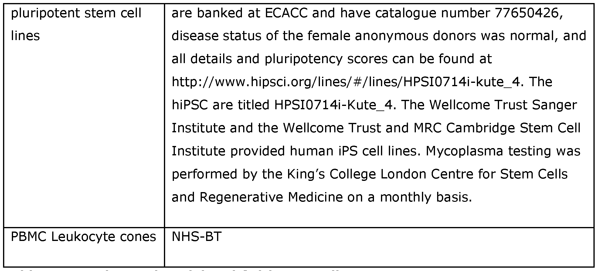
Table 1: Experimental Model and Subject Details


Table 2: Antibodies


Table 3: Cell culture reagents


Table 4: Kits used
EXAMPLES
Example 1 : Human intestinal organoids promote maturation of human systemic ILCP The translation of experimental approaches used in mice to distal human tissues, especially when studying rare populations like ILC, can be challenging. This makes the potential of in vitro human mucosal organoid systems particularly appealing. However, human bone marrow ILC precursors are poorly accessible. Thus, to translate the previously described murine ILCP-organoid system, readily available systemic ILCP were isolated from peripheral blood mononuclear cells (PBMC) instead of bone marrow, while human intestinal organoids (HIO) were derived from healthy KUTE-4 human induced pluripotent stem cells (McCracken et al., 2011). These GF structures are well-established and can be matured through addition of IL-2.
Heterogeneous systemic ILCPs (Figure la) were co-cultured with these hiPSC-derived epithelial-mesenchymal HIO (Figure lb). Co-culture with HIO significantly expanded (~20 fold) ILCPs relative to the number of cells seeded without HIO, and when compared to the number of ILCP seeded on day 1 (dl) (Figure 2a, b). As in murine co-cultures, human ILCP developed as putative group 2 (Figure 2c), group 3 (Figure 2d), and group 1 ILC (Figure 2e) when cultured with whole HIO supplemented with IL-2 and IL-7. This fold expansion and development occurred without requiring the addition of IL-ip or subset-specific cytokines, whereas previous in vitro systems required supplementation with IL- 1 p+ IL-23 for ILC3s, IL- 25+IL-33 for ILC2s, and IL-12+IL-18 for robust ILC1 development (Lim et al., 2017b), or of IL-15 and differential Notch ligands when matured from CD34+ hematopoietic stem cells (Hernandez et al., 2021). Instead, HIO alone significantly supported maturation of RORyt+ ILC Figure 2f) which significantly upregulated NKp44 (Figure 2g), recapitulating the difference in c-KIT+ ILC observed between periphery and gut by Lim and colleagues. These NKp44+/_ CCR6+/_ group 3 RORyt+ ILC produced low levels of IL-22 and IL-17A upon nonspecific PMA/Ionomycin stimulation, and significantly responded to additional 4h stimulation with IL-23 (Figure 2h), while putative group 1 ILC significantly upregulated expression of T- bet and IFN-y secretion in response to 4h stimulation with IL-18 (Figure 2e).
Example 2: Epithelial cells, not mesenchyme, drive robust ILC expansion and maturation These data suggest the human intestinal organoid microenvironment provides stimuli for baseline maturation of ILC subsets from ILCP. However, unlike the murine primary epithelial-only organoids, hiPSC-derived hindgut organoids co-develop with rich and complex native mesenchyme. These cells not only contribute to the maturation of epithelial cells (Stallmach et al., 1989), but are a known source of ILC-survival factor and CD127- ligand IL-7 (Xu et al., 2015). To assess whether epithelial cells or mesenchymal cells were
predominantly driving this ILC maturation, the matrix of HIO structures was digested with Collagenase, and a single cell suspension of mesenchyme was separated from 3D epithelial structures through serial gravity-gradient separation, resulting in a mesenchyme-depleted HIO fraction (MD-HIO) and an epithelial-depleted fibroblast-rich fraction (ED-FB) (Figure 1c). These fractions were cultured with ILCP for 14 days, and both fractions were able to maintain ILC viability and expansion (Figure 1c). However, the mesenchyme-depleted epithelial fraction unexpectedly yielded a dramatic increase in EpCAM’, CD45+, Lineage’ cells after culture (Figure Id), significantly increasing the fold-change expansion of seeded ILCP relative to those seeded with epithelial-depleted mesenchyme or without organoids in Matrigel only (Figure le). Moreover, while complete epithelial-mesenchymal HIO induced NKp44 expression in ~2-6% of group 3 ILC (Figure 2g), this upregulation was significantly greater in mesenchyme-depleted co-cultures (15-22%) (Figure If, 3a). Indeed, the epithelial-enriched MD-HIO fraction promoted patterns of ILC-subset maturation that more closely resembled the distribution of mature ILC in the healthy human intestine (Kramer et al., 2017) than the epithelial-depleted mesenchyme fraction (Figure 1g). The relative proportion of NKp44+ to NKp44’ putative ILC3 biased maturation towards an NCR’ population in GF, environmentally controlled MD-HIO (Figure If, g), in which ILCP could freely interact with the epithelium (Figure Ih).
The current gold standard for investigating murine and human ILC maturation in vitro relies on modified murine bone marrow stromal cells (Nakano, 1996). Therefore, the relatively poor ILCP expansion rates induced by hiPSC-derived intestinal mesenchyme were unexpected. We hypothesized that the increased cell number in mesenchyme-depleted HIO could represent proliferation of poorly differentiated, immature ILC, while stromal cells may promote a smaller but more mature ILC yield. To test this, unbiased stimulation with PMA/Ionomycin was performed in ILC derived from MD-HIO and ED-FB co-cultures. First, the maturation of putative Group 3 ILCs was assessed. Here, only the frequency of NKp44+ cells of RORyt+ ILC significantly expanded in MD-HIO, with the relative frequency of CCR6+, NKp44’, and T-bet+ ILC remaining constant in both conditions (Figure 3a). However, CCR6+ and NKp44+/’ ILC expressed significantly greater amounts of IL-22, and CCR6+ and NKp44’ ILC expressed significantly more IL-17A than did the respective corresponding populations derived from the epithelial-depleted mesenchymal fraction (Figure 3b). In fact, the frequency of IL-22+ ILC was either comparable or greater in the mesenchyme-depleted fractions with unbiased PMA/Ionomycin stimulation than in complete HIO co-cultures stimulated with IL-22-inducing IL-23 (Figure 2h). The proportion of putative group 1 CD56+/’, T-bet+ and Eomes+ ILC (Figure 4) was not only significantly decreased in the epithelial-depleted fraction (Figure 3d), but no expression of IFN-y was observed in either population in the absence of epithelial cells when stimulated with PMA/Ionomycin (Figure 3e). Comparable to group 3 ILC, the proportion of T-bet+ ILC and the number of IFN-y+ cells was greater in the absence of mesenchyme than in the absence of epithelium (Figure 2e).
Unlike group 1 and 3 ILC, the overall count (Figure 3f) and relative frequency (Figure 5a) of putative group 2 ILC (Figure 5b) was not significantly affected by the depletion of mesenchyme. Nevertheless, expression of IL-5 and IL-13 were both significantly and greatly increased in mesenchyme-depleted HIO fractions upon unbiased stimulation with PMA/Ionomycin (Figure 3g). The lack of difference in ILC2 numbers induced by epithelial depletion stands in contrast to findings in murine ILC2P transwell cultures, where the ILC2 number was significantly decreased upon physical separation from epithelial cells (Figure Id-f). To assess whether contact-dependent mechanisms were conserved in maturing human ILCP-derived ILC2, ILCP were also separated from mesenchyme depleted HIO (Figure 5c). Though the yield of ILC2 was still greater in transwell-separated co-cultures than in ILCP cultured without organoids, both the overall cell count and relative ILC2 frequency appeared to decrease in transwell culture, and these ILC2 failed to express IL-5 or IL-13 (Figure 5c). Finally, we aimed to assess whether the lack of cytokine expression in epithelial-depleted co-cultures was because ILC2 failed to mature without epithelial cells, or if this was the result of active ILC2 cytokine repression by mesenchyme either through secretion of repressive factors or via competitive depletion of necessary ligands. To test this, ILCP were first successfully matured with MD-HIO, then resulting EpCAM’ CD45+ Lineage’ ILC were FACS-purified and reseeded either with MD-HIO or swapped to ED-FB (Figure 5d). ILC reseeded without epithelial cells appeared to decrease their rate of proliferation, and while there was no difference in the expression of IL-13 (Figure 5e), the greater presence of mesenchyme resulted in a significant decrease in IL-5 expression by ILC2 (Figure 5e). This suggests that stable development of IL-13+ ILC2 may be promoted by intestinal epithelial contact, but ILC2 proliferation and expression of IL-5 may either be actively repressed by intestinal mesenchyme or require constant epithelial exposure.
Example 3: Human mucosal epithelial identity contributes to tissue-specific ILC maturation To assess if any tissue-characteristic ILC phenotypes could be captured in these human organoid systems (Figure 6a), ILCP were cultured either with hiPSC-derived small intestine or hiPSC-derived lung organoids. ILCP expanded in both co-cultures (Figure 6b), in which the absolute number of fibroblasts post mesenchyme-depletion remaining sufficiently consistent between conditions. HLO demonstrated comparable capacity to yield mature group 1 and group 3 cells that expressed IL-22, IL-17A, and IFN-y (Figure 6c). Both HIO and HLO yielded GATA3+ ILC that expressed IL-5 and IL-13 (Figure 6d). Much like ED-FB, HLO did not support maturation of IL-5+ as significantly as HLO. Similarly, a recently described c-KIT+, CRTh2l0W, IL-17A+ ILC2 population was additionally present in HIO, but not in HLO cultures, suggesting that much like the murine system, the human intestinal microenvironment may favour group 3 maturation, even in inducing ex-ILC2 to ILC3 plasticity in mature MD-HIO-derived ILC2. However, unlike in the murine system, co-culture with HIO or HLO alone was not sufficient to induce statistically significant differences in the frequency of CD25 and ST2 ILC within this GATA3+ putative ILC2 population (Figure 6e).
Finally, we assessed the capacity of the human organoids to recapitulate the gut-lung translocation approach. ILCP were matured with MD-HIO for two weeks, then FACS-purified and reseeded with MD-HIO, MD-HLO, or MD-HLO with human IL-33 neutralisation, as performed in the murine system. HiPSC-derived lung organoids did not significantly impact the frequency of CD25+ (Figure 6f). However, the anticipated increase in pulmonary ILC2 ST2 expression that was not captured by HIO and HLO co-culture alone was significantly observed upon transfer of MD-HIO-derived ILC2 to lung culture (Figure 6g). This effect was dampened by addition of neutralising IL-33.
Example 4: Antigen processing of the ILCs of the invention
The ILCs of the invention express HLA-DR, an MHC-II surface receptor (Figure 7). The ability of the ILCs of the invention to process antigens via MHC-II was tested by hydrolysis of DQ-BSA (which leads to fluorescent signal). The positive results are shown in Figure 8.
Example 5: Single cell RNAseq (scRNAseq) on the organoid-generated ILCs
From the co-cultures, CD45+ Lin- cells were isolated via fluorescence activated cell sorting (FACS). We identified 5 'super-clusters' of cells corresponding to a cytotoxic ILC1 population, two distinct ILC2 clusters (ILC2a and ILC2b), an ILC3 cluster, and a small cluster of cells which exhibited significantly upregulated expression of genes characteristic of regulatory T cells (Tregs) and a regulatory ILC (ILCreg) population previously described in mice (Wang et al., 2017) (Fig. 9A).
The helper-like ILC cell types (ILC1-3) were identified via determining expression patterns of key genes previously described to distinguish the different ILC family members in single cell RNA sequencing studies using human samples (Bjbrklund et al., 2016; Celia et al., 2019; Liu et al., 2021; Mazzurana et al., 2021). The significant enrichment of these genes in each cluster was determined via unbiased differential gene expression analysis comparing expression of the genes within each cluster to that of all other clusters (Figure 9B). Accordingly, the ILC1 population was identified via its enriched expression of key cytokines and chemokines including IFNG and CCL3 alongside their relatively high expression of genes such KLRC1, PRF1, and GNLY, previously shown to characterise cytotoxic populations of ILC1. The increased expression of the transcription factor GATA-3 (encoded via GATA3) and the cytokine signalling genes IL33 and IL17RB were used to identify the two ILC2 populations, alongside their upregulation of additional ILC2-associated genes such as HPGDS and KLRG1. Finally, ILC3s showed significantly greater enrichment of genes encoding their characteristic transcription factors (e.g., RORC), cell surface markers (e.g., KIT), and cytokine signalling pathways (e.g., IL23RA). These genes were found coupled to expression of markers previously shown to be highly expressed in both ILC3s and ILCls, including NCR1 and PECAM1, further confirming the identity of both clusters.
The annotation of the ILCreg cluster, however, was primarily performed using markers identified in mouse research on ILCregs as this population has yet to be extensively characterised in human patients, although an IL-10 expressing ILC population was identified in the human intestine (Wang et al., 2017). This included genes encoding their key cytokine, IL-10 IL10), the cell surface marker CD25 IL2RA) and the transcription factors ID3 and SOX4. Interestingly, the ILCreg cluster additionally expressed genes associated with human T-regulatory (Tregs) cells including FOXP3, RUNX1, IL1R1 and CTLA4. Importantly, the T-cell markers CD4 and CD3 were included within the lineage gating panel when sorting the ILCs from the co-cultures, and the lack of their expression was further confirmed at the RIMA level. Accordingly, these cells were deemed to be the human ILC equivalent of Tregs.
Using cell-specific enrichment scores for a human ILCP gene signature (Liu et al., 2021) we identified a small population of these precursor cells within the larger ILC3 super-cluster (Fig. 9C). Moreover, the cytotoxic ILC1 super-cluster showed distinct expression patterns of the transcription factors TBX21 and EOMES, suggesting this contained both ILC1 and NK- like cells (Fig. 9D). Overall, we found cells corresponding to the major ILC cell types expected to be observed in our co-culture model, alongside a putative population of human ILCregs.
To determine whether the ILCs exhibited an intestinal tissue specific phenotype, we generated cell-specific enrichment scores for three gene modules identified by Mazzurana et al., (2021) associated with blood/tonsillar circulatory ILCs (modll), colonic ILCs (mod3) or lung ILCs (mod34) (Mazzurana et al., 2021). Only ILC3s were examined here as Mazzurana et al., did not identify any distinct clusters of ILCls or ILC2s in the colon. The ILC3 supercluster was found to exhibit the greatest enrichment for the circulatory gene module (Fig. 9E), followed by the colonic gene module, with minimal cells showing enriched expression of lung ILC associated genes. Considering we observed the putative ILCP population within our ILC3s, we subsequently overlaid the enrichment scores onto the UMAP to determine any heterogeneity in the pattern of expression of these gene modules (Fig. 9F). This revealed that the ILC super-cluster could be clearly differentiated into those cells which exhibit a more circulatory phenotype compared to those which express a gene signature more closely resembling that of colonic ILC3s. As expected, there were minimal numbers of cells enriched for the lung module. Taken together, these data suggest that a substantial proportion of the ILC3s in this co-culture model differentiate away from a circulatory phenotype to acquire a colonic gene expression signature.
Experiment outline:
Circulatory innate lymphoid cell precursors (ILCPs; CD45+ Lin- CD56- CD127+ CRTh2- CKIT+ NKp46- KLRG1-) were isolated from the blood of age-matched male and female
donors (n = 1), placed into co-culture with iPSC-derived human intestinal organoids and allowed to expand for 14 days. After this 14-day period, four populations of ILCs were isolated from the co-cultures using FACS: ILC2 (CD45+ Lin- CRTh2+), ILC3 (CD45+ Lin- CRTh2+ CKIT+), ILC1/NK cells (CD45+ Lin- CRTh2- cKIT- CD56+/- CD161 + ) and 'other' ILC (CD45+ Lin- CRTh2- cKIT- CD56+/- CD161-). Each population was subsequently pooled in a 1 : 1 ratio of male to female cells to give a final total proportion of each ILC population at 25, 15, 35 and 15% respectively. These proportions were decided upon based on prior knowledge of the heterogeneity of each ILC subset, with those ILCs displaying greater heterogeneity contributing a larger percentage of the final cell counts. This pool of cells was then sequenced as single sample via 10X sequencing. scRNA-seq data processing and analysis:
Barcoding, cDNA synthesis, and library preparation were performed using Chromium™ Next GEM Single Cell 3', Library & Gel Bead Kit v3.1 according to the manufacturer's instructions. Libraries were sequenced by Novogene using NovaSeq with a target of 20,000 reads per cell. Pre-processing of single cell RNA-sequencing fastq files was performed using Cell Ranger v7.0.1 (10X Genomics), with reads aligned to the GRCh38 reference genome. Further data pre-processing was performed in R (v4.2.2) using the package Seurat (v4.3.0). Cells with less than 1,000 genes and greater than 10 % of transcripts derived from mitochondrial genes were removed as they were considered lysed/apoptotic. Moreover, cells with more than 7,500 genes or 40,000 UMIs were excluded as these were presumed to contain doublets. The filtered dataset was subsequently normalised using the Seurat SCTransform function (v2) in which the effects of the cell cycle were regressed out. The top 30 principal components were then used as input for Louvain clustering via the FindClusters() function in Seurat at resolution 0.2. UMAP was used for cluster visualisation. Differentially expressed genes were determined using the Wilcox test in the FindAIIMarkers() function in Seurat, and genes with log fold change >= 2.5 and a false discovery rate P < 0.05 were considered significantly differentially expressed. To visualise lowly expressed genes (TBX21 and EOMES) the plot_density function was used from the R package Nebulosa (vl.8.0). To determine the enrichment of different ILC gene signatures, the R package AUCell (vl.6.1) was used and the resultant scores were plotted for visualisation.
The results are shown in Figure 9 and the Table 5 below.
Table 5: Results of scRNAseq on the organoid-generated ILCs


Example 6: NK cells generated by co-culture of ILC precursors (ILCP) with intestinal organoids are cytotoxic
The cytotoxicity of NK cells generated by co-culture of ILC precursors (ILCP) with intestinal organoids was tested using standard methods. The positive results are shown in Figure 10.
Example 7: Generation of ILCs in human small intestine biopsy derived organoids Differentiated ILCs (including ILC1, ILC2, ILC3 and NK cells) were generated from ILC precursors (ILCP) after 15 days in culture with human small intestine biopsy derived organoids. The results are shown in Figure 11.
Example 8: Producing and testing ILCregs
Circulatory innate lymphoid cell precursors (ILCPs; CD45+ Lin- (CD3, CD14, CD19, CD20) CD56- CD127+ CRTh2- cKIT+ NKp46- KLRG1-) were isolated from the blood cones (n = 3), placed into co-culture with iPSC-derived human intestinal organoids, which were depleted of mesenchymal cells, and allowed to expand for 13 days. After this 13-day period, cells were analysed by flow cytometry for extracellular (CD45, EpCAM, Lin) and intracellular (Foxp3, CTLA-4) markers. The results are shown in Figures 12 and 13 and demonstrate that the ILCs express at the protein level markers associated with regulatory lymphocyte populations supporting the generation of a human population of ILCregs with this invention.
REFERENCES
Bando, J.K., Liang, H.E., and Locksley, R.M. (2015). Identification and distribution of developing innate lymphoid cells in the fetal mouse intestine. Nat. Immunol. 16, 153-160.
Bando JK, Gilfillan S, Di Luccia B, Fachi JL, Secca C, Celia M, Colonna M. ILC2s are the predominant source of intestinal ILC-derived IL-10. J Exp Med. 2020 Feb 3;217(2):e20191520. doi: 10.1084/jem.20191520. PMID: 31699824; PMCID: PMC7041711
Bjorklund, A. K., Forkel, M., Picelli, S., Konya, V., Theorell, J., Friberg, D., Sandberg, R., & Mjosberg, J. (2016). The heterogeneity of human CD127+ innate lymphoid cells revealed by single-cell RNA sequencing. Nature Immunology 2016 17:4, 17 4), 451-460. https://doi.org/10.1038/ni.3368.
Celia, M., Gamini, R., Secca, C., Collins, P. L., Zhao, S., Peng, V., Robinette, M. L., Schettini, J., Zaitsev, K., Gordon, W., Bando, J. K., Yomogida, K., Cortez, V., Fronick, C., Fulton, R., Lin, L. L., Gilfillan, S., Flavell, R. A., Shan, L., ... Colonna, M. (2019). Subsets of ILC3- ILCl-like cells generate a diversity spectrum of innate lymphoid cells in human mucosal tissues. Nature Immunology 2019 20:8, 20(8), 980-991. https://doi.org/10.1038/s41590-019-0425-y.
Dutton, E.E., Camelo, A., Sleeman, M., Herbst, R., Carlesso, G., Belz, G.T., and Withers, D.R. (2018). Characterisation of innate lymphoid cell populations at different sites in mice with defective T cell immunity [version 3; referees: 2 approved]. Wellcome Open Res. 2, 117.
Elmentaite, R., Kumasaka, N., Roberts, K., Fleming, A., Dann, E., King, H.W., Kleshchevnikov, V., Dabrowska, M., Pritchard, S., Bolt, L., et al. (2021). Cells of the human intestinal tract mapped across space and time. Nature 597, 250-255.
Gronke, K., Kofoed-Nielsen, M., and Diefenbach, A. (2017). Isolation and Flow Cytometry Analysis of Innate Lymphoid Cells from the Intestinal Lamina Propria. In Methods in Molecular Biology (Clifton, N .), pp. 255-265.
Heo, I., Dutta, D., Schaefer, D.A., lakobachvili, N., Artegiani, B., Sachs, N., Boonekamp, K.E., Bowden, G., Hendrickx, A.P.A., Willems, RJ.L., et al. (2018). Modelling Cryptosporidium infection in human small intestinal and lung organoids. Nat. Microbiol. 3, 814-823.
Hernandez, D.C., Juelke, K., Muller, N.C., Durek, P., Ugursu, B., Mashreghi, M.F., Ruckert, T., and Romagnani, C. (2021). An in vitro platform supports generation of human innate lymphoid cells from CD34+ hematopoietic progenitors that recapitulate ex vivo identity. Immunity 54, 2417-2432. e5.
Huang, Y., Guo, L., Qiu, J., Chen, X., Hu-Li, J., Siebenlist, U., Williamson, P.R., Urban, J.F., and Paul, W.E. (2015). IL-25-responsive, lineage-negative KLRG1 hi cells are multipotential "inflammatory" type 2 innate lymphoid cells. Nat. Immunol. 16, 161-169.
Huang, Y., Mao, K., Chen, X., Sun, M.A., Kawabe, T., Li, W., Usher, N., Zhu, J., Urban, J.F., Paul, W.E., et al. (2018). SIP-dependent interorgan trafficking of group 2 innate lymphoid cells supports host defense. Science (80-. ). 359, 114-119.
Jowett, G.M., Norman, M.D.A., Yu, T.T.L. et al. ILC1 drive intestinal epithelial and matrix remodelling. Nat. Mater. 20, 250-259 (2021). https://doi.org/10.1038/s41563-020-0783-8
Kilpinen, H., Goncalves, A., Leha, A., Afzal, V., Alasoo, K., Ashford, S., Bala, S., Bensaddek, D., Casale, F.P., Culley, O.J., et al. (2017). Common genetic variation drives molecular heterogeneity in human iPSCs. Nature 546, 370-375.
Konishi, S., Gotoh, S., Tateishi, K., Yamamoto, Y., Korogi, Y., Nagasaki, T., Matsumoto, H., Muro, S., Hirai, T., Ito, I., et al. (2016). Directed Induction of Functional Multi-ciliated Cells in Proximal Airway Epithelial Spheroids from Human Pluripotent Stem Cells. Stem Cell Reports 6, 18-25.
Kramer, B., Goeser, F., Lutz, P., Glassner, A., Boesecke, C., Schwarze-Zander, C., Kaczmarek, D., Nischalke, H.D., Branchi, V., Manekeller, S., et al. (2017). Compartmentspecific distribution of human intestinal innate lymphoid cells is altered in HIV patients under effective therapy. PLoS Pathog. 13.
Leha, A., Moens, N., Meleckyte, R., Culley, O.J., Gervasio, M.K., Kerz, M., Reimer, A., Cain, S.A., Streeter, I., Folarin, A., et al. (2016). A high-content platform to characterise human induced pluripotent stem cell lines. Methods 96, 85-96.
Lim, A. I., Li, Y., Lopez-Lastra, S., Stadhouders, R., Paul, F., Casrouge, A., Serafini, N., Puel, A., Bustamante, J., Surace, L., et al. (2017a). Systemic Human ILC Precursors Provide a Substrate for Tissue ILC Differentiation. Cell 168, 1086-1100. elO.
Lim, A. I., Li, Y., Lopez-Lastra, S., Stadhouders, R., Paul, F., Casrouge, A., Serafini, N., Puel, A., Bustamante, J., Surace, L., et al. (2017b). Systemic Human ILC Precursors Provide a Substrate for Tissue ILC Differentiation. Cell 168, 1086-1100. elO.
Liu, C., Gong, Y., Zhang, H., Yang, H., Zeng, Y., Bian, Z., Xin, Q., Bai, Z., Zhang, M., He, J., Yan, J., Zhou, J., Li, Z., Ni, Y., Wen, A., Lan, Y., Hu, H., & Liu, B. (2021). Delineating spatiotemporal and hierarchical development of human fetal innate lymphoid cells. Cell Research, 31(10). https://doi.org/10.1038/s41422-021-00529-2.
Mazzurana, L., Czarnewski, P., Jonsson, V., Wigge, L., Ringner, M., Williams, T. C., Ravindran, A., Bjbrklund, A. K., Safholm, J., Nilsson, G., Dahlen, S. E., Orre, A. C., Al- Ameri, M., Hbog, C., Hedin, C., Szczegielniak, S., Aimer, S., & Mjbsberg, J. (2021). Tissuespecific transcriptional imprinting and heterogeneity in human innate lymphoid cells revealed by full-length single-cell RNA-sequencing. Cell Research 2021 31:5, 31(5), 554- 568. https://doi.org/10.1038/s41422-020-00445-x.
McCracken, K.W., Howell, J.C., Wells, J.M., and Spence, J.R. (2011). Generating human intestinal tissue from pluripotent stem cells in vitro. Nat. Protoc. 6, 1920-1928.
McQualter, J.L., Yuen, K., Williams, B., and Bertoncello, I. (2010). Evidence of an epithelial stem/progenitor cell hierarchy in the adult mouse lung. Proc. Natl. Acad. Sci. U. S. A. 107, 1414-1419.
Nakano, T. (1996). In vitro development of hematopoietic system from mouse embryonic stem cells: A new approach for embryonic hematopoiesis. Int. J. Hematol. 65, 1-8.
Ricardo-Gonzalez, R.R., Van Dyken, S.J., Schneider, C., Lee, J., Nussbaum, J.C., Liang, H.E., Vaka, D., Eckalbar, W.L., Molofsky, A.B., Erie, D.J., et al. (2018a). Tissue signals imprint ILC2 identity with anticipatory function. Nat. Immunol. 19, 1093-1099.
Saluzzo, S., Gorki, A.D., Rana, B.M.J., Martins, R., Scanlon, S., Starkl, P., Lakovits, K., Hladik, A., Korosec, A., Sharif, O., et al. (2017). First-Breath-Induced Type 2 Pathways Shape the Lung Immune Environment. Cell Rep. 18, 1893-1905.
Sato, T., Vries, R.G., Snipped, H.J., van de Wetering, M., Barker, N., Stange, D.E., van Es, J.H., Abo, A., Kujala, P., Peters, P.J., et al. (2009). Single Lgr5 stem cells build crypt-villus structures in vitro without a mesenchymal niche. Nature 459, 262-265.
Sato, T., van Es, J.H., Snipped, H.J., Stange, D.E., Vries, R.G., van den Born, M., Barker, N., Shroyer, N.F., van de Wetering, M., and Clevers, H. (2011). Paneth cells constitute the niche for Lgr5 stem cells in intestinal crypts. Nature 469, 415-418.
Schroeder, J.-H., Meissl, K., Hromadova, D., Lo, J.W., Neves, J.F., Howard, J.K., Helmby, H., Powell, N., Strobl, B., and Lord, G.M. (2021). T-Bet Controls Cellularity of Intestinal Group 3 Innate Lymphoid Cells. Front. Immunol. 11, 3701.
Stallmach, A., Hahn, U., Mekker, H.J., Hahn, E.G., and Riecken, E.O. (1989). Differentiation of rat intestinal epithelial cells is induced by organotypic mesenchymal cells in vitro. Gut 30, 959-970.
Wang, S., Xia, P., Chen, Y., Qu, Y., Xiong, Z., Ye, B., Du, Y., Tian, Y., Yin, Z., Xu, Z., & Fan, Z. (2017). Regulatory Innate Lymphoid Cells Control Innate. Intestinal Inflammation. Cell, 171(1). https://doi.Org/10.1016/j.cell.2017.07.027
Xu, W., Domingues, R.G., Fonseca-Pereira, D., Ferreira, M., Ribeiro, H., Lopez-Lastra, S., Motomura, Y., Moreira-Santos, L., Bihl, F., Braud, V., et al. (2015). NFIL3 Orchestrates the emergence of common helper innate lymphoid cell precursors. Cell Rep. 10, 2043-2054.
Yusa, K., Rashid, S.T., Strick-Marchand, H., Varela, I., Liu, P.Q., Paschon, D.E., Miranda, E., Ordonez, A., Hannan, N.R.F., Rouhani, F.J., et al. (2011). Targeted gene correction of ol- antitrypsin deficiency in induced pluripotent stem cells. Nature 478, 391-394.
Claims
1. A human regulatory innate lymphoid cell (ILCreg) which expresses a detectable level of FOXP3 and/or CTLA4.
2. A human ILCreg according to claim 1, wherein the human ILCreg further expresses a detectable level of CD25 (IL2RA) and/or CD127.
3. A human ILCreg according to claim 1 or 2, wherein the human ILCreg further expresses detectable levels of one or more of CCR5, FGGY, GATA3, GZMB, IL10, IL1R1, IL2RA, IL2RB, KAT2B, LGLS3, PIM1, PRDM1, RUNX1, SOX4, TNFRSF18 and TRAF1.
4. A human ILCreg according to any one of the preceding claims, wherein the human ILCreg further expresses detectable levels of CCR5, FGGY, GATA3, GZMB, IL10, IL1R1, IL2RA, IL2RB, KAT2B, LGLS3, PIM1, PRDM1, RUNX1, SOX4, TNFRSF18 and TRAF1.
5. A human ILCreg according to any one of the preceding claims, wherein the human ILCreg does not express detectable levels of one or more of CD3, CD4, CD19, CD20, TCRap.
6. A human ILCreg according to any one of the preceding claims, wherein the human ILCreg does not express detectable levels of one or more of CD3, CD4, CD19, CD20, TCRap, and TCRyS.
7. A human ILCreg according to any one of the preceding claims, wherein the human ILCreg does not express detectable levels of CD3, CD4, CD19, CD20, TCRap, and TCRyS.
8. An in vitro population of human ILCregs, wherein the population comprises at least about two human ILCregs according to any one of claims 1-7.
9. An in vitro population according to claim 8, wherein the population comprises at least about 100 human ILCregs.
10. An in vitro population according to claim 8 or 9, wherein at least 10% of the human ILCregs in the in vitro population comprise an exogenous polynucleotide.
11. A pharmaceutical composition comprising a human ILCreg according to any one claims 1-7 or an in vitro population according to any one of claims 8-10 and a pharmaceutically or physiologically acceptable diluent and/or carrier.
12. A method of treating or preventing a disease in a subject, wherein the method comprises administering to the subject a human ILCreg according to any one claims 1-7, an in vitro population according to any one of claims 8-10 or a pharmaceutical composition according to claim 11.
13. A method according to claim 12, wherein the disease is an inflammatory disease.
14. A method according to claim 12, wherein the inflammatory disease is an allergy, an autoimmune disease, an infection or cancer.
15. A method according to any one of claims 12-14, wherein the subject is human.
16. A human ILCreg according to any one claims 1-7, an in vitro population according to any one of claims 8-10 or a pharmaceutical composition according to claim 11 for use in the treatment or prevention of a disease.
17. A method for expanding a human ILCreg according to any one claims 1-7 or an in vitro population according to any one of claims 8-10, the method comprising:
(a) co-culturing the human ILCreg or the in vitro population and at least one epithelial organoid, wherein the epithelial organoid comprises more epithelial cells than mesenchymal cells.
18. A method for producing a human ILCreg according to any one claims 1-7 or an in vitro population according to any one of claims 8-10, the method comprising:
(a) co-culturing human ILC precursors and at least one epithelial organoid, wherein the epithelial organoid comprises more epithelial cells than mesenchymal cells, to expand the human ILC precursors and differentiate the ILC precursors into the human ILCregs.
Applications Claiming Priority (4)
| Application Number | Priority Date | Filing Date | Title |
|---|---|---|---|
| PCT/EP2023/066317 WO2023242426A1 (en) | 2022-06-17 | 2023-06-16 | Method for expanding immune cells |
| EPPCT/EP2023/066317 | 2023-06-16 | ||
| GBGB2319529.0A GB202319529D0 (en) | 2023-12-19 | 2023-12-19 | Novel innate lymphoid cells (ILCS) |
| GB2319529.0 | 2023-12-19 |
Publications (1)
| Publication Number | Publication Date |
|---|---|
| WO2024256627A1 true WO2024256627A1 (en) | 2024-12-19 |
Family
ID=91782124
Family Applications (1)
| Application Number | Title | Priority Date | Filing Date |
|---|---|---|---|
| PCT/EP2024/066529 Ceased WO2024256627A1 (en) | 2023-06-16 | 2024-06-14 | Novel innate lymphoid cells (ilcs) |
Country Status (1)
| Country | Link |
|---|---|
| WO (1) | WO2024256627A1 (en) |
Citations (2)
| Publication number | Priority date | Publication date | Assignee | Title |
|---|---|---|---|---|
| US4458066A (en) | 1980-02-29 | 1984-07-03 | University Patents, Inc. | Process for preparing polynucleotides |
| US7446190B2 (en) | 2002-05-28 | 2008-11-04 | Sloan-Kettering Institute For Cancer Research | Nucleic acids encoding chimeric T cell receptors |
-
2024
- 2024-06-14 WO PCT/EP2024/066529 patent/WO2024256627A1/en not_active Ceased
Patent Citations (2)
| Publication number | Priority date | Publication date | Assignee | Title |
|---|---|---|---|---|
| US4458066A (en) | 1980-02-29 | 1984-07-03 | University Patents, Inc. | Process for preparing polynucleotides |
| US7446190B2 (en) | 2002-05-28 | 2008-11-04 | Sloan-Kettering Institute For Cancer Research | Nucleic acids encoding chimeric T cell receptors |
Non-Patent Citations (50)
| Title |
|---|
| "PCR Protocols: A Guide to Methods and Applications", 1990, ACADEMIC PRESS |
| "PCR Technology: Principles and Applications for DNA Amplification", 1992, FREEMAN PRESS |
| "Remington's Pharmaceutical Sciences", 2012 |
| BANDO JKGILFILLAN SDI LUCCIA BFACHI JLSÉCCA CCELLA MCOLONNA M.: "ILC2s are the predominant source of intestinal ILC-derived IL-10", J EXP MED., vol. 217, no. 2, 3 February 2020 (2020-02-03), pages e20191520 |
| BANDO, J.K.LIANG, H.E.LOCKSLEY, R.M.: "Identification and distribution of developing innate lymphoid cells in the fetal mouse intestine", NAT. IMMUNOL., vol. 16, 2015, pages 153 - 160 |
| BEAUCAGE, TETRA. LETT., vol. 22, 1981, pages 1859 |
| BITTNER ET AL., METH. ENZYMOL., vol. 153, 1987, pages 516 |
| BJORKLUND, A. K.FORKEL, M.PICELLI, S.KONYA, V.THEORELL, J.FRIBERG, D.SANDBERG, R.MJÖSBERG, J.: "The heterogeneity of human CD127+ innate lymphoid cells revealed by single-cell RNA sequencing", NATURE IMMUNOLOGY, vol. 17, no. 4, 2016, pages 451 - 460 |
| CELLA, M.GAMINI, R.SÉCCA, C.COLLINS, P. L.ZHAO, S.PENG, V.ROBINETTE, M. L.SCHETTINI, J.ZAITSEV, K.GORDON, W.: "Subsets of ILC3-ILC1-like cells generate a diversity spectrum of innate lymphoid cells in human mucosal tissues", NATURE IMMUNOLOGY, vol. 20, no. 8, 2019, pages 980 - 991, XP036853449, DOI: 10.1038/s41590-019-0425-y |
| DUTTON, E.E.CAMELO, A.SLEEMAN, M.HERBST, R.CARLESSO, G.BELZ, G.T.WITHERS, D.R.: "Characterisation of innate lymphoid cell populations at different sites in mice with defective T cell immunity [version 3; referees: 2 approved", WELLCOME OPEN RES., vol. 2, 2018, pages 117 |
| ECKERT ET AL., PCR METHODS AND APPLICATIONS, vol. 1, 1991, pages 17 |
| ELMENTAITE, R.KUMASAKA, N.ROBERTS, K.FLEMING, A.DANN, E.KING, H.W.KLESHCHEVNIKOV, V.DABROWSKA, M.PRITCHARD, S.BOLT, L. ET AL.: "Cells of the human intestinal tract mapped across space and time", NATURE, vol. 597, 2021, pages 250 - 255, XP037574981, DOI: 10.1038/s41586-021-03852-1 |
| GRONKE, K.KOFOED-NIELSEN, M.DIEFENBACH, A.: "Isolation and Flow Cytometry Analysis of Innate Lymphoid Cells from the Intestinal Lamina Propria", METHODS IN MOLECULAR BIOLOGY, 2017, pages 255 - 265 |
| HARRINGTON ET AL., NAT GENET., vol. 15, 1997, pages 345 |
| HEO, I.DUTTA, D.SCHAEFER, D.A.IAKOBACHVILI, N.ARTEGIANI, B.SACHS, N.BOONEKAMP, K.E.BOWDEN, G.HENDRICKX, A.P.A.WILLEMS, R.J.L. ET A: "Modelling Cryptosporidium infection in human small intestinal and lung organoids", NAT. MICROBIOL., vol. 3, 2018, pages 814 - 823, XP037535390, DOI: 10.1038/s41564-018-0177-8 |
| HERNANDEZ, D.C.JUELKE, K.MULLER, N.C.DUREK, P.UGURSU, B.MASHREGHI, M.F.RÜCKERT, T.ROMAGNANI, C.: "An in vitro platform supports generation of human innate lymphoid cells from CD34+ hematopoietic progenitors that recapitulate ex vivo identity", IMMUNITY, vol. 54, 2021, pages 2417 - 2432 |
| HUANG, Y.GUO, L.QIU, J.CHEN, X.HU-LI, J.SIEBENLIST, U.WILLIAMSON, P.R.URBAN, J.F.PAUL, W.E.: "IL-25-responsive, lineage-negative KLRG1 hi cells are multipotential ''inflammatory'' type 2 innate lymphoid cells", NAT. IMMUNOL., vol. 16, 2015, pages 161 - 169 |
| HUANG, Y.MAO, K.CHEN, X.SUN, M.A.KAWABE, T.LI, W.USHER, N.ZHU, J.URBAN, J.F.PAUL, W.E. ET AL.: "S1P-dependent interorgan trafficking of group 2 innate lymphoid cells supports host defense", SCIENCE (80-., vol. 359, 2018, pages 114 - 119 |
| JOWETT GERALDINE M. ET AL: "Organoids capture tissue-specific innate lymphoid cell development in mice and humans", CELL REPORTS, vol. 40, no. 9, 1 August 2022 (2022-08-01), US, pages 111281, XP093074149, ISSN: 2211-1247, DOI: 10.1016/j.celrep.2022.111281 * |
| JOWETT, G.M.NORMAN, M.D.A.YU, T.T.L. ET AL.: "ILC1 drive intestinal epithelial and matrix remodelling", NAT. MATER., vol. 20, 2021, pages 250 - 259, XP037419351, DOI: 10.1038/s41563-020-0783-8 |
| KILPINEN, H.GONCALVES, A.LEHA, A.AFZAL, V.ALASOO, K.ASHFORD, S.BALA, S.BENSADDEK, D.CASALE, F.P.CULLEY, O.J. ET AL.: "Common genetic variation drives molecular heterogeneity in human iPSCs", NATURE, vol. 546, 2017, pages 370 - 375 |
| KONISHI, S.GOTOH, S.TATEISHI, K.YAMAMOTO, Y.KOROGI, Y.NAGASAKI, T.MATSUMOTO, H.MURO, S.HIRAI, T.ITO, I. ET AL.: "Directed Induction of Functional Multi-ciliated Cells in Proximal Airway Epithelial Spheroids from Human Pluripotent Stem Cells", STEM CELL REPORTS, vol. 6, 2016, pages 18 - 25, XP055294072, DOI: 10.1016/j.stemcr.2015.11.010 |
| KRAMER, B.GOESER, F.LUTZ, P.GLASSNER, A.BOESECKE, C.SCHWARZE-ZANDER, C.KACZMAREK, D.NISCHALKE, H.D.BRANCHI, V.MANEKELLER, S. ET AL: "Compartment-specific distribution of human intestinal innate lymphoid cells is altered in HIV patients under effective therapy", PLOS PATHOG., vol. 13, 2017 |
| LEHA, A.MOENS, N.MELECKYTE, R.CULLEY, O.J.GERVASIO, M.K.KERZ, M.REIMER, A.CAIN, S.A.STREETER, I.FOLARIN, A. ET AL.: "A high-content platform to characterise human induced pluripotent stem cell lines", METHODS, vol. 96, 2016, pages 85 - 96, XP029430756, DOI: 10.1016/j.ymeth.2015.11.012 |
| LIM, A.I.LI, Y.LOPEZ-LASTRA, S.STADHOUDERS, R.PAUL, F.CASROUGE, A.SERAFINI, N.PUEL, A.BUSTAMANTE, J.SURACE, L. ET AL.: "Systemic Human ILC Precursors Provide a Substrate for Tissue ILC Differentiation", CELL, vol. 168, 2017, pages 1086 - 1100 |
| LIU, C.GONG, Y.ZHANG, H.YANG, H.ZENG, Y.BIAN, Z.XIN, Q.BAI, Z.ZHANG, M.HE, J.: "Delineating spatiotemporal and hierarchical development of human fetal innate lymphoid cells", CELL RESEARCH, vol. 31, no. 10, 2021, XP037578330, DOI: 10.1038/s41422-021-00529-2 |
| LOVE ET AL., COLD SPRING HARBOR PERSPECT. BIOL, vol. 2, no. 6, 2010, pages 002485 |
| MATTILA ET AL., NUCLEIC ACIDS RES., vol. 19, 1991, pages 967 |
| MAZZURANA, L.CZARNEWSKI, P.JONSSON, V.WIGGE, L.RINGNÉR, M.WILLIAMS, T. C.RAVINDRAN, A.BJ6RKLUND, A. K.SÄFHOLM, J.NILSSON, G.: "Tissue-specific transcriptional imprinting and heterogeneity in human innate lymphoid cells revealed by full-length single-cell RNA-sequencing", CELL RESEARCH, vol. 31, no. 5, 2021, pages 554 - 568, XP037441894, DOI: 10.1038/s41422-020-00445-x |
| MCCRACKEN, K.W.HOWELL, J.C.WELLS, J.M.SPENCE, J.R.: "Generating human intestinal tissue from pluripotent stem cells in vitro", NAT. PROTOC., vol. 6, 2011, pages 1920 - 1928, XP055210541, DOI: 10.1038/nprot.2011.410 |
| MCQUALTER, J.L.YUEN, K.WILLIAMS, B.BERTONCELLO, I.: "Evidence of an epithelial stem/progenitor cell hierarchy in the adult mouse lung", PROC. NATL. ACAD. SCI. U. S. A, vol. 107, 2010, pages 1414 - 1419 |
| MORITA HIDEAKI ET AL: "Induction of human regulatory innate lymphoid cells from group 2 innate lymphoid cells by retinoic acid", JOURNAL OF ALLERGY AND CLINICAL IMMUNOLOGY, vol. 143, no. 6, 22 January 2019 (2019-01-22), pages 2190, XP085705064, ISSN: 0091-6749, DOI: 10.1016/J.JACI.2018.12.1018 * |
| NAKANO, T.: "In vitro development of hematopoietic system from mouse embryonic stem cells: A new approach for embryonic hematopoiesis", INT. J. HEMATOL., vol. 65, 1996, pages 1 - 8, XP001106703, DOI: 10.1016/S0925-5710(96)00531-2 |
| NARANG ET AL., METH. ENZYMOL., vol. 68, 1979, pages 109 |
| QUEEN ET AL., IMMUNOL. REV., vol. 89, 1986, pages 49 - 68 |
| READ EMILY ET AL: "Co-Culture of Murine Small Intestine Epithelial Organoids with Innate Lymphoid Cells", JOURNAL OF VISUALIZED EXPERIMENTS, no. 181, 23 March 2022 (2022-03-23), US, pages 1 - 21, XP093072805, ISSN: 1940-087X, Retrieved from the Internet <URL:http://dx.doi.org/10.3791/63554> DOI: 10.3791/63554 * |
| RICARDO-GONZALEZ, R.R.VAN DYKEN, S.J.SCHNEIDER, C.LEE, J.NUSSBAUM, J.C.LIANG, H.E.VAKA, D.ECKALBAR, W.L.MOLOFSKY, A.B.ERLE, D.J. E: "Tissue signals imprint ILC2 identity with anticipatory function", NAT. IMMUNOL., vol. 19, 2018, pages 1093 - 1099, XP036599923, DOI: 10.1038/s41590-018-0201-4 |
| RIVIERE ET AL., PROC. NATL. ACAD. SCI., vol. 92, 1995, pages 6733 - 6737 |
| ROSENFELD ET AL., CELL, vol. 68, 1992, pages 143 |
| SALUZZO, S.GORKI, A.D.RANA, B.M.J.MARTINS, R.SCANLON, S.STARKL, P.LAKOVITS, K.HLADIK, A.KOROSEC, A.SHARIF, O. ET AL.: "First-Breath-Induced Type 2 Pathways Shape the Lung Immune Environment", CELL REP., vol. 18, 2017, pages 1893 - 1905 |
| SATO, T.VAN ES, J.H.SNIPPERT, H.J.STANGE, D.E.VRIES, R.G.VAN DEN BORN, M.BARKER, N.SHROYER, N.F.VAN DE WETERING, M.CLEVERS, H.: "Paneth cells constitute the niche for Lgr5 stem cells in intestinal crypts", NATURE, vol. 469, 2011, pages 415 - 418 |
| SATO, T.VRIES, R.G.SNIPPERT, H.J.VAN DE WETERING, M.BARKER, N.STANGE, D.E.VAN ES, J.H.ABO, A.KUJALA, P.PETERS, P.J. ET AL.: "Single Lgr5 stem cells build crypt-villus structures in vitro without a mesenchymal niche", NATURE, vol. 459, 2009, pages 262 - 265, XP093073401, DOI: 10.1038/nature07935 |
| SCHARF ET AL., RESULTS PROBL. CELL DIFFER., vol. 20, 1994, pages 125 |
| SCHROEDER, J.-H.MEISSL, K.HROMADOVÁ, D.LO, J.W.NEVES, J.F.HOWARD, J.K.HELMBY, H.POWELL, N.STROBL, B.LORD, G.M.: "T-Bet Controls Cellularity of Intestinal Group 3 Innate Lymphoid Cells", FRONT. IMMUNOL., vol. 11, 2021, pages 3701 |
| SMITH, ANNU. REV. MICROBIOL., vol. 49, 1995, pages 807 |
| STALLMACH, A.HAHN, U.MEKKER, H.J.HAHN, E.G.RIECKEN, E.O.: "Differentiation of rat intestinal epithelial cells is induced by organotypic mesenchymal cells in vitro", GUT, vol. 30, 1989, pages 959 - 970 |
| WANG SHUO ET AL: "Regulatory Innate Lymphoid Cells Control Innate Intestinal Inflammation", CELL, vol. 171, no. 1, 1 September 2017 (2017-09-01), Amsterdam NL, pages 201 - 216.e18, XP093123830, ISSN: 0092-8674, Retrieved from the Internet <URL:https://www.sciencedirect.com/science/article/pii/S0092867417308632?via%3Dihub> DOI: 10.1016/j.cell.2017.07.027 * |
| WANG, S.XIA, P.CHEN, Y.QU, Y.XIONG, Z.YE, B.DU, Y.TIAN, Y.YIN, Z.XU, Z.: "Regulatory Innate Lymphoid Cells Control Innate. Intestinal Inflammation", CELL, vol. 171, no. 1, 2017, XP093123830, DOI: 10.1016/j.cell.2017.07.027 |
| XU, W.DOMINGUES, R.G.FONSECA-PEREIRA, D.FERREIRA, M.RIBEIRO, H.LOPEZ-LASTRA, S.MOTOMURA, Y.MOREIRA-SANTOS, L.BIHL, F.BRAUD, V. ET : "NFIL3 Orchestrates the emergence of common helper innate lymphoid cell precursors", CELL REP., vol. 10, 2015, pages 2043 - 2054 |
| YUSA, K.RASHID, S.T.STRICK-MARCHAND, H.VARELA, I.LIU, P.Q.PASCHON, D.E.MIRANDA, E.ORDOFIEZ, A.HANNAN, N.R.F.ROUHANI, F.J. ET AL.: "Targeted gene correction of 1-antitrypsin deficiency in induced pluripotent stem cells", NATURE, vol. 478, 2011, pages 391 - 394, XP093149951, DOI: 10.1038/nature10424 |
Similar Documents
| Publication | Publication Date | Title |
|---|---|---|
| EP2853590B1 (en) | Method for producing antigen-specific t cells | |
| US11141471B2 (en) | Universal donor checkpoint inhibitor silenced/gene edited cord blood killer cells | |
| JP6126804B2 (en) | Cloning method of T cell receptor | |
| US11834502B2 (en) | Reducing immune inhibition induced by SIGLEC-15 | |
| JP2021534783A (en) | Method for producing chimeric antigen receptor-expressing cells | |
| CA3168932A1 (en) | Methods for activation and expansion of tumor infiltrating lymphocytes | |
| US20210207174A1 (en) | Genetic engineering of endogenous proteins | |
| CN112779223B (en) | Coupled chimeric antigen receptor cells and uses thereof | |
| KR20150080522A (en) | Mait-like cells and method for manufacturing same | |
| AU2022200647A1 (en) | Methods of making and using embryonic mesenchymal progenitor cells | |
| WO2024256627A1 (en) | Novel innate lymphoid cells (ilcs) | |
| EP4540372A1 (en) | Method for expanding immune cells | |
| WO2023242426A1 (en) | Method for expanding immune cells | |
| EP4212618A1 (en) | Method of producing vdelta1+ t cells | |
| RU2808595C1 (en) | Method of obtaining a culture of lymphocytes enriched in tumor-specific t-lymphocyte clones and cell cultures obtained using the specified method | |
| CN113574166B (en) | Mesodermal Killer (MK) Cells | |
| Sankaran | Understanding the impact of senescence on CAR-T cell manufacturing | |
| HK40119436A (en) | Genetically engineered placental mucosal-associated invariant t (mait) cells and uses thereof | |
| HK40118018A (en) | Method of producing vdelta1+ t cells | |
| Patil | The CXCL10/CXCR3 axis cross-talk between emerging T cell acute lymphoblastic leukemia and thymic epithelial cells | |
| CN119730876A (en) | Genetically engineered placental mucosa-associated constant T (MAIT) cells and uses thereof | |
| CN121057812A (en) | Human induced regulatory T cells, methods for their preparation, and pharmaceutical compositions for the treatment or prevention of T cell-related diseases | |
| HK40014033A (en) | Methods of making and using embryonic mesenchymal progenitor cells | |
| NZ795662A (en) | Methods of making and using embryonic mesenchymal progenitor cells |
Legal Events
| Date | Code | Title | Description |
|---|---|---|---|
| 121 | Ep: the epo has been informed by wipo that ep was designated in this application |
Ref document number: 24737868 Country of ref document: EP Kind code of ref document: A1 |
|
| NENP | Non-entry into the national phase |
Ref country code: DE |