WO2025037497A1 - Procédé de production d'un tissu de l'intestin grêle - Google Patents
Procédé de production d'un tissu de l'intestin grêle Download PDFInfo
- Publication number
- WO2025037497A1 WO2025037497A1 PCT/JP2024/024841 JP2024024841W WO2025037497A1 WO 2025037497 A1 WO2025037497 A1 WO 2025037497A1 JP 2024024841 W JP2024024841 W JP 2024024841W WO 2025037497 A1 WO2025037497 A1 WO 2025037497A1
- Authority
- WO
- WIPO (PCT)
- Prior art keywords
- cells
- small intestinal
- intestinal tissue
- tissue
- medium
- Prior art date
- Legal status (The legal status is an assumption and is not a legal conclusion. Google has not performed a legal analysis and makes no representation as to the accuracy of the status listed.)
- Pending
Links
Images




















Classifications
-
- A—HUMAN NECESSITIES
- A61—MEDICAL OR VETERINARY SCIENCE; HYGIENE
- A61L—METHODS OR APPARATUS FOR STERILISING MATERIALS OR OBJECTS IN GENERAL; DISINFECTION, STERILISATION OR DEODORISATION OF AIR; CHEMICAL ASPECTS OF BANDAGES, DRESSINGS, ABSORBENT PADS OR SURGICAL ARTICLES; MATERIALS FOR BANDAGES, DRESSINGS, ABSORBENT PADS OR SURGICAL ARTICLES
- A61L27/00—Materials for grafts or prostheses or for coating grafts or prostheses
- A61L27/36—Materials for grafts or prostheses or for coating grafts or prostheses containing ingredients of undetermined constitution or reaction products thereof, e.g. transplant tissue, natural bone, extracellular matrix
- A61L27/38—Materials for grafts or prostheses or for coating grafts or prostheses containing ingredients of undetermined constitution or reaction products thereof, e.g. transplant tissue, natural bone, extracellular matrix containing added animal cells
-
- C—CHEMISTRY; METALLURGY
- C12—BIOCHEMISTRY; BEER; SPIRITS; WINE; VINEGAR; MICROBIOLOGY; ENZYMOLOGY; MUTATION OR GENETIC ENGINEERING
- C12M—APPARATUS FOR ENZYMOLOGY OR MICROBIOLOGY; APPARATUS FOR CULTURING MICROORGANISMS FOR PRODUCING BIOMASS, FOR GROWING CELLS OR FOR OBTAINING FERMENTATION OR METABOLIC PRODUCTS, i.e. BIOREACTORS OR FERMENTERS
- C12M1/00—Apparatus for enzymology or microbiology
-
- C—CHEMISTRY; METALLURGY
- C12—BIOCHEMISTRY; BEER; SPIRITS; WINE; VINEGAR; MICROBIOLOGY; ENZYMOLOGY; MUTATION OR GENETIC ENGINEERING
- C12N—MICROORGANISMS OR ENZYMES; COMPOSITIONS THEREOF; PROPAGATING, PRESERVING, OR MAINTAINING MICROORGANISMS; MUTATION OR GENETIC ENGINEERING; CULTURE MEDIA
- C12N5/00—Undifferentiated human, animal or plant cells, e.g. cell lines; Tissues; Cultivation or maintenance thereof; Culture media therefor
- C12N5/06—Animal cells or tissues; Human cells or tissues
-
- C—CHEMISTRY; METALLURGY
- C12—BIOCHEMISTRY; BEER; SPIRITS; WINE; VINEGAR; MICROBIOLOGY; ENZYMOLOGY; MUTATION OR GENETIC ENGINEERING
- C12Q—MEASURING OR TESTING PROCESSES INVOLVING ENZYMES, NUCLEIC ACIDS OR MICROORGANISMS; COMPOSITIONS OR TEST PAPERS THEREFOR; PROCESSES OF PREPARING SUCH COMPOSITIONS; CONDITION-RESPONSIVE CONTROL IN MICROBIOLOGICAL OR ENZYMOLOGICAL PROCESSES
- C12Q1/00—Measuring or testing processes involving enzymes, nucleic acids or microorganisms; Compositions therefor; Processes of preparing such compositions
- C12Q1/02—Measuring or testing processes involving enzymes, nucleic acids or microorganisms; Compositions therefor; Processes of preparing such compositions involving viable microorganisms
-
- C—CHEMISTRY; METALLURGY
- C12—BIOCHEMISTRY; BEER; SPIRITS; WINE; VINEGAR; MICROBIOLOGY; ENZYMOLOGY; MUTATION OR GENETIC ENGINEERING
- C12Q—MEASURING OR TESTING PROCESSES INVOLVING ENZYMES, NUCLEIC ACIDS OR MICROORGANISMS; COMPOSITIONS OR TEST PAPERS THEREFOR; PROCESSES OF PREPARING SUCH COMPOSITIONS; CONDITION-RESPONSIVE CONTROL IN MICROBIOLOGICAL OR ENZYMOLOGICAL PROCESSES
- C12Q1/00—Measuring or testing processes involving enzymes, nucleic acids or microorganisms; Compositions therefor; Processes of preparing such compositions
- C12Q1/70—Measuring or testing processes involving enzymes, nucleic acids or microorganisms; Compositions therefor; Processes of preparing such compositions involving virus or bacteriophage
-
- G—PHYSICS
- G01—MEASURING; TESTING
- G01N—INVESTIGATING OR ANALYSING MATERIALS BY DETERMINING THEIR CHEMICAL OR PHYSICAL PROPERTIES
- G01N33/00—Investigating or analysing materials by specific methods not covered by groups G01N1/00 - G01N31/00
- G01N33/48—Biological material, e.g. blood, urine; Haemocytometers
- G01N33/50—Chemical analysis of biological material, e.g. blood, urine; Testing involving biospecific ligand binding methods; Immunological testing
- G01N33/53—Immunoassay; Biospecific binding assay; Materials therefor
- G01N33/569—Immunoassay; Biospecific binding assay; Materials therefor for microorganisms, e.g. protozoa, bacteria, viruses
-
- G—PHYSICS
- G01—MEASURING; TESTING
- G01N—INVESTIGATING OR ANALYSING MATERIALS BY DETERMINING THEIR CHEMICAL OR PHYSICAL PROPERTIES
- G01N33/00—Investigating or analysing materials by specific methods not covered by groups G01N1/00 - G01N31/00
- G01N33/48—Biological material, e.g. blood, urine; Haemocytometers
- G01N33/50—Chemical analysis of biological material, e.g. blood, urine; Testing involving biospecific ligand binding methods; Immunological testing
- G01N33/53—Immunoassay; Biospecific binding assay; Materials therefor
- G01N33/573—Immunoassay; Biospecific binding assay; Materials therefor for enzymes or isoenzymes
Definitions
- the present invention relates to a method for producing small intestinal epithelial cells or small intestinal tissue. More specifically, the present invention relates to a method for producing small intestinal epithelial cells or small intestinal tissue, which includes a step of culturing midgut cells under specific conditions, and uses of the small intestinal epithelial cells or small intestinal tissue obtained by the method.
- the intestine is one of the main parts of the digestive tract, and is an organ where many digestive glands open and perform digestion and absorption.
- the intestine can be broadly divided into the small intestine and large intestine, and the small intestine is further divided into the duodenum, jejunum, and ileum from the oral side.
- There is no fundamental treatment for small intestinal dysfunction and in many cases the only treatment available is symptomatic treatment such as restricting oral intake and administering total parenteral nutrition.
- small intestine transplantation is indicated.
- the small intestine used in small intestine transplantation has a complex immune defense function because it is an organ that digests and absorbs food by directly contacting the outside world, the digestive tract. Therefore, compared to other organ transplants, it is more difficult to control rejection reactions, and there are many problems with infectious complications after transplantation.
- Non-Patent Document 2 A method has also been reported in which intestinal stem cells derived from human iPS cells are cultured in a medium containing forskolin, and then EGF, PD98059, 5-aza-2'-dC, and A-83-01 are further added to the medium, followed by culture, to induce small intestinal epithelial cells.
- Non-Patent Document 3 organoids containing more than 40 crypt domains surrounding a central lumen lined with villus-like epithelium can be produced by isolating crypts from mouse small intestine and culturing the crypt preparation on Matrigel.
- small intestinal tissue chips can be produced by obtaining epithelial cells from human intestinal organoids derived from induced pluripotent stem cells and then incorporating the cells into a microengineered chip (Non-Patent Document 4).
- the constituent cells are limited to small intestinal epithelial cells, and the models do not contain mesenchymal cells that support the small intestinal epithelial cells, and there are many differences from the small intestinal tissue of the living body.
- the objective of the present invention is to provide a novel method for producing small intestinal epithelial cells or small intestinal tissue, in particular a method for producing a novel small intestinal tissue model that combines small intestinal epithelial cells and supporting mesenchymal cells.
- the present inventors first attempted to develop a novel method for inducing small intestinal epithelial cells by improving the method of Non-Patent Document 2.
- the process of inducing differentiation from midgut cells to small intestinal epithelial cells can be achieved by going through two steps: a step of inducing midgut cells to intestinal progenitor cells and a step of inducing the intestinal progenitor cells to small intestinal epithelial cells.
- midgut cells are cultured in the presence of a Wnt agonist and a BMP inhibitor
- the intestinal progenitor cells obtained in the first step are cultured in the presence of a GSK inhibitor, a ⁇ -secretase inhibitor, etc.
- Wnt agonists and BMP inhibitors are substances that are often used for replicating intestinal progenitor cells
- GSK inhibitors and ⁇ -secretase inhibitors are substances that are used immediately after induction into definitive endoderm cells in existing methods.
- the method of the present invention was surprising in that it was possible to produce small intestinal epithelial cells, even though these substances were used at a completely different time than in conventional methods.
- the inventors conducted further research and found that by carrying out the above-mentioned culture method in a microfluidic device, small intestinal tissue containing fibroblasts could be obtained, and further that by carrying out perfusion culture, small intestinal tissue could be obtained that is closer to the morphology of a living organism than that obtained by conventional methods.
- the inventors conducted further research based on these findings, and as a result, they were able to complete the present invention.
- a method for producing small intestinal epithelial cells or small intestinal tissue from midgut cells comprising: The method comprises: (1) culturing midgut cells in a medium containing a Wnt agonist, a BMP inhibitor and a growth factor to obtain intestinal progenitor cells; and (2) culturing the intestinal progenitor cells obtained in step (1) in a medium containing a growth factor, an adenylate cyclase activator, a MEK inhibitor, a DNA methylation inhibitor, a TGF- ⁇ inhibitor, a GSK3 ⁇ inhibitor and a ⁇ -secretase inhibitor to obtain small intestinal epithelial cells or small intestinal tissue.
- [4-1] The method according to any one of [1] to [3], wherein the growth factor in steps (1) and (2) comprises EGF.
- [4-2] The method according to [4-1], wherein the growth factor in step (1) includes IGF.
- [4-3] The method according to any one of [1] to [4-2], wherein at least one of the adenylate cyclase activators is forskolin.
- [4-4] The method according to any one of [1] to [4-3], wherein at least one of the MEK inhibitors is PD98059.
- [4-5] The method according to any one of [1] to [4-4], wherein at least one of the DNA methylation inhibitors is 5-aza-2'-deoxycytidine.
- [4-6] The method according to any one of [1] to [4-5], wherein at least one of the TGF- ⁇ inhibitors is A-83-01.
- [4-7] The method according to any one of [1] to [4-6], wherein at least one of the GSK3 ⁇ inhibitors is BIO.
- [4-8] The method according to any one of [1] to [4-7], wherein at least one of the ⁇ -secretase inhibitors is DAPT.
- steps (1) and (2) are carried out in a microfluidic device.
- steps (1) and (2) include a step of performing perfusion culture.
- [7] The method according to [6], wherein the perfusion culture is carried out using a peristaltic pump or a shaker.
- [8] The method according to any one of [1] to [7], comprising, prior to step (1), a step of inducing differentiation of definitive endoderm cells into midgut cells.
- [9-1] The method according to [8], further comprising the step of inducing differentiation of pluripotent stem cells into definitive endoderm cells.
- [9-2] The method according to [9-1], wherein the pluripotent stem cells are induced pluripotent stem cells or embryonic stem cells.
- [9-3] The method according to [9-1] or [9-2], wherein the pluripotent stem cells are human pluripotent stem cells.
- a small intestinal tissue or a microfluidic device carrying the small intestinal tissue which has all of the following characteristics (A) to (F): (A) Having absorptive epithelial cells; (B) Having fibroblasts; (C) Having collagen fibers; (D) Expressing one or more (preferably all) genes selected from the group consisting of TGFB1, Wnt5A and IGF2; (E) Expressing one or more (preferably all) genes selected from the group consisting of MMP1, COL3A1 and TIMP1; (F) Having drug metabolizing enzyme activity.
- [11-4] A small intestinal tissue according to any one of [11-1] to [11-3], having tight junctions (TJs), adherens junctions (AJs) and desmosomes (DSs), or a microfluidic device carrying the small intestinal tissue.
- TJs tight junctions
- AJs adherens junctions
- DSs desmosomes
- [12-1] A transplantation therapeutic agent comprising the small intestinal epithelial cells or small intestinal tissue according to any one of [10] to [11-4].
- a method for screening for a therapeutic or prophylactic agent for a viral infection comprising: (I) contacting the small intestinal tissue according to any one of [10] to [11-4] with a virus in the presence or absence of a test substance, and culturing the tissue; (II) evaluating the degree of infection of the small intestinal tissue with the virus; and (III) selecting the test substance as a candidate substance for a therapeutic or prophylactic drug for a viral infection when the degree of infection of the virus is evaluated to be lower in the presence of the test substance compared to the absence of the test substance in step (II).
- the virus is a virus belonging to the Coronaviridae family.
- a method for evaluating the degree of metabolism of a test substance comprising the steps of: (i) contacting a test substance with the small intestine tissue according to any one of [10] to [11-4]; (ii) measuring the activity level of a drug-metabolizing enzyme; and (iii) evaluating the degree of metabolism of the test substance by the drug-metabolizing enzyme based on the activity level of the drug-metabolizing enzyme in step (ii).
- the drug metabolizing enzyme is at least one selected from the group consisting of CYP3A4, UGT1A1 and CYP2C9.
- a method for treating or preventing a small intestinal disease comprising administering or transplanting an effective amount of the small intestinal epithelial cells or small intestinal tissue according to any one of [10] to [11-4] into a mammal.
- small intestinal epithelial cells can be produced from midgut cells using a method different from conventional methods. Furthermore, in a method using a microfluidic device, small intestinal epithelial cells and mesenchymal cells such as fibroblasts can be induced to differentiate from pluripotent stem cells simultaneously within a single microfluidic device.
- the mesenchymal cells produce extracellular matrix such as collagen and niche factors, allowing the small intestinal epithelial cells to exhibit polarity and functions similar to those of the living body.
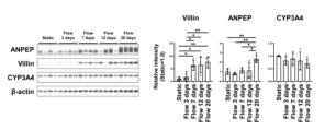
- Villin 1 (VIL1)
- MUC2 MUC2
- LGR5 OSTa
- ASBT CYP3A4 mRNA expression levels
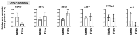
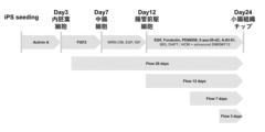
- protocol 4 Induction of differentiation into small intestinal tissue using a microfluidic device.
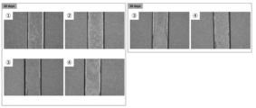
- the effect of medium perfusion on small intestine tissue chips Bright field images are shown on days 10, 17, and 25. Static culture and perfusion culture were compared. It was shown that medium perfusion results in the formation of three-dimensional small intestine-like tissue. It was shown that three-dimensional small intestine-like tissue can be formed not only by perfusion into the lower channel, but also by perfusion using a shaker. The effect of medium perfusion in small intestinal tissue chips.
- HE stained images are shown for static culture and perfusion culture conditions.
- static culture a single layer of small intestinal epithelial cells is formed.
- perfusion culture small intestinal crypt- and villi-like structures were observed, along with mucosal lamina basement-like structures and extracellular matrix-like structures below them. Effect of medium perfusion on small intestinal tissue chips.
- Perfusion confirmed that the Masson's trichrome-positive collagen layer was aligned on the basement membrane side.
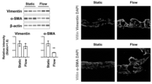
- the small intestinal epithelium layer is supported by the collagen layer. Since it is expected that fibroblasts are the main collagen-producing cells, an experiment was next conducted to confirm the presence of fibroblasts. Localization of fibroblasts in small intestine tissue chips. Results of automated capillary electrophoresis immunoassay for Vimentin, ⁇ -SMA, and ⁇ -actin (left) and immunostaining for Villin, Vimentin, and ⁇ -SMA (right). Expression of Vimentin, a marker for intestinal fibroblasts, was maintained and localized to the basolateral side by perfusion.
- ⁇ -SMA a marker for myofibroblasts
- n 3, error bars: standard deviation, **: P ⁇ 0.01.
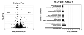
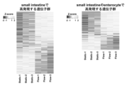
- RNA-seq analysis Comparison of the developed small intestinal tissue chip with live human small intestine (small intestinal total RNA) (RNA-seq analysis). Gene expression profiles were compared between perfusion-cultured small intestinal tissue chips and small intestinal total RNA. There were 4,510 genes whose expression was more than two-fold higher in the small intestinal tissue chip, and 5,462 genes whose expression was reduced to half or less in the small intestinal tissue chip. Comparison of the developed small intestine tissue chip with live human small intestine.
- ECM extracellular matrix
- MMP1, COL3A1, TIMP1 extracellular matrix
- TGFB1, WNT5A, IGF2 growth factors
- Heatmap showing changes in protein expression of drug metabolizing enzymes cytochrome P450 (CYP), uridine diphosphate glucuronosyltransferase (UGT), sulfotransferase (SULT), and carboxylesterase (CES) (left), and comparison of expression levels of CES2, which is highly expressed in the human small intestine, and CES1, which is highly expressed in the liver (right).
- CYP cytochrome P450
- UTT uridine diphosphate glucuronosyltransferase
- SULT sulfotransferase
- CES carboxylesterase
- POU5F1 undifferentiated cell marker POU class 5 homeobox 1
- MIXL1 mesendodermal cell marker Mix paired-like homeobox
- FOXA2 endodermal cell marker forkhead box A2
- VIL1 absorptive epithelial cell markers villin 1
- SI sucrase-isomaltase
- TXT mesodermal cell marker T-box transcription factor T
- VM mesenchymal cell markers vimentin
- LYVE1 lymphatic vessel endothelial hyaluronan receptor 1
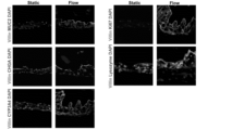
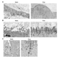
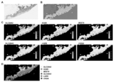
- Morphological analysis (electron microscopy analysis) of small intestinal tissue chips. The morphology of small intestinal tissue chips prepared under static culture conditions was compared with that of small intestinal tissue chips prepared under flow culture conditions. TJ (tight junction): tight junction, AJ (adherence junction): adherens junction, DS: desmosome. Scale bars: 6 ⁇ m (A), 600 nm (B, C). In situ gene expression profiling in small intestinal tissue chips. A shows an H&E stained image of the small intestinal tissue chip used in the analysis. B shows the cell morphology inferred from the position of the cell nucleus.
- C and D show the results of expression analysis in small intestinal tissue chips of absorptive epithelial cell markers solute carrier family 26 member 2 (SLC26A2) and solute carrier family 26 member 3 (SLC26A3), intestinal progenitor cell marker SRY-box transcription factor 9 (SOX9), stem cell markers leucine rich repeat containing G protein-coupled receptor 5 (LGR5) and Bestrophin 4 (BEST4), and neuroendocrine cell marker CHGA.
- SLC26A2 solute carrier family 26 member 2
- SLC26A3 solute carrier family 26 member 3
- SOX9 intestinal progenitor cell marker SRY-box transcription factor 9
- LGR5 stem cell markers leucine rich repeat containing G protein-coupled receptor 5
- BEST4 Bestrophin 4
- CHGA neuroendocrine cell marker
- C and D show the results of transcriptome analysis of vimentin (VIM), an intestinal fibroblast marker, platelet derived growth factor receptor alpha (PDGFRA), a telocyte or trophocyte marker, and frizzled-related protein (FRZB, a Wnt antagonist), in the small intestinal tissue chip.
- VIM vimentin
- PDGFRA platelet derived growth factor receptor alpha
- FRZB frizzled-related protein
- a and B show the results of subcluster analysis of the interstitial cell cluster. The interstitial cells were classified into three subclusters (A). In the figure, 0, 1, and 2 indicate subclusters 0, 1, and 2, respectively.
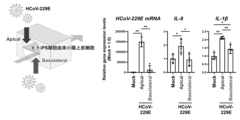
- Results of virus (HCoV-229E) infection experiments using small intestinal tissue chips (RNA-seq analysis).
- A shows the results when HCoV-229E was infected from the apical side (Apical) of a small intestinal tissue chip that had been perfused with medium (Mock vs Apical), and when HCoV-229E was infected from the basolateral side (Bock vs Basolateral).
- B and C show the changes in expression of host genes associated with viral infection in small intestinal tissue chips infected with HCoV-229E from the apical side.
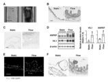
- A shows a bright field image on day 24. Scale bar: 500 ⁇ m.
- B shows a HE stained image. Scale bar: 50 ⁇ m.
- C shows an Alcian blue stained image. Scale bar: 50 ⁇ m.
- E shows immunostained images for VIL1 and VIM. Nuclei were stained with DAPI. Scale bar: 50 ⁇ m.
- F shows a Masson's trichrome stained image. Scale bar: 50 ⁇ m.
- A shows a bright field image on day 20.
- Scale bar 500 ⁇ m.
- B shows a HE stained image. Scale bar: 50 ⁇ m.
- C shows an Alcian blue stained image. Scale bar: 50 ⁇ m.
- E shows an immunostained image for VIL1 and VIM. Nuclei were stained with DAPI. Scale bar: 50 ⁇ m.
- F shows a Masson's trichrome stained image. Scale bar: 50 ⁇ m.
- the present invention provides a method for producing small intestinal epithelial cells or small intestinal tissue from midgut cells. Specifically, the method comprises: (1) culturing midgut cells in a medium containing a Wnt agonist, a BMP inhibitor and a growth factor to obtain intestinal progenitor cells; and (2) culturing the intestinal progenitor cells obtained in step (1) in a medium containing a growth factor, an adenylate cyclase activator, a MEK inhibitor, a DNA methylation inhibitor, a TGF- ⁇ inhibitor, a GSK3 ⁇ inhibitor and a ⁇ -secretase inhibitor to obtain small intestinal epithelial cells or small intestinal tissue (hereinafter, sometimes referred to as the "production method of the present invention").
- Midgut cells are cells that constitute the midgut, which is the basis of the small intestine that occurs during the developmental stage of vertebrates.
- “midgut cells” refers to cells that express CDX2 and have the ability to differentiate into intestinal progenitor cells.
- Small intestinal epithelial cells are cells that constitute the small intestinal epithelium in a living organism. Specific examples of small intestinal epithelial cells obtained by cell culture include absorptive epithelial cells that express Villin, goblet cells that express MUC2, intestinal stem cells that express LGR5, Paneth cells that express LYZ, enteroendocrine cells that express CHGA, M cells that express GP2, and Tuft cells that express DCLK1.
- small intestinal tissue refers to a structure that includes at least absorptive epithelial cells as small intestinal epithelial cells and has a layered structure composed of cells. Absorptive epithelial cells express Villin, and preferably also express SI and/or FABP2.
- cells includes “cell populations.” Furthermore, unless otherwise specified, “cells” refers to those obtained by cell culture. A cell population may be composed of one type of cell, or may be composed of two or more types of cells.
- small intestinal epithelial cells includes not only one type of small intestinal epithelial cells (e.g., absorptive epithelial cells), but also cell populations containing two or more types of small intestinal epithelial cells (e.g., a combination of absorptive epithelial cells, goblet cells, and intestinal stem cells).
- intestinal progenitor cells can be obtained by culturing midgut cells in a medium containing a Wnt agonist, a BMP inhibitor, and a growth factor.
- Intestinal progenitor cells are cells that arise from intestinal stem cells during the developmental stage of vertebrates and can differentiate into cells that constitute the small intestine, which are cells that have the ability to differentiate into small intestinal epithelial cells.
- intestinal progenitor cells refers to cells that are induced from midgut cells or intestinal stem cells and have the ability to differentiate into small intestinal epithelial cells.
- intestinal progenitor cells includes not only cells that do not have the ability to self-proliferate, but also cells that have the ability to self-proliferate (i.e., intestinal stem cells).
- Intestinal progenitor cells are typically cells that weakly express Villin, MUC2, LYZ, and CHGA.
- Wnt agonist refers to a substance that activates T-cell factor (TCF)/lymphoid enhancer actor (LEF)-mediated transcription in cells.
- Wnt agonists are not limited to Wnt family proteins, but include Wnt agonists that bind to and activate Frizzled receptor family members, inhibitors of intracellular ⁇ -catenin degradation, and TCF/LEF activators.
- Wnt agonists used in step (1) of the production method of the present invention include Wnt proteins, R-spondin, etc. It is preferable to use a combination of Wnt proteins (e.g., Wnt3A, etc.) and R-spondin (e.g., R-spondin3, etc.).
- Wnt proteins used in step (1) of the production method of the present invention include Wnt1, Wnt2, Wnt2B, Wnt3, Wnt3A, Wnt4, Wnt5A, Wnt5B, Wnt6, Wnt7A, Wnt7B, Wnt8A, Wnt8B, Wnt9A, Wnt9B, Wnt10A, Wnt10B, Wnt11, and Wnt16.
- Wnt3A is preferred.
- a combination of multiple types of Wnt proteins may be used.
- the R-spondins used in step (1) of the production method of the present invention include, for example, R-spondin 1, R-spondin 2, R-spondin 3, and R-spondin 4. Among these, R-spondin 3 is preferred. Multiple types of R-spondins may be used in combination.
- Bone morphogenetic proteins bind as dimeric ligands to a receptor complex consisting of two different receptor serine/threonine kinases, type I and type II receptors.
- the type II receptor phosphorylates the type I receptor, which activates the receptor kinase.
- the type I receptor then phosphorylates specific receptor substrates (SMADs), which result in transcriptional activation via signal transduction pathways.
- SADs specific receptor substrates
- BMP inhibitors are substances that bind to BMP molecules to form complexes that neutralize BMP activity, e.g., those that inhibit the binding of BMP molecules to BMP receptors.
- BMP inhibitors are also substances that bind to BMP receptors and inhibit the binding of BMP molecules to the receptor, acting as antagonists or inverse agonists.
- BMP inhibitors used in step (1) of the production method of the present invention include, for example, chordin-like proteins such as Noggin, Chordin, and chordin domains, follistatin-related proteins such as follistatin and follistatin domains, DAN-like proteins such as DAN and DAN cysteine-knot domains, sclerostin/SOST, decorin, ⁇ 2-macroglobulin, and the like.
- chordin-like proteins or DAN-like proteins are preferred, and chordin-like proteins (particularly Noggin) are more preferred.
- Chordin-like proteins and DAN-like proteins are diffusible proteins that bind to BMP molecules with various affinities and can inhibit the approach of BMP molecules to signaling receptors.
- Growth factors used in step (1) of the method of the present invention include, for example, epidermal growth factor (EGF), insulin-like growth factor (IGF), transforming growth factor- ⁇ (TGF- ⁇ ), fibroblast growth factor (FGF), brain derived neurotrophic factor (BDNF), and keratinocyte growth factor (KGF).
- FGF includes, for example, 23 types of proteins, FGF1 to FGF23.
- IGF may be IGF1 or IGF2.
- Growth factors used in step (1) of the method of the present invention are preferably EGF and IGF, and more preferably a combination of EGF and IGF (particularly IGF1). These growth factors may be used in combination.
- the concentration of EGF in the medium used in step (1) of the production method of the present invention is typically 5 ng/mL to 500 ng/mL, preferably 10 ng/mL to 300 ng/mL, more preferably 30 ng/mL to 100 ng/mL (particularly 50 ng/mL).
- the concentration of IGF1 in the medium used in step (1) of the production method of the present invention is typically 1 ng/mL to 200 ng/mL, preferably 2 ng/mL to 100 ng/mL, more preferably 10 ng/mL to 50 ng/mL (particularly 20 ng/mL).
- the concentration of the growth factor in the medium is selected appropriately.
- the culture period in step (1) of the method of the present invention is typically 3 to 13 days, preferably 4 to 10 days, and more preferably 5 to 7 days (particularly 6 days).
- the intestinal progenitor cells obtained in step (1) can be cultured in a medium containing a growth factor, an adenylate cyclase activator, a MEK inhibitor, a DNA methylation inhibitor, a TGF- ⁇ inhibitor, a GSK3 inhibitor, and a ⁇ -secretase inhibitor to obtain small intestinal epithelial cells or small intestinal tissue.
- Growth factors used in step (2) of the method of the present invention include the same as those used in step (1).
- the growth factor used in step (2) of the method of the present invention is preferably EGF. These growth factors may be used in combination.
- the concentration of EGF in the medium used in step (2) of the method of the present invention is typically 0.5 ng/mL to 100 ng/mL, preferably 1 ng/mL to 50 ng/mL, and more preferably 5 ng/mL to 30 ng/mL (particularly 10 ng/mL).
- concentration of the growth factor in the medium is appropriately selected.
- the adenylate cyclase activator used in step (2) of the production method of the present invention may be any that increases the intracellular cAMP concentration, such as forskolin, HJC0350, 8-Br-cAMP, adenosine 3',5'-cyclic monophosphate (cAMP), dibutyryl-cAMP (Bucladesine), etc. Of these, forskolin is preferred. These adenylate cyclase activators may be used in combination.
- the concentration of Forskolin in the medium used in step (2) of the production method of the present invention is typically 1 ⁇ M to 200 ⁇ M, preferably 3 ⁇ M to 100 ⁇ M, and more preferably 5 ⁇ M to 50 ⁇ M (particularly 15 ⁇ M).
- concentration of the adenylate cyclase activator in the medium is appropriately selected.
- MEK inhibitors used in step (2) of the production method of the present invention include PD98059 (2-(2-amino-3-methoxyphenyl)-4H-1-benzopyran-4-one), PD184352 (2-(2-chloro-4-iodoanilino)-N-(cyclopropylmethoxy)-3,4-difluorobenzamide), PD184161 (2-(2-chloro-4-iodophenylamino)-N-cyclopropylmethoxy-3,4-difluoro-5-bromobenzamide), PD0325901 (N-[(2R)-2,3-dihydroxypropoxy]-3,4-difluoro-2-(2-fluoro-4-iodoanilino)benzamide), U0126, MEK inhibitor I, MEK inhibitor II, MEK1/2 inhibitor II, and SL327. Among these, PD98059 is preferred. These MEK inhibitors may be used in combination with
- the concentration of PD98059 in the medium used in step (2) of the production method of the present invention is typically 0.5 ⁇ M to 100 ⁇ M, preferably 1 ⁇ M to 50 ⁇ M, and more preferably 5 ⁇ M to 30 ⁇ M (particularly 10 ⁇ M).
- concentration of the MEK inhibitor in the medium is appropriately selected.
- DNA methylation inhibitors used in step (2) of the production method of the present invention include 5-aza-2'-deoxycytidine (5-aza-2'-dC), 5-azacytidine, RG108, and zebularine. Among these, 5-aza-2'-dC is preferred. These DNA methylation inhibitors may be used in combination.
- the concentration of 5-aza-2'-dC in the medium used in step (2) of the production method of the present invention is typically 0.1 ⁇ M to 100 ⁇ M, preferably 0.5 ⁇ M to 50 ⁇ M, and more preferably 1 ⁇ M to 5 ⁇ M (particularly 2.5 ⁇ M).
- concentration of the DNA methylation inhibitor in the medium is appropriately selected.
- the TGF- ⁇ inhibitor used in step (2) of the production method of the present invention is, for example, SB431542 (4-(5-benzol[1,3]dioxol-5-yl-4-pyridin-2-yl-1H-imidazol-2-yl)-benzamide, 4-[4-(1,3-benzodioxol-5-yl)-5-(2-pyridinyl)-1H-imidazol-2-yl]-benzamide, 4-[4-(3,4-methylenedioxyphenyl)-5-(2-pyridyl)-1H-imidazol-2-yl]-benzamide, amide), A-83-01 (3-(6-methylpyridin-2-yl)-1-phenylthiocarbamoyl-4-quinolin-4-ylpyrazole), LDN193189 (4-[6-[4-(1-piperazinyl)phenyl]pyrazolo[1,5-a]pyrimidin-3-yl]
- the concentration of A-83-01 in the medium used in step (2) of the production method of the present invention is typically 10 nM to 10 ⁇ M, preferably 50 nM to 5 ⁇ M, and more preferably 100 nM to 500 nM (particularly 250 nM).
- concentration of the TGF- ⁇ inhibitor in the medium is appropriately selected.
- GSK3 ⁇ inhibitor refers to a substance that has inhibitory activity against GSK3 ⁇ (glycogen synthase kinase 3 ⁇ ).
- GSK3 glycogen synthase kinase 3 ⁇
- GSK3 glycogen synthase kinase 3 ⁇
- GSK3 glycogen synthase kinase 3 ⁇
- GSK3 glycogen synthase kinase 3 ⁇
- GSK3 glycogen synthase kinase 3 ⁇
- GSK3 glycogen synthase kinase 3 ⁇
- the "GSK3 ⁇ inhibitor” used in the present invention is not particularly limited as long as it has GSK3 ⁇ inhibitory activity, and may be a substance that has both GSK3 ⁇ inhibitory activity and GSK3 ⁇ inhibitory activity.
- the GSK3 ⁇ inhibitors used in step (2) of the production method of the present invention include, for example, CHIR98014 (2-[[2-[(5-nitro-6-aminopyridin-2-yl)amino]ethyl]amino]-4-(2,4-dichlorophenyl)-5-(1H-imidazol-1-yl)pyrimidine), CHIR99021 (6-[[2-[[4-(2,4-dichlorophenyl)-5-(4-methyl-1H-imidazol-2-yl)-2-pyrimidinyl]amino]ethyl]amino]nicotinonitrile), CP21R7 (3-(3 -amino-phenyl)-4-(1-methyl-1H-indol-3-yl)-pyrrole-2,5-dione), LY2090314 (3-[9-Fluoro-1,2,3,4-tetrahydro-2-(1-piperidin
- the concentration of BIO in the medium used in step (2) of the production method of the present invention is typically 0.1 ⁇ M to 100 ⁇ M, preferably 0.5 ⁇ M to 50 ⁇ M, and more preferably 1 ⁇ M to 5 ⁇ M (particularly 2.5 ⁇ M).
- concentration of the GSK3 ⁇ inhibitor in the medium is appropriately selected.
- the gamma-secretase inhibitors used in step (2) of the production method of the present invention include, for example, DAPT, DBZ, LY450139, BMS-708163, NIC5-15, GSI-953, ELND006, and CHF-5074. Of these, DAPT is preferred. These gamma-secretase inhibitors may be used in combination.
- the concentration of DAPT in the medium used in step (2) of the production method of the present invention is typically 0.2 ⁇ M to 200 ⁇ M, preferably 1 ⁇ M to 100 ⁇ M, and more preferably 2 ⁇ M to 10 ⁇ M (particularly 5 ⁇ M).
- concentration of the ⁇ -secretase inhibitor in the medium is appropriately selected.
- Each substance used in the manufacturing method of the present invention may be synthesized by a known method, or a commercially available product may be used.
- the compound is a protein or peptide such as a Wnt protein
- the target protein or peptide may be obtained from a cell that expresses it.
- Cells that express the target protein or peptide can be produced by inserting DNA encoding the target protein or peptide into a known expression vector and introducing the resulting expression vector into an appropriate host cell.
- Wnt protein-expressing cells include L cells (ATCC CRL-2647) that stably express mouse Wnt3a and L cells (ATCC CRL-2814) that stably express mouse Wnt5a.
- L cells expressing multiple types of proteins or peptides may be used, specifically, for example, L-WRN cells (ATCC CRL-3276) that express Wnt-3A, R-spondin 3, and noggin.
- the Wnt agonist e.g., a combination of Wnt protein and R-spondin, etc.
- BMP inhibitor e.g., chordin-like protein, etc.
- each substance used in the method of the present invention is a protein or peptide
- its origin is not particularly limited, but is preferably mammalian (e.g., human, mouse, rat, monkey, cow, horse, pig, dog, etc.).
- the proteins or peptides used in the method of the present invention include not only wild-type proteins or peptides, but also mutants thereof having similar functions.
- mutants include proteins or peptides that have a high identity (e.g., 80% or more, 85% or more, 90% or more, 91% or more, 92% or more, 93% or more, 94% or more, 95% or more, 96% or more, 97% or more, 98% or more, or 99% or more identity) to the amino acid sequence of a particular wild-type protein or peptide.
- a high identity e.g., 80% or more, 85% or more, 90% or more, 91% or more, 92% or more, 93% or more, 94% or more, 95% or more, 96% or more, 97% or more, 98% or more, or 99% or more identity
- the culture period in step (2) of the method of the present invention is not particularly limited, but is 5 days or more (e.g., 6 days, 7 days, 8 days, 9 days, 10 days or more). Since small intestinal epithelial cells and small intestinal tissue can be maintained by step (2), there is no particular upper limit, but it is typically 20 days or less (e.g., 19, 18, 17, 16 days or less). In one embodiment, the culture period in step (2) of the method of the present invention is 5 to 15 days, or 7 to 13 days.
- the basal medium used in the production method of the present invention is not particularly limited, but may be RPMI-1640 medium, EagleMEM (EMEM), Dulbecco's modified MEM, Glasgow's MEM (GMEM), ⁇ -MEM, 199 medium, IMDM, DMEM, Hybridoma Serum free medium, KnockOut TM DMEM, Advanced TM medium (e.g., Advanced MEM, Advanced RPMI, Advanced DMEM/F-12), Ham's Medium F-12, Ham's Medium F-10, Ham's Medium F12K, DMEM/F-12, ATCC-CRCM30, DM-160, DM-201, BME, Fischer, McCoy's 5A, Leibovitz's L-15, RITC80-7, MCDB105, MCDB107, MCDB131, MCDB153, MCDB201, NCTC109, NCTC135, Waymouth's Medium (e.g., Waymouth's MB752/1), CMRL medium (e
- StemFit registered trademark
- AK02 medium Alignomoto Co., Inc.
- StemFit registered trademark
- AK03 medium Alignomoto Co., Inc.
- StemFit registered trademark
- Basic03 medium CTS (registered trademark) KnockOut SR XenoFree Medium (Gibco)
- mTeSR1 medium TeSR1 medium
- Iscove's modified Dulbecco's medium GE Healthcare
- Improved MEM Thermo Fisher Scientific
- the medium used in step (2) of the present invention may be the above-mentioned basal medium (preferably Advanced DMEM/F-12) or a medium obtained by mixing the basal medium with a hepatocyte culture medium.
- basal medium preferably Advanced DMEM/F-12
- hepatocyte culture medium include HCM (LONZA), William's E Medium (Thermo Fisher Scientific), and Leibovitz's L-15 Medium (Thermo Fisher Scientific).
- the medium may contain serum, e.g., fetal bovine serum, one or more serum substitutes, such as Knockout Serum Replacement (KSR), N2 supplement (Invitrogen), B27 supplement (Invitrogen), albumin, transferrin, apotransferrin, fatty acids, insulin, collagen precursors, trace elements, 2-mercaptoethanol, 3'-thiolglycerol, and one or more substances, such as lipids, amino acids, L-glutamine, Glutamax (Invitrogen), non-essential amino acids, vitamins, growth factors, small molecules, antibiotics, antioxidants, pyruvate, buffers, inorganic salts, selenate, progesterone, and putrescine.
- serum substitutes such as Knockout Serum Replacement (KSR), N2 supplement (Invitrogen), B27 supplement (Invitrogen), albumin, transferrin, apotransferrin, fatty acids, insulin, collagen precursors, trace elements, 2-mercaptoethanol, 3'-thi
- microfluidic device means a cell culture device having microchannels, and the width and depth of the microchannels can be freely designed.
- the width and height of each microchannel of the microfluidic device used in the present invention is typically 10 ⁇ m to 10 mm, and preferably 100 ⁇ m to 1 mm.
- the microfluidic device used in the present invention is preferably a device having two upper and lower flow paths (upper and lower channels) separated by a semipermeable membrane.
- the material of the substrate of a microfluidic device having two upper and lower flow paths is typically PDMS (polydimethylsiloxane), but may be, for example, cycloolefin polymer, polymethyl methacrylate, cycloolefin copolymer, etc.
- the material of the membrane of a microfluidic device having two upper and lower flow paths is typically polyethylene terephthalate (PET), but may be, for example, plastic such as polycarbonate or polytetrafluoroethylene.
- PET polyethylene terephthalate
- the microfluidic device can be fabricated by known methods.
- the flow channels can be fabricated, for example, using soft lithography techniques (D. C. Duffy, J. C. McDonald, O. J. A. Schueller, G. M. Whitesides, Rapid prototyping of microfluidic systems in poly(dimethylsiloxane). Analytical Chemistry 70, 4974-4984 (1998).).
- the culture in the method of the present invention is preferably an adherent culture.
- adherent culture refers to culture under conditions that allow strong cell-substrate bonds to be formed between the cells and the culture equipment, etc.
- Culturing in a microfluidic device typically involves culturing cells using a device in which the upper channel is seeded with cells and the lower channel is filled with culture medium.
- the culture medium in the lower channel may be perfused.
- steps (1) and (2) of the method of the present invention are perfusion culture for the entire period.
- Perfusion culture is typically performed using a peristaltic pump or shaker.
- a peristaltic pump also known as a tube pump, is a pump that pumps liquid by squeezing a soft tube such as silicone with a roller.
- the lower channel of the microfluidic device is connected to the soft tube, and the medium in the lower channel is perfused by rotating the roller.
- small intestinal tissue can be produced at perfusion rates of 10 ⁇ L/hour, 30 ⁇ L/hour, and 100 ⁇ L/hour, so the perfusion rate is not particularly limited, but is preferably 10 ⁇ L/hour to 100 ⁇ L/hour.
- the expression level of certain genes varies depending on the perfusion rate, so it is also preferable to adjust the perfusion rate appropriately depending on the purpose.
- the shaker used in the present invention may be an orbital shaker, a reciprocating shaker, a figure-of-eight shaker, or a shaker that has the function of appropriately switching between these shaking methods.
- the shaking direction may be either horizontal or vertical, but is typically horizontal.
- the shaking speed is not particularly limited, and is typically in the range of 50 to 450 rpm, but is preferably in the range of 100 to 200 rpm.
- Culture vessels used in adhesion culture include those in which the surface of the vessel or the flow channels of the microfluidic device have been artificially treated to improve adhesion to cells (e.g., coating with basement membrane preparations, extracellular matrices such as fibronectin, laminin or fragments thereof, entactin, collagen, gelatin, synthemax, vitronectin, etc., or polymers such as polylysine and polyornithine, or surface treatments such as positive charge treatment).
- basement membrane preparations include, for example, Matrigel and Geltrex. Among these, culture vessels coated with Matrigel are preferred.
- laminin or a fragment thereof is also preferred.
- laminin or a fragment thereof include laminin-111 or a fragment thereof containing the E8 region, laminin-211 or a fragment thereof containing the E8 region (e.g., iMatrix-211), laminin-121 or a fragment thereof containing the E8 region, laminin-221 or a fragment thereof containing the E8 region, laminin-332 or a fragment thereof containing the E8 region, laminin-3A11 or a fragment thereof containing the E8 region, laminin-411 or a fragment thereof containing the E8 region (e.g., iMatrix-411), and laminin-421.
- laminin-511 or fragments containing its E8 region laminin-511 or fragments containing its E8 region (e.g., iMatrix-511, iMatrix-511 silk), laminin-521 or fragments containing its E8 region, laminin-213 or fragments containing its E8 region, laminin-423 or fragments containing its E8 region, laminin-523 or fragments containing its E8 region, laminin-212/222 or fragments containing its E8 region, laminin-522 or fragments containing its E8 region, etc.
- laminin-511 or fragments containing its E8 region e.g., iMatrix-511, iMatrix-511 silk
- laminin-521 or fragments containing its E8 region laminin-213 or fragments containing its E8 region
- laminin-423 or fragments containing its E8 region laminin-523 or fragments containing its E8 region
- cells may be cultured under feeder-free and/or xeno-free conditions.
- all steps may be performed under feeder-free and xeno-free conditions.
- cells may be cultured under feeder-free and/or xeno-free conditions.
- feeder-free refers to a medium or culture condition that does not contain other cell types (i.e., feeder cells) that play a supporting role and are used to prepare the culture conditions for the cells to be cultured.
- xeno-free refers to a medium or culture condition that does not contain components derived from organisms other than the organism species of the cells to be cultured.
- the culture temperature is not particularly limited, but is about 30 to 40°C, preferably about 37°C, and culture is performed in an atmosphere of CO2- containing air, with the CO2 concentration being preferably about 2 to 5%.
- the seeding density of the cells is not particularly limited as long as the cells can grow, and is typically 1.0 ⁇ 10 2 to 1.0 ⁇ 10 7 cells/cm 2 , preferably 1.0 ⁇ 10 3 to 1.0 ⁇ 10 6 cells/cm 2 , and more preferably 1.0 ⁇ 10 4 to 1.0 ⁇ 10 5 cells/cm 2 .
- the midgut cells used in the present invention can be obtained by known methods. Examples include a method of isolating from biological tissue (e.g., intestinal tissue, etc.) by known techniques, a method of inducing differentiation of definitive endoderm cells, and a method of obtaining from a company such as ATCC.
- Midgut cells can be isolated from biological tissue, for example, using a method that uses flow cytometry or mass cytometry with expressed antigens as an indicator, a magnetic cell separation method, an affinity column with a desired antigen immobilized, etc. It is preferable that the midgut cells used in the present invention are obtained by a method of inducing differentiation of definitive endoderm cells.
- definitive endoderm cells refers to cells that express SOX17 and FOXA2 and have the ability to differentiate into endodermal cells such as those of the digestive tract.
- the definitive endoderm cells used in the present invention can be obtained by known methods. For example, they can be isolated from biological tissues (e.g., intestinal tissues, etc.) by known techniques, by inducing differentiation of pluripotent stem cells, or by obtaining cells from companies such as ATCC. From biological tissues, midgut cells can be isolated, for example, using expression antigens (e.g., E-cadherin, CXCR4, CD55, etc.) as indicators, using methods that use flow cytometry or mass cytometry, magnetic cell separation methods, affinity columns with immobilized desired antigens, etc. It is preferable that the definitive endoderm cells used in the present invention are obtained by a method that induces differentiation of pluripotent stem cells.
- pluripotent stem cells refer to stem cells that can differentiate into tissues and cells with various different morphologies and functions in the body, and have the ability to differentiate into cells of any of the three germ layers (endoderm, mesoderm, and ectoderm).
- pluripotent stem cells used in the present invention include induced pluripotent stem cells (iPS cells), embryonic stem cells (ES cells), embryonic stem cells derived from cloned embryos obtained by nuclear transfer (ntES cells), multipotent germline stem cells (mGS cells), and embryonic germline stem cells (EG cells), with iPS cells (more preferably human iPS cells) being preferred.
- the pluripotent stem cells are ES cells or any cells derived from a human embryo, the cells may be cells produced by destroying an embryo or cells produced without destroying an embryo, but from an ethical point of view, cells produced without destroying an embryo are preferable.
- ES cells are stem cells that are pluripotent and have the ability to proliferate through self-renewal and are established from the inner cell mass of early mammalian embryos (e.g., blastocysts) such as humans and mice.
- ES cells were discovered in mice in 1981 (M.J. Evans and M.H. Kaufman (1981), Nature 292:154-156), and subsequently, ES cell lines were established in humans, monkeys, and other primates (J.A. Thomson et al. (1998), Science 282:1145-1147; J.A. Thomson et al. (1999), Science 282:1145-1147). (1995), Proc. Natl. Acad. Sci. USA, 92:7844-7848; J.A.
- ES cells can be established by extracting the inner cell mass from the blastocyst of a fertilized egg of a target animal and culturing the inner cell mass on fibroblast feeders.
- ES cells can be established using only a single blastomere from an embryo at the cleavage stage prior to the blastocyst stage (Chung Y. et al. (2008), Cell Stem Cell 2: 113-117) or from developmentally arrested embryos (Zhang X. et al. (2006), Stem Cells 24: 2669-2676.).
- nt ES cells are cloned embryo-derived ES cells produced by nuclear transfer technology, and have almost the same characteristics as fertilized egg-derived ES cells (Wakayama T. et al. (2001), Science, 292:740-743; S. Wakayama et al. (2005), Biol. Reprod., 72:932-936; Byrne J. et al. (2007), Nature, 450:497-502).
- nt ES (nuclear transfer ES) cells are ES cells established from the inner cell mass of a cloned embryo-derived blastocyst obtained by replacing the nucleus of an unfertilized egg with that of a somatic cell.
- nuclear transfer technology (Cibelli JB . et al. (1998), Nature Biotechnol., 16:642-646) in combination with ES cell production technology (mentioned above) is used (Syoka Wakayama et al. (2008), Experimental Medicine, Vol. 26, No. 5 (special issue), pp. 47-52).
- nuclear transfer the nucleus of a somatic cell is injected into an enucleated unfertilized egg of a mammal, and the egg is cultured for several hours to initialize the cell.
- ES cell lines used in the present invention for example, various mouse ES cell lines established by inGenious targeting laboratory, RIKEN (Riken) and the like can be used for mouse ES cells, and for human ES cell lines, various human ES cell lines established by University of Wisconsin, NIH, RIKEN, Kyoto University, National Center for Child Health and Development, Cellartis and the like can be used.
- human ES cell lines include CHB-1 to CHB-12 strains, RUES1 strain, RUES2 strain, HUES1 to HUES28 strains, etc. distributed by ESI Bio, H1 strain, H9 strain, etc. distributed by WiCell Research, and KhES-1 strain, KhES-2 strain, KhES-3 strain, KhES-4 strain, KhES-5 strain, SSES1 strain, SSES2 strain, SSES3 strain, etc. distributed by RIKEN.
- iPS cells are cells obtained by reprogramming mammalian somatic cells or undifferentiated stem cells by introducing specific factors (nuclear reprogramming factors).
- iPSCs established by Yamanaka et al. by introducing four factors, Oct3/4, Sox2, Klf4, and c-Myc, into mouse fibroblasts (Takahashi K, Yamanaka S., Cell, (2006) 126: 663-676); human cell-derived iPSCs established by introducing the same four factors into human fibroblasts (Takahashi K, Yamanaka S., et al.
- Nanog-iPSCs established by selecting using the expression of Nanog as an indicator after introducing the above four factors (Okita, K., Ichisaka, T., and Yamanaka, S. (2007). Nature 448, 313-317.); and iPSCs created by a method that does not include c-Myc (Nakagawa M, Yamanaka S., et al. Nature Biotechnology, (2008) 26, 101-106), iPSCs established by introducing six factors using a virus-free method (Okita K et al. Nat. Methods 2011 May;8(5):409-12, Okita K et al. Stem Cells. 31(3):458-66.), etc.
- induced pluripotent stem cells established by introducing four factors, OCT3/4, SOX2, NANOG, and LIN28, created by Thomson et al. (Yu J., Thomson JA. et al., Science (2007) 318: 1917-1920.), induced pluripotent stem cells created by Daley et al. (Park IH, Daley GQ. et al., Nature (2007) 451: 141-146), and induced pluripotent stem cells created by Sakurada et al. (JP Patent Publication No. 2008-307007) can also be used.
- human iPSC strains include RIKEN's HiPS-RIKEN-1A strain, HiPS-RIKEN-2A strain, HiPS-RIKEN-12A strain, Nips-B2 strain, etc., and Kyoto University's 253G1 strain, 253G4 strain, 1201C1 strain, 1205D1 strain, 1210B2 strain, 1383D2 strain, 1383D6 strain, 201B7 strain, 409B2 strain, 454E2 strain, 606A1 strain, 610B1 strain, 648A1 strain, 1231A3 strain, FfI-01s04 strain, etc.
- the induced pluripotent stem cells used in the method of the present invention may be cells derived from a patient with a hereditary disease (e.g., a patient with a hereditary small intestinal disease).
- a hereditary disease e.g., a patient with a hereditary small intestinal disease.
- Cells induced to differentiate from pluripotent stem cells derived from a patient with a hereditary small intestinal disease can serve as a disease model that reflects the pathology of the disease, and are therefore suitable for screening therapeutic or preventive drugs for the disease.
- pluripotent stem cells derived from a patient with a hereditary small intestinal disease can be genetically repaired by genome editing using the CRISPR-Cas system or the like, and then differentiated into the desired cells or tissues, making it possible to use the cells or tissues as therapeutic agents for the disease.
- mGS cells are pluripotent stem cells derived from the testis and are the source of spermatogenesis. Like ES cells, these cells can be induced to differentiate into cells of various lineages, and have the property that, for example, when transplanted into mouse blastocysts, chimeric mice can be produced (Kanatsu-Shinohara M. et al. (2003) Biol. Reprod., 69:612-616; Shinohara K. et al. (2004), Cell, 119:1001-1012).
- GDNF glial cell line-derived neurotrophic factor
- EG cells are established from primordial germ cells during the fetal period and have pluripotency similar to that of ES cells. They can be established by culturing primordial germ cells in the presence of substances such as LIF, bFGF, and stem cell factor (Matsui Y. et al. (1992), Cell, 70:841-847; J.L. Resnick et al. (1992), Nature, 359:550-551).
- the species from which the pluripotent stem cells are derived is not particularly limited, and may be, for example, cells from rodents such as rats, mice, hamsters, and guinea pigs; lagomorphs such as rabbits; ungulates such as pigs, cows, goats, and sheep; felines such as dogs and cats; and primates such as humans, monkeys, rhesus monkeys, marmosets, orangutans, and chimpanzees.
- the preferred species is human.
- pluripotent stem cells into midgut cells can be induced by known methods such as those described in Non-Patent Documents 1 to 4, Toyoda T, et al. Stem Cell Reports 2017, and International Publication No. 2017/047797.
- Pluripotent stem cells can be induced to differentiate into midgut cells by a method comprising: A) a step of inducing the differentiation of pluripotent stem cells into definitive endoderm cells; and B) a step of inducing the differentiation of definitive endoderm cells into midgut cells. Therefore, the production method of the present invention may include the above-mentioned step A) and/or step B) prior to step (1).
- the basal medium and medium additives used in steps A) and B) may be the same as those used in the method of the present invention.
- the basal medium used in step A) is preferably RPMI1640 medium.
- the basal medium used in step B) is preferably advanced DMEM/F12.
- the culture temperature in steps A) and B) is typically about 30 to 40°C, preferably about 37°C, and the culture is performed under an atmosphere of CO2- containing air, with a CO2 concentration of preferably about 2 to 5%. It is preferable to perform steps A) and B) by adhesion culture.
- the definition and method of adhesion culture are as described above.
- step B) is performed in a microfluidic device.
- the differentiation of pluripotent stem cells into definitive endoderm cells in step A) can be carried out, for example, by culturing the pluripotent stem cells in a medium containing a low dose of activin A.
- the medium may further contain a ROCK inhibitor or a GSK3 ⁇ inhibitor.
- the culture period is typically 2 to 8 days, preferably 2 to 4 days (particularly 3 days).
- the concentration of activin A in the medium used in step A) is, for example, 5 to 1000 ng/mL, preferably 20 to 500 ng/mL, and more preferably 50 to 150 ng/mL.
- the GSK3 ⁇ inhibitor used in step A) may be the same as those listed in step (2) of the production method of the present invention. Among them, CHIR99021 is preferred. When CHIR99021 is used as the GSK3 ⁇ inhibitor, the concentration in the medium is typically 0.5 to 5 ⁇ M, preferably 1 to 4 ⁇ M.
- ROCK inhibitors used in step A include Y-27632 (see, e.g., Ishizaki et al., Mol. Pharmacol. 57, 976-983 (2000); Narumiya et al., Methods Enzymol. 325, 273-284 (2000)), fasudil/HA1077 (see, e.g., Uenata et al., Nature 389: 990-994 (1997)), and SR3677 (see, e.g., Feng Y et al., J Med Chem. 51: 6642-6645 (2008)). ), GSK269962 (see, e.g., Stavenger RA et al., J Med Chem.
- Y-27632 is preferred.
- the concentration in the medium is typically 1 to 20 ⁇ M, preferably 5 to 15 ⁇ M.
- Insulin may further be added to the medium.
- the concentration of insulin in the medium is typically 0.01 to 20 ⁇ M, preferably 0.1 to 10 ⁇ M, and more preferably 0.5 to 5 ⁇ M.
- the concentration of insulin in the medium may be, but is not limited to, the concentration of insulin contained in the added B-27 supplement.
- the differentiation into midgut cells in step B) can be carried out, for example, by culturing definitive endoderm cells in a medium containing a growth factor.
- the culture period is typically 2 to 8 days, preferably 3 to 5 days (particularly 4 days).
- step B) is perfusion culture.
- step B) is perfusion culture for the entire period.
- the specific method of perfusion culture is as described above.
- Growth factors used in step B) include those listed in relation to step (1) of the production method of the present invention. Among them, EGF, KGF, FGF2, and FGF10 are preferred, and FGF2 is more preferred.
- the concentration of the growth factor in the medium is set appropriately depending on the type of growth factor used, but is typically about 0.1 nM to 1000 ⁇ M, preferably about 0.1 nM to 100 ⁇ M. In the case of EGF, the concentration is about 5 to 2000 ng/ml (i.e., about 0.8 to 320 nM), preferably about 5 to 1000 ng/ml (i.e., about 0.8 to 160 nM), and more preferably about 10 to 1000 ng/ml (i.e., about 1.6 to 160 nM).
- the concentration is about 5 to 2000 ng/ml (i.e., about 0.3 to 116 nM), preferably about 10 to 1000 ng/ml (i.e., about 0.6 to 58 nM), more preferably about 10 to 1000 ng/ml (i.e., about 0.6 to 58 nM).
- the concentration is typically 5 to 150 ng/mL, preferably 30 to 100 ng/mL, and particularly preferably about 50 ng/mL.
- steps A) and/or B) may involve culturing under feeder-free conditions and/or xeno-free conditions.
- the method of the present invention may include a step of recovering the target cells or tissues obtained in each step.
- the recovered cells may be cryopreserved using a cell cryopreservation solution.
- the cells recovered in the container may be counted using a cell counter, or may be labeled with an antibody against a cell surface marker and selected by flow cytometry, mass cytometry, magnetic cell separation, or the like.
- Small intestinal epithelial cells and small intestinal tissue Small intestinal epithelial cells, small intestinal tissue, or a microfluidic device carrying them can be obtained by step (2) of the production method of the present invention. Therefore, in another aspect of the present invention, there is also provided small intestinal epithelial cells, small intestinal tissue, or a microfluidic device carrying them obtained by the production method of the present invention ("obtained” can be read as “obtained” as appropriate).
- Small intestinal tissue obtained by the production method of the present invention typically has all of the following characteristics (A) to (F).
- A having absorptive epithelial cells;
- B having fibroblasts;
- C having collagen fibers;
- D expressing one or more (preferably all) genes selected from the group consisting of TGFB1, Wnt5A, and IGF2;
- E expressing one or more (preferably all) genes selected from the group consisting of MMP1, COL3A1, and TIMP1;
- F having drug metabolizing enzyme activity.
- the small intestine tissue of the present invention further has at least one (preferably all) of the following characteristics (G) to (K): (G) derived from pluripotent stem cells; (H) having goblet cells; (I) having Paneth cells; (J) having enteroendocrine cells; (K) having crypt-like structures and villi-like structures.
- the small intestinal tissue of the present invention may be characterized by expressing (L) a transporter gene.
- transporter genes include ABCB1/MDR1, ABCC1/MRP1, ABCC2/MRP2, ABCG2/BCRP, SLCO2B1/OATP2B1, SLC15A1/PEPT1, etc.
- the small intestinal tissue of the present invention expresses ABCB1/MDR1, ABCC1/MRP1, ABCC2/MRP2, ABCG2/BCRP, SLCO2B1/OATP2B1, and SLC15A1/PEPT1.
- the small intestinal tissue of the present invention is supported in a microfluidic device.
- the fibroblasts in the small intestinal tissue can be arranged in parallel along the flow path of the microfluidic device.
- the small intestinal tissue of the present invention may be tissue composed of uniform columnar epithelial cells and having microvilli arranged in an orderly fashion on its surface.
- the small intestinal tissue of the present invention may have tight junctions (TJs), adherens junctions (AJs), and desmosomes (DSs), and is preferably tissue in which TJs, AJs, and DSs are aligned.
- the small intestinal tissue of the present invention is tissue in which absorptive epithelial cells expressing SLC26A2 or SLC26A3 are mainly present on the apical side, and intestinal progenitor cells expressing SOX9 and intestinal stem cells expressing LGR5 are mainly present on the basal side.
- the present invention also provides small intestinal tissue having all of the above characteristics (A) to (F). Also provided is small intestinal tissue having at least any (preferably all) of the above characteristics (G) to (K) and/or the above characteristic (L). Such small intestinal tissue may be obtained by the production method of the present invention, or may be obtained by another method. Hereinafter, the small intestinal tissue may also be referred to as "small intestinal tissue of the present invention.”
- the terms “express” or “positive” a gene are used to mean at least “production of mRNA encoded by the gene,” but preferably also “production of a protein encoded by the mRNA.” Therefore, if the production of mRNA encoded by the gene is detected at least by the methods described in the Examples below (RT-qPCR or RNA-seq), the gene can be said to be expressed.
- the fibroblasts in the small intestinal tissue of the present invention are positive for Vimentin, an intestinal fibroblast marker, and have the ability to produce collagen.
- the fibroblasts are typically PDGFRA-positive cells.
- collagen fiber means a fiber formed by the assembly of multiple collagen molecules.
- the small intestinal tissue of the present invention has multiple types of fibroblasts, and in a preferred embodiment, the small intestinal tissue of the present invention has fibroblasts that express R-spondin 3 (RSPO3) and fibroblasts that express bone morphogenetic protein 5 (BMP5).
- RSPO3 R-spondin 3
- BMP5 bone morphogenetic protein 5
- the small intestinal tissue of the present invention expresses one or more (preferably all) genes selected from the group consisting of TGFB1, Wnt5A, and IGF2. These genes are genes that encode growth factors. Furthermore, the small intestinal tissue of the present invention typically has a higher expression level of one or more (preferably all) of these genes compared to the normal small intestine of a human adult.
- the small intestinal tissue of the present invention expresses one or more (preferably all) genes selected from the group consisting of MMP1, COL3A1, and TIMP1. These genes encode extracellular matrix (ECM) and factors regulating its degradation. Typically, the small intestinal tissue of the present invention has a higher expression level of one or more (preferably all) of these genes compared to the normal small intestine of a human adult.
- ECM extracellular matrix
- the expression level of a specific gene can be evaluated by the method described in the Examples below, specifically, by RNA-seq analysis using total RNA isolated from the small intestinal tissue of the present invention and total RNA from normal adult human small intestine (BioChain Institute, R1234226-50) as samples. If the TPM (transcripts per million) value for a specific gene obtained as a result of such RNA-seq analysis is higher in the small intestinal tissue of the present invention compared to the normal adult human small intestine, it can be said that the small intestinal tissue of the present invention has a higher expression level of the gene compared to the normal adult human small intestine.
- the drug metabolizing enzyme activity of the above characteristic (F) includes CYP3A4 activity, UGT1A1 activity, and CYP2C9 activity, but it is preferable that the small intestinal tissue of the present invention has at least CYP3A4 activity.
- Having drug metabolizing enzyme activity means exhibiting drug metabolizing enzyme activity at least by the method of the Examples described below.
- Crypts are tubular depressions located behind the villi in the small intestine, and in this specification, “crypt-like structures” refer to structures similar in appearance to the crypts of the small intestine. Villi are protrusions present in the circular folds of the inner wall of the small intestine, and in this specification, “villi-like structures” refer to structures similar in appearance to the villi of the small intestine.
- a transplantation therapeutic agent (hereinafter, sometimes referred to as the "transplantation therapeutic agent of the present invention") containing the small intestinal epithelial cells or small intestinal tissue of the present invention is provided.
- the present invention also includes a method for treating or preventing a small intestinal disease, in which an effective amount of the small intestinal epithelial cells or small intestinal tissue of the present invention is administered or transplanted into a mammalian subject to treatment or prevention (e.g., human, mouse, rat, monkey, cow, horse, pig, dog, etc.).
- a therapeutic or preventive agent for a disease also includes a medicine (or method) that can treat and prevent the disease.
- Small intestinal diseases include, for example, Crohn's disease, gastrointestinal polyposis, small intestinal tumors, protein-losing gastroenteropathy, malabsorption syndrome, intestinal diverticular disease, and Meckel's diverticulum.
- the small intestinal epithelial cells and small intestinal tissue of the present invention are effective in treating or preventing small intestinal diseases when transplanted into the affected area or into a site that has been resected for the treatment of the disease.
- the transplantation therapy agent of the present invention can be administered or transplanted into the living body of a subject in need thereof.
- the small intestinal epithelial cells or small intestinal tissue to be transplanted may be administered in a therapeutically or prophylactically effective amount, which may vary depending on factors such as the age, weight, size of the transplant site, and severity of the disease of the transplant subject, and is not particularly limited, and may be, for example, about 10 x 104 cells to 10 x 1011 cells in terms of the number of cells.
- small intestinal epithelial cells or tissue of the present invention are used as a transplantation therapy, it is desirable to use small intestinal epithelial cells or tissue derived from iPS cells established from somatic cells with the same or substantially the same HLA genotype of the recipient individual, from the viewpoint of preventing rejection reactions.
- substantially the same means that the HLA genotype matches the transplanted cells to such an extent that the immune response can be suppressed with an immunosuppressant, for example, somatic cells with an HLA type that matches the three loci HLA-A, HLA-B, and HLA-DR, or four loci including HLA-C. If sufficient cells cannot be obtained due to age, constitution, or other reasons, they can be transplanted in a state where they are embedded in a capsule such as polyethylene glycol or silicone, or in a porous container, to avoid rejection reactions.
- a capsule such as polyethylene glycol or silicone
- the small intestinal epithelial cells or small intestinal tissue of the present invention are produced as parenteral preparations such as injections, suspensions, or drops by mixing with a medicamentically acceptable carrier according to conventional methods.
- a method for producing a transplantation therapy agent which includes a step of formulating the small intestinal epithelial cells or small intestinal tissue of the present invention.
- Such a production method may include a step of preparing the small intestinal epithelial cells or small intestinal tissue of the present invention. It may also include a step of preserving the small intestinal epithelial cells or small intestinal tissue of the present invention.
- pharma- ceutically acceptable carriers examples include aqueous solutions for injection, such as physiological saline, isotonic solutions containing glucose and other adjuvants (e.g., D-sorbitol, D-mannitol, sodium chloride, etc.).
- aqueous solutions for injection such as physiological saline, isotonic solutions containing glucose and other adjuvants (e.g., D-sorbitol, D-mannitol, sodium chloride, etc.).
- the small intestinal epithelial cells and small intestinal tissue of the present invention may be compounded with, for example, buffers (e.g., phosphate buffer, sodium acetate buffer), soothing agents (e.g., benzalkonium chloride, procaine hydrochloride, etc.), stabilizers (e.g., human serum albumin, polyethylene glycol, etc.), preservatives, antioxidants, etc.
- buffers e.g., phosphate buffer, sodium acetate buffer
- the transplantation therapy agent of the present invention is provided in a frozen state under conditions normally used for cryopreservation of cells, and can be thawed when needed.
- it may further contain serum or a substitute thereof, an organic solvent (e.g., DMSO), etc.
- an organic solvent e.g., DMSO
- the concentration of serum or a substitute thereof is not particularly limited, but may be about 1 to about 30% (v/v), preferably about 5 to about 20% (v/v).
- the concentration of the organic solvent is not particularly limited, but may be 0 to about 50% (v/v), preferably about 5 to about 20% (v/v).
- the small intestinal epithelial cells or small intestinal tissue of the present invention can also be used in a method for screening candidate drugs that are useful for treating or preventing small intestinal diseases.
- a method for screening therapeutic or preventive drugs for diseases comprises a step of culturing the small intestinal epithelial cells or small intestinal tissue of the present invention in the presence or absence of a test substance.
- Such small intestinal diseases include those similar to the above-mentioned small intestinal diseases that are the target of treatment or prevention of the transplantation therapy agent of the present invention.
- the small intestinal tissue of the present invention typically expresses ANPEP (Alanyl aminopeptidase, membrane).
- ANPEP is known as an infection receptor for human coronavirus 229E (HCoV-229E).
- HCV-229E human coronavirus 229E
- the small intestinal tissue of the present invention may also express ACE2.
- ACE2 is known as an important infection receptor for several viruses belonging to the Coronaviridae family, such as SARS-CoV and SARS-CoV-2. Therefore, the small intestinal epithelial cells and small intestinal tissue of the present invention can be useful tools for research into the infection mechanisms of viruses that infect cells via ANPEP and/or ACE2, particularly viruses belonging to the Coronaviridae family, and for drug development.
- step (I) contacting the small intestinal tissue of the present invention with a virus in the presence or absence of a test substance and culturing the virus; (II) evaluating the level of infection of the small intestinal tissue with the virus; and (III) selecting the test substance as a candidate for a therapeutic or prophylactic drug for a viral infection when the level of infection is evaluated to be lower in the presence of the test substance compared to the absence of the test substance in step (II) (hereinafter also referred to as the "screening method of the present invention").
- the target virus in the screening method of the present invention is not limited as long as it has the ability to infect the small intestinal tissue of the present invention via cell surface receptors such as ANPEP and ACE2.
- viruses include viruses belonging to the Coronaviridae family (hereinafter sometimes referred to as "coronaviruses").
- viruses belonging to the Sarbecovirus subgenus that infect cells via ANPEP, ACE2, etc. are preferred, and in particular viruses belonging to the Coronaviridae family (hereinafter sometimes referred to as "coronaviruses”) (e.g., HCoV-229E, etc.), novel coronavirus (SARS-CoV-2), and severe acute respiratory syndrome coronavirus (SARS-CoV) are preferred.
- step (II) of the screening method of the present invention is a step of measuring the expression and/or secretion amounts of cytokines in small intestinal tissue and/or culture medium supernatant.
- CPE cytopathic effect
- a substance that can inhibit the binding of, for example, viral spike proteins or envelopes to cellular ANPEP and/or ACE2 can be used as a therapeutic or preventive drug for viral infections.
- a greater amount of virus is detected in the culture supernatant than in its absence, and the amount of virus in the culture supernatant is an indicator of the degree of viral infection. Therefore, another aspect of step (II) of the screening method of the present invention is a step of measuring the amount of virus in the culture supernatant.
- step (II) of the screening method of the present invention can be performed by isolating the virus from the small intestinal tissue of the present invention that has been contacted with the virus, and comparing the amount of the virus. In this case, if the amount of virus in the small intestinal tissue is lower in the presence of the test substance compared to its absence, the test substance is a candidate for a drug for treating or preventing a viral infection.
- the amount of virus can be measured, for example, by quantitative PCR such as competitive PCR or real-time PCR using viral genomic RNA. Alternatively, it can be measured by immunological assays (e.g., CLEIA, ELISA, FIA, RIA, Western blot, etc.) using antibodies that specifically recognize viral peptides (e.g., spike proteins, etc.). Commercially available virus quantification kits can also be used. Cytokine expression and secretion levels can also be measured in a similar manner.
- the small intestinal tissue of the present invention can also be used to evaluate the pharmacokinetics and toxicity of drugs.
- the small intestinal tissue of the present invention can be used to test, for example, the absorbability or membrane permeability of a test substance, drug interactions, induction of drug-metabolizing enzymes, induction of drug transporters, toxicity, etc.
- the small intestine tissue of the present invention typically expresses a high level of drug-metabolizing enzymes, and therefore the degree of drug metabolism can be evaluated using the activity of the drug-metabolizing enzymes as an index.
- the toxicity of a drug can be evaluated by analyzing (e.g., identifying) a drug metabolite produced as a result of an enzymatic reaction using the drug as a substrate by the drug-metabolizing enzymes.
- the present invention provides a method for assessing the extent of metabolism of a test substance, comprising:
- Step (i) can typically be carried out by adding a test substance to a medium in which the small intestinal tissue of the present invention is cultured, or by transferring the small intestinal tissue of the present invention to a medium to which the test substance has already been added.
- Drug metabolizing enzymes used in the present invention include, for example, cytochrome P450 (e.g., CYP3A4, CYP2C9, etc.), uridine diphosphate-glucuronosyltransferase (e.g., UGT1A1, UGT1A8, UGT1A10, etc.), and sulfotransferase (e.g., SULT1A3, etc.).
- cytochrome P450 e.g., CYP3A4, CYP2C9, etc.
- UGT1A1, UGT1A8, UGT1A10 e.g., uridine diphosphate-glucuronosyltransferase
- sulfotransferase e.g., SULT1A3, etc.
- CYP3A4, UGT1A1, and CYP2C9 are preferred, with CYP3A4 being particularly preferred.
- Step (ii) is typically a step of measuring the expression level of a drug-metabolizing enzyme.
- the expression level of a drug-metabolizing enzyme can be evaluated at the mRNA level or protein level. For example, if the mRNA level of the drug-metabolizing enzyme increases compared to before contact with the test substance or compared to contact with a negative control, it can be determined that at least a portion of the test substance has been metabolized.
- the degree of drug metabolism e.g., the proportion of drug metabolized, metabolic rate, etc.
- the small intestinal tissue of the present invention can also be contacted with a substance known to be metabolized by a drug-metabolizing enzyme (e.g., triazolam (metabolite; ⁇ -hydroxy-triazolam), diclofenac (metabolite; 4-hydroxydiclofenac), 7-hydroxycoumarin (metabolite; 7-hydroxycoumarin glucuronide), etc.) and the amount of the metabolite of the substance can be measured.
- a drug-metabolizing enzyme e.g., triazolam (metabolite; ⁇ -hydroxy-triazolam), diclofenac (metabolite; 4-hydroxydiclofenac), 7-hydroxycoumarin (metabolite; 7-hydroxycoumarin glucuronide), etc.
- the amount can be measured using a commercially available kit for measuring the activity of drug-metabolizing enzymes (e.g., P450-Glo TM CYP3A4 Assay, etc.).
- the amount of the test substance remaining in the culture medium can be measured, and the degree of absorption of the test substance can be evaluated based on the amount remaining.
- Methods for measuring the expression level of metabolic enzymes, the amount of metabolites, and the remaining amount of the test substance include, but are not limited to, mass spectrometry, liquid chromatography, immunoassays, etc.
- steps (I) and (i) above is not particularly limited, but is typically 1 hour to 10 days.
- Test substances used in the present invention include, for example, cell extracts, cell culture supernatants, microbial fermentation products, extracts derived from marine organisms, plant extracts, purified or crude proteins, peptides, non-peptide compounds, synthetic low molecular weight compounds, and natural compounds.
- test substances can also be obtained using any of the many approaches in combinatorial library methods known in the art, including (1) biological libraries, (2) synthetic library methods using deconvolution, (3) "one-bead one-compound” library methods, and (4) synthetic library methods using affinity chromatography selection. While the biological library method using affinity chromatography selection is limited to peptide libraries, the other four approaches are applicable to small molecule libraries of peptides, non-peptide oligomers, or compounds (Lam (1997) Anticancer Drug Des. 12:145-67). Examples of methods for the synthesis of molecular libraries can be found in the art (DeWitt et al. (1993) Proc. Natl. Acad. Sci. USA 90:6909-13; Erb et al.
- the microfluidic device consists of two layers of microchannels separated by a semipermeable membrane ( Figure 2).
- the microchannel layer was fabricated from polydimethylsiloxane (PDMS) using a soft lithography method (D. C. Duffy, J. C. McDonald, O. J. A. Schueller, G. M. Whitesides, Rapid prototyping of microfluidic systems in poly(dimethylsiloxane). Analytical Chemistry 70, 4974-4984 (1998).).
- PDMS prepolymer (Sylgard 184, Dow Corning) was cast in a 10:1 ratio of base to hardener against a mold consisting of a SU-8 2150 (MicroChem) pattern formed on a silicon wafer.
- the size of the microchannel was 1 mm wide and approximately 300 ⁇ m high.
- an access hole was drilled in the PDMS using a biopsy trephine with a tip diameter of 6 mm (Kai Corporation).
- a semipermeable polyethylene terephthalate (PET) membrane with 3.0 ⁇ m pores (Cat. No.
- both the top and bottom channel layers were placed on the glass slide, and a thin layer of the PDMS prepolymer was transferred onto the embossed PDMS surface.
- a PET membrane was placed on the top layer and then adhered to the bottom layer.
- the combined layers were left at room temperature for 1 day to remove air bubbles, and then placed in a 60 °C oven overnight to cure the PDMS prepolymer.
- the PDMS-based microfluidic device was sterilized by placing it under ultraviolet (UV) light for 1 h before cell culture.
- UV ultraviolet
- iPS induced pluripotent stem
- ES embryonic stem
- KhES-3 embryonic stem
- H9 WiCell Research Institute
- Human ES cells were used in accordance with the Guidelines for Establishment and Use of Human ES Cells from the Ministry of Education, Culture, Sports, Science and Technology of Japan, and the study was approved by an independent ethical committee.
- Human iPS cells were seeded on cell culture plates coated with Matrigel Growth Factor Reduced Basement Membrane (Cat. No. 354230; Corning). To induce definitive endoderm cell differentiation, human iPS cells were treated with 100 ng/mL Activin A (Cat. No. 338-AC-01M; R&D Systems) for 3 days in RPMI1640 medium (Cat. No. R8758-500; Sigma-Aldrich) supplemented with 1X B-27 Supplement Minus Vitamin A (Cat. No. 12587001; Thermo Fisher Scientific), 1X GlutaMAX (Cat. No. 35050-079; Thermo Fisher Scientific), and 1X Penicillin-Streptomycin.
- Activin A Cat. No. 338-AC-01M; R&D Systems
- RPMI1640 medium Cat. No. R8758-500; Sigma-Aldrich
- 1X B-27 Supplement Minus Vitamin A Cat. No. 12587001; Thermo Fisher Scientific
- definitive endoderm cells derived from human iPS cells were detached and suspended at 5 ⁇ 106 cells/mL in intestinal differentiation medium (advanced DMEM/F12 (catalog number 12634028; Thermo Fisher Scientific) supplemented with 2% FBS and 1X GlutaMAX) supplemented with 100 ng/mL FGF2 and 10 ⁇ M Y -27632 (catalog number 034-24024; Fujifilm Wako Pure Chemical Industries, Ltd.).
- the suspension medium (10 ⁇ L) was then injected into the Matrigel-coated upper channel of the microfluidic device.
- intestinal differentiation medium containing 100 ng/mL FGF2 and 10 ⁇ M Y-27632 was added to the upper channel of the microfluidic device and cultured for 24 h.
- human iPS cell-derived definitive endoderm cells were then treated with 100 ng/mL FGF2 (catalog number 160-0010-3; Katayama Chemical Industry) in intestinal differentiation medium for 3 days.
- EGF EGF
- IGF-1 IGF-1
- Noggin conditioned medium prepared by L-WRN cells
- definitive endoderm cells derived from human iPS cells were dissociated and suspended in intestinal differentiation medium at 0.3 ⁇ 106 cells/mL. The suspension medium was then seeded (500 ⁇ L/well) into Matrigel-coated 24-well plates.
- the growth factors or compounds used for intestinal differentiation in 24-well plates were the same as those used in the microfluidic device.
- RNA from small intestinal tissue chips was isolated using ISOGENE (catalog no. 319-90211; Nippon Gene). Human small intestinal total RNA was purchased from Takara Bio USA, Inc. (catalog no. 636539). cDNA was synthesized using the Superscript VILO cDNA Synthesis Kit (catalog no. 11754250; Thermo Fisher Scientific). RT-qPCR was performed with SYBR Green PCR Master Mix (catalog no. 4385614; Thermo Fisher Scientific) using the StepOnePlus real-time PCR system, QuantStudio 1, or QuantStudio 3 Real-Time PCR System (Thermo Fisher Scientific).
- Relative quantification of target mRNA levels was performed using the 2 ⁇ Ct method. Values were normalized to the housekeeping gene glyceraldehyde 3-phosphate dehydrogenase (GAPDH). Primer sequences are summarized in Tables 1-1 and 1-2. As for GAPDH, the primers having the sequences shown in Table 1-1 were used in the experiments other than Example 10, and the primers having the sequences shown in Table 1-2 were used only in Example 10.
- Sections were incubated in Tris-buffered saline containing 0.1%-Tween 20 surfactant at room temperature for 2 h in the presence or absence of primary antibodies. Sections were washed with PBS and incubated in Tris-buffered saline containing 0.1% Tween 20 detergent with secondary antibodies conjugated with Alexa Fluor 488 or 594 for 45 min at room temperature. Finally, sections were washed, mounted with Fluoro-KEEPER Antifade Reagent, Non-hardening Type with DAPI (cat. no. 12593-64; Nacalai Tesque), and analyzed using a BZ-X700 (Keyence Corporation) or FV3000 confocal microscope (Evident). Antibodies are summarized in Table 2.
- Trimmed reads were mapped to the human reference genome sequence (hg38) using STAR v2.7.9a (A. Dobin et al., STAR: ultrafast universal RNA-seq aligner. Bioinformatics 29, 15-21 (2013).) with gtf files from GENCODE (release 36, GRCh38.p13) (A. Frankish et al., GENCODE reference annotation for the human and mouse genomes. Nucleic Acids Research 47, D766-D773 (2019).).
- Raw counts of protein-coding genes were calculated using htseq-count v0.13.5 (S. Anders, P. T. Pyl, W. Huber, HTSeq-a Python framework to work with high-throughput sequencing data.
- ⁇ H&E staining> The small intestine tissue chip was fixed with 4% paraformaldehyde for 15 minutes. The small intestine tissue on the chip was harvested and used to prepare paraffin sections. Paraffin-embedded tissue sections and H&E staining were performed by Applied Medical Research.
- Single-cell cDNA libraries were prepared using 10X Genomics Chromium Next GEM Single Cell 3' Reagent Kits v3.1 (10X Genomics) according to the manufacturer's instructions. Briefly, microintestinal systems were trypsinized to dissociate into single cells. These cells were mixed and adjusted to a cell concentration of 1,000 cells/ ⁇ L before applying to the Chromium 10X Single Cell System (10X Genomics) to generate gel beads-in-emulsions (GEMs).
- cDNA libraries were prepared using Chromium Next GEM Single Cell 3' GEM, Library & Gel Bead Kit v3.1 (10X Genomics) according to the manufacturer's instructions. Sequencing was performed using a NovaSeq 6000 (Illumina).
- the 10X Genomics Cell Ranger pipeline (version 7.1.0) was used to demultiplex samples, align to the hg38 human reference genome, process barcodes/UMIs, and count genes in each cell.
- the gene count matrix data was processed as follows. Briefly, low-quality cells were excluded based on the following criteria: (I) the number of detected genes was 200 or less, (II) the number of UMI molecules detected in the cells was 100,000 or more, and (III) the proportion of mitochondrial genes was 12.5% or more.
- the filtered unique molecular identifier (UMI) feature barcode matrix was processed using ICARUS v.2.0 (Jiang, AD, et al., Nar Genomics and Bioinformatics. 5:lqad032 (2023)).
- the gene count matrix was normalized and scaled by a factor of 10,000. Dimensionality reduction by principal component analysis was performed using a set of 2,000 top variable genes (Seurat::FindVariableFeatures) (Hao, YH, et al., Nature Biotechnology. 42(2):293-304 (2023)). Clustering was performed using the Louvain clustering algorithm with 15 principal components and a k-nearest neighbor value of 20. Cell type annotation was performed by scType (Ianevski, A., et al., Nature Communications. 13(1):1246 (2022)). The accession number for the scRNA-seq data reported in this study is GSE244013.
- a scRNA-seq expression dataset of human fetal small intestine was obtained from a public database (ArrayExpress accession number E-MTAB-9536) (Elmentaite, R., et al., Nature. 597(7875):250-255 (2021)).
- RNA expression analysis at the single cell level was performed using Xenium (10x Genomics).
- the micro-small intestine system was fixed with 4% paraformaldehyde for 15 min.
- FFPE tissue sections (5 ⁇ m thick) were prepared on Xenium slides (10x Genomics) (with the assistance of the Tokushima Institute of Molecular Pathology). Fixation and tissue permeabilization were performed according to the manufacturer's protocol (Cat. No. CG000581, Rev A, 10x Genomics).
- Probe hybridization mix was prepared using re-designed panels (Xenium Human Colon Gene Expression and Xenium Human Skin Gene Expression panels) according to the user guide (Cat. No. CG000582, Rev A, 10x Genomics).
- the prepared probes were hybridized overnight at 50°C. Post-hybridization washes (37°C for 30 min), ligation (37°C for 2 h), and amplification (30°C for 2 h) were performed according to the user guide. Autofluorescence quenching and nuclear staining were performed in the dark. Prepared slides were used to perform hybridization and imaging of fluorescent probes using the Xenium Analyzer (on-board analysis: version 1.8.2.1, software: version 1.7.1.0, 10x Genomics). Output images and expression profiles were evaluated with Xenium Explorer (version 2.0.0, 10x Genomics). After running Xenium, H&E staining was performed on the Xenium slides. For quality control of the Xenium analysis, the output summary HTML file from the Xenium Analyzer was checked to ensure that no errors were reported. In situ gene expression profiling was supported by KOTAI Biotechnologies.
- HCV-229E Human coronavirus 229E (HCoV-229E) (catalog number VR-740) was purchased from the American Type Culture Collection (ATCC). HCoV-229E was produced in MRC-5 (catalog number CCL-171; ATCC) cells and stored at -80°C until use. MRC-5 cells (catalog number CCL-171; ATCC) were cultured in RPMI-1640 (Fujifilm Wako Pure Chemical Industries, Ltd.) supplemented with 10% fetal bovine serum and 1% penicillin/streptomycin. Medium containing HCoV-229E (0.1 MOI) was injected into the upper or lower channel of the small intestinal tissue on the chip. One day post-infection (1 dpi), the medium was replaced with fresh medium and then cultured for 3 days.
- Trimmed reads were mapped to the human reference genome sequence (hg38) using STAR ver 2.7.10a (Dobin, A., et al., Bioinformatics 29(1):15-21 (2019)) and GENCODE (release 32, GRCh38.p13) (Frankish, A., et al., Nucleic Acids Research. 47(D1):D766-D773 (2019)) GTF files. Unique and well-mapped reads were used for further analysis. Raw read counts were calculated using htseq-count ver. 2.0.2 (Anders, S., Pyl, PT, and Huber, W. Bioinformatics. 31(2):166-169 (2015)) and GENCODE GTF files.
- LC-MS/MS liquid chromatography tandem mass spectrometry
- the samples were filtered through Cosmonice Filter W with a pore size of 0.45 ⁇ m and then analyzed by LC-MS/MS to measure the concentrations of the substrate and its metabolites according to the standard curve.
- the detailed information of the mass spectrometer ionization mode and multiple reaction monitoring (MRM) transitions are summarized in Table 4.
- the residence time of each MRM transition was set to 100 ms.
- LC separation was performed at 40 °C using a Cosmocore® 2.6C18 packed column 2.1 mm I.D. ⁇ 50 mm (catalog no. 12631-41; Nacalai Tesque).
- the mobile phase conditions are summarized in Table 5.
- CYP3A4 activity was measured using the P450-GloTM CYP3A4 Assay Kit (catalog no. V9001; Promega). Small intestinal tissue on the chip was treated with luciferin-IPA, and luciferase intensity was measured using a luminometer (Lumat LB 9508; Berthold Technologies) according to the manufacturer's instructions.
- the alkylated protein samples were mixed with 20 ⁇ L of redissolved beads and a final concentration of 75% (v/v) ethyl alcohol. The mixed samples were washed twice with 80% ethyl alcohol, and the supernatant was discarded. The samples were then suspended in 100 ⁇ L of 50 mM Tris-HCl (pH 8.0) containing 2 ⁇ L of trypsin/Lys-C Mix (catalog no. V5072, Promega) and protein digestion was performed overnight at 37 °C.
- the digested samples were acidified with 20 ⁇ L of 5% trifluoroacetic acid (TFA) and desalted using an SDB-StageTip (J Rappsilber et al., Stop and go extraction tips for matrix-assisted laser desorption/ionization, nanoelectrospray, and LC/MS sample pretreatment in proteomics.
- TFA trifluoroacetic acid
- SDB-StageTip J Rappsilber et al., Stop and go extraction tips for matrix-assisted laser desorption/ionization, nanoelectrospray, and LC/MS sample pretreatment in proteomics.
- Peptide concentrations were measured using a Quantitative peptide assay kit (catalog number 23290, Thermo Fisher Scientific).
- a total of 200 ng of desalted peptides were loaded and separated on an Aurora column (length 250 mm, inner diameter 75 mm, IonOpticks) using a nanoElute (Bruker) and analyzed on a timsTOF Pro2 (Bruker).
- the mobile phase consisted of 0.1% formic acid (solution A) and 0.1% formic acid acetonitrile (solution B).
- the flow rate was 400 nL/min for 60 min with 2-17% solution B, 30 min with 17-25% solution B, 10 min with 25-37% solution B, 10 min with 37-80% solution B, and 10 min with 80% solution B (total 120 min).
- the spray voltage was 1400 V, and the interface heater temperature was 180 °C.
- the Parallel Accumulation Serial Fragmentation (PASEF) acquisition method combined with data-independent acquisition (DIA) was used (diaPASEF) ( F Meier et al., diaPASEF: parallel accumulation-serial fragmentation combined with data-independent acquisition. Nat Methods 17, 1229-1236 (2020).).
- the diaPASEF settings were precursor ion scan at 1.7 seconds per cycle, 16 diaPASEF scans, MS/MS isolation width at 25 m/z, precursor ion ranges at 400-1200 m/z, and ion mobility range at 0.57-1.47 V ⁇ s ⁇ cm -2 .
- DIA-NN v1.81 (PMID: 31768060, Demichev et al., 2020) with carbamidomethylation of cysteine as a fixed modification and acetylation of protein N-terminus and oxidation of methionine as variable modifications.
- Other DIA-NN parameters were Trypsin/P protease, missed cleavage of 1, peptide length of 7-30, precursor m/z of 300-1800, precursor charge of 1-4, fragment ion m/z of 200-1800, and 1% precursor FDR.
- PG.MaxLFQ was used as a representative protein area value for comparison of protein expression levels.
- Example 1 Preparation of small intestinal tissue chip To prepare a small intestinal tissue chip, a protocol for inducing differentiation from human iPS cells to small intestinal epithelial cells was investigated. Differentiation induction from human iPS cells to small intestinal epithelial cells was performed on a 24-well plate. Previously reported differentiation induction protocols using Activin A, FGF2, EGF, Forskolin, PD98059, 5-aza-2'-dC, and A-83-01 (Kabeya T, Mima S, Imakura Y, Miyashita T, Ogura I, Yamada T, Yasujima T, Yuasa H, Iwao T, Matsunaga T.
- condition 1 was a mixture of 32651148. and HCM as the basal medium in condition 2 (protocol 2); condition 3 (protocol 3) was a mixture of Wnt-3A, R-spondin 3, Noggin, BIO, and DAPT in addition to condition 1; and condition 4 (protocol 4) was a mixture of HCM and the basal medium in condition 3.
- condition 1 was a mixture of 32651148. and HCM as the basal medium in condition 2 (protocol 2);
- condition 3 was a mixture of Wnt-3A, R-spondin 3, Noggin, BIO, and DAPT in addition to condition 1;
- condition 4 was a mixture of HCM and the basal medium in condition 3.
- Human iPS cells were induced to differentiate into small intestinal tissue on a microfluidic device. Human iPS cells were cultured on a 12-well plate to definitive endoderm cells, which were then detached and seeded in the upper channel of the microfluidic device to induce differentiation into small intestinal tissue.
- Figure 4 shows bright-field images of cells cultured on the microfluidic device
- Figure 5 shows the amount of RNA extracted from cells collected after differentiation induction. When differentiation was induced under conditions 1 and 2, almost no cells survived on the microfluidic device, and only a small amount of RNA was collected. On the other hand, when differentiation was induced under conditions 3 and 4, cells survived on the microfluidic device.
- Example 2 Study of medium perfusion using small intestinal tissue chip The effect of medium perfusion was investigated using a small intestinal tissue chip. A peristaltic pump was connected to the lower channel of the microfluidic device, and medium was perfused. Gene expression analysis (measured by RT-qPCR analysis) showed no difference in the gene expression levels of the absorptive epithelial cell marker (Villin), goblet cell marker (MUC2), and intestinal stem cell marker (LGR5) that constitute small intestinal epithelial cells (Figure 7).
- Figure 8 is a bright-field image of the small intestinal tissue chip. In the small intestinal tissue chip cultured in a stationary state, the cells did not become three-dimensional, but in the small intestinal tissue chip that was perfused with medium, the cells formed a raised structure.
- Immunofluorescence staining was performed for the absorptive epithelial cell marker (Villin), goblet cell marker (MUC2), Paneth cell marker (lysozyme), neuroendocrine cell marker (CHGA), and proliferating cell marker (Ki67) that compose small intestinal epithelial cells (Figure 13).
- the expression level of Villin increased by medium perfusion and was localized to the apical side.
- a large number of MUC2-positive goblet cells, LYZ-positive Paneth cells, CHGA-positive neuroendocrine cells, and Ki67-positive proliferating cells were confirmed by medium perfusion (Figure 13).
- CYP3A4 expression was high in both stationary culture chips and perfusion cultured small intestinal tissue chips (Figure 13).
- Villin protein expression was at a high level even at a slower flow rate (10 ⁇ L/h) than the above-mentioned rate (30 ⁇ L/h), but the expression level decreased at a faster flow rate (100 ⁇ L/h).
- CYP3A4 protein expression was higher at a faster flow rate (100 ⁇ L/h).
- Alanyl aminopeptidase, membrane (ANPEP) protein expression was higher at the above-mentioned perfusion rate (30 ⁇ L/h).
- Example 3 Search for the cause of medium perfusion promoting differentiation in small intestinal tissue chips Further analysis was performed to explore the cause of the promotion of differentiation by medium perfusion in small intestinal tissue chips.
- Masson's trichrome staining (mainly staining collagen) was performed to examine the localization of collagen fibers ( Figure 16).
- Collagen fibers that support epithelial cells were aligned on the basolateral side of the epithelial cell layer by medium perfusion, and were localized similarly to the small intestine in vivo. Since collagen, the main component of collagen fibers, is produced by fibroblasts, the presence of fibroblasts in the small intestinal tissue chip was confirmed.
- Vimentin a fibroblast marker (measured by Jess)
- the protein expression level of Vimentin did not change with or without medium perfusion.
- Vimentin-positive cells were present in a disordered manner in the static culture group, but Vimentin-positive cells were localized on the basolateral side of the epithelial cell layer by medium perfusion ( Figure 17).
- the expression level of ⁇ -SMA, a myofibroblast marker was reduced by medium perfusion (immunofluorescence staining) ( Figure 17).
- immunofluorescence staining of COL1A1 showed that it colocalized with Vimentin (Fig.
- Example 4 Viral infection experiment using small intestinal tissue chip.
- Human coronavirus 229E uses aminopeptidase N (APN/CD13/ANPEP), which is highly expressed in the small intestine, as an infection receptor.
- ANPEP aminopeptidase N
- the expression level increased by medium perfusion (measured by Jess) ( Figure 20).
- HCoV-229E was infected from the upper channel (apical side) or the lower channel (basolateral side) of the small intestinal tissue chip that had been perfused with medium ( Figure 21).
- the amount of viral genome (HCoV-229E mRNA) detected within the cells was higher when infected from the apical side than when infected from the basolateral side.
- the immune response to viral infection (increased gene expression of IL-8 and IL-1 ⁇ ) was also higher in the group infected from the apical side than in the group infected from the basolateral side.
- Example 5 Comparison between small intestinal tissue chip and in vivo small intestine Small intestinal tissue chips perfused with medium and in vivo small intestinal total RNA were compared by RNA-seq analysis. As a result, the expression of 4,510 genes was increased in the small intestinal tissue chip compared to the small intestinal total RNA ( Figure 22). Enrichment analysis of genes with increased expression showed that the expression levels of genes related to collagen fibrils, extracellular matrix (ECM), and negative control of the apoptosis process were increased ( Figure 23). Furthermore, the expression levels of growth factors (TGFB1, WNT5A, IGF2), ECM, and its degradation regulators were increased in the small intestinal tissue chip (Figure 24). These results suggest that the expression levels of genes related to the regulation of ECM tend to be higher in the small intestinal tissue chip than in the in vivo small intestinal total RNA.
- ECM extracellular matrix
- Example 6 Proteome analysis Proteome analysis was performed on small intestine tissue chips that had been cultured statically or perfused. As a result, the protein expression levels of 469 genes increased and those of 344 genes decreased due to medium perfusion ( Figure 25, left). Next, the organs in which the genes whose protein expression levels increased due to perfusion culture (Flow) were expressed were investigated using the Human Protein Atlas ( Figure 25, right). As a result, it was found that the genes whose protein expression levels increased due to perfusion culture (Flow) were genes that were highly expressed in cell types present in the intestinal tract (gray column).
- cytochrome P450 cytochrome P450
- UCT uridine diphosphate glucuronosyltransferase
- SULT sulfotransferase
- CES carboxylesterase
- Example 7 Verification of optimization of culture conditions The effect of the period of perfusion culture (Flow) on the structure and function of the small intestine tissue chip was investigated. An outline of the protocol for inducing differentiation of pluripotent stem cells into small intestine epithelial cells is shown in Figure 29. The period of perfusion culture (Flow) of the small intestine tissue chip was shortened from the usual 20 days to 3, 7, or 12 days to examine whether it would affect the structure and function of the small intestine tissue chip. A bright field image of the small intestine tissue chip is shown in Figure 30.
- Example 8 Verification of the construction mechanism of the small intestine tissue chip
- the small intestine tissue chip was stained with Vimentin, a fibroblast marker ( Figure 33, upper right).
- Vimentin a fibroblast marker
- the orientation degree of Vimentin was calculated using FiberOri8single03 ( Figure 33, lower).
- the orientation intensity was 1.05 in the static culture group and 1.21 in the flow culture group, and the flow culture showed a higher value. From the above, it was shown that the Vimentin-positive fibroblasts in the small intestine tissue chip were oriented along the flow of the medium by medium flow.
- MMPs Matrix metalloproteinases
- Example 9 Gene expression analysis in small intestinal tissue chips perfused with medium
- RT-qPCR analysis was performed (Figure 35).
- the undifferentiated cell marker POU class 5 homeobox 1 (POU5F1) and the mesendodermal cell marker Mix paired-like homeobox (MIXL1) were high on days 0 and 4 of differentiation induction, and then gradually decreased ( Figures 35A and 35B). Then, differentiation was induced into endoderm cells and mesoderm cells by inducing differentiation in a medium containing 1-2% fetal bovine serum (FBS).
- FBS fetal bovine serum
- the endoderm cell marker forkhead box A2 (FOXA2) transiently increased around day 8 of differentiation induction, and then the gene expression of the absorptive epithelial cell markers villin 1 (VIL1) and sucrase-isomaltase (SI) increased ( Figure 35C).
- mesodermal cell marker T-box transcription factor T (TBXT) was transiently increased around day 8 of differentiation induction, followed by increased gene expression of mesenchymal cell markers vimentin (VIM) and lymphatic vessel endothelial hyaluronan receptor 1 (LYVE1) (Fig. 35D).
- Example 10 Comparative analysis of small intestinal tissue chips, human intestine, and Caco-2 cells. Gene expression was compared between Caco-2 cells, which are widely used as a small intestinal epithelial cell model, and human intestine by RT-qPCR analysis ( Figure 36). When gene expression of absorptive epithelial cell markers was examined, the gene expression levels of absorptive epithelial cell markers VIL1 and fatty acid-binding protein 2 (FABP2) were higher in small intestinal tissue chips than in Caco-2 cells ( Figure 36A).
- VIL1 and FBP2 fatty acid-binding protein 2
- goblet cell marker mucin 2 MUC2
- Paneth cell marker lysozyme LYZ
- neuroendocrine cell marker regenerating family member 4 REG4
- M cell marker glycoprotein 2 GP2
- DCLK1 tuft cell marker doublecortin like kinase 1
- cytochrome P450 family 3 subfamily A member 4 CYP3A4
- CYP family 2 subfamily C member 9 CYP2C9
- UDP glucuronosyltransferase family 1 member A1 UDP glucuronosyltransferase family 1 member A1
- SULT1B1 sulfotransferase family 1B member 1
- Example 11 Morphological analysis in small intestine tissue chips The effect of medium perfusion on the structure of absorptive epithelial cells was evaluated in more detail by electron microscopy.
- the small intestine epithelial cell layer was composed of uniform columnar epithelial cells, and microvilli were found to be neatly arranged on the surface ( Figure 37A, Figure 37B). In the static culture group, such mature epithelial cells were not observed.
- Example 12 In situ gene expression profiling in small intestinal tissue chips By in situ gene expression profiling at the single cell level, the localization of various small intestinal epithelial cell markers in small intestinal tissue chips perfused with medium was examined (Figure 38A, Figure 38B).
- the absorptive epithelial cell markers solute carrier family 26 member 2 (SLC26A2) and solute carrier family 26 member 3 (SLC26A3) were expressed on the apical side of the small intestinal tissue chip, while the intestinal progenitor cell marker SRY-box transcription factor 9 (SOX9) and the stem cell marker leucine rich repeat containing G protein-coupled receptor 5 (LGR5) were expressed on the basal side (Figure 38C, Figure 38D).
- SOX9 intestinal progenitor cell marker SRY-box transcription factor 9
- LGR5 stem cell marker leucine rich repeat containing G protein-coupled receptor 5
- Bestrophin 4 BEST4
- CHGA neuroendocrine cell marker
- PDGFRA platelet derived growth factor receptor alpha
- FRZB frizzled-related protein
- Example 13 Gene expression analysis of fibroblasts constituting the small intestine chip Among the various fibroblasts constituting the intestine, we verified what cell types are contained in the small intestine tissue chip.
- Subcluster 0 expressed R-spondin 3 (RSPO3) which is important for the maintenance of intestinal stem cells.
- Subclusters 1 and 2 expressed bone morphogenetic protein 5 (BMP5), which is important for small intestinal epithelial cell differentiation. From the above, it was found that fibroblasts expressing growth factors important for the maintenance and differentiation of small intestinal epithelial cells exist in the small intestine tissue chip.
- BMP5 bone morphogenetic protein 5
- Example 14 Viral infection experiment using small intestinal tissue chips (HCoV-229E infection experiment) A human coronavirus 229E (HCoV-229E) infection experiment was performed using a small intestine tissue chip, and RNA-seq analysis of the small intestine tissue chip after infection was performed (Figure 41).
- the small intestine tissue chip was perfused with medium and infected with HCoV-229E from the apical side, the expression of 182 genes changed, but when infected from the basolateral side, gene expression did not change ( Figure 41A).
- Host gene groups related to viral infection were elevated in the small intestine tissue chip infected with HCoV-229E from the apical side ( Figure 41B, Figure 41C).
- Example 15 Verification using various human pluripotent stem (ES) cells
- ES pluripotent stem
- small intestine tissue chips with a villi-like raised three-dimensional structure could be constructed using human ES cells H9 by the action of interstitial flow.
- the small intestine tissue chip induced to differentiate from H9 showed high mucus production and expression of absorptive epithelial cell markers (Fig. 43C, Fig. 43D).
- a fibroblast layer and collagen fibers were formed beneath the epithelial cell layer (Fig. 43E, Fig. 43F).
- small intestinal epithelial cells can be produced from midgut cells by a method different from the conventional method.
- small intestinal epithelial cells and mesenchymal cells such as fibroblasts can be induced to differentiate from pluripotent stem cells simultaneously in one microfluidic device.
- the mesenchymal cells produce extracellular matrix such as collagen and niche factors, allowing the small intestinal epithelial cells to exhibit polarity and functions similar to those of the living body.
Landscapes
- Health & Medical Sciences (AREA)
- Life Sciences & Earth Sciences (AREA)
- Chemical & Material Sciences (AREA)
- Engineering & Computer Science (AREA)
- Organic Chemistry (AREA)
- Biomedical Technology (AREA)
- Zoology (AREA)
- Immunology (AREA)
- Biotechnology (AREA)
- Wood Science & Technology (AREA)
- Bioinformatics & Cheminformatics (AREA)
- General Health & Medical Sciences (AREA)
- Microbiology (AREA)
- Molecular Biology (AREA)
- Biochemistry (AREA)
- Genetics & Genomics (AREA)
- Physics & Mathematics (AREA)
- Analytical Chemistry (AREA)
- Medicinal Chemistry (AREA)
- Proteomics, Peptides & Aminoacids (AREA)
- Urology & Nephrology (AREA)
- Hematology (AREA)
- Cell Biology (AREA)
- General Engineering & Computer Science (AREA)
- General Physics & Mathematics (AREA)
- Pathology (AREA)
- Virology (AREA)
- Biophysics (AREA)
- Food Science & Technology (AREA)
- Tropical Medicine & Parasitology (AREA)
- Sustainable Development (AREA)
- Botany (AREA)
- Chemical Kinetics & Catalysis (AREA)
- Dermatology (AREA)
- Oral & Maxillofacial Surgery (AREA)
- Transplantation (AREA)
- Epidemiology (AREA)
- Animal Behavior & Ethology (AREA)
- Public Health (AREA)
- Veterinary Medicine (AREA)
Abstract
La présente invention concerne un procédé de production de cellules épithéliales de l'intestin grêle ou d'un tissu de l'intestin grêle à partir de cellules de l'intestin moyen, le procédé comprenant : (1) une étape de culture de cellules de l'intestin moyen dans un milieu contenant un agoniste de Wnt, un inhibiteur de BMP et un facteur de croissance pour obtenir des cellules progénitrices intestinales ; et (2) une étape de culture des cellules progénitrices intestinales obtenues à l'étape (1) dans un milieu contenant un facteur de croissance, un activateur de l'adénylate cyclase, un inhibiteur de MEK, un inhibiteur de la méthylation de l'ADN, un inhibiteur de TGF-β, un inhibiteur de GSK3β et un inhibiteur de la γ-sécrétase pour obtenir des cellules épithéliales de l'intestin grêle ou un tissu de l'intestin grêle.
Applications Claiming Priority (2)
| Application Number | Priority Date | Filing Date | Title |
|---|---|---|---|
| JP2023-132372 | 2023-08-15 | ||
| JP2023132372 | 2023-08-15 |
Publications (1)
| Publication Number | Publication Date |
|---|---|
| WO2025037497A1 true WO2025037497A1 (fr) | 2025-02-20 |
Family
ID=94632821
Family Applications (1)
| Application Number | Title | Priority Date | Filing Date |
|---|---|---|---|
| PCT/JP2024/024841 Pending WO2025037497A1 (fr) | 2023-08-15 | 2024-07-09 | Procédé de production d'un tissu de l'intestin grêle |
Country Status (1)
| Country | Link |
|---|---|
| WO (1) | WO2025037497A1 (fr) |
Citations (15)
| Publication number | Priority date | Publication date | Assignee | Title |
|---|---|---|---|---|
| US5223409A (en) | 1988-09-02 | 1993-06-29 | Protein Engineering Corp. | Directed evolution of novel binding proteins |
| US20020103360A1 (en) | 1998-09-01 | 2002-08-01 | Yang Pan | Novel protein related to melanoma-inhibiting protein and uses thereof |
| WO2005037197A2 (fr) | 2003-10-06 | 2005-04-28 | Glaxo Group Limited | Preparation d'azabenzimidazoles 1,6-disubstitues comme inhibiteurs de kinases |
| WO2007069666A1 (fr) | 2005-12-13 | 2007-06-21 | Kyoto University | Facteur de reprogrammation nucleaire |
| US20080236610A1 (en) | 2004-04-10 | 2008-10-02 | Holger Bartels | Hair Rollers |
| WO2008118220A2 (fr) | 2006-11-28 | 2008-10-02 | Veritainer Corporation | Unité de détection de rayonnement pour monter un capteur de rayonnement sur une grue de contenant |
| WO2008124133A1 (fr) | 2007-04-07 | 2008-10-16 | Whitehead Institute For Biomedical Research | Reprogrammation de cellules somatiques |
| WO2008151058A2 (fr) | 2007-05-30 | 2008-12-11 | The General Hospital Corporation | Procédés de génération de cellules pluripotentes à partir de cellules somatiques |
| JP2008307007A (ja) | 2007-06-15 | 2008-12-25 | Bayer Schering Pharma Ag | 出生後のヒト組織由来未分化幹細胞から誘導したヒト多能性幹細胞 |
| US20090047263A1 (en) | 2005-12-13 | 2009-02-19 | Kyoto University | Nuclear reprogramming factor and induced pluripotent stem cells |
| WO2017047797A1 (fr) | 2015-09-18 | 2017-03-23 | 国立大学法人京都大学 | Procédé de production de cellules de bourgeon pancréatique |
| US20190300860A1 (en) * | 2016-07-11 | 2019-10-03 | Trustees Of Tufts College | Innervated Artificial Intestine Compositions |
| WO2020262492A1 (fr) * | 2019-06-25 | 2020-12-30 | 国立大学法人大阪大学 | Procédé de production de petites cellules du type épithélium intestinal |
| JP2021158938A (ja) * | 2020-03-31 | 2021-10-11 | 大日本印刷株式会社 | 小腸上皮細胞層を含む細胞構造物、その用途、及び、その製造方法 |
| JP2023132372A (ja) | 2022-03-10 | 2023-09-22 | 兵神装備株式会社 | ディスペンサシステム |
-
2024
- 2024-07-09 WO PCT/JP2024/024841 patent/WO2025037497A1/fr active Pending
Patent Citations (21)
| Publication number | Priority date | Publication date | Assignee | Title |
|---|---|---|---|---|
| US5403484A (en) | 1988-09-02 | 1995-04-04 | Protein Engineering Corporation | Viruses expressing chimeric binding proteins |
| US5571698A (en) | 1988-09-02 | 1996-11-05 | Protein Engineering Corporation | Directed evolution of novel binding proteins |
| US5223409A (en) | 1988-09-02 | 1993-06-29 | Protein Engineering Corp. | Directed evolution of novel binding proteins |
| US20020103360A1 (en) | 1998-09-01 | 2002-08-01 | Yang Pan | Novel protein related to melanoma-inhibiting protein and uses thereof |
| WO2005037197A2 (fr) | 2003-10-06 | 2005-04-28 | Glaxo Group Limited | Preparation d'azabenzimidazoles 1,6-disubstitues comme inhibiteurs de kinases |
| US20080236610A1 (en) | 2004-04-10 | 2008-10-02 | Holger Bartels | Hair Rollers |
| US20090047263A1 (en) | 2005-12-13 | 2009-02-19 | Kyoto University | Nuclear reprogramming factor and induced pluripotent stem cells |
| JP2008283972A (ja) | 2005-12-13 | 2008-11-27 | Kyoto Univ | 誘導多能性幹細胞の製造方法 |
| WO2007069666A1 (fr) | 2005-12-13 | 2007-06-21 | Kyoto University | Facteur de reprogrammation nucleaire |
| WO2008118220A2 (fr) | 2006-11-28 | 2008-10-02 | Veritainer Corporation | Unité de détection de rayonnement pour monter un capteur de rayonnement sur une grue de contenant |
| WO2008124133A1 (fr) | 2007-04-07 | 2008-10-16 | Whitehead Institute For Biomedical Research | Reprogrammation de cellules somatiques |
| WO2008151058A2 (fr) | 2007-05-30 | 2008-12-11 | The General Hospital Corporation | Procédés de génération de cellules pluripotentes à partir de cellules somatiques |
| WO2009006930A1 (fr) | 2007-06-15 | 2009-01-15 | Izumi Bio, Inc. | Cellules souches pluripotentes humaines induites à partir des cellules souches indifférenciées issues d'un tissu postnatal humain |
| WO2009007852A2 (fr) | 2007-06-15 | 2009-01-15 | Izumi Bio, Inc | Cellules multipotentes/pluripotentes et procédés s'y rapportant |
| WO2009006997A1 (fr) | 2007-06-15 | 2009-01-15 | Izumi Bio, Inc. | Cellules souches pluripotentes humaines et leur utilisation à des fins médicales |
| JP2008307007A (ja) | 2007-06-15 | 2008-12-25 | Bayer Schering Pharma Ag | 出生後のヒト組織由来未分化幹細胞から誘導したヒト多能性幹細胞 |
| WO2017047797A1 (fr) | 2015-09-18 | 2017-03-23 | 国立大学法人京都大学 | Procédé de production de cellules de bourgeon pancréatique |
| US20190300860A1 (en) * | 2016-07-11 | 2019-10-03 | Trustees Of Tufts College | Innervated Artificial Intestine Compositions |
| WO2020262492A1 (fr) * | 2019-06-25 | 2020-12-30 | 国立大学法人大阪大学 | Procédé de production de petites cellules du type épithélium intestinal |
| JP2021158938A (ja) * | 2020-03-31 | 2021-10-11 | 大日本印刷株式会社 | 小腸上皮細胞層を含む細胞構造物、その用途、及び、その製造方法 |
| JP2023132372A (ja) | 2022-03-10 | 2023-09-22 | 兵神装備株式会社 | ディスペンサシステム |
Non-Patent Citations (68)
| Title |
|---|
| A. DOBIN ET AL.: "STAR: ultrafast universal RNA-seq aligner", BIOINFORMATICS, vol. 29, 2013, pages 15 - 21 |
| A. FRANKISH ET AL.: "GENCODE reference annotation for the human and mouse genomes", NUCLEIC ACIDS RESEARCH, vol. 47, 2019, pages D766 - D773 |
| ANDERS, S.PYL, P.T.HUBER, W., BIOINFORMATICS, vol. 31, no. 2, 2015, pages 166 - 169 |
| CARELL ET AL., ANGEW. CHEM. INT. ED. ENGL., vol. 33, 1994, pages 2061 |
| CHO ET AL., SCIENCE, vol. 261, 1993, pages 1303 - 5 |
| CHUEH BHHUH DKYRTSOS CRHOUSSIN TFUTAI NTAKAYAMA S.: "Leakage-free bonding of porous membranes into layered microfluidic array systems", ANAL CHEM., vol. 79, no. 9, 1 May 2007 (2007-05-01), pages 3504 - 8 |
| CIBELLI J.B. ET AL., NATURE BIOTECHNOL., vol. 16, 1998, pages 642 - 646 |
| CULL ET AL., PROC. NATL. ACAD. SCI. USA, vol. 89, 1992, pages 1865 - 9 |
| CWIRLA ET AL., PROC. NATL. ACAD. SCI. USA, vol. 87, 1990, pages 6378 - 82 |
| D. C. DUFFYJ. C. MCDONALDO. J. A. SCHUELLERG. M. WHITESIDES: "Rapid prototyping of microfluidic systems in poly(dimethylsiloxane", ANALYTICAL CHEMISTRY, vol. 70, 1998, pages 4974 - 4984 |
| DEGUCHI, SAYAKA ET AL.: "AW-4 Construction of small intestine-like tissue by reproducing interstitial flow on-chip", ABSTRACTS OF THE 23RD CONGRESS OF THE JAPANESE SOCIETY FOR REGENERATIVE MEDICINE; MARCH 21-23, 2024, JAPANESE SOCIETY FOR REGENERATIVE MEDICINE, JP, 21 February 2024 (2024-02-21) - 23 March 2024 (2024-03-23), JP, pages 24, XP009561748 * |
| DEGUCHI, SAYAKA ET AL.: "Development of human iPS cell-derived micro small intestine tissue using interstitial flow", REGENERATIVE MEDICINE, JAPANESE SOCIETY FOR REGENERATIVE MEDICINE, JP, 21 February 2024 (2024-02-21) - 23 March 2024 (2024-03-23), JP , pages 460, XP009561747, ISSN: 1347-7919 * |
| DEWITT ET AL., PROC. NATL. ACAD. SCI. USA, vol. 90, 1993, pages 6909 - 13 |
| DOBIN, A. ET AL., BIOINFORMATICS, vol. 29, no. 1, 2019, pages 15 - 21 |
| ELMENTAITE, R. ET AL., NATURE, vol. 597, no. 7875, 2021, pages 250 - 255 |
| ERB ET AL., PROC. NATL. ACAD. SCI. USA, vol. 91, 1994, pages 11422 - 6 |
| F MEIER ET AL.: "diaPASEF: parallel accumulation-serial fragmentation combined with data-independent acquisition", NAT METHODS, vol. 17, 2020, pages 1229 - 1236 |
| FELICI, J. MOL. BIOL., vol. 222, 1991, pages 301 - 10 |
| FENG Y ET AL., J MED CHEM., vol. 51, 2008, pages 6642 - 6645 |
| FODOR, NATURE, vol. 364, 1993, pages 555 - 6 |
| FRANKISH, A. ET AL., NUCLEIC ACIDS RESEARCH, vol. 47, no. D1, 2019, pages D766 - D773 |
| HAO, Y.H. ET AL., NATURE BIOTECHNOLOGY, vol. 42, no. 2, 2023, pages 293 - 304 |
| HOUGHTEN, BIO/TECHNIQUES, vol. 13, 1992, pages 412 - 21 |
| HUANGFU D.MELTON, DA. ET AL., NATURE BIOTECHNOLOGY, vol. 26, no. 7, 2008, pages 795 - 797 |
| IANEVSKI, A. ET AL., NATURE COMMUNICATIONS., vol. 13, no. 1, 2022, pages 1246 |
| ISHIZAKI ET AL., MOL. PHARMACOL., vol. 57, 2000, pages 976 - 983 |
| J RAPPSILBER ET AL.: "Stop and go extraction tips for matrix-assisted laser desorption/ionization, nanoelectrospray, and LC/MS sample pretreatment in proteomics", ANAL CHEM, vol. 75, 2003, pages 663 - 670 |
| J. L. RESNICK ET AL., NATURE, vol. 359, 1992, pages 550 - 551 |
| J.A. THOMSON ET AL., BIOL. REPROD., vol. 55, 1996, pages 254 - 259 |
| J.A. THOMSON ET AL., PROC. NATL. ACAD. SCI. USA, vol. 92, 1995, pages 7844 - 7848 |
| J.A. THOMSON ET AL., SCIENCE, vol. 282, 1998, pages 1145 - 1147 |
| J.A. THOMSONV.S. MARSHALL, CURR. TOP. DEV. BIOL., vol. 38, 1998, pages 133 - 165 |
| JIANG, A.D. ET AL., NAR GENOMICS AND BIOINFORMATICS., vol. 5, 2023, pages lqad032 |
| KABEYA ET AL., DRUG METAB PHARMACOKINET., vol. 35, no. 4, 2020, pages 374 - 382 |
| KABEYA TMIMA SIMAKURA YMIYASHITA TOGURA IYAMADA TYASUJIMA TYUASA HIWAO TMATSUNAGA T.: "Pharmacokinetic functions of human induced pluripotent stem cell-derived small intestinal epithelial cells", DRUG METAB PHARMACOKINET., vol. 35, no. 4, August 2020 (2020-08-01), pages 374 - 382 |
| KANATSU-SHINOHARA M. ET AL., BIOL. REPROD., vol. 69, 2003, pages 612 - 616 |
| KECHIN, A. ET AL., JOURNAL OF COMPUTATIONAL BIOLOGY., vol. 24, no. 11, 2017, pages 1138 - 1143 |
| KIM JB.SCHOLER HR. ET AL., NATURE, vol. 454, 2008, pages 646 - 650 |
| LAM, ANTICANCER DRUG DES., vol. 12, 1997, pages 145 - 67 |
| LAM, NATURE, vol. 354, 1991, pages 82 - 4 |
| M. I. LOVEW. HUBERS. ANDERS: "Moderated estimation of fold change and dispersion for RNA-seq data with DESeq2", GENOME BIOLOGY, vol. 15, 2014 |
| M.J. EVANSM.H. KAUFMAN, NATURE, vol. 292, 1981, pages 154 - 156 |
| MATSUI Y. ET AL., CELL, vol. 70, 1992, pages 841 - 847 |
| NAKAJIMA ET AL., CANCER CHEMOTHER PHARMACOL., vol. 52, no. 4, 2003, pages 319 - 324 |
| NARUMIYA ET AL., METHODS ENZYMOL., vol. 325, 2000, pages 273 - 284 |
| OGAKI ET AL., STEM CELLS., vol. 31, no. 6, 2013, pages 1086 - 1096 |
| OKITA K ET AL., NAT. METHODS, vol. 8, no. 5, May 2011 (2011-05-01), pages 409 - 12 |
| OKITA K ET AL., STEM CELLS., vol. 31, no. 3, pages 458 - 66 |
| OKITA, K., ICHISAKA, T. AND YAMANAKA, S., NATURE, vol. 451, 2007, pages 141 - 146 |
| S. ANDERSP. T. PYLW. HUBER: "HTSeq-a Python framework to work with high-throughput sequencing data", BIOINFORMATICS, vol. 31, 2015, pages 166 - 169 |
| S. WAKAYAMA ET AL., BIOL. REPROD., vol. 72, 2005, pages 932 - 936 |
| SASAKI ET AL., PHARMACOL. THER., vol. 93, 2002, pages 225 - 232 |
| SATO ET AL., NATURE, vol. 459, no. 7244, 2009, pages 262 - 265 |
| SCOTTSMITH, SCIENCE, vol. 249, 1990, pages 404 - 90 |
| SHI Y.DING S. ET AL., CELL STEM CELL, vol. 3, 2008, pages 568 - 574 |
| SHINOHARA K. ET AL., CELL, vol. 119, 2004, pages 1001 - 1012 |
| STAVENGER RA ET AL., J MED CHEM., vol. 50, 2007, pages 2 - 5 |
| TAKAHASHI KYAMANAKA S. ET AL., CELL, vol. 131, 2007, pages 861 - 872 |
| TAKAHASHI KYAMANAKA S., CELL, vol. 126, 2006, pages 663 - 676 |
| TAKEBAYASHI, M. ET AL., JIKKEN IGAKU, vol. 26, no. 5, 2008, pages 41 - 46 |
| TOYODA T ET AL., STEM CELL REPORTS, 2017 |
| TS BATTH ET AL.: "Protein Aggregation Capture on Microparticles Enables Multipurpose Proteomics Sample Preparation", MOL CELL PROTEOMICS, vol. 18, 2019, pages 1027 - 1035 |
| UENATA ET AL., NATURE, vol. 389, 1997, pages 990 - 994 |
| WAKAYAMA T., SCIENCE, vol. 292, 2001, pages 740 - 743 |
| WORKMAN ET AL., CELL MOL GASTROENTEROL HEPATOL., vol. 5, no. 4, 2017, pages 669 - 677 |
| YU J.THOMSON JA. ET AL., SCIENCE, vol. 318, 2007, pages 1917 - 1920 |
| ZHANG X. ET AL., STEM CELLS, vol. 24, 2006, pages 2669 - 2676 |
| ZUCKERMANN ET AL., J. MED. CHEM., vol. 37, 1994, pages 1233 - 51 |
Similar Documents
| Publication | Publication Date | Title |
|---|---|---|
| KR102625361B1 (ko) | 간 유사 장기 조성물 및 이를 제조 및 사용하는 방법 | |
| CN109996870B (zh) | 从人多能干细胞产生中脑特异性类器官 | |
| KR102729404B1 (ko) | 간 유사 장기 조성물 및 이를 제조 및 사용하는 방법 | |
| US20250123274A1 (en) | Alveolar organoids, methods of making and uses thereof | |
| DK3042951T3 (en) | UNKNOWN PROCEDURE FOR INducing DOPAMINE-PRODUCING NEURAL PRECURSOR CELLS | |
| JP2021531018A (ja) | 肝胆膵組織およびその作製方法 | |
| JP6090855B2 (ja) | 心筋細胞および/または心筋前駆細胞増殖剤ならびに心筋細胞および/または心筋前駆細胞の増殖方法 | |
| WO2018038042A1 (fr) | Organoïde 2d pour l'infection et la culture de virus de la diarrhée humaine, et utilisation dudit organoïde 2d | |
| JP2022545516A (ja) | オルガノイド中胚葉系統多様化 | |
| AU2014333942A1 (en) | In vitro production of foregut stem cells | |
| CN104755607A (zh) | 多能哺乳动物细胞的体外胰腺分化 | |
| JP7848434B2 (ja) | インビトロで胸腺細胞を生成するための方法 | |
| CN111684058B (zh) | 从多能干细胞向肠上皮细胞的分化诱导方法 | |
| CN112585262B (zh) | 肠神经前体细胞的制造方法 | |
| US10323228B2 (en) | Differentiation of hepatocyte-like cells from stem cells | |
| US20220073882A1 (en) | Expansion culture method for cartilage or bone precursor cells | |
| US12467917B2 (en) | Reconstructing human early embryogenesis in vitro with pluripotent stem cells | |
| JP7682470B2 (ja) | オルガノイドの製造方法 | |
| WO2025037497A1 (fr) | Procédé de production d'un tissu de l'intestin grêle | |
| CN119698462A (zh) | 用于培养三维肠道类器官衍生的肠道干细胞群的具有明确化学组成的二维培养方法 | |
| WO2023017848A1 (fr) | Procédé de production de cellules progénitrices interstitielles rénales, cellules produisant de l'érythropoïétine et procédé de production de cellules produisant de la rénine | |
| WO2023002057A2 (fr) | Génération in vitro de structures cellulaires 3d organisées comprenant des structures de type embryon tête-tronc, à l'aide de facteurs de remodelage épigénétiques - plateforme microfluidique convenant à leur génération | |
| WO2021221166A1 (fr) | Cellules épithéliales respiratoires ciliées pseudostratifiées, procédé d'induction de différenciation et procédé de production | |
| Lee et al. | The Rho-associated kinase inhibitor fasudil can replace Y-27632 for use in human pluripotent stem cell research | |
| WO2025178087A1 (fr) | Procédé de production de cellules présentant une fonction de type cellule épithéliale intestinale élevée et une morphologie similaire à celle de l'intestin grêle à partir de cellules dérivées d'un cancer intestinal, et utilisation associée |
Legal Events
| Date | Code | Title | Description |
|---|---|---|---|
| 121 | Ep: the epo has been informed by wipo that ep was designated in this application |
Ref document number: 24854088 Country of ref document: EP Kind code of ref document: A1 |
|
| ENP | Entry into the national phase |
Ref document number: 2025540628 Country of ref document: JP Kind code of ref document: A |
|
| WWE | Wipo information: entry into national phase |
Ref document number: 2024854088 Country of ref document: EP |
|
| NENP | Non-entry into the national phase |
Ref country code: DE |