EP0061809A1 - Verfahren zur Identifizierung von Algen in Wasserproben und Vorrichtung zur Ausführung dieses Verfahrens - Google Patents
Verfahren zur Identifizierung von Algen in Wasserproben und Vorrichtung zur Ausführung dieses Verfahrens Download PDFInfo
- Publication number
- EP0061809A1 EP0061809A1 EP82200338A EP82200338A EP0061809A1 EP 0061809 A1 EP0061809 A1 EP 0061809A1 EP 82200338 A EP82200338 A EP 82200338A EP 82200338 A EP82200338 A EP 82200338A EP 0061809 A1 EP0061809 A1 EP 0061809A1
- Authority
- EP
- European Patent Office
- Prior art keywords
- fluorescence
- light
- flow cytometer
- light scattering
- parameters
- Prior art date
- Legal status (The legal status is an assumption and is not a legal conclusion. Google has not performed a legal analysis and makes no representation as to the accuracy of the status listed.)
- Withdrawn
Links
- 241000195493 Cryptophyta Species 0.000 title claims abstract description 29
- 238000000034 method Methods 0.000 title claims abstract description 27
- XLYOFNOQVPJJNP-UHFFFAOYSA-N water Substances O XLYOFNOQVPJJNP-UHFFFAOYSA-N 0.000 title claims abstract description 13
- 238000000149 argon plasma sintering Methods 0.000 claims abstract description 25
- 229930002875 chlorophyll Natural products 0.000 claims abstract description 19
- 235000019804 chlorophyll Nutrition 0.000 claims abstract description 19
- ATNHDLDRLWWWCB-AENOIHSZSA-M chlorophyll a Chemical compound C1([C@@H](C(=O)OC)C(=O)C2=C3C)=C2N2C3=CC(C(CC)=C3C)=[N+]4C3=CC3=C(C=C)C(C)=C5N3[Mg-2]42[N+]2=C1[C@@H](CCC(=O)OC\C=C(/C)CCC[C@H](C)CCC[C@H](C)CCCC(C)C)[C@H](C)C2=C5 ATNHDLDRLWWWCB-AENOIHSZSA-M 0.000 claims abstract description 19
- 239000007788 liquid Substances 0.000 claims abstract description 10
- 230000005284 excitation Effects 0.000 claims abstract description 9
- 239000012530 fluid Substances 0.000 claims abstract description 9
- 238000010186 staining Methods 0.000 claims description 11
- XKRFYHLGVUSROY-UHFFFAOYSA-N argon Substances [Ar] XKRFYHLGVUSROY-UHFFFAOYSA-N 0.000 claims description 9
- 239000000975 dye Substances 0.000 claims description 8
- 229910052786 argon Inorganic materials 0.000 claims description 7
- -1 argon ion Chemical class 0.000 claims description 6
- 230000035899 viability Effects 0.000 claims description 5
- 238000005259 measurement Methods 0.000 abstract description 7
- 210000004027 cell Anatomy 0.000 description 35
- 239000002245 particle Substances 0.000 description 19
- 241000894007 species Species 0.000 description 8
- 239000000126 substance Substances 0.000 description 7
- 238000005070 sampling Methods 0.000 description 4
- 210000004962 mammalian cell Anatomy 0.000 description 3
- 239000002352 surface water Substances 0.000 description 3
- 238000004458 analytical method Methods 0.000 description 2
- 238000013459 approach Methods 0.000 description 2
- 239000006285 cell suspension Substances 0.000 description 2
- 239000003344 environmental pollutant Substances 0.000 description 2
- 238000000684 flow cytometry Methods 0.000 description 2
- 239000010954 inorganic particle Substances 0.000 description 2
- 239000000203 mixture Substances 0.000 description 2
- 210000003463 organelle Anatomy 0.000 description 2
- 231100000719 pollutant Toxicity 0.000 description 2
- PRDFBSVERLRRMY-UHFFFAOYSA-N 2'-(4-ethoxyphenyl)-5-(4-methylpiperazin-1-yl)-2,5'-bibenzimidazole Chemical compound C1=CC(OCC)=CC=C1C1=NC2=CC=C(C=3NC4=CC(=CC=C4N=3)N3CCN(C)CC3)C=C2N1 PRDFBSVERLRRMY-UHFFFAOYSA-N 0.000 description 1
- JYJDKUOZAHYHLX-UHFFFAOYSA-N 2-amino-3-(2-phenylethenyl)phenol Chemical compound NC1=C(O)C=CC=C1C=CC1=CC=CC=C1 JYJDKUOZAHYHLX-UHFFFAOYSA-N 0.000 description 1
- FWBHETKCLVMNFS-UHFFFAOYSA-N 4',6-Diamino-2-phenylindol Chemical compound C1=CC(C(=N)N)=CC=C1C1=CC2=CC=C(C(N)=N)C=C2N1 FWBHETKCLVMNFS-UHFFFAOYSA-N 0.000 description 1
- SLPRBHHZHUNODC-UHFFFAOYSA-N 5-amino-2-[2-(4-aminophenyl)ethenyl]phenol Chemical compound C1=CC(N)=CC=C1C=CC1=CC=C(N)C=C1O SLPRBHHZHUNODC-UHFFFAOYSA-N 0.000 description 1
- 240000009108 Chlorella vulgaris Species 0.000 description 1
- 235000007089 Chlorella vulgaris Nutrition 0.000 description 1
- 241000195619 Euglena gracilis Species 0.000 description 1
- 241000199477 Ochromonas danica Species 0.000 description 1
- 241000206731 Phaeodactylum Species 0.000 description 1
- 241000195663 Scenedesmus Species 0.000 description 1
- 241001148696 Stichococcus Species 0.000 description 1
- 241000192589 Synechococcus elongatus PCC 7942 Species 0.000 description 1
- 230000004071 biological effect Effects 0.000 description 1
- 239000000498 cooling water Substances 0.000 description 1
- 238000012937 correction Methods 0.000 description 1
- 238000002474 experimental method Methods 0.000 description 1
- 239000007850 fluorescent dye Substances 0.000 description 1
- 239000001963 growth medium Substances 0.000 description 1
- 238000002347 injection Methods 0.000 description 1
- 239000007924 injection Substances 0.000 description 1
- 229910010272 inorganic material Inorganic materials 0.000 description 1
- 239000011147 inorganic material Substances 0.000 description 1
- 229910052743 krypton Inorganic materials 0.000 description 1
- 239000000463 material Substances 0.000 description 1
- 238000000386 microscopy Methods 0.000 description 1
- 230000007935 neutral effect Effects 0.000 description 1
- INAAIJLSXJJHOZ-UHFFFAOYSA-N pibenzimol Chemical class C1CN(C)CCN1C1=CC=C(N=C(N2)C=3C=C4NC(=NC4=CC=3)C=3C=CC(O)=CC=3)C2=C1 INAAIJLSXJJHOZ-UHFFFAOYSA-N 0.000 description 1
- 239000000049 pigment Substances 0.000 description 1
- 231100000614 poison Toxicity 0.000 description 1
- 230000007096 poisonous effect Effects 0.000 description 1
- 238000000926 separation method Methods 0.000 description 1
- 238000007873 sieving Methods 0.000 description 1
- 239000002351 wastewater Substances 0.000 description 1
- 238000003911 water pollution Methods 0.000 description 1
Images

Classifications
-
- G—PHYSICS
- G01—MEASURING; TESTING
- G01N—INVESTIGATING OR ANALYSING MATERIALS BY DETERMINING THEIR CHEMICAL OR PHYSICAL PROPERTIES
- G01N15/00—Investigating characteristics of particles; Investigating permeability, pore-volume or surface-area of porous materials
- G01N15/10—Investigating individual particles
- G01N15/14—Optical investigation techniques, e.g. flow cytometry
-
- C—CHEMISTRY; METALLURGY
- C12—BIOCHEMISTRY; BEER; SPIRITS; WINE; VINEGAR; MICROBIOLOGY; ENZYMOLOGY; MUTATION OR GENETIC ENGINEERING
- C12M—APPARATUS FOR ENZYMOLOGY OR MICROBIOLOGY; APPARATUS FOR CULTURING MICROORGANISMS FOR PRODUCING BIOMASS, FOR GROWING CELLS OR FOR OBTAINING FERMENTATION OR METABOLIC PRODUCTS, i.e. BIOREACTORS OR FERMENTERS
- C12M41/00—Means for regulation, monitoring, measurement or control, e.g. flow regulation
- C12M41/30—Means for regulation, monitoring, measurement or control, e.g. flow regulation of concentration
- C12M41/36—Means for regulation, monitoring, measurement or control, e.g. flow regulation of concentration of biomass, e.g. colony counters or by turbidity measurements
Definitions
- New method for identifying algae in water samples and apparatus for use in that method.
- the invention relates to a new method for identifying algae in water samples and apparatus for use in that method.
- Flow cytometry has been used in recent years for studying mammalian cells.
- the principle of a flow cytometer is, that cells are suspended in a liquid stream, whereupon this stream is passed through a laser beam.Light signals, providing information about cell size, structure and chemical composition are emitted from each cell and can be measured.
- biologists Using measurements of the binding of fluorescent dyes after fixation of the cells, and thus killing the cells, and the light scattering of these cells, biologists have been able to measure various biological properties and to differentiate among functional populations of cells.(See M.R. Melamed, P.F.Mullaney and M.L.Mendelsohn, Flow Cytometry and Sorting, 1979, pp.1-9 and 11-37, John Wiley & Sons, New York).
- plankton samples comprising phyto-plnakton(algae), zoo-plankton and dead matter, can be analyzed in a very short time, usually an exposure time to light of about 10 usec., while detailed information is obtained for about three of more parameters, thus providing a "fingerprint" of the cells and particles measured.
- surface waters such as lakes, rivers or even oceans, but also for municipal sewer systems, domestic water supplies and samples of waste
- the invention relates to a method for identifying algae in water samples, wherein an aqueous fluid, comprising algae, is passed through an adapted flow cytometer, using at least one excitation wavelength and determining at least three parameters out of the following list of parameters, using a high intensity light source for excitation: forward light scattering, perpendicular light scattering, backward light scattering, pulse length and/or shape of the scattered signals, chlorophyll fluorescence at different excitation wavelengths, fluorescence after staining of living cells with dyes, which do not affect the viability of the living cells, pulse length and/or shape of the fluorescence signals, at least one of the parameters being chlorophyll fluorescence, and the flow cytometer being adapted to detect all parameters to be measured.
- a flow cytometer for identifying algae which comprises at least one high intensity light source, means for providing a liquid jet, detectors for forward light scattering, backward light scattering and a system for detecting at least the chlorophyll fluorescence in light scattered perpendicularly, which system comprises a lens, optionally a partially reflecting mirror, for example reflecting 10% of the incident light, and for the light passing through said mirror a further dichroic mirror and a chlorophyll fluorescence detector arranged to receive the light passing through said dichroic mirror; optionally a perpendicular light scattering detector for the light reflected by the partially reflecting mirror, a yellow fluorescence detector for the light reflected by the said dichroic mirror, with appropriate band pass filters in front of each detector, and optionally a second dichroic mirror in front of the band pass filter of the'yellow fluorescence detector, of which the reflected light is received by a DNA fluorescence detector with a band pass filter in front thereof, each detector providing signals
- the high intensity light source is preferably in the form of a laser, and more preferably is comprised of two independent lasers, one of which is operated in the UV range and the other in the all lines mode.
- the signals from the detectors are channelled to a common computer in which they are combined for providing a multi dimensional presentation.
- Algae are much larger than mammalian cells, the latter being about 10 um, whereas the algae range from 1 um to 2 mm.
- Algae comrpise chlorophyll which can provide chlorophyll fluorescence.
- the latter will however disappear by chemical fixation of algae.
- Thus advantage is taken of the naturally occurring pigments in algae to identify different classes and/or species.
- special yellow fluorescence has been found at a wavelength of 530-560 nm after staining of the cells with the above- mentioned special dyes.
- each stream comprising different groups of cells, apart from the main fluid stream, which passes undisturbed.
- the groups of cells thus deflected are, are comprised in the positively charged droplets, deflected in a particular direction, and the negatively charged droplets, deflected in the opposite direction; the uncharged(neutral) droplets pass undeflected.None of these operations affect the viability of living cells.
- the most practical light source is a laser, such as a continuous argon ion laser or krypton ion laser, its beam having a width of at most 1 mm, preferably about 50 um, with special preference for an argon ion laser with wavelengths in the UV-region(350-361 nm) and at 458, 476, 488, 496 and/or 515 nm. If more than one excitation wavelength is used, beams with different wavelengths should be focussed at different points.
- the cells were diluted to about 3 x 10 6 cells/ml in a suitable growth medium at pH 8.Dyes as mentioned hereafter were added in a concentration of 10 or 20 ug/ml, whereafter the cells were incubated for at least 1 hour at room temperature.It will be obvious to those skilled in the art that the data mentioned above are subject to variation, provided the viability of the cells is not affected to a substantial degree and the desired staining is obtained.
- any chlorophyll fluorescence is determined above 620 nm(red)
- any DNA fluorescence is determined after staining with the dyes mentioned below, without chemical fixation, at 375-500 nm(blue) and any as yet unidentified organel is determined at 530-560 nm(yellow) in the preferred mode of the method according to the invention.
- forward light scattering provides information about the particle size
- perpendicular light scattering provides information about the particle structure
- backward light scattering provides information about particles with high refractive indices, e.g.
- the pulse length of the scatter signals(duration) provides information about the length of the particles and the shape of the scatter signals (intensity) provides information about the width of the particles.
- the chlorophyll fluorescence at different wavelengths is a suitable parameter for distinguishing algae of different classes.This fluorescence can not be measured after chemical fixation(in the course of which the living cells are killed), and can only be measured on living cells.
- Additional information can be obtained after staining the cells with special dyes, which can effectively be used without chemical fixation of living cells.
- these dyes are mentioned bisbenzimidazole derivatives, provided by Farbstoffwerke Hoechst as Hoechst"33258 and Hoechst 33342 and DAPI(hydroxystilbamine or 2-hydroxy-4,4'-diaminostilbene), which do not affect the viability of the living cells.
- DAPI hydroxystilbamine or 2-hydroxy-4,4'-diaminostilbene
- mirrors are used: for some measurements partially reflecting mirrors, which are not selective with respect to wavelength, are used and for others dichroic mirrors, i.e. mirrors which allow longer wavelengths(red) to pass, while reflecting the shorter wavelengths, are used.
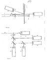
- Fig.la is a schematic side view and Fig.lb a schematic top view of a flow cytometer in accordance with the invention and Fig.2a and Fig.2b are graphical presentations of the results of the measurements obtained by plotting the chlorophyll fluorescence against the forward light scattering and the perpendicular light scattering respectively.
- Fig.la which is a side view of a flow cytometer of the invention, shows the following parts in cooperative display : a sample injection tube 1, which avoids turbulence, and wherein an orientation of the particles is obtained, positioned within a cuvette 2, in which a sheath fluid is introduced to provide a liquid jet 3, comprising the sheath fluid in which the particles are suspended.Onto this liquid jet 3 are projected two laser beams at different levels, i.e. an argon laser 4, comprising all lines, which is first passed through a prism 5 and subsequently through a.lens 6, before hitting the suspended particles.
- an argon laser 4 comprising all lines
- an argon laser 7, tuned to u.v.light, is passed through a lens 6 before hitting the suspended particles.
- a laser stop 8 is positioned in line at a point beyond the liquid jet.
- incident light is reflected by the suspended particles, suspended in the liquid jet.
- the light scattered in forward direction is detected by a forward light scatter detector 10 after the light has gone through a 515 nm band pass filter 9.
- the light scattered in backward direction is detected by a backward light scatter detector 12 after the light has gone through a 515 nm band pass filter 11.
- Fig.la provides a side view of the flow cytometer according to the invention
- Fig.lb provides a top view, showing the path of the perpendicular light scattering.Laser lights of argon ion lasers 4 and 7 hit liquid jet 3 and undeflected laser light is caught by laser stop 8.
- Perpendicularly scattered light passes through a lens 13 before hitting a 10% mirror 14, which reflects 10% of the incident light.
- the reflected light is detected by perpendicular light scatter detector 16 after the light has gone through a 515 nm band pass filter 15.
- the light going through mirror 14 hits a dichroic mirror 17, which reflects light with a wavelength of 550 nm and shorter, whereas the higher wavelengths pass.
- the passing light is detected by a chlorophyll fluorescence detector 19 after passing a filter 18, cutting at 620 nm.
- the reflected light hits in turn another dichroic mirror 20, which again reflects the shorter wavelengths of 450 nm and less, while passing the longer ones of 450 nm and more.
- the passing light is detected by a yellow fluorescence detector 22 after having passed through a yellow band pass filter 21.
- the reflected light is detected by a DN A fluorescence detector 24 after having passed a blue band pass filter 23.Thus this three dimensional lay-out of the flow cytometer according to the invention makes it possible to obtain detailed information about the particles present in the liquid jet in a very short time.
- Fig.2a and Fig.2b are graphs of chlorophyll fluorescence plotted against forward light scattering and perpendicular light scattering respectively, for which the data were obtained by the method according to the invention, using a previously prepared mixture of seven algae species.For identification purposes the data were initially taken for each algae species separately.These graphs show the effectiveness of the analysis by the method according to the invention.
- the seven algae species used were:
- the lens was the objective of a microscope with magnification lOx with long range, but any other lens of focussing mirror may be used instead.
- the prism may be replaced by a grating that may be holographic.
Landscapes
- Chemical & Material Sciences (AREA)
- Health & Medical Sciences (AREA)
- Life Sciences & Earth Sciences (AREA)
- Bioinformatics & Cheminformatics (AREA)
- Engineering & Computer Science (AREA)
- Analytical Chemistry (AREA)
- Biochemistry (AREA)
- General Health & Medical Sciences (AREA)
- Organic Chemistry (AREA)
- Zoology (AREA)
- Wood Science & Technology (AREA)
- Biotechnology (AREA)
- Dispersion Chemistry (AREA)
- Biomedical Technology (AREA)
- Physics & Mathematics (AREA)
- Microbiology (AREA)
- Sustainable Development (AREA)
- Pathology (AREA)
- Immunology (AREA)
- General Engineering & Computer Science (AREA)
- Genetics & Genomics (AREA)
- General Physics & Mathematics (AREA)
- Investigating, Analyzing Materials By Fluorescence Or Luminescence (AREA)
- Investigating Or Analysing Biological Materials (AREA)
Applications Claiming Priority (2)
| Application Number | Priority Date | Filing Date | Title |
|---|---|---|---|
| GB8109068 | 1981-03-23 | ||
| GB8109068A GB2097144A (en) | 1981-03-23 | 1981-03-23 | New method for identifying algae in water samples and apparatus for use in that method |
Publications (1)
| Publication Number | Publication Date |
|---|---|
| EP0061809A1 true EP0061809A1 (de) | 1982-10-06 |
Family
ID=10520587
Family Applications (1)
| Application Number | Title | Priority Date | Filing Date |
|---|---|---|---|
| EP82200338A Withdrawn EP0061809A1 (de) | 1981-03-23 | 1982-03-18 | Verfahren zur Identifizierung von Algen in Wasserproben und Vorrichtung zur Ausführung dieses Verfahrens |
Country Status (3)
| Country | Link |
|---|---|
| US (1) | US4500641A (de) |
| EP (1) | EP0061809A1 (de) |
| GB (1) | GB2097144A (de) |
Cited By (10)
| Publication number | Priority date | Publication date | Assignee | Title |
|---|---|---|---|---|
| EP0200851A3 (en) * | 1985-05-10 | 1988-05-04 | Becton, Dickinson And Company | Scatter/fluorescence beam splitter in a flow cytometry apparatus |
| EP0243300A3 (de) * | 1986-03-21 | 1989-11-23 | Ciba-Geigy Ag | Nachweis von Pilzen |
| EP0590775A1 (de) * | 1992-09-01 | 1994-04-06 | Becton, Dickinson and Company | Verfahren und Vorrichtung zum Nachweis von Mikroorganismen |
| DE4332163A1 (de) * | 1993-09-22 | 1995-03-23 | Kolibri Umweltanalytik Und On | Verfahren und Gerät zur Schadstoffanalyse von Gewässerproben |
| US8634077B2 (en) | 2008-10-01 | 2014-01-21 | East Carolina University | Methods and systems for optically characterizing a turbid material using a structured incident beam |
| GB2504981A (en) * | 2012-08-16 | 2014-02-19 | Natural Enviromental Res Council | Sensing apparatus and method for measuring algal growth |
| US9013692B2 (en) | 2008-06-12 | 2015-04-21 | East Carolina University | Flow cytometer apparatus for three dimensional difraction imaging and related methods |
| CN107228843A (zh) * | 2017-05-08 | 2017-10-03 | 绍兴市水环境科学研究院有限公司 | 一种应用于荧光法叶绿素a在线监测仪的校准方法 |
| CN109295185A (zh) * | 2018-09-05 | 2019-02-01 | 暨南大学 | 一种适用于单细胞真核藻类基因组大小的测定方法 |
| CN109810873A (zh) * | 2019-02-20 | 2019-05-28 | 暨南大学 | 一种不同粒径浮游甲藻分离器 |
Families Citing this family (31)
| Publication number | Priority date | Publication date | Assignee | Title |
|---|---|---|---|---|
| FR2566543B1 (fr) * | 1984-06-20 | 1988-02-26 | Commissariat Energie Atomique | Dispositif optique a rendement de collection eleve et cytofluorimetre en faisant application |
| US4622298A (en) * | 1984-08-09 | 1986-11-11 | Becton, Dickinson And Company | Detection and quantitation of microorganisms, leukocytes and squamous epithelial cells in urine |
| AU587534B2 (en) * | 1986-03-14 | 1989-08-17 | Luminis Pty Limited | Improvements in measurement of fluorescence |
| US4804850A (en) * | 1986-03-14 | 1989-02-14 | Luminis Pty. Limited | Measurement of fluorescence |
| SE454358B (sv) * | 1986-08-05 | 1988-04-25 | Trion Forskning & Utveckling | Sett att overvaka fermentering jemte anvendning av settet vid styrning av fermenteringsbetingelser |
| JPS63196854A (ja) * | 1987-02-10 | 1988-08-15 | Toa Medical Electronics Co Ltd | リンパ球亜群の測定方法およびその装置 |
| JPH02168160A (ja) * | 1988-12-22 | 1990-06-28 | Omron Tateisi Electron Co | 細胞選別装置 |
| NO930980L (no) * | 1993-03-18 | 1994-09-19 | Flowtech As | Optisk konfigurasjon for væskeströmcytofotometer |
| US5483469A (en) * | 1993-08-02 | 1996-01-09 | The Regents Of The University Of California | Multiple sort flow cytometer |
| CA2216645A1 (en) * | 1995-04-25 | 1996-11-21 | Irori | Remotely programmable matrices with memories and uses thereof |
| US5798222A (en) * | 1995-07-17 | 1998-08-25 | Guava Technologies, Inc. | Apparatus for monitoring substances in organisms |
| JP2973887B2 (ja) * | 1995-08-31 | 1999-11-08 | 株式会社島津製作所 | 核酸分子の分析方法及び装置 |
| US5818582A (en) * | 1996-09-19 | 1998-10-06 | Ciencia, Inc. | Apparatus and method for phase fluorometry |
| IES81138B2 (en) * | 1997-11-10 | 2000-04-05 | Jeacle Limited | Photometric analysis of water suspensions |
| JPH11295208A (ja) * | 1998-04-13 | 1999-10-29 | Sysmex Corp | 粒子撮像装置 |
| US6052187A (en) * | 1998-08-31 | 2000-04-18 | Containerless Research, Inc. | Hyperspectral polarization profiler for remote sensing |
| AU3081801A (en) | 1999-11-15 | 2001-05-30 | Chemclean Corporation | Bio-burden visualization system |
| US6649417B2 (en) | 2000-08-21 | 2003-11-18 | Ut-Battelle, Llc | Tissue-based standoff biosensors for detecting chemical warfare agents |
| US6569384B2 (en) | 2000-08-21 | 2003-05-27 | Ut-Battelle, Llc | Tissue-based water quality biosensors for detecting chemical warfare agents |
| US20040260157A1 (en) * | 2003-06-20 | 2004-12-23 | Montes Miguel A. | Method for automated screening of cervical/endocervical malignant and premalignant epithelial lesions using flow cytometry with HPV DNA fluorescent in-situ hybridization ( FISH) technology |
| US7258836B2 (en) * | 2003-10-20 | 2007-08-21 | Ut-Battelle, Llc | Freeze resistant buoy system |
| US7591979B2 (en) | 2003-10-20 | 2009-09-22 | Ut-Battelle, Llc | Enhanced monitor system for water protection |
| FI20115999A0 (fi) | 2011-10-11 | 2011-10-11 | Teknologian Tutkimuskeskus Vtt Oy | Optinen mittaus |
| US20140291550A1 (en) * | 2013-04-01 | 2014-10-02 | National Institute Of Standards And Technology | Flow cytometer systems and associated methods |
| CN106680186B (zh) * | 2016-11-02 | 2019-03-01 | 北京信息科技大学 | 一种流式细胞仪多类型散射光探测系统 |
| JP2018093758A (ja) * | 2016-12-09 | 2018-06-21 | 株式会社サタケ | 微生物の検査方法及びその装置 |
| CN108226112B (zh) * | 2017-12-27 | 2024-07-09 | 上海众毅工业控制技术有限公司 | 水体叶绿素a含量的测量装置及浊度补偿方法 |
| EP3821227B1 (de) * | 2018-07-10 | 2024-03-13 | Gerrit Jan Van Den Engh | System, vorrichtung und verfahren zur ausseraxialen beleuchtung in der durchflusszytometrie |
| DE102019131650A1 (de) * | 2019-11-22 | 2021-05-27 | Fraunhofer-Gesellschaft zur Förderung der angewandten Forschung e.V. | Verfahren zum Ermitteln und Optimieren des Gehalts von wenigstens einem Pflanzeninhaltsstoff von wenigstens einem Teil einer Pflanze |
| JP7785503B2 (ja) * | 2021-10-27 | 2025-12-15 | リオン株式会社 | 植物プランクトン分別システム |
| CN116519656B (zh) * | 2023-06-25 | 2023-09-12 | 广州盛安医学检验有限公司 | 一种荧光扫描装置 |
Citations (2)
| Publication number | Priority date | Publication date | Assignee | Title |
|---|---|---|---|---|
| US3785735A (en) * | 1972-01-19 | 1974-01-15 | Bio Physics Systems Inc | Photoanalysis method |
| US3916197A (en) * | 1973-11-28 | 1975-10-28 | Particle Technology Inc | Method and apparatus for classifying biological cells |
Family Cites Families (6)
| Publication number | Priority date | Publication date | Assignee | Title |
|---|---|---|---|---|
| GB1149064A (en) * | 1966-08-01 | 1969-04-16 | Int Research & Dev Co Ltd | Improvements in and relating to means for detecting malignant cells in human and animal tissue |
| US3928140A (en) * | 1974-05-10 | 1975-12-23 | Philip J Wyatt | Apparatus and process for testing microparticle response to its environment |
| US4061543A (en) * | 1976-03-19 | 1977-12-06 | General Electric Company | Bioassay for drugs and antibiotics |
| US4043669A (en) * | 1976-05-28 | 1977-08-23 | The United States Of America As Represented By The Secretary Of The Air Force | Light scattering test apparatus |
| US4203670A (en) * | 1977-04-21 | 1980-05-20 | Bromberg Nathan S | System and method of fluorescence polarimetry |
| SU734270A1 (ru) * | 1978-01-12 | 1980-05-15 | Ленинградский Институт Авиационного Приборостроения | Устройство дл флуоресцентного исследовани биологических объектов в водной пробе |
-
1981
- 1981-03-23 GB GB8109068A patent/GB2097144A/en not_active Withdrawn
-
1982
- 1982-03-18 EP EP82200338A patent/EP0061809A1/de not_active Withdrawn
- 1982-03-22 US US06/360,430 patent/US4500641A/en not_active Expired - Lifetime
Patent Citations (2)
| Publication number | Priority date | Publication date | Assignee | Title |
|---|---|---|---|---|
| US3785735A (en) * | 1972-01-19 | 1974-01-15 | Bio Physics Systems Inc | Photoanalysis method |
| US3916197A (en) * | 1973-11-28 | 1975-10-28 | Particle Technology Inc | Method and apparatus for classifying biological cells |
Non-Patent Citations (3)
| Title |
|---|
| IEEE TRANSACTION NUCLEAR SCIENCE, vol. 21, no. 1, February 1974, pages 714-717, New York (USA); * |
| THE JOURNAL OF HISTOCHEMISTRY AND CYTOCHEMISTRY, vol. 24, no. 1, 1976, pages 388-395, (USA); * |
| THE JOURNAL OF HISTOCHEMISTRY AND CYTOCHEMISTRY, vol. 25, no. 7, 1977, pages 836-844, (USA); * |
Cited By (12)
| Publication number | Priority date | Publication date | Assignee | Title |
|---|---|---|---|---|
| EP0200851A3 (en) * | 1985-05-10 | 1988-05-04 | Becton, Dickinson And Company | Scatter/fluorescence beam splitter in a flow cytometry apparatus |
| EP0243300A3 (de) * | 1986-03-21 | 1989-11-23 | Ciba-Geigy Ag | Nachweis von Pilzen |
| EP0590775A1 (de) * | 1992-09-01 | 1994-04-06 | Becton, Dickinson and Company | Verfahren und Vorrichtung zum Nachweis von Mikroorganismen |
| US5432061A (en) * | 1992-09-01 | 1995-07-11 | Becton Dickinson And Company | Method and apparatus for detecting microorganisms |
| DE4332163A1 (de) * | 1993-09-22 | 1995-03-23 | Kolibri Umweltanalytik Und On | Verfahren und Gerät zur Schadstoffanalyse von Gewässerproben |
| US9013692B2 (en) | 2008-06-12 | 2015-04-21 | East Carolina University | Flow cytometer apparatus for three dimensional difraction imaging and related methods |
| US8634077B2 (en) | 2008-10-01 | 2014-01-21 | East Carolina University | Methods and systems for optically characterizing a turbid material using a structured incident beam |
| GB2504981A (en) * | 2012-08-16 | 2014-02-19 | Natural Enviromental Res Council | Sensing apparatus and method for measuring algal growth |
| CN107228843A (zh) * | 2017-05-08 | 2017-10-03 | 绍兴市水环境科学研究院有限公司 | 一种应用于荧光法叶绿素a在线监测仪的校准方法 |
| CN109295185A (zh) * | 2018-09-05 | 2019-02-01 | 暨南大学 | 一种适用于单细胞真核藻类基因组大小的测定方法 |
| CN109295185B (zh) * | 2018-09-05 | 2022-03-22 | 暨南大学 | 一种适用于单细胞真核藻类基因组大小的测定方法 |
| CN109810873A (zh) * | 2019-02-20 | 2019-05-28 | 暨南大学 | 一种不同粒径浮游甲藻分离器 |
Also Published As
| Publication number | Publication date |
|---|---|
| GB2097144A (en) | 1982-10-27 |
| US4500641A (en) | 1985-02-19 |
Similar Documents
| Publication | Publication Date | Title |
|---|---|---|
| US4500641A (en) | Flow cytometer for identifying algae by chlorophyll fluorescence | |
| KR100849948B1 (ko) | X-염색체 또는 y-염색체를 가진 고순도 정자 개체군 | |
| US5552885A (en) | Measuring chamber for flow cytometer | |
| Dean et al. | High resolution dual laser flow cytometry. | |
| EP0177718B1 (de) | Verfahren und Vorrichtung zur Sortierung von mikroskopischen Partikeln | |
| Hercher et al. | Detection and discrimination of individual viruses by flow cytometry. | |
| Olson et al. | An inexpensive flow cytometer for the analysis of fluorescence signals in phytoplankton: chlorophyll and DNA distributions | |
| EP0229815B1 (de) | Messanordnung der lichtzerstreuung in biologischen zellen bei flusscytophotometern | |
| CA2992239C (en) | Cell analysis apparatus and methods | |
| EP0257559A2 (de) | Mehrfarbige Fluoreszenzanalyse mit Anregung einzelner Wellenlänge | |
| JPS5942885A (ja) | 生細胞選別方法及び生細胞選別装置 | |
| JPH07301628A (ja) | 容量測定細管血球計算のための装置及び方法 | |
| Steen | A microscope-based flow cytophotometer | |
| Van Dilla et al. | High-speed cell analysis and sorting with flow systems: biological applications and new approaches | |
| ATE167572T1 (de) | Flüssigkeitsverschmutzungfühler | |
| DE102017207262B4 (de) | Verfahren und Vorrichtung zur Bereitstellung einer eine gewünschte Zielprotein-Expression aufweisenden Zelllinie | |
| Mullaney et al. | Laser flow microphotometers for rapid analysis and sorting of individual mammalian cells | |
| Tanke et al. | Selection of defined cell types by flow-cytometric cell sorting | |
| Balfoort et al. | Flow cytometry: instrumentation and application in phytoplankton research | |
| Dean | Helpful hints in flow cytometry and sorting | |
| Steen | Flow cytometry instrumentation | |
| Boye et al. | The physical and biological basis for flow cytometry of Escherichia coli | |
| Pinkel | Flow sorting of cells and organelles | |
| van den Engh et al. | Flow cytometry in experimental hematology | |
| Salzman | Flow cytometry: the use of lasers for rapid analysis and separation of single biological cells |
Legal Events
| Date | Code | Title | Description |
|---|---|---|---|
| PUAI | Public reference made under article 153(3) epc to a published international application that has entered the european phase |
Free format text: ORIGINAL CODE: 0009012 |
|
| AK | Designated contracting states |
Designated state(s): AT BE CH DE FR GB IT LI LU NL SE |
|
| 17P | Request for examination filed |
Effective date: 19830325 |
|
| STAA | Information on the status of an ep patent application or granted ep patent |
Free format text: STATUS: THE APPLICATION IS DEEMED TO BE WITHDRAWN |
|
| 18D | Application deemed to be withdrawn |
Effective date: 19841218 |
|
| RIN1 | Information on inventor provided before grant (corrected) |
Inventor name: VAN DEN ENGH, GERRIT JAN Inventor name: VISSER, JOHANNES WILHELMUS MARIA Inventor name: TRASK, BARBARA JO |