EP0089692A2 - Alpha-Interferon GX-1 - Google Patents
Alpha-Interferon GX-1 Download PDFInfo
- Publication number
- EP0089692A2 EP0089692A2 EP83102893A EP83102893A EP0089692A2 EP 0089692 A2 EP0089692 A2 EP 0089692A2 EP 83102893 A EP83102893 A EP 83102893A EP 83102893 A EP83102893 A EP 83102893A EP 0089692 A2 EP0089692 A2 EP 0089692A2
- Authority
- EP
- European Patent Office
- Prior art keywords
- interferon
- alpha
- gene
- plasmid
- organism
- Prior art date
- Legal status (The legal status is an assumption and is not a legal conclusion. Google has not performed a legal analysis and makes no representation as to the accuracy of the status listed.)
- Granted
Links
Images

Classifications
-
- C—CHEMISTRY; METALLURGY
- C07—ORGANIC CHEMISTRY
- C07K—PEPTIDES
- C07K14/00—Peptides having more than 20 amino acids; Gastrins; Somatostatins; Melanotropins; Derivatives thereof
- C07K14/435—Peptides having more than 20 amino acids; Gastrins; Somatostatins; Melanotropins; Derivatives thereof from animals; from humans
- C07K14/52—Cytokines; Lymphokines; Interferons
- C07K14/555—Interferons [IFN]
- C07K14/56—IFN-alpha
Definitions
- the present invention relates to a cloned human gene, which specifies the biosynthesis of interferon. More particularly, the invention relates to a cloned human gene which specifies the biosynthesis of alpha-interferon Gx-1; to a plasmid containing such gene; to a microorganism transformed with such a plasmid; and to a polypeptide designated alpha-interferon Gx-1.
- interferon describes a family of animal proteins which possess antiviral and other potentially useful activities. Interferon is produced in vivo in minute amounts, in response to viral infections. Relatively small amounts of human interferons for research and clinical studies have been recovered from tissue cultures of human cells, which have been induced to make interferon by viruses or other inducing agents. These techniques are quite expensive, and the amounts of interferon which can be so produced are limited. Accordingly, there has been considerable interest in developing genetically engineered microorganisms, that are capable of efficiently producing interferon. The cloning of certain human interferon genes and the bacterial expression of interferon have been reported. Nagata, S. et al.
- Interferons are presently classified into the following three categories: leukocyte or alpha-interferon, fibroblast or beta-interferon, and immune or gamma- interferon.
- Current scientific evidence suggests that there are perhaps, ten to fifteen distinct alpha-interferons and only one form of beta-interferon.
- alpha-interferon genes have been cloned, and their nucleotide sequences published. Goedell, et al. Nature, 290, supra.
- alpha-interferon G the full-length alpha-interferon gene designated alpha-interferon G by prior investigators has not been reported, and only a portion of its nucleotide sequence has been disclosed. The bacterial production of the intact human alpha-interferon G protein has not been reported.
- alpha-interferon Gx-1 a novel human alpha-interferon gene, which is designated herein as alpha-interferon Gx-1, has been cloned, and characterized, and bacterial expression of the gene is described.
- the nucleotide sequence of the full length alpha-interferon Gx-1 gene and the amino acid sequence of the polypeptide specified by that gene are also reported herein.
- a portion of the nucleotide sequence reported here coresponds with the partial nucleotide sequence previously reported for alpha-interferon G, and it is therefore possible that the gene portion previously obtained and sequenced was a fragment of the gene here designated alpha-interferon Gx-l.
- the procedures leading to an alpha-interferon Gx-l-producing microorganism can be divided into the following six major stages, each of which is described more fully herein: (1) in vitro induction of human leukocytes to produce alpha-interferon Gx-1 messenger RNA (mRNA), (2) recovery and isolation of alpha-interferon Gx-1 mRNA, (3) in vitro synthesis of complementary DNA (cDNA), using the alpha-interferon Gx-1 mRNA as a template, (4) insertion of the cDNA into a suitable cloning vector and transformation of microbial cells with that cloning vector, (5) selection of microbial clones containing the alpha-interferon-Gx-1 gene, and (6) insertion of the cloned gene into a suitable expression vector and transformation of a suitable host microorganism with that expression vector.
- mRNA messenger RNA
- cDNA complementary DNA
- Alpha-interferons are produced by virus-treated leukocytes, and, although genes which specify alpha-interferons are present in the chromosomes of every cell in the body, for reasons discussed below, genes associated with alpha-interferons are most readily obtained from such virus-treated leukocytes.
- Eukaryotic genes are contained in the chromosomal DNA of the cell nuclei. This chromosomal DNA exists in a compact nucleoprotein complex called chromatin. Isolating a particular gene from eucaryotic chromosmoal DNA is a tedious and often unfeasible approach.
- messenger RNA mRNA
- mRNA messenger RNA
- the mRNA usually provides the desired genetic information in its most accessible form.
- Alpha-interferon mRNA may be recovered in useful quantities from leukocytes which have been treated with a virus or other inducing .agent.
- a virus or other inducing .agent Generally, the procedure described by Contell, K., et al., In Vitro, Waymouth E d., pp. 35-38, The Tissue Culture Association, Rockville, .MD (1974), has been used to induce leukocytes to produce alpha-interferon mRNA. This procedure involves suspending .red cell-free leukocytes (e.g., obtained by fractionating human blood) in a suitable nutrient medium and infecting with a suitable inducing agent, preferably a virus, such as Newcastle Disease Virus and incubating until sufficient interferon activity is obtained.
- a suitable inducing agent preferably a virus, such as Newcastle Disease Virus
- Interferon activity may be determined by a viral inhibition test, such as that described by Rubinstein, Familetti, Pestka, J. Virol, 37," 755-758 (1981). Induced leukocytes are advantageously washed and frozen prior to recovery of the mRNA.
- the alpha-interferon Gx-I mRNA produced by the induced cells is complementary to one of the two strands of the alpha-interferon Gx-1 gene, and may be employed as a template for the synthesis of complementary DNA (cDNA) as hereinafter described.
- cDNA complementary DNA
- RNA is inherently less stable than DNA, and is particularly subject to degradation by ribonucleases that are present in relatively high concentrations in human leukocytes. Therefore, the mRNA recovery procedures generally employ means for rapidly inactivating any ribonucleases which are present.
- RNA recovery of total RNA is initiated by disrupting the cells in the presence of.a ribonuclease- inactivating substance.
- Disruption of the cells may be accomplished by subjecting the cells to a lysing reagent, freezing/thawing, or mechanical disruption; preferably a combination thereof.
- a mixture of guanidine thiocyanate and a reducing agent, such as mercaptoethanol, has been found to function effectively as a lysing agent and a ribonuclease inactivator.
- the solid cell debris is removed, e.g. by centrifugation, and the RNA is precipitated from the resulting clarified solution.
- Precipitation is effected by known techniques, such as adding a water-immiscible alcohol, e.g. ethanol to the solution, in a precipitating amount.
- the solution is advantageously maintained at a low temperature, e.g., less than about 0°C during these procedures, to facilitate RN A precipitation.
- the alpha-interferon Gx-1 mRNA may be isolated from the precipitated RNA by any of several known techniques, or combinations thereof, e.g., see Adams, R.L.P., et al., Davidson's The Biochemistry of the Nucleic Acids, 8th Ed., pp. 52-58, Academic Press, Inc., N.Y. (1976). Density gradient centrifugation using a cesium chloride gradient, followed by phenol extraction can be used as a preliminary isolation technique. Glisin, et al., Biochemistry, 13, 2633 (1974).
- Affinity chromatography can be used to further purify alpha-interferon mRNA.
- Alpha-interferon mRNA is polyadenylated; therefore, it can readily be separated from non-adenylated RNA by chromatography on an oligo (dT)-cellulose column.
- oligo (dT)-cellulose column Green, M., et al., Arch. Biochem. Biophys., 172, 74 (1976).
- the mRNA can be fractionated by centrifugation through a sucrose gradient.
- Alpha-Interferon mRNA, migrating as about a 12S species, may be isolated by this procedure.
- Cell-free translation of the final purified mRNA (and intermediate fractions .if desired) can be employed to confirm that alpha-interferon mRNA has been obtained.
- a number of cell-free translational systems have been devised, such as wheat germ extract (Martial, J. et al., Proc. Nat. Acad. Sci. USA, 74, 1816 (1977), an mRNA-dependent reticulocyte lysate (Pelham, H.R.B., et al., Eur. J. Biochem., 67, 247 (1976), and oocytes from Xenopus laevis (Sloma, A., McCandliss, R. and Peska, S., Methods in Enzymology, Vol. 70 (1981). Translation of the recovered and isolated alpha-interferon G mRNA is preferably conducted in the X.laevis oocyte system.
- Translation in this system is accomplished by suspending oocytes obtained from healthy frogs in a suitable incubation medium, e.g. see Cavalieri, et al., Proc. Natl. Acad. Sci. USA., 74, 3287, (1977).
- a sterile water solution of the mRNA is injected into about ten oocytes using a micromanipulation device.
- the injected oocytes are incubated and then analyzed for interferon activity.
- the oocytes are analyzed by homogenizing them in their incubation medium, and centrifuging to remove the insoluble membranes, proteins and the like.
- the mRNA fractions having the highest activity by the X.laevis oocyte test can be combined to provide a template for cDNA synthesis.
- This procedure involves enzymatically constructing double-stranded DNA, which has a nucleotide base pair sequence identical to the functional sequence of the original chromosomal gene.
- the cDNA does not contain any noninformational segments (introns) which might be present on the eukaryotic gene, and thus can ultimately be transcribed and translated in prokaryotic systems.
- Synthesis of cDNA employs avian myeloblastosis virus reverse transcriptase. This enzyme catalyzes the synthesis of a single strand of DNA from deoxynucleoside triphosphates on the mRNA template. Kacian, D.L.,'et al., Proc. Nat'1 Acad. Sci. USA, 73, 2191 (1976).
- the poly r(A) tail of mRNA permits oligo (dT) (of about 12-18 nucleotides) to be used as a primer for cDNA synthesis.
- oligo (dT) of about 12-18 nucleotides
- a 32 P-containing deoxynucleoside triphosphate such as [a- 32 P]dCTP may be used advantageously for this purpose.
- the cDNA synthesis is generally conducted by combining the mRNA, the deoxynucleoside triphosphates, the oligo (dT) and the reverse transcriptase in a properly buffered solution.
- the solution also preferably contains small amounts of actinomycin D and dithiothreitol to promote full length synthesis. Kacian, D.L., et al. supra. This solution is incubated at an elevated temperature, e.g., about 40-50°C, for a time sufficient to allow formation of the cDNA copy, e.g. about 5-20 minutes.
- the conditions of the reaction are essentially as described by Kacian, D.L., et al., supra. After incubation, ethylenediaminetetraacetic acid is added to the solution, and the solution extracted with phenol: chloroform (1:1 by vol.). The aqueous phase is advantageously purified by gel filtration chromatography, .and the cDNA-mRNA complex in the eluate is precipitated with alcohol.
- the mRNA can be selectively hydrolyzed in the presence of the cDNA with dilute sodium hydroxide (about 0.1 M) at an elevated temperature, e.g., about 60-80°C for about 15-30 minutes. Neutralization of the alkaline solution and alcohol precipitation yields a single-stranded cDNA copy.
- dilute sodium hydroxide about 0.1 M
- the single-stranded cDNA copy has been shown to have a 5'-poly (dT) tail, and to have a 3' terminal hairpin structure, which provides a short segment of duplex DNA.
- This 3' hairpin structure can act as a primer for the synthesis of a complementary DNA strand. Synthesis of this complementary strand is conducted under essentially the same conditions as the synthesis of the cDNA copy, except that the Klenow fragment of DNA polymerase I (Klenow, H., et al., Eur. J. Biochem., 22, 371 (1971)) is substituted for reverse transcriptase.
- the duplex cDNA recovered by this procedure has a 3' loop, resulting from the 3' hairpin structure of the single-stranded cDNA copy.
- This 3' loop can be cleaved by digestion with the enzyme, Sl nuclease, using essentially the procedure of Ullrich, A., et al., supra.
- the Sl nuclease digest may be extracted with phenol-chloroform, and the resulting cDNA precipitated from the aqueous phase with alcohol.
- the intact double-stranded DNA, corresponding to an alpha-interferon gene may be isolated by.polyacrylamide gel electrophoresis, using essentially the procedure of Maniatis, et al., Biochemistry, 14, 3787 (1975). After staining the gel, e.g., with ethidium bromide to visualize restriction enzyme digests incorporated as molecular weight markers, a photographic film is exposed with the gel to locate the radioactively labelled double-stranded cDNA gene. The region of the gel containing DNA molecules between 500 and 1300 base pairs long is removed, and the DNA is eluted electrophoretically, essentially by the method of Smith, H.O., Methods of Enzymology, 65, 371 (1980).
- DNA corresponding in size-to the alpha-interferon Gx-1 gene (ca. 900 base pairs) is recovered by phenol-chloroform extraction of the electrophoretic eluate, followed by alcohol precipitation of the cDNA from the aqeuous phase.
- the double-stranded cDNA gene prepared as described above is generally inserted into a suitable cloning vector, which is used for transforming appropriate host cells.
- suitable cloning vectors include various plasmids and phages, and plasmids are generally preferred.
- the criteria for selecting a cloning vector include its size, its capability for replicating in the host cells, the presence of selectable genes, and the presence of'a site for insertion of the gene. With respect to its size, the vector is advantageously relatively small, to permit large gene insertions, and so as not to divert large amounts of cellular nutrients and energy to the production of unwanted macromolecules.
- the vector also includes an'intact replicon which remains functional after insertion of the gene.
- This replicon preferably directs the desired mode of replication of the plasmid, i.e., mult-iple copies or a single copy per cell, or a controllable number of copies per cell.
- Genes specifying one or more phenotypic properties, preferably antibiotic resistance, facilitate selection of transformants.
- the insertion site is advantageously a unique restriction site for a restriction endonuclease.
- a cloning vector meeting all of these criteria is the plasmid pB R 322.
- This plasmid is small (about 2.8 x 10 6 daltons), carries genes for ampicillin (amp) and tetracycline (tet) resistance, and is subject to relaxed replication in E.coli.
- the plasmid also has a restriction site for the endonuclease, PstI, which occurs within the amp gene.
- PstI the endonuclease
- the cDNA can be conveniently inserted into this plasmid by a homopolymeric tailing technique. Nelson, T., et al., Methods of Enzymology, 68, 41 (1980).
- Homopolymer tails e.g., poly-dC
- Homopolymer tails are'added to the 3'-hydroxyls of the interferon double-stranded cDNA gene, by reaction with the appropriate deoxynucleoside triphosphate, e.g., dCTP, in the presence of terminal deoxynucleotidyl transferase (Chang, L.M.S., et al., J. Biol. Chem., 246, 909 (1971)).
- the plasmid is opened by digestion with the appropriate endonuclease, and complementary homopolymer tails, e.g., poly dG, are added to the 3'-hydroxyls of the opened plasmid, using the identical homopolymer tailing technique e.g., using dGTP.
- the tailing reactions may be monitored, by employing radioactively labelled deoxynucleotide triphosphates e.g., [ 3 H]dCTP and C 3 H] dGTP, in the reactions.
- the reactions are conducted to provide tails about 10-20 nucleotides long.
- the tailed cDNA and plasmid are recovered, e.g., by phenol extraction followed by alcohol precipitation.
- the two . "tailed" DNA species are annealed by incubating a buffered solution of equimolar concentrations of the two species, to yield a recombinant plasmid containing the alpha-interferon Gx-1 gene.
- a suitable amp S , tet s strain of E.coli may be transformed with the recombinant plasmid, using essentially the method of Lederberg, J. Bacteriology, 119, 1072 (1974). Transformants are typically grown on a standard L-broth, containing about 50 ug/ml of tetracyline. Samples of colonies growing on the tetracycline-containing medium are then transferred to a second medium containing about 50 ug/ml of ampicillin. Because the pBR322 plasmid imparts tetracycline resistance to the cells, colonies growing on the tetracycline-containing medium must contain that plasmid. On the other hand, the ampicillin resistance of the pBR322 plasmid is destroyed by insertion of the gene, therefore, only tet R , amps colonies are selected for further analysis.
- a radioactively labelled DNA probe may advantageously be employed. Grunstein, M, et al., Proc. Nat'1. Acad. Sci. USA, 72, 3961-3965 (1975).
- a particularly preferred DNA probe is the tridecadeoxyribonucleotide probe described in copending U.S. patent application Serial No. 286,351 (Attorney Docket Number 1182-119A), herein incorporated by reference.
- That probe contains a thirteen-nucleotide sequence, which is complementary to a sequence common to known human'leukocyte and human fibroblast interferon genes.
- DNA from each colony (or from groups of colonies) is fixed to discrete zones of a nitrocellulose filter and denatured. A solution of the probe is applied thereto under hybridizing conditions. Unhybridized probe is washed from the filter, and colonies containing DNA to which the probe hybridized are identified by autoradiography.
- Positive clones may be cultivated on suitable growth media to obtain ample quantities of cells from which to extract the plasmid DNA.
- the plasmid DNA is-extracted, using conventional techniques, such as disruption of the cells, followed by phenol extraction, and alcohol precipitation.
- The'DNA may be separated, e.g. by electrophoresis or sucrose gradient sedimentation. Plasmid DNA containing inserts about 900 base pairs is selected for further characterization.
- the cloned gene may conveniently be excised from the plasmid DNA by digestion with the appropriate endonuclease.
- the excised gene is then characterized by restriction mapping and sequencing analysis.
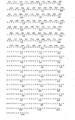
- the restriction map of the excised alpha-interferon Gx-1 gene of the present invention is shown in Fig. 1 of the drawings.
- the isolated gene consists of 1032 base pairs.
- the gene has noncoding regions at the 5' end (base pairs 1-85). and at the 3' end (base pairs 632-1032).
- the 5' end of the coding region (86-133 base pairs) includes a leader, and the mature alpha-interferon Gx-1 protein is specified by the region from base pair number 134 to base pair number 631.
- the nucleotide sequence of the gene can vary substantially. For example, portions or all of the gene could be chemically synthesized to yield DNA having a different nucleotide sequence than that shown in Figure 2, yet the amino acid sequence would be preserved, provided that the proper codon-amino acid assignments were observed. Having established the nucleotide sequence of the alpha-interferon Gx-1 gene and the amino acid sequence of the protein, the gene of present invention is not limited to a particular nucleotide sequence, but includes all variations thereof as permitted by the genetic code.
- a culture of E.coli cells containing the cloned alpha-interferon Gx-1 gene was analyzed for interferon activity by the viral inhibition method of Rubinstein, et al. supra, the cells were found to produce small amounts of interferon activity.
- This cell culture has been designated A3-26, and has been deposited with the American Type Culture Collection, Rockville, MD as ATCC No. 39063.
- expression vectors may be plasmids or phages; plasmids being preferred.
- an expression vector also contains properly positioned control signals which direct transcription and translation of the cloned gene. Guarante, L. et al., Cell, 20 543-553 (1980). Typically, a gene is inserted into an intact operon of a plasmid, and expression of the gene is controlled by that operon.
- lactose (lac) and tryptophan (trp) operons of E.coli have been used for this purpose. Roberts, T.M., et al., Proc. Nat'1 Acad. Sci. USA, 76, 760-764 (1979).
- E.coli as the bacterial host for recombinant DNA containing the alpha-interferon Gx-1 gene
- skilled molecular biologists will appreciate that other gram-negative bacteria, such as Pseudomonas; gram-positive bacteria, such as Bacillus; and higher unicellular organisms, such as yeasts and fungi can be employed for cloning and/or expression of the alpha-interferon Gx-1 gene.
- the starting material for the induction procedures was a residue from a plateletpheresis preparation. Each residue contained the white cells from 6 to 20 units of human blood admixed with contaminating red cells. Two residues were pooled and centrifuged in a bottle for 7 minutes at 1600 x g. Most of the red cells were removed from the bottom of the bottle with a pipet. 'The remaining volume was measured, and remaining red cells were lysed by rapid addition of ten volumes of 0.83% ammonium chloride.
- the white cells were collected by centrifugation' and suspended in 500 ml of Eagle's minimum essential medium (without phosphate buffer) supplemented with 10% heat-inactivated calf serum, 3 mg/ml of tricine and 25 ug/ml of neomycin. This gave a cell concentration of about 10 7 cells/ml.
- NDV Newcastle Disease Virus
- the cells of this aliquot were then removed by centrifugation, and the supernatant was assayed for interferon by its ability to protect human fibroblasts from cytopathic effects caused by Vesicular Stomatitis Virus.
- the titer was 25,000 IU/ml.
- the remaining 450 ml of the culture (5 hr after addition of NDV) was centrifuged.
- the supernatant was assayed for interferon.
- the titer was 6400 IU/ml.
- the cell pellet was washed with PBS (8.2 g/L sodium chloride, 0.22 g/L potassium chloride, 0.20 g/L monobasic potassium phosphate, 1.14 g/L dibasic sodium phosphate), and the washed pellet, weighing 4.9 grams, was frozen at -70°C.
- the lysis of induced cells was performed-in a fume hood.
- the cells from Example I were partially broken while kept frozen.
- Solution B The wash solution contained in 6 M ultrapure guanidine hydrochloride, 10 mM Na 2 EDTA, pH 7.0, and 10mM dithiothreitol. Because of the high purity of the guanidine hydrochloride used, no filtration of this solution was necessary, and it was stable when stored at room temperature. Again, care should be taken to avoid contact of this solution with the skin.
- the lysis of induced cells was performed in a fume hood.
- the cells from Example I were partially broken while kept frozen and placed in 20 ml of Solution A per gram (wet weight) of cells in the chamber of a high speed homogenizer.
- the homogenizer was operated at full speed for 2 minutes to lyse the cells.
- the lysate is then centrifuged for 10 minutes at 12,000 rpm in a Sorvall GSA rotor to, remove any debris present.
- the supernatant containing the RNA and other cellular components was acidified at pH 5 by addition of 0.04 volume of 1 N acetic acid, and the RNA was precipitated by adding 0.5 volume of absolute.ethanol. After mixing, the solution was kept at -20°C for at least 2 hours. Leaving the mixture at -20°C for much longer periods does not significantly increase the yield of RNA and results in the coprecipitation of larger amounts of protein, which may be difficult to redissolve in the next step.
- the precipitated RNA was collected by centrifugation for 10 min at 8,000 rpm . in a Sorvall GSA rotor.
- the first step in purifying the interferon mRNA involved affinity chromatography on an oligo (dT)-cellulose column, using the method of Green, M., et al., Arch. Biochem. Biophys., 172, 74 (1976).
- the RNA solution (30-50 A 260 units/ml) was adjusted to 0.5 M NaCl by addition of 0.11 volume of 5 M NaCl and then applied to a.column containing 5 g of oligo (dT)-cellulose (T-3, Collaborative Research) equilibrated with 10mM Tris-HCl, pH 7.4, and 0.5 M NaCl at a flow rate of about 20 ml/hour.
- Non-bound RNA was removed from the column by washing with the same buffer until the optical density was less than 0.08 A 260 unit/ml. Poly(A) RNA was then eluted with 10mM Tris-HCl, pH 7.4. The poly(A) was recycled over the column once more to decrease contamination by ribosomal and other non-poly(A) RNA's. After the first oligo(dT)-cellulose column chromatography, the RNA preparation was assayed for interferon mRNA activity by injection into Xenopus laevis oocytes as hereinafter described.
- the interferonmRNA so obtained was enriched by sucrose gradient centrifugation.
- Linear gradients were prepared by mixing equal volumes of 5% (w/v) and 20% sucrose in 0.02 M sodium acetate, pH 5, in the chambers of a Buchler gradient maker. After formation, the gradients were equilibrated at 4°C for at least 4 hours. Prior to loading, the RNA samples were heated to 80°C for 2 min and cooled rapidly in an ice bath to reduce aggregation. The samples were layered over the gradients and were centrifuged for 20 hours at 30,000 rpm at 4°C using an SW40 rotor (Beckman).
- the gradients were collected by use of an Isco Gradient Pump modified such that a flow cell is attached to a Gilford spectrophotometer. The gradients were analyzed by pumping 50% sucrose into the bottom of the tube. Fractions were collected, ethanol precipitated, dissolved in sterile water and assayed by injection into Xenopus laevis oocytes as hereinafter described. Leukocyte interferon mRNAs migrated approximately-as 12 S species.
- Xenopus laevis were obtained from Nasco, Ft. Atkinson, Wisconsin (#LM531LQ). The oocytes obtained from these frogs have been found to yield reproducible high titers of interferon. After the animal was placed in ice water for 30 min, it was sacrificed and the oocytes were removed and immediately placed in 150 ml of the following oocyte incubation media.
- the final pH was adjusted to pH 7.6 with HC1. and the solution was sterilized by filtration.
- oocytes were obtained by gently teasing apart the oocyte sacs with a blunt probe.
- the oocytes have a distinct animal and vegetal pole.
- the yolk-of the vegetal pole has a light white-green color and the animal pole is black. Although they vary in size, most oocytes were approximately 1 mm in diameter. The largest oocytes, approximately 1.2-1.5 mm in diameter, tend to leak after injection and were not used.
- the oocytes were stored at 5°C. Oocytes are capable of giving maximum interferon titer after 4-5 days storage at 5°C.
- Interferon mRNA from Example II was dissolved in sterile water at a concentration of 0.5-1 mg/ml, and the solution was kept on ice until injected.
- Injection needles were prepared by pulling five ul microdispenser tubes (#105G, Drummond Scientific) with a vertical pipette puller (Model 700B, David Kopf Instruments, Tujunga, California). The pulled needles were sealed at the tip. The tips were then broken off with fine scissors under a dissecting microscope to form the needle with a tip of 0.005-0.01 mm in diameter.
- the microdispenser capillaries consisted of uniform bore tubing with 0.27 mm equivalent to 50 nl (5.4 mm equivalent to 1 ul).
- RNA interferon mRNA
- two ul of interferon mRNA were placed in the bottom of a 60 x 15 mm round Petri dish filled with sterile mineral oil.
- the injection needle was then clamped to the end of a Brinkman micromanipulator (#06-15-00) and attached to tubing which was connected to a hydraulic pressure system. Positive pressure fills the needle up to 2 mm from the open tip.
- the needle was then placed through the mineral oil into the solution of mRNA.
- the solution was drawn into the tube by capillary action, with the help of negative pressure applied to the system.
- the sample filled 1/2-3/4 of the injection needle.
- a small air bubble remained between the RNA solution and the hydraulic fluid (sterile H 2 0).
- a small piece of graph paper with 1-mm divisions was placed on the needle and aligned with the meniscus.
- ten oocytes were aligned against the edge of a slide taped to a square (100 x 100 mm) petri dish.
- the animal or vegetal pole can be injected without affecting the final interferon titer.
- Positive pressure was applied to the system.
- Displacement of the meniscus was approximately 0.3 mm for each oocyte injected, which is equivalent to about 50 nl injected per oocyte.
- the needle was removed from the oocyte and the remaining 9 were injected in sequence.
- About 40-50 ng of interferon mRNA were injected per oocyte. It has been previously shown that this concentration is saturating.
- the ten oocytes were placed in 0.1 ml of fresh oocyte incubation media and incubated for 18 h at 23°C in a 1.5 ml sterile polypropylene conical tube.
- the oocytes were homogenized manually in the same tube in which they were incubated. This is important because some interferon is secreted into the incubation media.
- the extract was then centrifuged for 5 minutes in an Eppendorf centrifuge. Ninety ul of supernatant was carefully removed. A lipid layer formed at the top, which is toxic to the cells in the interferon assay. Therefore, as little of the lipid was removed as possible.
- the 90 ul of supernatant were then centrifuged again for 5 min. The supernatant was then assayed for interferon activity by the method of Rubinstein, et al. (supra).
- the interferon titer of the solution was 1260 U/ml.
- All buffers and salt solutions were autoclaved.
- the other solutions were prepared with sterile glass-distilled water and were stored in sterile containers. All stock solutions were stored frozen.
- the dried material was dissolved in a 2x reaction mixture consisting of 0.1 M Tris.HCl, pH 8.3, 140 mM KC1, 20 mM MgCl 2 , 1 mM dATP, 1 mM dCTP, 1 mM dGTP, 1mM TT P , and 0.4 mM DDT.
- This solution was kept on ice.
- To this solution were added mRNA (50 ug/ml, final concentration), oligo(dT) 12-18 (25 ug/ml), actinomycin D (40 ug/ml), AMV reverse transcriptase (800 units/ml), and enough water to dilute the 2x mix to lx. After 5 min.
- reaction mixture was incubated at 46° for 10 min. Following the incubation, EDTA was added to a final concentration of 25 mM.
- the solution was extracted one time with an equal volume of phenol:chloroform (1/1; v/v) and the aqueous phase was chromatographed on a column of Sephadex G-100 (0.7 x 20 cm), equilibrated with 10 mM Tris-HC1, pH 8.0, 1 mM EDTA, 0.1 M NaCl.
- the cDNA in the excluded volume was precipitated by addition of 0.1 volume of 2.4 M sodium acetate, pH 5, and 2.5 volumes of ethanol.
- the cDNA was sedimented by centrifugation, dissolved in 0.3 ml of 0.1 M NaOH, and incubated at 70° for 20 min. The solution was neutralized with 3.0 ml of 1.0 M HC1 and precipitated with ethanol as described above. The yield of cDNA was 10-20% of the mRNA used.
- EDTA was added to 25 mM, the solution was extracted once with an equal volume of phenol:chloroform (1/1; v/v), and the aqueous phase was chromatographed on a 0.7 x 20 cm column of Sephadex G-100 equilibrated with 10 mM Tris.HCl, pH 8.0, 1 mM EDTA, and 0.1 M NaCl. The DNA in the excluded fractions was precipitated with ethanol as described above. The yield of ds DNA was 50-100% of the amount of cDNA used as template.
- the ds cDNA contained a hairpin loop.
- the single-stranded loop was removed by digestion with Aspergillus oryzae Sl nuclease, prepared by the method of Vogt, Eur. J. Biochem., 33, 192 (1973).
- the ds cDNA was dissolved in water and 0.25 volume of 5x Sl buffer was added. An appropriate amount of Sl nuclease was added and the solution was incubated 20 min at 37°. (The amount of enzyme to be added must be determined empirically for each enzyme preparation since the activity varies from one preparation to another. This is done by measuring the decrease in TCA-precipitable counts from the ds cDNA.
- the ⁇ Sl-digested ds cDNA was extracted once with phenol:chloroform and the aqueous phase was precipitated with ethanol as described above.
- the double-stranded cDNA was layered onto a 12 ml 5 to 25% linear neutral sucrose gradient (50mM Tris, pH 7.5, 1mM EDTA) and centrifuged in a Beckman SW40 rotor-for 17 hours at 38,000 rpm (5°C). One ml. fractions were collected, and fractions 5-9 (from the top) were pooled. (See Norgard, et al., J. Biol. Chem., 255, 7665-7672 (1980)).
- the solution was extracted once with phenol:chloroform (1/1; v/v) and the aqueous phase was precipitated with ethanol.
- the dC-tailed ds cDNA produced by this procedure was then ready for-insertion into a dG-tailed pBR322 vector as hereinafter described.
- Plasmid pBR322 (Bolivar, F., et al., Gene, 2, 95 (1977)) (20ug) was cut with Pst I enzyme, and approximately twenty dG residues were added to the 3' ends according to the procedure of Roychoudhury, R., et al., Nucl. Acids Res., 3, 101 (1976). The reaction was conducted in a buffer consisting of 0.14 M potassium cacodylate; 0.03 M Tris-HC1, pH 7.0; lmM CoC1 2 and 0.01 mM dithiothreitol. The concentration of plasmid DNA 3'-hydroxyl groups was 40nM and a 3000 molar excess of 3 H -dG TP was employed. The reaction was catalyzed by 21 units of terminal transferase.
- the dC-tailed double-stranded cDNA from Example IV was annealed to the dG-tailed pBR322 by mixing equimolar (470 n moles) of the two species in 0.9 ml of an annealing buffer composed of 10mM Tris-HCl, pH 7.5; 100 mM NaCl and 2.5 mM Na 2 EDTA. This mixture was incubated for 10 minutes at 70°C in a water bath. The bath was then transferred to a 37°C room to allow slow cooling overnight. The next day, the bath was allowed to cool to room temperature. The resulting recombinant plasmid was used to transform E.coli cells.
- a culture of E.coli strain HB101 was grown overnight in 10 ml of LB medium containing 0.2% glucose. The next day, 50 ml of LB + 0.2% glucose medium was inoculated with 0.5 ml of the culture, and this culture was grown at 37°C until it had an absorbance of 0.3 at 595 nm. The culture was centrifuged in a Sorvall SS34 rotor for 10 minutes at 5000 rpm, and the resulting pellet was resuspended in 2 ml of cold 0.1 M MgCl 2 . The suspension was diluted to 25 ml with cold 0.1 M MgC1 2 and centrifuged for 10 minutes at 3000 rpm.
- the cells were then resuspended in 25 ml of a solution of 0.1 M CaC1 2 and incubated for one hour on ice. This suspension was again centrifuged for ten minutes at 3000 rpm and the cells were resuspended in 2.2 ml of the same buffer. At this point, 20 ul of the annealing mixture of Example V and 200 ul of the cell suspension were mixed together and incubated on ice for 20 minutes. The cells were then heat shocked for two minutes at 42.5°C. To the cells, 2.8 ml of LB + 0.2% glucose medium was added and the mixture was incubated at 37°C to allow expression.
- Clones were screened for the presence of the alpha-interferon gene by hybridization with a synthetic radiolabeled tridecadeoxyribonucleotide probe having the following nucleotide sequence, which was known to be homologous with portions of the human beta-interferon gene and several of the known alpha-interferon genes: The synthesis and use of this probe is described in copending U.S. patent application Serial Number 286,351.
- the supernatant was carefully removed, and to it was added 150 ul of isopropanol.
- the - DNA was precipitated for 30 minutes at -20°C, centrifuged for 10 minutes at 10,000 rpm and then resuspended in 100 ul of 0.3 N NaOH, 10 mM Tris (pH 8). This solution was incubated at 70°C for 30 minutes in order to hydrolyze RNA , and was then neutralized with the addition of 10 ul of 3N HC1 prior to ethanol precipitation of the DNA.
- the precipitated DNA was then centrifuged in an Eppendorf Microfuge for 10 minutes and the pellets were dried under vacuum.
- the DNA-containing pellet was dissolved in 20 ul of H 2 ). The yield per culture was approximately 100-200 ng of DNA.
- the filters were placed in sealed plastic bags containing 10 ml of 4XSSC, 10X Denhardts buffer (0.2% bovine serum albumin, 0.2% polyvinylpyrrolidone, and 0.2% Ficoll). A preincubation step at 65°C for 2 hours was employed to decrease nonspecific binding of radioactive probes. The solution was then removed and replaced with 4 ml of fresh 4XSSC, 10X Denhardts. The 32 p-labelled probe (described in Example I) was then added (4 x 10 6 cpm, specific activity 5 x 10 8 cpm/ug). Hybridization of the probe with the DNA bound to the filters continued overnight at 15°C.
- the filters were washed 5 times with 200.ml of 4XSSC at 15°C to remove the unhybridized probe.
- the filters were then dried prior to autoradiography with Kodak RP-5 film at -80°C. Approximately thirty groups of clones showed some degree of hyrbridization.
- the DNA was prepared using the following rapid plasmid procedure: Five ml of individual clones or 10 ml of a pool of 5 clones were grown overnight in L-broth plus 50 ug/ml tetracycline. After pelleting the cells at 5000 rpm for 10 minutes, they were frozen, thawed, dissolved in 2.0 ml at 25% sucrose, 50 mM Tris pH 8, and incubated for 5 minutes at room temperature. One half of a 5 mg/ml solution of lysozyme in 10mM Tris pH 8 was added and the solution was incubated for 5 minuts.
- Substantially pure, bacterially produced human alpha-interferon Gx-1 is obtained by cultivating the A3-26 cells in large scale, under protein-expression conditions, followed by lysis of the cells and extraction and purification of the alpha-interferon Gx-1 using known techniques (e.g., see Example M of UK Patent Application GB 2 079 291 A, published January 20, 1982).
Landscapes
- Chemical & Material Sciences (AREA)
- Health & Medical Sciences (AREA)
- Life Sciences & Earth Sciences (AREA)
- Organic Chemistry (AREA)
- General Health & Medical Sciences (AREA)
- Molecular Biology (AREA)
- Biochemistry (AREA)
- Biophysics (AREA)
- Zoology (AREA)
- Genetics & Genomics (AREA)
- Medicinal Chemistry (AREA)
- Gastroenterology & Hepatology (AREA)
- Proteomics, Peptides & Aminoacids (AREA)
- Toxicology (AREA)
- Preparation Of Compounds By Using Micro-Organisms (AREA)
- Medicines That Contain Protein Lipid Enzymes And Other Medicines (AREA)
- Micro-Organisms Or Cultivation Processes Thereof (AREA)
- Saccharide Compounds (AREA)
- Peptides Or Proteins (AREA)
Priority Applications (1)
| Application Number | Priority Date | Filing Date | Title |
|---|---|---|---|
| AT83102893T ATE50597T1 (de) | 1982-03-23 | 1983-03-23 | Alpha-interferon gx-1. |
Applications Claiming Priority (2)
| Application Number | Priority Date | Filing Date | Title |
|---|---|---|---|
| US36136482A | 1982-03-23 | 1982-03-23 | |
| US361364 | 1982-03-23 |
Publications (3)
| Publication Number | Publication Date |
|---|---|
| EP0089692A2 true EP0089692A2 (de) | 1983-09-28 |
| EP0089692A3 EP0089692A3 (en) | 1984-03-28 |
| EP0089692B1 EP0089692B1 (de) | 1990-02-28 |
Family
ID=23421737
Family Applications (1)
| Application Number | Title | Priority Date | Filing Date |
|---|---|---|---|
| EP83102893A Expired - Lifetime EP0089692B1 (de) | 1982-03-23 | 1983-03-23 | Alpha-Interferon GX-1 |
Country Status (5)
| Country | Link |
|---|---|
| EP (1) | EP0089692B1 (de) |
| JP (1) | JPH0822230B2 (de) |
| AT (1) | ATE50597T1 (de) |
| CA (1) | CA1210714A (de) |
| DE (1) | DE3381246D1 (de) |
Cited By (8)
| Publication number | Priority date | Publication date | Assignee | Title |
|---|---|---|---|---|
| FR2567540A1 (fr) * | 1984-07-13 | 1986-01-17 | Inst Genetiki I Selektsii | Procede de preparation d'un interferon leucocytaire humain alpha-2 |
| EP0136694A3 (en) * | 1983-10-04 | 1986-07-16 | Takeda Chemical Industries, Ltd. | Production of monomeric human gamma-interferon |
| EP0194006A1 (de) * | 1985-02-01 | 1986-09-10 | Imperial Chemical Industries Plc | Analoge Interferon-Polypeptide, Verfahren zu deren Herstellung und diese enthaltende pharmazeutische Zusammensetzungen |
| WO1986006744A1 (en) * | 1985-05-15 | 1986-11-20 | Kabivitrum Ab | A cDNA MOLECULE CODING FOR THE EXPRESSION OF AN INTERFERON alpha TYPE POLYPEPTIDE, A BACTERIAL OR CELLULAR HOST TRANSFORMED WITH SUCH MOLECULE AND A POLYPEPTIDE SHOWING INTERFERON ACTIVITY PREPARED BY SUCH HOST |
| EP0145390A3 (de) * | 1983-11-28 | 1987-01-21 | Takeda Chemical Industries, Ltd. | Methode zur Herstellung von nicht-glykosyliertem humanen Interleukin-2 Protein |
| EP0176299A3 (en) * | 1984-09-26 | 1987-07-15 | Takeda Chemical Industries, Ltd. | Mutual separation of proteins |
| EP0154186A3 (en) * | 1984-02-10 | 1987-12-09 | J.K. And Susie L. Wadley Research Institute And Blood Bank | Isolation of a novel interferon gene and expression thereof |
| US5256769A (en) * | 1984-09-26 | 1993-10-26 | Takeda Chemical Industries, Ltd. | Mutual separation of proteins |
Families Citing this family (2)
| Publication number | Priority date | Publication date | Assignee | Title |
|---|---|---|---|---|
| JPS60248185A (ja) * | 1984-02-10 | 1985-12-07 | ワドリ、テクナラジズ、インコーパレイティド | 新規インタ−フエロン遺伝子の分離およびその表現 |
| JPS61185189A (ja) * | 1985-02-08 | 1986-08-18 | Green Cross Corp:The | 新規インタ−フエロン−α↓1DNA配列、組み換えプラスミド及び形質転換体 |
Family Cites Families (1)
| Publication number | Priority date | Publication date | Assignee | Title |
|---|---|---|---|---|
| CH651308A5 (de) * | 1980-07-01 | 1985-09-13 | Hoffmann La Roche | Interferone und deren herstellung. |
-
1983
- 1983-03-01 CA CA000422646A patent/CA1210714A/en not_active Expired
- 1983-03-23 DE DE8383102893T patent/DE3381246D1/de not_active Expired - Fee Related
- 1983-03-23 EP EP83102893A patent/EP0089692B1/de not_active Expired - Lifetime
- 1983-03-23 JP JP58047349A patent/JPH0822230B2/ja not_active Expired - Lifetime
- 1983-03-23 AT AT83102893T patent/ATE50597T1/de active
Cited By (12)
| Publication number | Priority date | Publication date | Assignee | Title |
|---|---|---|---|---|
| EP0136694A3 (en) * | 1983-10-04 | 1986-07-16 | Takeda Chemical Industries, Ltd. | Production of monomeric human gamma-interferon |
| US4686284A (en) * | 1983-10-04 | 1987-08-11 | Takeda Chemical Industries, Ltd. | Production of monomeric human γ-interferon |
| EP0145390A3 (de) * | 1983-11-28 | 1987-01-21 | Takeda Chemical Industries, Ltd. | Methode zur Herstellung von nicht-glykosyliertem humanen Interleukin-2 Protein |
| EP0442538A1 (de) * | 1983-11-28 | 1991-08-21 | Takeda Chemical Industries, Ltd. | Hochgereinigtes Protein, seine Herstellung und Verwendung |
| US5464939A (en) * | 1983-11-28 | 1995-11-07 | Takeda Chemical Industries, Ltd. | Highly purified protein, production and use thereof |
| EP0751220A1 (de) * | 1983-11-28 | 1997-01-02 | Takeda Chemical Industries, Ltd. | Hochgereinigte Protein, dessen Herstellung und Verwendung |
| EP0154186A3 (en) * | 1984-02-10 | 1987-12-09 | J.K. And Susie L. Wadley Research Institute And Blood Bank | Isolation of a novel interferon gene and expression thereof |
| FR2567540A1 (fr) * | 1984-07-13 | 1986-01-17 | Inst Genetiki I Selektsii | Procede de preparation d'un interferon leucocytaire humain alpha-2 |
| EP0176299A3 (en) * | 1984-09-26 | 1987-07-15 | Takeda Chemical Industries, Ltd. | Mutual separation of proteins |
| US5256769A (en) * | 1984-09-26 | 1993-10-26 | Takeda Chemical Industries, Ltd. | Mutual separation of proteins |
| EP0194006A1 (de) * | 1985-02-01 | 1986-09-10 | Imperial Chemical Industries Plc | Analoge Interferon-Polypeptide, Verfahren zu deren Herstellung und diese enthaltende pharmazeutische Zusammensetzungen |
| WO1986006744A1 (en) * | 1985-05-15 | 1986-11-20 | Kabivitrum Ab | A cDNA MOLECULE CODING FOR THE EXPRESSION OF AN INTERFERON alpha TYPE POLYPEPTIDE, A BACTERIAL OR CELLULAR HOST TRANSFORMED WITH SUCH MOLECULE AND A POLYPEPTIDE SHOWING INTERFERON ACTIVITY PREPARED BY SUCH HOST |
Also Published As
| Publication number | Publication date |
|---|---|
| CA1210714A (en) | 1986-09-02 |
| JPH0822230B2 (ja) | 1996-03-06 |
| EP0089692A3 (en) | 1984-03-28 |
| DE3381246D1 (en) | 1990-04-05 |
| JPS58201798A (ja) | 1983-11-24 |
| EP0089692B1 (de) | 1990-02-28 |
| ATE50597T1 (de) | 1990-03-15 |
Similar Documents
| Publication | Publication Date | Title |
|---|---|---|
| US4530901A (en) | Recombinant DNA molecules and their use in producing human interferon-like polypeptides | |
| US4678751A (en) | Hybrid human leukocyte interferons | |
| EP0192811B1 (de) | Muteine biologisch aktiver Proteine mit verringertem Cysteingehalt, ihre Herstellung, Formulierungen, die solche Muteine enthalten sowie Strukturgene, Vektoren und Organismen, die zur Herstellung der genannten Muteine geeignet sind und deren Herstellung | |
| US4853332A (en) | Structural genes, plasmids and transformed cells for producing cysteine depleted muteins of biologically active proteins | |
| US5089400A (en) | Polypeptides and process for the production thereof | |
| EP0118617B1 (de) | Interleukin-2-Polypeptide | |
| EP0051873B1 (de) | Hybride menschliche Leukozyten-Interferone, Verfahren zu deren mikrobieller Herstellung, Zwischenverbindungen dafür und dieselben enthaltende Zusammensetzungen | |
| RU2092561C1 (ru) | Способ получения полипептида со свойствами гамма-интерферона человека | |
| US4588585A (en) | Human recombinant cysteine depleted interferon-β muteins | |
| Goeddel et al. | The structure of eight distinct cloned human leukocyte interferon cDNAs | |
| US4518584A (en) | Human recombinant interleukin-2 muteins | |
| EP0130564B1 (de) | Modifizierte (80-113) beta-Interferone | |
| JP2558422B2 (ja) | インターフェロンをコードする遺伝子 | |
| US4456748A (en) | Hybrid human leukocyte interferons | |
| CA1341583C (en) | Interferons and process for their preparation | |
| US4751077A (en) | Interferons with novel cysteine pattern | |
| EP0145390A2 (de) | Methode zur Herstellung von nicht-glykosyliertem humanen Interleukin-2 Protein | |
| GB2079291A (en) | Interferons and process for their preparation | |
| EP0206733A1 (de) | Geklontes menschliches Serumalbumin-Gen | |
| EP0089692B1 (de) | Alpha-Interferon GX-1 | |
| US4748233A (en) | Alpha-interferon Gx-1 | |
| US4695543A (en) | Alpha Interferon GX-1 | |
| Dukema et al. | [63] Cloning, expression, and purification of rat IFN-γ | |
| EP0125818A1 (de) | Geklontes Schafwachstumshormon-Gen | |
| Feinstein et al. | Family of human α-interferon-like sequences |
Legal Events
| Date | Code | Title | Description |
|---|---|---|---|
| PUAI | Public reference made under article 153(3) epc to a published international application that has entered the european phase |
Free format text: ORIGINAL CODE: 0009012 |
|
| AK | Designated contracting states |
Designated state(s): AT BE CH DE FR GB IT LI LU NL SE |
|
| PUAL | Search report despatched |
Free format text: ORIGINAL CODE: 0009013 |
|
| AK | Designated contracting states |
Designated state(s): AT BE CH DE FR GB IT LI LU NL SE |
|
| 17P | Request for examination filed |
Effective date: 19840829 |
|
| 17Q | First examination report despatched |
Effective date: 19860623 |
|
| GRAA | (expected) grant |
Free format text: ORIGINAL CODE: 0009210 |
|
| AK | Designated contracting states |
Kind code of ref document: B1 Designated state(s): AT BE CH DE FR GB IT LI LU NL SE |
|
| REF | Corresponds to: |
Ref document number: 50597 Country of ref document: AT Date of ref document: 19900315 Kind code of ref document: T |
|
| REF | Corresponds to: |
Ref document number: 3381246 Country of ref document: DE Date of ref document: 19900405 |
|
| ET | Fr: translation filed | ||
| REG | Reference to a national code |
Ref country code: CH Ref legal event code: PFA Free format text: BRISTOL-MYERS SQUIBB COMPANY (A DELAWARE CORPORATION) |
|
| RAP2 | Party data changed (patent owner data changed or rights of a patent transferred) |
Owner name: BRISTOL-MYERS SQUIBB COMPANY (A DELAWARE CORP.) |
|
| ITF | It: translation for a ep patent filed | ||
| REG | Reference to a national code |
Ref country code: FR Ref legal event code: CD |
|
| NLT2 | Nl: modifications (of names), taken from the european patent patent bulletin |
Owner name: BRISTOL-MYERS SQUIBB COMPANY (A DELAWARE CORP.) TE |
|
| PLBE | No opposition filed within time limit |
Free format text: ORIGINAL CODE: 0009261 |
|
| STAA | Information on the status of an ep patent application or granted ep patent |
Free format text: STATUS: NO OPPOSITION FILED WITHIN TIME LIMIT |
|
| BECH | Be: change of holder |
Free format text: 900228 *BRISTOL-MYERS CY |
|
| 26N | No opposition filed | ||
| ITTA | It: last paid annual fee | ||
| EPTA | Lu: last paid annual fee | ||
| EAL | Se: european patent in force in sweden |
Ref document number: 83102893.1 |
|
| PGFP | Annual fee paid to national office [announced via postgrant information from national office to epo] |
Ref country code: DE Payment date: 19950322 Year of fee payment: 13 |
|
| PGFP | Annual fee paid to national office [announced via postgrant information from national office to epo] |
Ref country code: CH Payment date: 19950323 Year of fee payment: 13 |
|
| PGFP | Annual fee paid to national office [announced via postgrant information from national office to epo] |
Ref country code: BE Payment date: 19950428 Year of fee payment: 13 |
|
| PGFP | Annual fee paid to national office [announced via postgrant information from national office to epo] |
Ref country code: FR Payment date: 19960126 Year of fee payment: 14 |
|
| PGFP | Annual fee paid to national office [announced via postgrant information from national office to epo] |
Ref country code: AT Payment date: 19960313 Year of fee payment: 14 |
|
| PGFP | Annual fee paid to national office [announced via postgrant information from national office to epo] |
Ref country code: GB Payment date: 19960314 Year of fee payment: 14 |
|
| PGFP | Annual fee paid to national office [announced via postgrant information from national office to epo] |
Ref country code: SE Payment date: 19960315 Year of fee payment: 14 |
|
| PGFP | Annual fee paid to national office [announced via postgrant information from national office to epo] |
Ref country code: NL Payment date: 19960328 Year of fee payment: 14 |
|
| PG25 | Lapsed in a contracting state [announced via postgrant information from national office to epo] |
Ref country code: LI Effective date: 19960331 Ref country code: CH Effective date: 19960331 Ref country code: BE Effective date: 19960331 |
|
| PGFP | Annual fee paid to national office [announced via postgrant information from national office to epo] |
Ref country code: LU Payment date: 19960401 Year of fee payment: 14 |
|
| BERE | Be: lapsed |
Owner name: BRISTOL-MYERS SQUIBB CY (A DELAWARE CORP.) Effective date: 19960331 |
|
| REG | Reference to a national code |
Ref country code: CH Ref legal event code: PL |
|
| PG25 | Lapsed in a contracting state [announced via postgrant information from national office to epo] |
Ref country code: DE Effective date: 19961203 |
|
| PG25 | Lapsed in a contracting state [announced via postgrant information from national office to epo] |
Ref country code: LU Free format text: LAPSE BECAUSE OF NON-PAYMENT OF DUE FEES Effective date: 19970323 Ref country code: GB Effective date: 19970323 Ref country code: AT Effective date: 19970323 |
|
| PG25 | Lapsed in a contracting state [announced via postgrant information from national office to epo] |
Ref country code: SE Effective date: 19970324 |
|
| PG25 | Lapsed in a contracting state [announced via postgrant information from national office to epo] |
Ref country code: NL Effective date: 19971001 |
|
| GBPC | Gb: european patent ceased through non-payment of renewal fee |
Effective date: 19970323 |
|
| PG25 | Lapsed in a contracting state [announced via postgrant information from national office to epo] |
Ref country code: FR Free format text: LAPSE BECAUSE OF NON-PAYMENT OF DUE FEES Effective date: 19971128 |
|
| NLV4 | Nl: lapsed or anulled due to non-payment of the annual fee |
Effective date: 19971001 |
|
| EUG | Se: european patent has lapsed |
Ref document number: 83102893.1 |
|
| REG | Reference to a national code |
Ref country code: FR Ref legal event code: ST |