EP0091005B1 - Methode zur Feststellung der Anwesenheit von GD3 Gangliosid - Google Patents
Methode zur Feststellung der Anwesenheit von GD3 Gangliosid Download PDFInfo
- Publication number
- EP0091005B1 EP0091005B1 EP83102865A EP83102865A EP0091005B1 EP 0091005 B1 EP0091005 B1 EP 0091005B1 EP 83102865 A EP83102865 A EP 83102865A EP 83102865 A EP83102865 A EP 83102865A EP 0091005 B1 EP0091005 B1 EP 0091005B1
- Authority
- EP
- European Patent Office
- Prior art keywords
- antibody
- cells
- melanoma
- glycolipid
- gangliosides
- Prior art date
- Legal status (The legal status is an assumption and is not a legal conclusion. Google has not performed a legal analysis and makes no representation as to the accuracy of the status listed.)
- Expired
Links
Images
Classifications
-
- G—PHYSICS
- G01—MEASURING; TESTING
- G01N—INVESTIGATING OR ANALYSING MATERIALS BY DETERMINING THEIR CHEMICAL OR PHYSICAL PROPERTIES
- G01N33/00—Investigating or analysing materials by specific methods not covered by groups G01N1/00 - G01N31/00
- G01N33/48—Biological material, e.g. blood, urine; Haemocytometers
- G01N33/50—Chemical analysis of biological material, e.g. blood, urine; Testing involving biospecific ligand binding methods; Immunological testing
- G01N33/53—Immunoassay; Biospecific binding assay; Materials therefor
- G01N33/575—Immunoassay; Biospecific binding assay; Materials therefor for cancer
- G01N33/5751—Immunoassay; Biospecific binding assay; Materials therefor for cancer of the skin, e.g. melanoma
-
- G—PHYSICS
- G01—MEASURING; TESTING
- G01N—INVESTIGATING OR ANALYSING MATERIALS BY DETERMINING THEIR CHEMICAL OR PHYSICAL PROPERTIES
- G01N33/00—Investigating or analysing materials by specific methods not covered by groups G01N1/00 - G01N31/00
- G01N33/48—Biological material, e.g. blood, urine; Haemocytometers
- G01N33/50—Chemical analysis of biological material, e.g. blood, urine; Testing involving biospecific ligand binding methods; Immunological testing
- G01N33/53—Immunoassay; Biospecific binding assay; Materials therefor
- G01N33/5308—Immunoassay; Biospecific binding assay; Materials therefor for analytes not provided for elsewhere, e.g. nucleic acids, uric acid, worms, mites
-
- Y—GENERAL TAGGING OF NEW TECHNOLOGICAL DEVELOPMENTS; GENERAL TAGGING OF CROSS-SECTIONAL TECHNOLOGIES SPANNING OVER SEVERAL SECTIONS OF THE IPC; TECHNICAL SUBJECTS COVERED BY FORMER USPC CROSS-REFERENCE ART COLLECTIONS [XRACs] AND DIGESTS
- Y10—TECHNICAL SUBJECTS COVERED BY FORMER USPC
- Y10S—TECHNICAL SUBJECTS COVERED BY FORMER USPC CROSS-REFERENCE ART COLLECTIONS [XRACs] AND DIGESTS
- Y10S436/00—Chemistry: analytical and immunological testing
- Y10S436/804—Radioisotope, e.g. radioimmunoassay
-
- Y—GENERAL TAGGING OF NEW TECHNOLOGICAL DEVELOPMENTS; GENERAL TAGGING OF CROSS-SECTIONAL TECHNOLOGIES SPANNING OVER SEVERAL SECTIONS OF THE IPC; TECHNICAL SUBJECTS COVERED BY FORMER USPC CROSS-REFERENCE ART COLLECTIONS [XRACs] AND DIGESTS
- Y10—TECHNICAL SUBJECTS COVERED BY FORMER USPC
- Y10S—TECHNICAL SUBJECTS COVERED BY FORMER USPC CROSS-REFERENCE ART COLLECTIONS [XRACs] AND DIGESTS
- Y10S436/00—Chemistry: analytical and immunological testing
- Y10S436/811—Test for named disease, body condition or organ function
- Y10S436/813—Cancer
-
- Y—GENERAL TAGGING OF NEW TECHNOLOGICAL DEVELOPMENTS; GENERAL TAGGING OF CROSS-SECTIONAL TECHNOLOGIES SPANNING OVER SEVERAL SECTIONS OF THE IPC; TECHNICAL SUBJECTS COVERED BY FORMER USPC CROSS-REFERENCE ART COLLECTIONS [XRACs] AND DIGESTS
- Y10—TECHNICAL SUBJECTS COVERED BY FORMER USPC
- Y10S—TECHNICAL SUBJECTS COVERED BY FORMER USPC CROSS-REFERENCE ART COLLECTIONS [XRACs] AND DIGESTS
- Y10S436/00—Chemistry: analytical and immunological testing
- Y10S436/815—Test for named compound or class of compounds
-
- Y—GENERAL TAGGING OF NEW TECHNOLOGICAL DEVELOPMENTS; GENERAL TAGGING OF CROSS-SECTIONAL TECHNOLOGIES SPANNING OVER SEVERAL SECTIONS OF THE IPC; TECHNICAL SUBJECTS COVERED BY FORMER USPC CROSS-REFERENCE ART COLLECTIONS [XRACs] AND DIGESTS
- Y10—TECHNICAL SUBJECTS COVERED BY FORMER USPC
- Y10S—TECHNICAL SUBJECTS COVERED BY FORMER USPC CROSS-REFERENCE ART COLLECTIONS [XRACs] AND DIGESTS
- Y10S436/00—Chemistry: analytical and immunological testing
- Y10S436/828—Protein A
Definitions
- the invention refers to a method of determining the presence of G D3 -disialoganglioside.
- This object is solved by a method comprising reacting antibody R 24 with the test sample and determining the extent of the reaction.
- the antigen detected by antibody R 24 is identified herein as G D3 a previously characterized disialoganglioside. In comparison with other cells and tissues, melanomas have high levels of G D3 . Thus, these antibodies are useful in determining whether a tissue sample is a melanoma or not. This is particularly important for characterizing lesions. These antibodies can also be used in determining concentrations of G D3 in serum, plasma, urine or other body fluids. This may aid in the early diagnosis of melanoma and possibly of other disorders where there are elevated glycolipid levels.
- Mouse monoclonal antibody R 24 (Dippold et al., Proc. Natl. Acad. Sci. 77: 6114-6118, 1980) has a high degree of specificity for human melanoma cells when tested on viable cultured cells using the PA-MHA [protein A-mixed hemagglutination assay] serological assay.
- the antigen detected by this antibody has been isolated from melanoma cells and shown to be G D3 ganglioside by compositional and partial structural analysis and by comparison with authentic G D3 by thin layer chromatography (TLC).
- Antibody R 24 reacts with authentic G D3 , but not with any other ganglioside tested.
- GMIA glycolipid-mediated immune adherence
- R 24 , C 5 , 1 12 , and N 9 have been previously described (1).
- R 24 and C 5 are IgG3 antibodies and 1 12 and N 9 are IgG2b and IgG1 antibodies, respectively.
- G D3 was obtained from Dr. Y-T. Li, Tulane University, New York (5).
- G M3 and G M2 were obtained from Drs. S. Kundu and D. M. Marcus, Baylor University, Houston, TX.
- G M1 , G D1a , G T1 were purchased from Supelco, Inc., Bellefonte, PA. Lactosylceramide was - purchased from Glycolipid Biochemical Co., Birmingham, AI.
- Reactivity of R 24 and C 5 with cell surface antigens of melanoma cells was determined with cultured cells growing in the wells of microtest plates (Falcon 3034) using a red cell rosetting method (3) in which indicator cells are human O red cells (RBC) to which Staphylococcus aureus protein A is conjugated (PA-MHA). 1 12 and N 9 were assayed using a modification of this method in which rabbit anti-mouse Ig-conjugated indicator cells were used (IgG-MHA).
- HBSS Hank's balanced salt solution
- Vibro cholerae neuraminidase Calbiochem
- ⁇ -galactosidase Sigma, Type VII
- Glycolipids were isolated initially by a modification of the method of Saito and Hakomori (6), and separated into neutral and acidic fractions by DEAE-Sephadex (Trademark) chromatography (7). Acidic glycolipids (gangliosides) were subsequently isolated directly from chloroform-methanol (C-M) extracts by DEAE-Sephadex chromatography and alkaline hydrolysis (7). Briefly, cells were homogenized in C-M (2:1) and after filtration were reextracted with C-M (1:1). After evaporating and redissolving the extract in C-M (1:2), it is filtered, evaporated and dialyzed against distilled ice water for 24 hours in the cold.
- C-M chloroform-methanol
- Lipid-bound sialic acid in cell pellets was determined on C:M (2:1 and 1:2) extracts after hydrolysis in 0.1 N HCI at 80° for 1 hour as described by Warren (10). Sugars were analysed after methanolysis (methanolic 1.0 N HCI at 100° for 16 hours) as their O-trifluoroacetate (11); N-acetylneuraminic acid was identified by the same procedure after methanolysis in 1.0 N HCI at 80° for 1.0 hour.
- Glycolipids (6 ⁇ g sialic acid) were dissolved, aliquoted into tubes (10x75 mm) and dried in a desiccator with P 2 0 5 in vacuo. To each tube, 200 ⁇ l of PBS was added, the sides of the tube scraped and the solutions sonicated for 15 min at 50°C. After transfer to a larger tube, 0.8 ml of PBS was added. The glycolipid solution was added slowly in a dropwise fashion to a 2% suspension of human O-RBC in PBS. After 1 hour at 37°C, with one mixing after 30 minutes, the cells were washed twice with PBS (12 ml each wash). Reactivity was tested by mixing a suspension of the treated RBC and appropriately diluted antibody R 24 (50 pl each) in microtiter plates. After 1-2 hours at 4°C, the agglutination reactions were scored visually.
- Glycolipids (6 pg sialic acid), dissolved in C-M (1:2), were aliquoted into tubes (6x50 mm) and dried as in-the previous assay.
- Antibody R 24 (1:2x10 4 ) was added (30 ⁇ l) and the tubes were vortexed, incubated for 30 minutes at room temperature, and then for 30 minutes at 4°C. Tubes were centrifuged for 20 minutes at 2000 rpm and the supernatants removed and serially diluted. These samples were immediately transferred to formaldehyde fixed SK-MEL 28 target cells. (The formaldehyde fixation was carried out by treating cells growing in the wells of microtest plates (Falcon 3034) with 0.33% HCHO in PBS. This treatment does not alter reactivity with antibody R 24 and provides a store of readily available source of target cells). Antibody reactivity was detected with the PA-MHA assay. Unabsorbed antibody served as a positive control.
- a solution of glycolipids in 95% ethanol was added to the wells of microtest plates (Falcon 3034; 10 pl per well) and the plates were dried in a desiccator in vacuo with P 2 O 5 for 45 minutes. Approximately 100 ng of lipid-bound sialic acid was found to be the optimal amount for efficient adsorption and maximal reactivity with antibody.
- Wells were then washed three times with PBS-2% GG-free FBS (10 ml/wash), and the plates blotted with gauze. Diluted antibody (in PBS with 5% GG-free FBS) was added to the wells and incubated for 45 minutes at room temperature.
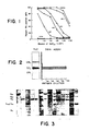
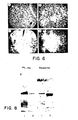
- SK-MEL-28 melanoma cells After treatment with neuraminidase (Vibrio cholerae), SK-MEL-28 melanoma cells no longer reacted with R 24 in PA-MHA assays (Table I). Reactivity with C 5 (an antibody with a serological specificity similar to that of R 24 (1)) was also lost. Reactivity with N 9 and 1 12 which recognize serologically unrelated determinants on glycoproteins of SK-MEL-28 was unaffected by neuraminidase. Enzyme-treated cells did not show non-specific reactivity with either Protein A- or with anti-mouse Ig-indicator cells.
- Glycolipids were isolated from culutured melanoma cells (SK-MEL 28) by chloroform-methanol (C-M) extraction and Florisil@ chromatography of their acetates as described by Saito and Hakomori (5) and the glycolipid preparation was fractionated into neutral and acidic components by DEAE-Sephadex chromatography. Inhibitory activity against R 24 antibody (assayed with PA-MHA) was found to reside entirely in the acidic glycolipid fractions.
- the isolated R 24 -reactive glycolipid was identified as by the following criteria:
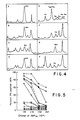
- G D3 could be detected in extracts of melanoma cell lines and melanoma tissue, but not in other sources (Table II). More sensitive assays (inhibition of PA-MHA and GMIA methods) showed that G D3 was detectable in a wider range of cells (bovine choroid, mouse eye, fetal and adult human lung, RAJI B-cell line, MOLT-4 T-cell line, RT-4 bladder cancer cell line and AJ astrocytoma cell line). A typical inhibition experiment is presented in Figure 5 and the data are summarized in Table II.
- Mouse monoclonal antibody R 24 which shows a high degree of serological specificity for cell surface antigens of melanoma cells, recognizes a disialoganglioside-G o3 . It might be significant that the mouse from which R 24 was developed had been extensively immunized over a period of six months with melanoma cells (SK-MEL-28) having a very high G D3 content.
- Two other monoclonal antibodies recognizing gangliosides have recently been described (17, 18). One reacts specifically with chicken neuronal cells and is directed against one of the higher gangliosides present in the GQ fraction (17); the second is directed against human colon carcinoma and recognizes an, as yet, uncharacterized monosialoganglioside (18).
- G D3 is a prominent ganglioside in cultured melanoma cells and in melanoma tissue. When compared with other cells, melanoma cells also possess relatively high total ganglioside levels. As shown by others, G D3 is present in small amounts in most mammalian tissues, but it is a major ganglioside in the retina, where it comprises between 30-40% of the gangliosides (19). In adult human brain, G D3 represents about 8-10% of the total ganglioside content (19).
- G D3 may be higher in fetal brain, considering that in fetal rat brain (15--17 days gestation) G D3 represents about 50% of the total ganglioside content, falling rapidly to about 10% by day 20 (20).
- Portoukalian and coworkers (21) have also reported that G D3 , identified by TLC and carbohydrate analysis, is a major constituent of melanomas. They showed that the proportion of G D3 varied from 31.0% to 57.2% of the ganglioside fraction in the four different melanoma specimens examined. From these results, as well as our own analysis, one can conclude that G D3 ganglisoide is a prominent component of malignant melanoma.
- T-cell line MOLT-4 showed a similar profile, and this may be another example of antigens shared by T-cells and cells of neuroectodermal origin e.g. Thy-1 (25).
- Thy-1 25
- Gangliosides derived from bovine choroid and mouse eye had more complex patterns, with G D3 being only one of three or four prominent components.
- G D3 ganglioside is by no means restricted to melanoma cells-it is ubiquitous. Yet using direct serological assays for cell surface antigens, only melanomas, choroidal melanocytes, and astrocytomas were reactive with R 24 (1). Even using sensitive absorption tests, only normal brain of other cells and tissues tested absorbed R 24 . A number of explanations for the apparent discrepancy between the serological finding and the biochemical data presented here can be suggested. First, it is possible that G D3 is not a cell surface constituent of most non-melanoma cells.
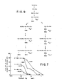
- G D3 is a biosynthetic precursor of other gangliosides ( Figure 9) and would therefore be located mainly within the cell, probably in the Golgi apparatus where the glycosyl transferases responsible for glycolipid synthesis are found (26, 27). As the biochemical studies were carried out on whole cell or tissues, the results are certainly compatible with this explanation. Another possibility is that G D3 is present at the cell surface of R 24 -negative cells but is not available for reaction with antibody. This phenomenon has been found with other cell membrane glycolipids: e.g. globoside is a major glycolipid of erythrocyte membrane but erythrocytes react only weakly with anti-globoside antibody (28).
- G D3 is not expressed on the surface of most non-melanoma cells in amounts that are detectable by the serological tests used. It is important to note that the cell types which reacted with antibody R 24 in both direct and absorption tests have both a high lipid-bound sialic acid content and have G D3 as a prominent ganglioside.
- melanomas have high levels of ⁇ -N-acetylgalactosaminidase which would result in increased degradation of G M2 and G D2 or that melanomas have elevated levels of certain sialyl transferases, resulting in increased synthesis of G D3 and G M3 .
- melanoma patients have increased serum sialyltransferase levels (29).
- Enzyme levels in tumor tissue have not yet been studied, although the fact that the glycoproteins of human melanoma cell lines have increased sialylation as compared to the glycoproteins of other cell types (30) suggests increased activity of this enzyme in melanoma.
- TLC thin layer chromatography
- MHA mixed hemagglutination assay
- C-M chloroform-methanol
- FBS fetal bovine serum
- NANA N-acetylneuraminic acid
- Gal D-galactose
- Glc D-glucose
- GaINAc N-acetyl-D-galactosamine
- Cer ceramide
- G M1 ⁇ Gal 1-3 GaINAc 1 ⁇ 4 ⁇ Gal (3 ⁇ 2 NANA) 1 ⁇ 4 Glc-Cer
- G M3 NANA 2 ⁇ 3 ⁇ Gal 1 ⁇ 4 Glc-Cer
- G D3 NANA 2 ⁇ 8 NANA 2 ⁇ 3 ⁇ Gal 1 ⁇ 4 Glc-Cer
- G D1a NANA 2 ⁇ 3 ⁇ Gal 1 ⁇ 3 GaINAc ⁇ 1 ⁇ 4 Gal (3 ⁇ 2 NANA) Glc-Cer
- G M2 ⁇ GalNAc 1 ⁇ 4 ⁇ Gal (3 ⁇ 2 NANA) G
Landscapes
- Health & Medical Sciences (AREA)
- Life Sciences & Earth Sciences (AREA)
- Immunology (AREA)
- Engineering & Computer Science (AREA)
- Molecular Biology (AREA)
- Biomedical Technology (AREA)
- Chemical & Material Sciences (AREA)
- Hematology (AREA)
- Urology & Nephrology (AREA)
- Food Science & Technology (AREA)
- Biochemistry (AREA)
- Cell Biology (AREA)
- Biotechnology (AREA)
- Medicinal Chemistry (AREA)
- Physics & Mathematics (AREA)
- Analytical Chemistry (AREA)
- Microbiology (AREA)
- General Health & Medical Sciences (AREA)
- General Physics & Mathematics (AREA)
- Pathology (AREA)
- Tropical Medicine & Parasitology (AREA)
- Medicines Containing Antibodies Or Antigens For Use As Internal Diagnostic Agents (AREA)
- Preparation Of Compounds By Using Micro-Organisms (AREA)
- Saccharide Compounds (AREA)
Claims (5)
Applications Claiming Priority (2)
| Application Number | Priority Date | Filing Date | Title |
|---|---|---|---|
| US06/365,065 US4507391A (en) | 1982-04-02 | 1982-04-02 | Method for detecting the presence of GD3 ganglioside |
| US365065 | 1999-07-30 |
Publications (2)
| Publication Number | Publication Date |
|---|---|
| EP0091005A1 EP0091005A1 (de) | 1983-10-12 |
| EP0091005B1 true EP0091005B1 (de) | 1986-08-13 |
Family
ID=23437333
Family Applications (1)
| Application Number | Title | Priority Date | Filing Date |
|---|---|---|---|
| EP83102865A Expired EP0091005B1 (de) | 1982-04-02 | 1983-03-23 | Methode zur Feststellung der Anwesenheit von GD3 Gangliosid |
Country Status (5)
| Country | Link |
|---|---|
| US (1) | US4507391A (de) |
| EP (1) | EP0091005B1 (de) |
| JP (1) | JPS58223066A (de) |
| CA (1) | CA1210689A (de) |
| DE (1) | DE3365236D1 (de) |
Families Citing this family (40)
| Publication number | Priority date | Publication date | Assignee | Title |
|---|---|---|---|---|
| US4808704A (en) * | 1981-09-30 | 1989-02-28 | Sloan-Kettering Institute For Cancer Research | Monoclonal antibodies to cell surface antigens of human malignant melanoma |
| CA1254846A (en) * | 1982-11-30 | 1989-05-30 | Sloan-Kettering Institute For Cancer Research | Monoclonal antibodies against melanocytes and melanomas |
| US4675287A (en) * | 1984-07-26 | 1987-06-23 | Scripps Clinic And Research Foundation | Monoclonal antibody directed to human ganglioside GD2 |
| US4708930A (en) * | 1984-11-09 | 1987-11-24 | Coulter Corporation | Monoclonal antibody to a human carcinoma tumor associated antigen |
| US4935495A (en) * | 1984-12-21 | 1990-06-19 | Oncogen | Monoclonal antibodies to the L6 glycolipid antigenic determinant found on human non-small cell lung carcinomas |
| US4906562A (en) * | 1984-12-21 | 1990-03-06 | Oncogen | Monocolonal antibodies and antigen for human non-small cell lung carcinomas |
| US4743543A (en) * | 1985-09-09 | 1988-05-10 | Coulter Corporation | Method for enhancing and/or accelerating immunoassay detection of human carcinoma tumor associated antigen in a pathology sample |
| US4844893A (en) * | 1986-10-07 | 1989-07-04 | Scripps Clinic And Research Foundation | EX vivo effector cell activation for target cell killing |
| US5104652A (en) * | 1986-11-13 | 1992-04-14 | Sloan-Kettering Institute For Cancer Research | Compositions and method for treatment of cancer using monoclonal antibody against GD3 ganglioside together with IL-2 |
| US4849509A (en) * | 1987-02-20 | 1989-07-18 | The Wistar Institute | Monoclonal antibodies against melanoma-associated antigens and hybrid cell lines producing these antibodies |
| EP0287916A1 (de) * | 1987-04-13 | 1988-10-26 | Otsuka Pharmaceutical Co., Ltd. | Immunoassay |
| US5006470A (en) * | 1987-04-16 | 1991-04-09 | Sloan-Kettering Institute For Cancer Research | Human monoclonal antibodies to cell surface antigens of melanoma |
| JP2754206B2 (ja) * | 1987-11-17 | 1998-05-20 | メクト株式会社 | α2→3結合を認識するモノクローナル抗体 |
| US5075218A (en) * | 1987-12-29 | 1991-12-24 | Biomira, Inc. | Screening for antibodies which bind carbohydrate epitopes of tumor-associated antigens, and uses thereof |
| JP2706777B2 (ja) * | 1988-02-19 | 1998-01-28 | メクト株式会社 | 非天然型gd▲下3▼を認識するモノクローナル抗体 |
| CA1337403C (en) * | 1988-03-28 | 1995-10-24 | Biomembrane Institute (The) | Methods for the production of antibodies and induction of immune responses to tumor-associated gangliosides by immunization with ganglioside lactones |
| US5173292A (en) * | 1988-06-14 | 1992-12-22 | Sloan-Kettering Institute For Cancer Research | Monoclonal antibodies which specifically recognize galactosyl-globoside, compositions containing same and methods of using same |
| US5242824A (en) * | 1988-12-22 | 1993-09-07 | Oncogen | Monoclonal antibody to human carcinomas |
| US5134075A (en) * | 1989-02-17 | 1992-07-28 | Oncogen Limited Partnership | Monoclonal antibody to novel antigen associated with human tumors |
| US5171665A (en) * | 1989-04-17 | 1992-12-15 | Oncogen | Monoclonal antibody to novel antigen associated with human tumors |
| US5980896A (en) * | 1989-06-30 | 1999-11-09 | Bristol-Myers Squibb Company | Antibodies reactive with human carcinomas |
| US6020145A (en) * | 1989-06-30 | 2000-02-01 | Bristol-Myers Squibb Company | Methods for determining the presence of carcinoma using the antigen binding region of monoclonal antibody BR96 |
| IL94872A (en) * | 1989-06-30 | 1995-03-30 | Oncogen | Monoclonal or chimeric antibodies reactive with human carcinomas, recombinant proteins comprising their antigen binding region, pharmaceutical compositions and kits comprising said antibodies and methods for imaging human carcinoma using same |
| US5624898A (en) | 1989-12-05 | 1997-04-29 | Ramsey Foundation | Method for administering neurologic agents to the brain |
| US6407061B1 (en) | 1989-12-05 | 2002-06-18 | Chiron Corporation | Method for administering insulin-like growth factor to the brain |
| EP0460607A3 (en) * | 1990-06-05 | 1992-04-01 | Bristol-Myers Squibb Company | Novel monoclonal antibody to novel antigen associated with human tumors |
| US5281710A (en) * | 1990-08-01 | 1994-01-25 | The Scripps Research Institute | Dynemicin analogs: synthesis, methods of preparation and use |
| US5792456A (en) * | 1994-08-04 | 1998-08-11 | Bristol-Myers Squibb Company | Mutant BR96 antibodies reactive with human carcinomas |
| US5728821A (en) * | 1994-08-04 | 1998-03-17 | Bristol-Myers Squibb Company | Mutant BR96 antibodies reactive with human carcinomas |
| RU2147373C1 (ru) * | 1998-06-19 | 2000-04-10 | Московский НИИ глазных болезней имени Гельмгольца | Способ выявления скрытого метастазирования при увеальной меланоме |
| RU2146823C1 (ru) * | 1998-09-29 | 2000-03-20 | Московский НИИ глазных болезней им.Гельмгольца | Способ прогнозирования клинического течения увеальной меланомы |
| US7273618B2 (en) * | 1998-12-09 | 2007-09-25 | Chiron Corporation | Method for administering agents to the central nervous system |
| RU2149404C1 (ru) * | 1999-03-24 | 2000-05-20 | Московский НИИ глазных болезней им. Гельмгольца | Способ прогнозирования клинического течения увеальной меланомы |
| RU2197731C2 (ru) * | 2001-02-26 | 2003-01-27 | Московский НИИ глазных болезней им. Гельмгольца | Способ прогнозирования клинического течения увеальной меланомы |
| RU2193200C1 (ru) * | 2001-05-23 | 2002-11-20 | Московский НИИ глазных болезней им. Гельмгольца | Способ прогнозирования клинического течения увеальной меланомы |
| RU2208789C1 (ru) * | 2002-10-28 | 2003-07-20 | Лихванцева Вера Геннадьевна | СПОСОБ ПРОГНОЗИРОВАНИЯ КЛИНИЧЕСКОГО ТЕЧЕНИЯ УВЕАЛЬНОЙ МЕЛАНОМЫ НА ОСНОВЕ МАРКЁРА АПОПТОЗА p53 |
| RU2208790C1 (ru) * | 2002-10-28 | 2003-07-20 | Лихванцева Вера Геннадьевна | СПОСОБ ПРОГНОЗИРОВАНИЯ КЛИНИЧЕСКОГО ТЕЧЕНИЯ УВЕАЛЬНОЙ МЕЛАНОМЫ НА ОСНОВЕ МАРКЕРА АПОПТОЗА Bax |
| RU2327420C1 (ru) * | 2006-12-12 | 2008-06-27 | ГУ Научно-исследовательский институт глазных болезней РАМН | Способ одновременной верификации и прогнозирования течения увеальной меланомы |
| WO2010065544A2 (en) * | 2008-12-01 | 2010-06-10 | The Johns Hopkins University | Diagnostic and treatment methods for cancer based on immune inhibitors |
| RU2698797C1 (ru) * | 2018-11-12 | 2019-08-30 | Федеральное государственное бюджетное учреждение "Национальный медицинский исследовательский центр глазных болезней имени Гельмгольца" Министерства здравоохранения Российской Федерации (ФГБУ "НМИЦ ГБ им. Гельмгольца" Минздрава России) | Способ прогнозирования неблагоприятного течения увеальной меланомы |
Family Cites Families (3)
| Publication number | Priority date | Publication date | Assignee | Title |
|---|---|---|---|---|
| DK36980A (da) * | 1979-01-30 | 1980-07-31 | Otsuka Pharma Co Ltd | Fremgangsmaade til bestemmelse af tumorforbundet glycobindingsholdigt stof og diagnosticering af cancer |
| US4331647A (en) * | 1980-03-03 | 1982-05-25 | Goldenberg Milton David | Tumor localization and therapy with labeled antibody fragments specific to tumor-associated markers |
| US4361544A (en) * | 1980-03-03 | 1982-11-30 | Goldenberg Milton David | Tumor localization and therapy with labeled antibodies specific to intracellular tumor-associated markers |
-
1982
- 1982-04-02 US US06/365,065 patent/US4507391A/en not_active Expired - Fee Related
-
1983
- 1983-03-23 DE DE8383102865T patent/DE3365236D1/de not_active Expired
- 1983-03-23 EP EP83102865A patent/EP0091005B1/de not_active Expired
- 1983-03-30 CA CA000424894A patent/CA1210689A/en not_active Expired
- 1983-04-01 JP JP58055318A patent/JPS58223066A/ja active Granted
Also Published As
| Publication number | Publication date |
|---|---|
| JPH0340833B2 (de) | 1991-06-20 |
| EP0091005A1 (de) | 1983-10-12 |
| DE3365236D1 (en) | 1986-09-18 |
| US4507391A (en) | 1985-03-26 |
| JPS58223066A (ja) | 1983-12-24 |
| CA1210689A (en) | 1986-09-02 |
Similar Documents
| Publication | Publication Date | Title |
|---|---|---|
| EP0091005B1 (de) | Methode zur Feststellung der Anwesenheit von GD3 Gangliosid | |
| Pukel et al. | GD3, a prominent ganglioside of human melanoma. Detection and characterisation by mouse monoclonal antibody. | |
| US4851511A (en) | Monoclonal antibody that specifically binds to disialosyl Lea | |
| Bremer et al. | Characterization of a glycosphingolipid antigen defined by the monoclonal antibody MBr1 expressed in normal and neoplastic epithelial cells of human mammary gland. | |
| US4965198A (en) | Monoclonal antibody and method of manufacturing hybridoma producing the same | |
| US4904596A (en) | Hybridoma antibody (FH6) defining a human cancer-associated difucoganglioside | |
| Fukushi et al. | A novel disialoganglioside (IV3NeuAcIII6NeuAcLc4) of human adenocarcinoma and the monoclonal antibody (FH9) defining this disialosyl structure | |
| Lloyd et al. | Cell surface accessibility of individual gangliosides in malignant melanoma cells to antibodies is influenced by the total ganglioside composition of the cells | |
| Le Pendu et al. | Monoclonal antibodies specific for type 3 and type 4 chain-based blood group determinants: relationship to the A1 and A2 subgroups | |
| Murayama et al. | Qualitative differences in position of sialylation and surface expression of glycolipids between murine lymphomas with low metastatic (Eb) and high metastatic (ESb) potentials and isolation of a novel disialoganglioside (GD1α) from Eb cells | |
| Kannagi et al. | Recent studies of glycolipid and glycoprotein profiles and characterization of the major glycolipid antigen in gastric cancer of a patient of blood group genotype pp (Tja-) first studied in 1951 | |
| GB2121417A (en) | Antigens and antibodies useful in the detection of cancer | |
| Miyoshi et al. | Detection of 4-O-acetyl-N-glycolylneuraminyl lactosylceramide as one of tumor-associated antigens in human colon cancer tissues by specific antibody | |
| Fredman et al. | Binding specificity of monoclonal antibodies to ganglioside, Fuc-GM1 | |
| Okada et al. | Glycolipid antigens with blood group I and i specificities from human adult and umbilical cord erythrocytes. | |
| EP0173663A2 (de) | Verwendung eines spezifischen tumorassoziierten Antigens, Sialosyllactotetraose, in Diagnose- oder Therapieverfahren bezüglich Krebskrankheiten | |
| Myoga et al. | Detection of patients with cancer by monoclonal antibody directed to lactoneotetraosylceramide (paragloboside) | |
| Siddiqui et al. | Effects of sodium butyrate, dimethyl sulfoxide, and retinoic acid on glycolipids of human rectal adenocarcinoma cells | |
| US5011920A (en) | Disialofucoganglioside immunogen and fucoganglioside monosialosyl Lea II | |
| Blaszczyk et al. | A fetal glycolipid expressed on adenocarcinomas of the colon | |
| Solomon et al. | A monoclonal antibody with reactivity to asialo GM1 and murine natural killer cells | |
| Nores et al. | Human heterophile antibodies recognizing epitopes present on insect glycolipids | |
| DE3785175T2 (de) | A-assoziierte h-antigene, spezifische monoklonale antikoerper und methoden zu deren verwendung zur blutgruppenbestimmung. | |
| US4888275A (en) | Diagnosing and monitoring cancer | |
| Schwarting et al. | The reactions of antibodies to paragloboside (lacto-N-neotetraosyl ceramide) with human erythrocytes and lymphocytes |
Legal Events
| Date | Code | Title | Description |
|---|---|---|---|
| PUAI | Public reference made under article 153(3) epc to a published international application that has entered the european phase |
Free format text: ORIGINAL CODE: 0009012 |
|
| AK | Designated contracting states |
Designated state(s): DE GB |
|
| 17P | Request for examination filed |
Effective date: 19840327 |
|
| GRAA | (expected) grant |
Free format text: ORIGINAL CODE: 0009210 |
|
| AK | Designated contracting states |
Kind code of ref document: B1 Designated state(s): DE GB |
|
| REF | Corresponds to: |
Ref document number: 3365236 Country of ref document: DE Date of ref document: 19860918 |
|
| PLBE | No opposition filed within time limit |
Free format text: ORIGINAL CODE: 0009261 |
|
| STAA | Information on the status of an ep patent application or granted ep patent |
Free format text: STATUS: NO OPPOSITION FILED WITHIN TIME LIMIT |
|
| 26N | No opposition filed | ||
| PGFP | Annual fee paid to national office [announced via postgrant information from national office to epo] |
Ref country code: GB Payment date: 19910218 Year of fee payment: 9 |
|
| PGFP | Annual fee paid to national office [announced via postgrant information from national office to epo] |
Ref country code: DE Payment date: 19910228 Year of fee payment: 9 |
|
| PG25 | Lapsed in a contracting state [announced via postgrant information from national office to epo] |
Ref country code: GB Effective date: 19920323 |
|
| GBPC | Gb: european patent ceased through non-payment of renewal fee | ||
| PG25 | Lapsed in a contracting state [announced via postgrant information from national office to epo] |
Ref country code: DE Effective date: 19921201 |