EP0106370A2 - Essais de liaison spécifiques utilisant des conjugués d'analyte-cytolysine - Google Patents
Essais de liaison spécifiques utilisant des conjugués d'analyte-cytolysine Download PDFInfo
- Publication number
- EP0106370A2 EP0106370A2 EP83110469A EP83110469A EP0106370A2 EP 0106370 A2 EP0106370 A2 EP 0106370A2 EP 83110469 A EP83110469 A EP 83110469A EP 83110469 A EP83110469 A EP 83110469A EP 0106370 A2 EP0106370 A2 EP 0106370A2
- Authority
- EP
- European Patent Office
- Prior art keywords
- analyte
- conjugate
- cytolysin
- melittin
- antibody
- Prior art date
- Legal status (The legal status is an assumption and is not a legal conclusion. Google has not performed a legal analysis and makes no representation as to the accuracy of the status listed.)
- Withdrawn
Links
- 230000009870 specific binding Effects 0.000 title claims abstract description 11
- 238000000159 protein binding assay Methods 0.000 title 1
- 239000012491 analyte Substances 0.000 claims abstract description 101
- 238000000034 method Methods 0.000 claims abstract description 59
- 239000003550 marker Substances 0.000 claims abstract description 55
- 239000000463 material Substances 0.000 claims abstract description 49
- 150000002632 lipids Chemical class 0.000 claims abstract description 43
- 239000011230 binding agent Substances 0.000 claims abstract description 26
- 238000006243 chemical reaction Methods 0.000 claims abstract description 17
- 230000035699 permeability Effects 0.000 claims abstract description 16
- 108010036176 Melitten Proteins 0.000 claims description 33
- 229960005156 digoxin Drugs 0.000 claims description 32
- VDXZNPDIRNWWCW-JFTDCZMZSA-N melittin Chemical compound NCC(=O)N[C@@H]([C@@H](C)CC)C(=O)NCC(=O)N[C@@H](C)C(=O)N[C@@H](C(C)C)C(=O)N[C@@H](CC(C)C)C(=O)N[C@@H](CCCCN)C(=O)N[C@@H](C(C)C)C(=O)N[C@@H](CC(C)C)C(=O)N[C@@H]([C@@H](C)O)C(=O)N[C@@H]([C@@H](C)O)C(=O)NCC(=O)N[C@@H](CC(C)C)C(=O)N1CCC[C@H]1C(=O)N[C@@H](C)C(=O)N[C@@H](CC(C)C)C(=O)N[C@@H]([C@@H](C)CC)C(=O)N[C@@H](CO)C(=O)N[C@H](C(=O)N[C@@H]([C@@H](C)CC)C(=O)N[C@@H](CCCCN)C(=O)N[C@@H](CCCNC(N)=N)C(=O)N[C@@H](CCCCN)C(=O)N[C@@H](CCCNC(N)=N)C(=O)N[C@@H](CCC(N)=O)C(=O)N[C@@H](CCC(N)=O)C(N)=O)CC1=CNC2=CC=CC=C12 VDXZNPDIRNWWCW-JFTDCZMZSA-N 0.000 claims description 32
- 108010056995 Perforin Proteins 0.000 claims description 31
- 102000004503 Perforin Human genes 0.000 claims description 31
- LTMHDMANZUZIPE-PUGKRICDSA-N digoxin Chemical compound C1[C@H](O)[C@H](O)[C@@H](C)O[C@H]1O[C@@H]1[C@@H](C)O[C@@H](O[C@@H]2[C@H](O[C@@H](O[C@@H]3C[C@@H]4[C@]([C@@H]5[C@H]([C@]6(CC[C@@H]([C@@]6(C)[C@H](O)C5)C=5COC(=O)C=5)O)CC4)(C)CC3)C[C@@H]2O)C)C[C@@H]1O LTMHDMANZUZIPE-PUGKRICDSA-N 0.000 claims description 31
- LTMHDMANZUZIPE-AMTYYWEZSA-N Digoxin Natural products O([C@H]1[C@H](C)O[C@H](O[C@@H]2C[C@@H]3[C@@](C)([C@@H]4[C@H]([C@]5(O)[C@](C)([C@H](O)C4)[C@H](C4=CC(=O)OC4)CC5)CC3)CC2)C[C@@H]1O)[C@H]1O[C@H](C)[C@@H](O[C@H]2O[C@@H](C)[C@H](O)[C@@H](O)C2)[C@@H](O)C1 LTMHDMANZUZIPE-AMTYYWEZSA-N 0.000 claims description 30
- LTMHDMANZUZIPE-UHFFFAOYSA-N digoxine Natural products C1C(O)C(O)C(C)OC1OC1C(C)OC(OC2C(OC(OC3CC4C(C5C(C6(CCC(C6(C)C(O)C5)C=5COC(=O)C=5)O)CC4)(C)CC3)CC2O)C)CC1O LTMHDMANZUZIPE-UHFFFAOYSA-N 0.000 claims description 30
- 102000004190 Enzymes Human genes 0.000 claims description 27
- 108090000790 Enzymes Proteins 0.000 claims description 27
- LPMXVESGRSUGHW-HBYQJFLCSA-N ouabain Chemical compound O[C@@H]1[C@H](O)[C@@H](O)[C@H](C)O[C@H]1O[C@@H]1C[C@@]2(O)CC[C@H]3[C@@]4(O)CC[C@H](C=5COC(=O)C=5)[C@@]4(C)C[C@@H](O)[C@@H]3[C@@]2(CO)[C@H](O)C1 LPMXVESGRSUGHW-HBYQJFLCSA-N 0.000 claims description 24
- 229960003343 ouabain Drugs 0.000 claims description 24
- LPMXVESGRSUGHW-UHFFFAOYSA-N Acolongiflorosid K Natural products OC1C(O)C(O)C(C)OC1OC1CC2(O)CCC3C4(O)CCC(C=5COC(=O)C=5)C4(C)CC(O)C3C2(CO)C(O)C1 LPMXVESGRSUGHW-UHFFFAOYSA-N 0.000 claims description 22
- LPMXVESGRSUGHW-GHYGWZAOSA-N Ouabain Natural products O([C@@H]1[C@@H](O)[C@@H](O)[C@@H](O)[C@H](C)O1)[C@H]1C[C@@H](O)[C@@]2(CO)[C@@](O)(C1)CC[C@H]1[C@]3(O)[C@@](C)([C@H](C4=CC(=O)OC4)CC3)C[C@@H](O)[C@H]21 LPMXVESGRSUGHW-GHYGWZAOSA-N 0.000 claims description 22
- 244000166550 Strophanthus gratus Species 0.000 claims description 22
- 239000000126 substance Substances 0.000 claims description 18
- YBJHBAHKTGYVGT-ZKWXMUAHSA-N (+)-Biotin Chemical compound N1C(=O)N[C@@H]2[C@H](CCCCC(=O)O)SC[C@@H]21 YBJHBAHKTGYVGT-ZKWXMUAHSA-N 0.000 claims description 16
- 102000002260 Alkaline Phosphatase Human genes 0.000 claims description 16
- 108020004774 Alkaline Phosphatase Proteins 0.000 claims description 16
- 239000012528 membrane Substances 0.000 claims description 16
- 239000007788 liquid Substances 0.000 claims description 15
- 102000004169 proteins and genes Human genes 0.000 claims description 15
- 108090000623 proteins and genes Proteins 0.000 claims description 15
- 229940079593 drug Drugs 0.000 claims description 12
- 239000003814 drug Substances 0.000 claims description 12
- 229960002685 biotin Drugs 0.000 claims description 11
- 239000011616 biotin Substances 0.000 claims description 11
- 210000003743 erythrocyte Anatomy 0.000 claims description 11
- BZTDTCNHAFUJOG-UHFFFAOYSA-N 6-carboxyfluorescein Chemical group C12=CC=C(O)C=C2OC2=CC(O)=CC=C2C11OC(=O)C2=CC=C(C(=O)O)C=C21 BZTDTCNHAFUJOG-UHFFFAOYSA-N 0.000 claims description 8
- 235000020958 biotin Nutrition 0.000 claims description 8
- 230000003993 interaction Effects 0.000 claims description 8
- 229940088594 vitamin Drugs 0.000 claims description 8
- 239000011782 vitamin Substances 0.000 claims description 8
- 235000013343 vitamin Nutrition 0.000 claims description 8
- 229930003231 vitamin Natural products 0.000 claims description 8
- 125000003277 amino group Chemical group 0.000 claims description 6
- 239000000427 antigen Substances 0.000 claims description 6
- 108091007433 antigens Proteins 0.000 claims description 6
- 102000036639 antigens Human genes 0.000 claims description 6
- 125000003178 carboxy group Chemical group [H]OC(*)=O 0.000 claims description 6
- 229940088597 hormone Drugs 0.000 claims description 6
- 239000005556 hormone Substances 0.000 claims description 6
- XZKIHKMTEMTJQX-UHFFFAOYSA-N 4-Nitrophenyl Phosphate Chemical compound OP(O)(=O)OC1=CC=C([N+]([O-])=O)C=C1 XZKIHKMTEMTJQX-UHFFFAOYSA-N 0.000 claims description 5
- 210000004027 cell Anatomy 0.000 claims description 5
- 125000002887 hydroxy group Chemical group [H]O* 0.000 claims description 5
- 239000002207 metabolite Substances 0.000 claims description 5
- 230000000813 microbial effect Effects 0.000 claims description 5
- 108090001008 Avidin Proteins 0.000 claims description 4
- 239000003344 environmental pollutant Substances 0.000 claims description 4
- 235000013305 food Nutrition 0.000 claims description 4
- 239000000575 pesticide Substances 0.000 claims description 4
- 150000003431 steroids Chemical class 0.000 claims description 4
- 239000003053 toxin Substances 0.000 claims description 4
- 231100000765 toxin Toxicity 0.000 claims description 4
- 108700012359 toxins Proteins 0.000 claims description 4
- DHMQDGOQFOQNFH-UHFFFAOYSA-N Glycine Natural products NCC(O)=O DHMQDGOQFOQNFH-UHFFFAOYSA-N 0.000 claims description 3
- 108090001090 Lectins Proteins 0.000 claims description 3
- 102000004856 Lectins Human genes 0.000 claims description 3
- KDXKERNSBIXSRK-UHFFFAOYSA-N Lysine Natural products NCCCCC(N)C(O)=O KDXKERNSBIXSRK-UHFFFAOYSA-N 0.000 claims description 3
- 239000004472 Lysine Substances 0.000 claims description 3
- 108091008324 binding proteins Proteins 0.000 claims description 3
- 210000004899 c-terminal region Anatomy 0.000 claims description 3
- 108091008039 hormone receptors Proteins 0.000 claims description 3
- 239000004471 Glycine Substances 0.000 claims description 2
- AYFVYJQAPQTCCC-GBXIJSLDSA-N L-threonine Chemical group C[C@@H](O)[C@H](N)C(O)=O AYFVYJQAPQTCCC-GBXIJSLDSA-N 0.000 claims description 2
- MTCFGRXMJLQNBG-UHFFFAOYSA-N Serine Chemical group OCC(N)C(O)=O MTCFGRXMJLQNBG-UHFFFAOYSA-N 0.000 claims description 2
- AYFVYJQAPQTCCC-UHFFFAOYSA-N Threonine Chemical group CC(O)C(N)C(O)=O AYFVYJQAPQTCCC-UHFFFAOYSA-N 0.000 claims description 2
- 239000004473 Threonine Chemical group 0.000 claims description 2
- 125000000404 glutamine group Chemical group N[C@@H](CCC(N)=O)C(=O)* 0.000 claims description 2
- 125000003630 glycyl group Chemical group [H]N([H])C([H])([H])C(*)=O 0.000 claims description 2
- 239000002523 lectin Substances 0.000 claims description 2
- 150000003722 vitamin derivatives Chemical class 0.000 claims 5
- 241000233866 Fungi Species 0.000 claims 3
- 239000002771 cell marker Substances 0.000 claims 3
- 125000003588 lysine group Chemical group [H]N([H])C([H])([H])C([H])([H])C([H])([H])C([H])([H])C([H])(N([H])[H])C(*)=O 0.000 claims 3
- 239000000439 tumor marker Substances 0.000 claims 3
- 241000700605 Viruses Species 0.000 claims 2
- 125000003275 alpha amino acid group Chemical group 0.000 claims 2
- 102000023732 binding proteins Human genes 0.000 claims 2
- 150000001615 biotins Chemical class 0.000 claims 2
- 239000003270 steroid hormone Substances 0.000 claims 2
- MTCFGRXMJLQNBG-REOHCLBHSA-N (2S)-2-Amino-3-hydroxypropansäure Chemical group OC[C@H](N)C(O)=O MTCFGRXMJLQNBG-REOHCLBHSA-N 0.000 claims 1
- 125000000539 amino acid group Chemical group 0.000 claims 1
- 150000001450 anions Chemical class 0.000 claims 1
- ZDXPYRJPNDTMRX-UHFFFAOYSA-N glutamine Natural products OC(=O)C(N)CCC(N)=O ZDXPYRJPNDTMRX-UHFFFAOYSA-N 0.000 claims 1
- 150000002500 ions Chemical class 0.000 claims 1
- 238000005259 measurement Methods 0.000 abstract description 9
- 238000001514 detection method Methods 0.000 abstract description 8
- 230000008569 process Effects 0.000 abstract description 4
- 230000001404 mediated effect Effects 0.000 abstract description 2
- 230000002596 correlated effect Effects 0.000 abstract 1
- 238000003556 assay Methods 0.000 description 34
- 229940088598 enzyme Drugs 0.000 description 25
- XKRFYHLGVUSROY-UHFFFAOYSA-N Argon Chemical compound [Ar] XKRFYHLGVUSROY-UHFFFAOYSA-N 0.000 description 22
- 239000000243 solution Substances 0.000 description 22
- 239000003599 detergent Substances 0.000 description 16
- 239000000203 mixture Substances 0.000 description 14
- 239000003153 chemical reaction reagent Substances 0.000 description 13
- 239000000523 sample Substances 0.000 description 13
- 238000004458 analytical method Methods 0.000 description 12
- 230000015572 biosynthetic process Effects 0.000 description 12
- 238000012360 testing method Methods 0.000 description 12
- 229910052786 argon Inorganic materials 0.000 description 11
- 230000027455 binding Effects 0.000 description 11
- 230000009089 cytolysis Effects 0.000 description 11
- 230000000694 effects Effects 0.000 description 11
- 125000006850 spacer group Chemical group 0.000 description 11
- 238000003786 synthesis reaction Methods 0.000 description 11
- FAPWRFPIFSIZLT-UHFFFAOYSA-M Sodium chloride Chemical compound [Na+].[Cl-] FAPWRFPIFSIZLT-UHFFFAOYSA-M 0.000 description 10
- 239000007983 Tris buffer Substances 0.000 description 10
- LENZDBCJOHFCAS-UHFFFAOYSA-N tris Chemical compound OCC(N)(CO)CO LENZDBCJOHFCAS-UHFFFAOYSA-N 0.000 description 10
- 239000011800 void material Substances 0.000 description 10
- XLYOFNOQVPJJNP-UHFFFAOYSA-N water Substances O XLYOFNOQVPJJNP-UHFFFAOYSA-N 0.000 description 10
- OKKJLVBELUTLKV-UHFFFAOYSA-N Methanol Chemical compound OC OKKJLVBELUTLKV-UHFFFAOYSA-N 0.000 description 9
- 239000012634 fragment Substances 0.000 description 9
- 238000002156 mixing Methods 0.000 description 9
- 210000002966 serum Anatomy 0.000 description 9
- 239000000758 substrate Substances 0.000 description 9
- HEDRZPFGACZZDS-UHFFFAOYSA-N Chloroform Chemical compound ClC(Cl)Cl HEDRZPFGACZZDS-UHFFFAOYSA-N 0.000 description 8
- 239000003795 chemical substances by application Substances 0.000 description 8
- 239000003431 cross linking reagent Substances 0.000 description 8
- 238000002360 preparation method Methods 0.000 description 8
- JQWHASGSAFIOCM-UHFFFAOYSA-M sodium periodate Chemical compound [Na+].[O-]I(=O)(=O)=O JQWHASGSAFIOCM-UHFFFAOYSA-M 0.000 description 8
- 239000001488 sodium phosphate Substances 0.000 description 8
- 229910000162 sodium phosphate Inorganic materials 0.000 description 8
- RYFMWSXOAZQYPI-UHFFFAOYSA-K trisodium phosphate Chemical compound [Na+].[Na+].[Na+].[O-]P([O-])([O-])=O RYFMWSXOAZQYPI-UHFFFAOYSA-K 0.000 description 8
- 239000012507 Sephadex™ Substances 0.000 description 7
- 238000002835 absorbance Methods 0.000 description 7
- LOKCTEFSRHRXRJ-UHFFFAOYSA-I dipotassium trisodium dihydrogen phosphate hydrogen phosphate dichloride Chemical compound P(=O)(O)(O)[O-].[K+].P(=O)(O)([O-])[O-].[Na+].[Na+].[Cl-].[K+].[Cl-].[Na+] LOKCTEFSRHRXRJ-UHFFFAOYSA-I 0.000 description 7
- 238000003018 immunoassay Methods 0.000 description 7
- 239000002953 phosphate buffered saline Substances 0.000 description 7
- 229920005654 Sephadex Polymers 0.000 description 6
- HVYWMOMLDIMFJA-DPAQBDIFSA-N cholesterol Chemical compound C1C=C2C[C@@H](O)CC[C@]2(C)[C@@H]2[C@@H]1[C@@H]1CC[C@H]([C@H](C)CCCC(C)C)[C@@]1(C)CC2 HVYWMOMLDIMFJA-DPAQBDIFSA-N 0.000 description 6
- 125000000524 functional group Chemical group 0.000 description 6
- 238000011534 incubation Methods 0.000 description 6
- 239000002244 precipitate Substances 0.000 description 6
- 108090000765 processed proteins & peptides Proteins 0.000 description 6
- 229920000936 Agarose Polymers 0.000 description 5
- 229920002684 Sepharose Polymers 0.000 description 5
- 239000000872 buffer Substances 0.000 description 5
- 230000008859 change Effects 0.000 description 5
- 230000000295 complement effect Effects 0.000 description 5
- 230000002101 lytic effect Effects 0.000 description 5
- 238000012544 monitoring process Methods 0.000 description 5
- KHIWWQKSHDUIBK-UHFFFAOYSA-N periodic acid Chemical compound OI(=O)(=O)=O KHIWWQKSHDUIBK-UHFFFAOYSA-N 0.000 description 5
- 102000004196 processed proteins & peptides Human genes 0.000 description 5
- 239000011541 reaction mixture Substances 0.000 description 5
- 239000011780 sodium chloride Substances 0.000 description 5
- 238000001042 affinity chromatography Methods 0.000 description 4
- 238000005119 centrifugation Methods 0.000 description 4
- 239000000499 gel Substances 0.000 description 4
- 238000005227 gel permeation chromatography Methods 0.000 description 4
- BDAGIHXWWSANSR-UHFFFAOYSA-N methanoic acid Natural products OC=O BDAGIHXWWSANSR-UHFFFAOYSA-N 0.000 description 4
- HEGSGKPQLMEBJL-RKQHYHRCSA-N octyl beta-D-glucopyranoside Chemical compound CCCCCCCCO[C@@H]1O[C@H](CO)[C@@H](O)[C@H](O)[C@H]1O HEGSGKPQLMEBJL-RKQHYHRCSA-N 0.000 description 4
- 239000003960 organic solvent Substances 0.000 description 4
- 229920001184 polypeptide Polymers 0.000 description 4
- 239000011347 resin Substances 0.000 description 4
- 229920005989 resin Polymers 0.000 description 4
- 239000011550 stock solution Substances 0.000 description 4
- 230000001225 therapeutic effect Effects 0.000 description 4
- 125000003396 thiol group Chemical group [H]S* 0.000 description 4
- XEKOWRVHYACXOJ-UHFFFAOYSA-N Ethyl acetate Chemical compound CCOC(C)=O XEKOWRVHYACXOJ-UHFFFAOYSA-N 0.000 description 3
- PEDCQBHIVMGVHV-UHFFFAOYSA-N Glycerine Chemical compound OCC(O)CO PEDCQBHIVMGVHV-UHFFFAOYSA-N 0.000 description 3
- ZMXDDKWLCZADIW-UHFFFAOYSA-N N,N-Dimethylformamide Chemical compound CN(C)C=O ZMXDDKWLCZADIW-UHFFFAOYSA-N 0.000 description 3
- 241000283973 Oryctolagus cuniculus Species 0.000 description 3
- 241000609499 Palicourea Species 0.000 description 3
- 102000057297 Pepsin A Human genes 0.000 description 3
- 108090000284 Pepsin A Proteins 0.000 description 3
- 239000012298 atmosphere Substances 0.000 description 3
- 230000001588 bifunctional effect Effects 0.000 description 3
- 229940098773 bovine serum albumin Drugs 0.000 description 3
- 239000008366 buffered solution Substances 0.000 description 3
- 235000012000 cholesterol Nutrition 0.000 description 3
- 230000008878 coupling Effects 0.000 description 3
- 238000010168 coupling process Methods 0.000 description 3
- 238000005859 coupling reaction Methods 0.000 description 3
- 230000029087 digestion Effects 0.000 description 3
- 239000012153 distilled water Substances 0.000 description 3
- VHJLVAABSRFDPM-QWWZWVQMSA-N dithiothreitol Chemical compound SC[C@@H](O)[C@H](O)CS VHJLVAABSRFDPM-QWWZWVQMSA-N 0.000 description 3
- 239000010408 film Substances 0.000 description 3
- 238000004519 manufacturing process Methods 0.000 description 3
- 230000003647 oxidation Effects 0.000 description 3
- 238000007254 oxidation reaction Methods 0.000 description 3
- 229940111202 pepsin Drugs 0.000 description 3
- 150000003904 phospholipids Chemical class 0.000 description 3
- 238000011533 pre-incubation Methods 0.000 description 3
- 238000000926 separation method Methods 0.000 description 3
- 239000007974 sodium acetate buffer Substances 0.000 description 3
- 239000012279 sodium borohydride Substances 0.000 description 3
- 229910000033 sodium borohydride Inorganic materials 0.000 description 3
- 239000010409 thin film Substances 0.000 description 3
- JQWAHKMIYCERGA-UHFFFAOYSA-N (2-nonanoyloxy-3-octadeca-9,12-dienoyloxypropoxy)-[2-(trimethylazaniumyl)ethyl]phosphinate Chemical compound CCCCCCCCC(=O)OC(COP([O-])(=O)CC[N+](C)(C)C)COC(=O)CCCCCCCC=CCC=CCCCCC JQWAHKMIYCERGA-UHFFFAOYSA-N 0.000 description 2
- HZAXFHJVJLSVMW-UHFFFAOYSA-N 2-Aminoethan-1-ol Chemical compound NCCO HZAXFHJVJLSVMW-UHFFFAOYSA-N 0.000 description 2
- NLMKTBGFQGKQEV-UHFFFAOYSA-N 2-[2-[2-[2-[2-[2-[2-[2-[2-[2-[2-[2-[2-[2-[2-[2-[2-[2-[2-(2-hexadecoxyethoxy)ethoxy]ethoxy]ethoxy]ethoxy]ethoxy]ethoxy]ethoxy]ethoxy]ethoxy]ethoxy]ethoxy]ethoxy]ethoxy]ethoxy]ethoxy]ethoxy]ethoxy]ethoxy]ethanol Chemical compound CCCCCCCCCCCCCCCCOCCOCCOCCOCCOCCOCCOCCOCCOCCOCCOCCOCCOCCOCCOCCOCCOCCOCCOCCOCCO NLMKTBGFQGKQEV-UHFFFAOYSA-N 0.000 description 2
- OSWFIVFLDKOXQC-UHFFFAOYSA-N 4-(3-methoxyphenyl)aniline Chemical compound COC1=CC=CC(C=2C=CC(N)=CC=2)=C1 OSWFIVFLDKOXQC-UHFFFAOYSA-N 0.000 description 2
- 108010088751 Albumins Proteins 0.000 description 2
- 102000009027 Albumins Human genes 0.000 description 2
- IJGRMHOSHXDMSA-UHFFFAOYSA-N Atomic nitrogen Chemical compound N#N IJGRMHOSHXDMSA-UHFFFAOYSA-N 0.000 description 2
- 108091003079 Bovine Serum Albumin Proteins 0.000 description 2
- 102000014914 Carrier Proteins Human genes 0.000 description 2
- 229920002307 Dextran Polymers 0.000 description 2
- 241000196324 Embryophyta Species 0.000 description 2
- 102000001554 Hemoglobins Human genes 0.000 description 2
- 108010054147 Hemoglobins Proteins 0.000 description 2
- 108091006905 Human Serum Albumin Proteins 0.000 description 2
- KFZMGEQAYNKOFK-UHFFFAOYSA-N Isopropanol Chemical compound CC(C)O KFZMGEQAYNKOFK-UHFFFAOYSA-N 0.000 description 2
- 239000000232 Lipid Bilayer Substances 0.000 description 2
- 241001465754 Metazoa Species 0.000 description 2
- 229910017974 NH40H Inorganic materials 0.000 description 2
- 206010028980 Neoplasm Diseases 0.000 description 2
- CDBYLPFSWZWCQE-UHFFFAOYSA-L Sodium Carbonate Chemical compound [Na+].[Na+].[O-]C([O-])=O CDBYLPFSWZWCQE-UHFFFAOYSA-L 0.000 description 2
- VMHLLURERBWHNL-UHFFFAOYSA-M Sodium acetate Chemical compound [Na+].CC([O-])=O VMHLLURERBWHNL-UHFFFAOYSA-M 0.000 description 2
- PXIPVTKHYLBLMZ-UHFFFAOYSA-N Sodium azide Chemical compound [Na+].[N-]=[N+]=[N-] PXIPVTKHYLBLMZ-UHFFFAOYSA-N 0.000 description 2
- WYURNTSHIVDZCO-UHFFFAOYSA-N Tetrahydrofuran Chemical compound C1CCOC1 WYURNTSHIVDZCO-UHFFFAOYSA-N 0.000 description 2
- 150000001299 aldehydes Chemical group 0.000 description 2
- 150000001408 amides Chemical group 0.000 description 2
- 150000001412 amines Chemical group 0.000 description 2
- 150000001413 amino acids Chemical class 0.000 description 2
- SOIFLUNRINLCBN-UHFFFAOYSA-N ammonium thiocyanate Chemical compound [NH4+].[S-]C#N SOIFLUNRINLCBN-UHFFFAOYSA-N 0.000 description 2
- 239000012131 assay buffer Substances 0.000 description 2
- 239000003659 bee venom Substances 0.000 description 2
- 230000008901 benefit Effects 0.000 description 2
- 201000011510 cancer Diseases 0.000 description 2
- 229910052799 carbon Inorganic materials 0.000 description 2
- 125000004432 carbon atom Chemical group C* 0.000 description 2
- 229940097217 cardiac glycoside Drugs 0.000 description 2
- 239000002368 cardiac glycoside Substances 0.000 description 2
- 239000007795 chemical reaction product Substances 0.000 description 2
- WORJEOGGNQDSOE-UHFFFAOYSA-N chloroform;methanol Chemical compound OC.ClC(Cl)Cl WORJEOGGNQDSOE-UHFFFAOYSA-N 0.000 description 2
- FDJOLVPMNUYSCM-WZHZPDAFSA-L cobalt(3+);[(2r,3s,4r,5s)-5-(5,6-dimethylbenzimidazol-1-yl)-4-hydroxy-2-(hydroxymethyl)oxolan-3-yl] [(2r)-1-[3-[(1r,2r,3r,4z,7s,9z,12s,13s,14z,17s,18s,19r)-2,13,18-tris(2-amino-2-oxoethyl)-7,12,17-tris(3-amino-3-oxopropyl)-3,5,8,8,13,15,18,19-octamethyl-2 Chemical compound [Co+3].N#[C-].N([C@@H]([C@]1(C)[N-]\C([C@H]([C@@]1(CC(N)=O)C)CCC(N)=O)=C(\C)/C1=N/C([C@H]([C@@]1(CC(N)=O)C)CCC(N)=O)=C\C1=N\C([C@H](C1(C)C)CCC(N)=O)=C/1C)[C@@H]2CC(N)=O)=C\1[C@]2(C)CCC(=O)NC[C@@H](C)OP([O-])(=O)O[C@H]1[C@@H](O)[C@@H](N2C3=CC(C)=C(C)C=C3N=C2)O[C@@H]1CO FDJOLVPMNUYSCM-WZHZPDAFSA-L 0.000 description 2
- 238000004440 column chromatography Methods 0.000 description 2
- 230000004154 complement system Effects 0.000 description 2
- 229920001577 copolymer Polymers 0.000 description 2
- 230000001461 cytolytic effect Effects 0.000 description 2
- 238000000502 dialysis Methods 0.000 description 2
- 238000009792 diffusion process Methods 0.000 description 2
- 238000010790 dilution Methods 0.000 description 2
- 239000012895 dilution Substances 0.000 description 2
- 239000008344 egg yolk phospholipid Substances 0.000 description 2
- 150000002148 esters Chemical class 0.000 description 2
- 230000007717 exclusion Effects 0.000 description 2
- 235000019253 formic acid Nutrition 0.000 description 2
- 238000004108 freeze drying Methods 0.000 description 2
- 238000002955 isolation Methods 0.000 description 2
- 238000002372 labelling Methods 0.000 description 2
- 230000014759 maintenance of location Effects 0.000 description 2
- 239000011159 matrix material Substances 0.000 description 2
- 230000007246 mechanism Effects 0.000 description 2
- 238000002439 negative-stain electron microscopy Methods 0.000 description 2
- 229920001467 poly(styrenesulfonates) Polymers 0.000 description 2
- 125000002924 primary amino group Chemical group [H]N([H])* 0.000 description 2
- 239000000047 product Substances 0.000 description 2
- 238000000746 purification Methods 0.000 description 2
- 125000001453 quaternary ammonium group Chemical group 0.000 description 2
- 230000009467 reduction Effects 0.000 description 2
- 230000002829 reductive effect Effects 0.000 description 2
- 230000035945 sensitivity Effects 0.000 description 2
- 239000001632 sodium acetate Substances 0.000 description 2
- 235000017281 sodium acetate Nutrition 0.000 description 2
- BEOOHQFXGBMRKU-UHFFFAOYSA-N sodium cyanoborohydride Chemical compound [Na+].[B-]C#N BEOOHQFXGBMRKU-UHFFFAOYSA-N 0.000 description 2
- GEHJYWRUCIMESM-UHFFFAOYSA-L sodium sulfite Chemical compound [Na+].[Na+].[O-]S([O-])=O GEHJYWRUCIMESM-UHFFFAOYSA-L 0.000 description 2
- 238000012289 standard assay Methods 0.000 description 2
- 229930002534 steroid glycoside Natural products 0.000 description 2
- 238000003860 storage Methods 0.000 description 2
- 239000006228 supernatant Substances 0.000 description 2
- 239000000725 suspension Substances 0.000 description 2
- ZFXYFBGIUFBOJW-UHFFFAOYSA-N theophylline Chemical compound O=C1N(C)C(=O)N(C)C2=C1NC=N2 ZFXYFBGIUFBOJW-UHFFFAOYSA-N 0.000 description 2
- 238000001291 vacuum drying Methods 0.000 description 2
- 230000007332 vesicle formation Effects 0.000 description 2
- 239000011715 vitamin B12 Substances 0.000 description 2
- LLXVXPPXELIDGQ-UHFFFAOYSA-N (2,5-dioxopyrrolidin-1-yl) 3-(2,5-dioxopyrrol-1-yl)benzoate Chemical compound C=1C=CC(N2C(C=CC2=O)=O)=CC=1C(=O)ON1C(=O)CCC1=O LLXVXPPXELIDGQ-UHFFFAOYSA-N 0.000 description 1
- YMXHPSHLTSZXKH-RVBZMBCESA-N (2,5-dioxopyrrolidin-1-yl) 5-[(3as,4s,6ar)-2-oxo-1,3,3a,4,6,6a-hexahydrothieno[3,4-d]imidazol-4-yl]pentanoate Chemical compound C([C@H]1[C@H]2NC(=O)N[C@H]2CS1)CCCC(=O)ON1C(=O)CCC1=O YMXHPSHLTSZXKH-RVBZMBCESA-N 0.000 description 1
- KIUMMUBSPKGMOY-UHFFFAOYSA-N 3,3'-Dithiobis(6-nitrobenzoic acid) Chemical compound C1=C([N+]([O-])=O)C(C(=O)O)=CC(SSC=2C=C(C(=CC=2)[N+]([O-])=O)C(O)=O)=C1 KIUMMUBSPKGMOY-UHFFFAOYSA-N 0.000 description 1
- BTJIUGUIPKRLHP-UHFFFAOYSA-M 4-nitrophenolate Chemical compound [O-]C1=CC=C([N+]([O-])=O)C=C1 BTJIUGUIPKRLHP-UHFFFAOYSA-M 0.000 description 1
- APKFDSVGJQXUKY-KKGHZKTASA-N Amphotericin-B Natural products O[C@H]1[C@@H](N)[C@H](O)[C@@H](C)O[C@H]1O[C@H]1C=CC=CC=CC=CC=CC=CC=C[C@H](C)[C@@H](O)[C@@H](C)[C@H](C)OC(=O)C[C@H](O)C[C@H](O)CC[C@@H](O)[C@H](O)C[C@H](O)C[C@](O)(C[C@H](O)[C@H]2C(O)=O)O[C@H]2C1 APKFDSVGJQXUKY-KKGHZKTASA-N 0.000 description 1
- 108010078791 Carrier Proteins Proteins 0.000 description 1
- 241000700199 Cavia porcellus Species 0.000 description 1
- 102000000989 Complement System Proteins Human genes 0.000 description 1
- 108010069112 Complement System Proteins Proteins 0.000 description 1
- 230000010777 Disulfide Reduction Effects 0.000 description 1
- 238000004435 EPR spectroscopy Methods 0.000 description 1
- LFQSCWFLJHTTHZ-UHFFFAOYSA-N Ethanol Chemical compound CCO LFQSCWFLJHTTHZ-UHFFFAOYSA-N 0.000 description 1
- JZNWSCPGTDBMEW-UHFFFAOYSA-N Glycerophosphorylethanolamin Natural products NCCOP(O)(=O)OCC(O)CO JZNWSCPGTDBMEW-UHFFFAOYSA-N 0.000 description 1
- 108090000288 Glycoproteins Proteins 0.000 description 1
- 102000003886 Glycoproteins Human genes 0.000 description 1
- 206010018910 Haemolysis Diseases 0.000 description 1
- 108010006464 Hemolysin Proteins Proteins 0.000 description 1
- 108010001336 Horseradish Peroxidase Proteins 0.000 description 1
- 102000008100 Human Serum Albumin Human genes 0.000 description 1
- 108060003951 Immunoglobulin Proteins 0.000 description 1
- 102000001706 Immunoglobulin Fab Fragments Human genes 0.000 description 1
- 108010054477 Immunoglobulin Fab Fragments Proteins 0.000 description 1
- 102000036675 Myoglobin Human genes 0.000 description 1
- 108010062374 Myoglobin Proteins 0.000 description 1
- 229910019142 PO4 Inorganic materials 0.000 description 1
- 108090000526 Papain Proteins 0.000 description 1
- 108010089814 Plant Lectins Proteins 0.000 description 1
- 239000004365 Protease Substances 0.000 description 1
- 108091006629 SLC13A2 Proteins 0.000 description 1
- VYPSYNLAJGMNEJ-UHFFFAOYSA-N Silicium dioxide Chemical compound O=[Si]=O VYPSYNLAJGMNEJ-UHFFFAOYSA-N 0.000 description 1
- 241000013033 Triso Species 0.000 description 1
- 239000013504 Triton X-100 Substances 0.000 description 1
- 229920004890 Triton X-100 Polymers 0.000 description 1
- 108010067973 Valinomycin Proteins 0.000 description 1
- 241001342522 Vampyrum spectrum Species 0.000 description 1
- 229930003779 Vitamin B12 Natural products 0.000 description 1
- 239000000654 additive Substances 0.000 description 1
- 125000003172 aldehyde group Chemical group 0.000 description 1
- 230000004075 alteration Effects 0.000 description 1
- 229940126575 aminoglycoside Drugs 0.000 description 1
- APKFDSVGJQXUKY-INPOYWNPSA-N amphotericin B Chemical compound O[C@H]1[C@@H](N)[C@H](O)[C@@H](C)O[C@H]1O[C@H]1/C=C/C=C/C=C/C=C/C=C/C=C/C=C/[C@H](C)[C@@H](O)[C@@H](C)[C@H](C)OC(=O)C[C@H](O)C[C@H](O)CC[C@@H](O)[C@H](O)C[C@H](O)C[C@](O)(C[C@H](O)[C@H]2C(O)=O)O[C@H]2C1 APKFDSVGJQXUKY-INPOYWNPSA-N 0.000 description 1
- 229960003942 amphotericin b Drugs 0.000 description 1
- 230000003321 amplification Effects 0.000 description 1
- 239000003957 anion exchange resin Substances 0.000 description 1
- 239000003242 anti bacterial agent Substances 0.000 description 1
- 229940088710 antibiotic agent Drugs 0.000 description 1
- 230000000890 antigenic effect Effects 0.000 description 1
- 206010003119 arrhythmia Diseases 0.000 description 1
- 229940125717 barbiturate Drugs 0.000 description 1
- HNYOPLTXPVRDBG-UHFFFAOYSA-N barbituric acid Chemical compound O=C1CC(=O)NC(=O)N1 HNYOPLTXPVRDBG-UHFFFAOYSA-N 0.000 description 1
- 239000011324 bead Substances 0.000 description 1
- 238000012455 bioassay technique Methods 0.000 description 1
- 239000013060 biological fluid Substances 0.000 description 1
- 239000012620 biological material Substances 0.000 description 1
- 239000012472 biological sample Substances 0.000 description 1
- 238000007413 biotinylation Methods 0.000 description 1
- 230000006287 biotinylation Effects 0.000 description 1
- 210000001124 body fluid Anatomy 0.000 description 1
- 239000010839 body fluid Substances 0.000 description 1
- 239000005388 borosilicate glass Substances 0.000 description 1
- 150000001718 carbodiimides Chemical class 0.000 description 1
- 125000002915 carbonyl group Chemical group [*:2]C([*:1])=O 0.000 description 1
- 230000000747 cardiac effect Effects 0.000 description 1
- 230000010261 cell growth Effects 0.000 description 1
- 238000011097 chromatography purification Methods 0.000 description 1
- 238000007398 colorimetric assay Methods 0.000 description 1
- 230000002860 competitive effect Effects 0.000 description 1
- FCFNRCROJUBPLU-UHFFFAOYSA-N compound M126 Natural products CC(C)C1NC(=O)C(C)OC(=O)C(C(C)C)NC(=O)C(C(C)C)OC(=O)C(C(C)C)NC(=O)C(C)OC(=O)C(C(C)C)NC(=O)C(C(C)C)OC(=O)C(C(C)C)NC(=O)C(C)OC(=O)C(C(C)C)NC(=O)C(C(C)C)OC1=O FCFNRCROJUBPLU-UHFFFAOYSA-N 0.000 description 1
- 239000000470 constituent Substances 0.000 description 1
- 238000011033 desalting Methods 0.000 description 1
- 238000011161 development Methods 0.000 description 1
- 238000002405 diagnostic procedure Methods 0.000 description 1
- 239000000539 dimer Substances 0.000 description 1
- 201000010099 disease Diseases 0.000 description 1
- 208000037265 diseases, disorders, signs and symptoms Diseases 0.000 description 1
- 238000005516 engineering process Methods 0.000 description 1
- 230000007613 environmental effect Effects 0.000 description 1
- 230000002255 enzymatic effect Effects 0.000 description 1
- 210000003617 erythrocyte membrane Anatomy 0.000 description 1
- 125000004185 ester group Chemical group 0.000 description 1
- 229940093499 ethyl acetate Drugs 0.000 description 1
- 235000019439 ethyl acetate Nutrition 0.000 description 1
- 210000002950 fibroblast Anatomy 0.000 description 1
- 239000012467 final product Substances 0.000 description 1
- 239000012847 fine chemical Substances 0.000 description 1
- 229940014144 folate Drugs 0.000 description 1
- OVBPIULPVIDEAO-LBPRGKRZSA-N folic acid Chemical compound C=1N=C2NC(N)=NC(=O)C2=NC=1CNC1=CC=C(C(=O)N[C@@H](CCC(O)=O)C(O)=O)C=C1 OVBPIULPVIDEAO-LBPRGKRZSA-N 0.000 description 1
- 235000019152 folic acid Nutrition 0.000 description 1
- 239000011724 folic acid Substances 0.000 description 1
- 238000004817 gas chromatography Methods 0.000 description 1
- 238000001502 gel electrophoresis Methods 0.000 description 1
- 229930182470 glycoside Natural products 0.000 description 1
- 150000002338 glycosides Chemical class 0.000 description 1
- 230000036541 health Effects 0.000 description 1
- 239000003228 hemolysin Substances 0.000 description 1
- 230000008588 hemolysis Effects 0.000 description 1
- NAQMVNRVTILPCV-UHFFFAOYSA-N hexane-1,6-diamine Chemical compound NCCCCCCN NAQMVNRVTILPCV-UHFFFAOYSA-N 0.000 description 1
- 230000002209 hydrophobic effect Effects 0.000 description 1
- 230000003053 immunization Effects 0.000 description 1
- 238000002649 immunization Methods 0.000 description 1
- 230000000984 immunochemical effect Effects 0.000 description 1
- 229940127121 immunoconjugate Drugs 0.000 description 1
- 230000002163 immunogen Effects 0.000 description 1
- 102000018358 immunoglobulin Human genes 0.000 description 1
- 229940029329 intrinsic factor Drugs 0.000 description 1
- 239000003041 laboratory chemical Substances 0.000 description 1
- 239000000787 lecithin Substances 0.000 description 1
- 235000010445 lecithin Nutrition 0.000 description 1
- 239000002502 liposome Substances 0.000 description 1
- 238000004811 liquid chromatography Methods 0.000 description 1
- 239000003120 macrolide antibiotic agent Substances 0.000 description 1
- 229940041033 macrolides Drugs 0.000 description 1
- 125000005439 maleimidyl group Chemical group C1(C=CC(N1*)=O)=O 0.000 description 1
- 238000004949 mass spectrometry Methods 0.000 description 1
- 230000004048 modification Effects 0.000 description 1
- 238000012986 modification Methods 0.000 description 1
- 208000010125 myocardial infarction Diseases 0.000 description 1
- 230000007935 neutral effect Effects 0.000 description 1
- 229910052757 nitrogen Inorganic materials 0.000 description 1
- 238000003199 nucleic acid amplification method Methods 0.000 description 1
- 125000002347 octyl group Chemical group [H]C([*])([H])C([H])([H])C([H])([H])C([H])([H])C([H])([H])C([H])([H])C([H])([H])C([H])([H])[H] 0.000 description 1
- 239000011368 organic material Substances 0.000 description 1
- 238000004806 packaging method and process Methods 0.000 description 1
- 238000012856 packing Methods 0.000 description 1
- 229940055729 papain Drugs 0.000 description 1
- 235000019834 papain Nutrition 0.000 description 1
- 239000013610 patient sample Substances 0.000 description 1
- 239000000813 peptide hormone Substances 0.000 description 1
- NBIIXXVUZAFLBC-UHFFFAOYSA-K phosphate Chemical compound [O-]P([O-])([O-])=O NBIIXXVUZAFLBC-UHFFFAOYSA-K 0.000 description 1
- 239000010452 phosphate Substances 0.000 description 1
- 150000008104 phosphatidylethanolamines Chemical class 0.000 description 1
- 239000002459 polyene antibiotic agent Substances 0.000 description 1
- 238000011085 pressure filtration Methods 0.000 description 1
- 230000002035 prolonged effect Effects 0.000 description 1
- 239000001397 quillaja saponaria molina bark Substances 0.000 description 1
- 230000009257 reactivity Effects 0.000 description 1
- 238000011084 recovery Methods 0.000 description 1
- 238000006894 reductive elimination reaction Methods 0.000 description 1
- 238000011160 research Methods 0.000 description 1
- 229930182490 saponin Natural products 0.000 description 1
- 150000007949 saponins Chemical class 0.000 description 1
- 238000012216 screening Methods 0.000 description 1
- 238000007423 screening assay Methods 0.000 description 1
- 229910000029 sodium carbonate Inorganic materials 0.000 description 1
- 239000012064 sodium phosphate buffer Substances 0.000 description 1
- 235000010265 sodium sulphite Nutrition 0.000 description 1
- 239000007787 solid Substances 0.000 description 1
- 238000001228 spectrum Methods 0.000 description 1
- 150000008143 steroidal glycosides Chemical class 0.000 description 1
- 108010075210 streptolysin O Proteins 0.000 description 1
- 239000012609 strong anion exchange resin Substances 0.000 description 1
- 239000004094 surface-active agent Substances 0.000 description 1
- YLQBMQCUIZJEEH-UHFFFAOYSA-N tetrahydrofuran Natural products C=1C=COC=1 YLQBMQCUIZJEEH-UHFFFAOYSA-N 0.000 description 1
- 229960000278 theophylline Drugs 0.000 description 1
- 229940126585 therapeutic drug Drugs 0.000 description 1
- -1 therapeutic drugs Substances 0.000 description 1
- 238000004809 thin layer chromatography Methods 0.000 description 1
- 125000005413 thiopyridyl group Chemical group 0.000 description 1
- 231100000331 toxic Toxicity 0.000 description 1
- 230000002588 toxic effect Effects 0.000 description 1
- 230000001052 transient effect Effects 0.000 description 1
- LASOLDQRFPFANT-UHFFFAOYSA-N tris(2-nitrophenyl) phosphate Chemical compound [O-][N+](=O)C1=CC=CC=C1OP(=O)(OC=1C(=CC=CC=1)[N+]([O-])=O)OC1=CC=CC=C1[N+]([O-])=O LASOLDQRFPFANT-UHFFFAOYSA-N 0.000 description 1
- 238000002211 ultraviolet spectrum Methods 0.000 description 1
- 239000002691 unilamellar liposome Substances 0.000 description 1
- FCFNRCROJUBPLU-DNDCDFAISA-N valinomycin Chemical compound CC(C)[C@@H]1NC(=O)[C@H](C)OC(=O)[C@@H](C(C)C)NC(=O)[C@@H](C(C)C)OC(=O)[C@H](C(C)C)NC(=O)[C@H](C)OC(=O)[C@@H](C(C)C)NC(=O)[C@@H](C(C)C)OC(=O)[C@H](C(C)C)NC(=O)[C@H](C)OC(=O)[C@@H](C(C)C)NC(=O)[C@@H](C(C)C)OC1=O FCFNRCROJUBPLU-DNDCDFAISA-N 0.000 description 1
- 235000019163 vitamin B12 Nutrition 0.000 description 1
Images
Classifications
-
- G—PHYSICS
- G01—MEASURING; TESTING
- G01N—INVESTIGATING OR ANALYSING MATERIALS BY DETERMINING THEIR CHEMICAL OR PHYSICAL PROPERTIES
- G01N33/00—Investigating or analysing materials by specific methods not covered by groups G01N1/00 - G01N31/00
- G01N33/48—Biological material, e.g. blood, urine; Haemocytometers
- G01N33/50—Chemical analysis of biological material, e.g. blood, urine; Testing involving biospecific ligand binding methods; Immunological testing
- G01N33/53—Immunoassay; Biospecific binding assay; Materials therefor
- G01N33/543—Immunoassay; Biospecific binding assay; Materials therefor with an insoluble carrier for immobilising immunochemicals
- G01N33/554—Immunoassay; Biospecific binding assay; Materials therefor with an insoluble carrier for immobilising immunochemicals the carrier being a biological cell or cell fragment, e.g. bacteria, yeast cells
-
- G—PHYSICS
- G01—MEASURING; TESTING
- G01N—INVESTIGATING OR ANALYSING MATERIALS BY DETERMINING THEIR CHEMICAL OR PHYSICAL PROPERTIES
- G01N33/00—Investigating or analysing materials by specific methods not covered by groups G01N1/00 - G01N31/00
- G01N33/48—Biological material, e.g. blood, urine; Haemocytometers
- G01N33/50—Chemical analysis of biological material, e.g. blood, urine; Testing involving biospecific ligand binding methods; Immunological testing
- G01N33/58—Chemical analysis of biological material, e.g. blood, urine; Testing involving biospecific ligand binding methods; Immunological testing involving labelled substances
- G01N33/585—Chemical analysis of biological material, e.g. blood, urine; Testing involving biospecific ligand binding methods; Immunological testing involving labelled substances with a particulate label, e.g. coloured latex
- G01N33/586—Liposomes, microcapsules or cells
-
- G—PHYSICS
- G01—MEASURING; TESTING
- G01N—INVESTIGATING OR ANALYSING MATERIALS BY DETERMINING THEIR CHEMICAL OR PHYSICAL PROPERTIES
- G01N33/00—Investigating or analysing materials by specific methods not covered by groups G01N1/00 - G01N31/00
- G01N33/48—Biological material, e.g. blood, urine; Haemocytometers
- G01N33/50—Chemical analysis of biological material, e.g. blood, urine; Testing involving biospecific ligand binding methods; Immunological testing
- G01N33/82—Chemical analysis of biological material, e.g. blood, urine; Testing involving biospecific ligand binding methods; Immunological testing involving vitamins or their receptors
-
- G—PHYSICS
- G01—MEASURING; TESTING
- G01N—INVESTIGATING OR ANALYSING MATERIALS BY DETERMINING THEIR CHEMICAL OR PHYSICAL PROPERTIES
- G01N33/00—Investigating or analysing materials by specific methods not covered by groups G01N1/00 - G01N31/00
- G01N33/48—Biological material, e.g. blood, urine; Haemocytometers
- G01N33/50—Chemical analysis of biological material, e.g. blood, urine; Testing involving biospecific ligand binding methods; Immunological testing
- G01N33/94—Chemical analysis of biological material, e.g. blood, urine; Testing involving biospecific ligand binding methods; Immunological testing involving narcotics or drugs or pharmaceuticals, neurotransmitters or associated receptors
- G01N33/9453—Cardioregulators, e.g. antihypotensives, antiarrhythmics
-
- Y—GENERAL TAGGING OF NEW TECHNOLOGICAL DEVELOPMENTS; GENERAL TAGGING OF CROSS-SECTIONAL TECHNOLOGIES SPANNING OVER SEVERAL SECTIONS OF THE IPC; TECHNICAL SUBJECTS COVERED BY FORMER USPC CROSS-REFERENCE ART COLLECTIONS [XRACs] AND DIGESTS
- Y10—TECHNICAL SUBJECTS COVERED BY FORMER USPC
- Y10S—TECHNICAL SUBJECTS COVERED BY FORMER USPC CROSS-REFERENCE ART COLLECTIONS [XRACs] AND DIGESTS
- Y10S435/00—Chemistry: molecular biology and microbiology
- Y10S435/966—Chemistry: molecular biology and microbiology involving an enzyme system with high turnover rate or complement magnified assay, e.g. multi-enzyme systems
-
- Y—GENERAL TAGGING OF NEW TECHNOLOGICAL DEVELOPMENTS; GENERAL TAGGING OF CROSS-SECTIONAL TECHNOLOGIES SPANNING OVER SEVERAL SECTIONS OF THE IPC; TECHNICAL SUBJECTS COVERED BY FORMER USPC CROSS-REFERENCE ART COLLECTIONS [XRACs] AND DIGESTS
- Y10—TECHNICAL SUBJECTS COVERED BY FORMER USPC
- Y10S—TECHNICAL SUBJECTS COVERED BY FORMER USPC CROSS-REFERENCE ART COLLECTIONS [XRACs] AND DIGESTS
- Y10S435/00—Chemistry: molecular biology and microbiology
- Y10S435/972—Modified antibody, e.g. hybrid, bifunctional
-
- Y—GENERAL TAGGING OF NEW TECHNOLOGICAL DEVELOPMENTS; GENERAL TAGGING OF CROSS-SECTIONAL TECHNOLOGIES SPANNING OVER SEVERAL SECTIONS OF THE IPC; TECHNICAL SUBJECTS COVERED BY FORMER USPC CROSS-REFERENCE ART COLLECTIONS [XRACs] AND DIGESTS
- Y10—TECHNICAL SUBJECTS COVERED BY FORMER USPC
- Y10S—TECHNICAL SUBJECTS COVERED BY FORMER USPC CROSS-REFERENCE ART COLLECTIONS [XRACs] AND DIGESTS
- Y10S436/00—Chemistry: analytical and immunological testing
- Y10S436/803—Stabe free radicals, e.g. spin immunoassay
-
- Y—GENERAL TAGGING OF NEW TECHNOLOGICAL DEVELOPMENTS; GENERAL TAGGING OF CROSS-SECTIONAL TECHNOLOGIES SPANNING OVER SEVERAL SECTIONS OF THE IPC; TECHNICAL SUBJECTS COVERED BY FORMER USPC CROSS-REFERENCE ART COLLECTIONS [XRACs] AND DIGESTS
- Y10—TECHNICAL SUBJECTS COVERED BY FORMER USPC
- Y10S—TECHNICAL SUBJECTS COVERED BY FORMER USPC CROSS-REFERENCE ART COLLECTIONS [XRACs] AND DIGESTS
- Y10S436/00—Chemistry: analytical and immunological testing
- Y10S436/811—Test for named disease, body condition or organ function
- Y10S436/813—Cancer
-
- Y—GENERAL TAGGING OF NEW TECHNOLOGICAL DEVELOPMENTS; GENERAL TAGGING OF CROSS-SECTIONAL TECHNOLOGIES SPANNING OVER SEVERAL SECTIONS OF THE IPC; TECHNICAL SUBJECTS COVERED BY FORMER USPC CROSS-REFERENCE ART COLLECTIONS [XRACs] AND DIGESTS
- Y10—TECHNICAL SUBJECTS COVERED BY FORMER USPC
- Y10S—TECHNICAL SUBJECTS COVERED BY FORMER USPC CROSS-REFERENCE ART COLLECTIONS [XRACs] AND DIGESTS
- Y10S436/00—Chemistry: analytical and immunological testing
- Y10S436/815—Test for named compound or class of compounds
- Y10S436/817—Steroids or hormones
-
- Y—GENERAL TAGGING OF NEW TECHNOLOGICAL DEVELOPMENTS; GENERAL TAGGING OF CROSS-SECTIONAL TECHNOLOGIES SPANNING OVER SEVERAL SECTIONS OF THE IPC; TECHNICAL SUBJECTS COVERED BY FORMER USPC CROSS-REFERENCE ART COLLECTIONS [XRACs] AND DIGESTS
- Y10—TECHNICAL SUBJECTS COVERED BY FORMER USPC
- Y10S—TECHNICAL SUBJECTS COVERED BY FORMER USPC CROSS-REFERENCE ART COLLECTIONS [XRACs] AND DIGESTS
- Y10S436/00—Chemistry: analytical and immunological testing
- Y10S436/827—Lectins
-
- Y—GENERAL TAGGING OF NEW TECHNOLOGICAL DEVELOPMENTS; GENERAL TAGGING OF CROSS-SECTIONAL TECHNOLOGIES SPANNING OVER SEVERAL SECTIONS OF THE IPC; TECHNICAL SUBJECTS COVERED BY FORMER USPC CROSS-REFERENCE ART COLLECTIONS [XRACs] AND DIGESTS
- Y10—TECHNICAL SUBJECTS COVERED BY FORMER USPC
- Y10S—TECHNICAL SUBJECTS COVERED BY FORMER USPC CROSS-REFERENCE ART COLLECTIONS [XRACs] AND DIGESTS
- Y10S436/00—Chemistry: analytical and immunological testing
- Y10S436/828—Protein A
-
- Y—GENERAL TAGGING OF NEW TECHNOLOGICAL DEVELOPMENTS; GENERAL TAGGING OF CROSS-SECTIONAL TECHNOLOGIES SPANNING OVER SEVERAL SECTIONS OF THE IPC; TECHNICAL SUBJECTS COVERED BY FORMER USPC CROSS-REFERENCE ART COLLECTIONS [XRACs] AND DIGESTS
- Y10—TECHNICAL SUBJECTS COVERED BY FORMER USPC
- Y10S—TECHNICAL SUBJECTS COVERED BY FORMER USPC CROSS-REFERENCE ART COLLECTIONS [XRACs] AND DIGESTS
- Y10S436/00—Chemistry: analytical and immunological testing
- Y10S436/829—Liposomes, e.g. encapsulation
-
- Y—GENERAL TAGGING OF NEW TECHNOLOGICAL DEVELOPMENTS; GENERAL TAGGING OF CROSS-SECTIONAL TECHNOLOGIES SPANNING OVER SEVERAL SECTIONS OF THE IPC; TECHNICAL SUBJECTS COVERED BY FORMER USPC CROSS-REFERENCE ART COLLECTIONS [XRACs] AND DIGESTS
- Y10—TECHNICAL SUBJECTS COVERED BY FORMER USPC
- Y10S—TECHNICAL SUBJECTS COVERED BY FORMER USPC CROSS-REFERENCE ART COLLECTIONS [XRACs] AND DIGESTS
- Y10S930/00—Peptide or protein sequence
- Y10S930/01—Peptide or protein sequence
- Y10S930/28—Bound to a nonpeptide drug, nonpeptide label, nonpeptide carrier, or a nonpeptide resin
Definitions
- This invention relates to a highly sensitive and rapid method of analysis for the quantitative determination of the amount of a specific analyte in liquid medium, and to novel analyte-cytolysin conjugates which alter the membrane permeability of vesicles containing marker material.
- Clinical laboratory chemical diagnostic tests are an important component of health care delivery.
- the utilization of these tests by physicians to monitor drug levels where only a narrow therapeutic range exists, to guide decisions on treatment and- surgical options, and to screen patients for the early detection of disease has rapidly increased the number of tests performed annually.
- speed, accuracy, and cost control are important objectives.
- the desire to measure such analytes as drugs, hormones, and metabolites at micromolar ( ⁇ M) to picomolar (pM) levels in complex body fluid matrices has led to the development of sophisticated test methodology which can be implemented by automated techniques at reasonable cost.
- vesicles containing sequestered detectable marker material can provide a stable, sensitive, and flexible measurement system for quantifying such medically important materials as cardiac glycosides, antibiotics, therapeutic drugs, hormones, and vitamins.
- methods of analysis for toxins, food and packaging additives, and environmental pollutants at extremely low concentration are required.
- Such systems require the preparation of lipid vesicles with specific analyte "tags" and also the use of the unstable, complex complement system to release the detectable marker material used to quantify the amount of analyte initially present. Long incubation periods are frequently required which increase analysis time.
- Hsia et al. [New York Academy Sci., Vol. 308, 139-148 (1978) and U.S. Patent 4,235,792] describe complement mediated immunoassay techniques wherein the lysis of lipid vesicle with a synthetic sensitizer incorporated in the lipid bilayer leads to the release of marker material, in particular of spin labelled molecules quantified by electron spin resonance techniques.
- the assay system requires the preparation of a specific lipid vesicle for each analyte of interest to mediate attack and lysis by the complement system in the presence of antibody.
- U.K. Patent Application 2069133A and U.S. Patent 4,342,826 describes a process for sequestering enzyme marker within lipid vesicles in a manner which enhances the so-called signal to noise ratio of the reagents.
- the lipid vesicles must be specifically labelled with either antigen or antibody to render them immunoreactive in the presence of complement.
- a sensitive, homogeneous assay to measure analytes in solution which utilizes novel analyte-cytolysin conjugates and vesicles containing marker material wherein the vesicle membrane permeability changes resulting from interaction with such conjugates can be modulated by analyte-specific binding agent.
- the assay also utilizes a standard vesicle preparation applicable for analysis of a wide variety of analytes by many different instrumental methods.
- one aspect of this invention involves-the synthesis of an analyte-cytolysin conjugate wherein at least one analyte derivative molecule is attached per cytolysin molecule such that said conjugate is capable of both reacting with binding agent of the analyte and altering vesicle membrane permeability to release marker material.
- Another aspect of this invention is a method for determining the amount of an analyte in liquid medium comprising the steps of:
- An antibody molecule is capable of functioning as an analyte in the present invention, but an antibody-cytolysin conjugate can have utility in a heterogeneous assay wherein a separation of reaction products occurs prior to the measurement step.
- Another aspect of this invention therefor involves a novel antibody-cytolysin conjugate.
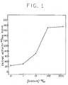
- Figure 1 is a standard assay curve to be used for digoxin analysis which relates the amount of marker material released from lipid vesicles by unbound ouabain-melittin conjugate to the concentration of digoxin initially present in the liquid medium.
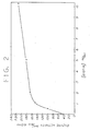
- Figure 2 is a standard assay curve to be used for analysis of biotin which relates the amount of marker material released from lipid vesicles by unbound biotin-melittin conjugate to the concentration of biotin initially present in the liquid medium.
- analyte is the substance, or group of substances, whose presence or amount in a liquid medium is to be determined, which additionally have the capability of being attached to a cytolysin substance to form a analyte-cytolysin conjugate;
- cytolysin is any substance or agent of molecular weight from about 1 00-100,000 daltons which can change the permeability of biomembranes;
- analyte derivative refers to both unmodified and chemically modified analyte molecules, and/or their chemical combinations with spacer arms, which can be chemically bound to cytolysin molecules by covalent, ionic, or other bonding techniques:
- spacer arm is a bifunctional molecule used to chemically bond an analyte derivative to a cytolysin molecules by covalent, ionic, or other bonding techniques:
- spacer arm is a bifunctional molecule used to chemically bond an analyte derivative to a cytolysin molecules by covalent,
- the present homogeneous method may be applied to the detection of any analyte for which a binding agent exists.
- the binding agent may consist of an antibody in the form of whole antiserum, an IgG fraction, as affinity-purified monospecific material, a monoclonal antibody, a monovalent antibody or of other specific binding proteins like lectins, hormone receptors, or serum transport proteins.
- the quantitative measurement aspect of the invention results from the fact that free analyte present in the test sample and the analyte-cytolysin conjugate are both capable of reacting in a competitive fashion with binding agent. In the absence of analyte, the concentration of the process components is adjusted such that there is no release of sequestered marker material from the vesicles.
- the determination of the amount of analyte initially present in the test sample can be carried out by correlation with either the amount of marker material released after a given contact time of reagents, or with the rate of release of such marker material under conditions which enable comparison with a standard curve produced for known amounts of the reagents.
- Contacting times of the reagents can vary from 10 seconds to one hour at temperatures in the range of from about 4° to 40°C and at a pH in the range of about 5-10, usually 6-8.
- the measurement can be carried out manually, or with reagents packaged to utilize automated analyzers.
- an aliquot containing an unknown amount of analyte is added to buffered incubation medium containing substrate for the enzyme which is used as marker material sequestered in the vesicles.
- a known amount of antibody specific for the analyte is added to the medium and briefly incubated prior to the addition of a known amount of analyte-cytolysin conjugate.
- a known amount of vesicle preparation is added and the amount of substrate converted by enzyme after diffusion into the vesicle is monitored as a function of time. Comparison with a standard curve relating analyte concentration with substrate conversion obtained under the same conditions of time intervals and reagent amounts enables the determination of the unknown amount of analyte.
- the antibody-cytolysin conjugate of the present invention can be used in a heterogeneous assay for the detection of any analyte which has antigenic or haptenic properties, i.e., the ability to elicit the formation of anti-analyte antibody when injected into a host.
- an antibody-cytolysin conjugate in a heterogeneous assay an aliquot containing an unknown amount of analyte is mixed with a buffered solution containing cytolysin labeled anti-analyte antibody (C-Ab).
- C-Ab cytolysin labeled anti-analyte antibody
- the mixture is then incubated during which time a fraction of the C-Ab will immunochemically bind to analyte while the other fraction of C-Ab will remain free.
- the bound and free C-Ab are separated from one another.
- the separation can be carried out using an affinity column which contains a packing material to which analyte is immobilized, either directly or through a spacer arm.
- the mixture is applied to the affinity column.
- the free C-Ab will bind to the immobilized analyte. Consequently, only the Analyte-(C-Ab) complex will elute from the column.
- the mixture can be applied to a size exclusion column such as Sepharose 4BC1, a crosslinked macroporous agarose in bead-form for gel permeation chromatography from Pharmacia Fine Chemicals.
- a size exclusion column such as Sepharose 4BC1
- the void volume containing the complex is collected.
- the eluate or void volume depending on which separation step is employed is then mixed with suspension of vesicles.
- the cytolysin moiety of the complex will effect a change in permeability of the vesicle membrane allowing marker material to be released in the external medium.
- the marker material will be either directly detectable (e.g., chromophores) or indirectly detectable (e.g., an enzyme which will combine with a specific substrate in the external medium to yield a detectable product).
- This invention can be applied to the detection and measurement of a broad variety of analytes to which binding agents are available such as drugs of biological and clinical importance, metabolites, vitamins, pesticides, steroids, peptide hormones and certain cancer markers.
- Analytes of particular interest include those drugs and hormones with either very low concentrations in biological fluids or with narrow therapeutic ranges.
- the cardiac steroid digoxin satisfies both criteria since levels below 0.8 ng/ml (nanograms per milliliter) in human serum are ineffective for treating cardiac arrhythmia while levels above 2.0 ng/ml are often toxic.
- Other analytes similarly present at low concentration or with narrow "therapeutic range include vitamin B12, folate, and most of the steroid, peptide, and protein hormones.
- Analytes such as myoglobin normally have very low levels in serum that can rise dramatically after myocardial infarction and are therefore indicative of this condition.
- Analytes such as microbial and cancer cell markers would generally be low in concentration since early detection (prior to prolonged cell growth) is highly desirable.
- Aminoglycoside drugs, barbiturate drugs, and many of the miscellaneous drugs such as theophylline are relatively high in concentration with ⁇ g/ml (microgram/milliter) levels but have narrow therapeutic ranges.
- Table I lists a variety of analytes of particular interest in practicing the instant invention.
- binding analogs of the analyte for example ouabain for digoxin
- ouabain for digoxin can be extremely advantageous by enabling reagents developed for one assay to be used in another for a group of related substances, and by providing choices of binding properties to those skilled in the art which simplify the isolation and purification of binding agents.
- cytolysins A large number of cytolysins exist which may be employed with the present invention. These cytolytic agents are commonly of natural origin; however synthetic cytolysins such as surfactants and analogs of naturally occurring agents can also be used. Cytolysins are also commonly referred to as hemolysins when red blood cells are involved as the vesicle. Molecular weights generally are in the range of 100-2000 for many of the synthetic cytolysins and 2000-100,000 for some of the naturally occurring agents. Many naturally occurring cytolysins are polypeptides and proteins, such as melittin and streptolysin O, respectively, while others are typically classified as glycosides (e.g.
- cytolysin includes the naturally occurring cytolysins, their derivatives, synthetic analogs, and synthetic cytolysins exemplified in Table II.
- the preferred cytolysin of this invention is melittin, since it is commercially available, well characterized in terms of structure and function, and of low molecular weight.
- Melittin is a 26 amino acid polypeptide in which the amino acids are linked together to form substituted amide bonds with characteristic side chains with the following structure based upon standard nomenclature Melittin is isolated from bee venom [E. Habermann, et al., Z. Physiol. Chem., Vol. 348, 37 (1967)] and has the demonstrated ability to lyse both erythrocytes and lipid vesicles [G. Sessu, et al., J. Biol. Chem., Vol. 244, 3575 (1969)].
- the present invention utilizes novel analyte-cytolysin conjugates which can be synthesized by covalently linking cytolysin materials with analytes.
- analyte-cytolysin conjugate also refers to analytes which are themselves cytolysins, and to cytolysins which are covalently linked either to derivatives of the analyte or to a binding analog of the analyte.
- Immunoassays for large molecular weight analytes, such as proteins, require that only one or more (but not all) binding sites be attached to the cytolysin.
- analyte protein may work as well in the present invention as the analyte proteins themselves.
- An immunoassay for digoxin is another case where a binding analog of the analyte (ouabain) works as well as the analyte itself since both contain common binding sites and are recognized by the same antidigoxin antibody.
- cytolysins Conditions for covalently attaching an analyte, including its derivatives and analogs, to a cytolysin depend upon the particular molecular architecture of both-types of molecules.
- cytolysins contain functional groups such as amines, amides, carboxyls, sulfhydryls, hydroxyls, aldehydes, and/or others, to which an analyte with its own appropriate functional groups could be attached directly or indirectly.
- the attachment chemistry may vary depending upon the functional groups involved, the number of analyte molecules to be bonded per cytolysin, and the desirability of including a spacer arm between the analyte and the cytolysin.
- the conjugate must retain the ability to react with binding agent of the analyte and to alter vesicle membrane permeability.
- possible sites of attachment include the hydroxyl group of the threonine and serine amino acid residues, the amino group of the lysine or N-terminal glycine residue, and the carbonyl group of the C-terminal glutamine residue of the polypeptide molecule.
- the analyte could be attached to the cytolysin through a spacer arm, such as 1,6-diaminohexane or other bifunctional crosslinking agents after periodate oxidation.
- a spacer arm can facilitate the synthesis of the conjugate, or insure that the conjugate will possess the requisite immunochemical reactivity.
- ouabain could be bonded through a spacer arm to the carboxyl' terminal of melittin or to one of the amino groups if the appropriate crosslinking agent is used.
- crosslinking agents both homo and heterobifunctional, have been described in the literature [Pierce Bio-Research Products Technical Bulletin, "Double-Agents”, Bifunctional Crosslinking Reagents, Pierce Chemical Co., Rockford, IL, U.S.A., 1982, Vol. 3] which could be used to couple ouabain to melittin.
- These crosslinking agents could be used with many different cytolysins and analytes of clinical importance.
- Carbodiimide crosslinking agents such as l-ethyl-3-(3-dimethylamino- p ropyl)carbodiimide, can also be employed to link analytes with carboxyl groups to amino groups on cytolysins or conversely, the amino groups of analytes to carboxyl groups on cytolysins.
- binding analogs of the analyte is illustrated by the observation that ouabain-melittin conjugates have higher lytic activity at lower conjugate concentrations than does melittin alone. This type of behavior is expected for other analyte-cytolysin conjugates and would increase the sensitivity and range of utility of the resulting assay.
- the synthesis of conjugates incorporating binding analogs of the analyte can also provide an assay for the analyte despite difficulty in purifying the analyte-cytolysin conjugate, as for example with digoxin-melittin.
- analyte-cytolysin conjugate The chemical reactions involved in bonding analyte molecules, or their derivatives or analogs, to the melittin polypeptide or other cytolysin are known to those skilled in the art.
- the important features of a particular analyte-cytolysin conjugate are that the conjugate be stable under conditions of storage and use, i.e., that it not release analyte or analyte derivative or analog, that it react with binding agent specific for the analyte, and that it alter vesicle membrane permeability to release marker material. While covalent bonds have been found to insure these properties, bonding can involve other types of interactions which result in stable combinations of analyte and cytolysin, such as ionic or hydrophobic bonds.
- conjugate means analyte molecule attached, directly or indirectly, to cytolysin molecule.
- One aspect of the present invention involves novel antibody-cytolysin conjugates which can be synthesized by covalently attaching cytolysin materials to antibody.
- Conditions for covalent attachment depend upon the particular molecular architecture of both types of molecules.
- cytolysins contain functional groups such as amines, amides, carboxyls, sulfhydryls, hydroxyls, aldehydes, etc. to which an antibody with its own appropriate functional groups could be attached directly or indirectly.
- the attachment chemistry will vary depending upon the functional groups involved and the desirability of including a spacer arm.
- any number of methods can be employed to attach the cytolysin to-the antibody.
- at least one cytolysin label should be coupled to each antibody.
- the free sulfhydryl groups present on Fab' fragments and the S-sulfonate group present on half-molecules (heavy chain-light chain dimers) provide specific reactive groups for covalent attachment of the cytolysin. Labeling of these groups is known not to affect the immunoreactivity of the antibody.
- Heterobifunctional crosslinking reagents having maleimido- or thiopyridyl- groups are useful for this purpose.
- the antibody must retain its ability to recognize and immunochemically bind to the analyte, and the cytolysin must retain its ability to alter vesicle membrane permeability.
- the use of whole antibody, Fab, Fab' and half-molecules is contemplated as being within the scope of the present invention. It is believed that the greatest sensitivity will be achieved using a monovalent antibody.
- Monovalent antibodies are produced by known methods. For example, Fab fragments are obtained by papain digestion of IgG; Fab * fragments are obtained by disulfide reduction of F(ab') 2 fragments obtained by pepsin digestion of IgG; half-molecules are formed by sulfitolysis of IgG. Intact IgG is sulfitolyzed with sodium sulfite (100 mM per mg of IgG) in the presence of 5,5'-dithiobis(2-nitrobenzoic acid) (2.5 mM per mg IgG), preferably in a buffered medium at room temperature under nitrogen, to yield S-sulfonated half-molecules of IgG. In general, it will be desirable to immunopurify the antibody prior to its use in an immunoassay. Again, the methods for isolation of IgG from animal serum and the methods for its immunopurification by affinity chromatography are known.
- Analytes as well as analyte-cytolysin conjugates
- their specific binding agents rapidly interact and combine to form a tightly bound complex.
- Antisera for the analytes shown above are known.
- the smaller analytes molecular weight less than 5000 daltons
- the antibody can be used in this assay as a serum fraction, a partially purified immunoglobulin fraction, as an immunopurified monospecific antibody fraction, or as monoclonal antibody.
- the preparation of these various antibody fractions are well known in the literature. [Weir, D.M., Handbook of Experimental Immunology, Blackwell Science Publication, Oxford 1978, pp. 6.1 to 10.5.]
- specific binding agents can be substituted for specific antibodies in these assays.
- physiological hormone-receptors extracted from tissue homogenates can be used in place of antibodies to detect and quantify hormones.
- glycoproteins specific plant or animal lectins can be used.
- Similar binding agents also exist for many vitamins (e.g., avidin/biotin and vitamin B 12 /intrinsic factor) which exhibit very high affinity.
- vitamins e.g., avidin/biotin and vitamin B 12 /intrinsic factor
- the detergent octyl glucoside is reported to produce vesicles of 1500-2000A in diameter and was found to be compatible with B-galactosidase and alkaline phosphatase, enzymes which have been utilized as sequestered marker materials.
- single wall ° lipid vesicles of 1500A are prepared by detergent removal.
- Egg lecithin dissolved in chloroform-methanol along with cholesterol dissolved in chloroform are dried to a thin film under a stream of argon.
- Organic solvent is removed by vacuum drying.
- the phospholipid-cholesterol film is solubilized in octyl B-glucopyranoside by vigorous mixing at room temperature.
- the marker material to be sequestered is then added either in dry form (e.g., 5-10 mg of alkaline phosphatase) or in liquid form (smallest volume possible). This mixture is then dialyzed against multiple changes of Tris 8 HCl.
- the solution is then chromatographed on a Sepharose 4B-CL column to remove residual detergent and to fractionate vesicles with sequestered marker from free marker material. All procedures are carried out with argon-flushed buffers. Vesicles eluting in the void volume of the column are pooled and stored at 4°C under argon. The vesicle peak is identified both by monitoring the eluant for absorbance at 280 nm and for radioactivity ( 14 C-phosphatidylcholine added as a vesicle identification marker).
- the vesicles prepared in this fashion have a 0 mean diameter of about 1500A when examined by negative stain electron microscopy.
- the internal volume of these vesicles calculated from the measured diameter is about 1.5 x 10 -15 ml .
- the vesicles are stored at a concentration of 5 mM lipid in 50 mM Triso•HCl, pH 7.8, under an atmosphere of argon.
- a leakage rate of about 7% per month is observed for sequestered 6-carboxyfluorescein or alkaline phosphatase.
- red blood cells and red blood cell ghosts can be used instead of artificially formed vesicles [DeGrado, W. F., et al., Biophys, J., Vol. 37, 329-338 (1982)].
- lysis by the analyte-cytolysin conjugate can be monitored by following the release of hemoglobin or other internal substances.
- electrodes containing lipid structures can also be used in conjunction with the analyte-cytolysin technology, since thin films of lipid can be perturbed by melittin [Kempf, C., et al., Biochemistry, Vol. 257, 2469-2476 (1982)] and stable .
- lipid films for electrochemical sensors have been described for valinomycin and amphotericin B [Thompson, M., et al., Anal. Chim. Acta, Vol. 117, 133-145 (1980)].
- Table IV lists some potential marker systems that can be sequestered within the lipid vesicles. The mode of detection of the marker upon release is also indicated in Table IV.
- the two major criteria to be met by a marker system are (1) the vesicle membrane must be relatively impermeable to the sequestered marker, and (2) the sequestered marker must not be detectable until released from within the internal space of the lipid vesicles and allowed to mix with the external milieu, or in the case of a sequestered enzyme system, until substrate diffuses into the vesicle as a result of an alternation in membrane permeability and is converted to a detectable form.
- the preferred method of conducting an assay is as follows: A known volume (5 ⁇ l to 500 ⁇ l) of sample containing the analyte to be measured (preferably 5 ⁇ l) is added to 1 to 5 ml of buffered solution (preferably 2 ml of 50 ⁇ M Triss•HCl, pH 7.8). If the marker system is an enzyme, this buffered solution should also contain substrate (e.g., the solution would contain 2 ⁇ M p-nitrophenyl phosphate when alkaline phosphatase is sequestered within the vesicles).
- a known amount of antibody or binding agent (preferably an amount approximately twice the suspected molar amount of unknown analyte) in the form of whole antiserum, an IgG-fraction, or as affinity-purified monospecific antibody is added.
- the volume of antibody solution should preferably be relatively small in comparison to the total assay volume ( ⁇ 5%).
- a known amount of analyte-cytolysin conjugate is added.
- the amount of analyte-cytolysin conjugate added should be approximately equal to the molar amount of antibody or binding agent used.
- an amount of lipid vesicles known to be in excess (preferably 10-100 femtomoles) containing a sequestered marker system is added.
- the conversion of substrate by enzyme is monitored continuously by a recording spectrophotometer by following-the production of color (p-nitrophenolate anion) at 410 nm.
- the greater the concentration of free digoxin in the assay system the greater the amount of unbound ouabain-melittin conjugate available for interaction with vesicles which results in an increased enzymatic activity as monitored by substrate conversion.
- the present invention can be used on automated analytical instruments such as the D u Pont aca TM discrete clinical analyzer with its analytical test packs (U.S. Patent Re 29,725).
- reagents such as antibody, analyte-cytolysin conjugate, vesicles containing marker material would be packaged separately in breakable compartments of an analytical test pack that is composed of pliable material.
- the aca TM analyzer would automatically conduct the analysis by taking the sequential steps of (1) recognizing the particular test to be performed; (2) dispensing a programmed volume of biological sample containing analyte into the analytical test pack; (3) adding a programmed volume of buffer; (4) releasing antibody, analyte-cytolysin, and vesicle reagents from compartments to contact sample; (5) mixing reagents and sampler (6) incubating the reaction mixture; (7) releasing anzyme substrate from its compartment to contact the reaction mixture; (8) mixing again; (9) measuring the rate or amount of enzyme released from vesicles; (10) converting the amount of enzyme released into concentration units of analyte using a preprogrammed mathematical transform; and (11) displaying the result to the instrument operator.
- the total time for analysis of a sample would be less than eight minutes, and successive samples could be processed at one-minute intervals or less.
- Analyte-cytolysin conjugates containing melittin are well suited for this type of automated procedure since the rate of vesicle lysis is rapid, producing measurable levels of released enzyme within seconds after mixing.
- assays requiring complement [Haga, et al., Biochem. Biophys. Res. Commun., 95, 187-192 (1980)] are not as easily automated since the kinetics of vesicle lysis are considerably slower requiring a thirty-minute incubation time.
- reaction mixture was added dropwise to 560 mg of bovine serum albumin dissolved in 20 ml of water which had been adjusted to pH 9.5 with 0.4 ml of 5% K 2 C0 3 .
- bovine serum albumin dissolved in 20 ml of water which had been adjusted to pH 9.5 with 0.4 ml of 5% K 2 C0 3 .
- 0.3 gm of sodium borohydride freshly dissolved in 20 ml of water, was added.
- 7.6 ml of 1 M formic acid was added to lower the pH to 6.5.
- pH was raised to 8.5 with the addition of 1.5 ml of 1 M NH 4 0H.
- the entire reaction mixture was then dialyzed against 5 changes (16 1 each) of distilled water followed by one change (16 1) of 0.015 M sodium phosphate, pH 7.8.
- the albumin-conjugated digoxin was provided to Cappel Laboratories, West Chester, Pennsylvania, for the production of antiserum by procedures well known in the art.
- Antibodies specific to digoxin were purified from the whole rabbit antiserum obtained from Cappel Laboratories in one step using affinity column chromatography.
- the affinity resin was composed of a digoxin derivative (ouabain) immobilized on an agarose matrix.
- Ouabain was attached to an agarose matrix through a protein (human serum albumin, HSA) spacer arm.
- the first step involved the synthesis of a ouabain-HSA conjugate.
- Ouabain (0.56 mmol dissolved in 20 ml of water) was oxidized with sodium metaperiodate (1.02 mmol) for 1 hour at room temperature in the dark. Quantitative oxidation was verified by thin layer chromatography on silica gel G plates developed in ethylacetate:methanol:H 2 0 (75:25:1 v/v).
- the excess periodate was removed by passing the aqueous mixture over a 3 ml column of DOWEX AG-lX8, a strong basic anion exchange resin with quaternary ammonium exchange groups attached to a styrene divinyl benzene copolymer lattice.
- Quantitative recovery of ouabain was verified by following radiolabeled (tritiated) ouabain.
- the solution of oxidized ouabain was buffered to pH 9.5 with the addition of 0.4 ml of 5% Na 2 CO 3 , and combined with 20 ml of HSA solution (28 mg/ml). After 45 minutes, the conjugate was reduced with the addition of 0.3 gm of sodium borohydride freshly dissolved in 20 ml of water.
- the ouabain-HSA conjugate was immobilized on Affi-Gel® 10 (Bio-Rad Laboratories), a beaded crosslinked agarose support with a 10 carbon atom neutral spacer arm terminating in N-hydroxysuccinimidyl ester for coupling, proteins and other molecules through amino groups, using the procedure described in the Bio-Rad manual. In this procedure, 25 ml of Affi-Gels 10 was washed with 75 ml of ice-cold isopropanol and then 75 ml of ice-cold water. The gel was added to the dialyzed ouabain-HSA conjugate and allowed to mix on a rocker overnight at 4°C.
- the excess active ester groups were quenched by adding 0.1 ml of 1 M ethanolamine, pH 8.0, for 1 hour at room temperature. Finally, the gel was washed extensively with distilled water, and then in turn: 500 ml of 0.5 M NaCl; 400 ml of 0.1 M glycine, pH 2.5; 300 ml 2.5 M NH 4 SCN; 1000 ml phosphate buffered saline which consists of 0.15M NaCl, 0.015M sodium phosphate, at pH 7.4. The resin was stored at 4°C in the presence of 0.02% sodium azide.
- Digoxin-specific antibodies were immunopurified directly from whole rabbit serum by 5 affinity column chromatography.
- the ouabain affinity resin of Example lb was packed into a column (0.7 x 15 cm) to a bed volume of 6 ml and equilibrated with phosphate-buffered saline.
- Antiserum (10 ml of Cappel a-digoxin serum at 4.5 mg/ml monospecific antibody) was applied at a flow rate of ⁇ 1 ml per minute.
- the column was washed with phosphate-buffered saline until the absorbance at 280 nm reached baseline ( ⁇ 0.01).
- Antibody was then eluted from the column with 60 ml of 3 M NH 4 SCN (pH 7.5) and immediately dialyzed against 4 x two liter changes of phosphate-buffered saline at 4°C.
- Ouabain was coupled through its sugar moiety to the lysine ⁇ amino groups of melittin.
- the reaction sequence involved is very similar to that described previously for the synthesis of digoxin and ouabain albumin conjugates. 200 ⁇ mol of ouabain plus 10 ⁇ C of ( 3 H)-ouabain dissolved in 10 ml of water was oxidized by the addition of 600 ⁇ mol of sodium metaperiodate for 60 minutes at 25°C. Excess periodate was then removed by passing the mixture through a column containing 3-ml Dowex 1-X8(C1), a strong anion exchange resin with quaternary ammonium exchange groups attached to a styrene divinylbenzene copolymer lattice.
- the oxidized ouabain solution was made 0.1 M in phosphate, pH 6.5, by the addition of 1 ml of 1 M sodium phosphate, pH 6.5. Melittin (10 mg) was then added as a solid. After 1 hour at 25°C, 100 ⁇ mol of sodium cyanoborohydride (6.28 mg) was added and the pH of the solution readjusted to 6.5. The coupling was allowed to proceed with mixing at 25°C for 3 days. During this time, a large precipitate was observed. The precipitate was separated from the supernatant by brief centrifugation at 10,000 x g. The supernatant which contained no protein (as determined by U.V. spectrum), but approximately 50% of the ( 3 H)-ouabain, was discarded.
- the precipitate was washed once with 10 ml of ice-cold water and collected by centrifugation. Finally, the precipitate was dissolved in 2 ml of 0.1 M sodium acetate buffer, pH 4.5, and chromatographed on a 1.5 x 40 cm column containing Sephadex G-25, a crosslinked beaded dextran for gel permeation chromatography, equilibrated in the same buffer. A single protein peak eluted in the void volume of the column. Ultraviolet spectrum of this peak clearly showed an altered spectrum from that exhibited by native melittin. Measurement of radiolabeled ouabain indicated that the conjugate contained, on the average, three ouabain molecules per melittin molecule.
- Single wall lipid vesicles of 1500 A diameter were prepared by a modified detergent removal procedure similar to that described in the literature [Mimms, L.T., et al., Biochemistry, Vol. 20, 833-840 (1981)].
- Egg lecithin (2.4 x 10- 5 mol) dissolved in 176 ⁇ l of 2:1 (v/v) chloroform-methanol along with cholesterol (1.2 x 10 -5 mol) dissolved in 44 ⁇ l of chloroform were dried to a thin film under a stream of argon in a 10 ml borosilicate glass test tube. Residual traces of organic solvent were removed by vacuum drying for 3-6 hours.
- the phospholipid-cholesterol film was solubilized in 3.75 ml of 200 mM octyl B-D-glucopyranoside by vigorous mixing at room temperature. Ten mg of lyophilized alkaline phosphatase was then dissolved in the solution of lipid and detergent. This mixture was immediately dialyzed against two 2 liter changes of 0.05 M Tris•HCl, pH 7.8, at 25°C.
- the solution was chromatographed on a 1.5 x 40 cm column containing Sepharose 4B-CL, crosslinked macroporous agarose in bead form for gel permeation chromatography, equilibrated in 0.05 M TriseHCl, pH 7.8, to remove residual detergent and to fractionate vesicles with entrapped enzyme from free enzyme. All procedures were carried out with argon-flushed buffers. Vesicles eluting in the void volume of the column were pooled and stored at 4°C under argon. The vesicle peak was identified both by monitoring the eluant for absorbance at 280 nm and radioactivity 14 C-phosphatidylcholine).
- the vesicles prepared in this fashion eluted in the void volume of the Sepharose 4B-Cl column and were determined to have a mean diameter of about 1500A when examined by negative stain electron microscopy. From the internal volume of these vesicles (1.5 x 10 -15 ml) and the concentration of enzyme (alkaline phosphatase) in the dialysis mixture, it can be calculated that there are about 14 molecules of alkaline phosphatase sequestered per vesicle. This value agreed very well with the experimental data in which the alkaline phosphatase activity eluting in the column void volume was measured before and after total lysis of the vesicles with detergent. The vesicles were stored at a concentration of 5 mM lipid in 50 mM Tris HC1, pH 7.8, under an atmosphere of argon.
- B-galactosidase can be encapsulated in lipid vesicles.
- the lipid was dissolved in 600pl of 200 octyl-B-D-glucopyranoside by vigorous mixing. To this lipid-detergent mixture was added 200 ⁇ l of a B-galactosidase solution at 3.2 mg/ml protein.
- the sample was chromatographed on a S ephadex G-25 column (1.5 x 40 cm) equilibrated in lOmM Tris•HCl, pH 7.6.
- the void volume fractions containing the lipid vesicles were pooled and rechromatographed on a Sepharose 4B-C1 column (1.5 x 40 cm) to remove unencapsulated enzyme.
- the lipid vesicles containing the entrapped B-galactosidase eluting in the void volume of the column were pooled and stored at 4°C under argon.
- B-galactosidase The release of B-galactosidase from the lipid vesicles upon lysis by either detergent or melittin was monitored by measuring the enzyme activity. For this, 50 ⁇ l of vesicles containing entrapped B-galactosidase was diluted into 1.5 ml of 0.15M sodium phosphate, pH 7.4 containing 2.5 mM o-nitrophenyl-galactopyranoside at 37°C. After recording the blank enzyme rate, a small amount of detergent (5 ⁇ l of 20% Triton-X-100) or melittin (50 nM) is added and the enzyme activity monitored again by following the change in absorbance at 405 nm.
- Another marker material which can be sequestered within lipid vesicles is 6-carboxyfluorescein.
- Eighty-eight microliters of egg yolk lecithin 100 mg/ml lipid in chloroform: methanol (2:1) was dried down in the bottom of a test tube under a stream of argon. Excess organic solvent was removed by lyophilization overnight.
- the lipid was dissolved in 600 ⁇ l of 200mM B-D-octylglucoside by vigorous mixing.
- the lipid-detergent mixture was fractionated through a Sephadex G-25 column (1.5 x 40 cm) equilibrated in 10mM Tris•HCl, pH 7.6, 60mM NaC1 to remove excess detergent and unincorporated 6-carboxyfluorescein.
- the void volume fractions contained the lipid vesicles with entrapped 6-carboxyfluorescein as identified by monitoring the absorbance at 473nm.
- the vesicles were immediately stored at 4°
- 6-carboxyfluorescein The release of 6-carboxyfluorescein from the lipid vesicles upon lysis by either detergent or cytolysin (melittin) was monitored by measuring the fluorescence enhancement. Twenty microliters of the vesicle fraction was diluted into 2ml of 0.15M NaCl, 0.015M sodium phosphate, pH 7.4. The steady state fluorescence was then recorded by exciting at 493nm and measuring the emission at 519nm. Addition of excess detergent (0.2% final concentration) or melittin (as little as 20 nanomolar) results in a 3-4 fold increase in fluorescence intensity of the 6-carboxyfluorescein. Vesicle stability-was monitored for 28 days with an observed leak rate of 7% 6-carboxyfluorescein released per month.
- Measurements were performed in a total assay volume of 2 ml of 0.05 M Tris HC1, pH 7.8 containing 2 mM p-nitrophenyl phosphate at 37°C.
- a standard stock solution of digoxin was prepared by dissolving digoxin in dimethylformamide at a concentration of 1 mg/ml.
- a working dilution of this digoxin stock solution was prepared by dilution to 10 ⁇ g/ml and 1 ⁇ g/ml with 0.05 M Tris•HCl, pH 7.8.
- Final concentrations of 1, 5, and 10 ng/ml digoxin in the assay buffer were achieved by adding 2 ⁇ l, 10 ⁇ l, and 20 ⁇ l of the 1 ⁇ g/ml digoxin to the 2 ml assay solutions, respectively.
- a final concentration of 100 ng/ml was obtained by adding 20 ⁇ l of the 10 ⁇ g/ml digoxin standard to the 2-ml assay volume and the 1000 ng/ml sample was obtained by adding 2 ⁇ l of the mg/ml digoxin stock solution to 2 ml of assay buffer.
- 25 ⁇ l of affinity-purified digoxin antibodies at 6.24 x 10⁻⁶ M in phosphate buffered saline were added.
- Vesicles containing entrapped alkaline phosphatase were formed as described in Example le. Assays were performed in a total assay volume of 2 mL of 0.05 M Tris•HCl, pH 7.8, containing 2mM p-nitrophenylphosphate at 37°C. Free biotin was added to the assay mixtures at concentrations of 0, 0.5, 1, 2, 5, and 10 ng/ml followed by 2 ⁇ l of a 1 mg/ml avidin stock solution (final concentration of avidin was 1.7 x 10 -8 M). After 2 minutes at 37°C, 25 ⁇ l of the melittin-biotin conjugate was added (final concentration about 1 x 10 -7 M).
- Monospecific antibodies to the analyte of interest are obtained from high affinity antiserum by affinity chromatography.
- the procedure for doing this is well known in the art on a specific example is described above in Example lc.
- the purified F(ab') 2 fraction is obtained using pepsin digestion and subsequent column chromatography. This methodology is also well known in the art. Briefly, 20 mg of affinity-purified antibodies dissolved in 2 ml of 0.1 M sodium acetate buffer, pH 4.5, are digested with 200 ug of pepsin for 16 hours at 37°C.
- the sample is clarified by centrifugation and then chromatographed on a 1.5 x 90 cm column containing Sephadex G-150, a crosslinked beaded dextran for gel permeation chromatography, equilibrated in 0.015 M sodium phosphate, pH 7.4, 0.15 M NaCl.
- the column fractions containing the F(ab') 2 fragments, identified by gel electrophoresis, are pooled and reconcentrated to 2 ml by pressure filtration.
- the F(ab') 2 fragments are then converted to their corresponding monovalent Fab'-fragments by reductive cleavage with dithiothreitol.
- the Fab'-fragments are coupled to melittin using the heterobifunctional crosslinking agent m-maleimido-benzoyl N-hydroxysuccinimide ester (MBS, purchased from Pierce Chemicals).
- MBS m-maleimido-benzoyl N-hydroxysuccinimide ester
- the amino residues of melittin are acylated by the active N-hydroxy-succinimidyl ester of MBS to form a covalent adduct, and then the free sulfhydryl groups on the Fab'-fragments form a covalent adduct with the maleimidyl moiety of MBS.
- the heterogeneous >assay using the antibody-cytolysin conjugate is performed as follows.
- a known volume of patient sample usually 5 ⁇ l to 500 ⁇ l, containing an unknown amount of analyte is mixed with a solution containing an amount of monovalent antibody-cytolysin conjugate known to be in excess over analyte.
- the conjugate will be present in approximately 100-fold molar excess over analyte.
- Analyte and conjugate are preincubated for a specific length of time, usually at least 5 minutes and not more than 30 minutes, at a fixed temperature between 4°C and 45°C, usually 22-25°C.
- a known volume (usually 5 ⁇ l to 500 ⁇ l) of this solution containing analyte and conjugate is passed through a column, preferably of dimensions 2mm x 10mm, consisting of analyte or analyte-analog immobilized on a porous support.
- Sufficient analyte-coupled (or analyte-analogue-coupled) support must be used to bind preferably all of the free conjugate.
- the column is eluted at a flow rate of 0.2 - 5.0 ml per minute with a suitable buffer, usually 1-5ml total volume.
- the fraction which elutes from the column contains conjugate complexed with analyte from patient serum.
- a suspension of vesicles containing marker material is added to this fraction.
- the permeability of the vesicle will increase, thereby releasing the marker material into the external milieu, thereby allowing the marker material to be detected.
Landscapes
- Health & Medical Sciences (AREA)
- Life Sciences & Earth Sciences (AREA)
- Engineering & Computer Science (AREA)
- Immunology (AREA)
- Molecular Biology (AREA)
- Biomedical Technology (AREA)
- Chemical & Material Sciences (AREA)
- Hematology (AREA)
- Urology & Nephrology (AREA)
- Cell Biology (AREA)
- Microbiology (AREA)
- Physics & Mathematics (AREA)
- Food Science & Technology (AREA)
- Medicinal Chemistry (AREA)
- Biotechnology (AREA)
- Analytical Chemistry (AREA)
- Biochemistry (AREA)
- General Health & Medical Sciences (AREA)
- General Physics & Mathematics (AREA)
- Pathology (AREA)
- Mycology (AREA)
- Cardiology (AREA)
- Heart & Thoracic Surgery (AREA)
- Bioinformatics & Cheminformatics (AREA)
- Pharmacology & Pharmacy (AREA)
- Investigating Or Analysing Biological Materials (AREA)
- Measuring Or Testing Involving Enzymes Or Micro-Organisms (AREA)
- Medicines Containing Antibodies Or Antigens For Use As Internal Diagnostic Agents (AREA)
- Medicines That Contain Protein Lipid Enzymes And Other Medicines (AREA)
- Peptides Or Proteins (AREA)
- Compounds Of Unknown Constitution (AREA)
Applications Claiming Priority (2)
| Application Number | Priority Date | Filing Date | Title |
|---|---|---|---|
| US06/435,455 US4517303A (en) | 1982-10-20 | 1982-10-20 | Specific binding assays utilizing analyte-cytolysin conjugates |
| US435455 | 1982-10-20 |
Publications (2)
| Publication Number | Publication Date |
|---|---|
| EP0106370A2 true EP0106370A2 (fr) | 1984-04-25 |
| EP0106370A3 EP0106370A3 (fr) | 1986-02-26 |
Family
ID=23728475
Family Applications (1)
| Application Number | Title | Priority Date | Filing Date |
|---|---|---|---|
| EP83110469A Withdrawn EP0106370A3 (fr) | 1982-10-20 | 1983-10-20 | Essais de liaison spécifiques utilisant des conjugués d'analyte-cytolysine |
Country Status (6)
| Country | Link |
|---|---|
| US (1) | US4517303A (fr) |
| EP (1) | EP0106370A3 (fr) |
| JP (1) | JPS5994069A (fr) |
| CA (1) | CA1206899A (fr) |
| DK (1) | DK481183A (fr) |
| GR (1) | GR79650B (fr) |
Cited By (4)
| Publication number | Priority date | Publication date | Assignee | Title |
|---|---|---|---|---|
| EP0475786A3 (en) * | 1990-09-13 | 1992-12-09 | Wako Pure Chemical Industries Ltd | Method for determination of antistreptolysin o |
| WO1998038511A1 (fr) * | 1997-02-26 | 1998-09-03 | Leppaeluoto Pekka Juhani | Dosage immunologique et trousse de dosage de la ouabaine; ouabaine marquee avec i125 ou un marqueur fluorescent |
| WO1998041535A3 (fr) * | 1997-03-18 | 1998-12-23 | Anmat Technology Limited | Activation de peptides |
| US8497353B2 (en) | 2008-02-14 | 2013-07-30 | 3M Innovative Properties Company | Polypeptides for microbial detection |
Families Citing this family (40)
| Publication number | Priority date | Publication date | Assignee | Title |
|---|---|---|---|---|
| JPH06105256B2 (ja) * | 1983-06-14 | 1994-12-21 | 株式会社東芝 | 免疫分析方法 |
| US4943636A (en) * | 1984-03-10 | 1990-07-24 | Cetus Corporation | Certain pyridyl-di-sulfide-alkylenecarbonxylate-ortho-nitro-phenylsulfonic acid esters useful for coupling biological materials |
| US4954637A (en) * | 1984-03-10 | 1990-09-04 | Cetus Corporation | Certain maleimide-N-alkylenecarboxylate-ortho-nitrobenzenesulfonic acid esters and derivatives useful for coupling biological materials |
| US4957735A (en) * | 1984-06-12 | 1990-09-18 | The University Of Tennessee Research Corporation | Target-sensitive immunoliposomes- preparation and characterization |
| US4708933A (en) * | 1984-06-12 | 1987-11-24 | Leaf Huang | Immunoliposome assay-methods and products |
| US5663306A (en) * | 1984-08-09 | 1997-09-02 | Chiron Corporation | Method of conjugating an activated ester to an amine-containing biological material |
| US4656129A (en) * | 1984-08-16 | 1987-04-07 | Becton Dickinson And Company | Assay for a ligand by use of supported binder and sac lysing agent |
| US4666830A (en) * | 1984-08-23 | 1987-05-19 | Wagner Daniel B | Assay employing sacs and sac lysing agent |
| WO1987004795A1 (fr) * | 1985-06-11 | 1987-08-13 | University Of Tennessee Research Corporation | Analyse immunologique de liposomes - procedes et produits |
| US4752572A (en) * | 1985-08-30 | 1988-06-21 | Eastman Kodak Company | Lipid vesicles containing labeled species and their analytical uses |
| US4689311A (en) * | 1985-09-30 | 1987-08-25 | Rhode Island Hospital | Screening antibodies for capacity to deliver toxin to target cells |
| US4874710A (en) * | 1986-02-20 | 1989-10-17 | Becton Dickinson And Company | Assay and product in which binder and liposomes are supported on a solid support |
| US4748112A (en) * | 1986-03-07 | 1988-05-31 | International Genetic Engineering, Inc. | Methods and compositions relating to regression-associated antigens |
| US5242823A (en) * | 1986-03-07 | 1993-09-07 | International Genetic Engineering, Inc. | Cloning of the 38kd Mycoplasma hyorhinis regression-associated antigen |
| IT1211749B (it) * | 1986-09-24 | 1989-11-03 | Bayer Aktinegesellschaft | Procedimento per la polimerizzazio ne di derivati acrilici |
| US4912208A (en) * | 1987-06-29 | 1990-03-27 | Abbott Laboratories | Fluorophores for encapsulation into liposomes |
| CA1335879C (fr) * | 1987-07-27 | 1995-06-13 | Bruce Andrew Cornell | Membranes dotees de recepteurs |
| US5766960A (en) * | 1987-07-27 | 1998-06-16 | Australian Membrane And Biotechnology Research Institute | Receptor membranes |
| US5086002A (en) * | 1987-09-07 | 1992-02-04 | Agen Biomedical, Ltd. | Erythrocyte agglutination assay |
| US5389523A (en) * | 1988-05-31 | 1995-02-14 | The United States Of Americas, As Represented By The Secretary Of Commerce | Liposome immunoanalysis by flow injection assay |
| US4973394A (en) * | 1988-09-02 | 1990-11-27 | Sri International | Immobilized valinomycin molecule for K+ sensor |
| GB2233977B (en) * | 1989-01-04 | 1993-03-31 | Michael Kehoe | Cytolytic streptolysin o mutants and uses |
| US5077210A (en) * | 1989-01-13 | 1991-12-31 | Eigler Frances S | Immobilization of active agents on substrates with a silane and heterobifunctional crosslinking agent |
| US5874316A (en) * | 1989-01-27 | 1999-02-23 | Australian Membrane Biotechnology Research Institute | Receptor membranes and ionophore gating |
| ES2102364T3 (es) * | 1989-01-27 | 1997-08-01 | Au Membrane & Biotech Res Inst | Membranas receptoras y control del paso a su traves por medio de ionoforos. |
| US5583003A (en) * | 1989-09-25 | 1996-12-10 | Agen Limited | Agglutination assay |
| WO1991017440A1 (fr) * | 1990-05-07 | 1991-11-14 | Immunomedics, Inc. | Procede ameliore de radiomarquage de fragments d'anticorps monovalents |
| CA2074151A1 (fr) * | 1991-07-23 | 1993-01-24 | Minoru Fujita | Procede de lysation de membranes de liposomes |
| US5440023A (en) * | 1992-09-18 | 1995-08-08 | Beckman Instruments, Inc. | Method for making valproic acid derivatives |
| US5756362A (en) * | 1993-10-12 | 1998-05-26 | Cornell Research Foundation, Inc. | Liposome-enhanced immunoaggregation assay and test device |
| US5789154A (en) * | 1993-10-12 | 1998-08-04 | Cornell Research Foundation, Inc. | Liposome-enhanced immunoassay and test device |
| US5775371A (en) * | 1995-03-08 | 1998-07-07 | Abbott Laboratories | Valve control |
| US5798218A (en) * | 1996-09-24 | 1998-08-25 | University Of British Columbia Innovation And Development Corporation | Compositions and methods for the specific detection of thy-1 antigen |
| US7074586B1 (en) * | 1999-06-17 | 2006-07-11 | Source Precision Medicine, Inc. | Quantitative assay for low abundance molecules |
| EP1192466B1 (fr) | 1999-06-23 | 2004-08-11 | Cornell Research Foundation, Inc. | Deshydration/rehydration de liposomes marques sur un dispositif d'essai |
| US6576460B1 (en) | 1999-10-28 | 2003-06-10 | Cornell Research Foundation, Inc. | Filtration-detection device and method of use |
| ATE478337T1 (de) | 2002-05-31 | 2010-09-15 | Cornell Res Foundation Inc | Methoden zum nachweis analyten in proben |
| US20090145485A1 (en) * | 2005-08-11 | 2009-06-11 | Eksigent Technologies, Llc | Microfluidic methods and apparatuses for fluid mixing and valving |
| US8003398B2 (en) * | 2007-03-27 | 2011-08-23 | E.I. De Pont De Nemours And Company | Methods and compositions for detecting glyphosate and metabolites thereof |
| CN114791491A (zh) * | 2022-04-19 | 2022-07-26 | 吉林大学 | 一种利用生物膜干涉技术快速检测金黄色葡萄球菌的方法 |
Family Cites Families (7)
| Publication number | Priority date | Publication date | Assignee | Title |
|---|---|---|---|---|
| US4235792A (en) * | 1977-04-14 | 1980-11-25 | The Governing Council Of The University Of Toronto | Immunological materials |
| US4193983A (en) * | 1978-05-16 | 1980-03-18 | Syva Company | Labeled liposome particle compositions and immunoassays therewith |
| DE3066899D1 (en) * | 1979-01-18 | 1984-04-19 | Unilever Plc | Method for detecting and determining proteinaceous specific binding agents and materials bindable thereto, test composition and testkit therefor |
| GB2059421A (en) * | 1979-10-03 | 1981-04-23 | Self C H | Assay method and reagents therefor |
| US4372745A (en) * | 1979-12-19 | 1983-02-08 | Electro-Nucleonics, Inc. | Chemical luminescence amplification substrate system for immunochemistry involving microencapsulated fluorescer |
| WO1981001883A1 (fr) * | 1979-12-19 | 1981-07-09 | Electro Nucleonics | Systeme de substrat a amplification chimique de la luminescence chimique pour l'immunochimie |
| US4342826A (en) * | 1980-02-04 | 1982-08-03 | Collaborative Research, Inc. | Immunoassay products and methods |
-
1982
- 1982-10-20 US US06/435,455 patent/US4517303A/en not_active Expired - Lifetime
-
1983
- 1983-10-18 CA CA000439211A patent/CA1206899A/fr not_active Expired
- 1983-10-19 DK DK481183A patent/DK481183A/da not_active Application Discontinuation
- 1983-10-19 JP JP58194473A patent/JPS5994069A/ja active Pending
- 1983-10-19 GR GR72747A patent/GR79650B/el unknown
- 1983-10-20 EP EP83110469A patent/EP0106370A3/fr not_active Withdrawn
Cited By (6)
| Publication number | Priority date | Publication date | Assignee | Title |
|---|---|---|---|---|
| EP0475786A3 (en) * | 1990-09-13 | 1992-12-09 | Wako Pure Chemical Industries Ltd | Method for determination of antistreptolysin o |
| WO1998038511A1 (fr) * | 1997-02-26 | 1998-09-03 | Leppaeluoto Pekka Juhani | Dosage immunologique et trousse de dosage de la ouabaine; ouabaine marquee avec i125 ou un marqueur fluorescent |
| US6190921B1 (en) | 1997-02-26 | 2001-02-20 | LEPPäLUOTO PEKKA JUHANI | Ouabain immunoassay and kit and ouabain labeled with 125I or fluorescent label |
| WO1998041535A3 (fr) * | 1997-03-18 | 1998-12-23 | Anmat Technology Limited | Activation de peptides |
| US8497353B2 (en) | 2008-02-14 | 2013-07-30 | 3M Innovative Properties Company | Polypeptides for microbial detection |
| US8936916B2 (en) | 2008-02-14 | 2015-01-20 | 3M Innovative Properties Company | Polypeptides for microbial detection |
Also Published As
| Publication number | Publication date |
|---|---|
| EP0106370A3 (fr) | 1986-02-26 |
| DK481183A (da) | 1984-04-21 |
| US4517303A (en) | 1985-05-14 |
| JPS5994069A (ja) | 1984-05-30 |
| GR79650B (fr) | 1984-10-31 |
| DK481183D0 (da) | 1983-10-19 |
| CA1206899A (fr) | 1986-07-02 |
Similar Documents
| Publication | Publication Date | Title |
|---|---|---|
| EP0106370A2 (fr) | Essais de liaison spécifiques utilisant des conjugués d'analyte-cytolysine | |
| Wisdom | Enzyme-immunoassay. | |
| US4434236A (en) | Immunoassay wherein labeled antibody is displaced from immobilized analyte-analogue | |
| AU748387B2 (en) | Neutralization of polycations in a chromatographic device for whole blood use | |
| US4629692A (en) | Immunoassay for nonenzymatically glucosylated proteins and protein fragments an index of glycemia | |
| US4650751A (en) | Protected binding assay avoiding non-specific protein interference | |
| US4302534A (en) | Chemiluminescent enzyme immunoassay | |
| EP0185738A1 (fr) | Essai immunologique par liposomes; procedes et produits | |
| JP2642342B2 (ja) | 固相拡散試験法 | |
| WO1986004682A1 (fr) | Reactif et procede d'analyse immunologique de liposomes | |
| JPS6488155A (en) | Immunological inspection for detecting antibody against antigen | |
| Miedema et al. | Determinations of proteins and hormones in serum by an immunoassay using antigen-enzyme conjugates | |
| EP0185722A1 (fr) | Anticorps polyclonaux, leur preparation et leur utilisation | |
| US20040023289A1 (en) | Method and device for obtaining and detecting immunologically active substances from the gas phase | |
| JP2517187B2 (ja) | アナライト変異体分析 | |
| US5210040A (en) | Process for coupling antibodies or antibody fragments to liposomes | |
| US5227311A (en) | Intrinsic factor to determine B12 | |
| EP0095089B1 (fr) | Méthode de test de fixation homogène, système réactif, nécessaire d'essai et appareil pour la réalisation du test | |
| JP3360174B2 (ja) | 酵素等を抗体等に結合させるホモバイファンクショナル試薬 | |
| US4971916A (en) | Liposome based homogeneous immunoassay for diagnostic tests | |
| US20050239145A1 (en) | Flow-through membrane assays for carbohydrates using labeled lectins | |
| EP0028133A2 (fr) | Procédé de détection et de dosage d'haptènes et d'antigènes | |
| CA1309344C (fr) | Dosage immunologique homogene a base de liposome pour epreuves diagnostiques | |
| EP0184701B1 (fr) | Procédé pour déterminer un ligand | |
| EP0345897B1 (fr) | Immunoessai hétérogène |
Legal Events
| Date | Code | Title | Description |
|---|---|---|---|
| PUAI | Public reference made under article 153(3) epc to a published international application that has entered the european phase |
Free format text: ORIGINAL CODE: 0009012 |
|
| AK | Designated contracting states |
Designated state(s): AT BE CH DE FR GB IT LI LU NL SE |
|
| PUAL | Search report despatched |
Free format text: ORIGINAL CODE: 0009013 |
|
| AK | Designated contracting states |
Designated state(s): AT BE CH DE FR GB IT LI LU NL SE |
|
| 17P | Request for examination filed |
Effective date: 19860825 |
|
| 17Q | First examination report despatched |
Effective date: 19880421 |
|
| STAA | Information on the status of an ep patent application or granted ep patent |
Free format text: STATUS: THE APPLICATION IS DEEMED TO BE WITHDRAWN |
|
| 18D | Application deemed to be withdrawn |
Effective date: 19890704 |
|
| RIN1 | Information on inventor provided before grant (corrected) |
Inventor name: LITCHFIELD, WILLIAM JOHN Inventor name: FREYTAG, WILLIAM J. |