EP0117154A2 - Système de réduction de bruit à mesure multiple utilisant l'identification du flanc d'impulsion de bruit et traitement de signal sélectif - Google Patents
Système de réduction de bruit à mesure multiple utilisant l'identification du flanc d'impulsion de bruit et traitement de signal sélectif Download PDFInfo
- Publication number
- EP0117154A2 EP0117154A2 EP84301118A EP84301118A EP0117154A2 EP 0117154 A2 EP0117154 A2 EP 0117154A2 EP 84301118 A EP84301118 A EP 84301118A EP 84301118 A EP84301118 A EP 84301118A EP 0117154 A2 EP0117154 A2 EP 0117154A2
- Authority
- EP
- European Patent Office
- Prior art keywords
- image signal
- processed image
- signal
- measurements
- processed
- Prior art date
- Legal status (The legal status is an assumption and is not a legal conclusion. Google has not performed a legal analysis and makes no representation as to the accuracy of the status listed.)
- Withdrawn
Links
Images

Classifications
-
- G—PHYSICS
- G01—MEASURING; TESTING
- G01N—INVESTIGATING OR ANALYSING MATERIALS BY DETERMINING THEIR CHEMICAL OR PHYSICAL PROPERTIES
- G01N23/00—Investigating or analysing materials by the use of wave or particle radiation, e.g. X-rays or neutrons, not covered by groups G01N3/00 – G01N17/00, G01N21/00 or G01N22/00
- G01N23/02—Investigating or analysing materials by the use of wave or particle radiation, e.g. X-rays or neutrons, not covered by groups G01N3/00 – G01N17/00, G01N21/00 or G01N22/00 by transmitting the radiation through the material
- G01N23/04—Investigating or analysing materials by the use of wave or particle radiation, e.g. X-rays or neutrons, not covered by groups G01N3/00 – G01N17/00, G01N21/00 or G01N22/00 by transmitting the radiation through the material and forming images of the material
-
- H—ELECTRICITY
- H04—ELECTRIC COMMUNICATION TECHNIQUE
- H04N—PICTORIAL COMMUNICATION, e.g. TELEVISION
- H04N23/00—Cameras or camera modules comprising electronic image sensors; Control thereof
- H04N23/30—Cameras or camera modules comprising electronic image sensors; Control thereof for generating image signals from X-rays
Definitions
- This invention relates generally to imaging systems such as x-ray systems, and more particularly the invention relates to the processing of images.derived from a plurality of measurements..
- images are constructed as a weighted sum of a plurality of measurements.
- a prime example are the recent new developments in x-ray imaging. Here measurements are made at'different energies and/or at different times. These measurements are then combined to provide selective images representing specific materials.
- a third example is energy-selective computerized tomography as described in the publication by R . E. Alvarez and A. Macovski, "Energy-Selective Reconstructions in X-ray Computerized Tomography," in Physics in Medicine & Biology, Vol. 21, pp. 733-744, 1976.
- sets of measurements are made at two energy spectra and distortion- free reconstructions are made which delineate the average atomic number and density of each region.
- a fourth example is hybrid subtraction described in U.S. Application 6-260694, filed 5/5/81 and in the publication by W. R. Brody, "Hybrid Subtraction for Improved Intravenous Arteriography,” in Radiology, Vol. 141, pp. 828-831, 1981.
- dual energy measurements are made before and after the administration of iodine. Each dual energy pair is used to eliminate soft tissue. In this way the resultant subtracted vessel images are immune to soft tissue motion.
- a system to improve the SNR of the hybrid subtraction and other multiple measurement systems has been developed by A. Macovski and is disclosed in U.S. Patent Application Serial No. 415,334 filed September 7, 1982 for "Multiple Measurement Noise Reducing System.”
- An array of measurements are taken of an object under different conditions. These are combined to select a specific aspect of the object. The same measurements are then combined in different weightings to provide a lower-noise image, without the desired selectivity.
- the improved selective image is formed by combining the selective image with the lower-noise image using the lower frequency components of the former and the higher frequency components of the latter.
- the components are weighted such as with the ratio of the derivative of the selective image to the derivative of the lower-noise image.
- Nonlinear thresholds are used for those cases where this ratio becomes an unreliable indicator of the correct amplitude of the high frequency components.
- One limitation of the Macovski system is the introduction of extraneous signals (herein termed "artifacts") by the low noise signal.
- the present invention is directed to an improvement in the Macovski system.
- the improvements eliminate severe artifact edges which may not otherwise be suppressed in the Macovski system.
- the locations of the artifact edges in the image signal are identified and the display signal is switched from the Macovski noise-reducing system (NRS) signal to the initial selective signal, S, or a signal derived therefrom.
- the artifact edges are identified by passing an artifact image.signal through a plurality of derivative operators in the horizontal and vertical directions to provide the two components of the spatial gradient vector, then taking the magnitude of the gradient at each point. This magnitude is then applied to a threshold detector. Since severe edges are of interest, a relatively large threshold is used. When the threshold is exceeded, thereby indicating a severe artifact edge in the NRS signal, the selective signal, S, or a signal derived therefrom is applied to the display.
- an object of the present invention is a multiple measurement noise reducing system in which severe artifacts are eliminated.
- Another object of the invention is a method of improving the image in a multiple measurement noise reducing system.
- a feature of the invention is the detection of artifact edge locations in an image signal and switching to a signal not having the artifacts at the detected locations.
- Figure 1 is a block diagram of an x-ray system which includes an x-ray tube 8 supplied by variable voltage power source 3.
- the x-rays are transmitted through object 1, normally the human anatomy.
- the transmitted rays are received by x-ray detector 4, such as an image intensifier and television camera, or a one- or two-dimensional array of scintillators, etc.
- the image signal 5 is applied to storage system 6 where a plurality of images 9, 10, 11, 12, etc., can be stored.
- a first image 9 is stored.
- a second image 10 is stored.
- images 9, 10, 11, 12, etc. are stored at different values of beam energy corresponding to different anode voltages 3.
- two voltages are used.
- different x-ray beam filters, not shown, can be added.
- the lines 9, 10, 11 and 12 representing an array of x-ray measurements of an object are processed in accordance with the Macovski system described above.
- These signals could be obtained, for-example, from the output of the television camera in a fluoroscopic system where an array of images are formed and stored, for example, on a magnetic disc or in a solid-state frame memory.
- These signals can represent x-ray images taken with different photon energy spectra or taken before and after the administration of a contrast agent.
- the multiple measurements are taken so as to provide the 'isolation or elimination of specific materials.
- One simple example involves the isolation of vessel images by subtracting measurements taken before and after the administration of an iodinated contrast agent.
- Other examples include measurements at different x-ray photon energy spectra which are then combined to eliminate or enhance specific materials.
- Weighted summer 13 takes the measurements and provides selective signal 17, containing the desired material properties. Unfortunately, however, signal 17 often has relatively poor SNR. The weighted sum usually involves negative weights which diminish the signal. The noise variances of each independent measurement-add, however, to provide a large variance, hence a low SNR. This SNR is considerably improved using low pass filter 15 which reduces the bandwidth. This noise reduction, however, is accomplished at the price of reduced resolution.
- a primary objective of this invention is to restore the resolution using a source of high frequency information with reduced noise.
- Weighted summing structure 14 Signals 9, 10, 11 and 12 are also applied to weighted summing structure 14. These weights can be chosen to maximize the signal-to-noise ratio for a particular component of the image. For example, for vessel imaging they can be chosen to maximize the signal-to-noise ratio for the iodinated contrast agent. This is in sharp distinction to weighted summing structure 13 where the weights are chosen to isolate iodine and eliminate the various tissue components. Ideally, the weights which maximize the SNR-will vary in different portions of the image, depending on the intervening tissue. Thus, the weights can be varied as the signals are scanned. However, for simplicity, the weights can be fixed to maximize the average SNR with relatively small degradation in performance.
- the high SNR weighted-sum signal, 18, is applied to high pass filter 16 which extracts the high frequency components.
- a preferred embodiment of this high pass filter is the complement of low pass filter 15.
- the sum of the normalized transmissions equals unity within the frequencies of interest.
- Combiner 21 combines the low frequency image or signal 19 with the high frequency image or signal 20 to obtain the processed image 22 which is displayed in display 23. Since signal 19 is low noise because of the filtering, and signal 20 is low noise because of the weightings, the processed signal 22 is a low noise version of the desired signal, having the full bandwidth or resolution.
- the degree of artifacts in the image can depend on the nature of the combiner 21.
- the combiner 21 can simply be an adder.
- the desired selected image will have the required low noise and high resolution.
- signal 20 contains the high frequency components of undesired structures.
- signal 20 contains the edges of undesired structures such as bone and various soft tissue organs and motion artifacts. Since these are only edges, however, a trained radiologist can ignore them and concentrate on the vessels.
- summer 13 weights the four signals to eliminate everything but iodine, while also subtracting soft tissue motion artifacts.
- Summer 14 can, for example, weight the signals to provide a low noise temporal subtraction, without concern about soft tissue motion artifacts. This can be accomplished by using a large positive weight for the low energy signal before iodine is administered, and a smaller positive weight for the high energy signals since it contains less of the iodine component. These weights are reversed for the measurements taken after the iodine administration to provide a temporal subtraction of everything except iodine.
- the multiple measurement noise reducing system of Macovski is modified whereby severe artifacts, ⁇ which may not be reliably prevented in the Macovski system, are eliminated by switching to the parameter selective signal or a signal derived therefrom in the image regions where the severe artifact edge is present.
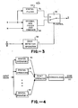
- the selective signal, S, 17 and the low noise signal, M, 18 are applied to the NRS filter and combiner shown generally at 30 and which correspond to the functional elements 15, 16, and 21 in Figure 2.
- the low noise signal, M, or a signal, D, which is the difference (M - S) or any other signal which includes the artifact edges, is applied to an edge detection operator 32 which identifies artifact edges and generates a signal, E, in response thereto.
- the signal E is applied to a switch 36 whereby the signal from the.NRS filter and combiner 30 is overridden upon detection of an artifact edge, and the selective signal, S, or a spatially filtered version thereof derived from the spatial filter 34, is applied as the output image signal, S. Accordingly, in the region of the artifact edge, as identified by the edge detection operator, the artifact generated in the NRS filter and combiner 30 is eliminated by disconnecting the NRS filter and combiner.
- Figure 4 is a functional block diagram of one embodiment of an edge detector operator 32 of Figure 3 and includes a plurality of derivative operators (or other edge filters) 40, 41, and 42, each receiving the input image signal, D or M, in the horizontal and vertical directions to provide the two components of the spatial gradient vector.
- the magnitude of the gradient vector at each point in the image signal as derived by the point operator 44 is then compared with a threshold.
- Other edge map techniques can be employed such as template filters known in the art to detect edge orientation and strength.
Landscapes
- Biochemistry (AREA)
- General Health & Medical Sciences (AREA)
- Signal Processing (AREA)
- Physics & Mathematics (AREA)
- Health & Medical Sciences (AREA)
- Life Sciences & Earth Sciences (AREA)
- Multimedia (AREA)
- Chemical & Material Sciences (AREA)
- Engineering & Computer Science (AREA)
- Analytical Chemistry (AREA)
- General Physics & Mathematics (AREA)
- Immunology (AREA)
- Pathology (AREA)
- Apparatus For Radiation Diagnosis (AREA)
- Image Processing (AREA)
- Analysing Materials By The Use Of Radiation (AREA)
Applications Claiming Priority (2)
| Application Number | Priority Date | Filing Date | Title |
|---|---|---|---|
| US468656 | 1983-02-22 | ||
| US06/468,656 US4499493A (en) | 1983-02-22 | 1983-02-22 | Multiple measurement noise reducing system using artifact edge identification and selective signal processing |
Publications (2)
| Publication Number | Publication Date |
|---|---|
| EP0117154A2 true EP0117154A2 (fr) | 1984-08-29 |
| EP0117154A3 EP0117154A3 (fr) | 1987-02-04 |
Family
ID=23860696
Family Applications (1)
| Application Number | Title | Priority Date | Filing Date |
|---|---|---|---|
| EP84301118A Withdrawn EP0117154A3 (fr) | 1983-02-22 | 1984-02-21 | Système de réduction de bruit à mesure multiple utilisant l'identification du flanc d'impulsion de bruit et traitement de signal sélectif |
Country Status (6)
| Country | Link |
|---|---|
| US (1) | US4499493A (fr) |
| EP (1) | EP0117154A3 (fr) |
| JP (1) | JPS59214433A (fr) |
| AU (1) | AU558490B2 (fr) |
| CA (1) | CA1214286A (fr) |
| IL (1) | IL71012A (fr) |
Cited By (3)
| Publication number | Priority date | Publication date | Assignee | Title |
|---|---|---|---|---|
| GB2170373A (en) * | 1984-12-28 | 1986-07-30 | Canon Kk | Image processing method |
| EP0518588A3 (en) * | 1991-06-10 | 1993-07-21 | General Electric Company | X-ray imaging system with improved spatial filter |
| EP0797347A3 (fr) * | 1996-03-22 | 1998-02-18 | Kabushiki Kaisha Toshiba | Appareil de formation d'images avec une unité de traitement d'images comprenant plusieurs filtres disposés en parallèle |
Families Citing this family (33)
| Publication number | Priority date | Publication date | Assignee | Title |
|---|---|---|---|---|
| US4541106A (en) * | 1984-02-22 | 1985-09-10 | General Electric Company | Dual energy rapid switching imaging system |
| US4729100A (en) * | 1984-08-28 | 1988-03-01 | Kabushiki Kaisha Toshiba | CT System which convolutes projection data with a frequency varying filter function |
| US4678996A (en) * | 1985-05-07 | 1987-07-07 | Picker International, Inc. | Magnetic resonance imaging method |
| GB2181330B (en) * | 1985-09-26 | 1990-05-09 | Toshiba Kk | X-ray inspection apparatus |
| US4827528A (en) * | 1985-11-15 | 1989-05-02 | Stanford University | Error-minimizing noise-reduction system |
| US4792900A (en) * | 1986-11-26 | 1988-12-20 | Picker International, Inc. | Adaptive filter for dual energy radiographic imaging |
| US4761819A (en) * | 1987-02-27 | 1988-08-02 | Picker International, Inc. | Adaptive noise reduction filter for reconstructed images |
| JP2536094B2 (ja) * | 1988-10-20 | 1996-09-18 | 富士ゼロックス株式会社 | カラ―画像処理装置 |
| US4918543A (en) * | 1989-01-24 | 1990-04-17 | Eastman Kodak Company | Apparatus for thresholding an image signal |
| EP0411911B1 (fr) * | 1989-08-02 | 1996-03-27 | Canon Kabushiki Kaisha | Appareil de traitement d'image |
| JP2993014B2 (ja) * | 1989-08-30 | 1999-12-20 | 富士ゼロックス株式会社 | 画像処理装置の画質制御方式 |
| JPH03185574A (ja) * | 1989-12-15 | 1991-08-13 | Fuji Xerox Co Ltd | 多値画像フィルタ処理装置 |
| US5273040A (en) * | 1991-11-14 | 1993-12-28 | Picker International, Inc. | Measurement of vetricle volumes with cardiac MRI |
| KR950003035B1 (ko) * | 1992-04-17 | 1995-03-29 | 주식회사 금성사 | 영상신호의 천이영역 강조장치 및 방법 |
| US5738096A (en) * | 1993-07-20 | 1998-04-14 | Biosense, Inc. | Cardiac electromechanics |
| US5391199A (en) * | 1993-07-20 | 1995-02-21 | Biosense, Inc. | Apparatus and method for treating cardiac arrhythmias |
| JP3372611B2 (ja) * | 1993-10-18 | 2003-02-04 | キヤノン株式会社 | 映像伝送システム、映像処理装置及び映像処理方法 |
| US5830144A (en) * | 1995-03-28 | 1998-11-03 | Vesely; Ivan | Tracking data sheath |
| US6246898B1 (en) | 1995-03-28 | 2001-06-12 | Sonometrics Corporation | Method for carrying out a medical procedure using a three-dimensional tracking and imaging system |
| US5797849A (en) * | 1995-03-28 | 1998-08-25 | Sonometrics Corporation | Method for carrying out a medical procedure using a three-dimensional tracking and imaging system |
| US5868673A (en) * | 1995-03-28 | 1999-02-09 | Sonometrics Corporation | System for carrying out surgery, biopsy and ablation of a tumor or other physical anomaly |
| US5795298A (en) * | 1995-03-28 | 1998-08-18 | Sonometrics Corporation | System for sharing electrocardiogram electrodes and transducers |
| US5817022A (en) * | 1995-03-28 | 1998-10-06 | Sonometrics Corporation | System for displaying a 2-D ultrasound image within a 3-D viewing environment |
| US5718241A (en) * | 1995-06-07 | 1998-02-17 | Biosense, Inc. | Apparatus and method for treating cardiac arrhythmias with no discrete target |
| US5954665A (en) * | 1995-06-07 | 1999-09-21 | Biosense, Inc. | Cardiac ablation catheter using correlation measure |
| US6125213A (en) * | 1997-02-17 | 2000-09-26 | Canon Kabushiki Kaisha | Image processing method, an image processing apparatus, and a storage medium readable by a computer |
| US6019725A (en) * | 1997-03-07 | 2000-02-01 | Sonometrics Corporation | Three-dimensional tracking and imaging system |
| US6490474B1 (en) * | 1997-08-01 | 2002-12-03 | Cardiac Pathways Corporation | System and method for electrode localization using ultrasound |
| US6950689B1 (en) | 1998-08-03 | 2005-09-27 | Boston Scientific Scimed, Inc. | Dynamically alterable three-dimensional graphical model of a body region |
| US6917697B2 (en) * | 2001-05-08 | 2005-07-12 | Ge Medical Systems Global Technology Company, Llc | Method and apparatus to automatically determine tissue cancellation parameters in X-ray dual energy imaging |
| US20050261571A1 (en) * | 2004-05-21 | 2005-11-24 | Willis Nathaniel P | 3-D ultrasound navigation during radio-frequency ablation |
| US7590303B2 (en) * | 2005-09-29 | 2009-09-15 | Samsung Electronics Co., Ltd. | Image enhancement method using local illumination correction |
| JP5106978B2 (ja) * | 2007-10-15 | 2012-12-26 | ジーイー・メディカル・システムズ・グローバル・テクノロジー・カンパニー・エルエルシー | X線ct装置 |
Family Cites Families (3)
| Publication number | Priority date | Publication date | Assignee | Title |
|---|---|---|---|---|
| US4445226A (en) * | 1981-05-05 | 1984-04-24 | The Board Of Trustees Of The Leland Stanford Junior University | Multiple-energy X-ray subtraction imaging system |
| NL8105223A (nl) * | 1981-11-18 | 1983-06-16 | Philips Nv | Inrichting voor verschilbeeldbepaling. |
| US4463375A (en) * | 1982-09-07 | 1984-07-31 | The Board Of Trustees Of The Leland Standford Junior University | Multiple-measurement noise-reducing system |
-
1983
- 1983-02-22 US US06/468,656 patent/US4499493A/en not_active Expired - Lifetime
-
1984
- 1984-02-17 CA CA000447734A patent/CA1214286A/fr not_active Expired
- 1984-02-20 AU AU24730/84A patent/AU558490B2/en not_active Ceased
- 1984-02-21 IL IL71012A patent/IL71012A/xx unknown
- 1984-02-21 EP EP84301118A patent/EP0117154A3/fr not_active Withdrawn
- 1984-02-22 JP JP59032254A patent/JPS59214433A/ja active Pending
Cited By (6)
| Publication number | Priority date | Publication date | Assignee | Title |
|---|---|---|---|---|
| GB2170373A (en) * | 1984-12-28 | 1986-07-30 | Canon Kk | Image processing method |
| GB2170373B (en) * | 1984-12-28 | 1989-03-15 | Canon Kk | Image processing apparatus |
| EP0518588A3 (en) * | 1991-06-10 | 1993-07-21 | General Electric Company | X-ray imaging system with improved spatial filter |
| US5347590A (en) * | 1991-06-10 | 1994-09-13 | General Electric Company | Spatial filter for an image processing system |
| EP0797347A3 (fr) * | 1996-03-22 | 1998-02-18 | Kabushiki Kaisha Toshiba | Appareil de formation d'images avec une unité de traitement d'images comprenant plusieurs filtres disposés en parallèle |
| US5912744A (en) * | 1996-03-22 | 1999-06-15 | Kabushiki Kaisha Toshiba | Image forming apparatus having image processing unit containing a plurality of filters arranged in parallel to each other |
Also Published As
| Publication number | Publication date |
|---|---|
| JPS59214433A (ja) | 1984-12-04 |
| IL71012A0 (en) | 1984-05-31 |
| US4499493A (en) | 1985-02-12 |
| EP0117154A3 (fr) | 1987-02-04 |
| CA1214286A (fr) | 1986-11-18 |
| AU2473084A (en) | 1984-08-30 |
| IL71012A (en) | 1988-11-15 |
| AU558490B2 (en) | 1987-01-29 |
Similar Documents
| Publication | Publication Date | Title |
|---|---|---|
| US4499493A (en) | Multiple measurement noise reducing system using artifact edge identification and selective signal processing | |
| US4503461A (en) | Multiple measurement noise reducing system using space-variant filters | |
| EP1328903B1 (fr) | Procede et dispositif de correction des defauts dans une image numerique et de filtrage du bruit | |
| EP0105619A2 (fr) | Système de réduction de bruit à mesure multiple | |
| US4792900A (en) | Adaptive filter for dual energy radiographic imaging | |
| US4445226A (en) | Multiple-energy X-ray subtraction imaging system | |
| US5963676A (en) | Multiscale adaptive system for enhancement of an image in X-ray angiography | |
| US9791384B2 (en) | X-ray imaging apparatus and method for generating X-ray image | |
| US6760401B2 (en) | Apparatus and method for processing of digital images | |
| US10475181B2 (en) | Image processing apparatus, image processing method, and image processing program | |
| Levin et al. | Digital subtraction angiography: principles and pitfalls of image improvement techniques | |
| Ishida et al. | Development of a new digital radiographic image processing system | |
| US4611341A (en) | Multiple-energy x-ray substraction imaging system | |
| Macovski et al. | Measurement-dependent filtering: a novel approach to improved SNR | |
| Wagner | Low contrast sensitivity of radiologic, CT, nuclear medicine, and ultrasound medical imaging systems | |
| US10438327B2 (en) | Image processing apparatus, image processing method, and image processing program | |
| US4658410A (en) | Stereo X-ray subtraction apparatus | |
| US12573010B2 (en) | Image processing apparatus, radiation imaging system, image processing method, and non-transitory computer-readable storage medium | |
| US20040228444A1 (en) | X-ray diagnostic apparatus with image computer for direction filtering | |
| Kruger | Image data acquisition, processing, storage and display | |
| Macovski | Iodine imaging using spectral analysis | |
| Ikeda et al. | Development and evaluation of a digital radiography system using a large-area flat panel detector | |
| Mistretta et al. | Absorption edge fluoroscopy | |
| JPH0534710B2 (fr) | ||
| Mistretta | Current practice and future directions in digital subtraction angiography |
Legal Events
| Date | Code | Title | Description |
|---|---|---|---|
| PUAI | Public reference made under article 153(3) epc to a published international application that has entered the european phase |
Free format text: ORIGINAL CODE: 0009012 |
|
| AK | Designated contracting states |
Designated state(s): AT BE CH DE FR GB IT LI LU NL SE |
|
| PUAL | Search report despatched |
Free format text: ORIGINAL CODE: 0009013 |
|
| AK | Designated contracting states |
Kind code of ref document: A3 Designated state(s): AT BE CH DE FR GB IT LI LU NL SE |
|
| STAA | Information on the status of an ep patent application or granted ep patent |
Free format text: STATUS: THE APPLICATION IS DEEMED TO BE WITHDRAWN |
|
| 18D | Application deemed to be withdrawn |
Effective date: 19870805 |
|
| RIN1 | Information on inventor provided before grant (corrected) |
Inventor name: NISHIMURA, DWIGHT GEORGE |