EP0121831A2 - Dispositif de mesure de particules diffusantes - Google Patents
Dispositif de mesure de particules diffusantes Download PDFInfo
- Publication number
- EP0121831A2 EP0121831A2 EP84103037A EP84103037A EP0121831A2 EP 0121831 A2 EP0121831 A2 EP 0121831A2 EP 84103037 A EP84103037 A EP 84103037A EP 84103037 A EP84103037 A EP 84103037A EP 0121831 A2 EP0121831 A2 EP 0121831A2
- Authority
- EP
- European Patent Office
- Prior art keywords
- carrier body
- arrangement according
- indicator
- light
- particles
- Prior art date
- Legal status (The legal status is an assumption and is not a legal conclusion. Google has not performed a legal analysis and makes no representation as to the accuracy of the status listed.)
- Withdrawn
Links
Images

Classifications
-
- A—HUMAN NECESSITIES
- A61—MEDICAL OR VETERINARY SCIENCE; HYGIENE
- A61B—DIAGNOSIS; SURGERY; IDENTIFICATION
- A61B5/00—Measuring for diagnostic purposes; Identification of persons
- A61B5/145—Measuring characteristics of blood in vivo, e.g. gas concentration or pH-value ; Measuring characteristics of body fluids or tissues, e.g. interstitial fluid or cerebral tissue
- A61B5/1455—Measuring characteristics of blood in vivo, e.g. gas concentration or pH-value ; Measuring characteristics of body fluids or tissues, e.g. interstitial fluid or cerebral tissue using optical sensors, e.g. spectral photometrical oximeters
- A61B5/14551—Measuring characteristics of blood in vivo, e.g. gas concentration or pH-value ; Measuring characteristics of body fluids or tissues, e.g. interstitial fluid or cerebral tissue using optical sensors, e.g. spectral photometrical oximeters for measuring blood gases
- A61B5/14556—Measuring characteristics of blood in vivo, e.g. gas concentration or pH-value ; Measuring characteristics of body fluids or tissues, e.g. interstitial fluid or cerebral tissue using optical sensors, e.g. spectral photometrical oximeters for measuring blood gases by fluorescence
-
- A—HUMAN NECESSITIES
- A61—MEDICAL OR VETERINARY SCIENCE; HYGIENE
- A61B—DIAGNOSIS; SURGERY; IDENTIFICATION
- A61B3/00—Apparatus for testing the eyes; Instruments for examining the eyes
- A61B3/0008—Apparatus for testing the eyes; Instruments for examining the eyes provided with illuminating means
-
- A—HUMAN NECESSITIES
- A61—MEDICAL OR VETERINARY SCIENCE; HYGIENE
- A61B—DIAGNOSIS; SURGERY; IDENTIFICATION
- A61B5/00—Measuring for diagnostic purposes; Identification of persons
- A61B5/145—Measuring characteristics of blood in vivo, e.g. gas concentration or pH-value ; Measuring characteristics of body fluids or tissues, e.g. interstitial fluid or cerebral tissue
- A61B5/1455—Measuring characteristics of blood in vivo, e.g. gas concentration or pH-value ; Measuring characteristics of body fluids or tissues, e.g. interstitial fluid or cerebral tissue using optical sensors, e.g. spectral photometrical oximeters
- A61B5/1459—Measuring characteristics of blood in vivo, e.g. gas concentration or pH-value ; Measuring characteristics of body fluids or tissues, e.g. interstitial fluid or cerebral tissue using optical sensors, e.g. spectral photometrical oximeters invasive, e.g. introduced into the body by a catheter
-
- G—PHYSICS
- G01—MEASURING; TESTING
- G01N—INVESTIGATING OR ANALYSING MATERIALS BY DETERMINING THEIR CHEMICAL OR PHYSICAL PROPERTIES
- G01N21/00—Investigating or analysing materials by the use of optical means, i.e. using sub-millimetre waves, infrared, visible or ultraviolet light
- G01N21/62—Systems in which the material investigated is excited whereby it emits light or causes a change in wavelength of the incident light
- G01N21/63—Systems in which the material investigated is excited whereby it emits light or causes a change in wavelength of the incident light optically excited
- G01N21/64—Fluorescence; Phosphorescence
- G01N21/6428—Measuring fluorescence of fluorescent products of reactions or of fluorochrome labelled reactive substances, e.g. measuring quenching effects, using measuring "optrodes"
-
- A—HUMAN NECESSITIES
- A61—MEDICAL OR VETERINARY SCIENCE; HYGIENE
- A61B—DIAGNOSIS; SURGERY; IDENTIFICATION
- A61B5/00—Measuring for diagnostic purposes; Identification of persons
- A61B5/68—Arrangements of detecting, measuring or recording means, e.g. sensors, in relation to patient
- A61B5/6801—Arrangements of detecting, measuring or recording means, e.g. sensors, in relation to patient specially adapted to be attached to or worn on the body surface
- A61B5/6813—Specially adapted to be attached to a specific body part
- A61B5/6814—Head
- A61B5/6821—Eye
-
- G—PHYSICS
- G01—MEASURING; TESTING
- G01N—INVESTIGATING OR ANALYSING MATERIALS BY DETERMINING THEIR CHEMICAL OR PHYSICAL PROPERTIES
- G01N21/00—Investigating or analysing materials by the use of optical means, i.e. using sub-millimetre waves, infrared, visible or ultraviolet light
- G01N21/75—Systems in which material is subjected to a chemical reaction, the progress or the result of the reaction being investigated
- G01N21/77—Systems in which material is subjected to a chemical reaction, the progress or the result of the reaction being investigated by observing the effect on a chemical indicator
- G01N2021/7769—Measurement method of reaction-produced change in sensor
- G01N2021/7786—Fluorescence
Definitions
- the invention relates to an arrangement for measuring diffusing particles, which has at least one monochromatic radiation source and a light measuring device, as well as at least one material-delimited, permeable by the particles to be measured, provided with an optical fluorescence indicator, the indicator is optically changeable by the particles.
- Arrangements of the specified type are used to measure diffusing particles, such as the blood-containing oxygen and its partial pressure p02 or the carbonic acid and its partial pressure pC02, invasively or non-invasively.
- diffusing particles such as the blood-containing oxygen and its partial pressure p02 or the carbonic acid and its partial pressure pC02
- the indicator spaces, OPTODES for short, permeable by the particles to be measured are brought into active connection with the measurement object and the change in the measurement light is measured by the optode.
- Electrodes as used for measurement in the known prior art like all electrodes, have an aging process and often have to be serviced with considerable effort. Furthermore, the wire feeds for the electrodes have to be guided onto the contact lens, which leads to impairment of the patient. In addition, the miniaturization of electrodes is only possible to a limited extent. Other types of particles, such as metabolic products from the metabolism of the eye, or other particles, such as cations or anions, are also available at this measurement location. So far, however, they could not be measured there because suitable sensors are missing.
- a carrier body which has depressions in the surface adjacent to the lid, in which indicator spaces (optodes) are arranged.
- a particularly gentle and at the same time intensive irradiation of the optodes is achieved in that light guides are provided within the carrier body, which lead from one or more coupling surfaces arranged on the edge of the carrier body to the optodes.
- a monochromatic radiation source for example a light-emitting diode and the associated radiation receiver, can be arranged in the carrier body, so that the radiation of the optodes and the measurement of the radiation change by the optodes take place directly.
- a monochromatic radiation source for example a light-emitting diode and the associated radiation receiver
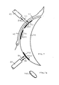
- depressions 1010, 1011,... are arranged in the surface 11 of a carrier body that lies against the eyelid, the rear side of which can be reflective and inserted into the optodes 110, 111 ...
- Such optodes consist, for example, of plastic films, FIG. 1a, in which indicators are arranged which, when irradiated with light of a specific wavelength dependent on the indicator, produce fluorescent light.
- the fluorescent light or “Measuring light” 30 has a different color than the test light 31.
- This measuring light 30, which in FIG. 1, like the test light 31, is transported by a light guide 20, which can have several fibers, can be easily separated from the measuring light by known optical means be separated and proven. The arrangement required for this is known from the art and is not specifically drawn.
- the indicator which generates this fluorescent light is in turn influenced by a particle type to be measured, for example oxygen, which originates from the inner surface of the eyelid 40 and which is in a diffusion equilibrium with the capillary oxygen in the mucous membrane of the eyelid, and changes its fluorescent light output accordingly Concentration of oxygen.
- a particle type to be measured for example oxygen, which originates from the inner surface of the eyelid 40 and which is in a diffusion equilibrium with the capillary oxygen in the mucous membrane of the eyelid, and changes its fluorescent light output accordingly Concentration of oxygen.
- the measurement for p02 and pC02 takes place through the eyelid using the optodes 110, 111 .., so that there are no falsifications by the atmosphere. This is possible because the fluorescent light from the indicator can be easily extracted from the reflected radiation.
- the optodes can easily be made from biocompatible substances, so that neither toxic nor contaminating processes are triggered by the measurement.
- optically decoupling the optodes by means of reflecting or absorbing layers, the radiation into the retina that is possible during the measurement can be switched off.
- the intensity of the fluorescent light measured in this way is multiplied by a correction factor, which is determined by calibration.
- the deviation of the partial prints from the arterial prints can be eliminated by a further correction.
- an optode 112 can also be arranged in the open eyelid slot. Furthermore, several optodes, in particular for simultaneous measurement of several particle types or physical parameters, such as for example the oxygen concentration, the CO 2 partial pressure, the pH value, the GLucose concentration, the temperature. and the like, may be arranged on the device.
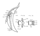
- the arrangement according to FIG. 2 is used when the p02 value, the pH value or the pC02 value are measured with the lid open and the lighting is not done through the eyelid.
- the depression 1010 is arranged on the edge of the carrier body 10 to such an extent that the lid 40 permanently covers this optode.
- the light guide 20 transporting the test light 31 and the measurement light 30 irradiates a coupling surface 105, which is the end surface of a light guide 106 which guides the radiation onto the optode 110.
- the light runs, for example, over a mirror 107 of the optode holder 108
- the measuring light then emanating from the optode runs back in the same way.
- the arrangement 1100 in FIG. 2a which is a one-piece design of the individual parts: coupling surface (105), light guide (106), mirror (107), optode holder (108), can, for example, be manufactured by itself and in a carrier body provided with the corresponding channels 10 be used.
- FIG. 3 shows an arrangement in which supply lines a, b, c are admitted onto the carrier body. This makes it possible to arrange color-emitting light-emitting diodes 120 and photo receivers 121 in the carrier body, which over Lines a, b are fed and their signals are taken off via lines b, c.
- the arrangements 1200, 1210 form light-emitting diode 120, mirror 122, photo receiver 121 and optode holder 123, which are embedded in the carrier body 10.
Landscapes
- Health & Medical Sciences (AREA)
- Life Sciences & Earth Sciences (AREA)
- Physics & Mathematics (AREA)
- General Health & Medical Sciences (AREA)
- Biophysics (AREA)
- Veterinary Medicine (AREA)
- Optics & Photonics (AREA)
- Public Health (AREA)
- Animal Behavior & Ethology (AREA)
- Surgery (AREA)
- Molecular Biology (AREA)
- Medical Informatics (AREA)
- Pathology (AREA)
- Heart & Thoracic Surgery (AREA)
- Biomedical Technology (AREA)
- Engineering & Computer Science (AREA)
- Chemical & Material Sciences (AREA)
- Spectroscopy & Molecular Physics (AREA)
- Immunology (AREA)
- Chemical Kinetics & Catalysis (AREA)
- Biochemistry (AREA)
- Analytical Chemistry (AREA)
- General Physics & Mathematics (AREA)
- Nuclear Medicine, Radiotherapy & Molecular Imaging (AREA)
- Ophthalmology & Optometry (AREA)
- Measurement Of The Respiration, Hearing Ability, Form, And Blood Characteristics Of Living Organisms (AREA)
- Investigating Or Analysing Materials By The Use Of Chemical Reactions (AREA)
- Investigating, Analyzing Materials By Fluorescence Or Luminescence (AREA)
- Investigating Or Analysing Biological Materials (AREA)
Applications Claiming Priority (2)
| Application Number | Priority Date | Filing Date | Title |
|---|---|---|---|
| DE19833313047 DE3313047A1 (de) | 1983-04-12 | 1983-04-12 | Anordnung zur messung von diffundierenden partikeln |
| DE3313047 | 1983-04-12 |
Publications (2)
| Publication Number | Publication Date |
|---|---|
| EP0121831A2 true EP0121831A2 (fr) | 1984-10-17 |
| EP0121831A3 EP0121831A3 (fr) | 1987-11-11 |
Family
ID=6196041
Family Applications (1)
| Application Number | Title | Priority Date | Filing Date |
|---|---|---|---|
| EP84103037A Withdrawn EP0121831A3 (fr) | 1983-04-12 | 1984-03-20 | Dispositif de mesure de particules diffusantes |
Country Status (4)
| Country | Link |
|---|---|
| US (1) | US4597392A (fr) |
| EP (1) | EP0121831A3 (fr) |
| JP (1) | JPS59207135A (fr) |
| DE (1) | DE3313047A1 (fr) |
Cited By (4)
| Publication number | Priority date | Publication date | Assignee | Title |
|---|---|---|---|---|
| WO1990012534A1 (fr) * | 1989-04-26 | 1990-11-01 | Glynn Christopher J | Dispositif pour controler les fonctions du corps |
| EP0567447A1 (fr) * | 1992-04-23 | 1993-10-27 | AVL Medical Instruments AG | Arrangement de sonde pour la détermination directe ou indirecte de paramètre physique ou chimique |
| US5881879A (en) * | 1997-02-07 | 1999-03-16 | Baxter International Inc. | System for preserving and delivering gas-containing solutions |
| WO1999040839A1 (fr) * | 1998-02-13 | 1999-08-19 | Centre National De La Recherche Scientifique (Cnrs) | Mesure optique d'un marqueur fluorescent dans les bronches |
Families Citing this family (33)
| Publication number | Priority date | Publication date | Assignee | Title |
|---|---|---|---|---|
| CA1305873C (fr) * | 1987-05-26 | 1992-08-04 | Howel Gwynne Giles | Methode et instrument pour le dosage de la concentration d'alcool chez les humains |
| AT388248B (de) * | 1987-07-20 | 1989-05-26 | Avl Verbrennungskraft Messtech | Deckschicht, vorzugsweise zur probenseitigen anbringung an optischen ionensensoren |
| EP0312293A3 (fr) * | 1987-10-16 | 1990-03-14 | O.C.T. Optical Chemical Technologies Limited | Dispositif capteur pour analyse |
| US4922919A (en) * | 1988-03-22 | 1990-05-08 | Duke University | Method for measuring ocular oxidative metabolism |
| US5452723A (en) * | 1992-07-24 | 1995-09-26 | Massachusetts Institute Of Technology | Calibrated spectrographic imaging |
| US5560356A (en) * | 1994-02-23 | 1996-10-01 | Vitrophage, Inc. | Diagnostic system and method using an implanted reflective device |
| US6108576A (en) * | 1996-03-18 | 2000-08-22 | The Research Foundation Of City College Of New York | Time-resolved diffusion tomographic 2D and 3D imaging in highly scattering turbid media |
| US6120460A (en) * | 1996-09-04 | 2000-09-19 | Abreu; Marcio Marc | Method and apparatus for signal acquisition, processing and transmission for evaluation of bodily functions |
| US6544193B2 (en) * | 1996-09-04 | 2003-04-08 | Marcio Marc Abreu | Noninvasive measurement of chemical substances |
| HU226183B1 (en) | 1999-08-26 | 2008-06-30 | Novartis Ag | Ophthalmic lens and sensor system for measuring the concentration of an analyte in an ocular fluid |
| GB0016841D0 (en) * | 2000-07-07 | 2000-08-30 | Stanley Christopher J | Optical device for measurement of analytes in tears |
| US20040152963A1 (en) * | 2001-04-27 | 2004-08-05 | March Wayne Front | Apparatus for measuring blood glucose concentrations |
| US6650915B2 (en) * | 2001-09-13 | 2003-11-18 | Fovioptics, Inc. | Non-invasive measurement of blood analytes using photodynamics |
| US20050010091A1 (en) * | 2003-06-10 | 2005-01-13 | Woods Joe W. | Non-invasive measurement of blood glucose using retinal imaging |
| KR101205935B1 (ko) * | 2002-04-22 | 2012-11-28 | 마시오 마크 아우렐리오 마틴스 애브리우 | 뇌 온도 터널의 단부에 있는 피부에 배치하기 위한 지지 구조체 |
| US9848815B2 (en) | 2002-04-22 | 2017-12-26 | Geelux Holdings, Ltd. | Apparatus and method for measuring biologic parameters |
| US8849379B2 (en) * | 2002-04-22 | 2014-09-30 | Geelux Holdings, Ltd. | Apparatus and method for measuring biologic parameters |
| US8328420B2 (en) | 2003-04-22 | 2012-12-11 | Marcio Marc Abreu | Apparatus and method for measuring biologic parameters |
| US6895264B2 (en) * | 2002-08-26 | 2005-05-17 | Fovioptics Inc. | Non-invasive psychophysical measurement of glucose using photodynamics |
| US10227063B2 (en) | 2004-02-26 | 2019-03-12 | Geelux Holdings, Ltd. | Method and apparatus for biological evaluation |
| KR20060044058A (ko) * | 2004-11-11 | 2006-05-16 | 삼성전자주식회사 | 투과반사를 이용한 혈중성분 측정 장치 및 방법 |
| CA2627278A1 (fr) | 2005-10-24 | 2007-05-03 | Marcio Marc Abreu | Appareil et procede de mesure de parametres biologiques |
| US8971978B2 (en) | 2012-08-21 | 2015-03-03 | Google Inc. | Contact lens with integrated pulse oximeter |
| EP4079242B1 (fr) * | 2013-03-19 | 2025-10-08 | Surgisense Corporation | Appareils, systèmes et procédés de détermination de l'oxygénation d'un tissu |
| WO2015054681A1 (fr) | 2013-10-11 | 2015-04-16 | Marcio Marc Abreu | Procédé et appareil pour évaluation biologique |
| WO2015106180A1 (fr) | 2014-01-10 | 2015-07-16 | Marcio Marc Abreu | Dispositifs pour surveiller et appliquer un traitement au niveau d'un tunnel cérébral abreu |
| CN106102566A (zh) * | 2014-01-10 | 2016-11-09 | 马尔西奥·马克·阿布雷乌 | 用于测量abreu脑热通道的红外输出的装置 |
| WO2015112776A2 (fr) | 2014-01-22 | 2015-07-30 | Marcio Marc Abreu | Dispositifs conçus pour effectuer un traitement au niveau d'un tunnel de température cérébrale d'abreu |
| WO2015154094A1 (fr) | 2014-04-05 | 2015-10-08 | Zand Jason M | Appareil, systèmes et procédés permettant la cartographie de l'oxygénation tissulaire |
| WO2016054079A1 (fr) | 2014-09-29 | 2016-04-07 | Zyomed Corp. | Systèmes et procédés pour la détection et la mesure du glucose sanguin du sang et d'autres analytes à l'aide du calcul de collision |
| JP2018512915A (ja) | 2015-03-10 | 2018-05-24 | マーシオ マーク アブリュー | Abtt終端の温度を測定するためのデバイス、装置、システム、及び方法 |
| US9554738B1 (en) | 2016-03-30 | 2017-01-31 | Zyomed Corp. | Spectroscopic tomography systems and methods for noninvasive detection and measurement of analytes using collision computing |
| WO2018187693A1 (fr) | 2017-04-06 | 2018-10-11 | TearDX LLC | Dispositifs oculaires et leurs procédés d'utilisation |
Family Cites Families (17)
| Publication number | Priority date | Publication date | Assignee | Title |
|---|---|---|---|---|
| US3769963A (en) * | 1972-03-31 | 1973-11-06 | L Goldman | Instrument for performing laser micro-surgery and diagnostic transillumination of living human tissue |
| US3893444A (en) * | 1972-07-20 | 1975-07-08 | Univ California | Non-invasively measuring arterial oxygen tension |
| US3769961A (en) * | 1972-07-20 | 1973-11-06 | I Fatt | Conjunctival device |
| USRE29703E (en) * | 1974-01-17 | 1978-07-18 | The Regents Of The University Of California | Non-invasively measuring arterial oxygen tension |
| US4014321A (en) * | 1974-11-25 | 1977-03-29 | March Wayne F | Non-invasive glucose sensor system |
| US4041932A (en) * | 1975-02-06 | 1977-08-16 | Fostick Moshe A | Method for monitoring blood gas tension and pH from outside the body |
| DE2508637C3 (de) * | 1975-02-28 | 1979-11-22 | Max-Planck-Gesellschaft Zur Foerderung Der Wissenschaften E.V., 3400 Goettingen | Anordnung zur optischen Messung von Blutgasen |
| JPS5834129B2 (ja) * | 1975-10-29 | 1983-07-25 | ミノルタ株式会社 | ガンテイミヤクハケイ |
| DE2632710C3 (de) * | 1976-07-21 | 1979-11-08 | Max-Planck-Gesellschaft Zur Foerderung Der Wissenschaften E.V., 3400 Goettingen | Anordnung zur optischen Messung von Stoffkonzentrationen |
| CA1089252A (fr) * | 1976-10-18 | 1980-11-11 | John M. Sperinde | Catheter optique ne necessitant pas une calibration individuelle |
| DE2833356A1 (de) * | 1978-07-29 | 1980-02-14 | Max Planck Gesellschaft | Verfahren zur optischen messung von stoffkonzentrationen |
| US4344438A (en) * | 1978-08-02 | 1982-08-17 | The United States Of America As Represented By The Department Of Health, Education And Welfare | Optical sensor of plasma constituents |
| FR2443233A1 (fr) * | 1978-12-04 | 1980-07-04 | Essilor Int | Verre de contact pour l'electroretinographie |
| DE2915367A1 (de) * | 1979-04-14 | 1980-10-30 | Max Planck Gesellschaft | Optische indikatormessung |
| DE2927814C2 (de) * | 1979-07-10 | 1982-08-05 | Hellige Gmbh, 7800 Freiburg | Verfahren und Vorrichtung zur transkutanen photometrischen Bestimmung der Konzentration von Bestandteilen im Blut |
| US4350163A (en) * | 1980-05-29 | 1982-09-21 | Ford Jr Norman C | Method and apparatus for analyzing contaminants in aqueous humor |
| US4454643A (en) * | 1981-04-20 | 1984-06-19 | Thoratec Laboratories Corporation | Combination membraning tool, package and calibration unit for eyelid sensor or the like |
-
1983
- 1983-04-12 DE DE19833313047 patent/DE3313047A1/de not_active Ceased
-
1984
- 1984-03-20 EP EP84103037A patent/EP0121831A3/fr not_active Withdrawn
- 1984-04-02 US US06/597,105 patent/US4597392A/en not_active Expired - Lifetime
- 1984-04-12 JP JP59071936A patent/JPS59207135A/ja active Granted
Cited By (8)
| Publication number | Priority date | Publication date | Assignee | Title |
|---|---|---|---|---|
| WO1990012534A1 (fr) * | 1989-04-26 | 1990-11-01 | Glynn Christopher J | Dispositif pour controler les fonctions du corps |
| GB2248297A (en) * | 1989-04-26 | 1992-04-01 | Glynn Christopher J | Device for use in real-time monitoring of human or animal bodily functions |
| GB2248297B (en) * | 1989-04-26 | 1993-11-24 | Glynn Christopher J | Device for use in real-time monitoring of human or animal bodily functions. |
| EP0686372A1 (fr) * | 1989-04-26 | 1995-12-13 | GLYNN, Christopher James | Dispositif de surveillance de fonctions corporelles |
| EP0567447A1 (fr) * | 1992-04-23 | 1993-10-27 | AVL Medical Instruments AG | Arrangement de sonde pour la détermination directe ou indirecte de paramètre physique ou chimique |
| US5881879A (en) * | 1997-02-07 | 1999-03-16 | Baxter International Inc. | System for preserving and delivering gas-containing solutions |
| WO1999040839A1 (fr) * | 1998-02-13 | 1999-08-19 | Centre National De La Recherche Scientifique (Cnrs) | Mesure optique d'un marqueur fluorescent dans les bronches |
| FR2774887A1 (fr) * | 1998-02-13 | 1999-08-20 | Centre Nat Rech Scient | Capteur optique sur substrat de silicium et application a la mesure in situ d'un marqueur fluorescent dans les petites bronches |
Also Published As
| Publication number | Publication date |
|---|---|
| EP0121831A3 (fr) | 1987-11-11 |
| JPH0433456B2 (fr) | 1992-06-03 |
| DE3313047A1 (de) | 1984-10-18 |
| JPS59207135A (ja) | 1984-11-24 |
| US4597392A (en) | 1986-07-01 |
Similar Documents
| Publication | Publication Date | Title |
|---|---|---|
| DE3313047A1 (de) | Anordnung zur messung von diffundierenden partikeln | |
| DE602004011471T2 (de) | Optoelektronische Vorrichtung zum schnellen diagnostischen Assay | |
| DE2851138C2 (fr) | ||
| AT391949B (de) | Die optische messung von stoffkonzentrationen | |
| EP0093927B1 (fr) | Dispositif de mesure spectrale dans les vaisseaux sanguins | |
| DE69408976T2 (de) | Glukose fluoreszenz monitor und verfahren | |
| DE2508637C3 (de) | Anordnung zur optischen Messung von Blutgasen | |
| DE2632556C2 (de) | Lichtzuführung für eine Vorrichtung zur optischen Messung von Stoffkonzentrationen | |
| DE69219767T2 (de) | Verfahren und vorrichtung zur überwachung der glukosekonzentration | |
| DE102007003341B4 (de) | Okularsensor und Messsystem zum Nachweis eines Analyten in einer Augenflüssigkeit | |
| CH665345A5 (de) | Sonde zum bestimmen des sauerstoffpartialdruckes. | |
| EP2317909B1 (fr) | Système médical avec lecteur de codes-barres compact pour consommables | |
| DE3528369A1 (de) | Spektralphotometer und spektralphotometrisches verfahren | |
| EP0030610B1 (fr) | Procédé et dispositif de détermination quantitative de substances optiquement actives | |
| DE2215984B2 (de) | Katheter | |
| DE3432157A1 (de) | Bildaufnahmevorrichtung fuer ein endoskop | |
| EP0555645A1 (fr) | Dispositif pour détecter la carie dentaire | |
| DE112019004578T5 (de) | Optischer verteilungssteckverbinder und endoskopsystem | |
| EP0049905A1 (fr) | Dispositif pour mesurer la couleur des gencives | |
| DE602004006396T2 (de) | Analyse einer zusammenstellung mit beobachtung | |
| DE69731327T2 (de) | Gerät und Verfahren zur Feststellung der Urinfarbe | |
| DE102017001484A1 (de) | Verfahren und Anordnung zum Kalibrieren von Vorrichtungen zur Erkennung von Blut oder Blutbestandteilen in einer Flüssigkeit | |
| DE3825352A1 (de) | Verfahren und vorrichtung zur bestimmung von lokalen farbstoff-konzentrationen und von streuparametern in tierischen und menschlichen geweben | |
| DE3243919A1 (de) | Kolorimetrieverfahren und -vorrichtung | |
| DE2204433A1 (de) | Mit einem Katheter verbundenes Mehrfach-Photometer |
Legal Events
| Date | Code | Title | Description |
|---|---|---|---|
| PUAI | Public reference made under article 153(3) epc to a published international application that has entered the european phase |
Free format text: ORIGINAL CODE: 0009012 |
|
| AK | Designated contracting states |
Designated state(s): AT CH FR GB IT LI LU NL SE |
|
| PUAL | Search report despatched |
Free format text: ORIGINAL CODE: 0009013 |
|
| AK | Designated contracting states |
Kind code of ref document: A3 Designated state(s): AT CH FR GB IT LI LU NL SE |
|
| 17P | Request for examination filed |
Effective date: 19880406 |
|
| STAA | Information on the status of an ep patent application or granted ep patent |
Free format text: STATUS: THE APPLICATION IS DEEMED TO BE WITHDRAWN |
|
| 18D | Application deemed to be withdrawn |
Effective date: 19900504 |
|
| RIN1 | Information on inventor provided before grant (corrected) |
Inventor name: LUEBBERS, DIETRICH WERNER, PROF. DR. Inventor name: OPITZ, NORBERT, DR. DIPL.-PHYS. |