EP0127394A2 - Mutants de lignées de cellules T humaines produisant un facteur immunosupresseur et procédé d'obtention de ces mutants - Google Patents
Mutants de lignées de cellules T humaines produisant un facteur immunosupresseur et procédé d'obtention de ces mutants Download PDFInfo
- Publication number
- EP0127394A2 EP0127394A2 EP84303351A EP84303351A EP0127394A2 EP 0127394 A2 EP0127394 A2 EP 0127394A2 EP 84303351 A EP84303351 A EP 84303351A EP 84303351 A EP84303351 A EP 84303351A EP 0127394 A2 EP0127394 A2 EP 0127394A2
- Authority
- EP
- European Patent Office
- Prior art keywords
- cells
- cell
- suppressor
- factor
- 20sif
- Prior art date
- Legal status (The legal status is an assumption and is not a legal conclusion. Google has not performed a legal analysis and makes no representation as to the accuracy of the status listed.)
- Withdrawn
Links
Images







Classifications
-
- C—CHEMISTRY; METALLURGY
- C07—ORGANIC CHEMISTRY
- C07K—PEPTIDES
- C07K14/00—Peptides having more than 20 amino acids; Gastrins; Somatostatins; Melanotropins; Derivatives thereof
- C07K14/435—Peptides having more than 20 amino acids; Gastrins; Somatostatins; Melanotropins; Derivatives thereof from animals; from humans
- C07K14/52—Cytokines; Lymphokines; Interferons
-
- A—HUMAN NECESSITIES
- A61—MEDICAL OR VETERINARY SCIENCE; HYGIENE
- A61F—FILTERS IMPLANTABLE INTO BLOOD VESSELS; PROSTHESES; DEVICES PROVIDING PATENCY TO, OR PREVENTING COLLAPSING OF, TUBULAR STRUCTURES OF THE BODY, e.g. STENTS; ORTHOPAEDIC, NURSING OR CONTRACEPTIVE DEVICES; FOMENTATION; TREATMENT OR PROTECTION OF EYES OR EARS; BANDAGES, DRESSINGS OR ABSORBENT PADS; FIRST-AID KITS
- A61F2/00—Filters implantable into blood vessels; Prostheses, i.e. artificial substitutes or replacements for parts of the body; Appliances for connecting them with the body; Devices providing patency to, or preventing collapsing of, tubular structures of the body, e.g. stents
- A61F2/02—Prostheses implantable into the body
-
- A—HUMAN NECESSITIES
- A61—MEDICAL OR VETERINARY SCIENCE; HYGIENE
- A61K—PREPARATIONS FOR MEDICAL, DENTAL OR TOILETRY PURPOSES
- A61K35/00—Medicinal preparations containing materials or reaction products thereof with undetermined constitution
- A61K35/12—Materials from mammals; Compositions comprising non-specified tissues or cells; Compositions comprising non-embryonic stem cells; Genetically modified cells
- A61K35/28—Bone marrow; Haematopoietic stem cells; Mesenchymal stem cells of any origin, e.g. adipose-derived stem cells
-
- A—HUMAN NECESSITIES
- A61—MEDICAL OR VETERINARY SCIENCE; HYGIENE
- A61L—METHODS OR APPARATUS FOR STERILISING MATERIALS OR OBJECTS IN GENERAL; DISINFECTION, STERILISATION OR DEODORISATION OF AIR; CHEMICAL ASPECTS OF BANDAGES, DRESSINGS, ABSORBENT PADS OR SURGICAL ARTICLES; MATERIALS FOR BANDAGES, DRESSINGS, ABSORBENT PADS OR SURGICAL ARTICLES
- A61L27/00—Materials for grafts or prostheses or for coating grafts or prostheses
- A61L27/36—Materials for grafts or prostheses or for coating grafts or prostheses containing ingredients of undetermined constitution or reaction products thereof, e.g. transplant tissue, natural bone, extracellular matrix
-
- A—HUMAN NECESSITIES
- A61—MEDICAL OR VETERINARY SCIENCE; HYGIENE
- A61P—SPECIFIC THERAPEUTIC ACTIVITY OF CHEMICAL COMPOUNDS OR MEDICINAL PREPARATIONS
- A61P37/00—Drugs for immunological or allergic disorders
- A61P37/02—Immunomodulators
- A61P37/06—Immunosuppressants, e.g. drugs for graft rejection
-
- C—CHEMISTRY; METALLURGY
- C12—BIOCHEMISTRY; BEER; SPIRITS; WINE; VINEGAR; MICROBIOLOGY; ENZYMOLOGY; MUTATION OR GENETIC ENGINEERING
- C12N—MICROORGANISMS OR ENZYMES; COMPOSITIONS THEREOF; PROPAGATING, PRESERVING, OR MAINTAINING MICROORGANISMS; MUTATION OR GENETIC ENGINEERING; CULTURE MEDIA
- C12N5/00—Undifferentiated human, animal or plant cells, e.g. cell lines; Tissues; Cultivation or maintenance thereof; Culture media therefor
- C12N5/06—Animal cells or tissues; Human cells or tissues
- C12N5/0602—Vertebrate cells
- C12N5/0634—Cells from the blood or the immune system
- C12N5/0647—Haematopoietic stem cells; Uncommitted or multipotent progenitors
-
- A—HUMAN NECESSITIES
- A61—MEDICAL OR VETERINARY SCIENCE; HYGIENE
- A61K—PREPARATIONS FOR MEDICAL, DENTAL OR TOILETRY PURPOSES
- A61K35/00—Medicinal preparations containing materials or reaction products thereof with undetermined constitution
- A61K35/12—Materials from mammals; Compositions comprising non-specified tissues or cells; Compositions comprising non-embryonic stem cells; Genetically modified cells
- A61K2035/124—Materials from mammals; Compositions comprising non-specified tissues or cells; Compositions comprising non-embryonic stem cells; Genetically modified cells the cells being hematopoietic, bone marrow derived or blood cells
-
- A—HUMAN NECESSITIES
- A61—MEDICAL OR VETERINARY SCIENCE; HYGIENE
- A61K—PREPARATIONS FOR MEDICAL, DENTAL OR TOILETRY PURPOSES
- A61K38/00—Medicinal preparations containing peptides
Definitions
- This invention relates generally to new mutant cell lines and methods for producing them, and more specifically to a mutant human T cell line and its subclones which secrete high titer suppressive factor.
- lymphokines soluble factors
- the profile of factors reported include migration inhibitory factor (2,3), lymphotoxin (4,5), transfer factor (6,7), lymphocyte proliferation inhibitory factor (8,9) and interferon (10,11).
- lymphokines have been strongly impeded by two major limitations. Firstly, due to the heterogeneity of the cell populations used to prepare conventional lymphokines, it is extremely difficult to assign a given biological activity to a distinct molecule. Secondly, the amounts of a given lymphokine in conventional preparations are usually too small to allow a precise biochemical analysis of the biologically active molecules. Moreover, since lymphocytes have a finite and usually short lifetime in culture, it is impossible to obtain repeatedly homogenous lymphokine preparations for characterization studies. These difficulties with regard to the study of lymphokines have caused workers in the field to experiment with cell lines and hybridomas.
- T cell growth factor T cell growth factor
- T-T hybridomas have been constructed (13-17) to immortalize lymphokine-secreting T cells. Since hybridomas are TCGF independent, some investigators in the field have thought they would serve as better sources than cloned T cell lines for preparing larger quantities of lymphokines for biochemical characterization.
- Human lymphoid cell lines also produce a number of factors similar to mediators of cellular immunity released by lymphocytes.
- Gillis, et al., (18) showed that the human T cell line Jurkat produces large quantities of human interleukin 2 (IL2) upon stimulation with mitogen.
- Farrar, et al., (19) showed that the mouse thymoma EL4 secretes high concentrations of mouse IL2 upon stimulation by the mutagen phorbol myristate acetate.
- CEM human lymphoid cell line CCRF-CEM
- Vesole Yoshida, et al., J. Immunol., 117, 518 (1976)
- CEM among other cell lines
- Irigoyen, et al. describe the preparation of a 6-thioguanine (6T) resistant mutant of the CEM cell line and the subsequent fusing of this mutant with lectin- stimulated human T cells to produce human-human T cell hybridomas. No mention is made of the source of the parent C E M cell line.
- the method used for production of the mutant involved an initial mutation by ethyl methanesulfonate followed by exposure to increasing concentrations of 6T (1.25 ⁇ g - 5 ⁇ g/ml), after which the mutant cells were maintained in 16.7 ⁇ g/ml of 6-thioguanine.
- Irigoyen, et al. is the only group known to Applicant which has reported on the characteristics of a 6T-resistant mutant CEM line.
- Their mutant line CEM-T15 (the only one discussed in the reference) was described as lacking the OKT3 antigen and having a chromosome number (karotype) of 35-47, the same. as the parent CEM line used in its production.
- CEM-T15 Irigoyen's parent CEM line and the mutant line CEM-T15 both grew normally in the presence of 10- 8 M aminopterin (0.004 ug/ml), both died in the presence of 10- 7 M aminopterin (0.04 ug/ml), and there was differential growth (CEM grew, while CEM-T15 died) at 3.3x10 -8 M and 6.6xlO- 8 M aminopterin (0.013 and 0.026 ug/ml, respectively).
- the reference also indicates that CEM-T15"inhibits the background level of P F C (plaque forming cells) generated in cultures containing B cells and PWM (pokeweed mitogen)." This result indicates that CEM-T15 inhibits B cell differentiation. No information is given regarding any action on T cell differentiation.
- a method producing a stable 6-thioguanine resistant mutant of a lymphoblastoid cell line secreting an enhanced level of lymphokine which comprises treating said lymphoblastoid cell line with from 20 to 30 ⁇ g/ml of 6-thioguanine for a period of time sufficient to kill at least 90% of the cells and isolating the viable cells.
- This method is applicable to lymphoblastoid cell lines of human or animal origin, as well as to lymphoblastoid cell lines of T cell or B cell origin.
- a stable 6-thioguanine resistant mutant of the lymphoblastoid cell line CEM which secretes constitutively a high titer of a non-mitogenic, non-cytotoxic T-cell suppressor inducer factor.
- a non-mitogenic, non-cytotoxic T cell suppressor factor which suppresses at least 90% of T cell proliferation to nitogenic, stimulation at dilution of up to 2 x 10 -4 and whose suppressive activity is blocked by the monosaccharide rhamnose.
- a novel stable 6-thioguanine resistantmutant of the lymphoblastoid cell line CCRF-CEM (designated 6T-CM) which secretes constitutively into the growth medium a high titer of a non-mitogenic, non-ctyotoxic-T cell suppressor-inducer factor (SIF) which induces production of a T cell suppressor factor (TSF).
- the cell-free suernatant of the particular novel mutant suppresses at least 90% of mitogen-induced T cell proliferation at dilutions as high as 10- 6 , while the cell-free supernatant of its most productive subclone exhibits such suppression at a dilution up to 10- 9 .
- the supernatants do not suppress mitogen-induced B cell proliferation at these dilutions.
- This titer has remained constant for over 8 months.
- the mechanism of action, the cellular target, and the biochemical characterization of the factors responsible for the observed suppression have been determined.
- the factor SIF also exhibits T cell proliferation suppression in vivo.
- mutant as used herein in reference to the subject mutant and its subclones means that the mutant and its subclones continue to secrete high titer SIF for at least six months. Additionally, the "stable" mutant and subclones do not suffer any appreciable change in their karotype over this same period. It should be understood in general that mutants or subclones of mutants which are not stable are not practically useful, since they will not serve as reliable sources of lymphokines in general or SIF in particular.
- non-mitogenic means that the SIF secreted by the subject mutant and its subclones has no observed mitogenic effect on human T cells.
- non cytotoxic means that the SIF secreted by the subject mutant and its subclones has no observed cytotoxic effect on human T cells.
- suppression and its variants as used herein mean a diminution in the activity which is “suppressed”, which diminution may be partial or complete.
- Mutant cell lines according to the invention are useful not only for secretion of suppressor-inducer factor, but also as potential partners in hybridoma formation.
- aminopterin sensitivity is extremely important, because human lymphocytes (and thus human hybridomas produced from human lymphocytes) are much more sensitive to aminopterin than corresponding mouse lymphocytes or hybridomas. Accordingly, in order to differentially select human hybridomas using HAT medium, smaller amounts of amino p terin must be used in the medium to ensure survival of the desired hybridomas and hence the unfused cell line must be very sensitive to the material.
- the SIF and TSF produced as part of the subject invention are useful for suppressing T cell proliferation both in vitro and in vivo.
- mutant cell line 6T-CEM includes that designated 6T-CEM-20
- method for preparing the cell lines and the subclones comprising treating said T cells in vitro with an effective T cell proliferation suppressing amount of either the suppressor inducer factor or the T cell suppressor factor.
- the invention also includes the suppressor inducer factor or T cell suppressor factor, or pharmaceutical compositions containing them, for use in vivo in suppressing T cell proliferation.
- the particular mutant cell line 6T-CEM was produced by treating the parent line CEM (obtained from the American Type Culture Collection) with an initial high concentration of 6T (about 20-30 ug/ml of 6T) for a period of time sufficient to kill at least 90% of the cells: the small percentage of surviving cells after seven days treatment with 30 ⁇ g/ml of 6T constituted the mutant cell line 6T-CEM.
- the parent line CEM obtained from the American Type Culture Collection
- an initial high concentration of 6T about 20-30 ug/ml of 6T
- no additional mutagen was used and the cells were exposed to an initial high concentration of 6T rather than progressively increasing concentrations. It appears that the treatment of the parent line with a high concentration of 6T from the beginning was significant in yielding the subject mutant cell line.
- Prior art references dealing with mutating procedures have taught one to begin with low concentrations of mutating agent and progressively increase the amount.
- the Irigoyen, et al., reference is typical.
- the particular cell line was obtained, it was possible by techniques known in the art to subclone the particular mutant cell line to obtain subclones having higher levels of suppressor-inducer factor production. Both the particular mutant cell line and its subclones possess the OKT3 antigen and may be detected with OKT3 monoclonal antibody (31).
- This SIF-secreting mutant cell line is surprising not only in view of the state of the art, but also in view of Applicant's unsuccessful attempts to produce such a mutant cell line of CEM using azaguanine.
- the parental line CEM secretes only low levels of an uncharacterized suppressive factor, as set out in Vesole, et al.
- the Okada, et al., reference noted above, which discloses an azaguanine-resistant mutant of CEM indicates only that T-T hybridomas prepared using this cell line secreted IL-2 and a helper factor. No information is presented regarding any factors which might be secreted by the azaguanine-resistant mutant CEM itself.
- the method used above for producing the mutant cell line 6T-CEM is also effective to produce stable mutants of other lymphoblastoid cell lines which have enhanced production of lymphokines over the parent cell line used for production of the mutant.
- the lymphoblastoid cell line HSB (ATCC CCL 120.1) produces an uncharacterized suppressive factor which suppresses mitogen-induced T cell differentiation only at a dilution of 10- 1 .
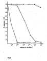
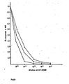
- FIG. 1 Suppression of T cell proliferation by supernatants of CEM, 6T-CEM and its subclone 6T-CEM-20. 10 5 cells/well of T lymphocytes were stimulated with 0.1% phytohemagglutinin (PHA). Cell free supernatants from CEM (A), 6T-CEM (o) and 6T-CEM-20 (x) were added at different dilutions as indicated in the figure. Proliferation was quantitated by measuring 3 H-thymidine uptake on day 4.
- PHA phytohemagglutinin
- the 3 H-thymidine uptake for T cells alone in PHA was 12021 ⁇ 1216 cpm.
- PHA-P Purified phytohenagglutinin
- PWM Pokeweed mitogen
- mitomycin C mitomycin C
- C57/B6 and DBA/2 mice were obtained from the Jackson Laboratory.
- Growth medium for human cell lines consisted of a-minimum essential medium (a-MEM) (Gibco) supplemented with penicillin (Gibco, 50 ⁇ g/ml), streptomycin (Gibco, 100 pg/ml), L-glutamine (Gibco, 2.0 mM) and 2-10% fetal calf serum (FCS, Gibco).
- a-MEM a-minimum essential medium
- FCS 2-10% fetal calf serum
- the medium for the T cell proliferation assay was the same except RPMI 1640 (Gibco) was used instead of a-MEM; 10% FCS was used at all times.
- RPMI 1640 Gibco
- FCS 10% FCS was used at all times.
- the monosaccharides fructose, NAG, rhamnose, and a-methyl mannoside, and the prostaglandin inhibitor indomethacin were obtained from Sigma.
- 6-thioguanine (6T) resistant mutant 6T-CEM and the subclone 6T-CEM-20 The 6-thioguanine (2-amino-6-mercaptopurine; 6T) was obtained from Sigma (Cat. No. A-4882). 100 mg of 6T was dissolved in 100 ml of distilled water. 1 or 2 drops of 10 N NaOH were added to help the 6T to dissolve completely. The pH of the final solution is about 9.
- the lymphoblastic leukemic line CEM (CCRF-CEM, ATCC CCL 119) was obtained from the American Type Culture Collection, Rockville, MD, and was maintained in 90% a-MEM and 10% FCS. CEM cells at log phase (10 6 cells/well) were treated with various concentrations of 6T and viability was assayed 7 days later (Table 1). Cells treated with 30 ug/ml of 6T were selected for further studies. The remaining viable cells (4%) were grown in 25 mm culture flasks. until a significant number of cells was observed, after which dead cells were removed by a Ficoll-Hypaque gradient. The viable cells were divided into 2 parts.
- the growth characteristics, karotype, sensitivity to aminopterin, level of suppressor activity and effects of supernatants on other immunological assays are listed in Table 3 for CEM, 6T-CEM, 6T-CEM-20, and Az-CEM (an azaguanine-resistant CEM mutant).
- the line 6T-CEM-20 has been showing a consistent level of suppressor inducer factor secretion for greater than eight months, indicating no loss of chromosome(s) involved in the secretion of the factor.
- the stability data are shown in Table 4.
- 6T-CEM cells were routinely maintained in a-MEM containing 10% FCS.
- For production of the suppressive supernatants from 6T-CEM cells were washed and suspended at 2x10 5 cells/ml or 10 6 cells/ml in a-MEM with 2%. FCS. Supernatant was harvested 48 hours later and the functional activities were assessed.
- the standard procedure for the production of the lymphocyte derived suppressor factor TSF involved incubation of the lymphocyte population of interest at 3x10 6 cells/ml in the presence.of 1/500 dilution pf 6T-20SIF (the suppressor-inducer factor produced by the subclone 6T-CEM-20). After 24 hours, cells were washed 3-4 times and then set up at 1x10 6 cells/ml in RPMI 1640 with 2% FCS. Supernatants were harvested 72 hours afterwards unless otherwise indicated.
- Peripheral blood lymphocytes were obtained from the blood of normal donors by using standard Ficoll-Hypaque gradient techniques (21). The T and non-T cells were separated by standard sheep red blood cell (SRB C ) rosetting technique (21). Briefly, 5x10 6 /ml P BL were incubated with 1% neuraminidase-treated SRBC, and rosetted cells were separated from non-rosetted cells on a F icoll-Hypaque gradient. The rosetted cells were designated as T cells. Purified B lymphocytes were obtained from non-rosetted cells by removal of residual T cells using the Pan T monoclonal antibody OKT11 and complement.
- Purified OKT4 + and OKT8 + cells were obtained by treating T lymphocytes (10 7 /ml) with 1 ⁇ g of the respective monoclonal antibody, followed by complement lysing. Dead cells were removed by Ficoll-Hypaque gradient and purity of the subset was assayed with the ORTHO Spectrum III cytofluorograph (Ortho Diagnostic Systems) using fluorescent labeled monoclonal antibodies. Adherent cells were removed by putting 10 ml of lymphocytes at 10 6 /ml on a 100 mm Petri dish (Falcon). Cells were incubated at 37°C for 2-3 hours. The nonadherent cells were recovered from the medium and the subsequent washes with gentle pipetting.
- Adherent cells were released from the plate by the addition of ethylenediamine tetraacetic acid (EDTA, 0.2mM, Gibco). Employing these techniques, T lymphocytes were usually 95% OKTll + and 1-2% OKMl + before removal of adherent cells, and 96% OKTll + and OKMl - after removal of adherent cells. B cells were OKTll - , 68% Ig+ and 20% OKM1 + before removal of adherent cells, and 78% Ig + and 2% OKMl + after removal of adherent cells. OKTll staining was done by direct fluorescent technique using FITC-conjugated monoclonal antibody.
- Ig staining was done by using FITC-conjugated goat anti-human Ig (Meloy), and OKMl staining was done by indirect fluorescent staining technique using OKM1 and FITC-conjugated goat anti-mouse Ig (Meloy). All OK monoclonal antibodies were supplied by Ortho Diagnostic Systems (31).
- T cell proliferation 7x10 4 - 10 5 human T lymphocytes were cultured in U bottom microtiter plates (Flow) in the presence of 0.1% PHA-P or 1/64 PWM for 4 days. 3 H thymidine (0.1 ⁇ Ci/well) was added for the last 6 hours of the culture period.
- T cell dependent B cell proliferation 2x10 4 T cells and 10 5 B cells were cultured together in the presence of 1/64 dilution of PWM for 4 days. 3 H thymidine was added for the last 6 hours of the culture period. B cell proliferation was carried out exactly the same manner with no T cells added to the culture.
- MLC Mixed lymphocyte culture
- Human 5x10 4 or 10 5 human T lymphocytes were cultured with 10 5 mit-C-treated (30 ⁇ g/ml per 10 7 cells for 30 minutes at 37°C) allogenic non-rosetted cells, autologous non-rosetted cells, or DBA spleen cells (xenogenic cells) for 5 days. 3 H thymidine was added for the last 12 hours of the culture period.
- Mouse 2x10 5 C57/B6 spleen cells were cultured with 2x10 5 mit-C-treated DBA spleen cells for 5 days and 3 H thymidine was added for the last 12 hours of the culture period. Percent response was calculated according to the same formula used for lymphocyte proliferation assays.
- T lymphocytes pretreated with 6T-20SIF or medium were cocultured with 5x10 4 fresh T cells together with 10 4 mit-C treated SB cells as stimulator cells. Cells were cultured for 5 days and 3 H thymidine was added for the last 6 hours of the culture period.
- Cytotoxic T cell assay Purified T lymphocytes were suspended at 106/ml in RPMI containing 5% human AB serum. 100 ⁇ l of cells were put into U bottom microtiter wells together with 10 4 mit-C treated SB cells (30 ⁇ g mit-C for 30 min.) and various dilutions of TSF. After 3 days, 10 4 51 C r labeled SB cells were added to each well and the plates were spun at 1000 g for 10 min. After 4 hours of incubation at 37°, 100 ⁇ l of supernatant was removed and counted in a Beckman Gamma counter. The % cytotoxicity was calculated using the following formula:
- Enzymatic digestion Trypsin was added to 6T-20SIF at a final concentration of 0.1% and incubated for 2 hours at 37° C . Enzymatic reaction was stopped by the addition of FCS to a final concentration of 10%. Controls were set up consisting of medium substituted for. the trypsin. Ammonium sulfate precipitation. Cell-free supernatant was centrifuged at 10,000 rpm for 20 minutes to remove any debris. Ammonium sulfate was then added gradually to give a final 50% ammonium sulfate saturation. After stirring for 45 minutes, the solution was centrifuged at 10,000 rpm for 30 minutes. The precipitate, which contained the suppressor activity, was exhaustively dialyzed against PBS. All steps were carried out in the cold and the pH was maintained between pH 7.0 and pH 7.4.
- 6T-CEM the thioguanine resistant mutant of the lymphoblastoid cell line CEM, secretes into the growth medium a high titered suppressive factor which inhibits T cell proliferation to PHA to the 95% level (10/10 experiments).
- the parental line CEM used herein also secretes a suppressor factor, but the titer of this factor is about 1000 fold lower than the titer of SIF produced by 6T-CEM.
- 6T-CEM By subcloning 6T-CEM, there was obtained one clone 6T-CEM-20 which produced 10 fold higher suppressive activity than 6T-CEM (Fig. 1).
- 6T-CEM-20 has been showing stable levels of factor production for over 8 months.
- the supernatant of 6T-CEM-20 is referred to herein as 6T-20SIF.
- the suppressive activity (50% response) of 6T-20SIF has also been observed at a dilution of 10- 9 .
- T cells were fractionated into OKT4 + and OKTB + subpopulations by monoclonal antibody-mediated complement cytolysis. Dead cells were removed on a Ficoll-Hypaque gradient. The purified cells were 95% viable and showed 90-95% purity as tested by fluorescent antibody labelling and measured by the ORTHO Spectrum III cytoflorometer.
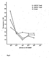
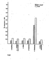
- the differential effects of 6T-20SIF on OKT4 (helper/ inducer) and OKT8 (suppressor/cytotoxic) subsets are shown in Fig. 2. 6T-20SIF suppressed equally well the proliferation of both the OKT4 + and the OKT8 + populations (31).
- 6T-20SIF did not suppress via a cytotoxic mechanism.
- the number of cells staining positively in the presence of trypan blue did not increase when cultured 3-7 days without mitogen or alloantigen.
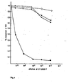
- 6T-20SIF Effect of 6T-20SIF on B cell proliferation (Fig. 3).
- 6T-20SIF suppressed B cell proliferation only at 10 -1 dilution, whereas almost 100% suppression of T cell proliferation was observed at 10- 5 dilution.
- T cell dependent B cell proliferation was partially suppressed by 6T-20SIF at 10 -4 dilution. Similar results were obtained in two other experiments. 6T-20SIF did not suppress PWM induced Ig synthesis at 10- 1 dilution.
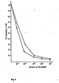
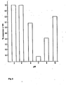
- T lymphocytes were treated with medium or 10 -5 -10 -1 dilutions of 6T-20SIF at 10 7 cells/ml for 8 to 24 hours at 37°. Cells were then washed 3-4 times and set up for PHA induced proliferation assay as described herein. As can be seen in Fig. 5, T lymphocytes that were in contact with 10- 3 dilution of 6T-20SIF for 8 hours or with 10 -4 dilution of 6T-20SIF for 24 hours did not proliferate in response to PHA stimulation.
- 6T-20SIF was also tested in a mouse spleen cell mixed lymphocyte culture experiment. 2x10 5 C57/B6 spleen cells were cultured with various concentrations of 6T-20SIF in the presence of 2x10 5 mit-C treated DBA cells. The suppressive effect observed as shown in Fig. 7 was very similar to that of human lymphocytes. 6T-20SI F thus does not exhibit species restriction.
- T lymphocytes were incubated at 3x10 6 cells/ml with different dilutions of 6T-2,OSIF or growth medium for 24 hours. Cells were washed extensively and then set up in coculture experiments with 5x10 4 fresh allogenic T cells in a PHA-induced proliferation assay or with 2.5x10 4 T cells and 10 5 B cells in a PWM induced T cell dependent B cell proliferation assay.
- T lymphocytes exposed to 10- 4 dilution of 6T-20SIF for 24 hours suppressed 90-95% T cell proliferation in a coculture assay (suppression observed in 3 out of 3 experiments).
- T cell induced B cell proliferation was partially suppressed at around 10- 2 dilution.
- the effect of 6T-20SIF treated T cells on fresh T cell proliferation in a one way mixed lymphocyte culture (MLC) stimulated by mit-C treated SB cells is shown in Fig. 12.
- 6T-20SIF pretreated T cells suppressed T cell proliferation in a one way MLC in a dose-related fashion with the pattern of suppression similar to the inhibition of mitogenic stimulation.
- T lymphocytes Production of suppressor factor by 6T-20SIF treated lymphocytes .(Fig. 13, Table 5). After 24 hours of exposure to 6T-20SIF or growth medium followed by extensive washing, T lymphocytes were set up at 1x10 6 cells/ml in RPMI+2% FCS. Supernatants were harvested at various times to test for suppressor activities in a PHA induced T cell proliferation assay. Fig. 13 depicts the results of this time-course experiment. Significant amounts of suppressor activity were released into the medium at 24 hours, whereas production of T cell suppressor factor (TSF) reached maximum level at around 48-72 hours (3 out of 3 experiments).
- TNF T cell suppressor factor
- TSF Production of TSF by different lymphoid populations and the effect of various treatments and culture conditions on the production are shown in Table 5.
- the purified T cell population is the major producer of TSF, while the depletion of residual adherent cells did not affect the amount of factor produced (2 out of 2 experiments).
- Both the OKT4 + population and the OKTB + population are potent producers.
- a purified B cell population (less than 0.1% of T cells) produces about half of what purified T cells would have produced under the same culture condition (2 out of 2 experiments).
- TSF shows serum dependency. Serum-free medium did not support full production, whereas the presence of 2% FCS was sufficient for maximum secretion of TSF by T lymphocytes. Mit-C treatment and irradiation at 6000 R abolished almost completely the capacity of 6T-20SIF treated cells to elaborate TSF. Irradiation at 2000 R or 3000 R caused only partial abolition of TSF production.
- TSF Mechanism of action of TSF (Fig. 14, Table 6).
- the suppressive effects of TSF on PHA induced T cell proliferation, PWM induced T cell dependent B cell proliferation, and PWM induced B cell proliferation are shown in F ig. 14.
- TSF suppressed 85-90% PHA induced T cell proliferation at around 10- 3 dilution but showed only partial suppression on T cell dependent B cell proliferation at 10- 2 dilution.
- Proliferation of PWM stimulated B lymphocytes was only slightly suppressed at 10- 1 dilution (3 out of 3 experiments).
- the pattern of suppressive activities is comparable to that of 6T-20SIF. Similar to 6T-20SIF, TSF has no suppressive effect on the generation of plaque- forming cells.
- TSF The effect of TSF on the generation of cytotoxic T cells is shown in Table 6.
- TSF differential suppressive effects of TSF on various functional subsets of lymphocytes indicate that TSF suppresses via a noncytotoxic mechanism. This was further substantiated by studies showing that TSF did not augment the number of cells staining positively in the presence of trypan blue dye when cultured 3-5 days without mitogen (3 out of 3 experiments). Similar findings were also true for 6T-20SIF.
- TSF-induced suppression (Fig. 15). TSF was added to a PHA induced T cell proliferation assay at various times after culture initiation. When added at mid-culture period, suppressive activity was reduced by 50%, while no suppressive effect was observed when TSF was added at the last day of the culture period. The presence of TSF at the early phase of culture period is thus required for maximum suppression, which is similar to what was observed with 6T-20SIF. However, if TSF was added at the beginning of a culture period, there is little difference in percent suppression observed in a 3 or 4 day PHA-induced T cell proliferation assay. 6T-20SIF, on the other hand, did not show much suppressive effect on T cell proliferation in a 3 day lectin induced mitogenesis assay. A minimum of 4 day culture period is required for 6T-20SIF to exert maximum suppression.
- the subject 6-thioguanine resistant mutant of the human lymphoblastoid cell line CEM secretes constitutively into the growth medium high titered immunosuppressive factor.
- the cell-free supernatant 6T-20SIF at dilutions as high as 10- 9 suppressed at least 90% of T cell proliferation to both mitogenic and antigenic stimulations.
- T cell induced B cell proliferation was suppressed only by lower dilutions of the supernatant (10 -3 ) and mitogen stimulated B cell proliferation was only -partially affected at dilutions of 10 - 1 .
- the parental line CEM also secretes into the growth medium low levels of previously uncharacterized suppressive factor. As shown by Vesole, et al., CEM supernatant caused a 50% inhibition in mitogenically-induced T cell proliferation at a dilution of 10 -0.8 .
- the parental CE M line used by Applicant was able to achieve the same degree of inhibition at a dilution of 5X10 -3 , although this fact was not previously known.
- the subject mutant cell line and its most productive subclones are capable of showing this same degree of inhibition at dilutions of 10- 6 and 10- 9 , respectively.
- 6T-CEM-20, 6T-20SIF suppressed equally well the proliferation of both OKT4 + and OKT8+ populations to PHA stimulation. Allogenic, xenogenic and autologous stimulation of T lymphocytes by mit-C treated stimulated cells were suppressed in similar fashion as were mitogenic stimulation. 6T-20SIF did not exhibit species restriction since it also suppressed mouse spleen cell proliferation to both mitogenic and allogenic stimulations.
- 6T-20SIF is optimally present at the initiation of the culture period. No suppressive effect was observed when 6T-20SIF was added 48 hours after culture initiation, indicating that 6T-20SIF is probably involved in suppressing the early stages of mitogen induced proliferation with little or no effect on the terminal stages.
- the continuous presence of 6T-20SIF throughout the culture period is not required, since cells that had been exposed for 8 to 24 hours to 6T-20SIF did not respond to PHA induced proliferation. This cannot be caused by the presence of residual 6T-20SIF, since the medium from the last wash contained no suppressive activity.
- 6T-20SIF suppressed more profoundly PHA induced T cell proliferation in a 4- day culture than a 3-day culture, although the degree of PHA induced proliferation in the absence of 6T-20SIF did not show significant differences between the two culture periods.
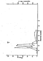
- Partial purification by Sephacryl® S-200 gel chromatography indicates that 6T-20SIF is a high molecular weight protein of around 110,000 dalton.
- the activity and optical density profile did not change in the presence of mercaptoethanol or 0.1 M salt.
- SDS gel electrophoresis of the active fraction showed a major band of protein around 110,000 dalton.
- TSF T cell specific suppressor factor
- the prior art suppressor agent cyclosporin is highly toxic.
- the potent suppressive effect of 6T-20SIF on T cell proliferation is believed to be mediated via the induction of the lymphocyte specific TSF because: 1) The targets of action are identical for the two factors. Both 6T-20SIF and TSF suppressed 90-95% T cell proliferation to mitogenic and antigenic stimulation at high dilutions of 10- 9 and 10- 3 respectively. T cell dependent B cell proliferation to mitogen was partially suppressed by both at lower dilutions of 10 -4 and 10- 2 . Mitogenic induced B cell proliferation and B cell differentiation were not suppressed by either: 2) The time course of suppression indicates that TSF and not 6T-20SIF is the immediate suppressor factor.
- TSF direct suppression was exerted on lymphocytes at the initiation of the culture period and maximum suppression could be detected 72 hours later, the time when PHA induced T cell proliferation almost reached its maximum level (under the culture conditions herein PHA induced proliferation usually peaked at day 4); 3)
- the suppressive activities of both 6T-20SIF and TSF were blocked by the monosaccharide rhamnose but not by other monosaccharides such as fructose, NAG, or a-methyl mannoside or by the prostaglandin inhibitor indomethacin.
- rhamnose Since the presence of rhamnose did not affect the level of 6T-20SIF induced TSF production, it is unlikely that rhamnose blocked the lymphocyte suppressive activity of 6T-20SIF by interfering with its ability to induce TSF. Therefore, the target for rhamnose-mediated blockage is TSF.
- TSF activity detected was due merely to residual 6T-20SIF that was bound loosely onto the lymphocyte surface and was then gradually released into the supernatant during the subsequent culture period, since lymphocytes that were treated with 30 ug/ml of mit-C after exposure to 6T-20SIF did not elaborate TSF into the growth medium. Lymphocytes that were irradiated at 2000-3000 R showed partial loss of TSF producing capabilities whereas 6000 R irradiation abrogated TSF production completely. It appears that active cellular metabolism is required for TSF synthesis.
- TSF suppressed 90% of T cell proliferation with little or no effect on the generation of cytotoxic T cells or T cell dependent Ig synthesis. This can be explained by the hypothesis that TSF suppressed mainly the rapidly proliferating population triggered by mitogenic or allogenic stimulation without affecting the functional activities of all the subsets. Studies with monoclonal antibodies indicate that while both OKT4 + and OKT8 + populations respond equally well to allogenic and mitogenic stimulations, only a subset within the OKTS + population would become cytotoxic effector cells upon stimulation by class I MHC antigens (22).
- TSF inhibits synthesis of interleukin-2 (IL-2) receptors in suppressed mouse T cells but does not inhibit IL-2 production. This observation may explain the results described herein.
- IL-2 interleukin-2
- TSF The functional characteristics of TSF distinguish it quite clearly from other soluble factors produced by lymphocytes during Concanavalin A (con A) or PWM stimulation (25-27). Firstly, it suppresses predominantly T cell proliferation with little or no effect of B cell proliferation or Ig secretion. Secondly, it suppresses T cell proliferation in an MLC assay but does not affect the generation of cytotoxic T cells in the same experiment. Thirdly, the concentration of TSF required to produce maximum suppression on T cell proliferation is much lower than other soluble factors described so far (10- 3 as compared to 1/2 or 1/10 dilutions).
- SISS-T human soluble suppressor of T cell proliferation (25) and lipid suppressor substance (LSS) (29).
- SISS-T was secreted by PBL upon activation by concanavalin A (Con A), had a MW of 30-45,000 dalton and suppressed mitogenic and antigenic stimulated T cell proliferation at around 1/10 dilution. SISS-T had little effect on B cell proliferation and differentiation. The suppressive activity of SISS-T was blocked by the monosaccharide NAG but not by rhamnose, a-methyl mannoside, or frucose. SISS-T was partially inactivated at 56° but stable during prolonged exposure to acid or base.
- LSS was secreted constitutively by an uncloned human cell line established from a cutaneous T cell lymphoma (29) and was active at 1/1000 dilution. A similar material was secreted by antigen- activated conventional cultures of human T cells, which was active at 1/10 dilution. It appears that TS F is dissimilar to SISS-T since TSF is not stable in prolonged exposure to acid or base and has a much larger molecular weight. TSF suppressed 90-95% T cell proliferation at 10- 3 dilution whereas SISS-T inhibited 40-70% T cell proliferation at 10- 1 dilution.
- TSF is a protein or glycoprotein-like molecule which precipitated out in the 50% ammonium sulfate fraction.
- 6T-20SIF is also different from the proliferation inhibitory factor (PIF) secreted by PHA activated lymphocytes (8,9).
- PIF proliferation inhibitory factor
- the activity of PIF is limited to the cells of the same species from which PIF is derived, whereas 6T-20SIF exhibits no species restriction. 6T-20SIF suppresses, with similar potency, both human PBL and mouse spleen cell proliferation to allogenic stimulation.
- Supernatants from mitogen activated lymphocytes usually contain a spectrum of biologically active soluble mediators like migration inhibitory factor MIF (2,3), lymphotoxin (4,5) and interferon (10,30).
- MIF migration inhibitory factor 2,3
- lymphotoxin lymphotoxin
- interferon 10,30.
- the functional characteristics, the biochemical and biophysical properties preclude the possibility that TSF is identical to any of the molecules mentioned above. It should be noted that 6T-20SIF is the only non-mitogenic suppressor-inducer material known, in contrast to such prior art materials as PHA.
- lymphocytes from a patient has been previously used to diagnose various autoimmune diseases such as systemic lupus erythematosis, see G.M. Kammer et al. in the Journal of Immunology, Volume 130, page 1706 (1983); R.S. Krakauer et al. in Clinical Immunology And Immunopathology, Volume 14, page 327 (1979); and D.B. Kaufman, et al. in Clinical Immunology and Immunopathology, Volume 13, page 9 (1979).
- autoimmune diseases such as systemic lupus erythematosis
- the 6-thioguanine resistant mutant of the human lymphoblastoid cell line CEM secretes a high titered immunosuppressive factor referred to herein as SIF which suppresses via inducing human T lymphocytes to secrete a T cell suppressor factor TSF.
- SIF titered immunosuppressive factor
- This endogenous suppressor factor is probably, by far, more active than any T cell suppressor factor ever described in the literature. It is reasonable to expect that such an endogenous factor may play an important physiological role in regulating the immune response.
- the SIF of the present invention may be used to accurately and precisely diagnose rheumatoid arthritis in humans. This can be done in a relatively simple, inexpensive and fast procedure as described below.
- the following protocol takes into account fluctuation in TSF production among normals by different age groups, day- to-day variability in responses to SIF among normal individuals and the consistency of the testing itself.
- this protocol involves taking whole blood from the subject, isolating the mononuclear (white) cells and rosetting the mononuclear cells with sheep red blood cells (SRBC) to isolate the E + (erythrocyte rosetting) cells and testing production of TSF by the E + cells after induction with the SIF of the invention (the A test) and testing the response of an identical sample of E + cells after incubation with both SIF and TSF (referred to as the B test).
- the B test is used to be certain that the particular patient being evaluated is not a "non - responder" to the patient's own TSF.
- T lymphocytes from patients are suspended at 10 6 per ml in RPMI 1640 and 10% FCS and responses to SIF or TSF are tested in a PHA induced T cell proliferation assay as described in #9 below with the patients' T lynphocytes as responders.
- this aspect of the present invention comprises a method of diagnosing rheumatoid arthritis in a human subject which comprises i) treating T lymphocytes from the blood of the subject with a standard and effective T cell suppressor factor-inducing concentration of the suppressor inducer factor of the present invention, ii) determining the activity of the produced T cell suppressor factor, and iii) comparing the activity of the T cell suppressor factor found in step ii) with control values of T cell suppressor activity induced by the supressor inducer factor of the present invention which controls are obtained from human normals and humans known to have rheumatoid arthritis.
- Transplantation of tissue from a donor animal to a host animal may involve two types of cell-mediated responses.
- the first is termed the graft-versus-host disease (GvHD), which has clinical implications especially in bone marrow transplantation, and the second is host-vrsus-graft disease (HvGD), allograft rejection or simply rejection which has important implications in the transplant of kidneys, livers and other soft tissues.
- GvHD graft-versus-host disease
- HvGD host-vrsus-graft disease
- Allograft rejection or simply rejection which has important implications in the transplant of kidneys, livers and other soft tissues.
- the cellular basis for such transplant phenomenon is described in Chapter 4 of "The Cellular Basis of the Immune Response", second edition, by Edward S. Golub, Sinauer Associates, S underland, Massachusetts (1981). Both responses pose serious problems for the host recipient of the grafted tissue. References include R. Korngold et al in the J. Exp. Med.
- the suppressor inducer factor of the present invention may be used in vitro to treat a donor tissue,.e.g., bone marrow, to decrease or eliminate GvHD in the recipient host and can also be used to reduce or eliminate allograft rejection, e.g., by in vivo administration.
- bone marrow is withdrawn from a h istocompatible donor (HL A -compatible or -identical and mixed lymphocyte culture matched) and implanted, e.g., injected intravenously, into the host.
- HL A h istocompatible donor
- implanted e.g., injected intravenously, into the host.
- this involves use of bone marrow from a donor to a sibling host.
- the reason for the transplantation of bone marrow is either the presence in the host of malignant cells in the immune system, e.g., in acute lymphocytic leukemia, acute non-lymphocytic leukemia or chronic nyelogenous leukemia, or congenital blood disease, e.g., aplastic anemia or severe combined immuno deficiency, and the therapy is replacement of the bone marrow stem cells with healthy cells from the donor.
- the bone marrow is destroyed, usually by total-body irradiation. Maturation of the transplanted cells will gradually replace both the lymphoid and henopoietic systems of the host.
- immature stem cells the precursors of lymphoid cells and red cells which are transplanted will not recognize the host tissues as foreign and will mature into functionally useful cells for the host.
- the problem comes about when mature T cells are carried along with the grafted bone marrow. Once in the host they proceed to attack the host as foreign causing GvDH.
- a method has not yet been devised for separating the desirable immature stem cells in the graft from the mature T cells.
- the suppressor-type T cells are the first to be activated and this situation results in severe immunosuppression and possibly interference with the maturation of the desirable immature stem cells resulting in what is termed "Acute GvHD".
- Acute GvHD lasts for about one hundred days and can be treated by the administration of chemotherapeutic and immunomodulating agents such as methothrexate, heterologous, antithymocyte globulin, corticosteroids and cyclosporin A. These agents are selected for their inhibitory effect on functional T lymphocytes. However, such non-specific agents must be given for several months after the transplant and adverse side effects include hepatic and renal toxicity.
- chemotherapeutic and immunomodulating agents such as methothrexate, heterologous, antithymocyte globulin, corticosteroids and cyclosporin A.
- “Chronic GvHD” occurs about one hundred days after bone marrow transplantation and is thought to be caused by proliferation of mature helper T-lymphocytes from the donor. Such helper cells then attack the host and the host will exhibit symptoms similar to progressive systemic sclerosis, lupus or Sjögren's syndrome.
- another aspect of the present invention is in the method for transplanting bone marrow from a donor mammal to a host mammal which comprises the steps of destroying the bone marrow of the host, e.g., by irradiating the host mammal withdrawing the bone marrow graft from the donor mammal and implanting, e.g., injecting, the graft tissue into the host, the improvement which comprises treating the graft tissue, e.g., in vitro, with the suppressor inducer factor of the invention.
- This method is particularly suited to the transplantation of bone marrow, e.g., in humans. This method is demonstrated by the following experimentation.
- mice To demonstrate the efficacy of the suppressor inducer factor of the present invention in bone marrow transplantation, it is necessary to use as a model, a system wherein any significant number of mature T cells transplanted into the host would cause an acute GvHD reaction and wherein the genotype (haplotype) of the surviving host could be analyzed to determine whether the engraftment has taken place. A healthy surviving host with the genotype of the donor in the hemopoietic and lymphoid systems would indicate the success of the transplant since such could not be possible unless acute GvHD had been eliminated.
- the H-2 complex is a linked series of genes in a small segment of chromosome 17 of the mouse.
- the offspring (Ft) from C57 BL/ 6( H -2 b ) and DBA/2( H -2 d ) parents designated by the strain B 6 D 2 F I (H-2 b-d ) was used as the host recipient.
- One of its parents (C57BL/6) was used as the donor.
- Mice were purchased from Jackson Laboratories of Bar Harbor, Maine and were 8-10 weeks old at the start of the experiment. The host mice were irradiated with 137 Cs at a dose of 102 rads per minute for a total dose of 950 rads.
- Bone marrow suspensions were obtained by . aspirating phosphate-buffered saline through the marrow cavities of femors of donor mice. Cells were washed three times in RPMI and suspended in RPMI + 10% fetal calf serum for treatment.
- Example VIII The dialyzed 50% ammonium sulfate fraction of the suppressor inducer factor of the present invention described in Example VIII was prepared.
- the control supernatant was a 50% ammonium sulfate fraction was prepared from the Az-CEM line described in Table 3 above. The control supernatant showed no detectable suppressive effect on T cell proliferation.
- the SIF is used in the present invention in bone marrow transplantation preferably in an amount of at least about 1:1000 v/v (SIF/cells).
- a further aspect of the present invention is a method for transplanting tissue from a donor to a host mammal where the host is immunologically competent which comprises the steps of removing the tissue from the donor mammal and implanting the tissue in the host while suppressing the immunological response of the host with the administration of the suppressor inducer factor of the present invention.
- the amount of the suppressor inducer factor of the present invention can be titrated by the attending physician, the amount utilized will generally be in the range of about 0.1 to 1.0 ml of the 50% ammonium sulfate precipitated suppressor inducer factor described in Example VIII above per kilogram of body weight of the host per day. Such an amount of suppressor inducer factor of the present invention would be administered starting about one day before the transplant and daily thereafter for about two weeks.
Landscapes
- Health & Medical Sciences (AREA)
- Life Sciences & Earth Sciences (AREA)
- Chemical & Material Sciences (AREA)
- Engineering & Computer Science (AREA)
- General Health & Medical Sciences (AREA)
- Immunology (AREA)
- Biomedical Technology (AREA)
- Organic Chemistry (AREA)
- Veterinary Medicine (AREA)
- Bioinformatics & Cheminformatics (AREA)
- Zoology (AREA)
- Animal Behavior & Ethology (AREA)
- Public Health (AREA)
- Medicinal Chemistry (AREA)
- Genetics & Genomics (AREA)
- Biotechnology (AREA)
- Cell Biology (AREA)
- Hematology (AREA)
- Transplantation (AREA)
- Developmental Biology & Embryology (AREA)
- Wood Science & Technology (AREA)
- Chemical Kinetics & Catalysis (AREA)
- Oral & Maxillofacial Surgery (AREA)
- Pharmacology & Pharmacy (AREA)
- Biochemistry (AREA)
- Epidemiology (AREA)
- Microbiology (AREA)
- General Engineering & Computer Science (AREA)
- Proteomics, Peptides & Aminoacids (AREA)
- Molecular Biology (AREA)
- Biophysics (AREA)
- Botany (AREA)
- Gastroenterology & Hepatology (AREA)
- Dermatology (AREA)
- Toxicology (AREA)
- Virology (AREA)
- General Chemical & Material Sciences (AREA)
- Nuclear Medicine, Radiotherapy & Molecular Imaging (AREA)
- Cardiology (AREA)
- Heart & Thoracic Surgery (AREA)
Applications Claiming Priority (2)
| Application Number | Priority Date | Filing Date | Title |
|---|---|---|---|
| US49590883A | 1983-05-18 | 1983-05-18 | |
| US495908 | 1983-05-18 |
Publications (2)
| Publication Number | Publication Date |
|---|---|
| EP0127394A2 true EP0127394A2 (fr) | 1984-12-05 |
| EP0127394A3 EP0127394A3 (fr) | 1987-09-23 |
Family
ID=23970473
Family Applications (1)
| Application Number | Title | Priority Date | Filing Date |
|---|---|---|---|
| EP84303351A Withdrawn EP0127394A3 (fr) | 1983-05-18 | 1984-05-17 | Mutants de lignées de cellules T humaines produisant un facteur immunosupresseur et procédé d'obtention de ces mutants |
Country Status (11)
| Country | Link |
|---|---|
| EP (1) | EP0127394A3 (fr) |
| JP (1) | JPS6016590A (fr) |
| AU (1) | AU569079B2 (fr) |
| CA (1) | CA1228312A (fr) |
| DK (1) | DK244384A (fr) |
| ES (1) | ES8603569A1 (fr) |
| FI (1) | FI841988L (fr) |
| IL (1) | IL71857A (fr) |
| NO (1) | NO841953L (fr) |
| YU (1) | YU86784A (fr) |
| ZA (1) | ZA843747B (fr) |
Cited By (3)
| Publication number | Priority date | Publication date | Assignee | Title |
|---|---|---|---|---|
| US4705687A (en) * | 1985-06-17 | 1987-11-10 | Ortho Pharmaceutical (Canada) Ltd. | Treatment of autoimmune diseases such as rheumatoid arthritis with suppressor factor |
| EP0249502A3 (fr) * | 1986-06-12 | 1989-03-15 | The Board Of Trustees Of The Leland Stanford Junior University | Facteur immunosuppresseur soluble et cellules suppressives |
| EP0329760A4 (en) * | 1987-08-11 | 1990-12-27 | Univ South Carolina | Suppressive t lymphocyte cell lines and supernatant derivative thereof |
Families Citing this family (2)
| Publication number | Priority date | Publication date | Assignee | Title |
|---|---|---|---|---|
| AU621066B2 (en) * | 1987-08-11 | 1992-03-05 | Medical University Of South Carolina | Suppressive t lymphocyte cell lines and supernatant derivative thereof |
| JPS6434592U (fr) * | 1987-08-26 | 1989-03-02 |
Family Cites Families (1)
| Publication number | Priority date | Publication date | Assignee | Title |
|---|---|---|---|---|
| US4594325A (en) * | 1981-03-26 | 1986-06-10 | The Regents Of The University Of Calif. | High fusion frequency fusible lymphoblastoid cell line |
-
1984
- 1984-05-16 NO NO841953A patent/NO841953L/no unknown
- 1984-05-17 IL IL71857A patent/IL71857A/xx not_active IP Right Cessation
- 1984-05-17 ZA ZA843747A patent/ZA843747B/xx unknown
- 1984-05-17 AU AU28345/84A patent/AU569079B2/en not_active Expired - Fee Related
- 1984-05-17 CA CA000454530A patent/CA1228312A/fr not_active Expired
- 1984-05-17 EP EP84303351A patent/EP0127394A3/fr not_active Withdrawn
- 1984-05-17 DK DK244384A patent/DK244384A/da not_active Application Discontinuation
- 1984-05-17 ES ES532593A patent/ES8603569A1/es not_active Expired
- 1984-05-17 FI FI841988A patent/FI841988L/fi not_active Application Discontinuation
- 1984-05-18 JP JP59100353A patent/JPS6016590A/ja active Pending
- 1984-05-18 YU YU00867/84A patent/YU86784A/xx unknown
Cited By (4)
| Publication number | Priority date | Publication date | Assignee | Title |
|---|---|---|---|---|
| US4705687A (en) * | 1985-06-17 | 1987-11-10 | Ortho Pharmaceutical (Canada) Ltd. | Treatment of autoimmune diseases such as rheumatoid arthritis with suppressor factor |
| EP0206719A3 (fr) * | 1985-06-17 | 1988-06-08 | Ortho Pharmaceutical (Canada) Ltd | Traitement de maladies auto-immunes, telle que l'arthrite rhumatoide, avec un facteur suppresseur |
| EP0249502A3 (fr) * | 1986-06-12 | 1989-03-15 | The Board Of Trustees Of The Leland Stanford Junior University | Facteur immunosuppresseur soluble et cellules suppressives |
| EP0329760A4 (en) * | 1987-08-11 | 1990-12-27 | Univ South Carolina | Suppressive t lymphocyte cell lines and supernatant derivative thereof |
Also Published As
| Publication number | Publication date |
|---|---|
| ZA843747B (en) | 1986-01-29 |
| JPS6016590A (ja) | 1985-01-28 |
| NO841953L (no) | 1984-11-19 |
| FI841988A7 (fi) | 1984-11-19 |
| AU569079B2 (en) | 1988-01-21 |
| YU86784A (en) | 1991-02-28 |
| EP0127394A3 (fr) | 1987-09-23 |
| DK244384D0 (da) | 1984-05-17 |
| FI841988A0 (fi) | 1984-05-17 |
| FI841988L (fi) | 1984-11-19 |
| AU2834584A (en) | 1984-11-22 |
| CA1228312A (fr) | 1987-10-20 |
| IL71857A0 (en) | 1984-09-30 |
| IL71857A (en) | 1987-10-30 |
| DK244384A (da) | 1984-11-19 |
| ES532593A0 (es) | 1985-12-16 |
| ES8603569A1 (es) | 1985-12-16 |
Similar Documents
| Publication | Publication Date | Title |
|---|---|---|
| US4381295A (en) | Monoclonal antibody to human helper T cells and methods of preparing same | |
| US4657760A (en) | Methods and compositions using monoclonal antibody to human T cells | |
| Phillips et al. | Natural killer cells activated in a human mixed lymphocyte response culture identified by expression of Leu-11 and class II histocompatibility antigens. | |
| Raghavachar et al. | T lymphocyte control of human eosinophilic granulopoiesis. Clonal analysis in an idiopathic hypereosinophilic syndrome. | |
| EP0018795B1 (fr) | Lignée cellulaire hybride produisant un anticorps monoclonal, anticorps et procédé pour sa préparation, composition thérapeutique le contenant et ses applications diagnostiques et thérapeutiques | |
| CA1149281A (fr) | Lignee cellulaire hybride utilisee pour produire un anticorps monoclonal contre les cellules humaines t | |
| EP0025722B1 (fr) | Anticorps monoclonal contre les cellules T humaines cytotoxiques et suppressives et son procédé de préparation | |
| Chilosi et al. | Soluble interleukin-2 receptors in the sera of patients with hairy cell leukemia: relationship with the effect of recombinant alpha-interferon therapy on clinical parameters and natural killer in vitro activity | |
| Welte et al. | OKT8 antibody inhibits OKT3-induced IL 2 production and proliferation in OKT8+ cells. | |
| CA1219231A (fr) | Anticorps monoclonal regroupant les lymphocytes t "helper" et "killer" et methode d'utilisation | |
| US4658020A (en) | Monoclonal antibody to human helper T cells | |
| US4728614A (en) | Mutant human T cell line producing immunosuppressive factor and method for obtaining such mutants | |
| US4515895A (en) | Hybrid cell line for producing monoclonal antibody to human helper T cells | |
| US4652447A (en) | Methods and compositions using monoclonal antibody to human helper T cells | |
| Hoffmann et al. | Endotoxin-induced serum factor controlling differentiation of bone-marrow-derived lymphocytes. | |
| US4816404A (en) | Late differentiation antigens associated with helper T lymphocyte function | |
| CA1228312A (fr) | Lignee de cellules t humaines mutantes produisant un facteur immunosuppressif et methode pour obtenir lesdits mutants | |
| Lau et al. | A mutant human T-cell line producing immunosuppressive factor (s) | |
| Komiyama et al. | Childhood acute lymphoblastic leukemia with natural killer activity. Clinical and cellular features of three cases | |
| US4515894A (en) | Hybrid cell line for producing monoclonal antibody to human T cells | |
| US5229494A (en) | Receptor for natural killer and non-specific cytotoxic cells | |
| TAGAWA et al. | Phenotypical heterogeneity of Japanese adult T‐cell leukaemia | |
| US4803262A (en) | Hybrid cell line for producing monoclonal antibody to human cytotoxic and suppressor T cells, antibody, and methods | |
| US4637983A (en) | Hybrid cell line for producing monoclonal antibody to human cytotoxic and suppressor T cells, antibody, and methods | |
| O'Keefe et al. | Variation in accessory cell requirements in human mixed lymphocyte response to leukaemic cell lines |
Legal Events
| Date | Code | Title | Description |
|---|---|---|---|
| PUAI | Public reference made under article 153(3) epc to a published international application that has entered the european phase |
Free format text: ORIGINAL CODE: 0009012 |
|
| AK | Designated contracting states |
Designated state(s): AT BE CH DE FR GB IT LI NL SE |
|
| PUAL | Search report despatched |
Free format text: ORIGINAL CODE: 0009013 |
|
| AK | Designated contracting states |
Kind code of ref document: A3 Designated state(s): AT BE CH DE FR GB IT LI NL SE |
|
| 17P | Request for examination filed |
Effective date: 19880303 |
|
| STAA | Information on the status of an ep patent application or granted ep patent |
Free format text: STATUS: THE APPLICATION IS DEEMED TO BE WITHDRAWN |
|
| 18D | Application deemed to be withdrawn |
Effective date: 19891201 |
|
| RIN1 | Information on inventor provided before grant (corrected) |
Inventor name: LAU, CATHERINE Y. |