EP0129425A2 - Méthode et appareil de mesure de la hémostase et de la thrombolyse - Google Patents
Méthode et appareil de mesure de la hémostase et de la thrombolyse Download PDFInfo
- Publication number
- EP0129425A2 EP0129425A2 EP84304055A EP84304055A EP0129425A2 EP 0129425 A2 EP0129425 A2 EP 0129425A2 EP 84304055 A EP84304055 A EP 84304055A EP 84304055 A EP84304055 A EP 84304055A EP 0129425 A2 EP0129425 A2 EP 0129425A2
- Authority
- EP
- European Patent Office
- Prior art keywords
- tube
- blood
- blood sample
- causing
- flow
- Prior art date
- Legal status (The legal status is an assumption and is not a legal conclusion. Google has not performed a legal analysis and makes no representation as to the accuracy of the status listed.)
- Granted
Links
- 230000023597 hemostasis Effects 0.000 title claims abstract description 20
- 230000002537 thrombolytic effect Effects 0.000 title claims abstract description 10
- 238000000034 method Methods 0.000 title claims description 26
- 210000004369 blood Anatomy 0.000 claims abstract description 82
- 239000008280 blood Substances 0.000 claims abstract description 82
- 230000017531 blood circulation Effects 0.000 claims abstract description 9
- 238000006243 chemical reaction Methods 0.000 claims abstract description 8
- 230000000025 haemostatic effect Effects 0.000 claims abstract description 8
- 239000007788 liquid Substances 0.000 claims abstract description 8
- 229940030225 antihemorrhagics Drugs 0.000 claims abstract description 7
- 230000007423 decrease Effects 0.000 claims abstract description 5
- 239000012530 fluid Substances 0.000 claims description 13
- FAPWRFPIFSIZLT-UHFFFAOYSA-M Sodium chloride Chemical compound [Na+].[Cl-] FAPWRFPIFSIZLT-UHFFFAOYSA-M 0.000 claims description 4
- 238000005086 pumping Methods 0.000 claims description 4
- 230000000740 bleeding effect Effects 0.000 abstract description 7
- 230000003287 optical effect Effects 0.000 abstract description 7
- 238000003756 stirring Methods 0.000 abstract description 3
- 229940057995 liquid paraffin Drugs 0.000 abstract 1
- 239000000523 sample Substances 0.000 description 22
- 208000032843 Hemorrhage Diseases 0.000 description 6
- 208000034158 bleeding Diseases 0.000 description 6
- -1 paraffin oil Chemical class 0.000 description 6
- 238000005259 measurement Methods 0.000 description 5
- XLYOFNOQVPJJNP-UHFFFAOYSA-N water Substances O XLYOFNOQVPJJNP-UHFFFAOYSA-N 0.000 description 5
- 239000004215 Carbon black (E152) Substances 0.000 description 4
- 239000005662 Paraffin oil Substances 0.000 description 4
- 239000004698 Polyethylene Substances 0.000 description 4
- 210000000601 blood cell Anatomy 0.000 description 4
- 239000003527 fibrinolytic agent Substances 0.000 description 4
- 229930195733 hydrocarbon Natural products 0.000 description 4
- 150000002430 hydrocarbons Chemical class 0.000 description 4
- QSHDDOUJBYECFT-UHFFFAOYSA-N mercury Chemical compound [Hg] QSHDDOUJBYECFT-UHFFFAOYSA-N 0.000 description 4
- 229910052753 mercury Inorganic materials 0.000 description 4
- 238000012544 monitoring process Methods 0.000 description 4
- 229920000573 polyethylene Polymers 0.000 description 4
- 229960000103 thrombolytic agent Drugs 0.000 description 4
- 208000007536 Thrombosis Diseases 0.000 description 3
- 210000004204 blood vessel Anatomy 0.000 description 3
- KAQKFAOMNZTLHT-OZUDYXHBSA-N prostaglandin I2 Chemical compound O1\C(=C/CCCC(O)=O)C[C@@H]2[C@@H](/C=C/[C@@H](O)CCCCC)[C@H](O)C[C@@H]21 KAQKFAOMNZTLHT-OZUDYXHBSA-N 0.000 description 3
- HTTJABKRGRZYRN-UHFFFAOYSA-N Heparin Chemical compound OC1C(NC(=O)C)C(O)OC(COS(O)(=O)=O)C1OC1C(OS(O)(=O)=O)C(O)C(OC2C(C(OS(O)(=O)=O)C(OC3C(C(O)C(O)C(O3)C(O)=O)OS(O)(=O)=O)C(CO)O2)NS(O)(=O)=O)C(C(O)=O)O1 HTTJABKRGRZYRN-UHFFFAOYSA-N 0.000 description 2
- 241000005139 Lycium andersonii Species 0.000 description 2
- 108010023197 Streptokinase Proteins 0.000 description 2
- 230000004872 arterial blood pressure Effects 0.000 description 2
- 210000004027 cell Anatomy 0.000 description 2
- 239000003795 chemical substances by application Substances 0.000 description 2
- 238000006073 displacement reaction Methods 0.000 description 2
- 230000000466 effect on haemostasis Effects 0.000 description 2
- 230000000694 effects Effects 0.000 description 2
- 229960001123 epoprostenol Drugs 0.000 description 2
- 229960002897 heparin Drugs 0.000 description 2
- 229920000669 heparin Polymers 0.000 description 2
- 239000004033 plastic Substances 0.000 description 2
- 229920003023 plastic Polymers 0.000 description 2
- 229920003229 poly(methyl methacrylate) Polymers 0.000 description 2
- 239000004926 polymethyl methacrylate Substances 0.000 description 2
- 229960005202 streptokinase Drugs 0.000 description 2
- 206010067484 Adverse reaction Diseases 0.000 description 1
- 206010003178 Arterial thrombosis Diseases 0.000 description 1
- KRKNYBCHXYNGOX-UHFFFAOYSA-K Citrate Chemical compound [O-]C(=O)CC(O)(CC([O-])=O)C([O-])=O KRKNYBCHXYNGOX-UHFFFAOYSA-K 0.000 description 1
- KCXVZYZYPLLWCC-UHFFFAOYSA-N EDTA Chemical compound OC(=O)CN(CC(O)=O)CCN(CC(O)=O)CC(O)=O KCXVZYZYPLLWCC-UHFFFAOYSA-N 0.000 description 1
- 239000000853 adhesive Substances 0.000 description 1
- 230000001070 adhesive effect Effects 0.000 description 1
- 230000006838 adverse reaction Effects 0.000 description 1
- 238000013019 agitation Methods 0.000 description 1
- 239000003146 anticoagulant agent Substances 0.000 description 1
- 229940127219 anticoagulant drug Drugs 0.000 description 1
- 230000015271 coagulation Effects 0.000 description 1
- 238000005345 coagulation Methods 0.000 description 1
- 239000013068 control sample Substances 0.000 description 1
- 238000013481 data capture Methods 0.000 description 1
- 230000007547 defect Effects 0.000 description 1
- 238000010586 diagram Methods 0.000 description 1
- 208000037265 diseases, disorders, signs and symptoms Diseases 0.000 description 1
- 208000035475 disorder Diseases 0.000 description 1
- 208000009190 disseminated intravascular coagulation Diseases 0.000 description 1
- 230000002489 hematologic effect Effects 0.000 description 1
- 208000031169 hemorrhagic disease Diseases 0.000 description 1
- 238000005286 illumination Methods 0.000 description 1
- 238000000338 in vitro Methods 0.000 description 1
- 238000001727 in vivo Methods 0.000 description 1
- 238000011835 investigation Methods 0.000 description 1
- 239000000463 material Substances 0.000 description 1
- 230000037361 pathway Effects 0.000 description 1
- 230000010412 perfusion Effects 0.000 description 1
- 230000002572 peristaltic effect Effects 0.000 description 1
- 229920001296 polysiloxane Polymers 0.000 description 1
- 229920001343 polytetrafluoroethylene Polymers 0.000 description 1
- 239000004810 polytetrafluoroethylene Substances 0.000 description 1
- 239000011780 sodium chloride Substances 0.000 description 1
- 125000006850 spacer group Chemical group 0.000 description 1
- 230000002269 spontaneous effect Effects 0.000 description 1
- 229910001220 stainless steel Inorganic materials 0.000 description 1
- 239000010935 stainless steel Substances 0.000 description 1
- 238000001356 surgical procedure Methods 0.000 description 1
- 208000011580 syndromic disease Diseases 0.000 description 1
- 238000012360 testing method Methods 0.000 description 1
- 230000000007 visual effect Effects 0.000 description 1
- 239000002699 waste material Substances 0.000 description 1
Images


Classifications
-
- G—PHYSICS
- G01—MEASURING; TESTING
- G01N—INVESTIGATING OR ANALYSING MATERIALS BY DETERMINING THEIR CHEMICAL OR PHYSICAL PROPERTIES
- G01N33/00—Investigating or analysing materials by specific methods not covered by groups G01N1/00 - G01N31/00
- G01N33/48—Biological material, e.g. blood, urine; Haemocytometers
- G01N33/483—Physical analysis of biological material
- G01N33/487—Physical analysis of biological material of liquid biological material
- G01N33/49—Blood
- G01N33/4905—Determining clotting time of blood
Definitions
- This invention relates generally to methods and apparatus for measuring haemostasis, that is to say cessation of bleeding, and to certain other methods and apparatus useful in connection with the determination of haemostasis.
- apparatus for monitoring the haemostasic reaction of a blood sample which apparatus comprises a container for the blood sample, means for causing the blood sample to flow through a tube, for example a capillary tube, a support for the tube, and a needle having a substantially parallel-sided shank mounted for movement to pierce a hole in a wall, and preferably in both walls of the tube, substantially perpendicular to its surface, and means for monitoring the blood flow through the hole.
- Apparatus as described abave preferably includes an optical system of some kind for viewing the tube under magnification, for example an optical microscope, an optical projection system for projecting an image of the tube onto a screen, or, preferably a closed-circuit television system, to enable the operator to determine the precise positioning of the tube and needle, so that the needle pierces the tube perpendicularly to its surface.
- an optical system of some kind for viewing the tube under magnification for example an optical microscope
- an optical projection system for projecting an image of the tube onto a screen
- preferably a closed-circuit television system to enable the operator to determine the precise positioning of the tube and needle, so that the needle pierces the tube perpendicularly to its surface.
- the provision of such an optical system also assists the clinician to visualise the bleeding and verify the recorded measurements with visual observation.
- apparatus for monitoring the haemostasis of a blood sample comprises a container for the sample, means for causing the blood sample to flow through a tube, for example a capillary tube, means for piercing a hole in the tube, and means for observing under magnification the site of piercing of the tube to observe the haemostasis of the blood.
- a support for the tube is preferably in the form of a jig adapted to hold the tube in a fixed position, and to support a parallel-sided needle for movement towards and away from the tube.
- a clearance hole for the needle is preferably provided in the support opposite the needle.
- a flow-path is preferably provided in the region of the intended hole for a saline solution to wash blood away from the region of the hole as it exits the tube, to facilitate observation by the optical system.
- a method of determining the haemostasis time of a blood sample comprises causing the blood to flow through a tube under a substantially constant pressure, piercing a hole in the tube whilst the blood is flowing in it, and measuring the time necessary for blood flow through the hole to substantially cease.
- the pattern of pressure change in the blood vessel is monitored to give an indication of the nature of the haemostatic reaction. Observation of the shape of the pressure trace as haemostasis takes place can give a useful indication of various haemostatic disorders.
- apparatus for measuring the haemostasis time of a blood sample which apparatus comprises a container for the blood sample, means for causing the blood sample to flow through a tube, for example a capillary tube, a support for the tube, means for causing a hole to be pierced in the tube, and means for controlling the back-pressure applied to the blood in the tube, to enable the said back-pressure to be maintained at substantially the venous or arterial pressure.
- a convenient measure of when the blood flow through the hole has ceased can be provided by monitoring the pressure applied to the blood sample, for example in the blood container.
- a further and particularly serious defect of the haemostasis measuring techniqe suggested by Blakley et al is that the pump used causes severe and unquantifiable damage to the blood corpuscles, which tends to render the results obtained irrelevant to the in vivo situation or non-reproducible.
- Any pumping mechanism such as the roller-type peristaltic pump used by Blakley et al will cause physical damage to the blood cells, and thus the likelihood of inconsistent results. Even if physical damage to blood cells cannot be observed, any agitation of this kind is likely to cause the release of ADP, which has an effect on haemostasis.
- a method of pumping a sensitive liquid ie. one containing components susceptible to mechanical damage
- a sensitive liquid for example a blood sample
- a method of pumping a sensitive liquid comprises providing a columnar container for the blood sample, having an outlet for the blood at the lower end thereof, and causing a displacing fluid, preferably one which is not miscible with the liquid to be pumped for example a liquid hydrocarbon such as paraffin oil, to pass into the lower end of the columnar container, to simultaneously stir the liquid, and displace the blood through the outlet.
- the displacing fluid can be pumped into the columnar container by any suitable means, for example using a syringe pump.
- the diameter of the columnar container is preferably such that the droplets of the immiscible displacing fluid which form in the column are substantial in size in comparison with the column diameter, and thereby have a substantial stirring effect on all the bulk of liquid in the column.
- apparatus for measuring blood haemostasis comprises a container 1 for the blood provided with a water jacket 2 having a water inlet 4 and a water outlet 5. Water at a constant temperature of 37°C may be passed through the waterjacket. Again, this assists consistency of the results measured.
- the container 1 has an outlet 6 for blood at its lower end, in the form of a tube received in a rubber bung. The bung also receives an inlet 7 for a displacing fluid, for example paraffin oil.
- Received in a further bung 8 in the upper end of the container 1 is a Y-tube, having arms 10 and 11.
- Arm 10 is used for introducing the blood sample into the container 10. After the blood sample is introduced, an amount of the displacing fluid, paraffin oil, is also introduced, to form a layer on the surface of the blood. The arm 10 is then sealed.
- Arm 11 is sealed to the atmosphere, and contains a pressure transducer to enable measurement of the blood flow to be recorded.
- the blood outlet 6 and the fluid inlet 7 may be concentric.
- the inlet 7 is connected to a conventional syringe pump 12, which may be driven by a conventional motor 13.
- the pressure transducer 14 in arm 11 of the Y-tube 9 may be connected to conventional data capture apparatus, which may, for example include a chart recorder 15.
- a tube 16 which is of polyethylene which could alternatively be made of some other inert plastics material such as polytetrafluoroethylene has an external diameter of 1.00 mm and an internal diameter of 0.5 mm, is connected to outlet 6 from the blood container 1, and passes through a jig 17, which will be described in more detail hereafter with reference to Figures 2 and 3.
- waste blood is passed into a closed drainage vessel 18.
- Vessel 18 has a tube 19 connected to its outlet which dips into a pool of mercury. Adjustment of the height of the mercury column in the tube 19 thus provides a readily measurable and variable back-pressure to the blood in the tube 16.
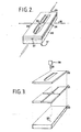
- Figures 2 and 3 illustrate in more detail the jig and tube-piercing arrangement used in the apparatus of Figure 1.
- a support for the tubing 16 includes a lower support block 20 of polymethylmethacrylate, with small hole 23, having a diameter of approximately 0.15 mm provided therein.
- Four polymethylmethacrylate spacer blocks 21 are secured to the support block 20 with adhesive, and define between them four channels, 24,25,26, and 27.
- the jig assembly is completed by a cover plate 22.
- the polyethylene tubing 16 is passed through the channels 24 and 26 defined by the-spacer blocks 21, so that it covers the hole 23 (tube 16 is shown broken in Figure 2 for clarity).
- a saline solution at a constant temperature of 37°C is supplied to inlet 27, and a suction supply to passageway 25, to remove the saline, and blood washed from the tube 16, to permit observation.
- a stainless steel needle 28 having a parallel-sided shank, 0.14 mm in diameter, and a length of approximately 15 mm is affixed to a needle-support 29, which is mounted on a slide (not shown) for movement towards and away from the hole 23, substantially normally to the block 20.
- the polyethylene tubing 16 can be manipulated within the block 17 so that the needle 28 enters the tube substantially perpendicular and pierces a through hole in it, ie. pierces the tube 16 such that the parallel-sided portion of the shank passes completely through the tube, so as to produce in both walls of the tube a hole having a size which is completely reproducible.
- the needle holder 29 is fitted onto the adjustable lens holder of an inverted microscope (WILD).
- WILD inverted microscope
- the microscope is illustrated schematically in Figure 1.
- the position of the needle 28 with respect to the tubing 16 is observed by means of a closed-circuit television system, illustrated schematically at 30 in Figure 1, coupled to the microscope. Because of the optical system, great precision can be obtained in the positioning of the holes perpendicular to the tubing.
- the syringe 12 is filled with light paraffin oil, and the container 1 with anticoagulated blood (for example buffered with heparin citrate, or EDTA).
- a new tube 16 is applied to the outlet 6 of tube 1, and cause to pass through jig 17, and into vessel 18.
- a layer of hydrocarbon oil is floated on the surface of blood, using branch 10 of the Y tube 9, and branch 10 is then sealed.
- Figure 4 shows a trace of the pressure indicated by the chart recorder 15 for two samples, a control sample of normal blood, (indicated as C on Figure 4) and a sample containing prostacyclin added to the blood (PGI 2 on Figure 4). It can be seen that the sample to which prostacyclin has been added shows a substantially increased bleeding time.
- the pumping technique utilising displacement by hydrocarbon oil has been found to cause no appreciable damage to the red cells or previous techniques have been found to cause considerable damage, and I have even found that a number of other fluids which might have been expected to be inert and therefore as effective as hydrocarbon oil in fact cause considerable damage.
- silicone fluid has been found to cause some damage to red cells.
- the technique allows described above is suitable for determination of the haemostasis reaction of non-anticoagulated blood that is to say, blood to which no anti-coagulant has been added, provided that the blood sample is used immediately after being taken.
- the technique indicates that haemostasis is generally complete in from 3 to 5 minutes.
- the apparatus according to the invention may also be utilised for the measurement of thrombolysis of the blood, ie., the time taken for a thrombus to be dissolved.
- thrombolytic agents for example streptokinase, or eurokinase.
- streptokinase or eurokinase.
- These agents are used clincially, to treat arterial thrombosis, and the amount of the agents needed for a particular patient is found to vary widely, depending on a number of factors, in particular the concentration in the blood of certain antibodies. In patients of this kind, it is essential that estimates be made as rapidly as possible of the amount of thrombolytic agents required.
- anticoagulated eg. heparin containing
- a thrombolytic agent such as streptokinase or eurokinase
- this is done by causing the blood flow in the tube to cease when haemostasis occurs, for example by stopping the syringe pump motor 13. The way in which the pressure in the tube (16) decreases with time is then observed.
- a method of measuring thrombolytic activity of an anticoagulated blood sample comprises causing the blood to flow through a tube under substantially constant pressure, piercing a hole in the tube whilst the blood is flowing in it, causing blood flow in the tube to cease when haemostasis occurs, and observing the decrease in pressure in the tube with time, to determine the thrombolytic activity of the blood.
- the pressure will in general remain steady when the initial haemostatic plug has formed, but will decrease as the thrombus dissolves, at a rate proportional to the leakage through the holes.
- thrombolysis can be tested using only a very small volume of blood.
- a similar method can be employed without the addition of thrombolytic agents, to determine the spontaneous thrombolytic activity of a blood sample. Such measurement can be useful in the investigation of clinical conditions such as disseminated intravascular coagulation syndrome.
Landscapes
- Health & Medical Sciences (AREA)
- Life Sciences & Earth Sciences (AREA)
- Engineering & Computer Science (AREA)
- Biomedical Technology (AREA)
- Chemical & Material Sciences (AREA)
- Hematology (AREA)
- Physics & Mathematics (AREA)
- Food Science & Technology (AREA)
- Molecular Biology (AREA)
- Urology & Nephrology (AREA)
- Ecology (AREA)
- Biophysics (AREA)
- Medicinal Chemistry (AREA)
- Analytical Chemistry (AREA)
- Biochemistry (AREA)
- General Health & Medical Sciences (AREA)
- General Physics & Mathematics (AREA)
- Immunology (AREA)
- Pathology (AREA)
- Investigating Or Analysing Biological Materials (AREA)
Applications Claiming Priority (2)
| Application Number | Priority Date | Filing Date | Title |
|---|---|---|---|
| GB8316332 | 1983-06-15 | ||
| GB838316332A GB8316332D0 (en) | 1983-06-15 | 1983-06-15 | Measuring haemotasis |
Related Child Applications (2)
| Application Number | Title | Priority Date | Filing Date |
|---|---|---|---|
| EP19880116753 Division EP0308994B1 (fr) | 1983-06-15 | 1984-06-15 | Méthode pour pomper du sang |
| EP88116753.0 Division-Into | 1988-10-10 |
Publications (3)
| Publication Number | Publication Date |
|---|---|
| EP0129425A2 true EP0129425A2 (fr) | 1984-12-27 |
| EP0129425A3 EP0129425A3 (en) | 1986-02-19 |
| EP0129425B1 EP0129425B1 (fr) | 1989-08-23 |
Family
ID=10544282
Family Applications (2)
| Application Number | Title | Priority Date | Filing Date |
|---|---|---|---|
| EP19880116753 Expired - Lifetime EP0308994B1 (fr) | 1983-06-15 | 1984-06-15 | Méthode pour pomper du sang |
| EP84304055A Expired EP0129425B1 (fr) | 1983-06-15 | 1984-06-15 | Méthode et appareil de mesure de la hémostase et de la thrombolyse |
Family Applications Before (1)
| Application Number | Title | Priority Date | Filing Date |
|---|---|---|---|
| EP19880116753 Expired - Lifetime EP0308994B1 (fr) | 1983-06-15 | 1984-06-15 | Méthode pour pomper du sang |
Country Status (3)
| Country | Link |
|---|---|
| EP (2) | EP0308994B1 (fr) |
| DE (2) | DE3486291T2 (fr) |
| GB (1) | GB8316332D0 (fr) |
Cited By (8)
| Publication number | Priority date | Publication date | Assignee | Title |
|---|---|---|---|---|
| WO1988002116A1 (fr) * | 1986-09-10 | 1988-03-24 | Peter Gorog | Dispositif d'analyse du sang |
| WO1991002976A1 (fr) * | 1989-08-18 | 1991-03-07 | Xylum Corporation | Dispositif a cassette jetable de manipulation du sang pour la mesure des hemostases |
| WO1991014943A1 (fr) * | 1990-03-23 | 1991-10-03 | Peter Gorog | Appareil et procede de simulation de formation d'un thrombus arteriel |
| WO1993012423A1 (fr) * | 1991-12-19 | 1993-06-24 | Diana Gorog | Mesure de l'activite thrombolytique du sang |
| US5296379A (en) * | 1990-03-23 | 1994-03-22 | Peter Gorog | Apparatus and method for modeling arterial thrombus formations |
| US5316730A (en) * | 1992-12-07 | 1994-05-31 | Xylum Corporation | Disposable cartridge for investigating physical properties of blood |
| US5339830A (en) * | 1992-01-21 | 1994-08-23 | Blake Joseph W Iii | Blood coagulation test system |
| US6004819A (en) * | 1996-11-04 | 1999-12-21 | Xylum Corporation | Blood testing device |
Families Citing this family (1)
| Publication number | Priority date | Publication date | Assignee | Title |
|---|---|---|---|---|
| GB2254423A (en) * | 1990-12-07 | 1992-10-07 | Alpha Dyffryn Cyfyngedig | Analytical apparatus with agitation of samples |
Family Cites Families (5)
| Publication number | Priority date | Publication date | Assignee | Title |
|---|---|---|---|---|
| GB1283787A (en) * | 1969-04-15 | 1972-08-02 | Beckman Instruments Inc | Liquid analyzer apparatus |
| US3768496A (en) * | 1972-04-27 | 1973-10-30 | Brian A Hills | Two phase blood pump |
| JPS4982304U (fr) * | 1972-11-02 | 1974-07-17 | ||
| EP0046345A3 (fr) * | 1980-08-15 | 1982-03-03 | Ortho Diagnostic Systems Inc. | Dispositif d'écoulement hydrodynamique contrôlé dans un système cytométrique à écoulement |
| GB2096329B (en) * | 1981-03-25 | 1985-09-18 | Born Gustave Victor Rudolph | Method and apparatus for the in vitro study of the occlusive properties of blood |
-
1983
- 1983-06-15 GB GB838316332A patent/GB8316332D0/en active Pending
-
1984
- 1984-06-15 DE DE19843486291 patent/DE3486291T2/de not_active Expired - Fee Related
- 1984-06-15 EP EP19880116753 patent/EP0308994B1/fr not_active Expired - Lifetime
- 1984-06-15 EP EP84304055A patent/EP0129425B1/fr not_active Expired
- 1984-06-15 DE DE8484304055T patent/DE3479533D1/de not_active Expired
Cited By (15)
| Publication number | Priority date | Publication date | Assignee | Title |
|---|---|---|---|---|
| JPH07101219B2 (ja) * | 1986-09-10 | 1995-11-01 | ゴーロッグ、ピーター | 血液検査装置 |
| AU609060B2 (en) * | 1986-09-10 | 1991-04-26 | Xylum Corporation | Blood testing device |
| WO1988002116A1 (fr) * | 1986-09-10 | 1988-03-24 | Peter Gorog | Dispositif d'analyse du sang |
| US5925569A (en) * | 1986-09-10 | 1999-07-20 | Xylum Corporation | Blood testing device and method |
| WO1991002976A1 (fr) * | 1989-08-18 | 1991-03-07 | Xylum Corporation | Dispositif a cassette jetable de manipulation du sang pour la mesure des hemostases |
| US5047211A (en) * | 1989-08-18 | 1991-09-10 | Xylum Corporation | Disposable blood handling cassette device for measuring haemostasis |
| WO1991014943A1 (fr) * | 1990-03-23 | 1991-10-03 | Peter Gorog | Appareil et procede de simulation de formation d'un thrombus arteriel |
| GB2245061A (en) * | 1990-03-23 | 1991-12-18 | Peter Gorog | Modelling thrombosis formation |
| US5296379A (en) * | 1990-03-23 | 1994-03-22 | Peter Gorog | Apparatus and method for modeling arterial thrombus formations |
| WO1993012423A1 (fr) * | 1991-12-19 | 1993-06-24 | Diana Gorog | Mesure de l'activite thrombolytique du sang |
| US5599718A (en) * | 1991-12-19 | 1997-02-04 | Gorog; Diana | Measurement of the thrombolytic activity of blood |
| US5339830A (en) * | 1992-01-21 | 1994-08-23 | Blake Joseph W Iii | Blood coagulation test system |
| USRE36054E (en) * | 1992-12-07 | 1999-01-19 | Xylum Corporation | Disposable cartridge for investigating physical properties of blood |
| US5316730A (en) * | 1992-12-07 | 1994-05-31 | Xylum Corporation | Disposable cartridge for investigating physical properties of blood |
| US6004819A (en) * | 1996-11-04 | 1999-12-21 | Xylum Corporation | Blood testing device |
Also Published As
| Publication number | Publication date |
|---|---|
| DE3486291T2 (de) | 1994-09-29 |
| DE3479533D1 (en) | 1989-09-28 |
| EP0308994A3 (fr) | 1991-05-15 |
| DE3486291D1 (de) | 1994-04-28 |
| EP0129425A3 (en) | 1986-02-19 |
| EP0308994B1 (fr) | 1994-03-23 |
| EP0129425B1 (fr) | 1989-08-23 |
| EP0308994A2 (fr) | 1989-03-29 |
| GB8316332D0 (en) | 1983-07-20 |
Similar Documents
| Publication | Publication Date | Title |
|---|---|---|
| CA1341179C (fr) | Dispositif d'analyse pour le sang | |
| US5854423A (en) | Apparatus and method for assessment of visco-elasticity and shear adherence strength properties of blood clots | |
| US3918908A (en) | Method for prothrombin testing | |
| US3720097A (en) | Apparatus and method for measuring mammalian blood viscosity | |
| JP2945473B2 (ja) | 鬱血測定のための使い捨て血液操作カセット装置 | |
| US6043871A (en) | System and method for measuring blood platelet function | |
| US5792660A (en) | Comparative determinants of viscosity in body fluids obtained with probes providing increased sensitivity | |
| US20100139375A1 (en) | Apparatus and method for measuring rheological properties of blood | |
| Carr Jr | In vitro assessment of platelet function | |
| US3951606A (en) | Apparatus for prothrombin testing | |
| EP0677169B1 (fr) | Procede et dispositif permettant de mesurer la tendance a la formation de thrombus | |
| EP0129425A2 (fr) | Méthode et appareil de mesure de la hémostase et de la thrombolyse | |
| McKenzie et al. | Clinical utility of available methods for determining platelet function | |
| US4797369A (en) | Method and apparatus for detecting a blood clot | |
| EP2817099A2 (fr) | Dispositif de perfusion, appareil correspondant l'utilisant et méthode permettant d'analyser la pathologie thrombotique-ischémique et hémorragique | |
| Kenny et al. | Measurement of erythrocyte filterability using washed-erythrocyte and whole-blood methods | |
| US6004819A (en) | Blood testing device | |
| US3518057A (en) | Method and apparatus for thrombus formation time determinations | |
| Matrai et al. | Biophysics | |
| EP2010903B1 (fr) | Appareil et procede destines aux diagnostic, pronostic et surveillance pharmacologique de la pathologie thrombotique-ischemique et hemorragique de l'appareil cardio-vasculaire | |
| Glover et al. | Effect of shear stress on clot structure formation | |
| KR102893038B1 (ko) | 혈전탄성도 테스트 장치 및 방법 | |
| US5783447A (en) | Hypercoagulability comparative determinants obtained using detection systems with variable force-induced energy inputs | |
| EP0111551B1 (fr) | Procede et dispositif permettant une mesure rapide et directe de la viscosite sanguine | |
| Abts et al. | Ultrasound detection of aggregation of red cells in blood flow |
Legal Events
| Date | Code | Title | Description |
|---|---|---|---|
| PUAI | Public reference made under article 153(3) epc to a published international application that has entered the european phase |
Free format text: ORIGINAL CODE: 0009012 |
|
| AK | Designated contracting states |
Designated state(s): DE FR GB IT SE |
|
| PUAL | Search report despatched |
Free format text: ORIGINAL CODE: 0009013 |
|
| AK | Designated contracting states |
Designated state(s): DE FR GB IT SE |
|
| 17P | Request for examination filed |
Effective date: 19860807 |
|
| 17Q | First examination report despatched |
Effective date: 19880212 |
|
| GRAA | (expected) grant |
Free format text: ORIGINAL CODE: 0009210 |
|
| AK | Designated contracting states |
Kind code of ref document: B1 Designated state(s): DE FR GB IT SE |
|
| ITF | It: translation for a ep patent filed | ||
| REF | Corresponds to: |
Ref document number: 3479533 Country of ref document: DE Date of ref document: 19890928 |
|
| ET | Fr: translation filed | ||
| PLBE | No opposition filed within time limit |
Free format text: ORIGINAL CODE: 0009261 |
|
| STAA | Information on the status of an ep patent application or granted ep patent |
Free format text: STATUS: NO OPPOSITION FILED WITHIN TIME LIMIT |
|
| 26N | No opposition filed | ||
| ITTA | It: last paid annual fee | ||
| EAL | Se: european patent in force in sweden |
Ref document number: 84304055.1 |
|
| REG | Reference to a national code |
Ref country code: GB Ref legal event code: 732E |
|
| REG | Reference to a national code |
Ref country code: FR Ref legal event code: TP |
|
| PGFP | Annual fee paid to national office [announced via postgrant information from national office to epo] |
Ref country code: GB Payment date: 20010521 Year of fee payment: 18 |
|
| PGFP | Annual fee paid to national office [announced via postgrant information from national office to epo] |
Ref country code: SE Payment date: 20010523 Year of fee payment: 18 Ref country code: FR Payment date: 20010523 Year of fee payment: 18 |
|
| PGFP | Annual fee paid to national office [announced via postgrant information from national office to epo] |
Ref country code: DE Payment date: 20010627 Year of fee payment: 18 |
|
| REG | Reference to a national code |
Ref country code: GB Ref legal event code: IF02 |
|
| PG25 | Lapsed in a contracting state [announced via postgrant information from national office to epo] |
Ref country code: GB Free format text: LAPSE BECAUSE OF NON-PAYMENT OF DUE FEES Effective date: 20020615 |
|
| PG25 | Lapsed in a contracting state [announced via postgrant information from national office to epo] |
Ref country code: SE Free format text: LAPSE BECAUSE OF NON-PAYMENT OF DUE FEES Effective date: 20020616 |
|
| PG25 | Lapsed in a contracting state [announced via postgrant information from national office to epo] |
Ref country code: DE Free format text: LAPSE BECAUSE OF NON-PAYMENT OF DUE FEES Effective date: 20030101 |
|
| EUG | Se: european patent has lapsed | ||
| GBPC | Gb: european patent ceased through non-payment of renewal fee |
Effective date: 20020615 |
|
| PG25 | Lapsed in a contracting state [announced via postgrant information from national office to epo] |
Ref country code: FR Free format text: LAPSE BECAUSE OF NON-PAYMENT OF DUE FEES Effective date: 20030228 |
|
| REG | Reference to a national code |
Ref country code: FR Ref legal event code: ST |