EP0168933A2 - Nachweis und/oder Identifizierung von Mikroorganismen in einer Probe mittels Biolumineszenz oder anderer exogenen genetisch eingeführten Markierungssubstanzen - Google Patents
Nachweis und/oder Identifizierung von Mikroorganismen in einer Probe mittels Biolumineszenz oder anderer exogenen genetisch eingeführten Markierungssubstanzen Download PDFInfo
- Publication number
- EP0168933A2 EP0168933A2 EP85303913A EP85303913A EP0168933A2 EP 0168933 A2 EP0168933 A2 EP 0168933A2 EP 85303913 A EP85303913 A EP 85303913A EP 85303913 A EP85303913 A EP 85303913A EP 0168933 A2 EP0168933 A2 EP 0168933A2
- Authority
- EP
- European Patent Office
- Prior art keywords
- dna
- donor
- vector
- luciferase
- genetic
- Prior art date
- Legal status (The legal status is an assumption and is not a legal conclusion. Google has not performed a legal analysis and makes no representation as to the accuracy of the status listed.)
- Granted
Links
Images








Classifications
-
- C—CHEMISTRY; METALLURGY
- C12—BIOCHEMISTRY; BEER; SPIRITS; WINE; VINEGAR; MICROBIOLOGY; ENZYMOLOGY; MUTATION OR GENETIC ENGINEERING
- C12Q—MEASURING OR TESTING PROCESSES INVOLVING ENZYMES, NUCLEIC ACIDS OR MICROORGANISMS; COMPOSITIONS OR TEST PAPERS THEREFOR; PROCESSES OF PREPARING SUCH COMPOSITIONS; CONDITION-RESPONSIVE CONTROL IN MICROBIOLOGICAL OR ENZYMOLOGICAL PROCESSES
- C12Q1/00—Measuring or testing processes involving enzymes, nucleic acids or microorganisms; Compositions therefor; Processes of preparing such compositions
- C12Q1/02—Measuring or testing processes involving enzymes, nucleic acids or microorganisms; Compositions therefor; Processes of preparing such compositions involving viable microorganisms
- C12Q1/04—Determining presence or kind of microorganism; Use of selective media for testing antibiotics or bacteriocides; Compositions containing a chemical indicator therefor
-
- C—CHEMISTRY; METALLURGY
- C12—BIOCHEMISTRY; BEER; SPIRITS; WINE; VINEGAR; MICROBIOLOGY; ENZYMOLOGY; MUTATION OR GENETIC ENGINEERING
- C12N—MICROORGANISMS OR ENZYMES; COMPOSITIONS THEREOF; PROPAGATING, PRESERVING, OR MAINTAINING MICROORGANISMS; MUTATION OR GENETIC ENGINEERING; CULTURE MEDIA
- C12N15/00—Mutation or genetic engineering; DNA or RNA concerning genetic engineering, vectors, e.g. plasmids, or their isolation, preparation or purification; Use of hosts therefor
- C12N15/09—Recombinant DNA-technology
- C12N15/11—DNA or RNA fragments; Modified forms thereof; Non-coding nucleic acids having a biological activity
- C12N15/52—Genes encoding for enzymes or proenzymes
-
- C—CHEMISTRY; METALLURGY
- C12—BIOCHEMISTRY; BEER; SPIRITS; WINE; VINEGAR; MICROBIOLOGY; ENZYMOLOGY; MUTATION OR GENETIC ENGINEERING
- C12Q—MEASURING OR TESTING PROCESSES INVOLVING ENZYMES, NUCLEIC ACIDS OR MICROORGANISMS; COMPOSITIONS OR TEST PAPERS THEREFOR; PROCESSES OF PREPARING SUCH COMPOSITIONS; CONDITION-RESPONSIVE CONTROL IN MICROBIOLOGICAL OR ENZYMOLOGICAL PROCESSES
- C12Q1/00—Measuring or testing processes involving enzymes, nucleic acids or microorganisms; Compositions therefor; Processes of preparing such compositions
- C12Q1/68—Measuring or testing processes involving enzymes, nucleic acids or microorganisms; Compositions therefor; Processes of preparing such compositions involving nucleic acids
- C12Q1/6897—Measuring or testing processes involving enzymes, nucleic acids or microorganisms; Compositions therefor; Processes of preparing such compositions involving nucleic acids involving reporter genes operably linked to promoters
Definitions
- the invention herein described concerns analytical methods, test means, compositions, and methods of preparation thereof for detecting the presence of bacteria in a test sample.
- the invention pertains to the determination of bacteria in areas related to health care, such as in the analysis of human excretory products or body fluids, (i.e., urine, blood, feces) for the purpose of aiding diagnosis, or to detect bacterial contamination in foodstuffs. In these and other fields it is important to rapidly, accurately and economically detect the presence of specific bacteria.
- Enumeration and identification of microorganisms as well as the determination of their susceptibility to antibiotics are the main goals of diagnostic medical microbiology. Numerous techniques, tests and media have been developed in order to achieve these goals. However, none of the currently applied tests allow the fulfillment of these tasks by a short-term (i.e., minutes or hours) procedure. A total viable count of bacteria requires 18-24 hrs using present state of the art methods. The estimation of microbial biomass through the determination of adenosine triphosphate (ATP) content is not specific and does not allow the detection of less than 10 4-5 bacteria per ml.. Enumeration of a specific species of bacteria usually requires selective media and long periods of incubation.
- ATP adenosine triphosphate
- ATP can be degraded by enzymes in the test sample, ATP can originate from nonbacterial sources such as tissues of the host, radioactive materials present a biohazard, and enzymes with similar activity can arise from the host.
- Particle counting instrumentation has also been applied to detection of bacteria. Such instrumentation measures pertubations in an electrical current across a small orifice caused by the presence of particles in a fluid flowing through the orifice. Besides requiring the use of a complex and expensive apparatus, this method is highly nonspecific and requires a high level of care and particle-free conditions.
- Plasmids are autonomous extra-chromosomal genetic units consisting of circular strands of DNA which are found in most bacteria and some eukaryotes. Bacteriophages are DNA viruses which parasitize only bacteria. Hybrid genetic vectors can then be introduced into selected microbial hosts, which in turn serve as potential factories for the production of large amounts of the cloned DNA.
- a non-expressing or low-expressing cloning vector represents a ready source of the foreign genetic material which can then be isolated and introduced in turn into a suitable expression vector (i.e., a specially prepared or selected plasmid).
- the foreign DNA is placed in a suitable location in an expression vector where the indigenous genetic sequence is such that the foreign genetic information will be transcribed (i.e., mRNA produced from the foreign DNA) and translated (i.e., protein synthesis from the mRNA template) and the desired product coded in the foreign DNA obtained.
- Transformed microorganisms containing such expression vectors serve as factories for the manufacture of the foreign-DNA product.
- Restriction endonucleases are site-specific endonucleases which primarily cleave double-stranded DNA, but in some instances cleave single-stranded DNA.
- Class II restriction endonucleases cleave at specific sequences.
- Class I restriction endonucleases appear to cleave DNA randomly and produce heterogeneous products.
- Various restriction endonucleases produce DNA fragments of different lengths and types.
- restriction endonucleases cleave both DNA strands at the same point and produce the so-called "blunt end” DNA fragments.
- other restriction endonucleases cleave one DNA strand several nucleotides away from the cleavage on the complimentary strand and produce "cohesive end” DNA fragments. Consequently, an accomplished practitioner of the recombinant DNA art can, by creative selection of endonucleases for treatment of subject DNA, obtain desired DNA fragments which can then be joined together by the action of a DNA ligase.
- nucleotides For ligation purposes, it may be desirable to add nucleotides to the ends of cut DNA fragments and to add complementary deoxyribonucleotides to the ends of the cloning vector (i.e., in the process known as homopolymeric tailing).
- Another general method for obtaining a desired DNA fusion product is the addition of "adapter” or "linker fragments" to the ends of either or both the cloning vector or the DNA fragments to be cloned.
- Linker fragments are small sections of DNA that contain one or more recognition sequences for restriction endonucleases.
- Foreign DNA material can be obtained for insertion into a transfer vector by several ways. For example, DNA fragments directly obtained from the parental source can be inserted into the appropriate vector. Another method is to obtain mRNA from an active synthesis location in the parent system and to then enzymatically synthesize (reverse transcriptase) a single-stranded complementary DNA strand from the isolated mRNA. A double-stranded DNA molecule is then synthesized from the single-stranded template. Double-stranded DNA obtained in this fashion is known as complementary DNA (cDNA). Once a desired DNA sequence is known, genes can also be chemically synthesized in vitro for cloning/expression purposes in microbial, tissue, or cell culture systems.
- cDNA complementary DNA
- a preferred embodiment of the invention employs the luminescent system from Virbio fischeri.
- the technique we have developed is by no means limited to that system. Any exogenous genetically-introduced system showing marked increase of expression in host bacteria whose presence or absence is to be determined can also be used.
- U.S. Patent No. 3,930,956 involves the transfer of raw bacterial DNA to an auxotroph. The detection of phage-induced lysis of bacteria is described in U.S. Patent No. 4,104,126.
- Bioluminescence is found in microorganisms [i.e., some bacteria (mostly marine forms, e.g., Vibrio fischeri), fungi, and dinoflagellates], insects (e.g., the firefly, Photinus pyradis), some crustaceans (i.e., Cypridine hilgendorfi), jellyfish, worms and other invertebrates and even in mammals.
- the biochemical mechanism of luminescence is known to vary (i.e., the luminescence system found in bacteria is different from that found in fireflies and dinoflagellates), light production in living organisms is most frequently catalyzed by the enzyme luciferase.
- Bacterial luciferase is a mixed function oxidase, consisting of two different subunits each with a molecular weight of approximately 40,000 daltons.
- the synthesis of the enzymes participating in the luminescence system is regulated by a small sensory molecule, named autoinducer.
- autoinducer a small sensory molecule
- the autoinducer is accumulated in the growth medium.
- induction of the luminescence system occurs, resulting in approximately 1000 fold increase in light production.
- the preferred element of the new test is a fragment of DNA carrying the luminescence system of a luminescent bacterium, usually a marine bacterium, e.g., Vibrio fischeri.
- An extracellular DNA fragment carrying the luminescence genes does not, of course, luminesce but upon transferring it to a suitable living host by transduction or transformation, the host's genetic and synthetic machinery can utilize the DNA fragment thereby causing light to be emitted.
- Intracellular gene segments can also be used if in the test organisms they are poorly expressed or unexpressed and only become expressed after some genetic transfer of exogenous DNA to the organism whose presence or absence is to be determined. This latter method employs conjugation, i.e., mating and genetic transfer from one cell to another.
- DNA transfer occurs among a group of strains of the same species or among closely related species. Some factors acting to limit such transfer include the presence of DNA restriction-modification systems in many bacterial species and/or their strains, the dependence on host factors for the replication of the introduced DNA and appropriate bacteriophage receptors in the bacterial wall upon which bacteriophage can adsorb and thereby properly inject their genetic material into the host.
- Conjugation and transformation are not particularly strain specific. Some plasmids can be transferred by conjugation not only to the same species but also to related ones. There are even several plasmids which can be transferred to distant species by conjugation. Transformation is potentially even broader than conjugation since a wide range of bacteria take up extracellular DNA. Many, if not most, that are at present not known to do so may in fact be transformable under specific conditions. For example, E. coli does not normally take up DNA efficiently. However, after calcium shock this bacterium can be induced to do so at a high level of efficiency. A limiting factor in using transformation is the ability of the foreign DNA to express, replicate or integrate its genetic information into the host genome.
- transduction is quite strain or species specific. Some bacteriophages infect several species of bacteria which are usually close relatives; most infect only a particular subset of strains of a single species. By using different kinds of strains of bacteriophage which infect subsets, it is possible to arrange the strains of a bacterial species into classification schemes. This is called phage typing.
- Recombinant DNA technology and molecular genetics allow the introduction of genes whose products are easily assayable.
- a gene such as lac z of Escherichia coli into a bacterium can lead to expression of the introduced gene.
- a bacteriophage is used to introduce the gene, then the course of the subsequent infection can be measured by analyzing the extent of beta-galactosidase formation. Even though the bacterial cell itself may possess a gene for this enzyme, these measurements can be performed because the endogenous level can be depressed by appropriate media and the propagation of the bacteriophage genetic material leads to multiple copies of the gene.
- the luminescent system of Vibrio fischeri is particularly useful and illustrative of our method for ascertaining the presence or absence of a given bacterial species. Only a few bacterial species contain genes allowing them to convert chemical energy to light. Most of these species are marine. The vast majority of bacterial species do not contain such systems and are dark. If the genes for light production are introduced and express themselves in a species with no such capability, then emission of light will result. The background interference in such a system is extremely low (chemical luminescence). Since very low levels of light can be detected and measured, tests based on this method should be sensitive and quantitative.
- exogenous DNA containing a luminescence system or other genetic system derived from a suitable donor organism is transferred by transformation, transduction, or conjugation (genetic means) to a host organism lacking or containing relatively low levels of endogenous DNA.
- the entry of the donor genetic material into the organism which it is desired to determine leads to the expression of the introduced system.
- the expression of such genes allows one to detect the presence and numbers of such organisms.
- antibiotics are added at the time of entry, the susceptibility of the host organism to the antibiotic can be determined.
- a preferred embodiment of the invention involves the transference of a luminescent system, or part thereof, obtained from a luminescent bacterium (or other source), and used as a source for transferring this system into a non-luminescent host microorganism with subsequent expression of luciferase or other system in the host and concomitant production of light or other marker.
- the invention can also be used to detect antibiotics in a test sample, to assay microorganisms for antibiotic susceptibility, or to assay compositions for antibiotic activity.
- any other genetic system which is absent or poorly expressed until entry into the bacteria whose presence it is desired to ascertain may be used.
- introduction of genes coding for enzymes or proteins e.g., beta-galactosidase of E. coli or the egg albumin protein from chickens
- enzymes or proteins e.g., beta-galactosidase of E. coli or the egg albumin protein from chickens
- enzymatic assay e.g., beta-galactosidase of E. coli or the egg albumin protein from chickens
- immunoassay e.g., immunoassay
- Luminescent bacteria are mainly marine, a typical example being Vibrio fischeri.
- the DNA containing a luminescence system derived from a luminescent bacterium, firefly, dinoflagellate, or luminescent fungi, etc. may be natural, in which case it is derived as such from the luminescent organism.
- the DNA containing a luminescence system or part thereof derived from a luminescent bacterium or other source may be an artificial recombinant or synthesized DNA with the luminescence system or part thereof being derived from the luminescent source.
- the medium may be desirable to include in the medium an amount of autoinducer of the luminescent bacterium from which the luminescence system or part thereof is derived, in order to avoid any prolonged incubation time which would otherwise be required for the formation of the autoinducer inside the host organism.
- an aliphatic aldehyde to the milieu in order to accelerate or increase the expression of the luminescence system or part thereof derived from a luminescent bacterium.
- a foreign genetic marker system other than a luminescence system is introduced into a suitable host, it may be advantageous to introduce intermediates, precursors, enzymatic substrates, or other ingredients to facilitate or enhance expression of the marker genetic system or detection of the expression.
- the DNA containing a luminescence system or part thereof derived from a luminescent bacterium or other source is transcribed by RNA polymerase to form mRNA that can then be translated into the luminescence system proteins.
- the luminescence level of even a single bacterium can be detected with, for example, the aid of a scintillation counter.
- luminescence is produced upon interaction between two non-luminescent components, namely the organism to be determined and said DNA containing a luminescence system or parts thereof derived from a luminescent bacterium or other source. Consequently, any luminescence that exceeds the weak background chemiluminescence is conclusive of the presence of the host organism. Conversely, the absence of any luminescence beyond the background level of chemiluminescence is conclusive of the absence of the host organism. Such a technique has never been suggested before.
- the transfer of the DNA containing a luminescence or other marker system or parts thereof derived from a suitable donor to the host organism may be mediated by transformation, transduction or conjugation.
- a large number of microbial genera serve as hosts for bacteriophages and retain or accept plasmids.
- Examples of such genera are: Escherichia, Aerobacter, Salmonella, Shigella, Klebsiella, Proteus, Pseudomonas, Staphylococcus, Streptococcus, Chlamydia, Mycoplasma, Pneumococcus, Neisseria, Clostridium, Bacillus, Corynebacterium, Mycobacterium, Camphybacter, Vibrio, Serratia, Enterobacter, Providencia, Chromobacterium, Brucella, Yersinia,. Haemophilus, and Bordetella.
- transduction bacteriophages are constructed that are specific to the bacteria to be determined. If desired, a series of tests may be run each with a different bacteriophage, either sequentially or simultaneously.
- Some bacteriophages may be constructed by packaging DNA containing a luminescence system or parts thereof or other donor marker system derived from a luminescent bacterium or other suitable source in the proper bacteriophage capsid.
- the bacteriophage may be constructed by recombinant DNA technology using restriction endonuclease with or without subsequent packaging.
- the bacteriophages containing all or part of the luminescence or other system may be constructed through naturally occurring genetic recombinational mechanism. This can also involve the use of both artificial recombination and natural recombination.
- the invention provides bacterial strains containing a luminescence system or parts thereof in a transferable replicon.
- the replicon may be the bacterial chromosome or a plasmid.
- the luminescence system or parts thereof may be introduced by a bacteriophage or by genetic recombination, either natural or artificial, such that the luminescence genes are covalently linked to the transferred genetic material.
- any other genetic system whose primary or secondary products can be measured as described earlier can be used in a like fashion.
- luminescence is given as a relevant and particular example of the more general principle underlying this invention.
- Hfr's high frequency of conjugation
- Hfr's or donors can be made through genetic recombination-, either natural or artificial, or through the addition of genetic material such that the Hfr or donor carries the luminescence system or parts thereof.
- the host organisms perform metabolic activities that are involved in the formation of luminescence or other donor systems; this includes protein synthesis and a process that generates a reducing power.
- Antimicrobial agents that affect these processes or alter cell integrity abolish or reduce the formation of the donor genetic system (i.e., luminescence, etc.). Consequently it is possible in accordance with the invention to determine the susceptibility of bacteria to various antibacterial agents, e.g., antibiotics.
- the specific type or group of bacteria to be tested are subjected to DNA transfer as specified in the presence of a given antibacterial agent, and the kinetics of the expressed donor genetic system (i.e., luminescence), which are a function of the protein synthesis capacity and the general metabolism of the host bacteria are then determined.
- the kinetics of the expressed donor genetic system i.e., luminescence
- simultaneous tests are run, one in the absence of any antibacterial agent and several others each in the presence of a particular antibacterial agent.
- Restriction endonucleases were purchased from BRL, New England Biolabs, Boehringer and Amersham. Digestion of DNA by such enzymes was according to the suggestion of New England Biolabs catalogue (1982) except that 100 p g/ml gelatin replaced bovine serum albumin in all cases. DNA concentration did not exceed 2 ⁇ g/20 ul.
- DNA's digested by restriction endonucleases were precipitated by the addition of one-tenth volume of 3N sodium acetate and 2.2 volumes of distilled 96% ethanol. After freezing at -70°C in a dry ice-ethanol bath, the Eppendorf tubes were centrifuged for 10 minutes in an Eppendorf centrifuge, the supernate carefully removed and discarded, the pellet washed with 100 ⁇ l 70X ethanol, centrifuged for 3 minutes, the supernate removed and discarded, and the pellet dried for 10 minutes in a dessicator under vacuum. The dry pellets were resuspended in water.
- Ligations were performed in 20 ⁇ l containing 4 ⁇ 1 of a five-fold concentrated kinase-linker buffer (Maniatis et al., loc. cit.) and 1 ⁇ l of DNA ligase (New England Biolabs., Catalogue No. 202; 400 units/ml) at 25° C for 12-16 hours.
- the DNA concentration was usually 0.5 - 1.0 ⁇ g/20 ⁇ l reaction.
- the bacterial strain to be transformed is grown overnight at 37°C in LB medium. It is diluted 1:200 into fresh LB and grown with aeration at 37°C until its absorbance, A, at 600 nM is 0.5.
- the culture (original volume) is chilled on ice, then centrifuged in a Beckman centrifuge at 7000 rpm for 7 minutes at 0°C in a JA 20 rotor.
- the cells are resuspended in the same volume of ice cold 0.1 M MgCl 2 , and centrifuged as before.
- the cell pellet is resuspended in one-half of the original volume of 0.1 M CaCl 2 and kept on ice for 30 minutes.
- the suspension is centrifuged as before and resuspended in one-tenth of the original volume of 0.1 M CaCl 2 .
- a 0.2 ml portion of these concentrated CaCl 2 treated cells is mixed with 1-100 ⁇ l of solution containing 0.1-2 ⁇ g of DNA. The mixture is held at 0°C for 30 minutes, heated to 42°C for 3 minutes in a water bath and again placed on ice for an additional 20 minutes. Then 0.8 ml of LB medium is added and the culture placed at 37°C for 1 hour to allow the expression of antibiotic resistance essentially without cell division since the cells are recovering from the calcium shock. Portions of the culture are placed on LB plates (containing the appropriate antibiotics). Controls consist of untransformed but Ca ++ treated cells and cells transformed with a known amount of an uncleaved plasmid.
- DNA 8 ⁇ g from Vibrio fischeri strain MJ1 was cleaved with 30 units of restriction endonuclease Sail for 3 hrs at 37°C.
- DNA (6 ⁇ g) of plasmid pBR322 was similarly digested. Samples were tested by agarose gel electrophoresis to check that the digestions had gone to near completion. The cleaved DNA's were precipitated with sodium acetate-ethanol and the dry pellets resuspended in H 2 0. A 2 ⁇ g amount of V. fischeri DNA was ligated to 0.5 g of pBR322 DNA using 400 units of DNA ligase.
- the reaction was terminated by adding 200 ⁇ l H 2 0 and 40 ⁇ 3l STE saturated-phenol. The mixture was vortexed and centrifuged. The supernate was precipitated with sodium acetate-ethanol and the pellet of ligated DNA was resuspended in 40 ⁇ 1 of H 2 0.
- Strain MM294 was grown and prepared for transformation.
- the control transformation contained 1 ⁇ g pBR322 DNA (loc. cit.). After transformation the cells were plated on LB plates containing ampicillin. Cells without added DNA gave no colonies. 2.64 x 10 transformants/ ⁇ g pBR322 DNA were obtained in the control transformation. The ligated mixture gave 3.2 x 10 transformant colonies/ 11 g bacterial DNA and 6 x 10 total transformants were obtained.
- By checking for the insertional inactivation of the tetracycline resistance gene inserts of foreign DNA inactivate the gene since the SalI site is in this gene) it was found that about 5X of these transformants contain inserts of V. fischeri DNA. Thus about 3000 inserts were obtained. Seven of the colonies were luminescent.
- pACYC184 plasmid DNA similarly prepared, was cleaved by SalI and analyzed as above. After ligation and transformation into MM294 E, coli cells, colonies that were resistant to chloramphenicol (from pACYC184) and that gave light (30°C) were analyzed by standard methods.
- a strain containing a plasmid (pAChv-1) that has the backbone of pACYC184 and the 8 Kb light producing segment inserted in the SalI site of pACYC184 was kept, and placed in permanent deposit (ATCC 53133).
- Bacteriophage XCharon 30 DNA (0.3 P g), purified as described by Maniatis (loc. cit.), was cleaved with restriction endonuclease Sa1I and ligated to pBTK5 DNA (0.5 ⁇ g) obtained according to IX above, similarly cleaved. After ligation the DNA was precipitated by sodium acetate-ethanol, the pellet dried and resuspended in 5 ⁇ l H 2 0 .
- Packaging mix for DNA was prepared by the method of B. Hohn as given in Maniatis (loc. cit.). The packaging of the ligated DNA was carried out according to the protocol of Maniatis (loc. cit.) and plated with MM294 grown to stationary phase in FT medium. Plaques were checked for light production by transferring them to scintillation vials containing 1 ml FT medium, 0.1 ml MM294 grown on FT medium. Individual plaques had been transferred through the use of a Pasteur pipette to 1 ml of 10 mM MgS0 4 . A 0.1 ml of this was transferred to a vial.
- Phage strains giving light were plaque purified twice, plate lysates were made and the phage preparations stored over several drops of chloroform. These phages were then characterized for light production during infection of strain MM294 growing in FT medium at 27°C. Autoinducer was added to a duplicate vial (Nealson, K.H., Platt, T., and Hastings, J.W., 1970, Cellular control of the synthesis and activity of the bacterial luminescent system. J.
- ⁇ L1, XL35 and ⁇ L40 produce light at times considerably later than the others, the light produced is relatively little and the amount of light emitted is greatly increased if the bacteriophage infection takes place in the presence of autoinducer.
- Phage strains ⁇ L4, ⁇ L5, ⁇ L28 and XL32 produce light within 15-20 minutes after infection, and the amount of light emitted increases with time and reaches a maximum after about one hour. There is no significant effect of autoinducer on the amount of light produced by these phages.
- the second possible orientation has the regulatory gene of the lux operon nearer the N gene of ⁇ and the aldehyde and luciferase genes to the J gene side. Since the direction of transcription of the aldehyde and luciferase genes is the same as that of the P L promoter of ⁇ , these genes should be transcribed not only from their own promoter but even more so from P L . Thus, the addition of autoinducer to cultures of bacteria infected by these kind of phages should and does have little or no effect on light production.
- Phage DNA from ⁇ L4 and ⁇ L28 was prepared as described by Maniatis (op. cit.) using CsCl gradient purified phage as the source of DNA.
- X Charon 30 has the normal ⁇ sequence until approximately base pair 23616 (Hendrix R.W., Roberts, J.W., Stahl, F.W., and Weisberg, R.A., 1983: Lambda II, Cold Spring Harbor Laboratory, Cold Spring Harbor, New York; Appendix II, pp 519-676) and then the deletion b1007 which extends from approximately base pair 23616 to 28312. From base 28312 until base 34449 the sequence is again normal. At base 34449 an earlier sequence repeats itself and this repeat starts with base pair 22346. The repeat continues until base pair 23616 and then is deleted (b1007) until base pair 28312.
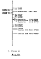
- the pattern of restriction enzyme fragments obtained from PstI cleavage in ⁇ Charon 30 can be calculated from the description of its DNA provided above. After removal of the internal Sa1I fragment and insertion of the lux operon Sall fragment this pattern is, of course, altered.
- the PstI sites until the base pair 22425 will be identical to normal ⁇ and also ⁇ Charon 30. There will be a fragment in ⁇ Charon 30 of 4888 base pairs generated by the segment between the site at 22425 and 32009 (contains deletion b1007 which removes 4696 base pairs). This fragment will also occur in the "luminescent" phage described here. However, the DNA insert will alter the pattern between the PstI sites at base pair 22425 and the end (normal ⁇ coordinates).
- PstI site of the lux operon is near the last PstI site in ⁇ , PstI cleavage will generate a small fragment of about 700 base pairs and a large fragment of about 18 - 19 kilo bases. This would be the expected cleavage pattern for XL1, X L35 and X L40.
- the PstI site of the lux fragment is toward the distal end of the ⁇ , then two fragments of about equal size (approximately 9 and 11 kilobase) should result.
- ⁇ Charon 30 has four SalI sites. These four are divided into 2 pairs. Within each pair of sites, the sites are separated by only 499 base pairs while the distance between pairs is close to 7000 base pairs.
- ⁇ Charon 30 was used as a vector and cleaved with SalI restriction endonuclease to generate ⁇ L4 and ⁇ L28, the fragment between the pairs will be replaced by the lux operon fragment. What is less clear is whether the recombinant phages contain 2, 3, or 4 Sa1I sites.
- the junction between the lux operon and ⁇ DNA was at the most proximal and most distal Sall sites of ⁇ . If there are 4 SalI sites then the lux fragment is joined to the two innermost SalI sites of XCharon 30. Three Sa1I sites indicate that a pair of SalI sites still exists at one end of the insert and a single Sa1I site at the other.
- the 7Kb (kilobase) internal fragment of ⁇ Charon 30 must always be removed because, if it were not, the addition of the lux operon would lead to a phage DNA molecule that is too long to be packaged.
- ⁇ Charon 30 the sites at 16420 and 18683 exist while the sites at 25684 and 27262 have been deleted.
- the site at 32801 occurs twice in ⁇ Charon 30 since there are 2 pairs of SalI sites rather than the single pair in ⁇ wild type.
- ⁇ Charon 30 there is a ScaI site at base pair 28391 and at base pair 35819, besides those at 16420 and 18683.
- ⁇ Charon 30 gives fragments of 16420, 2263, 9708, 7428 and 10938 base pairs after ScaI digestion.
- a recombinant phage having the lux operon and only 2 SaII sites remaining, should give fragments of 16420, 2263 and one of about 28000 base pairs after ScaI digestion.
- the presence of 3 SalI sites will give either a pattern of 16420, 2263, 9708 and approximately 19000 base pairs, or 16420, 2263, approximately 18500 and 10938 base pairs after ScaI digestion, depending on whether the pair of SalI sites still remaining is proximal or distal respectively.
- the presence of both pairs of SalI sites will yield fragments of 16420, 2263, 9708, approximately 9300 and 10938 base pairs after ScaI cleavage.
- Escherichia coli K12 strain AT2446 which is an Hfr was made lysogenic for the luminescent bacteriophage ⁇ L28. This was accomplished by simultaneously infecting AT2446 with ⁇ L28 and ⁇ cI + .
- phage is necessary to provide integration functions, the ⁇ att site for integration, and an active repressor gene to allow lysogenization: all three of these are lacking in X L28.
- Non-lysogenic cells were eliminated by subsequently challenging the cells with X clb2 which cannot itself lysogenize and which kills non-lysogens. Survivors that give off light, produce phages after induction by UV light, and carry luminescent genes were isolated.
- Luminescence in vivo of aliquots placed in scintillation vials was measured by a photomultiplier photometer similar to that described by Mitchell & Hastings (Mitchell, G.W., and Hastings, J. W., 1971: A Stable inexpensive solid state, photomultiplier photometer. Anal.
- the luminescence was expressed in quanta per second by using the Hastings and Weber standard (Hastings, J.W., and G. Weber, 1963: Total quantum flux of isotropic sources. J. Opt. Amer. 53:1410-1415).
- V. fischeri MJ-1 cells were grown in ASWRP liquid medium with shaking at 22°C. Late logarithmic phase cultures (80-100 Klett units, Filter 66) that are highly luminescent were centrifuged at 10,000 g at 4°C for 15 minutes. The supernatant fluid was sterilized by passage through a 0.22 ⁇ m pore size membrane filter (Millipore Corp. Bedford, Mass.). These preparations were stored at 4°C for up to 30 days without loss of activity.
- the bioassay for the V. fischeri autoinducer was performed using the methods previously described by Nealson (Nealson, K.H., Platt, T., and Hastings, J.W., 1970: Cellular control of the synthesis and activity of the bacterial luminescent system. J. Bacteriol. 104: 313-322).
- the assay was performed according to Miller (op. cit.) using chloroform and SDS (sodium lauryl sulfate) to permeabilize the cells to o-nitrophenyl galactoside (ONPG).
- constructions i.e., plasmid and bacteriophage
- ATCC American Type Culture Collection
- Rockville Rockville
- MD 20852 U.S.A.
- Example 1 Determination of Escherichia coli strain W3110 by Transduction with phage XL28.
- E. coli W3110 cells were grown in liquid FT medium to early logarithmic phase of growth at 30°C.
- the culture was diluted tenfold and duplicate 1 ml samples were transferred to sterile vials.
- the L28 (ATCC No. 40183) was prepared as described above in Part XI of Materials and Methods.
- the vials were incubated at 25°C in the scintillation counter and the luminescence was determined as a function of time as described above in Part XIII of Materials and Methods.
- Fig. 1 shows the luminescence after 60 and 120 minutes of incubation as a function of E. coli concentration.
- the background count was 15,000 cpm.
- Example 2 Determination of Escherichia coli strain W3110 by transduction with phage ⁇ L4.
- E. coli cells were grown in liquid FT medium.
- the culture was diluted in saline (100 ml) to give final cell concentrations of 10 1 , 10 2 , 10 3 and 10 4 cells/ml.
- 100 ml of each sample was filtered through 0.45 micron Millipore filters and the filters were placed face upward in sterile scintillation vials.
- To each vial was added 1 ml of liquid medium containing ⁇ L4 at 7 x 10 PFU/ml, the ⁇ L4 having been prepared as described above in Part XI of Materials and Methods.
- the luminescence of the vials was determined after 40, 60 and 100 minutes of incubation at 22°C, as described above in Part XIII of Materials and Methods.
- Fig. 2 shows the relation between the light emission (CPM) and the concentration of E. coli cells.
- Example 3 Determination of Escherichia coli strain CSH1 by transduction with ⁇ cI857 plac5.
- Escherichia coli strain CSH1 was grown with shaking in FT medium at 37°C. Numbers of cells per ml were determined by viable counts on LB plates after a 20 hr incubation at 37°C. Expotentially growing cells were doubly diluted (dilutions of 2) with FT sterile medium. To each vial, isopropyl thiogalactoside (Sigma catalogue number 15502) was added to give a final concentration of 100 mM.
- ⁇ cI857 S7 plac5 bacteriophage was added to give a final concentration of 10 plaque forming units/ml. Final volume was 2 ml.
- the vials were incubated with shaking at 37°C for 45 min.
- 2 ml of Z buffer (Miller, op. cit.) were added followed by the addition of 0.1 ml chloroform and 0.05ml of SDS (0.1%).
- the contents of the vial were mixed with the aid of a Vortex mixer (30 sec) and 0.8 ml of ONPG (8 mg/ml) were added to each vial.
- ONPG was made up in 0.1M sodium phosphate buffer, pH 7.0.
- the vials were incubated with slight shaking at 37°C for 60 min.
- Figure 8 shows the amount of beta-galactosidase formed as determined by enzymatic assay as a function of cell concentration.
- Example 4 Determination of Escherichia coli K strain MM294 by transformation with pBTKS.
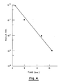
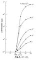
- Recombinant DNA pBTK5 was prepared from the luminescence system fragment of Vibrio fischeri DNA and plasmid pBR322 as described above in Part IX of Materials and Methods. 1 ⁇ g of pBTK5 DNA was used to transform different concentrations of E. coli K strain MM294 in 0.2 ml of CaCl 2 . After transformation, the cells were put in a scintillation vial containing 0.8 ml of LB and 0.2 ml of LB medium containing autoinducer prepared in accordance with Part XIV of Materials and Methods. Cell concentrations in different vials were 10 5 , 10 6 , 10 7 , and 10 8 cells/ml. Fig. 3 shows the kinetics of light emission for the four different concentrations of E. coli cells and Fig. 4 shows the correlation between the cell concentration and the onset of light emissions in these experiments. The background light emission was 1000 cpm.
- Transformation was performed as in Example 3 and the transformed bacteria were subsequently incubated in the presence of various antibiotics.
- Fig. 5 shows the kinetics of luminescence with and without the antibiotics. The background count emission was 1000 cpm.
- Transformation was performed as in Example 3, using as would be recipients Escherichia coli strain MM294, Aerobacter aerogenes strain 62-1, Proteus vulgaris and Staphylococcus albus.
- Fig. 6 shows the level of light emitted (cpm) as a function of time, and it is seen therefrom that only E. coli was transformed while the remaining three species remain unaffected. This demonstrates the specificity of transformation with the recombinant DNA pBTK5. The background count was 1000 cpm.
- Example 7 Determination of Escherichia coli strain W3110 by conjunction.
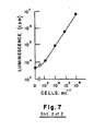
- the HfrH (high frequency of conjugation) strain containing the luminescence gene was constructed as described in XII (Materials and Methods). This strain was grown in LB medium containing streptomycin (2.5 ⁇ g/ml) for 2 hrs, at 37°C. The cells were washed once and diluted into LB medium to give a final cell density of 10 7 cells/ml. To 1 ml aliquots of this culture, different concentrations of (non-luminescent) E. coli strain W3110 cells were added. The mixed cultures were incubated without shaking at 37°C for 30 minutes and then the cultures were taken for luminescence determinations in a scintillation counter at 24°C. Fig. 7 shows the luminescence developed in the presence of different concentrations of E. coli W3110 recipient cells as a function of time (from time zero).
- Example 8 General method for the isolation, cloning, and transfer of exogenous genes into microbial host organisms, and detection of the recipient host using the expression of the introduced donor gene systems.
- luciferase and S -galactosidase can be introduced into host organisms for detection purposes in accordance with the invention.
- the system or enzyme can be chosen because of the ease with which its presence may be determined or because it is unique and absent in the organisms to be ascertained.
- the luciferase system previously described has both these qualities and is a preferred embodiment of the invention.
- Genes encoding proteins whose presence can be directly detected by their absorption of light at certain wavelengths e.g., hemoglobin
- ELISA radioactively labelled or linked to conveniently determined enzymes
- biotin to other compounds such as biotin
- genes encoding enzymes whose product or products can be measured directly or indirectly or in which the disappearance of one of its substrates can be similarly measured may be more suitable. Examples of such enzymes are given below in several Examples.
- Measurements might be based on evolution of gas or the reduction of the concentration of a dissolved gas, changes in pH, products or substrates that can be directly measured by spectrophotometry or chromatography or indirectly by coupled reactions or by antibody detection of a secondary complex (e.g., biotin-avidin) or through the use of radioactively labelled compounds.
- a secondary complex e.g., biotin-avidin
- Such measurements for identification purposes are given by means of illustration only and are not intended to limit the scope of the present invention, but to indicate the types of systems that might be used in conjunction with our method.
- Representative enzymes, proteins or enzyme systems can be utilized for detection of microorganisms after the relevant genetic structures have been assembled. Such assembly may involve natural recombination events or in vitro recombinant DNA techniques or both. There are numerous possibilities and strategies consistent with the present invention.
- the DNA of donor organisms i.e., bacterium, yeast, etc.
- the restriction endonuclease Sau3A to yield a set of overlapping fragments encompassing the whole donor genome. Fragments of a particular size may be prepared by centrifugation through glycerol or sucrose gradients or by agarose gel electrophoresis.
- fragments contain protruding 5' single strand ends with the structure 5'GATC-double stranded DNA and can then be ligated by T4 DNA ligase to appropriate vector molecules cut with enzymes (BamHI, BglII, Bc1I Sau3A) that created appropriate and compatible single strand ends.
- enzymes BamHI, BglII, Bc1I Sau3A
- An example of such a vector is ⁇ Charon 28 (Maniatis et al., op. cit.) cut with restriction endonuclease BamHI.
- the ligated DNA can then be transformed, subsequent to packaging if desired, into an appropriate host (lacking a restriction system).
- Viral DNA containing sequences for the desired exogenous system or enzyme can be detected either through enzymatic activity, immunological techniques or by synthetic or natural DNA probes and nucleic acid hybridization. Many of the above techniques of cloning and detection are detailed in Maniatis et al. (op. cit.). Subsequently, the donor gene is subcloned and then connected to a powerful bacterial promoter (e.g., plac of Escherichia coli or PRof bacteriophage ⁇ ).
- a powerful bacterial promoter e.g., plac of Escherichia coli or PRof bacteriophage ⁇ .
- the promoter-gene segment can be transferred by recombination, natural, in vitro or both, to a conjugative plasmid (e.g., F factor or RP4), a transformable element (e.g., plasmid pBR322) or to an appropriate bacteriophage (e.g., ⁇ or T4).
- a conjugative plasmid e.g., F factor or RP4
- a transformable element e.g., plasmid pBR322
- an appropriate bacteriophage e.g., ⁇ or T4
- the expressed donor gene or gene system thus transferred to a receptive host microorganism can then be used to identify the host organism.
- a suitable method for monitoring the activity of the donor genetic system in the host organism is selected (i.e., light production with luciferase; end product or intermediate of enzymatic reaction; gas evolution, etc.).
- the isolation, cloning, and transfer of foreign genes from a donor organism into a receptive host microorganism and identification of the host microorganism by monitoring expression of the foreign gene system contained therein is effected.
- Example 9 Method for the use of alcohol dehydrogenase or other NAD oxidoreductases gene systems obtained from a foreign donor and transferred to a receptive host microorganism for detection of the host.
- Alcohol dehydrogenase (E.C. 1.1.1.1; Alcohol:NAD oxidoreductase) is an enzyme that is formed by many species such as yeast, man and Bacillus.
- the gene or genes coding for this enzyme can be isolated by recombinant DNA techniques. Using the method of Example 8, the DNA of yeast as a donor organism is isolated, treated by restriction endonuclease, cloned, connected to an effective bacterial promotor, and transferred by an appropriate vector, preferably a bacteriophage, to a receptive host microorganism.
- the ADH gene can then be used to detect the presence of specific microorganisms by techniques and strategy similar to those presented earlier for bacterial luciferase and beta-galactosidase.
- ADH In the case of ADH, light production will not be directly monitored; rather the conversion of NAD to NADH would be measured.
- ADH catalyses a reaction whose products are NADH, H and acetaldehyde.
- the presence and concentration of NADH can be determined by adsorbance of light at 340 nm or by bacterial luciferase in the presence of an appropriate aldehyde (e.g., C 14 aldehyde) (Stanley, P.E. in Methods in Enzymology, volume LVII edited by M.A. DeLuca, Academic Press 1978, New York, San Francisco, London, pp. 215-222).
- NAD oxidoreductases There are many other NAD oxidoreductases that would be as suitable as ADH (enzymes of E.C. classes 1.1.1, 1.2.1, 1.3.1, 1.4.1, 1.5.1 and 1.8.1) for identification of host microorganisms.
- L-alanine dehydrogenase (E.C.1.4.1.1., L-alanine: NAD oxidoreductase) also yields NADH from NAD
- this enzyme liberates NH 3 from L-alanine when it is converted to pyruvate and NH 3 may be the product measured rather than NADH.
- Other oxidoreductases use 0 2 as the electron acceptor in place of NAD (E.C. classes 1.1.3, 1.2.3, 1.3.3 and 1.4.3) and the disappearance of dissolved oxygen might be followed by, for example, an oxygen electrode.
- Example 10 Method for the use of genetic systems coating for transferase obtained from a foreign donor and transferred to a receptive host microorganism for detection of the host.
- Transferase enzyme systems can also be used to identify microorganisms in conjunction with the invention herein described.
- the DNA of a suitable donor organism containing the selected transferase gene(s) is isolated, treated by restriction endonucleases, cloned, connected to an effective bacterial promotor, and transferred by an appropriate vector (preferably a bacteriophage), to a receptive host microorganism.
- L-tyrosine:2-oxoglutarate animotransferase yields p-hydroxyphenylpyruvate from L-tyrosine in the presence of 2-oxoglutarate and the appearance of p-hydroxyphenylpyruvate can be monitored spectrophotometrically at 330 nm (Hadar, R., Slonim, A., and Kuhn, J., 1976. Role of D-tryptophan oxidase in D-tryptophan utilization by Escherichia coli. J. Bacteriol. 125:1096-1104).
- a second example of a transferase is galactokinase (ATP: -D-galactose-1-phosphotransferase; E.C.2.7.1.6) whose activity can be followed by disappearance of ATP or appearance of P-D-galactose-1-phosphate.
- ATP -D-galactose-1-phosphotransferase
- Other detection modes can be readily adapted to the particular selected transferase.
- Example 11 Method for the use of h Y drolase, lyase, or isomerase genetic systems obtained from a foreign donor and transferred to a receptive host microorganism for detection of the host.
- hydrolase, lyase, and isomerase enzyme systems can be used to identify microorganisms in conjunction with the present invention.
- DNA of a suitable donor organism containing the selected enzyme is isolated, treated by restriction endonucleases, cloned, connected to an effective bacterial promotor, and transferred by an appropriate vector (preferably a bacteriophage), to a receptive host microorganism.
- alkaline phosphatase (E.C.3.1.3.1, orthophosphoric monoester phosphohydrolase) activity can be determined in a number of ways, one of which is by release of p-nitrophenol from p-nitrophenylphosphate.
- p-nitrophenol concentration can be determined spectrophotometrically at 420 nm at alkaline pH in the same way that o-nitrophenol was used for determination of -galactosidase (E.C.3.2.1.23) activity.
- Lyases such as aspartate-I-decarboxylase (E.C.4.1.1.11, L-aspartate 1-carboxylyase) and ornithine decarboxylase (E.C.4.1.1.17, L-ornithine carboxylyase) are equally suitable since the liberation of C0 2 (as H 2 C0 3 ) can be determined by changes in pH.
- Isomerases such as glutamate racemase (E.C.5.1.1.3) can also be employed. In the case of glutamate racemase the conversion of D-glutamate to L-glutamate can be followed by coupling the system to L-glutamate oxidase following conventional protocols.
- Example 12 Method for the use of oxidase genetic systems obtained from a foreign donor and transferred to a receptive host microorganism for detection of the host.
- bacteriophage genes can sometimes become linked to bacterial genes through substitution and/or addition.
- some bacteriophage envelope accidently DNA coming from a source other than the bacteriophage i.e. with chromosomal, plasmid or other bacteriophage species.
- the former is called specialized transduction, the latter generalized transduction. Both of these phenomena lead to the transfer of DNA from one bacterium to another.
- Phages can be constructed that carry desired genes and in which no essential bacteriophage genes have been lost. Such bacteriophages will efficiently introduce these cloned genes if an appropriate host is present. If elements for their expression exist, these genes will be highly expressed since many copies of the bacteriophage chromosome are made during the growth of the phage.
- the fragment of DNA carrying the luminescence genes of V, fischeri was transferred to the Escherichia coli bacteriophage ⁇ from the plasmid pBTK5. Transference of other donor genetic systems can be similarly effected by appropriate constructions.
- the phage Upon infection, the phage adsorb to the specific bacteria capable of acting as host, and the encapsulated DNA of the phage head is injected into the bacterial cell. Once the DNA is inside the bacterium it can make use of the host functions and it is transcribed and translated to produce luminescence enzymes (luciferase and those necessary for aldehyde synthesis) or other introduced enzyme systems.
- luminescence enzymes luciferase and those necessary for aldehyde synthesis
- a luminescent system is a preferred embodiment of the invention. Since neither the phage, nor the bacteria emit light by themselves, the level of light subsequent to infection can be used to detect the presence of bacteria and the amount of light emitted reflects the number of phage-bacterium complexes. In the presence of excess phage the amount of light reflects the concentration of bacteria. Using this technique less than 10 4 Escherichia coli cells/ml can be detected within one hour (Fig. 1). When a sample containing bacteria was concentrated by filtration through Millipore HA 0.45 micron filters, concentrations as low as 10 cells/ml could be detected.
- the presence of bacteria can also be detected by enzymatic means using transduction as illustrated in Figure 3.
- the presence of sufficient bacterial cells and the infection thereof by a bacteriophage carrying a gene coding for beta-galactosidase results in the de novo synthesis of beta-galactosidase whose presence and activity may be determined by enzymatic assay.
- Bacteriophage generally can only grow on some or all strains within a given single species of bacteria. Occasionally a given type of bacteriophage can infect several species but these are almost always found to be closely related.
- the test for bacteria can be made as specific as desired. This means that not only can the presence of bacteria be detected in this test but also the presence or absence of specific types of bacteria can be rapidly determined. For example, if the detection of Escherichia coli is desired, a mixture of phages whose host ranges span all known strains of this species and which carry luminescence genes, would allow the rapid specific detection of this species.
- bacteria can be detected by transforming them with a compatible DNA containing bioluminescence or other donor genes.
- the presence of bacteria, their approximate numbers, their taxonomic position and their susceptibility to antibiotics can be determined through this technique.
- Fig. 5 shows the experimental results obtained. Kanamycin (30 ⁇ g/ml) and chloramphenicol (30 ⁇ g/ml) entirely prevented the production of light. Tetracycline (10 p g/ml) caused a 90 y decrease versus that of the control without antibiotic. Ampicillin (30 ⁇ g/ml) actually stimulates light production.
- ampicillin would not inhibit or only slightly inhibit light emission was expected because pBTK5 caries a determinant for ampicillin resistance.
- the unexpected stimulation may be due to selection for increased replication of the entering plasmid or to increased transcription of the light genes via the bla. gene (ampicillin resistance) promoter.
- an antibiotic chloramphenicol, tetracycline and kanamycin in these experiments
- Resistance is also rapidly detected.
- the activity of an antibiotic can be rapidly ascertained as a function of its concentration (tetracycline versus chloramphenicol and kanamycin). A suboptimal concentration or partial resistance is also detected by this test (tetracycline).
- An additional feature of transformation is its specificity. As shown above, two strains of Escherichia coli were readily detected. When an experiment was conducted in which Escherichia coli, Proteus vulgaris, Staphylococcus albus and Aerobacter aerogenes were transformed by pBTK5 DNA, only Escherichia coli emitted light (Fig. 6). Since Aerobacter and Proteus are quite closely related to Escherichia coli, this shows that this test has a good deal of specificity.
- plasmids are circular DNA molecules whose presence is often superfluous to the cell's health. Not all plasmids are able to promote conjugation: only those that have transfer genes are able.
- the plasmid exists as a free-non-integrated molecule it usually mediates its own transfer from donor to recipient and only occasionally the transfer of other plasmids or of the bacterial chromosome.
- a conjugating plasmid is integrated into the chromosomes, it primarily transfers the chromosome and only rarely succeeds in transferring itself. The range of possible recipients depends on the conjugating plasmid itself. For example, plasmid RP4 can transfer itself between quite distantly related gram negative bacterial while F + can transfer itself to many fewer, closely related types.
- Escherichia coli K12 strain AT2446 that is an Hfr was made doubly lysogenic as described in Part XII of Material and Methods.
- AT2446 is an HfrH strain and hence transfers its DNA to a recipient starting from 0 minutes.
- the donor DNA is transferred clockwise with thr (Bachmann, B. J. and K. B. Low, 1980: Linkage map of Escherichia coli K12, edition 6. Microbiol. Rev. 44:1-56) the first bacterial chromosomal marker and malB at 91 minutes about the last. Since the attachment site of bacteriophage X is at 17 minutes, HfrH should transfer the lysogenic virus (in this case viruses) fairly early and at a high efficiency.
- Example 6 An experiment illustrating the use of conjugation and luminescence is described in Example 6 and as shown in Fig. 7, the amount of light emitted is directly related to the concentration of the recipient cells at a given donor concentration. That the relation between emitted light is linearly related to recipient concentration is illustrated in the insert of Fig. 7.
- Antibiotics like chloramphenicol (10 p g/ml) and streptomycin (100 11 g/ml) totally inhibited the increase in emitted light when they were included during mating.
- Conjugation is not limited to Hfr's or lysogenic bacteriophages. Any strain can be used which contains a luminescence system, or parts thereof, or other donor genetic system which is absent or poorly expressed in the host organism, in a transferable replicon.
- the replicon may be the bacterial chromosome or a plasmid.
- the luminescence system or parts thereof, or other genetic system may be introduced by a bacteriophage or by genetic recombination, either natural or artificial, such that the luminescence or other genes are covalently linked to the transferred genetic material.
- conjugation is analagous to the use of light coupled transduction and transformation described above.
Landscapes
- Health & Medical Sciences (AREA)
- Life Sciences & Earth Sciences (AREA)
- Chemical & Material Sciences (AREA)
- Engineering & Computer Science (AREA)
- Genetics & Genomics (AREA)
- Organic Chemistry (AREA)
- Zoology (AREA)
- Wood Science & Technology (AREA)
- Proteomics, Peptides & Aminoacids (AREA)
- Biotechnology (AREA)
- General Engineering & Computer Science (AREA)
- Bioinformatics & Cheminformatics (AREA)
- Molecular Biology (AREA)
- Biophysics (AREA)
- Biomedical Technology (AREA)
- Physics & Mathematics (AREA)
- Biochemistry (AREA)
- Microbiology (AREA)
- General Health & Medical Sciences (AREA)
- Immunology (AREA)
- Analytical Chemistry (AREA)
- Toxicology (AREA)
- Plant Pathology (AREA)
- Measuring Or Testing Involving Enzymes Or Micro-Organisms (AREA)
- Micro-Organisms Or Cultivation Processes Thereof (AREA)
- Investigating Or Analysing Materials By The Use Of Chemical Reactions (AREA)
- Preparation Of Compounds By Using Micro-Organisms (AREA)
- Enzymes And Modification Thereof (AREA)
Priority Applications (1)
| Application Number | Priority Date | Filing Date | Title |
|---|---|---|---|
| AT85303913T ATE88760T1 (de) | 1984-06-05 | 1985-06-03 | Nachweis und/oder identifizierung von mikroorganismen in einer probe mittels biolumineszenz oder anderer exogenen genetisch eingefuehrten markierungssubstanzen. |
Applications Claiming Priority (4)
| Application Number | Priority Date | Filing Date | Title |
|---|---|---|---|
| IL72026 | 1984-06-05 | ||
| IL72026A IL72026A0 (en) | 1984-06-05 | 1984-06-05 | Bioluminescent detection of microorganisms |
| IL75347 | 1985-05-30 | ||
| IL7534785A IL75347A (en) | 1985-05-30 | 1985-05-30 | Method for detecting and identifying microorganisms in a biological test sample using bioluminescence or other exogenous genetically-introduced marker |
Publications (3)
| Publication Number | Publication Date |
|---|---|
| EP0168933A2 true EP0168933A2 (de) | 1986-01-22 |
| EP0168933A3 EP0168933A3 (en) | 1987-08-26 |
| EP0168933B1 EP0168933B1 (de) | 1993-04-28 |
Family
ID=26321307
Family Applications (1)
| Application Number | Title | Priority Date | Filing Date |
|---|---|---|---|
| EP85303913A Expired - Lifetime EP0168933B1 (de) | 1984-06-05 | 1985-06-03 | Nachweis und/oder Identifizierung von Mikroorganismen in einer Probe mittels Biolumineszenz oder anderer exogenen genetisch eingeführten Markierungssubstanzen |
Country Status (9)
| Country | Link |
|---|---|
| EP (1) | EP0168933B1 (de) |
| JP (2) | JPH0634757B2 (de) |
| AT (1) | ATE88760T1 (de) |
| CA (1) | CA1277931C (de) |
| DE (1) | DE3587299T2 (de) |
| DK (1) | DK249485A (de) |
| ES (5) | ES8801853A1 (de) |
| FI (1) | FI852220L (de) |
| NO (1) | NO852232L (de) |
Cited By (36)
| Publication number | Priority date | Publication date | Assignee | Title |
|---|---|---|---|---|
| EP0202688A3 (en) * | 1985-05-24 | 1987-09-02 | Enzo Biochem, Inc. | Method and composition for detecting analyte moieties |
| WO1988000617A1 (en) * | 1986-07-22 | 1988-01-28 | Boyce Thompson Institute For Plant Research | Use of bacterial luciferase structural genes for cloning and monitoring gene expression in microorganisms and for tagging and identification of genetically engineered organisms |
| WO1988004326A1 (en) * | 1986-12-01 | 1988-06-16 | Mcdonnell Douglas Corporation | Method of identifying unknown organisms |
| EP0288306A1 (de) * | 1987-04-24 | 1988-10-26 | Bioscience International, Inc. | Diagnostika und Impfstoffe für Mycobacteria in der Volksgesundheit, in der medizinischen und veterinären Praxis |
| EP0252558A3 (de) * | 1986-07-04 | 1989-03-22 | SCLAVO S.p.A. | DNS-Element von Corynebacterium diphteriae mit typischen Eigenschaften eines IS-Insertionselementes |
| EP0341477A1 (de) * | 1988-05-11 | 1989-11-15 | Chisso Corporation | Verfahren zur Herstellung von einem Aequorinphotoprotein |
| DE3833628A1 (de) * | 1988-10-03 | 1990-04-12 | Genlux Forschungsgesellschaft | Verfahren zum nachweis und zur identifikation toxischer substanzen mit hilfe klonierter mikroorganismen |
| DE3902982A1 (de) * | 1989-02-01 | 1990-08-02 | Genlux Forschungsgesellschaft | Verfahren zum nachweis von quecksilber mit hilfe von durch quecksilber zu erhoehter biolumineszenz angeregter mikroorganismen |
| WO1992002633A1 (en) * | 1990-08-09 | 1992-02-20 | Amersham International Plc | Methods for rapid microbial detection |
| EP0439354A3 (en) * | 1990-01-24 | 1992-06-17 | Amoco Corporation | Signal generating moiety and method for use |
| EP0437537A4 (en) * | 1988-10-04 | 1992-08-12 | Dna Plant Technology Corporation | Bacterial detection by phage transduction of detectable phenotype |
| US5225324A (en) * | 1987-04-24 | 1993-07-06 | Bioscience International, Inc. | Diagnostics for mycobacteria in public health, medical, and veterinary practice |
| WO1998023737A1 (en) * | 1996-11-28 | 1998-06-04 | Universiteit Van Amsterdam | Production of in vivo labeled single chain synthetic antibody fragments |
| WO1999000517A3 (en) * | 1997-06-27 | 1999-05-14 | Univ Cambridge Tech | Biosensor materials and methods |
| WO1999045152A1 (en) * | 1998-03-06 | 1999-09-10 | E.I. Du Pont De Nemours And Company | Methods for identifying polycationic, peptide-like compounds with antibacterial activity |
| WO2004079015A1 (ja) * | 2003-03-07 | 2004-09-16 | Ebara Corporation | 大腸菌の検出方法及び大腸菌検出用ファージ |
| US6806079B1 (en) | 1990-07-10 | 2004-10-19 | Medical Research Council | Methods for producing members of specific binding pairs |
| US7063943B1 (en) | 1990-07-10 | 2006-06-20 | Cambridge Antibody Technology | Methods for producing members of specific binding pairs |
| US7132247B1 (en) | 1998-09-17 | 2006-11-07 | Regents Of The University Of Minnesota | Composite devices incorporating biological material and methods |
| US7635666B1 (en) | 1990-07-10 | 2009-12-22 | Medical Research Council | Methods for producing members of specific binding pairs |
| US7745023B2 (en) | 2003-08-08 | 2010-06-29 | Regents Of The University Of Minnesota | Structured material for the production of hydrogen |
| US7972773B2 (en) | 2002-04-12 | 2011-07-05 | Colorado School Of Mines | Method for detecting concentrations of a target bacterium that uses phages to infect target bacterial cells |
| US8092990B2 (en) | 2005-03-31 | 2012-01-10 | Colorado School Of Mines | Apparatus and method for detecting microscopic organisms using bacteriophage |
| US8216780B2 (en) | 2002-04-12 | 2012-07-10 | Microphage (Tm) Incorporated | Method for enhanced sensitivity in bacteriophage-based diagnostic assays |
| US8455186B2 (en) | 2007-06-15 | 2013-06-04 | MicroPhage™ Incorporated | Method of detection of microorganisms with enhanced bacteriophage amplification |
| US8481302B2 (en) | 2008-11-03 | 2013-07-09 | General Electric Company | Total bacteria monitoring system |
| US8697434B2 (en) | 2008-01-11 | 2014-04-15 | Colorado School Of Mines | Detection of phage amplification by SERS nanoparticles |
| US8829473B1 (en) | 2013-03-13 | 2014-09-09 | GeneWeave Biosciences, Inc. | Systems and methods for detection of cells using engineered transduction particles |
| US9388453B2 (en) | 2013-03-13 | 2016-07-12 | GeneWeave Biosciences, Inc. | Non-replicative transduction particles and transduction particle-based reporter systems |
| US9441204B2 (en) | 2008-04-03 | 2016-09-13 | Colorado School Of Mines | Compositions and methods for detecting Yersinia pestis bacteria |
| WO2019055457A1 (en) * | 2017-09-12 | 2019-03-21 | Biocapital Holdings, Llc | BIOLOGICAL DEVICES FOR PRODUCING OXIDIZED ZINC AND THEIR APPLICATIONS |
| US10351893B2 (en) | 2015-10-05 | 2019-07-16 | GeneWeave Biosciences, Inc. | Reagent cartridge for detection of cells |
| EP3524686A1 (de) * | 2014-04-24 | 2019-08-14 | Geneweave Biosciences Inc. | Verfahren zum nachweis von antibiotika-resistenten zellen unter verwendung von gentechnisch hergestellten transduktionspartikel |
| CN110951764A (zh) * | 2019-12-10 | 2020-04-03 | 首都医科大学附属北京地坛医院 | 一种表达荧光素酶的产酸克雷伯菌及其用途 |
| CN112266928A (zh) * | 2020-09-21 | 2021-01-26 | 西北大学 | 一种检测多重耐药质粒水平转移的方法 |
| US11077444B2 (en) | 2017-05-23 | 2021-08-03 | Roche Molecular Systems, Inc. | Packaging for a molecular diagnostic cartridge |
Families Citing this family (10)
| Publication number | Priority date | Publication date | Assignee | Title |
|---|---|---|---|---|
| JPS6434286A (en) * | 1987-07-29 | 1989-02-03 | Kikkoman Corp | Production of luciferase |
| JPS6434289A (en) * | 1987-07-29 | 1989-02-03 | Kikkoman Corp | Novel recombinant dna |
| JPH02174697A (ja) * | 1988-09-30 | 1990-07-06 | Sekisui Chem Co Ltd | 発光測定方式のバイオアッセイ法 |
| JP3615810B2 (ja) * | 1994-12-05 | 2005-02-02 | 東北電子産業株式会社 | 細菌の検出方法及び検出装置 |
| JP3270722B2 (ja) * | 1996-09-27 | 2002-04-02 | オルガノ株式会社 | 細菌の検出方法及び検出装置 |
| JP3973876B2 (ja) * | 2001-10-30 | 2007-09-12 | 株式会社三菱化学ヤトロン | 新規の細菌分析方法 |
| JP6300222B2 (ja) * | 2013-09-25 | 2018-03-28 | 国立大学法人東京工業大学 | 遺伝子組換えウイルスによる微生物の迅速検出法 |
| ES2872526T3 (es) * | 2014-06-13 | 2021-11-02 | Geneweave Biosciences Inc | Detección de células independiente del crecimiento |
| US11873524B2 (en) * | 2018-01-12 | 2024-01-16 | Laboratory Corporation Of America Holdings | Methods and systems for the rapid detection of Salmonella using infectious agents |
| US10968491B2 (en) | 2018-12-05 | 2021-04-06 | GeneWeave Biosciences, Inc. | Growth-independent detection of cells |
Family Cites Families (9)
| Publication number | Priority date | Publication date | Assignee | Title |
|---|---|---|---|---|
| US3930956A (en) * | 1973-10-29 | 1976-01-06 | The Regents Of The University Of Michigan | Method for the genetic detection of microorganisms |
| US4038143A (en) * | 1973-10-29 | 1977-07-26 | The Regents Of The University Of Michigan | Test kit for the genetic detection of microorganisms |
| FR2423541A1 (fr) * | 1978-04-18 | 1979-11-16 | Pasteur Institut | Procede de tri parmi une population de micro-organismes, notamment de bacteries, de ceux qui sont porteurs ou producteurs d'un constituant intracellulaire particulier, notamment d'un fragment d'acide nucleique determine |
| EP0035831B1 (de) * | 1980-03-07 | 1986-01-08 | Imperial Chemical Industries Plc | Verfahren zur Herstellung genetisch modifizierter Mikroorganismen |
| JPS58126789A (ja) * | 1981-12-29 | 1983-07-28 | Kyowa Hakko Kogyo Co Ltd | レースレオニンの製造法 |
| ATE48156T1 (de) * | 1982-11-01 | 1989-12-15 | Miles Inc | Verfahren zur heterologen klonierung eines gens in einem bacillus-mikroorganismus. |
| US4581335A (en) * | 1982-12-01 | 1986-04-08 | Texas A&M University System | Process for producing a cloned luciferase-synthesizing microorganism |
| JPS59196098A (ja) * | 1983-04-23 | 1984-11-07 | Ajinomoto Co Inc | 発酵法によるl−トリプトフアンの製造法 |
| CA1338856C (en) * | 1983-07-05 | 1997-01-21 | Jannis Stavrianopoulos | In vivo labelling of polynucleotide sequences |
-
1985
- 1985-05-30 CA CA000482862A patent/CA1277931C/en not_active Expired - Lifetime
- 1985-06-03 EP EP85303913A patent/EP0168933B1/de not_active Expired - Lifetime
- 1985-06-03 FI FI852220A patent/FI852220L/fi not_active Application Discontinuation
- 1985-06-03 DE DE8585303913T patent/DE3587299T2/de not_active Expired - Fee Related
- 1985-06-03 NO NO852232A patent/NO852232L/no unknown
- 1985-06-03 DK DK249485A patent/DK249485A/da not_active Application Discontinuation
- 1985-06-03 AT AT85303913T patent/ATE88760T1/de not_active IP Right Cessation
- 1985-06-03 JP JP60120297A patent/JPH0634757B2/ja not_active Expired - Fee Related
-
1986
- 1986-02-17 ES ES552110A patent/ES8801853A1/es not_active Expired
-
1987
- 1987-10-16 ES ES557769A patent/ES8900026A1/es not_active Expired
-
1988
- 1988-03-16 ES ES557825A patent/ES8900020A1/es not_active Expired
- 1988-03-16 ES ES557826A patent/ES8900021A1/es not_active Expired
- 1988-06-01 ES ES557840A patent/ES8900143A1/es not_active Expired
- 1988-07-11 JP JP63171065A patent/JPS6427479A/ja active Pending
Cited By (52)
| Publication number | Priority date | Publication date | Assignee | Title |
|---|---|---|---|---|
| EP0202688A3 (en) * | 1985-05-24 | 1987-09-02 | Enzo Biochem, Inc. | Method and composition for detecting analyte moieties |
| EP0252558A3 (de) * | 1986-07-04 | 1989-03-22 | SCLAVO S.p.A. | DNS-Element von Corynebacterium diphteriae mit typischen Eigenschaften eines IS-Insertionselementes |
| WO1988000617A1 (en) * | 1986-07-22 | 1988-01-28 | Boyce Thompson Institute For Plant Research | Use of bacterial luciferase structural genes for cloning and monitoring gene expression in microorganisms and for tagging and identification of genetically engineered organisms |
| WO1988004326A1 (en) * | 1986-12-01 | 1988-06-16 | Mcdonnell Douglas Corporation | Method of identifying unknown organisms |
| EP0288306A1 (de) * | 1987-04-24 | 1988-10-26 | Bioscience International, Inc. | Diagnostika und Impfstoffe für Mycobacteria in der Volksgesundheit, in der medizinischen und veterinären Praxis |
| WO1988008456A1 (en) * | 1987-04-24 | 1988-11-03 | Bioscience International, Inc. | Diagnostics and vaccines for mycobacteria in public health, medical and veterinary practice |
| US5225324A (en) * | 1987-04-24 | 1993-07-06 | Bioscience International, Inc. | Diagnostics for mycobacteria in public health, medical, and veterinary practice |
| EP0341477A1 (de) * | 1988-05-11 | 1989-11-15 | Chisso Corporation | Verfahren zur Herstellung von einem Aequorinphotoprotein |
| DE3833628A1 (de) * | 1988-10-03 | 1990-04-12 | Genlux Forschungsgesellschaft | Verfahren zum nachweis und zur identifikation toxischer substanzen mit hilfe klonierter mikroorganismen |
| EP0437537A4 (en) * | 1988-10-04 | 1992-08-12 | Dna Plant Technology Corporation | Bacterial detection by phage transduction of detectable phenotype |
| US5447836A (en) * | 1988-10-04 | 1995-09-05 | Dna Plant Technology Corporation | Bacterial detection by phage transduction of ice nucleation and other phenotypes |
| DE3902982A1 (de) * | 1989-02-01 | 1990-08-02 | Genlux Forschungsgesellschaft | Verfahren zum nachweis von quecksilber mit hilfe von durch quecksilber zu erhoehter biolumineszenz angeregter mikroorganismen |
| EP0439354A3 (en) * | 1990-01-24 | 1992-06-17 | Amoco Corporation | Signal generating moiety and method for use |
| US7723270B1 (en) | 1990-07-10 | 2010-05-25 | Medical Research Council | Methods for producing members of specific binding pairs |
| US7635666B1 (en) | 1990-07-10 | 2009-12-22 | Medical Research Council | Methods for producing members of specific binding pairs |
| US7732377B2 (en) | 1990-07-10 | 2010-06-08 | Medical Research Council | Methods for producing members of specific binding pairs |
| US6806079B1 (en) | 1990-07-10 | 2004-10-19 | Medical Research Council | Methods for producing members of specific binding pairs |
| US6916605B1 (en) | 1990-07-10 | 2005-07-12 | Medical Research Council | Methods for producing members of specific binding pairs |
| US7063943B1 (en) | 1990-07-10 | 2006-06-20 | Cambridge Antibody Technology | Methods for producing members of specific binding pairs |
| US7662557B2 (en) | 1990-07-10 | 2010-02-16 | Medical Research Council | Methods for producing members of specific binding pairs |
| US5498525A (en) * | 1990-08-09 | 1996-03-12 | Amersham International Plc | Methods for rapid microbial detection |
| US5723330A (en) * | 1990-08-09 | 1998-03-03 | Merck Patent Gmbh | Genetically engineered reporter bacteria for the detection of bacteriophage |
| WO1992002633A1 (en) * | 1990-08-09 | 1992-02-20 | Amersham International Plc | Methods for rapid microbial detection |
| WO1998023737A1 (en) * | 1996-11-28 | 1998-06-04 | Universiteit Van Amsterdam | Production of in vivo labeled single chain synthetic antibody fragments |
| US6849442B1 (en) | 1997-06-27 | 2005-02-01 | Cambridge University Technical Services Limited | Biosensor materials and methods |
| WO1999000517A3 (en) * | 1997-06-27 | 1999-05-14 | Univ Cambridge Tech | Biosensor materials and methods |
| WO1999045152A1 (en) * | 1998-03-06 | 1999-09-10 | E.I. Du Pont De Nemours And Company | Methods for identifying polycationic, peptide-like compounds with antibacterial activity |
| US7132247B1 (en) | 1998-09-17 | 2006-11-07 | Regents Of The University Of Minnesota | Composite devices incorporating biological material and methods |
| US8216780B2 (en) | 2002-04-12 | 2012-07-10 | Microphage (Tm) Incorporated | Method for enhanced sensitivity in bacteriophage-based diagnostic assays |
| US7972773B2 (en) | 2002-04-12 | 2011-07-05 | Colorado School Of Mines | Method for detecting concentrations of a target bacterium that uses phages to infect target bacterial cells |
| WO2004079015A1 (ja) * | 2003-03-07 | 2004-09-16 | Ebara Corporation | 大腸菌の検出方法及び大腸菌検出用ファージ |
| US7745023B2 (en) | 2003-08-08 | 2010-06-29 | Regents Of The University Of Minnesota | Structured material for the production of hydrogen |
| US8092990B2 (en) | 2005-03-31 | 2012-01-10 | Colorado School Of Mines | Apparatus and method for detecting microscopic organisms using bacteriophage |
| US8455186B2 (en) | 2007-06-15 | 2013-06-04 | MicroPhage™ Incorporated | Method of detection of microorganisms with enhanced bacteriophage amplification |
| US8697434B2 (en) | 2008-01-11 | 2014-04-15 | Colorado School Of Mines | Detection of phage amplification by SERS nanoparticles |
| US9441204B2 (en) | 2008-04-03 | 2016-09-13 | Colorado School Of Mines | Compositions and methods for detecting Yersinia pestis bacteria |
| US8481302B2 (en) | 2008-11-03 | 2013-07-09 | General Electric Company | Total bacteria monitoring system |
| US9481903B2 (en) | 2013-03-13 | 2016-11-01 | Roche Molecular Systems, Inc. | Systems and methods for detection of cells using engineered transduction particles |
| US8829473B1 (en) | 2013-03-13 | 2014-09-09 | GeneWeave Biosciences, Inc. | Systems and methods for detection of cells using engineered transduction particles |
| US9752200B2 (en) | 2013-03-13 | 2017-09-05 | GeneWeave Biosciences, Inc. | Non-replicative transduction particles and transduction particle-based reporter systems |
| US9771622B2 (en) | 2013-03-13 | 2017-09-26 | GeneWeave Biosciences, Inc. | Non-replicative transduction particles and transduction particle-based reporter systems |
| US10227662B2 (en) | 2013-03-13 | 2019-03-12 | GeneWeave Biosciences, Inc. | Non-replicative transduction particles and transduction particle-based reporter systems |
| US10227663B2 (en) | 2013-03-13 | 2019-03-12 | GeneWeave Biosciences, Inc. | Non-replicative transduction particles and transduction particle-based reporter systems |
| US9388453B2 (en) | 2013-03-13 | 2016-07-12 | GeneWeave Biosciences, Inc. | Non-replicative transduction particles and transduction particle-based reporter systems |
| EP3524686A1 (de) * | 2014-04-24 | 2019-08-14 | Geneweave Biosciences Inc. | Verfahren zum nachweis von antibiotika-resistenten zellen unter verwendung von gentechnisch hergestellten transduktionspartikel |
| US10351893B2 (en) | 2015-10-05 | 2019-07-16 | GeneWeave Biosciences, Inc. | Reagent cartridge for detection of cells |
| US11077444B2 (en) | 2017-05-23 | 2021-08-03 | Roche Molecular Systems, Inc. | Packaging for a molecular diagnostic cartridge |
| WO2019055457A1 (en) * | 2017-09-12 | 2019-03-21 | Biocapital Holdings, Llc | BIOLOGICAL DEVICES FOR PRODUCING OXIDIZED ZINC AND THEIR APPLICATIONS |
| US10995345B2 (en) | 2017-09-12 | 2021-05-04 | Biocapital Holdings, Llc | Biological devices for producing oxidized zinc and applications thereof |
| US11781157B2 (en) | 2017-09-12 | 2023-10-10 | Biocapital Holdings, Llc | Biological devices for producing oxidized zinc and applications thereof |
| CN110951764A (zh) * | 2019-12-10 | 2020-04-03 | 首都医科大学附属北京地坛医院 | 一种表达荧光素酶的产酸克雷伯菌及其用途 |
| CN112266928A (zh) * | 2020-09-21 | 2021-01-26 | 西北大学 | 一种检测多重耐药质粒水平转移的方法 |
Also Published As
| Publication number | Publication date |
|---|---|
| ES8801853A1 (es) | 1988-02-16 |
| DE3587299D1 (de) | 1993-06-03 |
| ES557826A0 (es) | 1988-10-01 |
| NO852232L (no) | 1985-12-06 |
| JPH0634757B2 (ja) | 1994-05-11 |
| FI852220A7 (fi) | 1985-12-06 |
| DE3587299T2 (de) | 1993-08-05 |
| EP0168933B1 (de) | 1993-04-28 |
| ATE88760T1 (de) | 1993-05-15 |
| ES557840A0 (es) | 1989-01-16 |
| ES8900021A1 (es) | 1988-10-01 |
| FI852220L (fi) | 1985-12-06 |
| FI852220A0 (fi) | 1985-06-03 |
| ES557769A0 (es) | 1988-07-01 |
| ES552110A0 (es) | 1988-02-16 |
| JPS6143998A (ja) | 1986-03-03 |
| ES557825A0 (es) | 1988-10-01 |
| CA1277931C (en) | 1990-12-18 |
| ES8900020A1 (es) | 1988-10-01 |
| ES8900026A1 (es) | 1988-07-01 |
| EP0168933A3 (en) | 1987-08-26 |
| ES8900143A1 (es) | 1989-01-16 |
| JPS6427479A (en) | 1989-01-30 |
| DK249485D0 (da) | 1985-06-03 |
| DK249485A (da) | 1985-12-06 |
Similar Documents
| Publication | Publication Date | Title |
|---|---|---|
| US4861709A (en) | Detection and/or identification of microorganisms in a test sample using bioluminescence or other exogenous genetically-introduced marker | |
| EP0168933B1 (de) | Nachweis und/oder Identifizierung von Mikroorganismen in einer Probe mittels Biolumineszenz oder anderer exogenen genetisch eingeführten Markierungssubstanzen | |
| Nicas et al. | Isolation and characterization of transposon-induced mutants of Pseudomonas aeruginosa deficient in production of exoenzyme S | |
| Moreillon et al. | Two bactericidal targets for penicillin in pneumococci: autolysis-dependent and autolysis-independent killing mechanisms | |
| Simmon et al. | Degradation of bacteriophage lambda deoxyribonucleic acid after restriction by Escherichia coli K-12 | |
| McEwen et al. | Chromosomal mutations of Escherichia coli that alter expression of conjugative plasmid functions. | |
| US4968619A (en) | Modified microorganisms and method of preparing and using same | |
| Calvo et al. | Regulation of branched-chain amino acid biosynthesis in Salmonella typhimurium: isolation of regulatory mutants | |
| Dubnau et al. | Suppression of promoter mutations by the pleiotropic supX mutations | |
| Dauenhauer et al. | Cloning and expression in Escherichia coli of Serratia marcescens genes encoding prodigiosin biosynthesis | |
| Liberfarb et al. | Isolation, characterization, and genetic analysis of mutator genes in Escherichia coli B and K-12 | |
| Alper et al. | Cyclic 3', 5'-adenosine monophosphate phosphodiesterase mutants of Salmonella typhimurium | |
| SU1200853A3 (ru) | Способ конструировани гибридной плазмиды,содержащей генетический код термостойкой альфа-амилазы | |
| Burman et al. | Resistance of Escherichia coli to penicillins v. physiological comparison of two isogenic strains, one with chromosomally and one with episomally mediated ampicillin resistance | |
| US5658748A (en) | Streptococcus thermophilus strains and their use | |
| CN113337627B (zh) | 基于CRISPR/Cas12a的免标记可视化检测副溶血弧菌基因的方法 | |
| Matsumoto et al. | A generalized transducing phage of Serratia marcescens | |
| US4332897A (en) | Novel bacteriophage and method for preparing same | |
| Guiso et al. | Expression and regulation of lactose genes carried by plasmids | |
| Karkhoff-Schweizer et al. | Utilization of a mim-Dlac transposable element to create an α-complementation and regulated expression system for cloning in Pseudomonas aeruginosa | |
| Mizobuchi et al. | Abortive infection by bacteriophage BF23 due to the colicin Ib factor. I: Genetic studies of nonrestricted and amber mutants of bacteriophage BF23 | |
| Merkel et al. | Relationship between “chloramphenicol reductase activity” and chloramphenicol resistance in Escherichia coli | |
| Rigby et al. | Construction of intergeneric hybrids using bacteriophage P1CM: transfer of the Klebsiella aerogenes ribitol dehydrogenase gene to Escherichia coli | |
| EP0625191A1 (de) | Für mycobakterien spezifische reporter-mycobakteriophagen | |
| Guterman et al. | Rifampicin supersensitivity of rho strains of E. coli, and suppression by sur mutation |
Legal Events
| Date | Code | Title | Description |
|---|---|---|---|
| PUAI | Public reference made under article 153(3) epc to a published international application that has entered the european phase |
Free format text: ORIGINAL CODE: 0009012 |
|
| AK | Designated contracting states |
Designated state(s): AT BE CH DE FR GB IT LI LU NL SE |
|
| PUAL | Search report despatched |
Free format text: ORIGINAL CODE: 0009013 |
|
| AK | Designated contracting states |
Kind code of ref document: A3 Designated state(s): AT BE CH DE FR GB IT LI LU NL SE |
|
| 17P | Request for examination filed |
Effective date: 19871003 |
|
| 17Q | First examination report despatched |
Effective date: 19890605 |
|
| DIN1 | Information on inventor provided before grant (deleted) | ||
| RAP1 | Party data changed (applicant data changed or rights of an application transferred) |
Owner name: CTI RESEARCH AG |
|
| RAP1 | Party data changed (applicant data changed or rights of an application transferred) |
Owner name: BIOLUME LIMITED Owner name: AMERSHAM INTERNATIONAL PLC |
|
| GRAA | (expected) grant |
Free format text: ORIGINAL CODE: 0009210 |
|
| AK | Designated contracting states |
Kind code of ref document: B1 Designated state(s): AT BE CH DE FR GB IT LI LU NL SE |
|
| REF | Corresponds to: |
Ref document number: 88760 Country of ref document: AT Date of ref document: 19930515 Kind code of ref document: T |
|
| REF | Corresponds to: |
Ref document number: 3587299 Country of ref document: DE Date of ref document: 19930603 |
|
| ITF | It: translation for a ep patent filed | ||
| PG25 | Lapsed in a contracting state [announced via postgrant information from national office to epo] |
Ref country code: LU Free format text: LAPSE BECAUSE OF NON-PAYMENT OF DUE FEES Effective date: 19930630 |
|
| ET | Fr: translation filed | ||
| PLBE | No opposition filed within time limit |
Free format text: ORIGINAL CODE: 0009261 |
|
| STAA | Information on the status of an ep patent application or granted ep patent |
Free format text: STATUS: NO OPPOSITION FILED WITHIN TIME LIMIT |
|
| 26N | No opposition filed | ||
| EAL | Se: european patent in force in sweden |
Ref document number: 85303913.9 |
|
| REG | Reference to a national code |
Ref country code: CH Ref legal event code: PUEA Free format text: AMERSHAM INTERNATIONAL PLC;BIOLUME LIMITED TRANSFER- BIOLUME LIMITED;MERCK PATENT GMBH |
|
| REG | Reference to a national code |
Ref country code: FR Ref legal event code: TP |
|
| NLS | Nl: assignments of ep-patents |
Owner name: MERCK PATENT GMBH;BIOLUME LIMITED |
|
| REG | Reference to a national code |
Ref country code: GB Ref legal event code: 732E |
|
| REG | Reference to a national code |
Ref country code: GB Ref legal event code: IF02 |
|
| PGFP | Annual fee paid to national office [announced via postgrant information from national office to epo] |
Ref country code: GB Payment date: 20030528 Year of fee payment: 19 |
|
| PGFP | Annual fee paid to national office [announced via postgrant information from national office to epo] |
Ref country code: SE Payment date: 20030604 Year of fee payment: 19 |
|
| PGFP | Annual fee paid to national office [announced via postgrant information from national office to epo] |
Ref country code: FR Payment date: 20030610 Year of fee payment: 19 |
|
| PGFP | Annual fee paid to national office [announced via postgrant information from national office to epo] |
Ref country code: AT Payment date: 20030611 Year of fee payment: 19 |
|
| PGFP | Annual fee paid to national office [announced via postgrant information from national office to epo] |
Ref country code: DE Payment date: 20030612 Year of fee payment: 19 |
|
| PGFP | Annual fee paid to national office [announced via postgrant information from national office to epo] |
Ref country code: CH Payment date: 20030616 Year of fee payment: 19 |
|
| PGFP | Annual fee paid to national office [announced via postgrant information from national office to epo] |
Ref country code: NL Payment date: 20030630 Year of fee payment: 19 |
|
| PGFP | Annual fee paid to national office [announced via postgrant information from national office to epo] |
Ref country code: BE Payment date: 20030902 Year of fee payment: 19 |
|
| PG25 | Lapsed in a contracting state [announced via postgrant information from national office to epo] |
Ref country code: GB Free format text: LAPSE BECAUSE OF NON-PAYMENT OF DUE FEES Effective date: 20040603 Ref country code: AT Free format text: LAPSE BECAUSE OF NON-PAYMENT OF DUE FEES Effective date: 20040603 |
|
| PG25 | Lapsed in a contracting state [announced via postgrant information from national office to epo] |
Ref country code: SE Free format text: LAPSE BECAUSE OF NON-PAYMENT OF DUE FEES Effective date: 20040604 |
|
| PG25 | Lapsed in a contracting state [announced via postgrant information from national office to epo] |
Ref country code: LI Free format text: LAPSE BECAUSE OF NON-PAYMENT OF DUE FEES Effective date: 20040630 Ref country code: CH Free format text: LAPSE BECAUSE OF NON-PAYMENT OF DUE FEES Effective date: 20040630 Ref country code: BE Free format text: LAPSE BECAUSE OF NON-PAYMENT OF DUE FEES Effective date: 20040630 |
|
| BERE | Be: lapsed |
Owner name: *BIOLUME LTD Effective date: 20040630 Owner name: *MERCK PATENT G.M.B.H. Effective date: 20040630 |
|
| PG25 | Lapsed in a contracting state [announced via postgrant information from national office to epo] |
Ref country code: NL Free format text: LAPSE BECAUSE OF NON-PAYMENT OF DUE FEES Effective date: 20050101 Ref country code: DE Free format text: LAPSE BECAUSE OF NON-PAYMENT OF DUE FEES Effective date: 20050101 |
|
| GBPC | Gb: european patent ceased through non-payment of renewal fee |
Effective date: 20040603 |
|
| EUG | Se: european patent has lapsed | ||
| EUG | Se: european patent has lapsed | ||
| REG | Reference to a national code |
Ref country code: CH Ref legal event code: PL |
|
| PG25 | Lapsed in a contracting state [announced via postgrant information from national office to epo] |
Ref country code: FR Free format text: LAPSE BECAUSE OF NON-PAYMENT OF DUE FEES Effective date: 20050228 |
|
| NLV4 | Nl: lapsed or anulled due to non-payment of the annual fee |
Effective date: 20050101 |
|
| REG | Reference to a national code |
Ref country code: FR Ref legal event code: ST |