EP0216854B1 - Anticorps monoclonaux et analyse - Google Patents
Anticorps monoclonaux et analyse Download PDFInfo
- Publication number
- EP0216854B1 EP0216854B1 EP86902121A EP86902121A EP0216854B1 EP 0216854 B1 EP0216854 B1 EP 0216854B1 EP 86902121 A EP86902121 A EP 86902121A EP 86902121 A EP86902121 A EP 86902121A EP 0216854 B1 EP0216854 B1 EP 0216854B1
- Authority
- EP
- European Patent Office
- Prior art keywords
- cells
- protein
- antibody
- cell
- activated
- Prior art date
- Legal status (The legal status is an assumption and is not a legal conclusion. Google has not performed a legal analysis and makes no representation as to the accuracy of the status listed.)
- Expired - Lifetime
Links
Images


Classifications
-
- C—CHEMISTRY; METALLURGY
- C07—ORGANIC CHEMISTRY
- C07K—PEPTIDES
- C07K14/00—Peptides having more than 20 amino acids; Gastrins; Somatostatins; Melanotropins; Derivatives thereof
- C07K14/435—Peptides having more than 20 amino acids; Gastrins; Somatostatins; Melanotropins; Derivatives thereof from animals; from humans
- C07K14/705—Receptors; Cell surface antigens; Cell surface determinants
-
- C—CHEMISTRY; METALLURGY
- C07—ORGANIC CHEMISTRY
- C07K—PEPTIDES
- C07K16/00—Immunoglobulins [IG], e.g. monoclonal or polyclonal antibodies
- C07K16/18—Immunoglobulins [IG], e.g. monoclonal or polyclonal antibodies against material from animals or humans
- C07K16/28—Immunoglobulins [IG], e.g. monoclonal or polyclonal antibodies against material from animals or humans against receptors, cell surface antigens or cell surface determinants
-
- C—CHEMISTRY; METALLURGY
- C07—ORGANIC CHEMISTRY
- C07K—PEPTIDES
- C07K16/00—Immunoglobulins [IG], e.g. monoclonal or polyclonal antibodies
- C07K16/18—Immunoglobulins [IG], e.g. monoclonal or polyclonal antibodies against material from animals or humans
- C07K16/28—Immunoglobulins [IG], e.g. monoclonal or polyclonal antibodies against material from animals or humans against receptors, cell surface antigens or cell surface determinants
- C07K16/30—Immunoglobulins [IG], e.g. monoclonal or polyclonal antibodies against material from animals or humans against receptors, cell surface antigens or cell surface determinants from tumour cells
- C07K16/3046—Stomach, Intestines
-
- Y—GENERAL TAGGING OF NEW TECHNOLOGICAL DEVELOPMENTS; GENERAL TAGGING OF CROSS-SECTIONAL TECHNOLOGIES SPANNING OVER SEVERAL SECTIONS OF THE IPC; TECHNICAL SUBJECTS COVERED BY FORMER USPC CROSS-REFERENCE ART COLLECTIONS [XRACs] AND DIGESTS
- Y10—TECHNICAL SUBJECTS COVERED BY FORMER USPC
- Y10S—TECHNICAL SUBJECTS COVERED BY FORMER USPC CROSS-REFERENCE ART COLLECTIONS [XRACs] AND DIGESTS
- Y10S435/00—Chemistry: molecular biology and microbiology
- Y10S435/8215—Microorganisms
- Y10S435/948—Microorganisms using viruses or cell lines
-
- Y—GENERAL TAGGING OF NEW TECHNOLOGICAL DEVELOPMENTS; GENERAL TAGGING OF CROSS-SECTIONAL TECHNOLOGIES SPANNING OVER SEVERAL SECTIONS OF THE IPC; TECHNICAL SUBJECTS COVERED BY FORMER USPC CROSS-REFERENCE ART COLLECTIONS [XRACs] AND DIGESTS
- Y10—TECHNICAL SUBJECTS COVERED BY FORMER USPC
- Y10S—TECHNICAL SUBJECTS COVERED BY FORMER USPC CROSS-REFERENCE ART COLLECTIONS [XRACs] AND DIGESTS
- Y10S435/00—Chemistry: molecular biology and microbiology
- Y10S435/975—Kit
-
- Y—GENERAL TAGGING OF NEW TECHNOLOGICAL DEVELOPMENTS; GENERAL TAGGING OF CROSS-SECTIONAL TECHNOLOGIES SPANNING OVER SEVERAL SECTIONS OF THE IPC; TECHNICAL SUBJECTS COVERED BY FORMER USPC CROSS-REFERENCE ART COLLECTIONS [XRACs] AND DIGESTS
- Y10—TECHNICAL SUBJECTS COVERED BY FORMER USPC
- Y10S—TECHNICAL SUBJECTS COVERED BY FORMER USPC CROSS-REFERENCE ART COLLECTIONS [XRACs] AND DIGESTS
- Y10S436/00—Chemistry: analytical and immunological testing
- Y10S436/807—Apparatus included in process claim, e.g. physical support structures
- Y10S436/808—Automated or kit
-
- Y—GENERAL TAGGING OF NEW TECHNOLOGICAL DEVELOPMENTS; GENERAL TAGGING OF CROSS-SECTIONAL TECHNOLOGIES SPANNING OVER SEVERAL SECTIONS OF THE IPC; TECHNICAL SUBJECTS COVERED BY FORMER USPC CROSS-REFERENCE ART COLLECTIONS [XRACs] AND DIGESTS
- Y10—TECHNICAL SUBJECTS COVERED BY FORMER USPC
- Y10S—TECHNICAL SUBJECTS COVERED BY FORMER USPC CROSS-REFERENCE ART COLLECTIONS [XRACs] AND DIGESTS
- Y10S530/00—Chemistry: natural resins or derivatives; peptides or proteins; lignins or reaction products thereof
- Y10S530/863—Chemistry: natural resins or derivatives; peptides or proteins; lignins or reaction products thereof involving IgM
- Y10S530/864—Monoclonal
Definitions
- This invention relates to monoclonal antibodies, to diagnostic methods employing monoclonal antibodies, and to the detection of activated B-cells.
- B-lymphocytes are involved in the humoral, or circulating, immune response to the extracellular phases of bacterial and viral infection. When activated by exposure to specific antigenic or mitogenic stimuli, human B-lymphocytes proliferate and subsequently differentiate into antibody secreting cells. During the stages of activation, B-cells increase in size, synthesize both DNA and RNA, and undergo changes in cell surface structures. Neoplastic changes in B-cells, e.g., leukemias or lymphomas of B-cell origin, also induce changes in the expression of cell surface antigens J. Immunol. 126: 1941-1947 (1981).
- B-LAST-1 B-cell restricted antigen
- EBV Epstein-Barr Virus
- B-LAST-1 is a single chain polypeptide with a molecular weight of 45 kd.
- B-LAST-1 is also found on chronic lymphocytic leukemia (CLL) cells and poorly differentiated lymphoma cells.
- CLL chronic lymphocytic leukemia
- the isotype of anti-B-LAST-1 monoclonal antibody of murine origin was reported to be IgG2b.
- BB-1 B-cell antigen
- the present invention provides a monoclonal antibody recognizing an antigenic determinant on activated human B-cells, the antigen being characterized in that it is a protein distinct from B-LAST-1 (described in Thorley-Lawson et al., (1982) Cell 30 :415-425) and from BB-1 (described in Yokochi et al., (1982) J.Immunol 128 :823-827).
- the monoclonal antibody of the invention is a monoclonal antibody which is substantially unreactive with unstimulated human B-cells, said antibody recognising an antigenic determinant on activated human-B-cells wherein said antigenic determinant is characterised in that it is a protein distinct from B-LAST - 1 and BB - 1, in that it is a protein on the surface of activated B-cells having an apparent molecular weight of approximately 75,000 daltons under reducing conditions and 67,000 daltons under non-reducing conditions and in that it is obtained from peripheral or splenic human B-cells activated with Epstein-Barr virus, pokeweed mitogen, protein A or anti-immunoglobulin, or from Burkitt's lymphoma cell line Ramos or the plasma cell leukemia line RPMI 8226.
- the present invention also provides a substantially pure protein having an antigenic determinant recognised by the monoclonal antibody as defined above.
- the protein has an antigenic determinant or determinants substantially identical to determinants of a single-chain polypeptide having an apparent molecular weight of approximately 75,000 daltons under reducing conditions and 67,000 daltons under non-reducing conditions, the single-chain polypeptide being a protein on the surface of activated human B-cells.
- the invention provides a process for preparing the antigenic protein which comprises culturing peripheral or splenic human B-cells activated with Epstein-Barr virus, pokeweed mitogen, protein A, or anti-immunoglobulin, or culturing Burkitt's lymphoma cell line Ramos or the plasma cell leukemia line RPMI 8226, and isolating the protein from these cells.
- kits useful for assaying a biological sample for the presence of cells expressing the antigen of the invention and for assaying a biological sample for the presence of antibody to the cells expressing the antigen of the invention contain one or more containers, each holding separately detectably labeled or unlabeled antibody or antigen of the invention, and in another compartment, a means for detecting the formation of immunocomplexes.
- Normal human splenic B-lymphocytes were cultured at 1.5 X 10⁶ cells/ml in RPMI 1640 supplemented with 10% FCS, 2mM glutamine, 1mM sodium pyruvate in tissue culture flasks for 1, 3, and 6 days with four different stimuli.
- Pokeweed mitogen PWM: at a final concentration of 1:300. For days, 1, 3, and 6 whole splenic mononuclear cells were used.
- Anti-Ig Affinity purified rabbit anti-human Ig was coupled to Affigel 702 beads, specificity was checked by testing Affigel 702 beads which has been conjugated to bovine serum albumin, anti-B1, or anti-B2 antibody, none of which showed any B cell stimulation.
- anti-Ig beads were incubated with highly purified B cells which were obtained by lysing the E rosette negative fraction of splenic mononuclear cells with anti-Mo1, anti-Mo2, anti-T4, and anti-T8 followed by complement.
- the day 6 stimulation utilized unfractionated splenic mononuclear cells.
- 3) Protein A Protein A was used at a final concentration of 10 g/ml.
- highly purified B cells were cultured for 1 and 3 days at a concentration of 1.5 X 10⁶/ml.
- the day 6 stimulation utilized unfractionated splenic mononuclear cells.
- EBV The E rosette negative fraction of splenic mononuclear cells were cultured with EBV (1:4 diluted supernatant from the EBV-producing marmoset cell line B955) for 1, 3, and 6 days. Prior to phenotypic analysis of all activated samples, the cells were harvested and lysed with anti-Mo1, anti-Mo2, anti-T4, and anti-T8 followed by complement to clear monocytes and T-cells respectively and enrich the B-cell fraction from the samples.
- a 6 week old female BALB/C mouse was immunized i.p. with 5 X 10⁶ cryopreserved B cell diffuse histiocytic lymphoma (DHL) cells in phosphate-buffered saline (PBS).
- DHL B cell diffuse histiocytic lymphoma
- PBS phosphate-buffered saline
- These tumor cells were of B cell origin in that they expressed monoclonal cell surface IgM, k, as well as the B cell associated antigens Ia, B1, and B4.
- these tumor cells were unreactive with monoclonal antibodies directed against the common acute leukemia antigen (CALLA); T cell antigens T3, T4, T8, and T11; and the myeloid/monocyte antigens Mo1, Mo2, and MY7.
- CALLA common acute leukemia antigen
- T3, T4, T8, and T11 T3, T4, T8, and T11
- Mo1, Mo2, and MY7 myeloid
- mice Twenty eight days later, the animal was boosted with 5 X 10⁶ tumor cells i.v. and somatic cell hybridization was carried out 4 days later by the method of Kohler and Milstein (Nature, (1977) 256 :495) with modifications as described in Nadler et al., J.Immunol. (1980) 125 :570.
- Mouse splenocytes (1.5 X 10⁸) were fused with 30% polyethylene glycol and Dulbecco's MEM with 2 X 10⁷ P3/NSl/l-Ag4-l myeloma cells. After fusion, cells were cultured in aminopterin-containing medium at 37° in a 5% CO2 humid atmosphere.
- Hybrid clone anti-B5 was found to react with the immunizing DHL cells, several other B cell DHL cell lines, but was unreactive with fractionated peripheral blood mononuclear cells.
- Hybrid clone anti-B5 was then subcloned three times by limiting dilution and passaged into BALB/C mice to produce a malignant ascites.
- Supernatant and ascites anti-B5 were shown to have a similar reactivity pattern by indirect immunofluorescence.
- the B5 ascites demonstrated reactivity with the immunizing DHL cells to a dilution of 1/20,000 that diminished to background at 1/50,000.
- the B5 antibody was determined to be of the murine IgM isotype. In all subsequent experiments, B5 ascites were used.
- Hybridoma B5 The anti-B5-producing hybridoma cell line, designated Hybridoma B5, has been deposited in the American Type Culture Collection, Rockville, MD, and given ATCC Accession No. HB 8716.
- B5 was expressed on activated B cells
- splenic mononuclear cells enriched for B cells were stimulated for 3 days with anti-Ig conjugated to beads.
- Viable cells were harvested and labelled with directly fluoresceinated anti-B1 and directly biotinylated anti-B5 developed with Texas-Red-Avidin, and then evaluated by dual laser flow cytometric analysis.
- panel A unstimulated cells only expressed the B1 antigen and failed to express B5. After three days in culture with anti-Ig, all cells expressing B5 co-expressed the B1 antigen.
- B5 is expressed on cell lines and tumor cells of B-cell origin. As shown in Table 2, anti-B5 was reactive with cell lines of B lineage including all EBV transformed lymphoblastoid B cell lines, Burkitt's lymphoma lines, four of five DHL lines and the plasma cell leukemia cell line RPMI 8226.
- B5 antigen was limited to cells which correspond to the mid stages of normal B cell differentiation.
- Cells from patients with T cell derived ALL, CLL, and T cell non-Hodgkin's lymphoma, including lymphoblastic lymphoma and DHL were unreactive with anti-B5.
- the B5 antigen was also not expressed on cells from patients with acute myeloblastic leukemia (AML).
- PBMC peripheral blood mononuclear cells isolated by Ficoll-Hypaque density sedimentation expressed the antigen, (Table 4) whereas they demonstrated significant reactivity with monoclonal antibodies directed against B-cell, T-cell, and monocyte antigens.
- T-cells isolated by E rosetting similarly lacked detectable B5 expression.
- the E rosette negative fraction containing B-cells, monocytes, and null cells was further enriched for B-cells by adherence.
- the B-cell-enriched PBMC were stained with directly fluoresceinated anti-B1 and directly biotin-conjugated anti-B5 developed with Avidin Texas Red.
- cells were examined as shown in Figure 3, before (Panel A) and after 3 days of stimulation with anti-Ig antibody (Panel B).
- Panel A very few dual labelled cells were observed, whereas in Panel B clearly demonstrated B1+B5+ cells could be detected.
- monocytes were stained with directly fluoresceinated anti-Mol and directly biotin-conjugated anti-B5 developed with Avidin Texas Red (Panel C) and similarly analyzed.
- B-cells were prepared by E rosetting, adherence, and then lysis of the remaining cells with anti-T-cell (T4 and T8) and anti-monocyte (Mo1 and Mo2) antibodies and complement.
- T cells isolated by E rosetting were cultured with PHA for six days. Viable cells were isolated and the cell surface phenotype examined after 2 and 6 days of stimulation. These cells were uniformly T cells by their strong expression of T11 and were activated as determined by their expression of Ia (30% of cells expressed Ia at day 6) and IL-2 receptor (90% of cells expressed IL-2R at day 2, 70% at day 6). These activated T-cells demonstrated no detectable B5 antigen. Similarly monocytes were activated overnight with PHA-leukocyte conditioned medium (PHA-LCM), and although these cells strongly expressed Mo1, Mo2, and Ia, they did not express B5.
- PHA-LCM PHA-leukocyte conditioned medium
- Burkitt's lymphoma cell line Ramos and the plasma cell leukemia cell line RPMI 8226, were used for the isolation of the B5 cell surface antigen.
- the iodinated cells obtained from this procedure were washed twice with cold PBS and lysed on ice with cell lysis buffer (50mM Tris HCl, 0.4M NaCl, 1% Triton® X-100, 2mM DMSF, 5mM EDTA, 50mM iodoacetamide, pH 8). After 30 minutes the lysate was centrifuged at 800g for 10 minutes to remove unlysed cells, nuclei, and other insoluble material. The supernatant was frozen at -80°C until analyzed by immunoprecipitation.
- Cell supernatants and cell lysates were centrifuged at 10,000g for 30 minutes and transferred to fresh test tubes. Prior to immunoprecipitation the cell lysates were mixed with 20mg of rabbit anti-human Ig antibody and pre-cleared four times; twice, for 1 hour at 4°C, with Pansorbin once with Sepharose® 4B beads and finally with preformed complex of rabbit anti-mouse antibody and an irrelevant mouse immunoglobulin. The pre-cleared samples were mixed with either: 1) Anti-B5 rabbit anti-mouse Ig complexes. 2) An irrelevant monoclonal IgM complex with rabbit anti-mouse Ig.
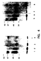
- the precipitates were analyzed by 10% SDS-polyacrylamide gel electrophoresis under non-reducing (lanes 1-5) and reducing (50mM dithiothreitol) (lanes 6-10) conditions.
- Ramos cell lines Anti-B5 conjugated to Sepharose® 4B (lanes 1 and 5) was compared with an irrelevant antibody conjugated to Sepharose® 4B (lanes 2 and 6).
- RPMI 8226 cell line Anti-B5 rabbit anti-mouse Ig preformed complexes (lanes 4 and 8) were compared to irrelevant antibody rabbit anti-mouse Ig preformed complexes (lanes 3 and 7).
- the monoclonal antibody of the invention can be labeled with a detectable label, e.g., a radiolabel by conventional procedures, and provide a quantitative measurement of activated B-cells in biological samples or in vivo .
- a detectable label e.g., a radiolabel
- the monoclonal antibody of the invention can be used to detect the presence of these cell types in biological samples.
- the monoclonal antibody of the invention can be used as a diagnostic aid in characterizing the cell type of various lymphomas and leukemias arising from B-cells.
- in vivo imaging using radiolabeled monoclonal antibody of the invention can provide a noninvasive means for detecting and localizing these cell types, e.g., lymphoid tumors.
- the monoclonal antibody of the invention will also be useful in defining the role of activated B-cells in autoimmune diseases, infections and other diseases which are characterized by activated B-cells, e.g., organ rejection.
Landscapes
- Health & Medical Sciences (AREA)
- Chemical & Material Sciences (AREA)
- Life Sciences & Earth Sciences (AREA)
- Organic Chemistry (AREA)
- Immunology (AREA)
- General Health & Medical Sciences (AREA)
- Medicinal Chemistry (AREA)
- Proteomics, Peptides & Aminoacids (AREA)
- Biochemistry (AREA)
- Biophysics (AREA)
- Molecular Biology (AREA)
- Genetics & Genomics (AREA)
- Cell Biology (AREA)
- Gastroenterology & Hepatology (AREA)
- Toxicology (AREA)
- Zoology (AREA)
- Preparation Of Compounds By Using Micro-Organisms (AREA)
- Micro-Organisms Or Cultivation Processes Thereof (AREA)
- Peptides Or Proteins (AREA)
- Medicines Containing Antibodies Or Antigens For Use As Internal Diagnostic Agents (AREA)
Abstract
Claims (21)
- Anticorps monoclonal qui est pratiquement non réactif avec des lymphocytes B humains non stimulés, ledit anticorps reconnaissant un déterminant antigénique sur des lymphocytes B humains activée, ledit déterminant antigénique étant caractérisé en ce qu'il consiste en une protéine distincte de B-LAST 1 et BB - 1, en ce qu'il consiste en une protéine sur la surface des lymphocytes B activés possédant un poids moléculaire apparent d'approximativement 75 000 daltons dans des conditions réductrices et 67 000 daltons dans des conditions non réductrices, et en ce qu'il est obtenu à partir de lymphocytes B humains périphériques ou spléniques activés avec le virus d'Epstein-Barr, l'agent mitogène de phytolaque, la protéine A ou une anti-immunoglobuline, ou bien à partir de la lignée de cellules de lymphome de Burkitt Ramos ou de la lignée de leucémie à plasmocytes RPMI 8226.
- Anticorps suivant la revendication 1, ledit anticorps étant pratiquement non réactif avec des lymphocytes T stimulés ou au repos et des monocytes, ou des néoplasmes non issus de lymphocytes B, ou bien des néoplasmes issus de lymphocytes B correspondant à des stades de différenciation autres que le stade intermédiaire de différenciation des lymphocytes B.
- Anticorps suivant la revendication 1 ou la revendication 2, ledit anticorps possédant l'isotype IgM.
- Anticorps produit par l'hybridome ayant reçu le numéro de dépôt ATCC HB8716.
- Anticorps suivant l'une quelconque des revendications 1 à 4, ledit anticorps étant marqué avec un marqueur détectable.
- Anticorps suivant la revendication 5, ledit marqueur consistant en un radiomarqueur.
- Cellule d'hybridome capable de produire l'anticorps monoclonal suivant l'une quelconque des revendications 1 à 4.
- Protéine pratiquement pure possédant un déterminant antigénique reconnu par l'anticorps monoclonal suivant l'une quelconque des revendications 1 à 4.
- Protéine pratiquement pure suivant la revendication 8, possédant un ou plusieurs déterminants antigéniques pratiquement identiques à des déterminants d'un polypeptide monocaténaire possédant un poids moléculaire apparent d'approximativement 75 000 daltons dans des conditions réductrices et 67 000 daltons dans des conditions non réductrices, ledit polypeptide monocaténaire étant une protéine sur la surface de lymphocytes B humains activés.
- Protéine suivant la revendication 9, qui est obtenue à partir de lymphocytes B humains périphériques ou spléniques activés avec le virus d'Epstein-Barr, l'agent mitogène de phytolaque, là protéine A ou une anti-immunoglobuline, ou bien à partir de la lignée de cellules de lymphome de Burkitt Ramos ou de la lignée de leucémie à plasmocytes RPMI 8226.
- Protéine suivant l'une quelconque des revendications 8, 9 et 10, qui est marquée de manière détectable.
- Protéine suivant l'une quelconque des revendications 8, 9 et 10, qui est liée à une phase insoluble.
- Procédé de préparation de la protéine suivant la revendication 10, qui comprend la culture de lymphocytes B humains périphériques ou spléniques activés avec le virus d'Epstein-Barr, l'agent mitogène de phytolaque, la protéine A ou une anti-immunoglobuline, ou bien la culture de la lignée de cellules de lymphome de Burkitt Ramos ou de la lignée de leucémie à plasmocytes RPMI 8226, et l'isolement de ladite protéine desdites cellules.
- Méthode d'analyse d'un échantillon biologique pour déterminer la présence de cellules exprimant la protéine suivant la revendication 8, qui comprend la mise en incubation dudit échantillon avec l'anticorps monoclonal suivant la revendication 1 et l'étape consistant à déterminer si un complexe immun a été formé ou non.
- Méthode suivant la revendication 14, dans laquelle l'échantillon comprend du sang périphérique humain, de la moelle osseuse ou du tissu lymphoïde.
- Méthode suivant la revendication 14, dans laquelle l'échantillon comprend des lymphocytes B humains, ou bien des néoplasmes issus de lymphocytes B.
- Méthode d'analyse d'un échantillon biologique pour déterminer la présence d'anticorps contre des cellules exprimant la protéine suivant la revendication 8, qui comprend la mise en incubation dudit échantillon avec la protéine suivant la revendication 8 et l'étape consistant à déterminer si un complexe immun est formé ou non.
- Méthode suivant la revendication 17, dans laquelle l'échantillon comprend du sang humain.
- Kit utile pour l'analyse d'un échantillon pour déterminer la présence d'un anticorps contre des cellules exprimant la protéine suivant la revendication 8, ledit kit étant compartimenté pour recevoir de manière étroitement confinée à l'intérieur de ce kit un ou plusieurs récipients, ce kit comprenant un premier récipient contenant la protéine suivant la revendication 8 et un second récipient contenant un moyen de détection de la formation d'un complexe immun entre ledit anticorps et ladite protéine.
- Kit utile pour l'analyse d'un échantillon pour déterminer la présence de cellules exprimant la protéine suivant la revendication 8, ledit kit étant compartimenté pour recevoir de manière étroitement confinée à l'intérieur de ce kit un ou plusieurs récipients, ce kit comprenant un premier récipient contentant l'anticorps monoclonal suivant la revendication 1 et un second récipient contenant un moyen de détection de formation d'un complexe immun entre ledit anticorps et ladite protéine.
- Utilisation d'un anticorps monoclonal tel que défini dans la revendication 1 dans la préparation d'un réactif de diagnostic pour la détection de sites contenant des lymphocytes B activés chez un mammifère.
Applications Claiming Priority (3)
| Application Number | Priority Date | Filing Date | Title |
|---|---|---|---|
| US06/708,176 US4692405A (en) | 1985-03-05 | 1985-03-05 | Monoclonal antibodies to antigen on activated human B-cells and assay therefor, protein antigenic determinant therefor and method of making same |
| PCT/US1986/000460 WO1986005188A1 (fr) | 1985-03-05 | 1986-03-04 | Anticorps monoclonaux et analyse |
| US708176 | 1996-09-06 |
Publications (3)
| Publication Number | Publication Date |
|---|---|
| EP0216854A1 EP0216854A1 (fr) | 1987-04-08 |
| EP0216854A4 EP0216854A4 (fr) | 1990-06-27 |
| EP0216854B1 true EP0216854B1 (fr) | 1994-07-06 |
Family
ID=24844693
Family Applications (1)
| Application Number | Title | Priority Date | Filing Date |
|---|---|---|---|
| EP86902121A Expired - Lifetime EP0216854B1 (fr) | 1985-03-05 | 1986-03-04 | Anticorps monoclonaux et analyse |
Country Status (5)
| Country | Link |
|---|---|
| US (1) | US4692405A (fr) |
| EP (1) | EP0216854B1 (fr) |
| JP (1) | JPH085918B2 (fr) |
| DE (1) | DE3689948T2 (fr) |
| WO (1) | WO1986005188A1 (fr) |
Families Citing this family (13)
| Publication number | Priority date | Publication date | Assignee | Title |
|---|---|---|---|---|
| US5015568A (en) * | 1986-07-09 | 1991-05-14 | The Wistar Institute | Diagnostic methods for detecting lymphomas in humans |
| US5066581A (en) * | 1987-02-25 | 1991-11-19 | The Trustees Of Columbia University In The City Of New York | Differentiation antigen, nda3, associated with the receptor for b cell growth factor |
| FR2634126B1 (fr) * | 1988-07-13 | 1992-04-17 | Unicet Laboratoires | Anticorps monoclonal specifique pour un antigene de surface des cellules b activees et/ou des cellules t transformees par l'htlv-i, lignee de cellules d'hybridomes produisant cet anticorps, procede pour la detection de cellules t transformees par l'htlv-i et/ou de cellules b activees |
| CA2007305A1 (fr) * | 1989-01-09 | 1990-07-09 | Nicole Suciu-Foca | Antigene de differenciation, nda4, associe au recepteur du facteur de croissance des cellules b |
| CA2011099A1 (fr) * | 1989-04-19 | 1990-10-19 | Stephen C. Wardlaw | Mesure de la reactivite des lymphocytes a des antigenes specifiques dans le sang |
| US5441871A (en) * | 1989-06-01 | 1995-08-15 | Health Research, Inc. | Monoclonal antibody reactive to human leukemia and lymphoma cells and methods of using same for diagnosis and treatment |
| US5100378A (en) * | 1989-06-09 | 1992-03-31 | Neorx Corporation | Enhancement of target cell localization of lymphoid cells |
| US6071716A (en) * | 1990-10-01 | 2000-06-06 | Dana-Farber Cancer Institute | DNA encoding, B7, a new member of the IG superfamily with unique expression on activated and neoplastic B cells |
| CA2147010A1 (fr) * | 1992-10-19 | 1994-04-28 | Stuart F. Schlossman | Elimination immunologique de cellules tumorales de la moelle osseuse au moyen de microspheres et d'anticorps monoclonaux |
| US5534430A (en) * | 1993-08-31 | 1996-07-09 | Hitachi Chemical Company Limited | Cell strain capable of multiplying an Epstein-Barr virus |
| JPH0779793A (ja) * | 1993-09-14 | 1995-03-28 | Sumitomo Electric Ind Ltd | B70分子に対するモノクローナル抗体 |
| AR083044A1 (es) | 2010-09-27 | 2013-01-30 | Regeneron Pharma | Anticuerpos anti-cd48 y usos de los mismos |
| US9533039B2 (en) | 2010-09-27 | 2017-01-03 | Regeneron Pharmaceuticals, Inc. | Methods of treating systemic lupus erythematosus (SLE) using anti-CD48 antibodies |
Family Cites Families (1)
| Publication number | Priority date | Publication date | Assignee | Title |
|---|---|---|---|---|
| US4444744A (en) * | 1980-03-03 | 1984-04-24 | Goldenberg Milton David | Tumor localization and therapy with labeled antibodies to cell surface antigens |
-
1985
- 1985-03-05 US US06/708,176 patent/US4692405A/en not_active Expired - Lifetime
-
1986
- 1986-03-04 EP EP86902121A patent/EP0216854B1/fr not_active Expired - Lifetime
- 1986-03-04 DE DE3689948T patent/DE3689948T2/de not_active Expired - Lifetime
- 1986-03-04 JP JP61501630A patent/JPH085918B2/ja not_active Expired - Lifetime
- 1986-03-04 WO PCT/US1986/000460 patent/WO1986005188A1/fr not_active Ceased
Non-Patent Citations (4)
| Title |
|---|
| CHEMICAL ABSTRACTS, vol. 102, no. 3, 21 january 1985, Columbus, OH (US); T.A. WALDMAN et al., p. 524, no. 22586x# * |
| CHEMICAL ABSTRACTS, vol. 102, no. 7, 18 February 1985, Columbus, OH (US); L.K.L. JUNG et al., p. 450, no. 60448m# * |
| CHEMICAL ABSTRACTS, vol. 95, no. 1, 06 July 1981, Columbus, OH (US); R.S. GOODENOW, p. 317, no. 3099m# * |
| JOURNAL OF IMMUNOLOGY, vol. 133, no. 2, August 1984; T.F. TEDDER et al., pp. 678-683# * |
Also Published As
| Publication number | Publication date |
|---|---|
| WO1986005188A1 (fr) | 1986-09-12 |
| US4692405A (en) | 1987-09-08 |
| DE3689948T2 (de) | 1994-10-13 |
| JPS62502614A (ja) | 1987-10-08 |
| JPH085918B2 (ja) | 1996-01-24 |
| EP0216854A4 (fr) | 1990-06-27 |
| EP0216854A1 (fr) | 1987-04-08 |
| DE3689948D1 (de) | 1994-08-11 |
Similar Documents
| Publication | Publication Date | Title |
|---|---|---|
| US4361549A (en) | Complement-fixing monoclonal antibody to human T cells, and methods of preparing same | |
| Carrel et al. | Subsets of human Ia-like molecules defined by monoclonal antibodies | |
| US4515893A (en) | Hybrid cell line for producing complement-fixing monoclonal antibody to human T cells | |
| US4657760A (en) | Methods and compositions using monoclonal antibody to human T cells | |
| US4381295A (en) | Monoclonal antibody to human helper T cells and methods of preparing same | |
| EP0030815B1 (fr) | Lignée de cellules hybrides pour la préparation d'anticorps monoclonaux contre un antigène prothymocyte humain; anticorps; procédé pour préparer cet anticorps, applications diagnostiques et thérapeutiques et compositions pharmaceutiques contenant cet anticorps | |
| US4658019A (en) | Complement-fixing monoclonal antibody to human T cells | |
| CA1149281A (fr) | Lignee cellulaire hybride utilisee pour produire un anticorps monoclonal contre les cellules humaines t | |
| EP0030814B1 (fr) | Lignée de cellules hybrides pour la préparation d'anticorps monoclonaux contre un antigène thymocyte précoce humain; anticorps; procédé pour préparer cet anticorps; applications diagnostiques et thérapeutiques et compositions pharmaceutiques contenant cet anticorps | |
| EP0025722B2 (fr) | Anticorps monoclonal contre les cellules T humaines cytotoxiques et suppressives et son procédé de préparation | |
| JPH0159871B2 (fr) | ||
| US4654210A (en) | Methods and compositions using complement fixing monoclonal antibody to human T cells | |
| US4692405A (en) | Monoclonal antibodies to antigen on activated human B-cells and assay therefor, protein antigenic determinant therefor and method of making same | |
| Fitchen et al. | Monoclonal antibodies to HLA-A, B, and Ia-like antigens inhibit colony formation by human myeloid progenitor cells. | |
| JPH05501648A (ja) | ヘルパー誘導物質およびサブレッサー誘導物質ce4+リンパ球を識別する単クローン性抗体 | |
| US4658020A (en) | Monoclonal antibody to human helper T cells | |
| US4515895A (en) | Hybrid cell line for producing monoclonal antibody to human helper T cells | |
| US4652447A (en) | Methods and compositions using monoclonal antibody to human helper T cells | |
| US4818689A (en) | Late differentiation antigen associated with helper T lymphocyte function | |
| US4661446A (en) | Monoclonal antibodies and method of immunizing therewith | |
| Bankert et al. | Screening and replica plating of anti-hapten hybridomas with a transfer template hemolytic spot assay | |
| US4515894A (en) | Hybrid cell line for producing monoclonal antibody to human T cells | |
| US4803262A (en) | Hybrid cell line for producing monoclonal antibody to human cytotoxic and suppressor T cells, antibody, and methods | |
| US4680383A (en) | Monoclonal antibody to human T cells | |
| US4806349A (en) | Hybrid cell line for producing monoclonal antibody to a human prothymocyte antigen, antibody, and methods |
Legal Events
| Date | Code | Title | Description |
|---|---|---|---|
| PUAI | Public reference made under article 153(3) epc to a published international application that has entered the european phase |
Free format text: ORIGINAL CODE: 0009012 |
|
| 17P | Request for examination filed |
Effective date: 19861110 |
|
| AK | Designated contracting states |
Kind code of ref document: A1 Designated state(s): DE FR GB IT |
|
| A4 | Supplementary search report drawn up and despatched |
Effective date: 19900627 |
|
| 17Q | First examination report despatched |
Effective date: 19911213 |
|
| GRAA | (expected) grant |
Free format text: ORIGINAL CODE: 0009210 |
|
| ITF | It: translation for a ep patent filed | ||
| AK | Designated contracting states |
Kind code of ref document: B1 Designated state(s): DE FR GB IT |
|
| REF | Corresponds to: |
Ref document number: 3689948 Country of ref document: DE Date of ref document: 19940811 |
|
| ET | Fr: translation filed | ||
| PLBE | No opposition filed within time limit |
Free format text: ORIGINAL CODE: 0009261 |
|
| STAA | Information on the status of an ep patent application or granted ep patent |
Free format text: STATUS: NO OPPOSITION FILED WITHIN TIME LIMIT |
|
| 26N | No opposition filed | ||
| REG | Reference to a national code |
Ref country code: GB Ref legal event code: IF02 |
|
| PGFP | Annual fee paid to national office [announced via postgrant information from national office to epo] |
Ref country code: GB Payment date: 20050223 Year of fee payment: 20 |
|
| PGFP | Annual fee paid to national office [announced via postgrant information from national office to epo] |
Ref country code: FR Payment date: 20050321 Year of fee payment: 20 |
|
| PGFP | Annual fee paid to national office [announced via postgrant information from national office to epo] |
Ref country code: IT Payment date: 20050330 Year of fee payment: 20 |
|
| PGFP | Annual fee paid to national office [announced via postgrant information from national office to epo] |
Ref country code: DE Payment date: 20050502 Year of fee payment: 20 |
|
| PG25 | Lapsed in a contracting state [announced via postgrant information from national office to epo] |
Ref country code: GB Free format text: LAPSE BECAUSE OF EXPIRATION OF PROTECTION Effective date: 20060303 |
|
| REG | Reference to a national code |
Ref country code: GB Ref legal event code: PE20 |