EP0244730A1 - Dispositif destiné à la désintégration de concréments présents dans le corps d'un être vivant - Google Patents
Dispositif destiné à la désintégration de concréments présents dans le corps d'un être vivant Download PDFInfo
- Publication number
- EP0244730A1 EP0244730A1 EP87106094A EP87106094A EP0244730A1 EP 0244730 A1 EP0244730 A1 EP 0244730A1 EP 87106094 A EP87106094 A EP 87106094A EP 87106094 A EP87106094 A EP 87106094A EP 0244730 A1 EP0244730 A1 EP 0244730A1
- Authority
- EP
- European Patent Office
- Prior art keywords
- ray
- customer
- shock wave
- shock
- wave generator
- Prior art date
- Legal status (The legal status is an assumption and is not a legal conclusion. Google has not performed a legal analysis and makes no representation as to the accuracy of the status listed.)
- Granted
Links
- 230000035939 shock Effects 0.000 claims abstract description 23
- 230000006870 function Effects 0.000 claims abstract description 6
- 230000000737 periodic effect Effects 0.000 claims abstract description 3
- 230000029058 respiratory gaseous exchange Effects 0.000 claims description 11
- 230000004807 localization Effects 0.000 claims description 5
- 230000001960 triggered effect Effects 0.000 claims description 4
- 238000011156 evaluation Methods 0.000 claims description 2
- 238000001514 detection method Methods 0.000 claims 1
- 238000006073 displacement reaction Methods 0.000 abstract 1
- 230000015654 memory Effects 0.000 description 4
- 208000000913 Kidney Calculi Diseases 0.000 description 2
- 206010029148 Nephrolithiasis Diseases 0.000 description 2
- 230000000694 effects Effects 0.000 description 2
- 208000009911 Urinary Calculi Diseases 0.000 description 1
- 210000001015 abdomen Anatomy 0.000 description 1
- 201000001883 cholelithiasis Diseases 0.000 description 1
- 230000006378 damage Effects 0.000 description 1
- 208000001130 gallstones Diseases 0.000 description 1
- 210000003734 kidney Anatomy 0.000 description 1
- 230000000630 rising effect Effects 0.000 description 1
- 238000002560 therapeutic procedure Methods 0.000 description 1
- 208000008281 urolithiasis Diseases 0.000 description 1
Images
Classifications
-
- A—HUMAN NECESSITIES
- A61—MEDICAL OR VETERINARY SCIENCE; HYGIENE
- A61B—DIAGNOSIS; SURGERY; IDENTIFICATION
- A61B6/00—Apparatus or devices for radiation diagnosis; Apparatus or devices for radiation diagnosis combined with radiation therapy equipment
- A61B6/12—Arrangements for detecting or locating foreign bodies
-
- A—HUMAN NECESSITIES
- A61—MEDICAL OR VETERINARY SCIENCE; HYGIENE
- A61B—DIAGNOSIS; SURGERY; IDENTIFICATION
- A61B17/00—Surgical instruments, devices or methods
- A61B17/22—Implements for squeezing-off ulcers or the like on inner organs of the body; Implements for scraping-out cavities of body organs, e.g. bones; for invasive removal or destruction of calculus using mechanical vibrations; for removing obstructions in blood vessels, not otherwise provided for
- A61B17/225—Implements for squeezing-off ulcers or the like on inner organs of the body; Implements for scraping-out cavities of body organs, e.g. bones; for invasive removal or destruction of calculus using mechanical vibrations; for removing obstructions in blood vessels, not otherwise provided for for extracorporeal shock wave lithotripsy [ESWL], e.g. by using ultrasonic waves
- A61B17/2256—Implements for squeezing-off ulcers or the like on inner organs of the body; Implements for scraping-out cavities of body organs, e.g. bones; for invasive removal or destruction of calculus using mechanical vibrations; for removing obstructions in blood vessels, not otherwise provided for for extracorporeal shock wave lithotripsy [ESWL], e.g. by using ultrasonic waves with means for locating or checking the concrement, e.g. X-ray apparatus, imaging means
-
- A—HUMAN NECESSITIES
- A61—MEDICAL OR VETERINARY SCIENCE; HYGIENE
- A61B—DIAGNOSIS; SURGERY; IDENTIFICATION
- A61B6/00—Apparatus or devices for radiation diagnosis; Apparatus or devices for radiation diagnosis combined with radiation therapy equipment
- A61B6/54—Control of apparatus or devices for radiation diagnosis
- A61B6/541—Control of apparatus or devices for radiation diagnosis involving acquisition triggered by a physiological signal
-
- A—HUMAN NECESSITIES
- A61—MEDICAL OR VETERINARY SCIENCE; HYGIENE
- A61B—DIAGNOSIS; SURGERY; IDENTIFICATION
- A61B17/00—Surgical instruments, devices or methods
- A61B2017/00681—Aspects not otherwise provided for
- A61B2017/00694—Aspects not otherwise provided for with means correcting for movement of or for synchronisation with the body
- A61B2017/00699—Aspects not otherwise provided for with means correcting for movement of or for synchronisation with the body correcting for movement caused by respiration, e.g. by triggering
Definitions
- the invention relates to a device for smashing concrements located in the body of a living being with a shock wave generator and with an X-ray examination device for locating the concrements.
- DE-OS 31 22 056 describes such a device, which is used, for example, to smash urinary stones, kidney stones, gall stones or the like.
- a shock wave is generated as a shock wave generator, for example by spark discharge, which is concentrated on the concrement and comminutes it.
- the device is connected to an X-ray examination device for better location so that it can be determined exactly whether the calculus is in the focal point of the focusing chamber.
- the individual images of a stereo image pair are stored individually in the image memory or integrated over several images in the television clock.
- the problem arises that, especially in the treatment of kidney stones, they are moved during breathing, so that an exact localization of the concrements cannot take place.
- the invention is based on the object of creating a device of the type mentioned at the outset, which makes it possible to carry out very precise localization of the concretions, so that the concretions are comminuted in a manner which is gentle on the tissue.
- the Device has at least one customer for periodic bodily functions of the patient, that the customer is connected via an evaluation circuit to a control device which is designed in such a way that X-ray images are generated for striking features of the body function and that after the localization the shock waves are triggered due to trigger pulses that are assigned to the times of the x-ray image generation.
- a control device which is designed in such a way that X-ray images are generated for striking features of the body function and that after the localization the shock waves are triggered due to trigger pulses that are assigned to the times of the x-ray image generation.
- the breathing phase can be taken into account if a transducer is used for breathing. Movement within a phase is reliably avoided if the sensor is a pneumatic belt.
- the heart rate can also be taken into account if an EKG electrode is used as a further consumer and the trigger pulses for the x-ray image and shock wave generation are supplied to the control device via an AND circuit.
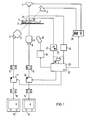
- FIG. 1 shows a device for crushing concrements located in the body of a living being with an X-ray examination device, which has two X-ray tubes 1 and 2, which generate X-ray beams that penetrate an examiner 4 to be treated on a patient couch 3 and onto the entrance fluorescent screens falling from X-ray image intensifiers 5 and 6.
- the x-ray tube 1 and the x-ray image intensifier 6 are arranged in such a way that the central beam of the x-ray beam of the x-ray tube 1 falls perpendicularly on the examiner 4 (a.p. projection).
- the x-ray tube 2 and the x-ray image intensifier 5 are arranged obliquely such that the central beam of the x-ray tube 2 intersects the central beam of the x-ray tube 1 in a target area within the examiner 4 at an angle of, for example, 45 °.
- fluoroscopic images are obtained from two different projection directions, so that the examiner 4 can be moved through the patient couch 3 in such a way that the concrements are located in the target area.
- the output signals of the television cameras 7 and 8 coupled to the X-ray image intensifier 5 and 6 are read into two image memories 11 and 12 via two analog / digital converters (A / D converters 9, 10).
- the output signals can be viewed on two monitors 15, 16 via two digital-to-analog converters (D / A converters 13, 14). If the concrements are now in the target area, they appear in the middle of the screens of monitors 15 and 16.
- a shock wave generator 17 which is shown schematically in FIG. 1, is provided for crushing the concrements located in the target area. It generates shock waves in a known manner, which lead to destruction of the concretions if, for example, they are in the focal point of the shock wave generator 17.
- the X-ray examination chungs worn and the shock wave generator 17 are so firmly connected to each other that the shock waves are focused in the target area of the X-ray examination device.
- the shock wave generator 17 is connected to a control circuit 19.
- a respirator 20 is attached to the examining person 4 and consists for example of a pneumatic belt wrapped around the belly of the examining person 4.
- a circuit 21 is connected to the consumer 20, which has an adjuster 22 for a threshold value.
- ECG electrodes 23, which are connected to an EKG circuit 24, are also arranged on the examiner 4.
- the output signals of the circuits 21 and 24 are fed to a control device 25, which is connected to a high voltage generator 26 for controlling the X-ray tubes 1 and 2, to the image memories 11 and 12 and to the control circuit 19 for the shock wave generator 17.
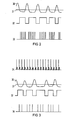
- the mode of operation of the device shown in FIG. 1 is explained in more detail using the time profiles shown in FIGS. 2 and 3.
- the customer 20 supplies an output signal 30 corresponding to the breathing of the examiner 4.
- a threshold 31 preselected by the adjuster 22 effects, for example, an output signal 32 of the circuit 21 by means of a comparator. Due to, for example, the rising leading edge of the rectangular pulses or also with a time delay, the control device 25 effects this
- An x-ray pulse is triggered by the high-voltage generator 26 and the x-ray images are simultaneously stored in the image memories 11 and 12. This ensures that the examination recordings are always taken in the same breathing phase.
- the output signal 32 of the circuit 21 switches through the control device 25 generates an enable signal for the control circuit 19 of the shock wave generator 17, which then periodically generates shock waves within the rectangular pulses, which are represented by the curve 33 as needle-shaped pulses.
- X-ray images are generated during the examination phase, which occur during a certain breathing phase, and during this same breathing phase, in which there is little or no movement, for example, of the kidney, several shock waves are generated which hit the concretions in a targeted manner and thus almost completely shred.
- triggering from the EKG can also take place simultaneously with the triggering on the breathing phase, the curve profile 34 of which is shown in FIG. 3.
- trigger pulses are generated from the EKG, for example from the so-called R wave, by the EKG circuit, which are fed to the control device 25.
- This signal is coupled together with the output signal 32 of the circuit 21 in an AND gate, so that a combination signal results therefrom which is used to control the X-ray examination device and the shock wave generator. It follows that a recording or shock wave is only generated during the breathing phase determined by the output signal 32 and during the occurrence of the R wave in the EKG signal, as can be seen from the curve profile shown in FIG. 3 by the needle pulses 35.
Landscapes
- Health & Medical Sciences (AREA)
- Life Sciences & Earth Sciences (AREA)
- Medical Informatics (AREA)
- Engineering & Computer Science (AREA)
- Nuclear Medicine, Radiotherapy & Molecular Imaging (AREA)
- Surgery (AREA)
- Animal Behavior & Ethology (AREA)
- Veterinary Medicine (AREA)
- Biomedical Technology (AREA)
- Heart & Thoracic Surgery (AREA)
- Public Health (AREA)
- Molecular Biology (AREA)
- Radiology & Medical Imaging (AREA)
- General Health & Medical Sciences (AREA)
- Physics & Mathematics (AREA)
- Biophysics (AREA)
- High Energy & Nuclear Physics (AREA)
- Optics & Photonics (AREA)
- Pathology (AREA)
- Orthopedic Medicine & Surgery (AREA)
- Vascular Medicine (AREA)
- Physiology (AREA)
- Apparatus For Radiation Diagnosis (AREA)
- Surgical Instruments (AREA)
Applications Claiming Priority (2)
| Application Number | Priority Date | Filing Date | Title |
|---|---|---|---|
| DE3615564 | 1986-05-09 | ||
| DE3615564 | 1986-05-09 |
Publications (2)
| Publication Number | Publication Date |
|---|---|
| EP0244730A1 true EP0244730A1 (fr) | 1987-11-11 |
| EP0244730B1 EP0244730B1 (fr) | 1990-07-04 |
Family
ID=6300408
Family Applications (1)
| Application Number | Title | Priority Date | Filing Date |
|---|---|---|---|
| EP87106094A Expired - Lifetime EP0244730B1 (fr) | 1986-05-09 | 1987-04-27 | Dispositif destiné à la désintégration de concréments présents dans le corps d'un être vivant |
Country Status (3)
| Country | Link |
|---|---|
| EP (1) | EP0244730B1 (fr) |
| JP (1) | JPH045150Y2 (fr) |
| DE (1) | DE3763533D1 (fr) |
Cited By (6)
| Publication number | Priority date | Publication date | Assignee | Title |
|---|---|---|---|---|
| EP0296349A3 (fr) * | 1987-06-24 | 1989-03-15 | Dornier Medizintechnik Gmbh | Déclencheur respiratoire |
| EP0324948A3 (fr) * | 1988-01-21 | 1989-10-25 | Dornier Medizintechnik Gmbh | Dispositif de réduction pour concrétions |
| EP0336620A3 (en) * | 1988-03-31 | 1990-04-25 | Kabushiki Kaisha Toshiba | Apparatus for destroying calculuses |
| EP0460536A1 (fr) * | 1990-05-31 | 1991-12-11 | Kabushiki Kaisha Toshiba | Dispositif pour la lithotripsie |
| US5419327A (en) * | 1992-12-07 | 1995-05-30 | Siemens Aktiengesellschaft | Acoustic therapy means |
| DE10260594B4 (de) * | 2002-12-23 | 2012-06-06 | Dornier Medtech Systems Gmbh | Vorrichtung zum extrakorporalen Erzeugen von fokussierten Stoßwellen |
Families Citing this family (1)
| Publication number | Priority date | Publication date | Assignee | Title |
|---|---|---|---|---|
| JP4928739B2 (ja) * | 2004-06-25 | 2012-05-09 | 株式会社東芝 | X線診断装置及びx線撮像方法 |
Citations (4)
| Publication number | Priority date | Publication date | Assignee | Title |
|---|---|---|---|---|
| DE496749C (de) * | 1927-09-10 | 1930-04-30 | Hans Appelrath Dr | Verfahren und Vorrichtung zur stereoskopischen Roentgenaufnahme von sich periodisch bewegenden Organen des menschlichen oder tierischen Koerpers |
| DE2722252A1 (de) * | 1977-05-17 | 1978-11-23 | Dornier System Gmbh | Einrichtung zur raeumlichen ortung von konkrementen |
| GB2002987A (en) * | 1977-07-15 | 1979-02-28 | Emi Ltd | Improvements in or relating to radiography |
| EP0081051A1 (fr) * | 1981-11-25 | 1983-06-15 | Dornier Gmbh | Dispositif de déclenchement pour ondes de choc pour buts thérapeutiques |
Family Cites Families (3)
| Publication number | Priority date | Publication date | Assignee | Title |
|---|---|---|---|---|
| DE3122056A1 (de) * | 1981-06-03 | 1982-12-23 | Siemens AG, 1000 Berlin und 8000 München | Einrichtung zum zertruemmern von im koerper eines lebewesens befindlichen konkrementen |
| JPS618686A (ja) * | 1984-06-25 | 1986-01-16 | Oki Electric Ind Co Ltd | 適応ビ−ム・フオ−マを用いるソ−ナ−方式 |
| DE3426398C1 (de) * | 1984-07-18 | 1987-11-12 | Dornier System Gmbh, 7990 Friedrichshafen | Vorrichtung zum räumlichen Orten und Positionieren von Konkrementen |
-
1987
- 1987-04-27 EP EP87106094A patent/EP0244730B1/fr not_active Expired - Lifetime
- 1987-04-27 DE DE8787106094T patent/DE3763533D1/de not_active Expired - Lifetime
- 1987-05-06 JP JP1987067792U patent/JPH045150Y2/ja not_active Expired
Patent Citations (4)
| Publication number | Priority date | Publication date | Assignee | Title |
|---|---|---|---|---|
| DE496749C (de) * | 1927-09-10 | 1930-04-30 | Hans Appelrath Dr | Verfahren und Vorrichtung zur stereoskopischen Roentgenaufnahme von sich periodisch bewegenden Organen des menschlichen oder tierischen Koerpers |
| DE2722252A1 (de) * | 1977-05-17 | 1978-11-23 | Dornier System Gmbh | Einrichtung zur raeumlichen ortung von konkrementen |
| GB2002987A (en) * | 1977-07-15 | 1979-02-28 | Emi Ltd | Improvements in or relating to radiography |
| EP0081051A1 (fr) * | 1981-11-25 | 1983-06-15 | Dornier Gmbh | Dispositif de déclenchement pour ondes de choc pour buts thérapeutiques |
Cited By (8)
| Publication number | Priority date | Publication date | Assignee | Title |
|---|---|---|---|---|
| EP0296349A3 (fr) * | 1987-06-24 | 1989-03-15 | Dornier Medizintechnik Gmbh | Déclencheur respiratoire |
| EP0324948A3 (fr) * | 1988-01-21 | 1989-10-25 | Dornier Medizintechnik Gmbh | Dispositif de réduction pour concrétions |
| EP0336620A3 (en) * | 1988-03-31 | 1990-04-25 | Kabushiki Kaisha Toshiba | Apparatus for destroying calculuses |
| US5054469A (en) * | 1988-03-31 | 1991-10-08 | Kabushiki Kaisha Toshiba | Apparatus for destroying calculuses |
| EP0460536A1 (fr) * | 1990-05-31 | 1991-12-11 | Kabushiki Kaisha Toshiba | Dispositif pour la lithotripsie |
| US5243985A (en) * | 1990-05-31 | 1993-09-14 | Kabushiki Kaisha Toshiba | Lithotrity apparatus having a missed-shot preventive function |
| US5419327A (en) * | 1992-12-07 | 1995-05-30 | Siemens Aktiengesellschaft | Acoustic therapy means |
| DE10260594B4 (de) * | 2002-12-23 | 2012-06-06 | Dornier Medtech Systems Gmbh | Vorrichtung zum extrakorporalen Erzeugen von fokussierten Stoßwellen |
Also Published As
| Publication number | Publication date |
|---|---|
| DE3763533D1 (de) | 1990-08-09 |
| JPS62183808U (fr) | 1987-11-21 |
| EP0244730B1 (fr) | 1990-07-04 |
| JPH045150Y2 (fr) | 1992-02-14 |
Similar Documents
| Publication | Publication Date | Title |
|---|---|---|
| DE69208141T2 (de) | Vorrichtung zum Zerstören von Konkrementen | |
| DE4241161C2 (de) | Akustische Therapieeinrichtung | |
| DE4125950C1 (fr) | ||
| DE19733838C2 (de) | Vorrichtung zur Behandlung mit akustischen Stosswellen | |
| DE4202302C2 (de) | Computer-Tomograph | |
| EP0081051B2 (fr) | Dispositif de déclenchement pour ondes de choc pour buts thérapeutiques | |
| DE3426398C1 (de) | Vorrichtung zum räumlichen Orten und Positionieren von Konkrementen | |
| DE3543867C2 (fr) | ||
| DE3607949C2 (fr) | ||
| DE10158519B4 (de) | Stoß- und Druckwellen-Therapiegerät | |
| DE4315282A1 (de) | Verwendung einer akustischen Druckimpulsquelle | |
| DE10102317A1 (de) | Verfahren und Vorrichtung zur Beaufschlagung des Körpers eines Lebeswesens mit Druckwellen | |
| DE3119295A1 (de) | Einrichtung zum zerstoeren von konkrementen in koerperhoehlen | |
| DE69018853T2 (de) | Vorrichtung zum Zerstören von Konkrementen. | |
| DE3840077A1 (de) | Lithotriptor | |
| EP0372119B2 (fr) | Lithotripteur | |
| DE3328039C2 (de) | Einrichtung zum beruehrungslosen zertruemmern eines im koerper eines lebewesens befindlichen konkrements | |
| EP0273256A1 (fr) | Appareil pour la fragmentation à distance de concrétions | |
| DE19548000C1 (de) | Vorrichtung zur Ortung von Konkrementen im Körper eines Patienten | |
| EP0355178B1 (fr) | Appareil pour la destruction à distance de concrétions dans le corps d'un être vivant | |
| DE69013735T2 (de) | Mit Ultraschallwellen funktionierendes Stosswellenlithotripsiegerät. | |
| DE68911724T2 (de) | Nierensteinzertrümmerer. | |
| EP0400196B1 (fr) | Transducteur d'ondes de choc pour destruction de concrétions | |
| EP0244730B1 (fr) | Dispositif destiné à la désintégration de concréments présents dans le corps d'un être vivant | |
| EP0257199B1 (fr) | Dispositif pour la destruction de calculs |
Legal Events
| Date | Code | Title | Description |
|---|---|---|---|
| PUAI | Public reference made under article 153(3) epc to a published international application that has entered the european phase |
Free format text: ORIGINAL CODE: 0009012 |
|
| AK | Designated contracting states |
Kind code of ref document: A1 Designated state(s): DE FR GB NL |
|
| 17P | Request for examination filed |
Effective date: 19871204 |
|
| 17Q | First examination report despatched |
Effective date: 19890307 |
|
| GRAA | (expected) grant |
Free format text: ORIGINAL CODE: 0009210 |
|
| AK | Designated contracting states |
Kind code of ref document: B1 Designated state(s): DE FR GB NL |
|
| REF | Corresponds to: |
Ref document number: 3763533 Country of ref document: DE Date of ref document: 19900809 |
|
| ET | Fr: translation filed | ||
| GBT | Gb: translation of ep patent filed (gb section 77(6)(a)/1977) | ||
| PLBE | No opposition filed within time limit |
Free format text: ORIGINAL CODE: 0009261 |
|
| STAA | Information on the status of an ep patent application or granted ep patent |
Free format text: STATUS: NO OPPOSITION FILED WITHIN TIME LIMIT |
|
| 26N | No opposition filed | ||
| PGFP | Annual fee paid to national office [announced via postgrant information from national office to epo] |
Ref country code: GB Payment date: 19930319 Year of fee payment: 7 |
|
| PGFP | Annual fee paid to national office [announced via postgrant information from national office to epo] |
Ref country code: FR Payment date: 19930423 Year of fee payment: 7 |
|
| PGFP | Annual fee paid to national office [announced via postgrant information from national office to epo] |
Ref country code: NL Payment date: 19930430 Year of fee payment: 7 |
|
| PG25 | Lapsed in a contracting state [announced via postgrant information from national office to epo] |
Ref country code: GB Effective date: 19940427 |
|
| PG25 | Lapsed in a contracting state [announced via postgrant information from national office to epo] |
Ref country code: NL Effective date: 19941101 |
|
| NLV4 | Nl: lapsed or anulled due to non-payment of the annual fee | ||
| GBPC | Gb: european patent ceased through non-payment of renewal fee |
Effective date: 19940427 |
|
| PG25 | Lapsed in a contracting state [announced via postgrant information from national office to epo] |
Ref country code: FR Effective date: 19941229 |
|
| REG | Reference to a national code |
Ref country code: FR Ref legal event code: ST |
|
| PGFP | Annual fee paid to national office [announced via postgrant information from national office to epo] |
Ref country code: DE Payment date: 20000619 Year of fee payment: 14 |
|
| PG25 | Lapsed in a contracting state [announced via postgrant information from national office to epo] |
Ref country code: DE Free format text: LAPSE BECAUSE OF NON-PAYMENT OF DUE FEES Effective date: 20020201 |