EP0251478B1 - Endoscope à balayage pour applications chirurgicales - Google Patents
Endoscope à balayage pour applications chirurgicales Download PDFInfo
- Publication number
- EP0251478B1 EP0251478B1 EP87304589A EP87304589A EP0251478B1 EP 0251478 B1 EP0251478 B1 EP 0251478B1 EP 87304589 A EP87304589 A EP 87304589A EP 87304589 A EP87304589 A EP 87304589A EP 0251478 B1 EP0251478 B1 EP 0251478B1
- Authority
- EP
- European Patent Office
- Prior art keywords
- prism
- movable
- angle
- lens
- head
- Prior art date
- Legal status (The legal status is an assumption and is not a legal conclusion. Google has not performed a legal analysis and makes no representation as to the accuracy of the status listed.)
- Expired
Links
- 230000003287 optical effect Effects 0.000 claims description 14
- 208000027418 Wounds and injury Diseases 0.000 description 2
- 230000006378 damage Effects 0.000 description 2
- 239000000835 fiber Substances 0.000 description 2
- 208000014674 injury Diseases 0.000 description 2
- 238000005286 illumination Methods 0.000 description 1
- 238000000034 method Methods 0.000 description 1
- 239000013307 optical fiber Substances 0.000 description 1
Images
Classifications
-
- A—HUMAN NECESSITIES
- A61—MEDICAL OR VETERINARY SCIENCE; HYGIENE
- A61B—DIAGNOSIS; SURGERY; IDENTIFICATION
- A61B1/00—Instruments for performing medical examinations of the interior of cavities or tubes of the body by visual or photographical inspection, e.g. endoscopes; Illuminating arrangements therefor
- A61B1/00163—Optical arrangements
- A61B1/00174—Optical arrangements characterised by the viewing angles
- A61B1/00183—Optical arrangements characterised by the viewing angles for variable viewing angles
-
- A—HUMAN NECESSITIES
- A61—MEDICAL OR VETERINARY SCIENCE; HYGIENE
- A61B—DIAGNOSIS; SURGERY; IDENTIFICATION
- A61B1/00—Instruments for performing medical examinations of the interior of cavities or tubes of the body by visual or photographical inspection, e.g. endoscopes; Illuminating arrangements therefor
- A61B1/00064—Constructional details of the endoscope body
- A61B1/00071—Insertion part of the endoscope body
- A61B1/0008—Insertion part of the endoscope body characterised by distal tip features
- A61B1/00096—Optical elements
-
- G—PHYSICS
- G02—OPTICS
- G02B—OPTICAL ELEMENTS, SYSTEMS OR APPARATUS
- G02B23/00—Telescopes, e.g. binoculars; Periscopes; Instruments for viewing the inside of hollow bodies; Viewfinders; Optical aiming or sighting devices
- G02B23/24—Instruments or systems for viewing the inside of hollow bodies, e.g. fibrescopes
- G02B23/2407—Optical details
- G02B23/2423—Optical details of the distal end
-
- A—HUMAN NECESSITIES
- A61—MEDICAL OR VETERINARY SCIENCE; HYGIENE
- A61B—DIAGNOSIS; SURGERY; IDENTIFICATION
- A61B1/00—Instruments for performing medical examinations of the interior of cavities or tubes of the body by visual or photographical inspection, e.g. endoscopes; Illuminating arrangements therefor
- A61B1/06—Instruments for performing medical examinations of the interior of cavities or tubes of the body by visual or photographical inspection, e.g. endoscopes; Illuminating arrangements therefor with illuminating arrangements
- A61B1/0623—Instruments for performing medical examinations of the interior of cavities or tubes of the body by visual or photographical inspection, e.g. endoscopes; Illuminating arrangements therefor with illuminating arrangements for off-axis illumination
-
- A—HUMAN NECESSITIES
- A61—MEDICAL OR VETERINARY SCIENCE; HYGIENE
- A61B—DIAGNOSIS; SURGERY; IDENTIFICATION
- A61B1/00—Instruments for performing medical examinations of the interior of cavities or tubes of the body by visual or photographical inspection, e.g. endoscopes; Illuminating arrangements therefor
- A61B1/06—Instruments for performing medical examinations of the interior of cavities or tubes of the body by visual or photographical inspection, e.g. endoscopes; Illuminating arrangements therefor with illuminating arrangements
- A61B1/07—Instruments for performing medical examinations of the interior of cavities or tubes of the body by visual or photographical inspection, e.g. endoscopes; Illuminating arrangements therefor with illuminating arrangements using light-conductive means, e.g. optical fibres
Definitions
- This invention relates to surgical microtelescopes, and more particularly to a device of that type in which the scanning angle can be varied while the device is in place.
- Scanning microtelescopes such as arthrascopes and cystoscopes are frequently used to examine the interior of the body by threading an optical observation head through a narrow passage in the body.
- the optical observation head In order to minimize the discomfort to the patient and permit access to a maximum number of body areas the optical observation head must be extremely small, with a diameter of as little as 3mm. It is obviously very difficult to place any movable objective lens system in such a small enclosure, and even more difficult to actuate it through the long flexible catheter which connects the head to the eyepiece on the outside of the body.
- the present invention makes it possible to examine a body cavity at several set-off angles, while the instrument is in place, by providing the objective lens system with a split prism whose parts can be moved with respect to one another.
- This invention provides an objective lens system for surgical microtelescopes which has a variable offset angle.
- the invention provides the foregoing improvement by means of an optical system including a split prism whose parts are relatively movable.
- a surgical microtelescope comprising: an elongated sterilizable observation head having mounted therein an optical system including a first prism and a second prism being movable with respect to said first prism and a lens so that in a first position of said movable prism, an image with a first angle of view is transmitted through said first prism and said second movable prism to said lens; an elongated optical conduit adjacent said lens; an eyepiece receiving from said elongated optical conduit the image obtained from said head; actuating means connected to said movable prism and extending between said head and the eyepiece end of said conduit for imparting relative movement to said movable prism from said eyepiece end of said conduit, said relative movement of said movable prism being such as to vary the angle between the axis of the field of view of said optical system and axis of said head characterised in that, a third prism is provided and in that said second movable prism has a partially mirrored surface so disposed that in a second
- a partially mirrored prism can be translated between two fixed prisms so as to interact with one of the feed prisms in one position, and with the other in another position. This arrangement allows the field of view to be switched between two fixed angles, and it dispenses with the requirement of a stiff actuator.
- split prisms make it possible to confine the motion of the parts within the objective lens system to a very small space without loss of field width or image quality.
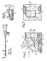
- scanning microtelescopes such as cystoscope conventionally consist of a head 11 attached to a tube 12 terminating in an eyepiece 14.
- the head 11 and tube 12 are inserted into the body of the patient, with the eyepiece 14 remaining outside the body of the patient for observation by the physician.
- a light source 16 is conventially mounted in the eyepiece 14, and its light is transmitted through optical fibers 18 to the head 11.
- the objective assembly of the head 11 consists of a negative lens 20, a prism 22, and a positive lens 24.
- the positive lens 24 transmits the image seen by the negative lens 20 to an optical conduit located within the tube 12.
- the image may be transmitted to the eyepiece 14 through this optical conduit by lens relays, fiber optics, a gradient index relay rod, or any other suitable conventional optical apparatus.
- the angle ⁇ between the axis 26 of the head 11 and the axis 28 of the field of view of the lens 20 is on the order of 70°.
- cystoscopes are manufactured with a view angle of 70° or 30° for the most common purposes.
- the typical diameter of the head 11 for surgical microtelescopes is on the order of 3mm, and that its walls must be thick enough to be sturdy and to be easily sterilizable. Consequently, the components of the objective system must be extremely small and precisely aligned. Also, the field of view of the lens 20 must have a rather wide angle in order to afford the physician a useful view. For these reasons, conventional means such as tiltable mirrors cannot be used to vary the angle ⁇ in any given cystoscope, and the instant has to be removed and another reinserted if the physician desires different view angles --- a procedure which is not only cumbersome for the physician, but also uncomfortable for the patient and a potential source of injury.
- the present invention makes practical a surgical microtelescope in which the field-of-view angle ⁇ can be changed by the physician, between two fixed settings, without removing the instrument.
- the objective system of this invention is similar to that of the prior art except that the prism 22 of Fig. 1b is replaced by a split prism assembly.
- the split prism assembly interposed between the positive lens 120 and the negative lenses 122, 124 (which look, respectively, through transparent windows 126, 128) consists of three prisms 130, 132, 134, of which only two are active at any given tine.
- the negative lenses 122, 124 may be omitted in certain applications without departing from the invention.
- the central prism 130 is transversely reciprocable by alternately pulling the flexible wires 136, 138.
- the minor face 140 of prism 130 is silvered on half of its area (142) and clear on the other half (144).
- the image seen by lens 124 through window 128 (at an ⁇ min angle of 30°) is transmitted through prisms 134 and 130, as shown in dotted lines, to lens 120.
- the mirrored surface 142 blocks the image from window 128 and instead reflects the image from window 126 (at the ⁇ max angle of 70°) through lens 122 and prism 132 into lens 120.
- the two images are neither rotated nor inverted with respect to one another, so that no eyepiece compensation is needed.
Landscapes
- Health & Medical Sciences (AREA)
- Life Sciences & Earth Sciences (AREA)
- Physics & Mathematics (AREA)
- Surgery (AREA)
- Optics & Photonics (AREA)
- Biomedical Technology (AREA)
- Animal Behavior & Ethology (AREA)
- Radiology & Medical Imaging (AREA)
- Nuclear Medicine, Radiotherapy & Molecular Imaging (AREA)
- Engineering & Computer Science (AREA)
- Biophysics (AREA)
- Heart & Thoracic Surgery (AREA)
- Medical Informatics (AREA)
- Molecular Biology (AREA)
- Pathology (AREA)
- General Health & Medical Sciences (AREA)
- Public Health (AREA)
- Veterinary Medicine (AREA)
- Astronomy & Astrophysics (AREA)
- General Physics & Mathematics (AREA)
- Instruments For Viewing The Inside Of Hollow Bodies (AREA)
- Endoscopes (AREA)
Claims (2)
- Endoscope chirurgical comprenant :(a) une tête d'observation allongée stérilisable (11) dans laquelle est monté un système optique comportant un premier prisme (134), un deuxième prisme (130) déplaçable par rapport audit premier prisme (134) et une lentille (120) de sorte que, dans une première position dudit prisme déplaçable (130), une image suivant un premier angle de visée est transmise,à travers ledit premier prisme et ledit deuxième prisme déplaçable (134),à ladite lentille (120) ;(b) un conduit optique allongé adjacent à la dite lentille (120) ;(c) un oculaire (14) recevant, par ledit conduit optique allongé l'image fournie par ladite tête (11) ;(d) des moyens de manoeuvre (136,138) connectés audit prisme déplaçable (130) et s'étendant entre ladite tête (11) et l'extrémité côté oculaire dudit conduit pour communiquer un mouvement relatif audit prisme déplaçable (130) à partir de ladite extrémité côté oculaire dudit conduit, ledit mouvement relatif dudit prisme déplaçable (130) ayant pour effet de modifier l'angle entre l'axe du champ de vision dudit système optique et l'axe de ladite tête;caractérisé en ce que
un troisième prisme (130) est prévu, et en ce que ledit deuxième prisme déplaçable présente une surface dont une partie est réfléchissante, disposée de sorte que, dans une deuxième position dudit deuxième prisme déplaçable (130), une image suivant un deuxième angle de visée différent du premier angle de visée est transmise, à travers ledit troisième prisme (132) et ledit deuxième prisme déplaçable (130), à ladite lentille(120). - Endoscope suivant la revendication 1, dans lequel ledit deuxième prisme déplaçable (130) est déplaçable de façon coulissante entre lesdits premier et deuxième prismes (132,134).
Applications Claiming Priority (2)
| Application Number | Priority Date | Filing Date | Title |
|---|---|---|---|
| US866452 | 1986-05-22 | ||
| US06/866,452 US4697577A (en) | 1986-05-22 | 1986-05-22 | Scanning microtelescope for surgical applications |
Publications (2)
| Publication Number | Publication Date |
|---|---|
| EP0251478A1 EP0251478A1 (fr) | 1988-01-07 |
| EP0251478B1 true EP0251478B1 (fr) | 1991-08-14 |
Family
ID=25347661
Family Applications (1)
| Application Number | Title | Priority Date | Filing Date |
|---|---|---|---|
| EP87304589A Expired EP0251478B1 (fr) | 1986-05-22 | 1987-05-22 | Endoscope à balayage pour applications chirurgicales |
Country Status (5)
| Country | Link |
|---|---|
| US (1) | US4697577A (fr) |
| EP (1) | EP0251478B1 (fr) |
| JP (1) | JPS62284626A (fr) |
| CA (1) | CA1278934C (fr) |
| DE (1) | DE3772121D1 (fr) |
Cited By (1)
| Publication number | Priority date | Publication date | Assignee | Title |
|---|---|---|---|---|
| US9572478B2 (en) | 2008-07-25 | 2017-02-21 | Karl Storz Imaging, Inc. | Swing prism endoscope |
Families Citing this family (72)
| Publication number | Priority date | Publication date | Assignee | Title |
|---|---|---|---|---|
| US4916534A (en) * | 1987-04-28 | 1990-04-10 | Olympus Optical Co., Ltd. | Endoscope |
| US4838247A (en) * | 1988-10-06 | 1989-06-13 | Baxter International, Inc. | Dual-view arthroscope |
| FR2637698A1 (fr) * | 1988-10-10 | 1990-04-13 | Lasag Ag | Dispositif catadioptrique monolithique |
| EP0362767A1 (fr) * | 1988-10-06 | 1990-04-11 | Lasag Ag | Dispositif catadioptrique monolithique |
| US4941457A (en) * | 1989-08-17 | 1990-07-17 | Olympus Optical Co., Ltd. | Endoscope using an optical guide twisted on the tip side to have the visual field direction and curvature axis coincide with each other |
| JP3012373B2 (ja) * | 1991-06-05 | 2000-02-21 | 旭光学工業株式会社 | 内視鏡の先端部 |
| US5288283A (en) * | 1993-01-25 | 1994-02-22 | Lisco, Inc. | Doorway exerciser |
| US5743846A (en) * | 1994-03-17 | 1998-04-28 | Olympus Optical Co., Ltd. | Stereoscopic endoscope objective lens system having a plurality of front lens groups and one common rear lens group |
| US5554100A (en) * | 1994-03-24 | 1996-09-10 | United States Surgical Corporation | Arthroscope with shim for angularly orienting illumination fibers |
| US6184923B1 (en) * | 1994-11-25 | 2001-02-06 | Olympus Optical Co., Ltd. | Endoscope with an interchangeable distal end optical adapter |
| EP0920276B1 (fr) * | 1996-08-12 | 2003-08-20 | MGB Endoskopische Geräte GmbH Berlin | Endoscope rigide avec eclairage |
| US5807237A (en) * | 1997-03-31 | 1998-09-15 | Tindel; Nathaniel L. | Endoscopic device |
| DE69922791T2 (de) * | 1998-02-19 | 2005-12-08 | California Institute Of Technology, Pasadena | Gerät zur bereitstellung eines spherischen sehfeldes während endoskopischen eingriffen |
| FR2783610B1 (fr) * | 1998-09-18 | 2000-11-24 | Tokendo Sarl | Endoscope rotatif rigide a visee distale deviee et mise au point proximale reglable |
| US6110105A (en) * | 1999-02-03 | 2000-08-29 | Durell & Gitelis, Inc. | Variable view arthroscope |
| DE19927816A1 (de) * | 1999-06-18 | 2001-01-04 | Winter & Ibe Olympus | Endoskop mit verstellbarem Bauelement |
| GB2354836B (en) * | 1999-09-28 | 2003-06-04 | Keymed | Improvements relating to borescopes and endoscopes with variable direction of view |
| US6364830B1 (en) | 1999-11-30 | 2002-04-02 | Durell & Gitelis, Inc. | Variable view arthroscope |
| JP3345645B2 (ja) * | 2000-06-20 | 2002-11-18 | 東京大学長 | 体腔内観察装置 |
| US6638216B1 (en) | 2000-08-30 | 2003-10-28 | Durell & Gitelis, Inc. | Variable view arthroscope |
| US7175593B2 (en) * | 2000-08-30 | 2007-02-13 | Durell & Gitelis, Inc. | Variable view arthroscope with charge coupled device |
| US6929603B2 (en) * | 2000-08-30 | 2005-08-16 | Durell & Gitelis, Inc. | Variable view arthroscope |
| US6695774B2 (en) | 2001-01-19 | 2004-02-24 | Endactive, Inc. | Apparatus and method for controlling endoscopic instruments |
| US6701181B2 (en) | 2001-05-31 | 2004-03-02 | Infraredx, Inc. | Multi-path optical catheter |
| US20060199999A1 (en) * | 2001-06-29 | 2006-09-07 | Intuitive Surgical Inc. | Cardiac tissue ablation instrument with flexible wrist |
| US6817974B2 (en) | 2001-06-29 | 2004-11-16 | Intuitive Surgical, Inc. | Surgical tool having positively positionable tendon-actuated multi-disk wrist joint |
| US20060178556A1 (en) | 2001-06-29 | 2006-08-10 | Intuitive Surgical, Inc. | Articulate and swapable endoscope for a surgical robot |
| US6648817B2 (en) | 2001-11-15 | 2003-11-18 | Endactive, Inc. | Apparatus and method for stereo viewing in variable direction-of-view endoscopy |
| FR2832516B1 (fr) * | 2001-11-19 | 2004-01-23 | Tokendo Sarl | Endoscopes rotatifs a visee distale deviee |
| US6663559B2 (en) * | 2001-12-14 | 2003-12-16 | Endactive, Inc. | Interface for a variable direction of view endoscope |
| EP3498213A3 (fr) | 2002-12-06 | 2019-07-03 | Intuitive Surgical Operations, Inc. | Poignée fléxible pour outil chirurgical |
| US20040254424A1 (en) * | 2003-04-15 | 2004-12-16 | Interscience, Inc. | Integrated panoramic and forward view endoscope |
| US7381183B2 (en) * | 2003-04-21 | 2008-06-03 | Karl Storz Development Corp. | Method for capturing and displaying endoscopic maps |
| US20050054895A1 (en) * | 2003-09-09 | 2005-03-10 | Hoeg Hans David | Method for using variable direction of view endoscopy in conjunction with image guided surgical systems |
| US7232409B2 (en) * | 2003-11-20 | 2007-06-19 | Karl Storz Development Corp. | Method and apparatus for displaying endoscopic images |
| US7344494B2 (en) * | 2004-02-09 | 2008-03-18 | Karl Storz Development Corp. | Endoscope with variable direction of view module |
| KR100573132B1 (ko) * | 2004-02-14 | 2006-04-24 | 삼성에스디아이 주식회사 | 유기 전계 발광 표시장치 및 그 제조 방법 |
| US9615772B2 (en) * | 2004-02-20 | 2017-04-11 | Karl Storz Imaging, Inc. | Global endoscopic viewing indicator |
| US7427263B2 (en) * | 2004-03-03 | 2008-09-23 | Karl Storz Development Corp. | Method and interface for operating a variable direction of view endoscope |
| US7374533B2 (en) * | 2004-03-26 | 2008-05-20 | Karl Storz Development Corp. | Tip structure for variable direction of view endoscope |
| US9033871B2 (en) | 2004-04-07 | 2015-05-19 | Karl Storz Imaging, Inc. | Gravity referenced endoscopic image orientation |
| IL170404A (en) * | 2004-08-26 | 2012-03-29 | C2Cure Inc | Wireless determination of endoscope orientation |
| US9182577B2 (en) * | 2005-01-21 | 2015-11-10 | Karl Storz Imaging, Inc. | Variable direction of view instrument with distal image sensor |
| US7909756B2 (en) | 2005-01-26 | 2011-03-22 | Karl Storz Imaging, Inc. | Illumination system for variable direction of view instruments |
| US7221522B2 (en) * | 2005-01-28 | 2007-05-22 | Karl Storz Development Corp. | Optical system for variable direction of view instrument |
| US7896803B2 (en) * | 2005-02-14 | 2011-03-01 | Karl Storz Imaging, Inc. | Variable direction of view instrument with on-board actuators |
| US7967742B2 (en) * | 2005-02-14 | 2011-06-28 | Karl Storz Imaging, Inc. | Method for using variable direction of view endoscopy in conjunction with image guided surgical systems |
| DE202005008788U1 (de) * | 2005-05-27 | 2006-08-10 | Karl Storz Gmbh & Co. Kg | Endoskop, insbesondere für die Tracheotomie |
| GB0613576D0 (en) * | 2006-07-10 | 2006-08-16 | Leuven K U Res & Dev | Endoscopic vision system |
| US8460195B2 (en) * | 2007-01-19 | 2013-06-11 | Sunnybrook Health Sciences Centre | Scanning mechanisms for imaging probe |
| CN101686827B (zh) * | 2007-01-19 | 2014-08-13 | 桑尼布鲁克健康科学中心 | 具有组合的超声和光学成像装置的成像探头 |
| DE102009059004A1 (de) * | 2009-12-17 | 2011-06-22 | Olympus Winter & Ibe GmbH, 22045 | Objektiv mit zwei Blickrichtungen mit Prisma |
| DE102009025659A1 (de) | 2009-06-17 | 2010-12-23 | Karl Storz Gmbh & Co. Kg | Endoskoprohr |
| DE102009049143B3 (de) * | 2009-10-12 | 2010-12-30 | Sopro-Comeg Gmbh | Endoskop |
| US9375139B2 (en) * | 2010-07-29 | 2016-06-28 | Cannuflow, Inc. | Arthroscopic system |
| DE102010040990A1 (de) * | 2010-09-17 | 2012-03-22 | Henke-Sass, Wolf Gmbh | Endoskop mit variabler Blickrichtung |
| DE102010040992A1 (de) * | 2010-09-17 | 2012-03-22 | Henke-Sass, Wolf Gmbh | Endoskop mit variabler Blickrichtung |
| DE102010063230A1 (de) * | 2010-12-16 | 2012-06-21 | Karl Storz Gmbh & Co. Kg | Endoskop mit einstellbarer Blickrichtung |
| TWI539925B (zh) * | 2011-01-18 | 2016-07-01 | Medical Intubation Tech Corp | An endoscopic image pickup assembly having two or more illumination directions |
| DE102011005255A1 (de) * | 2011-03-08 | 2012-09-13 | Olympus Winter & Ibe Gmbh | Vorrichtung zur Umschaltung einer Blickrichtung eines Videoendoskops |
| DE102011007880A1 (de) * | 2011-04-21 | 2012-10-25 | Karl Storz Gmbh & Co. Kg | Lichtleitereinrichtung für ein Endoskop zum Leiten von Beleuchtungslicht |
| EP2731488B1 (fr) | 2011-07-12 | 2017-05-03 | Anderson, David, Ryan | Système d'imagerie intra-orale |
| JP5399587B2 (ja) | 2011-08-10 | 2014-01-29 | オリンパスメディカルシステムズ株式会社 | 医療機器 |
| US9655679B2 (en) * | 2012-05-01 | 2017-05-23 | University Of Maryland | Actuated steerable probe and systems and methods of using same |
| DE102012219434A1 (de) * | 2012-10-24 | 2014-04-24 | Olympus Winter & Ibe Gmbh | Endoskop mit seitlicher Illumination, Verwendung und Verfahren |
| DE102013218229B4 (de) | 2013-09-11 | 2015-08-20 | Olympus Winter & Ibe Gmbh | Endoskop mit einstellbarer Blickrichtung |
| WO2015064521A1 (fr) * | 2013-10-30 | 2015-05-07 | オリンパスメディカルシステムズ株式会社 | Dispositif de capture d'image |
| DE102015101624A1 (de) | 2015-02-04 | 2016-08-04 | Olympus Winter & Ibe Gmbh | Endoskop |
| US10201267B2 (en) | 2016-05-24 | 2019-02-12 | Phoenix Spine Holdings, Inc. | Methods and apparatus for facilitating direct visualized rhizotomy |
| US10376382B2 (en) | 2016-11-16 | 2019-08-13 | Phoenix Spine Holdings, Inc. | Methods and apparatus for facilitating a posterior lumbar interbody fusion procedure |
| US11076902B2 (en) | 2018-02-22 | 2021-08-03 | Phoenix Spine Holdings, Inc. | Locking screw assembly for facilitating direct lateral interbody fusion procedures |
| DE102019100147B4 (de) * | 2019-01-04 | 2022-07-28 | Olympus Winter & Ibe Gmbh | Videoendoskop |
Family Cites Families (11)
| Publication number | Priority date | Publication date | Assignee | Title |
|---|---|---|---|---|
| US1680491A (en) * | 1928-08-14 | Reinhold h | ||
| US1932470A (en) * | 1931-01-28 | 1933-10-31 | Lehmann Herbert | Optical telescope for medical and technical purposes |
| US2927574A (en) * | 1954-05-28 | 1960-03-08 | Richard Wolf | Endoscope for the observation of ventricles of the body or the like |
| US2783757A (en) * | 1954-10-21 | 1957-03-05 | Richard Wolf | Endoscope for the observation of ventricles of the body or the like |
| US3548808A (en) * | 1967-11-22 | 1970-12-22 | Olympus Optical Co | Device for switching the direction of the observation in an endoscope |
| DE2347914C3 (de) * | 1972-09-25 | 1979-01-11 | Olympus Optical Co., Ltd., Tokio | Endoskop mit einer Bildübertragungs-Faseroptik und einem ersten und drehbaren zweiten Reflexionselement |
| JPS49131144A (fr) * | 1973-04-19 | 1974-12-16 | Canon Kk | |
| JPS584481Y2 (ja) * | 1973-06-23 | 1983-01-26 | オリンパス光学工業株式会社 | ナイシキヨウシヤヘンカンコウガクケイ |
| JPS569712A (en) * | 1979-07-06 | 1981-01-31 | Olympus Optical Co Ltd | Visual field direction changing optical system for slender image transmission system |
| EP0050922B1 (fr) * | 1980-10-27 | 1985-01-16 | Sumitomo Electric Industries Limited | Système d'observation pour l'inspection de trous de faible diamétre |
| JPS5812642A (ja) * | 1981-07-16 | 1983-01-24 | オリンパス光学工業株式会社 | 硬性内視鏡 |
-
1986
- 1986-05-22 US US06/866,452 patent/US4697577A/en not_active Expired - Fee Related
-
1987
- 1987-05-21 CA CA000537608A patent/CA1278934C/fr not_active Expired - Lifetime
- 1987-05-22 JP JP62125641A patent/JPS62284626A/ja active Pending
- 1987-05-22 EP EP87304589A patent/EP0251478B1/fr not_active Expired
- 1987-05-22 DE DE8787304589T patent/DE3772121D1/de not_active Expired - Lifetime
Cited By (1)
| Publication number | Priority date | Publication date | Assignee | Title |
|---|---|---|---|---|
| US9572478B2 (en) | 2008-07-25 | 2017-02-21 | Karl Storz Imaging, Inc. | Swing prism endoscope |
Also Published As
| Publication number | Publication date |
|---|---|
| CA1278934C (fr) | 1991-01-15 |
| DE3772121D1 (de) | 1991-09-19 |
| US4697577A (en) | 1987-10-06 |
| EP0251478A1 (fr) | 1988-01-07 |
| JPS62284626A (ja) | 1987-12-10 |
Similar Documents
| Publication | Publication Date | Title |
|---|---|---|
| EP0251478B1 (fr) | Endoscope à balayage pour applications chirurgicales | |
| US5613936A (en) | Stereo laparoscope apparatus | |
| US5976076A (en) | Stereo laparoscope with synchronized optics | |
| EP0494134B1 (fr) | Endoscopique video | |
| US5846185A (en) | High resolution, wide field of view endoscopic viewing system | |
| US6949069B2 (en) | Endoscope | |
| JP2989237B2 (ja) | 光学装置 | |
| EP0951861B1 (fr) | Instrument médical de surveillance | |
| US4364629A (en) | Operation microscope having a small working distance | |
| US5612816A (en) | Endoscopic attachment for a stereoscopic viewing system | |
| AU771368B2 (en) | Visual aid in the form of telescopic spectacles with an automatic focussing device | |
| US4664486A (en) | Variable magnification endoscope | |
| US5295477A (en) | Endoscopic operating microscope | |
| JP3032214B2 (ja) | 手術用顕微鏡 | |
| AU2001271570A1 (en) | Endoscope | |
| GB2066082A (en) | Operating endoscope | |
| US5385138A (en) | Stereo endoscope for inserting into body cavities | |
| US4723842A (en) | Stereoscopic surgery microscope for eye surgery with selective image reversal | |
| US6106456A (en) | Device for examining cavities using an endoscope | |
| US5222482A (en) | Binocular endoscope with depth perception and fixed focal plane | |
| US3608998A (en) | Variable power endoscope | |
| US7423807B2 (en) | Ophthalmoscopic stereomicroscope with correction component | |
| US6309348B1 (en) | Endomicroscope system | |
| JP3980672B2 (ja) | 屈曲した覗き方向を有する立体内視鏡 | |
| JPH06167658A (ja) | 立体内視鏡 |
Legal Events
| Date | Code | Title | Description |
|---|---|---|---|
| PUAI | Public reference made under article 153(3) epc to a published international application that has entered the european phase |
Free format text: ORIGINAL CODE: 0009012 |
|
| AK | Designated contracting states |
Kind code of ref document: A1 Designated state(s): CH DE FR GB LI |
|
| 17P | Request for examination filed |
Effective date: 19880608 |
|
| RAP1 | Party data changed (applicant data changed or rights of an application transferred) |
Owner name: BAXTER INTERNATIONAL INC. (A DELAWARE CORPORATION) |
|
| 17Q | First examination report despatched |
Effective date: 19900124 |
|
| GRAA | (expected) grant |
Free format text: ORIGINAL CODE: 0009210 |
|
| AK | Designated contracting states |
Kind code of ref document: B1 Designated state(s): CH DE FR GB LI |
|
| REF | Corresponds to: |
Ref document number: 3772121 Country of ref document: DE Date of ref document: 19910919 |
|
| ET | Fr: translation filed | ||
| PLBE | No opposition filed within time limit |
Free format text: ORIGINAL CODE: 0009261 |
|
| STAA | Information on the status of an ep patent application or granted ep patent |
Free format text: STATUS: NO OPPOSITION FILED WITHIN TIME LIMIT |
|
| 26N | No opposition filed | ||
| REG | Reference to a national code |
Ref country code: CH Ref legal event code: PUE Owner name: ENDOMEDIX CORPORATION |
|
| REG | Reference to a national code |
Ref country code: GB Ref legal event code: 732E |
|
| REG | Reference to a national code |
Ref country code: FR Ref legal event code: TP |
|
| PGFP | Annual fee paid to national office [announced via postgrant information from national office to epo] |
Ref country code: GB Payment date: 19940518 Year of fee payment: 8 |
|
| PGFP | Annual fee paid to national office [announced via postgrant information from national office to epo] |
Ref country code: FR Payment date: 19940524 Year of fee payment: 8 Ref country code: DE Payment date: 19940524 Year of fee payment: 8 |
|
| PGFP | Annual fee paid to national office [announced via postgrant information from national office to epo] |
Ref country code: CH Payment date: 19940525 Year of fee payment: 8 |
|
| REG | Reference to a national code |
Ref country code: CH Ref legal event code: PUE Owner name: SPECTRUM MEDSYSTEMS CORP. |
|
| REG | Reference to a national code |
Ref country code: GB Ref legal event code: 732E |
|
| REG | Reference to a national code |
Ref country code: FR Ref legal event code: TP |
|
| PG25 | Lapsed in a contracting state [announced via postgrant information from national office to epo] |
Ref country code: GB Effective date: 19950522 |
|
| PG25 | Lapsed in a contracting state [announced via postgrant information from national office to epo] |
Ref country code: LI Effective date: 19950531 Ref country code: CH Effective date: 19950531 |
|
| GBPC | Gb: european patent ceased through non-payment of renewal fee |
Effective date: 19950522 |
|
| REG | Reference to a national code |
Ref country code: CH Ref legal event code: PL |
|
| PG25 | Lapsed in a contracting state [announced via postgrant information from national office to epo] |
Ref country code: DE Effective date: 19960201 |
|
| PG25 | Lapsed in a contracting state [announced via postgrant information from national office to epo] |
Ref country code: FR Effective date: 19960229 |
|
| REG | Reference to a national code |
Ref country code: FR Ref legal event code: ST |
|
| REG | Reference to a national code |
Ref country code: FR Ref legal event code: ST |